Embed Size (px)
Citation preview
Cellular/Molecular
S-SCAM/MAGI-2 Is an Essential Synaptic ScaffoldingMolecule for the GluA2-Containing Maintenance Poolof AMPA Receptors
Eric Danielson,1 Nanyan Zhang,1 Jacob Metallo,1 Kanwardeep Kaleka,2 Seung Min Shin,1 Nashaat Gerges,2
and Sang H. Lee1
1Department of Pharmacology and Toxicology, 2Department of Cell Biology, Anatomy and Neurobiology, Medical College of Wisconsin, Milwaukee,Wisconsin 53226-0509
Synaptic plasticity, the cellular basis of learning and memory, involves the dynamic trafficking of AMPA receptors (AMPARs) into andout of synapses. One of the remaining key unanswered aspects of AMPAR trafficking is the mechanism by which synaptic strength ispreserved despite protein turnover. In particular, the identity of AMPAR scaffolding molecule(s) involved in the maintenance of GluA2-containing AMPARs is completely unknown. Here we report that the synaptic scaffolding molecule (S-SCAM; also called membrane-associated guanylate kinase inverted-2 and atrophin interacting protein-1) plays the critical role of maintaining synaptic strength.Increasing S-SCAM levels in rat hippocampal neurons led to specific increases in the surface AMPAR levels, enhanced AMPAR-mediatedsynaptic transmission, and enlargement of dendritic spines, without significantly effecting GluN levels or NMDA receptor (NMDAR)EPSC. Conversely, decreasing S-SCAM levels by RNA interference-mediated knockdown caused the loss of synaptic AMPARs, which wasfollowed by a severe reduction in the dendritic spine density. Importantly, S-SCAM regulated synaptic AMPAR levels in a manner,dependent on GluA2 not GluA1, sensitive to N-ethylmaleimide-sensitive fusion protein interaction, and independent of activity. Further,S-SCAM increased surface AMPAR levels in the absence of PSD-95, while PSD-95 was dependent on S-SCAM to increase surface AMPARlevels. Finally, S-SCAM overexpression hampered NMDA-induced internalization of AMPARs and prevented the induction of longterm-depression, while S-SCAM knockdown did not. Together, these results suggest that S-SCAM is an essential AMPAR scaffoldingmolecule for the GluA2-containing pool of AMPARs, which are involved in the constitutive pathway of maintaining synaptic strength.
IntroductionEfficient synaptic transmission relies on the precise organizationof various proteins including neurotransmitter receptors, ionchannels, signaling enzymes, and cytoskeletal elements (Okabe,2007; Sheng and Hoogenraad, 2007). Furthermore, activity-dependent modification of synaptic strength, or synaptic plastic-ity, demands a fine-tuned orchestration of recruitment andremoval of proteins at synapses. For example, long-term poten-tiation (LTP) and long-term depression (LTD) are mediated byactivity-dependent trafficking of AMPA receptors (AMPARs) toand out of excitatory synapses (Collingridge et al., 2004; Derkachet al., 2007; Shepherd and Huganir, 2007; Kessels and Malinow,2009). Synaptic scaffolding proteins (S-SCAMs) play importantroles in the assembly of the macro-signaling complexes and traf-
ficking of synaptic proteins. The membrane-associated guanylatekinase (MAGUK) family of proteins is one of the most well stud-ied synaptic scaffolds at the postsynaptic density (PSD). Amongthe PSD-MAGUKs, PSD-95 (SAP-90) represents the prototypi-cal member and is involved in the various aspects of excitatorysynaptic transmission, including synapse maturation, AMPARtrafficking, and synaptic plasticity (Elias and Nicoll, 2007; Xu,2011).
S-SCAM was first identified as a protein interacting withSAPAP (also called GKAP; Kim et al., 1997; Hirao et al., 1998).S-SCAM is also known as membrane-associated guanylate kinaseinverted-2 (MAGI-2) (Wu et al., 2000) or atrophin interactingprotein-1 (Wood et al., 1998). The molecular organization ofS-SCAM is in an inverse configuration to PSD-95, and is com-posed of six PDZ domains, one guanylate kinase (GK) domain,and two WW domains. Studies have shown that S-SCAM inter-acts with numerous PSD-95-binding proteins including NMDAreceptor (NMDAR) (Hirao et al., 1998), ErbB4 (Buxbaum et al.,2008), Neuroligin (Iida et al., 2004), Kif1B (Mok et al., 2002), andtransmembrane AMPAR regulatory proteins (TARPs) (Deng etal., 2006). S-SCAM also binds proteins that have no known in-teraction with PSD-95, which include Axin (Hirabayashi et al.,2004), �-catenin (Nishimura et al., 2002), �-dystroglycan(Sumita et al., 2007), and neuroligin 2 (Sumita et al., 2007). Theseoverlapping protein–protein interaction profiles suggest that
Received Jan. 3, 2012; revised March 21, 2012; accepted March 29, 2012.Author contributions: E.D., K.K., S.M.S., N.G., and S.H.L. designed research; E.D., N.Z., J.M., and S.H.L. performed
research; S.M.S. contributed unpublished reagents/analytic tools; E.D., N.Z., K.K., N.G., and S.H.L. analyzed data;S.H.L. wrote the paper.
This work was supported by the Whitehall foundation (S.H.L.) and U.S. National Institute of Health GrantsMH078135 (S.H.L.) and AG032320 (N.G.). We thank Dr. Yutaka Hata (Tokyo Medical and Dental University) forproviding S-SCAM expression plasmids and Dr. Qing-song Liu for advice on electrophysiology and data analyses.
Correspondence should be addressed to: Dr. Sang H. Lee, Department of Pharmacology and Toxicology, BSB 608,Medical College of Wisconsin, 8701 Watertown Plank Road, Milwaukee, WI 53226-0509. E-mail: [email protected].
DOI:10.1523/JNEUROSCI.0025-12.2012Copyright © 2012 the authors 0270-6474/12/326967-14$15.00/0
The Journal of Neuroscience, May 16, 2012 • 32(20):6967– 6980 • 6967
S-SCAM may play both similar and distinct roles, compared withPSD-95, in the molecular organization of PSDs and, in particu-lar, AMPAR regulation. There are three S-SCAM isoforms ofS-SCAM-�, -�, and -�, which are generated by differential trans-lational initiations from multiple sites (Hirao et al., 2000). Micelacking the longest variant S-SCAM-� died within 24 h after birth(Iida et al., 2007), indicating that S-SCAM is an essential protein.Interestingly, hippocampal culture neurons prepared from thesemutant mice showed abnormal elongated dendritic spines (Iidaet al., 2007), suggesting that S-SCAM may function in the den-dritic spine dynamics.
In addition to the probable role of S-SCAM in the molecularorganization of PSDs and synaptic transmission, recent geneticstudies uncovered S-SCAM gene mutations in patients with neu-rological diseases such as schizophrenia (Walsh et al., 2008) andinfantile spasms (IS) (Marshall et al., 2008). Despite the potentialimportance of S-SCAM in synaptic biology and neurological dis-eases, little is currently known for the function of S-SCAM. Here,we report a novel and essential role of S-SCAM in the organiza-tion of PSDs and excitatory synaptic transmission.
Materials and MethodsExpression and RNAi construct. Myc- or GFP-tagged S-SCAM expressionconstructs were obtained from Dr. Y. Hata (Tokyo Medical and DentalUniversity, Tokyo, Japan). GluA1 and GluA2 shRNAs were as describedpreviously (Lee et al., 2004). S-SCAM shRNA construct was created by clon-ing the following hairpin sequence GTACAGAACCTGAGCCATATTCAAGAGATATGGCTCAGGTTCTGTAC into pSUPER or pSUPER.neo�gfp vectors (OligoEngine). S-SCAM rescue construct wasgenerated by introducing silent mutations in S-SCAM RNAi targetingsequence via site-directed mutagenesis and confirmed by nucleotidesequencing. PSD-95 shRNA is as described previously (Nakagawa etal., 2004).
Neuron transfection and immunocytochemistry. Dissociated hippocam-pal neuron culture was prepared from E18 embryos of either sex asdescribed previously (Lee et al., 2002) and grown in Neurobasal mediasupplemented with B27. Neuron transfection was performed at DIV 14using Lipofectamine 2000 reagent (Invitrogen) as described previously(Lee et al., 2004). Cold methanol (�20°C for 10 min) fixation was usedfor the staining of PSD-95, Shank, GluN1, GluN2B, and GKAP. Forcostaining of �-Gal (or GFP) and S-SCAM, GKAP, or PSD-95, neuronswere incubated first in 2% formaldehyde/4% sucrose/1 � PBS for 2 minfollowed by cold methanol for 10 min. Surface AMPAR staining wasperformed as described previously (Lee et al., 2004). Rabbit polyclonalanti-S-SCAM antibodies were prepared by injecting rabbits with purifiedrecombinant protein covering the WW domains of S-SCAM (aminoacids 303– 405) (Hirao et al., 1998), affinity purified, and used at 0.01�g/ml concentration for immunocytochemistry or Western blotting. Wealso tested a commercial rabbit polyclonal anti-MAGI-2 antibody (1:1000 dilution; Sigma) that is prepared from different antigens (aminoacids 554 –571), which is specific to S-SCAM/MAGI-2. Both antibodiesproduced very similar immunostaining patterns. However, to eliminateany potential cross-reactivity problem, immunostaining data obtainedfrom the commercial antibody were shown. Primary antibodies and theirdilution used for immunocytochemistry are as follows: mouse anti-�-Gal(1:1000; Promega), rabbit anti-�-Gal (1:5000; Abcam), mouse anti-PSD-95(1:500; clone K28/43, UC Davis/NIH NeuroMab), rabbit anti- GKAP (1:250; Kim et al., 1997), mouse anti-NR2B (1:100, clone N59/36; UC Davis/NIH NeuroMab), mouse anti-pan-SAPAP (1: 250, clone N127/31; UCDavis/NIH NeuroMab), mouse anti-NMDAR1 (1:100; BD PharMingen),mouse anti-Shank (1:250; Naisbitt et al., 1999), mouse anti-Bassoon (1:200;Stressgen), mouse anti-synaptophysin (SVP-38, 1:1000; Sigma), rabbitanti-HA (1:100; Santa Cruz Biotechnology), mouse anti-HA (1:400, clone12CA5; Roche), mouse anti-myc (1:100, clone 9E10; Santa Cruz Biotechnol-ogy), rabbit anti-myc (1:100; Cell Signaling Technology), rabbit anti-GluA1(5 �g/ml; Oncogene), and mouse anti-GluA2 (5 �g/ml; Millipore Biosci-ence Research Reagents). Bound primary antibodies were visualized by Al-
exa Fluor 488 (1:250; Invitrogen), Cy3-conjugated (1:500; JacksonImmunoResearch Laboratories), and/or Alexa Fluor 647 (1:200; Invitrogen)secondary antibodies.
Internalization assay. AMPAR internalization assay was performed us-ing GluA2 antibody-recognizing extracellular epitope (MAB397; Milli-pore), and data analyses were done as described previously (Lee et al.,2004). Briefly, after live labeling surface GluA2 with the antibody (10�g/ml) in the conditioned medium for 10 min at 37°C, neurons wereincubated for 2 min with conditioned medium (control) or conditionedmedium plus NMDA (50 �M) or AMPA (100 �M). After a brief wash inNeurobasal medium, neurons were further incubated for 8 min in theconditioned medium before fixation and staining.
Confocal microscopy and image analyses. Image acquisition was per-formed using a Nikon C1 plus laser scanning confocal microscope. Ac-quired z-series stack images were converted to projection images (withmaximal projection option) for analysis using MetaMorph software(Molecular Devices) or in-house custom software. Images were analyzedin a double-blind manner. To measure the puncta number per givenlength of dendrites, per image, five dendritic segments (�5 to 10 �m inlength each) were selected from transfected and neighboring nontrans-fected neurons, respectively. After applying threshold, only puncta with�4 pixel sizes were counted. All data collected were transferred to Mi-crosoft Excel for computation.
Statistical analysis. All values represent means � SEM, unless other-wise indicated. All transfection experiments were repeated at least threetimes. Statistical significance for pairs was analyzed by the Student’s t test(unpaired, two-tailed, assuming unequal variance), unless otherwise in-dicated in the figure legends. ANOVA with Tukey’s post hoc test was usedfor group comparisons. Cumulative plot data were analyzed by Kolmo-grov–Smirnov test (K-S test). p � 0.05 was considered significant.
Electrophysiology. For miniature EPSC (mEPSC) measurement, weused dissociated culture hippocampal neurons (plated at the density of75 k cells per coverslip), which were transfected at DIV 14 with GFP alone(pEGFP-C1), GFP � S-SCAM, or pSUPER.neo�gfp plasmid-expressingS-SCAM shRNA. At 3 d post-transfection, transfected pyramidal neu-rons were identified by GFP fluorescence and morphological inspection.All recordings were performed at 25°C. Whole-cell patch recordings wereperformed by voltage-clamping neurons at �70 mV in bathing solutioncontaining the following (in mM): 119 NaCl, 5 KCl, 2 CaCl2, 2 MgCl2, 30glucose, 10 HEPES, pH7.4, and �300 mOsm, and containing tetrodo-toxin (1 �M; Tocris Bioscience) and bicuculline (20 �M; Tocris Biosci-ence), continuously perfused at the rate of �0.5 ml/min. Internalsolution was composed of the following (in mM): 140 K�gluconate, 5 KCl,2 MgCl2, 4 Mg�ATP, 0.3 Na2�GTP, 0.2 EGTA, 10 HEPES, and adjusted topH7.2 and �290 mOsm. Micropipettes with tip resistance of 4 –7 M�were used. mEPSCs were acquired through a MultiClamp 700B amplifier(Molecular Devices), filtered at 2 kHz, and digitized at 10 kHz, using the“gap-free” protocol. mEPSCs were detected and analyzed with Mini-Analyses software (Synaptosoft) by setting the amplitude threshold to 5pA (usually root mean square � three values lower than 4), and furtherfiltered by selecting mini events of 10 –90% rise time �3 ms. Cumulativeprobability plots were generated by combining mini events from all re-corded neurons and analyzed for statistical significance by the K-S test.
For whole-cell paired recording experiments, we used slices cultureprepared from the hippocampi of 7-d-old Sprague Dawley rats of eithersex. Slices of 400 �m thickness were prepared using a tissue chopper,transferred onto MilliCell culture plate inserts (Millipore), and culturedfor 4 – 6 d in MEM supplemented with 1 �g/ml insulin, 0.0012% ascorbicacid, 20% horse serum, 1 mM L-glutamine, 1 mM CaCl2, 30 mM HEPES,13 mM D-glucose, and 5.2 mM NaHCO3 with Pen/Strep. The media waschanged every other day. Sindbis virus-expressing GFP-S-SCAM (a giftfrom Dr. Yutaka Hata) (Nishimura et al., 2002) was injected into the CA1region of hippocampal slices using a Toohey Spritzer microinjectionsystem. Electrophysiological recordings were performed 15–24 h postin-fection. A single slice was removed from the insert and placed in therecording chamber and continuously perfused with artificial CSF con-taining the following (in mM): 119 NaCl, 2.5 KCl, 1 NaH2PO4, 11 glucose,26 NaHCO3, 4 MgCl2, 4 CaCl2, and 290 mOsm and 50 �M picrotoxin and2 �M chloroadenosine, bubbled with a mixture of 5% CO2 and 95% O2.
6968 • J. Neurosci., May 16, 2012 • 32(20):6967– 6980 Danielson et al. • S-SCAM and Maintenance of Synaptic AMPA Receptors
Stimulating electrodes (two conductor platinum/iridium cluster micro-electrode, 25 �m diameter; FHC) were placed �200 �m on either side ofthe recording cell. One pathway was used to induce LTD, the other servedas a control pathway. The patch pipette was filled with internal solutioncontaining (in mM): 115 CsMeSO3, 20 CsCl, 10 HEPES, 2.5 MgCl2, 4Na2-ATP, 0.4 Na-GTP, 10 Na-phosphocreatine, 0.6 EGTA, 5 QX314, pH7.2, and 290 mOsm, and had tip resistance of 3– 6 M�. For rectificationexperiments, spermine (0.1 mM) was added in the internal solution.AMPA EPSCs were recorded at �60 mV and measured as peak inwardcurrent within a 60 ms window after stimulation. NMDA EPSCs wererecorded at �40 mV and measured 60 –100 ms after the initiation of theEPSC. Rectification index was calculated by dividing peak AMPA ampli-tudes measured at �60 mV by those at �40 mV. Stimulation pulses wereprovided at 0.3 Hz. Sixty traces were averaged for AMPA and NMDAEPSCs. For LTD experiments, at least 8 min of stable baseline responseswere collected before LTD induction. LTD-inducing stimulus consisted
of 200 stimulation pulses at 1 Hz while holdingcells at �40 mV. For paired pulse ratio (PPR),two consecutive stimuli were given 25 ms, 50ms, 100 ms, and 200 ms apart with a 2 s intervalbetween time points. At least 60 traces wereaveraged.
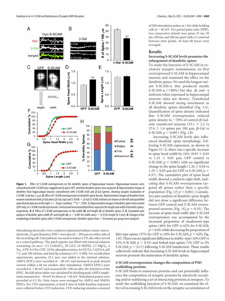
ResultsIncreasing S-SCAM levels promotes theenlargement of dendritic spinesTo study the function of S-SCAM in ex-citatory synaptic transmission, we firstoverexpressed S-SCAM in hippocampalneurons and examined the effect on thedendritic spines. We used the longest vari-ant S-SCAM-� that produced mostlyS-SCAM-� (�80%) but also -�, and -�isoforms when expressed in hippocampalneurons (data not shown). TransfectedS-SCAM showed strong enrichment inall dendritic spines identified (Fig. 1A).Quantification of spine density indicatedthat S-SCAM overexpression reducedspine density to �70% of control �-Gal-only transfected neurons (53.1 � 2.2 vs37.6 � 1.6 spines per 100 �m, �-Gal vsS-SCAM; p � 0.001) (Fig. 1B).
Increasing S-SCAM levels also influ-enced dendritic spine morphology. Fol-lowing S-SCAM expression, as shown inFigure 1C–E, there was a specific increasein spine head width by 24% (0.93 � 0.03vs 1.15 � 0.05 �m, GFP control vsS-SCAM; p � 0.001) with no significantchange in the spine length (1.26 � 0.04 vs1.29 � 0.05 �m for GFP vs S-SCAM; p 0.57). The cumulative plot of spine headwidth showed a uniform right shift, indi-cating that S-SCAM overexpression tar-geted all spines rather than a specificpopulation (Fig. 1F; p � 0.001). Cumula-tive plot analysis of dendritic spine lengthdid not show a significant difference be-tween GFP control and S-SCAM overex-pressed neurons (Fig. 1G; p 0.35). Theincrease of spine head width after S-SCAMoverexpression was accompanied by theincreased proportion of mushroom-typespines (46% for GFP vs 62% for S-SCAM;p�0.05) while decreasing the proportion of
thin-type spines (37% for GFP vs 24% for S-SCAM; p � 0.05; Fig.1H). There was no significant difference in stubby-type (16% GFP vs12% S-SCAM; p 0.33) and forked-type spines (1% GFP vs 2%S-SCAM; p 0.17) following S-SCAM transfection. These resultscollectively indicate that increasing S-SCAM levels in hippocampalneurons promote the maturation of dendritic spines.
S-SCAM overexpression changes the composition of PSDscaffolding proteinsS-SCAM binds to numerous proteins and can potentially influ-ence the composition of synaptic proteins by selectively recruit-ing and/or stabilizing a set of interacting proteins at synapses. Tostudy the scaffolding function of S-SCAM, we examined the ef-fect of increasing S-SCAM levels on the synaptic accumulation of
Figure 1. Effect of S-SCAM overexpression on the dendritic spines of hippocampal neurons. Hippocampal neurons werecotransfected with S-SCAM (myc-tagged) and �-gal or GFP, and their dendritic spines were analyzed. A, Representative images ofdendrites from hippocampal neurons cotransfected with S-SCAM (red) and �-Gal (green), showing synaptic localization ofS-SCAM. Scale bar, 5 �m. B, Effect of S-SCAM overexpression on dendritic spine density. Representative images of dendrites fromneurons transfected with �-Gal alone (�-Gal, top) and S-SCAM � �-Gal (S-SCAM, bottom) are shown on the left and quantifiedspine density data are on the right. n � 20 per condition. ***p � 0.001. C, Representative images of dendritic spines from control(GFP only) or S-SCAM-transfected neurons. Vertical and horizontal dotted lines represent the length and width of dendritic spines,respectively. D, E, Effect of S-SCAM overexpression on the width (D) and length (E) of dendritic spines. F, G, Cumulative plotanalyses of dendritic spine width (F ) and length (G). p � 0.001 for width and p 0.35 for length (K-S test). H, Changes in themorphology of dendritic spines after S-SCAM overexpression. Dendritic spines from �10 neurons per group were analyzed.
Danielson et al. • S-SCAM and Maintenance of Synaptic AMPA Receptors J. Neurosci., May 16, 2012 • 32(20):6967– 6980 • 6969
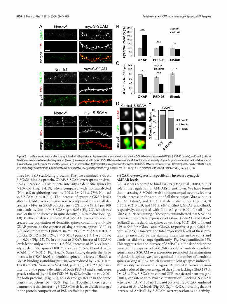
three key PSD scaffolding proteins. First we examined a directS-SCAM-binding protein, GKAP. S-SCAM overexpression dras-tically increased GKAP puncta intensity at dendritic spines by�2.5-fold (Fig. 2A,B), when compared with nontransfected(Non-txf) neighboring neurons (100 � 3 vs 261 � 27%, Non-txfvs S-SCAM; p � 0.001). The increase of synaptic GKAP levelsafter S-SCAM overexpression was accompanied by a small de-crease (�14%) in GKAP puncta density (78 � 3 vs 67 � 4 per 100�m dendrite, Non-txf vs S-SCAM; p � 0.05) (Fig. 2C), which wassmaller than the decrease in spine density (�40% reduction; Fig.1B). Further analyses indicated that S-SCAM overexpression in-creased the population of dendritic spines containing multipleGKAP puncta at the expense of single puncta spines (GFP vsS-SCAM, spines with 1 puncta, 84 � 2 vs 73 � 2%; p � 0.001; 2puncta, 15 � 2 vs 24 � 2%; p � 0.001; 3 puncta, 2 � 1 vs 3 � 1%;p 0.04) (Fig. 2D,E). In contrast to GKAP, increased S-SCAMlevels led to only a modest (�1.2-fold) increase of PSD-95 inten-sity at dendritic spines (100 � 2 vs 122 � 5%, Non-txf vs S-SCAM; p � 0.001) (Fig. 2A,B). Surprisingly, despite the largeincrease in GKAP levels at dendritic spines, the levels of Shank, aGKAP-binding scaffolding protein, were reduced by 17% (100 �4 vs 83 � 4%, Non-txf vs S-SCAM; p � 0.01) (Fig. 2A,B). Fur-thermore, the puncta densities of both PSD-95 and Shank weregreatly reduced (by 44% for PSD-95; by 62% for Shank; p � 0.001for both proteins) (Fig. 2C), to a degree greater than the spinedensity reduction (by �30%; Fig. 1B).Together, these resultsdemonstrate that increasing S-SCAM levels led to drastic changesin the protein composition of PSD scaffolding proteins.
S-SCAM overexpression specifically increases synapticAMPAR levelsS-SCAM was reported to bind TARPs (Deng et al., 2006), but itsrole in the regulation of AMPARs is unknown. We have foundthat increasing S-SCAM levels in hippocampal neurons led to adrastic increase in the amount of all three major GluA subunits(GluA1, GluA2, and GluA3) at dendritic spines (Fig. 3A,B)(170 � 9, 210 � 9, and 140 � 9% for GluA1, GluA2, and GluA3,respectively, compared with Non-txf; p � 0.001 for all threeGluAs). Surface staining of these proteins indicated that S-SCAMincreased the surface expression of GluA1 (sGluA1) and GluA2(sGluA2) at the dendritic spines as well (Fig. 3C,D) 238 � 14 and229 � 9% for sGluA1 and sGluA2, respectively; p � 0.001 forboth sGluAs). However, the total expression levels of these pro-teins, as measured by the staining intensities in the soma anddendrites, did not change significantly (Fig. 3A; quantified in 3B).This suggests that the increase of AMPARs in the dendritic spinecame at the expense of AMPARs localized outside dendriticspines. Since S-SCAM overexpression promoted the maturationof dendritic spines, we also examined the number of dendriticspines lacking sGluA2, which measures silent synapses indirectly.Remarkably, as shown in a Figure 3E, S-SCAM overexpressiongreatly reduced the percentage of the spines lacking sGluA2 (7 �2 vs 25 � 7%, S-SCAM vs control GFP-transfected neurons; p �0.001), consistent with synapse maturation. Blocking NMDARactivity with APV (100 �M) did not prevent the S-SCAM-inducedincrease of sGluA2 levels (Fig. 3F,G; p 0.42), indicating that theincrease of AMPAR by S-SCAM overexpression is an activity-
Figure 2. S-SCAM overexpression affects synaptic levels of PSD proteins. A, Representative images showing the effect of S-SCAM overexpression on GKAP (top), PSD-95 (middle), and Shank (bottom).Dendrites of nontransfected neighboring neurons (Non-txf) are compared with those of S-SCAM-transfected neurons. B, Quantification of intensity of synaptic puncta normalized to Non-txf neurons. C,Quantification of synaptic puncta density of PSD proteins. n�25 per condition. D, Representative images demonstrating the effect of S-SCAM overexpression, versus GFP control, on the number of GKAP punctapresent in a single dendritic spine. E, Quantification of the number of GKAP puncta per spine. ***p � 0.001, **p � 0.01, *p � 0.05 compared with Non-txf. Scale bars: A, 5 �m; D, 0.5 �m.
6970 • J. Neurosci., May 16, 2012 • 32(20):6967– 6980 Danielson et al. • S-SCAM and Maintenance of Synaptic AMPA Receptors
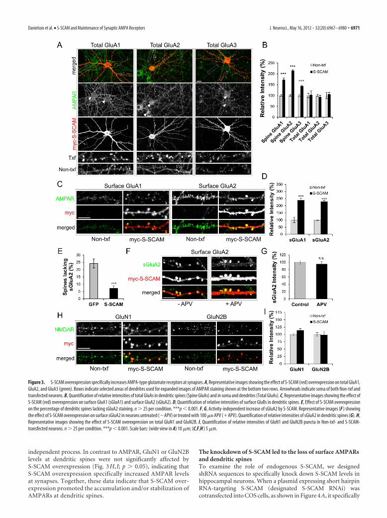
independent process. In contrast to AMPAR, GluN1 or GluN2Blevels at dendritic spines were not significantly affected byS-SCAM overexpression (Fig. 3H, I; p � 0.05), indicating thatS-SCAM overexpression specifically increased AMPAR levelsat synapses. Together, these data indicate that S-SCAM over-expression promoted the accumulation and/or stabilization ofAMPARs at dendritic spines.
The knockdown of S-SCAM led to the loss of surface AMPARsand dendritic spinesTo examine the role of endogenous S-SCAM, we designedshRNA sequences to specifically knock down S-SCAM levels inhippocampal neurons. When a plasmid expressing short hairpinRNA-targeting S-SCAM (designated S-SCAM RNAi) wascotransfected into COS cells, as shown in Figure 4A, it specifically
Figure 3. S-SCAM overexpression specifically increases AMPA-type glutamate receptors at synapses. A, Representative images showing the effect of S-SCAM (red) overexpression on total GluA1,GluA2, and GluA3 (green). Boxes indicate selected areas of dendrites used for expanded images of AMPAR staining shown at the bottom two rows. Arrowheads indicate soma of both Non-txf andtransfected neurons. B, Quantification of relative intensities of total GluAs in dendritic spines (Spine GluAs) and in soma and dendrites (Total GluAs). C, Representative images showing the effect ofS-SCAM (red) overexpression on surface GluA1 (sGluA1) and surface GluA2 (sGluA2). D, Quantification of relative intensities of surface GluRs in dendritic spines. E, Effect of S-SCAM overexpressionon the percentage of dendritic spines lacking sGluA2 staining. n � 25 per condition. ***p � 0.001. F, G, Activity-independent increase of sGluA2 by S-SCAM. Representative images (F ) showingthe effect of S-SCAM overexpression on surface sGluA2 in neurons untreated (�APV) or treated with 100 �M APV (�APV). Quantification of relative intensities of sGluA2 in dendritic spines (G). H,Representative images showing the effect of S-SCAM overexpression on total GluN1 and GluN2B. I, Quantification of relative intensities of GluN1 and GluN2B puncta in Non-txf- and S-SCAM-transfected neurons. n � 25 per condition. ***p � 0.001. Scale bars: (wide view in A) 10 �m; (C,F,H ) 5 �m.
Danielson et al. • S-SCAM and Maintenance of Synaptic AMPA Receptors J. Neurosci., May 16, 2012 • 32(20):6967– 6980 • 6971
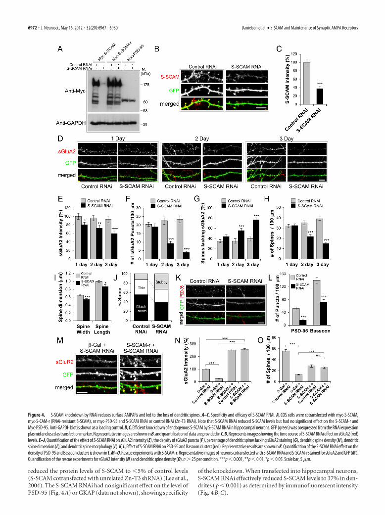
reduced the protein levels of S-SCAM to �5% of control levels(S-SCAM cotransfected with unrelated Zn-T3 shRNA) (Lee et al.,2004). The S-SCAM RNAi had no significant effect on the level ofPSD-95 (Fig. 4A) or GKAP (data not shown), showing specificity
of the knockdown. When transfected into hippocampal neurons,S-SCAM RNAi effectively reduced S-SCAM levels to 37% in den-drites (p � 0.001) as determined by immunofluorescent intensity(Fig. 4B,C).
Figure 4. S-SCAM knockdown by RNAi reduces surface AMPARs and led to the loss of dendritic spines. A–C, Specificity and efficacy of S-SCAM RNAi. A, COS cells were cotransfected with myc-S-SCAM,myc-S-CAM-r (RNAi-resistant S-SCAM), or myc-PSD-95 and S-SCAM RNAi or control RNAi (Zn-T3 RNAi). Note that S-SCAM RNAi reduced S-SCAM levels but had no significant effect on the S-SCAM-r andMyc-PSD-95. Anti-GAPDH blot is shown as a loading control. B, C, Efficient knockdown of endogenous S-SCAM by S-SCAM RNAi in hippocampal neurons. GFP (green) was coexpressed from the RNAi expressionplasmid and used as transfection marker. Representative images are shown in B, and quantification of data are provided in C. D, Represents images showing the time course of S-SCAM RNAi effect on sGluA2 (red)levels. E–J, Quantification of the effect of S-SCAM RNAi on sGluA2 intensity (E), the density of sGluA2 puncta (F ), percentage of dendritic spines lacking sGluA2 staining (G), dendritic spine density (H ), dendriticspine dimension (I ), and dendritic spine morphology (J ). K, L, Effect of S-SCAM RNAi on PSD-95 and Bassoon clusters (red). Representative results are shown in K. Quantification of the S-SCAM RNAi effect on thedensityofPSD-95andBassoonclusters isshowninL.M–O,RescueexperimentswithS-SCAM-r.RepresentativeimagesofneuronscotransfectedwithS-SCAMRNAiandS-SCAM-rstainedforsGluA2andGFP(M ).Quantification of the rescue experiments for sGluA2 intensity (N ) and dendritic spine density (O). n � 25 per condition. ***p � 0.001, **p � 0.01, *p � 0.05. Scale bar, 5 �m.
6972 • J. Neurosci., May 16, 2012 • 32(20):6967– 6980 Danielson et al. • S-SCAM and Maintenance of Synaptic AMPA Receptors
We first examined the effect of S-SCAM RNAi on GluA2,a major subunit of AMPARs in the hippocampal neurons.S-SCAM RNAi reduced both the intensity and number of sGluA2puncta significantly (Fig. 4D–F). The reduction of sGluA2 inten-sity started to show as early as 1 d post-transfection of S-SCAMRNAi (100 � 7 vs 80 � 6%, control vs S-SCAM RNAi; p � 0.05)and reached 60% of control levels 3 d post-transfection (Fig. 4E).The reduction of sGluA2 puncta density became statistically sig-nificant 2 d post-transfection (23 � 2 vs 9 � 1 per 100 �mdendrite, control vs S-SCAM RNAi; p � 0.001) (Fig. 4F) and wasreduced to 17% of control level after 3 d (23 � 2 vs 4 � 1 per 100�m dendrite, control vs S-SCAM RNAi; p � 0.001). The reduc-tion in sGluA2 was accompanied by a significant increase in theproportion of dendritic spines lacking sGluA2 staining (after 3 d,40 � 4 vs 76 � 4%, control vs S-SCAM RNAi; p � 0.001) (Fig.4G). These data clearly indicate that S-SCAM RNAi removessGluA2 from dendritic spines.
S-SCAM RNAi also had a dramatic effect on dendritic spinesFig. 4B,D; GFP channels). Knockdown of S-SCAM reduced thedendritic spine density (at 3 d post-transfection, 39 � 2 vs 15 � 1per 100 �m dendrite, control vs S-SCAM RNAi; p � 0.001) (Fig.4H) and the size of dendritic spines (p � 0.001 for width and p �0.05 for length) (Fig. 4 I). Furthermore, S-SCAM knockdownincreased the proportion of stubby spines (13 vs 33%, control vsS-SCAM RNAi; p � 0.001) at the expense of mushroom-typemature spines (55 vs 39% control vs S-SCAM RNAi; p � 0.05)(Fig. 4 J). These data suggest that losing S-SCAM from synapsespromotes the collapse of mushroom-type dendritic spines tostubby ones, which are eliminated eventually. Consistent with thereduction in the number of dendritic spines, S-SCAM RNAigreatly reduced the puncta density of PSD-95 (52 � 5 vs 16 � 2per 100 �m dendrite, control vs S-SCAM RNAi; p � 0.001) (Fig.4K,L) and Bassoon (141 � 9 vs 73 � 8 per 100 �m dendrite,control vs S-SCAM RNAi; p � 0.001) (Fig. 4L), indicating thereduction of overall synapse numbers.
To further confirm the specificity of S-SCAM RNAi effect, weperformed a “rescue” experiment with S-SCAM RNAi-resistantS-SCAM (Fig. 4A, designated S-SCAM-r). When rescuingS-SCAM-r was coexpressed with S-SCAM RNAi, the amount ofsGluR2 was increased to a level similar to S-SCAM overexpres-sion (257 � 7 vs 252 � 8%, S-SCAM RNAi � S-SCAM-r vsS-SCAM-r � Control RNAi; p 0.67) (Fig. 4M,N) and thenumber of dendritic spines was similarly restored (25 � 1 vs 28 �2 per 100 �m dendrite, S-SCAM RNAi � S-SCAM-r vsS-SCAM-r � Control RNAi; p 0.26) (Fig. 4O). Thus, the effectof S-SCAM RNAi on AMPARs and dendritic spines was specifi-cally related to the loss of S-SCAM proteins and not caused bynonspecific effect of RNAi. Together, these data point out thatS-SCAM is an essential scaffolding molecule for the stabilization/maintenance of AMPARs and dendritic spines.
S-SCAM levels regulate the AMPAR component of excitatorysynaptic transmissionTo measure directly the effect of changing S-SCAM levels onAMPAR-mediated synaptic transmission, we measured AMPARmEPSCs after transfecting dissociated hippocampal culture neu-rons with S-SCAM or S-SCAM RNAi constructs. IncreasingS-SCAM levels enhanced AMPA mEPSC amplitudes (17.4 � 0.9vs 23.4 � 1.0 pA, GFP control vs S-SCAM; p � 0.001) (Fig.5A–C), while S-SCAM RNAi reduces AMPA mEPSC amplitudes(14.5 � 0.9 pA; p � 0.01 compared with GFP control) (Fig.5A–C). We did not observe significant changes in the AMPAmEPSC frequency after S-SCAM overexpression (3.9 � 0.5 vs
4.6 � 1.0 Hz, GFP control vs S-SCAM; p 0.35) (Fig. 5D),despite the reduction in dendritic spine density. This is likely dueto the “unsilencing” of silent synapses by acquiring AMPAR (Fig.3E). In contrast, S-SCAM RNAi greatly reduced mEPSC fre-quency (1.7 � 0.4 Hz; p � 0.005), consistent with the reductionin synapse numbers after S-SCAM RNAi (Fig. 4H). Therefore,S-SCAM levels directly influence synaptic AMPAR levels.
S-SCAM overexpression in CA1 pyramidal neurons of hip-pocampal slices cultured by sindbis virus infection drasticallyincreased AMPAR-mediated synaptic transmission measured at�60 mV (26.5 � 4.2 vs 51.0 � 5.5 pA, uninfected neighboringneuron vs S-SCAM-infected; p � 0.001) (Fig. 5E,F). In contrast,we did not detect significant changes in the NMDAR-mediated re-sponses measured at �40 mV (16.5 � 2.5 vs 17.4 � 2.6 pA, unin-fected vs S-SCAM-infected; p 0.77) (Fig. 5E,G), indicating thatS-SCAM overexpression did not affect NMDAR-mediated synaptictransmission significantly. Importantly, S-SCAM-infected neuronsshowed a significant increase in the AMPA/NMDA ratio (1.9 � 0.2vs 3.3.0 � 0.4, uninfected vs S-SCAM-infected; p � 0.005) (Fig. 5H),indicating that S-SCAM overexpression specifically increased theAMPA component of excitatory synaptic transmission. We did notfind a significant difference in the PPR of S-SCAM-infected CA1neurons from uninfected neighboring neurons (1.7 � 0.3 vs1.5 � 0.2 at 50 ms interval, 1.4 � 0.1 vs 1.4 � 0.2 at100 msinterval, uninfected vs S-SCAM infected; p � 0.4) (Fig. 5 I, J),indicating that S-SCAM overexpression did not change presyn-aptic function significantly.
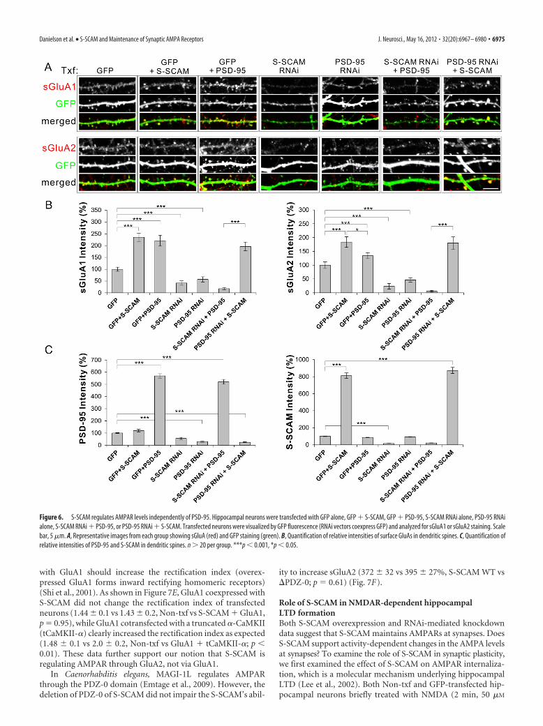
S-SCAM regulates synaptic AMPAR independently of PSD-95Our data and literature indicate that both S-SCAM and PSD-95can regulate synaptic AMPAR levels. Do they play redundant orindependent roles? To address this question, we performed “re-ciprocal rescuing” experiments. When S-SCAM or PSD-95 wasoverexpressed in hippocampal neurons (with a GFP txf marker),both proteins increased sGluA1 levels comparably (Fig. 6A,B)(100 � 8 vs 235 � 18 or 221 � 43%, non-txf vs S-SCAM orPSD-95; p � 0.001). Surface GluA2 levels were also increased byboth S-SCAM and PSD-95 overexpression, although S-SCAMwas more effective than PSD-95 (100 � 15 vs 184 � 19 or 134 �11%, non-txf vs S-SCAM or PSD-95, p � 0.001; S-SCAM vsnon-txf, p � 0.05; S-SCAM vs PSD-95). On the other hand,S-SCAM RNAi and PSD-95 RNAi reduced both sGluA levelsdrastically (for sGluA1, 41 � 9 vs 57 � 11%; for sGluA2, 23 � 10vs 46 � 7%, S-SCAM RNAi vs PSD-95 RNAi; p � 0.001 com-pared with GFP control for both RNAi). Importantly, overex-pression of PSD-95 in the presence of S-SCAM RNAi did notincrease or restore either sGluA subunit level at synapses (18 �5% for GluA1, 6 � 3% for GluA2). In sharp contrast, overexpres-sion of S-SCAM in the presence of PSD RNAi not only rescuedthe loss of PSD-95 but also increased both sGluA levels at den-dritic spines comparable to S-SCAM overexpression alone(sGluA1, 196 � 18 vs 235 � 18%; sGluA2, 184 � 19 vs 180 �22%,;PSD-95 RNAi � S-SCAM vs S-SCAM; for both sGluAs, p �0.001 and p � 0.1, compared with PSD-95 RNAi and to S-SCAMalone, respectively) (Fig. 6A,B). Quantification of relative inten-sities of PSD-95 and S-SCAM verified that both protein overex-pressions reached �5-fold higher levels than the endogenousprotein levels, regardless of the presence of RNAi (Fig. 6C).Therefore, S-SCAM increased surface AMPARs independently ofPSD-95, while PSD-95 was dependent on S-SCAM to exert itseffect on AMPARs. These data suggest that S-SCAM is an indis-pensable scaffolding molecule for AMPARs.
Danielson et al. • S-SCAM and Maintenance of Synaptic AMPA Receptors J. Neurosci., May 16, 2012 • 32(20):6967– 6980 • 6973
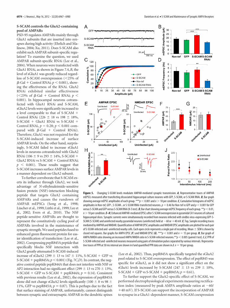
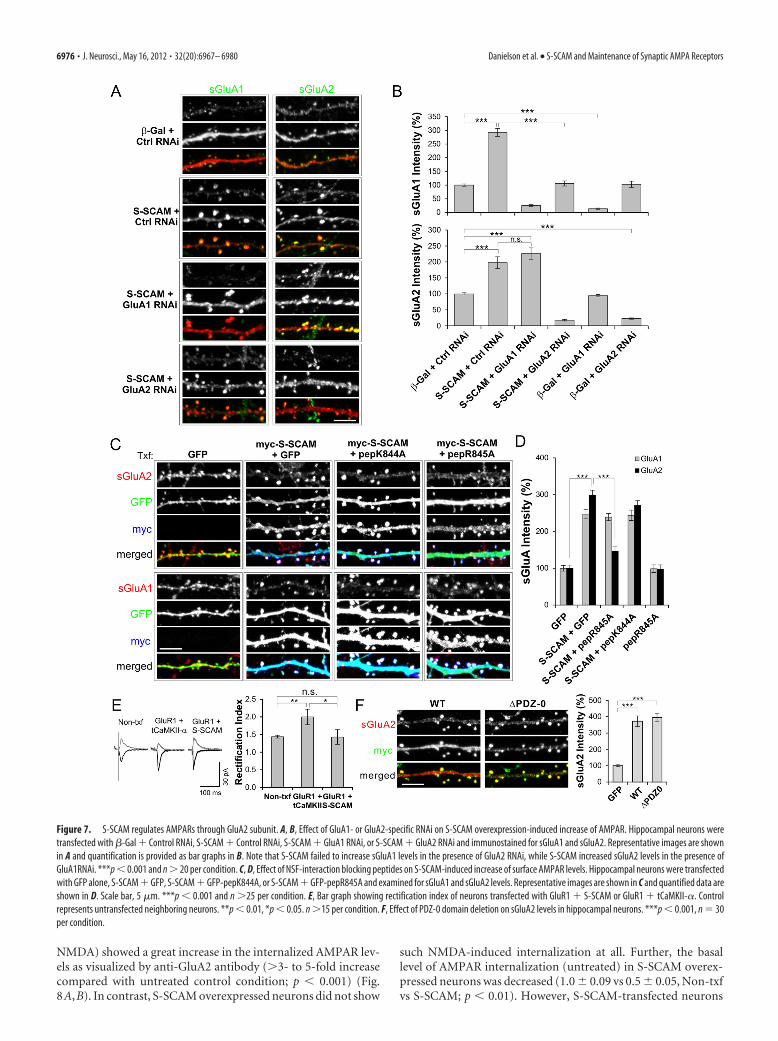
S-SCAM controls the GluA2-containingpool of AMPARsPSD-95 regulates AMPARs mainly throughGluA1 subunits that are inserted into syn-apses during high activity (Ehrlich and Ma-linow, 2004; Xu, 2011). Does S-SCAM alsoexhibit such AMPAR subunit-specific regu-lation? To examine the question, we usedAMPAR subunit-specific RNAi (Lee et al.,2004). When neurons were transfected withGluA1 RNAi, as shown in Figure 7A,B, thelevel of sGluA1 was greatly reduced regard-less of S-SCAM overexpression (�25% of�-Gal � Control RNAi; p � 0.001), show-ing the effectiveness of the RNAi. GluA2RNAi exhibited similar effectiveness(�23% of �-Gal � Control RNAi; p �0.001). In hippocampal neurons cotrans-fected with GluA1 RNAi and S-SCAM,sGluA2 levels were significantly increased toa level comparable to that of S-SCAM �Control RNAi (226 � 18 vs 198 � 18%,S-SCAM � GluA1 RNAi vs S-SCAM �Control RNAi, p 0.28; p � 0.001 com-pared with �-Gal � Control RNAi).Therefore, GluA1 was not required for theS-SCAM-induced increase of surfaceAMPAR levels. On the other hand, surpris-ingly, S-SCAM failed to increase sGluA1levels in neurons cotransfected with GluA2RNAi (106 � 9 vs 293 � 14%, S-SCAM �GluA2 RNAi vs S-SCAM � Control RNAi;p � 0.001). These results suggest thatS-SCAM increases surface AMPAR levels ina manner dependent on GluA2 subunit.
To further corroborate that S-SCAM ex-erts its influence through GluA2, we tookadvantage of N-ethylmaleimide-sensitivefusion protein (NSF)-interaction blockingpeptide that targets GluA2 containingAMPARs and causes the rundown ofAMPAR mEPSCs (Song et al., 1998;Luscher et al., 1999; Luthi et al., 1999; Lee etal., 2002; Evers et al., 2010). The NSFpeptide-sensitive AMPARs are thought torepresent the constitutively cycling pool ofAMPARs involved in the maintenance ofsynaptic strength. We used peptides fused toenhanced green fluorescent protein for eas-ier identification of transfection (Lee et al.,2002). Coexpressing pepR845A peptide thatspecifically blocks NSF interaction withGluA2 greatly attenuated S-SCAM-inducedincrease of sGluA2 (299 � 13 vs 147 � 11%, S-SCAM � GFP vsS-SCAM � pepR845A; p � 0.001) (Fig. 7C,D). In contrast, the neg-ative control peptide pepK844A that does not interfere with NSF orAP2 interaction had no significant effect (299 � 13 vs 270 � 13%,S-SCAM � GFP vs S-SCAM � pepK844A; p 0.14). Consistentwith previous results (Lee et al., 2002), the expression of pepR845Aalone did not change sGluA2 levels significantly (100 � 8 vs 98 �11%, GFP vs pepR845A; p 0.87). This is perhaps due to the factthat surface staining of AMPAR, unfortunately, cannot distinguishbetween synaptic and extrasynaptic AMPAR in the dendritic spines
(Lee et al., 2002). Thus, pepR845A specifically targeted the sGluA2pool related to S-SCAM overexpression. The effect of pepR845 wasspecific for sGluA2, as it did not show a significant effect on thesGluA1 levels increased by S-SCAM (247 � 13 vs 239 � 10%,S-SCAM � GFP vs S-SCAM � pepR845A; p 0.61).
To further support the GluA2-specific effect of S-SCAM, weperformed electrophysiological experiments measuring rectifica-tion index (measured by peak AMPA amplitude ratios at �60/�40 mV). If S-SCAM can support the incorporation of AMPARto synapse in a GluA1-dependent manner, S-SCAM coexpression
Figure 5. Changing S-SCAM levels modulate AMPAR-mediated synaptic transmission. A, Representative traces of AMPARmEPSCs measured after transfecting dissociated hippocampal culture neurons with GFP, S-SCAM, or S-SCAM RNAi. B, Bar graphshowing average mEPSC amplitudes of each group. ***p � 0.001 and n � 10 per condition. C, Cumulative histograms of mEPSCamplitudes in Non-txf, GFP-, S-SCAM-, or S-SCAM RNAi-transfected neurons. p 0.46 for Non-txf vs GFP and p � 0.001 for GFPversus S-SCAM and GFP versus S-SCAM RNAi (K-S test). D, Bar chart showing average mEPSC frequency of each group. **p � 0.01,n � 10 per condition. E–H, Enhanced AMPAR-mediated EPSCs after S-SCAM overexpression in pyramidal CA1 neurons of culturedhippocampal slices. Synaptic currents were simultaneously recorded from neurons infected with sindbis virus-expressing GFP-S-SCAM (S-SCAM) and uninfected nearby pyramidal neurons (uninfected) held at �60 or �40 mV. E, Top, Sample recording tracesmediated by AMPAR and NMDAR. Quantification of AMPAR EPSC amplitudes and NMDAR EPSC amplitudes are plotted for each pairof S-SCAM-infected and -uninfected nearby cells. Each open circle represents a single pair of recording. Mean � SEM is shown byclosed red squares. Bar graphs for AMPA EPSC (F ) and NMDAR EPSC (G). ***p � 0.001 and n 15 per group. H, Bar graph ofAMPA/NMDA ratio showing an increased AMPA/NMDA ratio in S-SCAM-infected neurons.**p � 0.005 (paired t test). I, J, PPR ofS-SCAM-infected and -uninfected neurons measured using pairs of stimulation pulses separated by various intervals. Representa-tive traces of PPR at 50 ms interval are shown in I and quantified PPR data are shown in J. n 10 per group.
6974 • J. Neurosci., May 16, 2012 • 32(20):6967– 6980 Danielson et al. • S-SCAM and Maintenance of Synaptic AMPA Receptors
with GluA1 should increase the rectification index (overex-pressed GluA1 forms inward rectifying homomeric receptors)(Shi et al., 2001). As shown in Figure 7E, GluA1 coexpressed withS-SCAM did not change the rectification index of transfectedneurons (1.44 � 0.1 vs 1.43 � 0.2, Non-txf vs S-SCAM � GluA1,p 0.95), while GluA1 cotransfected with a truncated �-CaMKII(tCaMKII-�) clearly increased the rectification index as expected(1.48 � 0.1 vs 2.0 � 0.2, Non-txf vs GluA1 � tCaMKII-�; p �0.01). These data further support our notion that S-SCAM isregulating AMPAR through GluA2, not via GluA1.
In Caenorhabditis elegans, MAGI-1L regulates AMPARthrough the PDZ-0 domain (Emtage et al., 2009). However, thedeletion of PDZ-0 of S-SCAM did not impair the S-SCAM’s abil-
ity to increase sGluA2 (372 � 32 vs 395 � 27%, S-SCAM WT vsPDZ-0; p 0.61) (Fig. 7F).
Role of S-SCAM in NMDAR-dependent hippocampalLTD formationBoth S-SCAM overexpression and RNAi-mediated knockdowndata suggest that S-SCAM maintains AMPARs at synapses. DoesS-SCAM support activity-dependent changes in the AMPA levelsat synapses? To examine the role of S-SCAM in synaptic plasticity,we first examined the effect of S-SCAM on AMPAR internaliza-tion, which is a molecular mechanism underlying hippocampalLTD (Lee et al., 2002). Both Non-txf and GFP-transfected hip-pocampal neurons briefly treated with NMDA (2 min, 50 �M
Figure 6. S-SCAM regulates AMPAR levels independently of PSD-95. Hippocampal neurons were transfected with GFP alone, GFP � S-SCAM, GFP � PSD-95, S-SCAM RNAi alone, PSD-95 RNAialone, S-SCAM RNAi � PSD-95, or PSD-95 RNAi � S-SCAM. Transfected neurons were visualized by GFP fluorescence (RNAi vectors coexpress GFP) and analyzed for sGluA1 or sGluA2 staining. Scalebar, 5 �m. A, Representative images from each group showing sGluA (red) and GFP staining (green). B, Quantification of relative intensities of surface GluAs in dendritic spines. C, Quantification ofrelative intensities of PSD-95 and S-SCAM in dendritic spines. n � 20 per group. ***p � 0.001, *p � 0.05.
Danielson et al. • S-SCAM and Maintenance of Synaptic AMPA Receptors J. Neurosci., May 16, 2012 • 32(20):6967– 6980 • 6975
NMDA) showed a great increase in the internalized AMPAR lev-els as visualized by anti-GluA2 antibody (�3- to 5-fold increasecompared with untreated control condition; p � 0.001) (Fig.8A,B). In contrast, S-SCAM overexpressed neurons did not show
such NMDA-induced internalization at all. Further, the basallevel of AMPAR internalization (untreated) in S-SCAM overex-pressed neurons was decreased (1.0 � 0.09 vs 0.5 � 0.05, Non-txfvs S-SCAM; p � 0.01). However, S-SCAM-transfected neurons
Figure 7. S-SCAM regulates AMPARs through GluA2 subunit. A, B, Effect of GluA1- or GluA2-specific RNAi on S-SCAM overexpression-induced increase of AMPAR. Hippocampal neurons weretransfected with �-Gal � Control RNAi, S-SCAM � Control RNAi, S-SCAM � GluA1 RNAi, or S-SCAM � GluA2 RNAi and immunostained for sGluA1 and sGluA2. Representative images are shownin A and quantification is provided as bar graphs in B. Note that S-SCAM failed to increase sGluA1 levels in the presence of GluA2 RNAi, while S-SCAM increased sGluA2 levels in the presence ofGluA1RNAi. ***p � 0.001 and n � 20 per condition. C, D, Effect of NSF-interaction blocking peptides on S-SCAM-induced increase of surface AMPAR levels. Hippocampal neurons were transfectedwith GFP alone, S-SCAM � GFP, S-SCAM � GFP-pepK844A, or S-SCAM � GFP-pepR845A and examined for sGluA1 and sGluA2 levels. Representative images are shown in C and quantified data areshown in D. Scale bar, 5 �m. ***p � 0.001 and n �25 per condition. E, Bar graph showing rectification index of neurons transfected with GluR1 � S-SCAM or GluR1 � tCaMKII-�. Controlrepresents untransfected neighboring neurons. **p � 0.01, *p � 0.05. n �15 per condition. F, Effect of PDZ-0 domain deletion on sGluA2 levels in hippocampal neurons. ***p � 0.001, n 30per condition.
6976 • J. Neurosci., May 16, 2012 • 32(20):6967– 6980 Danielson et al. • S-SCAM and Maintenance of Synaptic AMPA Receptors
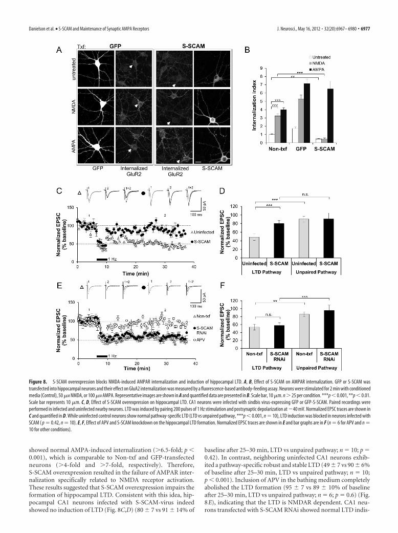
showed normal AMPA-induced internalization (�6.5-fold; p �0.001), which is comparable to Non-txf and GFP-transfectedneurons (�4-fold and �7-fold, respectively). Therefore,S-SCAM overexpression resulted in the failure of AMPAR inter-nalization specifically related to NMDA receptor activation.These results suggested that S-SCAM overexpression impairs theformation of hippocampal LTD. Consistent with this idea, hip-pocampal CA1 neurons infected with S-SCAM-virus indeedshowed no induction of LTD (Fig. 8C,D) (80 � 7 vs 91 � 14% of
baseline after 25–30 min, LTD vs unpaired pathway; n 10; p 0.42). In contrast, neighboring uninfected CA1 neurons exhib-ited a pathway-specific robust and stable LTD (49 � 7 vs 90 � 6%of baseline after 25–30 min, LTD vs unpaired pathway; n 10;p � 0.001). Inclusion of APV in the bathing medium completelyabolished the LTD formation (95 � 7 vs 89 � 10% of baselineafter 25–30 min, LTD vs unpaired pathway; n 6; p 0.6) (Fig.8E), indicating that the LTD is NMDAR dependent. CA1 neu-rons transfected with S-SCAM RNAi showed normal LTD indis-
Figure 8. S-SCAM overexpression blocks NMDA-induced AMPAR internalization and induction of hippocampal LTD. A, B, Effect of S-SCAM on AMPAR internalization. GFP or S-SCAM wastransfected into hippocampal neurons and their effect on GluA2 internalization was measured by a fluorescence-based antibody-feeding assay. Neurons were stimulated for 2 min with conditionedmedia (Control), 50 �M NMDA, or 100 �M AMPA. Representative images are shown in A and quantified data are presented in B. Scale bar, 10 �m. n � 25 per condition. ***p � 0.001, **p � 0.01.Scale bar represents 10 �m. C, D, Effect of S-SCAM overexpression on hippocampal LTD. CA1 neurons were infected with sindbis virus-expressing GFP or GFP-S-SCAM. Paired recordings wereperformed in infected and uninfected nearby neurons. LTD was induced by pairing 200 pulses of 1 Hz stimulation and postsynaptic depolarization at �40 mV. Normalized EPSC traces are shown inC and quantified in D. While uninfected control neurons show normal pathway-specific LTD (LTD vs unpaired pathway, ***p � 0.001, n 10), LTD induction was blocked in neurons infected withSCAM ( p 0.42, n 10). E, F, Effect of APV and S-SCAM knockdown on the hippocampal LTD formation. Normalized EPSC traces are shown in E and bar graphs are in F (n 6 for APV and n 10 for other conditions).
Danielson et al. • S-SCAM and Maintenance of Synaptic AMPA Receptors J. Neurosci., May 16, 2012 • 32(20):6967– 6980 • 6977
tinguishable from Non-txf neighboring neurons (57 � 5 vs 53 �6% of baseline after 25–30 min, S-SCAM RNAi vs Non-txf; n 10, p 0.61) (Fig. 8E,F). Together, these results suggest thatS-SCAM is not involved in the regulation of LTD-formingAMPAR pool.
DiscussionIn this study, we elucidate a novel and essential function ofS-SCAM as a synaptic AMPAR scaffold. From overexpressionexperiments, we found that increasing S-SCAM levels promotesenlargement of dendritic spines, changes the molecular compo-sition of PSD proteins, enhances AMPAR-mediated synaptictransmission, and blocks the induction of hippocampal LTD.Conversely, knockdown studies demonstrated that the loss ofS-SCAM decreases synaptic AMPAR levels, reduces the size ofdendritic spines, and severely reduces excitatory synapse num-bers. Therefore, S-SCAM is an important scaffolding moleculeinvolved in the synaptic protein organization and the control ofsynaptic AMPAR.
The most significant finding here is the novel and essentialrole of S-SCAM in the maintenance of AMPAR at synapses. Oneof the prevailing hypotheses in the AMPAR trafficking is that twoseparate pools of AMPARs contribute to differential aspects inAMPAR trafficking: constitutive and regulated pathways (Shi etal., 2001; Derkach et al., 2007; Shepherd and Huganir, 2007; Kes-sels and Malinow, 2009). The former is important for the main-tenance of synaptic strength during protein turnover, while thelatter is involved in changing synaptic AMPAR levels during syn-aptic plasticity. In this model, different AMPAR subunits playleading roles in the differential phases of AMPAR trafficking:GluA1 plays a dominant role in the activity-dependent insertionof AMPAR during LTP, while GluA2 is involved in the mainte-nance of synaptic AMPAR levels and activity-dependent removalof AMPAR from synapses during LTD. Accumulating evidencesupports that PSD-95 is important for the regulated pathways.For example, PSD-95 overexpression strongly enhanced AMPAreceptor-mediated synaptic transmission by molecular mecha-nisms similar to LTP, in that it drives GluA1-containing recep-tors to synapses, converts silent synapses to functional synapses,occludes LTP, and enhances LTD (El-Husseini et al., 2000; Sch-nell et al., 2002; Beïque and Andrade, 2003; Stein et al., 2003;Ehrlich and Malinow, 2004; Nakagawa et al., 2004; Elias et al.,2006). On the other hand, knockdown of PSD-95 reduced syn-aptic AMPAR levels (Nakagawa et al., 2004; Prange et al., 2004),and impaired LTD (Ehrlich et al., 2007; Xu et al., 2008) withoutaffecting synapse numbers significantly (Beïque and Andrade,2003; Elias et al., 2006).
In contrast to the regulated pathway, the identity of scaffold-ing protein(s) involved in the constitutive pathway has remainedunclear. Our results suggest that S-SCAM is the scaffolding mol-ecule important for the constitutive pathway. Multiple lines ofevidence support this notion: First, S-SCAM increases synapticAMPAR levels through a GluA2-dependent mechanism, asshown by GluA subunit-specific knockdown and NSF-peptideexperiments (Fig. 5). Second, the increase of AMPAR by S-SCAMis an activity-independent process (Fig. 3E,F), consistent withthe delivery mechanism of GluA2 receptors replacing GluA1 (Shiet al., 2001). Third, S-SCAM is required for maintaining AMPARlevels even in the presence of PSD-95 overexpression, as indicatedby knockdown experiments (Figs. 4, 6). Fourth, the knockdownof S-SCAM led to the loss of excitatory synapses (Fig. 4H), whileS-SCAM overexpression did not increase the number of excit-atory synapses (PSD-95 overexpression increases it). Fifth,
S-SCAM does not support activity-dependent changes of synap-tic AMPA receptor levels, as demonstrated by the blockade ofLTD after overexpression and by normal LTD after knockdown(Fig. 8). Thus, a pool of synaptic AMPARs anchored by S-SCAMis expected to play a “house-keeping” role during synaptic plas-ticity. Together, we propose a model in which S-SCAM stabilizesa maintenance pool of AMPARs at synapses and PSD-95 (to-gether with other PSD-MAGUKs) regulates a plasticity-relatedpool of AMPAR.
How does S-SCAM serve a differential function from PSD-95in the regulation of AMPA receptors and dendritic spines? Wespeculate that it arises from the overlapping but differential pro-tein–protein interaction profile and/or post-translational modi-fications of S-SCAM protein. For example, S-SCAM does nothave a palmitoylation site and lacks AKAP interaction, both ofwhich were shown to be important for activity-dependentAMPAR internalization and LTD (El-Husseini et al., 2002; Bhat-tacharyya et al., 2009). In addition, S-SCAM interacts withN-cadherins through �-catenin (Nishimura et al., 2002; Okabe etal., 2003), which may provide a mechanism for its stabilization atsynapses during plastic changes. It is intriguing that S-SCAMpromoted dendritic spine enlargements in the absence of in-creased synaptic levels of Shank (Fig. 2C) and SPAR (data notshown), critical molecules involved in dendritic spine enlarge-ment/maturation (Pak et al., 2001; Sala et al., 2001). S-SCAMmay increase dendritic spines through GluA2 subunits, whoseextracellular interaction with N-cadherin mediates dendriticspine enlargement (Passafaro et al., 2003; Saglietti et al., 2007).Further studies are necessary to examine these possibilities.
Our finding that S-SCAM plays an important role in the reg-ulation AMPAR is not without precedents. In C. elegans, theinteraction of MAGI-1L (long form of MAGI-1) with the AMPA-like subunit GLR-2 has shown to be important for the synapticlocalization of GLR-1/2 (Emtage et al., 2009). However, the mo-lecular details of the action are different from the vertebrate sys-tem. While GLR-1/2 directly binds to MAGI-1L through PDZinteraction, vertebrate GluAs interact with S-SCAM indirectly viaTARP (Deng et al., 2006), like PSD-95. Furthermore, the firstPDZ domain (PDZ-0) is important for the synaptic GLR regula-tion by MAGI-1L in C. elegans, but PDZ-0 is not important forAMPAR regulation (Fig. 7F ). Further detailed studies on theS-SCAM–TARP interaction are necessary to establish molec-ular mechanisms underlying S-SCAM-mediated regulation ofAMPARs.
It is remarkable that S-SCAM did not increase sGluA1 levels inneurons with GluA2 knockdown, suggesting a GluA2-dependentmechanism. Since GluA2 plays an important role in the assemblyand synaptic incorporation of heteromeric AMPAR (Wentholdet al., 1996; Lu et al., 2009), GluA2 knockdown may have causeda poor assembly and/or trafficking of GluA1-containing recep-tors to neuronal surface. However, this is unlikely the case in ourcondition, since GluA2 knockdown showed normal sGluA1 lev-els (Fig. 7B). Consistent with this, homomeric GluA1 receptorswere detected at synapses shortly after GluA2 deletion (Lu et al.,2009). Furthermore, S-SCAM failed to accommodate GluA1 ho-momeric receptors at the synapses (Fig. 7E). Therefore, these dataindicate that S-SCAM regulates AMPAR in a GluA2-dependentmanner. At present, the molecular bases of GluA2 dependence ofthe S-SCAM action are unclear, as TARP binds all GluA subunits.However, one could imagine that GluA2-specific binding proteinssuch as GRIP, NSF, PICK1, and/or yet unidentified protein(s) maywork together with S-SCAM to provide a GluA2-dependent mech-anism at synapses. Another intriguing question is what role S-SCAM
6978 • J. Neurosci., May 16, 2012 • 32(20):6967– 6980 Danielson et al. • S-SCAM and Maintenance of Synaptic AMPA Receptors
plays in the control of inhibitory synaptic transmission, sinceS-SCAM is also present in inhibitory synapses (Sumita et al., 2007).
Previously, mutant mice lacking the expression of S-SCAM-�did not show drastic abnormalities in dendritic spine density,albeit they showed elongated spine length (Iida et al., 2007).However, the S-SCAM-� knock-out mice still express otherS-SCAM isoforms of � and �. S-SCAM-� is the most abundantform expressed in the forebrain (Hirao et al., 2000; Deng et al.,2006). The main difference between the � and � isoform is thepresence of PDZ-0 domain. As PDZ-0 mutant increasedsGluR2 levels as effectively as WT (Fig. 7F), PDZ-0 domain is notrequired for the S-SCAM-mediated regulation of AMPAR. Thus,it is highly likely that the remaining S-SCAM-� (and possibly �)in the S-SCAM-� knock-out mice is sufficient to control AMPARand dendritic spine.
In addition to uncovering the role of S-SCAM in excitatorysynaptic transmission, our study also contributes to the under-standing of the pathobiology of neurological diseases, sinceS-SCAM gene duplication and deletion were found in individualswith schizophrenia and IS, respectively (Marshall et al., 2008;Walsh et al., 2008). Although these discoveries do not establish acausal relationship between S-SCAM and schizophrenia and/orIS per se, our results provide valuable insight into the pathologi-cal bases of these diseases. Here we have demonstrated thatchanging levels of S-SCAM profoundly affects synaptic transmis-sion and synaptic plasticity. For example, increasing levels ofS-SCAM enhances AMPAR-mediated synaptic transmission,but, at the same time, renders neurons less adaptable for at leastone type of synaptic plasticity, LTD. Conversely, decreasedS-SCAM levels profoundly reduce the number of excitatory syn-apses and weaken synaptic transmission. Incidentally, elevatedlevels of S-SCAM, a highly likely situation under S-SCAM geneduplication, caused the reduction in the number of dendriticspines, which is consistent with postmortem studies on the brainsof patients with schizophrenia (Garey et al., 1998; Glantz andLewis, 2000; Sweet et al., 2009; Penzes et al., 2011). Furthermore,aberrant Neuregulin-1 (NRG-1)/ErbB4 receptor signaling is oneof the prevailing hypotheses in schizophrenia research (Mei andXiong, 2008). Since S-SCAM also binds to ErbB4 receptorsthrough PDZ interaction (Buxbaum et al., 2008), further studieson the role of S-SCAM in the NRG-1/ErbB4 signaling will pro-vide important clues on a better understanding pathologicalbases of the disorder.
ReferencesBeïque JC, Andrade R (2003) PSD-95 regulates synaptic transmission and
plasticity in rat cerebral cortex. J Physiol 546:859 – 867.Bhattacharyya S, Biou V, Xu W, Schluter O, Malenka RC (2009) A critical
role for PSD-95/AKAP interactions in endocytosis of synaptic AMPAreceptors. Nat Neurosci 12:172–181.
Buxbaum JD, Georgieva L, Young JJ, Plescia C, Kajiwara Y, Jiang Y, MoskvinaV, Norton N, Peirce T, Williams H, Craddock NJ, Carroll L, Corfas G,Davis KL, Owen MJ, Harroch S, Sakurai T, O’Donovan MC (2008) Mo-lecular dissection of NRG1-ERBB4 signaling implicates PTPRZ1 as a po-tential schizophrenia susceptibility gene. Mol Psychiatry 13:162–172.
Collingridge GL, Isaac JT, Wang YT (2004) Receptor trafficking and synap-tic plasticity. Nat Rev Neurosci 5:952–962.
Deng F, Price MG, Davis CF, Mori M, Burgess DL (2006) Stargazin andother transmembrane AMPA receptor regulating proteins interact withsynaptic scaffolding protein MAGI-2 in brain. J Neurosci 26:7875–7884.
Derkach VA, Oh MC, Guire ES, Soderling TR (2007) Regulatory mecha-nisms of AMPA receptors in synaptic plasticity. Nat Rev Neurosci8:101–113.
Ehrlich I, Malinow R (2004) Postsynaptic density 95 controls AMPA recep-tor incorporation during long-term potentiation and experience-drivensynaptic plasticity. J Neurosci 24:916 –927.
Ehrlich I, Klein M, Rumpel S, Malinow R (2007) PSD-95 is required for activity-driven synapse stabilization. Proc Natl Acad Sci U S A 104:4176–4181.
El-Husseini AE, Schnell E, Chetkovich DM, Nicoll RA, Bredt DS (2000)PSD-95 involvement in maturation of excitatory synapses. Science290:1364 –1368.
El-Husseini AE, Schnell E, Dakoji S, Sweeney N, Zhou Q, Prange O, Gauthier-Campbell C, Aguilera-Moreno A, Nicoll RA, Bredt DS (2002) Synapticstrength regulated by palmitate cycling on PSD-95. Cell 108:849–863.
Elias GM, Nicoll RA (2007) Synaptic trafficking of glutamate receptors byMAGUK scaffolding proteins. Trends Cell Biol 17:343–352.
Elias GM, Funke L, Stein V, Grant SG, Bredt DS, Nicoll RA (2006) Synapse-specific and developmentally regulated targeting of AMPA receptors by afamily of MAGUK scaffolding proteins. Neuron 52:307–320.
Emtage L, Chang H, Tiver R, Rongo C (2009) MAGI-1 modulates AMPAreceptor synaptic localization and behavioral plasticity in response toprior experience. PLoS One 4:e4613.
Evers DM, Matta JA, Hoe HS, Zarkowsky D, Lee SH, Isaac JT, Pak DT (2010)Plk2 attachment to NSF induces homeostatic removal of GluA2 duringchronic overexcitation. Nat Neurosci 13:1199 –1207.
Garey LJ, Ong WY, Patel TS, Kanani M, Davis A, Mortimer AM, Barnes TR,Hirsch SR (1998) Reduced dendritic spine density on cerebral corticalpyramidal neurons in schizophrenia. J Neurol Neurosurg Psychiatry65:446 – 453.
Glantz LA, Lewis DA (2000) Decreased dendritic spine density on prefron-tal cortical pyramidal neurons in schizophrenia. Arch Gen Psychiatry57:65–73.
Hirabayashi S, Nishimura W, Iida J, Kansaku A, Kishida S, Kikuchi A, TanakaN, Hata Y (2004) Synaptic scaffolding molecule interacts with axin.J Neurochem 90:332–339.
Hirao K, Hata Y, Ide N, Takeuchi M, Irie M, Yao I, Deguchi M, Toyoda A,Sudhof TC, Takai Y (1998) A novel multiple PDZ domain-containingmolecule interacting with N-methyl-D-aspartate receptors and neuronalcell adhesion proteins. J Biol Chem 273:21105–21110.
Hirao K, Hata Y, Yao I, Deguchi M, Kawabe H, Mizoguchi A, Takai Y (2000)Three isoforms of synaptic scaffolding molecule and their characteriza-tion. Multimerization between the isoforms and their interaction withN-methyl-D-aspartate receptors and SAP90/PSD-95-associated protein.J Biol Chem 275:2966 –2972.
Iida J, Hirabayashi S, Sato Y, Hata Y (2004) Synaptic scaffolding molecule isinvolved in the synaptic clustering of neuroligin. Mol Cell Neurosci27:497–508.
Iida J, Ishizaki H, Okamoto-Tanaka M, Kawata A, Sumita K, Ohgake S, SatoY, Yorifuji H, Nukina N, Ohashi K, Mizuno K, Tsutsumi T, Mizoguchi A,Miyoshi J, Takai Y, Hata Y (2007) Synaptic scaffolding molecule alpha isa scaffold to mediate N-methyl-D-aspartate receptor-dependent RhoAactivation in dendrites. Mol Cell Biol 27:4388 – 4405.
Kessels HW, Malinow R (2009) Synaptic AMPA receptor plasticity and be-havior. Neuron 61:340 –350.
Kim E, Naisbitt S, Hsueh YP, Rao A, Rothschild A, Craig AM, Sheng M(1997) GKAP, a novel synaptic protein that interacts with the guanylatekinase-like domain of the PSD-95/SAP90 family of channel clusteringmolecules. J Cell Biol 136:669 – 678.
Lee SH, Liu L, Wang YT, Sheng M (2002) Clathrin adaptor AP2 and NSFinteract with overlapping sites of GluR2 and play distinct roles in AMPAreceptor trafficking and hippocampal LTD. Neuron 36:661– 674.
Lee SH, Simonetta A, Sheng M (2004) Subunit rules governing the sortingof internalized AMPA receptors in hippocampal neurons. Neuron43:221–236.
Lu W, Shi Y, Jackson AC, Bjorgan K, During MJ, Sprengel R, Seeburg PH,Nicoll RA (2009) Subunit composition of synaptic AMPA receptors re-vealed by a single-cell genetic approach. Neuron 62:254 –268.
Luscher C, Xia H, Beattie EC, Carroll RC, von Zastrow M, Malenka RC, NicollRA (1999) Role of AMPA receptor cycling in synaptic transmission andplasticity. Neuron 24:649 – 658.
Luthi A, Chittajallu R, Duprat F, Palmer MJ, Benke TA, Kidd FL, Henley JM,Isaac JT, Collingridge GL (1999) Hippocampal LTD expression involvesa pool of AMPARs regulated by the NSF-GluR2 interaction. Neuron24:389 –399.
Marshall CR, et al. (2008) Infantile spasms is associated with deletion ofthe MAGI2 gene on chromosome 7q11.23-q21.11. Am J Hum Genet83:106 –111.
Danielson et al. • S-SCAM and Maintenance of Synaptic AMPA Receptors J. Neurosci., May 16, 2012 • 32(20):6967– 6980 • 6979
Mei L, Xiong WC (2008) Neuregulin 1 in neural development, synapticplasticity and schizophrenia. Nat Rev Neurosci 9:437– 452.
Mok H, Shin H, Kim S, Lee JR, Yoon J, Kim E (2002) Association of thekinesin superfamily motor protein KIF1Balpha with postsynapticdensity-95 (PSD-95), synapse-associated protein-97, and synaptic scaf-folding molecule PSD-95/discs large/zona occludens-1 proteins. J Neuro-sci 22:5253–5258.
Naisbitt S, Kim E, Tu JC, Xiao B, Sala C, Valtschanoff J, Weinberg RJ, WorleyPF, Sheng M (1999) Shank, a novel family of postsynaptic density pro-teins that binds to the NMDA receptor/PSD-95/GKAP complex and cor-tactin. Neuron 23:569 –582.
Nakagawa T, Futai K, Lashuel HA, Lo I, Okamoto K, Walz T, Hayashi Y,Sheng M (2004) Quaternary structure, protein dynamics, and synapticfunction of SAP97 controlled by L27 domain interactions. Neuron44:453– 467.
Nishimura W, Yao I, Iida J, Tanaka N, Hata Y (2002) Interaction of synapticscaffolding molecule and �-catenin. J Neurosci 22:757–765.
Okabe S (2007) Molecular anatomy of the postsynaptic density. Mol CellNeurosci 34:503–518.
Okabe T, Nakamura T, Nishimura YN, Kohu K, Ohwada S, Morishita Y,Akiyama T (2003) RICS, a novel GTPase-activating protein for Cdc42and Rac1, is involved in the beta-catenin-N-cadherin and N-methyl-D-aspartate receptor signaling. J Biol Chem 278:9920 –9927.
Pak DT, Yang S, Rudolph-Correia S, Kim E, Sheng M (2001) Regulation ofdendritic spine morphology by SPAR, a PSD-95-associated RapGAP.Neuron 31:289 –303.
Passafaro M, Nakagawa T, Sala C, Sheng M (2003) Induction of dendriticspines by an extracellular domain of AMPA receptor subunit GluR2.Nature 424:677– 681.
Penzes P, Cahill ME, Jones KA, VanLeeuwen JE, Woolfrey KM (2011) Den-dritic spine pathology in neuropsychiatric disorders. Nat Neurosci14:285–293.
Prange O, Wong TP, Gerrow K, Wang YT, El-Husseini A (2004) A balancebetween excitatory and inhibitory synapses is controlled by PSD-95 andneuroligin. Proc Natl Acad Sci U S A 101:13915–13920.
Saglietti L, Dequidt C, Kamieniarz K, Rousset MC, Valnegri P, Thoumine O,Beretta F, Fagni L, Choquet D, Sala C, Sheng M, Passafaro M (2007)Extracellular interactions between GluR2 and N-cadherin in spine regu-lation. Neuron 54:461– 477.
Sala C, Piech V, Wilson NR, Passafaro M, Liu G, Sheng M (2001) Regulationof dendritic spine morphology and synaptic function by Shank andHomer. Neuron 31:115–130.
Schnell E, Sizemore M, Karimzadegan S, Chen L, Bredt DS, Nicoll RA (2002)Direct interactions between PSD-95 and stargazin control synapticAMPA receptor number. Proc Natl Acad Sci U S A 99:13902–13907.
Sheng M, Hoogenraad CC (2007) The postsynaptic architecture of excit-atory synapses: a more quantitative view. Annu Rev Biochem 76:823– 847.
Shepherd JD, Huganir RL (2007) The cell biology of synaptic plasticity:AMPA receptor trafficking. Annu Rev Cell Dev Biol 23:613– 643.
Shi S, Hayashi Y, Esteban JA, Malinow R (2001) Subunit-specific rules gov-erning AMPA receptor trafficking to synapses in hippocampal pyramidalneurons. Cell 105:331–343.
Song I, Kamboj S, Xia J, Dong H, Liao D, Huganir RL (1998) Interaction ofthe N-ethylmaleimide-sensitive factor with AMPA receptors. Neuron21:393– 400.
Stein V, House DR, Bredt DS, Nicoll RA (2003) Postsynaptic density-95mimics and occludes hippocampal long-term potentiation and enhanceslong-term depression. J Neurosci 23:5503–5506.
Sumita K, Sato Y, Iida J, Kawata A, Hamano M, Hirabayashi S, Ohno K, PelesE, Hata Y (2007) Synaptic scaffolding molecule (S-SCAM) membrane-associated guanylate kinase with inverted organization (MAGI)-2 is asso-ciated with cell adhesion molecules at inhibitory synapses in rathippocampal neurons. J Neurochem 100:154 –166.
Sweet RA, Henteleff RA, Zhang W, Sampson AR, Lewis DA (2009) Reduceddendritic spine density in auditory cortex of subjects with schizophrenia.Neuropsychopharmacology 34:374 –389.
Walsh T, et al. (2008) Rare structural variants disrupt multiple genes inneurodevelopmental pathways in schizophrenia. Science 320:539 –543.
Wenthold RJ, Petralia RS, Blahos J II, Niedzielski AS (1996) Evidence formultiple AMPA receptor complexes in hippocampal CA1/CA2 neurons.J Neurosci 16:1982–1989.
Wood JD, Yuan J, Margolis RL, Colomer V, Duan K, Kushi J, Kaminsky Z,Kleiderlein JJ, Sharp AH, Ross CA (1998) Atrophin-1, the DRPLA geneproduct, interacts with two families of WW domain-containing proteins.Mol Cell Neurosci 11:149 –160.
Wu X, Hepner K, Castelino-Prabhu S, Do D, Kaye MB, Yuan XJ, Wood J, RossC, Sawyers CL, Whang YE (2000) Evidence for regulation of the PTENtumor suppressor by a membrane-localized multi-PDZ domain contain-ing scaffold protein MAGI-2. Proc Natl Acad Sci U S A 97:4233– 4238.
Xu W (2011) PSD-95-like membrane associated guanylate kinases (PSD-MAGUKs) and synaptic plasticity. Curr Opin Neurobiol 21:306 –312.
Xu W, Schluter OM, Steiner P, Czervionke BL, Sabatini B, Malenka RC(2008) Molecular dissociation of the role of PSD-95 in regulating synap-tic strength and LTD. Neuron 57:248 –262.
6980 • J. Neurosci., May 16, 2012 • 32(20):6967– 6980 Danielson et al. • S-SCAM and Maintenance of Synaptic AMPA Receptors