Embed Size (px)
Citation preview
RT 4912 Review RT 4912 Review (A)(A)
Rex T. Christensen MHA RT (R) (MR) (CT) (ARRT) Rex T. Christensen MHA RT (R) (MR) (CT) (ARRT) CIIPCIIP
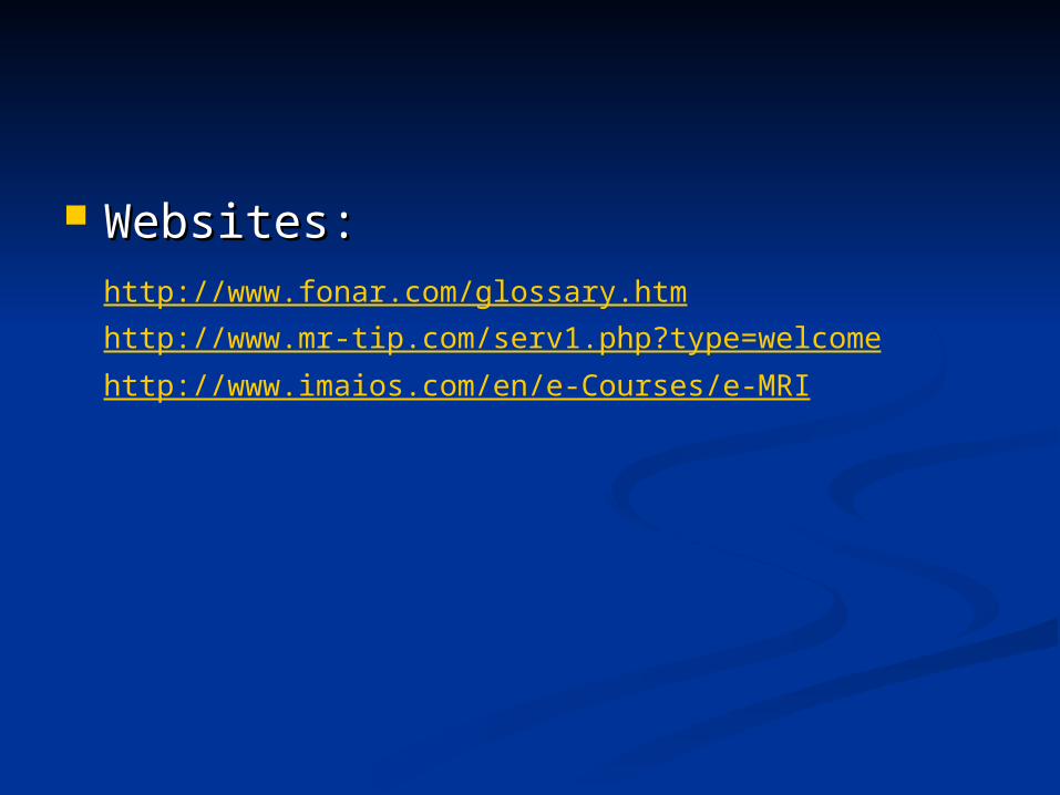
Websites: Websites: http://www.fonar.com/glossary.htm
http://www.mr-tip.com/serv1.php?type=welcomehttp://www.imaios.com/en/e-Courses/e-MRI
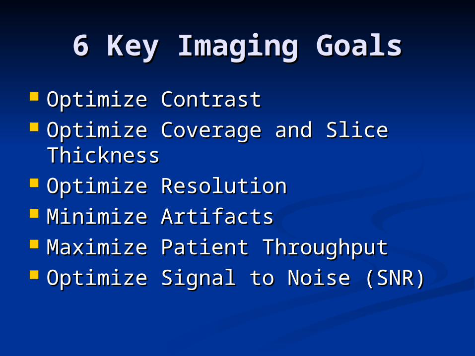
6 Key Imaging Goals6 Key Imaging Goals
Optimize ContrastOptimize Contrast Optimize Coverage and Slice Optimize Coverage and Slice
ThicknessThickness Optimize ResolutionOptimize Resolution Minimize ArtifactsMinimize Artifacts Maximize Patient ThroughputMaximize Patient Throughput Optimize Signal to Noise (SNR)Optimize Signal to Noise (SNR)
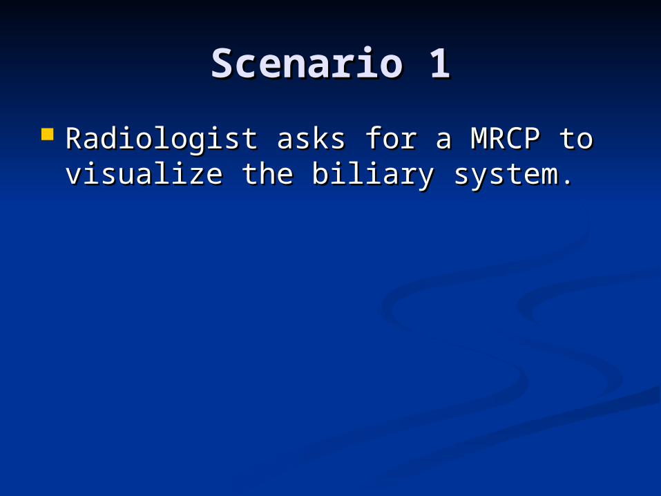
Scenario 1Scenario 1
Radiologist asks for a MRCP to Radiologist asks for a MRCP to visualize the biliary system.visualize the biliary system.
Scenario 2Scenario 2
Radiologist wants a sagittal T2 FSE Radiologist wants a sagittal T2 FSE fatsat knee with better signal and fatsat knee with better signal and better resolution.better resolution.
Scenario 3Scenario 3
A coronal T1 wrist is too grainyA coronal T1 wrist is too grainy
Scenario 4Scenario 4
A FSE T2 fatsat brachial plexus does A FSE T2 fatsat brachial plexus does not get good fat saturation.not get good fat saturation.
Scenario 5Scenario 5
FSE T2 IACs have low spatial FSE T2 IACs have low spatial resolutionresolution
Build a Build a Protocol/Optimize It!Protocol/Optimize It!
Brain w/wo contrastBrain w/wo contrast Routine kneeRoutine knee Routine L-spineRoutine L-spine
Advantages/Advantages/DisadvantagesDisadvantages
Changing FOVChanging FOV Changing Slice ThicknessChanging Slice Thickness Changing ETLChanging ETL Changing NSA/NEXChanging NSA/NEX Changing TEChanging TE Changing BandwidthChanging Bandwidth Changing TRChanging TR
If this…..then this…..If this…..then this…..
P. 136 and P.137 MRI in P. 136 and P.137 MRI in PracticePractice
I have a question?I have a question?

Student QuestionsStudent Questions
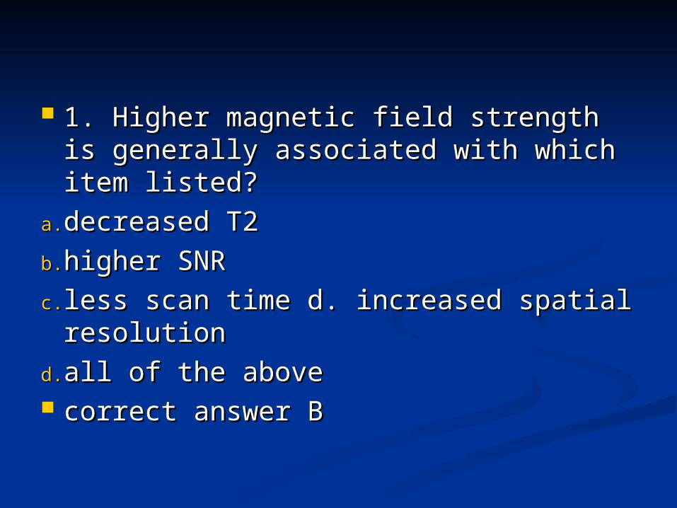
1. Higher magnetic field strength is 1. Higher magnetic field strength is generally associated with which item generally associated with which item listed?listed?
a.a. decreased T2 decreased T2
b.b. higher SNR higher SNR
c.c. less scan time d. increased spatial less scan time d. increased spatial resolution resolution
d.d. all of the aboveall of the above correct answer Bcorrect answer B
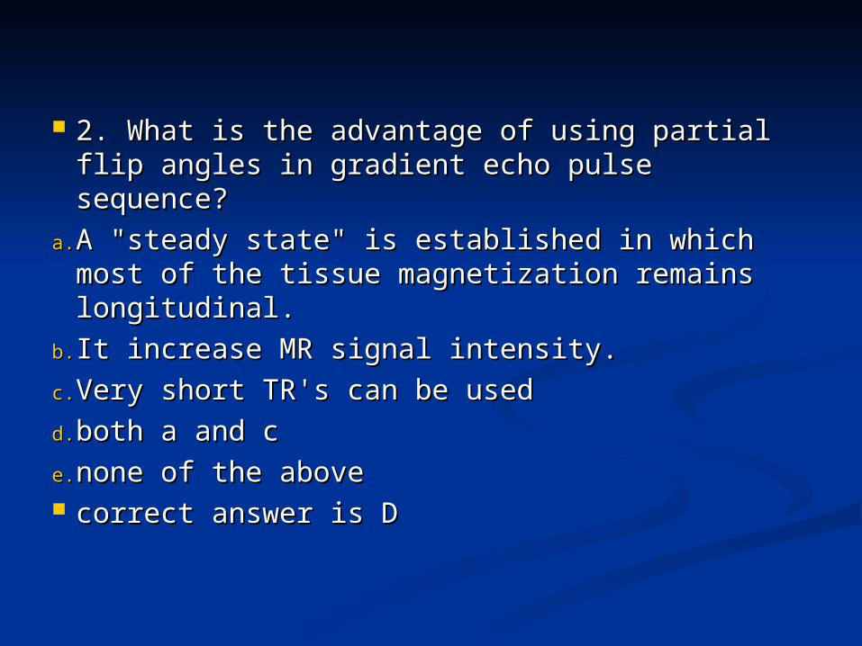
2. What is the advantage of using partial flip 2. What is the advantage of using partial flip angles in gradient echo pulse sequence?angles in gradient echo pulse sequence?
a.a. A "steady state" is established in which most A "steady state" is established in which most of the tissue magnetization remains of the tissue magnetization remains longitudinal.longitudinal.
b.b. It increase MR signal intensity.It increase MR signal intensity.
c.c. Very short TR's can be usedVery short TR's can be used
d.d. both a and cboth a and c
e.e. none of the abovenone of the above correct answer is Dcorrect answer is D
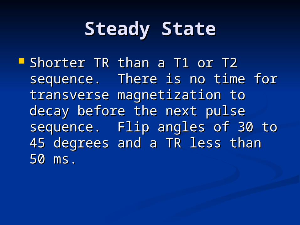
Steady StateSteady State
Shorter TR than a T1 or T2 Shorter TR than a T1 or T2 sequence. There is no time for sequence. There is no time for transverse magnetization to decay transverse magnetization to decay before the next pulse sequence. Flip before the next pulse sequence. Flip angles of 30 to 45 degrees and a TR angles of 30 to 45 degrees and a TR less than 50 ms.less than 50 ms.
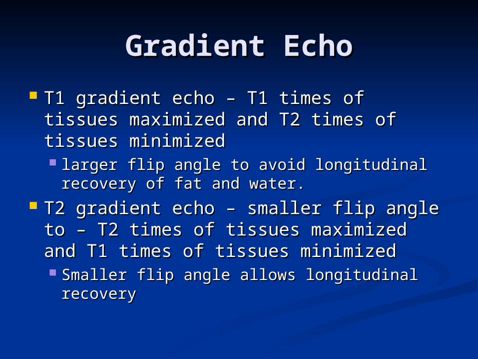
Gradient EchoGradient Echo
T1 gradient echo – T1 times of tissues T1 gradient echo – T1 times of tissues maximized and T2 times of tissues maximized and T2 times of tissues minimizedminimized larger flip angle to avoid longitudinal larger flip angle to avoid longitudinal
recovery of fat and water.recovery of fat and water. T2 gradient echo – smaller flip angle to T2 gradient echo – smaller flip angle to
– T2 times of tissues maximized and T1 – T2 times of tissues maximized and T1 times of tissues minimizedtimes of tissues minimized Smaller flip angle allows longitudinal Smaller flip angle allows longitudinal
recoveryrecovery
3. Which of the following results from an 3. Which of the following results from an increase in the number of averages of increase in the number of averages of an MRI scan?an MRI scan?
a.a. decreased scan time decreased scan time
b.b. increased SNR increased SNR
c.c. increased artifact increased artifact
d.d. increase spatial resolution increase spatial resolution
e.e. decreased spatial resolutiondecreased spatial resolution correct answer is Bcorrect answer is B
4. T2-weighted images are perfomed 4. T2-weighted images are perfomed with:with:
a.a. short TE and short TR short TE and short TR
b.b. short TE and long TR short TE and long TR
c.c. long TE and long TR long TE and long TR
d.d. TE equals TR TE equals TR
e.e. none of the abovenone of the above correct answer is Ccorrect answer is C
5. If the technologist decreases the field 5. If the technologist decreases the field of view it will:of view it will:
a.a. results in an image that depicts more results in an image that depicts more area area
b.b. improves spatial resolution improves spatial resolution
c.c. increase Signal to noise ratioincrease Signal to noise ratio
d.d. increases T1 weighting increases T1 weighting
e.e. both B and Dboth B and D correct answer is Bcorrect answer is B
6. On single slice gradient echo 6. On single slice gradient echo images, rapidly flowing blood appears:images, rapidly flowing blood appears:
a.a. dark dark
b.b. bright bright
c.c. dark or bright dark or bright
d.d. depends on the vessel depends on the vessel
e.e. both dark and bright both dark and bright correct answer is Bcorrect answer is B
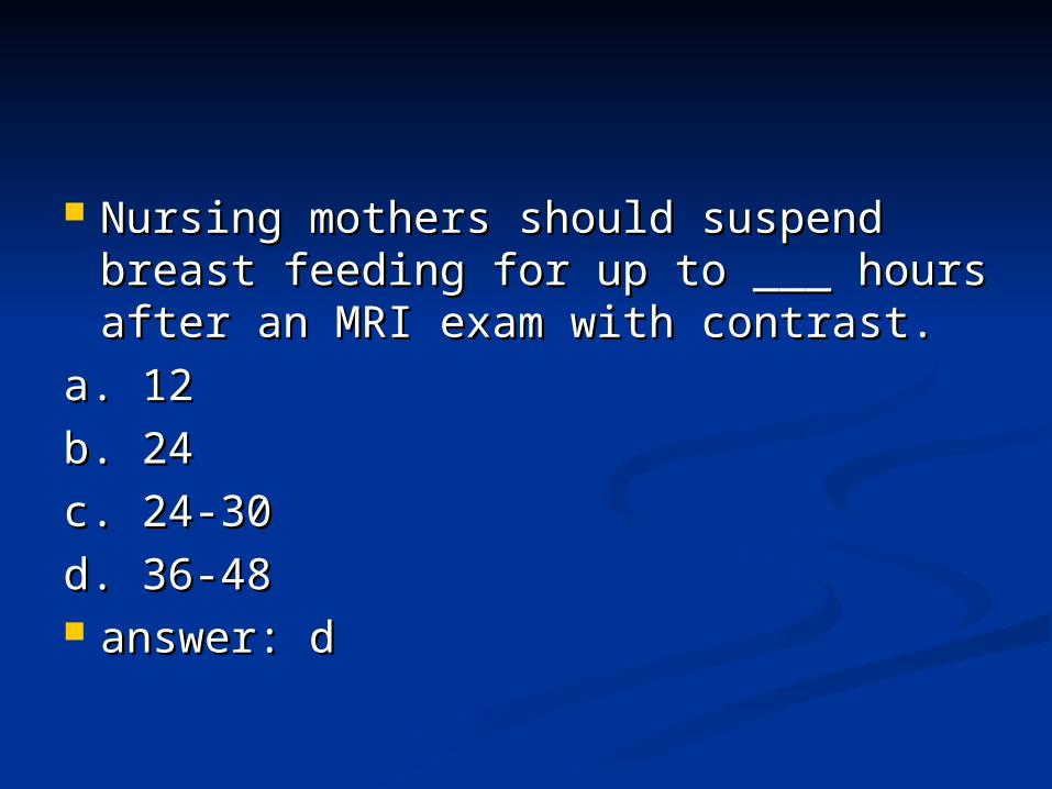
Nursing mothers should suspend Nursing mothers should suspend breast feeding for up to ___ hours breast feeding for up to ___ hours after an MRI exam with contrast.after an MRI exam with contrast.
a. 12a. 12
b. 24b. 24
c. 24‑30c. 24‑30
d. 36‑48d. 36‑48 answer: danswer: d
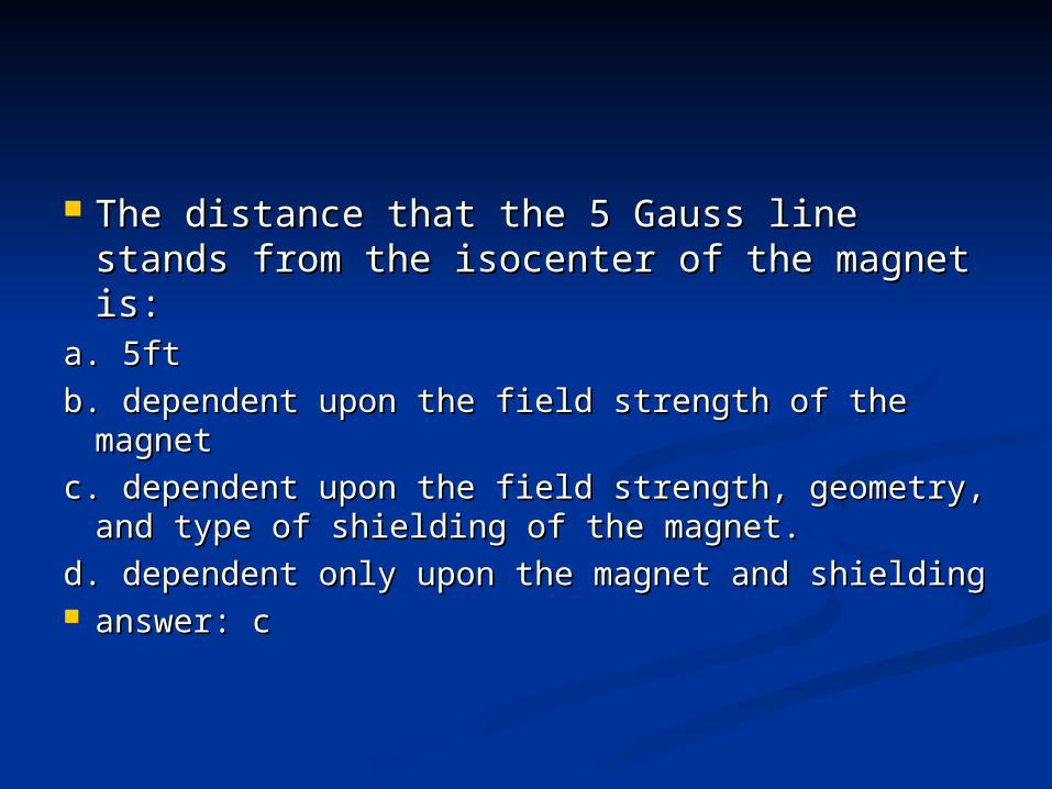
The distance that the 5 Gauss line stands The distance that the 5 Gauss line stands from the isocenter of the magnet is:from the isocenter of the magnet is:
a. 5fta. 5ft
b. dependent upon the field strength of the b. dependent upon the field strength of the magnetmagnet
c. dependent upon the field strength, geometry, c. dependent upon the field strength, geometry, and type of shielding of the magnet.and type of shielding of the magnet.
d. dependent only upon the magnet and shieldingd. dependent only upon the magnet and shielding answer: canswer: c
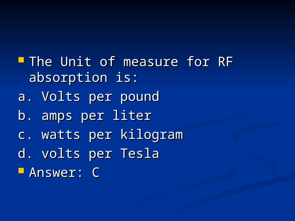
The Unit of measure for RF The Unit of measure for RF absorption is:absorption is:
a. Volts per pounda. Volts per pound
b. amps per literb. amps per liter
c. watts per kilogramc. watts per kilogram
d. volts per Teslad. volts per Tesla Answer: CAnswer: C
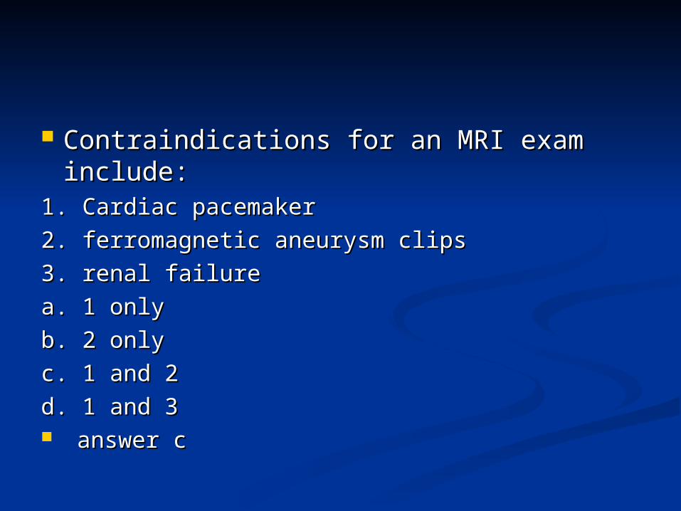
Contraindications for an MRI exam Contraindications for an MRI exam include:include:
1. Cardiac pacemaker1. Cardiac pacemaker
2. ferromagnetic aneurysm clips2. ferromagnetic aneurysm clips
3. renal failure3. renal failure
a. 1 onlya. 1 only
b. 2 onlyb. 2 only
c. 1 and 2c. 1 and 2
d. 1 and 3d. 1 and 3 answer canswer c
A joint effusion of the left knee would appear as what on a T2 weighted scan?a. Dark area around the jointb. Unremarkablec. Bright area around the jointd. Similar signal with the surrounding
soft tissue
Answer: CAnswer: C
Inversion recovery sequences are primarily used to identifya. Pathologyb. Anatomyc. Multiple sclerosis plaquesd. Fluid
Answer: CAnswer: C
Gadolinium contrast shortens which imaging sequencea. T2b. IRc. T1d. FLAIR
Answer: CAnswer: C
What scanning sequence is used to evaluate for strokes?a. T2*b. FLAIRc. Diffusion Weighted Imagingd. T1
Answer: CAnswer: C
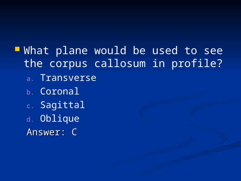
What plane would be used to see the corpus callosum in profile?a. Transverseb. Coronalc. Sagittald. Oblique
Answer: CAnswer: C
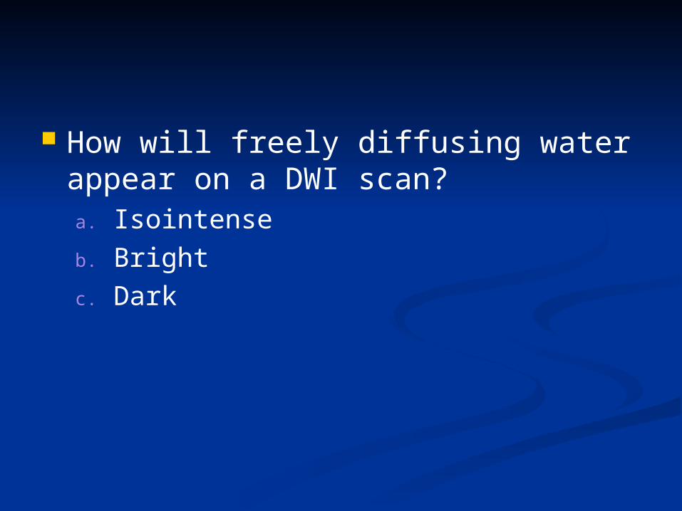
How will freely diffusing water appear on a DWI scan?a. Isointenseb. Brightc. Dark
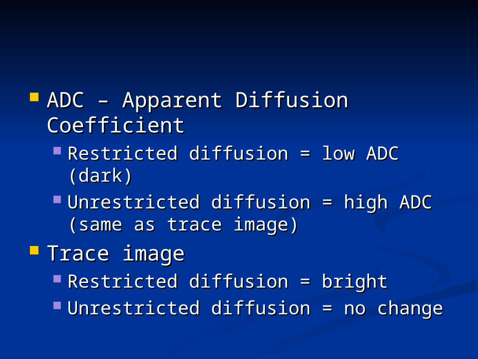
ADC – Apparent Diffusion CoefficientADC – Apparent Diffusion Coefficient Restricted diffusion = low ADC (dark)Restricted diffusion = low ADC (dark) Unrestricted diffusion = high ADC Unrestricted diffusion = high ADC
(same as trace image)(same as trace image) Trace imageTrace image
Restricted diffusion = brightRestricted diffusion = bright Unrestricted diffusion = no changeUnrestricted diffusion = no change
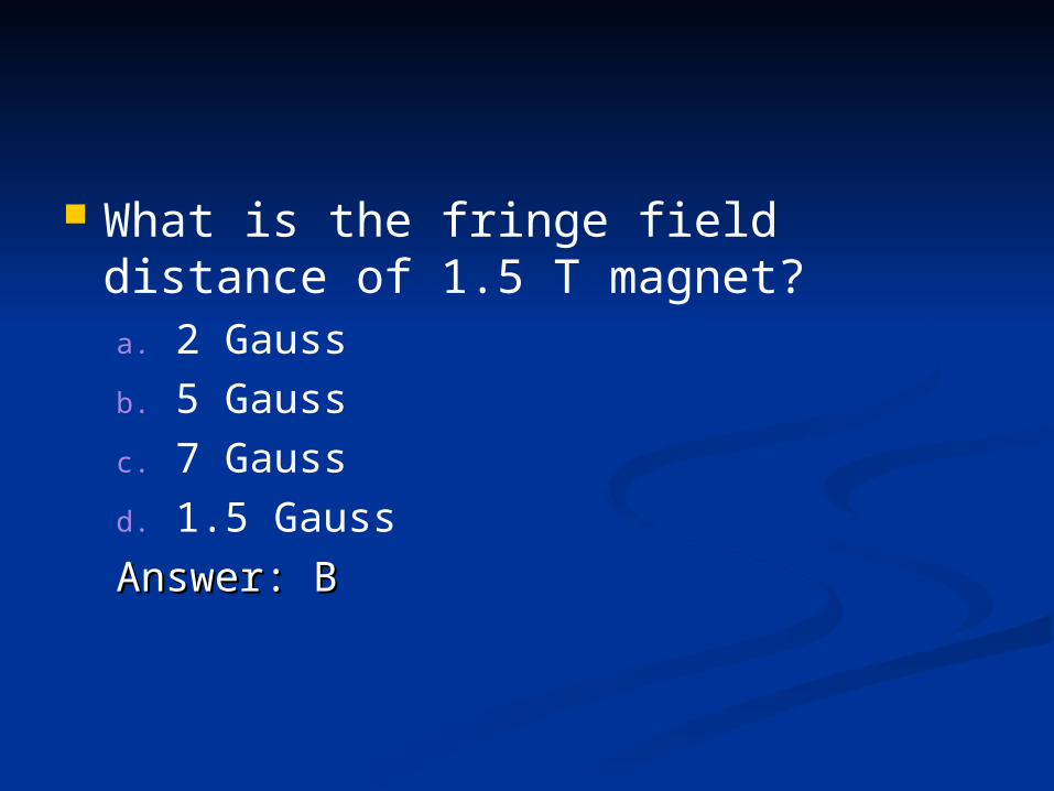
What is the fringe field distance of 1.5 T magnet?a. 2 Gaussb. 5 Gaussc. 7 Gaussd. 1.5 Gauss
Answer: BAnswer: B
On a FLAIR or T2 weighted sequence a subdural hematoma will appeara. Hypointenseb. Isointensec. Hyperintense

References:References:
Westbrook, Catherine and Kaut, Carolyn (1998). MRI In Westbrook, Catherine and Kaut, Carolyn (1998). MRI In Practice, 3rd Edition, Malden, MA: Blackwell Science, Inc.Practice, 3rd Edition, Malden, MA: Blackwell Science, Inc.