Embed Size (px)
Citation preview

This journal is c The Royal Society of Chemistry 2013 Chem. Commun.
Cite this: DOI: 10.1039/c2cc37536b
Tuning and assembling metal nanostructureswith DNA
Amit Kumar, Jae-Ho Hwang, Sumit Kumar and Jwa-Min Nam*
Here, we highlight the strategies for the synthesis and tuning of a variety of metal nanostructures and
nanoassemblies with oligonucleotides and their applications. We have discussed the importance of and
need for various metal nanostructures and the role and suitability of DNA in building these nanostructures
and nanoassemblies. A large part of this article is devoted to the discussion of DNA-mediated synthetic
methods for metal nanostructures. The synthetic strategies are categorized into two groups – strategy 1
that uses DNA as the ligand for metal nanoparticles and subsequent assembly or modification and
strategy 2 that uses DNA template-directed assemblies of metal nanoparticles.
1. Introduction
Nature uses a number of different functional molecules foreach specific and essential job in order to sustain the life ofvarious living species on earth. Nanostructures originated fromthe self-assembly of such biomolecules have been exploredenormously for their importance in several areas of science.1
Nature’s master biomolecule, namely ‘deoxyribonucleic acid(DNA)’, is a genetic material in the cell, which specificallyencodes the biosynthesis of various proteins with a very highdegree of accuracy, and proteins are necessary for almost all thebiological functions. Structurally, DNA is composed of two
purine bases [adenine (A) and guanine (G)] and two pyrimidinebases [thymine (T) and cytosine (C)].2 Specific complementarybinding of A with T and C with G (Watson–Crick pairing) inassociation with other supramolecular interactions lead to theformation of helical duplex structure of double-stranded DNA.2
A specific base sequence and the supramolecular forces in DNAalso lead to the formation of triplex and quadruplex secondarystructures.3 Inspired by the unique inherent properties of DNA,scientists have exploited DNA to synthetically build the nano-structures of well-controlled size and shape. This led to thedevelopment of DNA nanotechnology.1,4–7 Credit must also begiven to the development of customized synthetic methods forDNA (solid-phase synthesis) and modern purification andanalytical tools.8 The field of DNA nanotechnology is developedto the extent of synthetically building desired 2D or 3D
Department of Chemistry, Seoul National University, 599 Gwanak-ro, Gwanak-gu,
Seoul 151-747, South Korea. E-mail: [email protected]
Amit Kumar
Amit Kumar received his MScfrom Indian Institute of Techno-logy, Roorkee (India), in 2004followed by a PhD degree insynthetic organic chemistry inthe year 2009 from the Depart-ment of Chemistry, IndianInstitute of Technology, Kanpur(India). Currently, he is a post-doctoral fellow in the researchgroup of Professor Jwa-Min Namat Seoul National University(South Korea). His research ismainly focused on the synthesis
and applications of plasmonic nanostructures and carbonnanomaterials.
Jae-Ho Hwang
Jae-Ho Hwang obtained his BS(2009) from the Department ofChemistry at Seoul NationalUniversity (South Korea). Hejoined Professor Nam’s lab in2009 and now he is a PhDcandidate. His current researchsubjects mainly focus on thedesign and synthesis ofplasmonic nanostructures andtheir use for biosensingapplications.
Received 16th October 2012,Accepted 14th December 2012
DOI: 10.1039/c2cc37536b
www.rsc.org/chemcomm
ChemComm
FEATURE ARTICLE
Dow
nloa
ded
by C
alif
orni
a In
stitu
te o
f T
echn
olog
y on
25
Janu
ary
2013
Publ
ishe
d on
17
Dec
embe
r 20
12 o
n ht
tp://
pubs
.rsc
.org
| do
i:10.
1039
/C2C
C37
536B
View Article OnlineView Journal

Chem. Commun. This journal is c The Royal Society of Chemistry 2013
nanoobjects with complexity using a DNA-tile based approachor one-pot DNA-origami strategy.5,9 Many of the synthesizedDNA-nanostructures have found applications in materialschemistry, biotechnology and medical science.4,9–11 Manyplasmonic nanostructures, originated from the assembly ofmetal nanoparticles (NPs), are used in the fields such asbiosensing and surface-enhanced Raman scattering (SERS)applications.12,13 The assemblies of NPs have been implementedby random aggregation,14,15 control over the number of ligandson each nanoparticle16–18 and utilizing steric effects.19–21
However, high yielding and reproducible synthesis ofsuch nanostructures of a well-defined geometry has been achallenge. Since the introduction of single-stranded DNA-functionalized gold nanoparticles (AuNPs)/nanoclusters andtheir self-assembly,16,22 significant progress has been made inutilizing DNA for the construction of well-defined metalnanoassemblies.9–13,23 Such kinds of DNA-mediated assemblingprocesses of metal NPs have enormous applications in thefields such as medical science, plasmonics, and nanoelectronics.The present review focuses on the synthetic strategies adopted byscientists over the years for the utilization of DNA in theformation of metal nanostructures and nanoassemblies of desir-able size and shape.
1.1 Need for well-defined metal nanostructures
Well-defined and controlled assemblies of metal NPs havefound applications in the fields such as medical science(diagnostics and therapeutics),24,25 electronics,26,27 optics28
and catalysis.29 Metal nanostructures have unique features of
possessing surface plasmon resonance and originating fromthe collective electron oscillation within nanoscale dimensions.The surface plasmon energy of nanostructures is highly depen-dent on their size, shape and composition, resulting in tunablelight–matter interaction optical response. Controlled synthesisof metal nanostructures gives the opportunity to tune theoptical properties of metal nanostructures which is highlydesirable for their applications. Theoretical methods including‘Mie theory’ have been developed in order to predict therelationship between size/shape and optical properties of metalnanostructures.30,31 In multi-particle assembly, plasmoniccoupling between metal particles results in the enhancedelectric fields at a particular region usually designated as a‘hot-spot’.31 Correct positioning of NPs is crucial to harness themaximum enhancement of electric field, which has been thefocus of recent research effort in the development of highlysensitive SERS probes.32,33
In SERS, an analyte molecule, present right at the ‘hot-spot’of metal nano-assembly, gives the enhanced Raman signal andcan be detected even in very low amounts, down to the single-molecule level.34,35 Plasmonic theory-based principles can beused to design the desired metal nanostructures possessingthe required optical properties, but the synthesis of targetedplasmonic structures with high precision and high yieldis quite challenging. Over time, synthetic chemists have devel-oped expertise in synthesizing metal NPs in a variety of sizesand shapes such as zero dimensional (0D) spherical,36 1D wireor rod-like,37–39 2D plate-like or polygonal,40–42 3D polyhedraland many more complex metal nanostrucures.43–49 Cheng and
Sumit Kumar
Sumit Kumar received his MSc inChemistry from Indian Instituteof Technology, Guwahati (India).He joined Professor Nam’s lab in2010 as a PhD student. Hiscurrent research subjects aremainly the design and synthesesof plasmonic nanostructures andcarbon nanomaterials and theirapplications.
Jwa-Min Nam
Jwa-Min Nam received his BSand MS in Chemistry fromHanyang University, Korea. Heobtained his PhD in Chemistryfrom Northwestern Universityand worked as a postdoctoralfellow at University of California,Berkeley. Jwa-Min joined theDepartment of Chemistry, SeoulNational University, in 2006 asan Assistant Professor and waspromoted to Associate Professorin 2010. Jwa-Min received thePresidential Young Scientist
Award from the President of the Republic of Korea, the YoungInorganic Chemist Award from the Korean Chemical Society, theOutstanding Basic Science Research Achievement Award from theMinistry of Education, Science and Technology, Korea, the Victor K.LaMer Award from the American Chemical Society, and theCollegiate Inventors Award from the National Inventors Hall ofFame, USA. Jwa-Min served as a consultant for Nanosphere, Inc.and as a scientific advisory board member for Medifron DBT. Hismajor interests include the synthesis and optics of plasmonicnanostructures, nanocarriers for bio-imaging and delivery,nanoprobe-tethered lipid bilayers, cella nanostructure interfaces,graphene chemistry and biosensors.
Feature Article ChemComm
Dow
nloa
ded
by C
alif
orni
a In
stitu
te o
f T
echn
olog
y on
25
Janu
ary
2013
Publ
ishe
d on
17
Dec
embe
r 20
12 o
n ht
tp://
pubs
.rsc
.org
| do
i:10.
1039
/C2C
C37
536B
View Article Online

This journal is c The Royal Society of Chemistry 2013 Chem. Commun.
coworkers, in a review article, have attempted to make aperiodic chart keeping various nanostructures as ‘plasmonicatoms’ in different rows and columns taking their shapes intoaccount.12 The self-assembly of metal NPs results in the for-mation of organized nanostructures under thermo-dynamically favourable conditions and/or by externally applyingother stimuli such as forces (electrostatic, magnetic, flow, etc.),directing agents and templates.23,50
1.2 Role of DNA in the assembly of nanostructures
The choice of organic ligands for NPs is not only crucial fortheir stability in solution state but also to provide desiredfunctionalities on the metal NP surface for various applica-tions.51,52 The structural features of stabilizing organic ligandson metal NPs play very important roles in their directedassembly. Intermolecular interactions such as hydrogen bond-ing and electrostatic interaction among organic ligands presenton different NPs affect and direct the self-assembled struc-tures.53 Inherently, DNA has the affinity towards metals due tothe presence of nitrogenous bases and anionic phosphategroups. Due to such kinds of electron-rich structures, DNAcan efficiently serve as a stabilizing ligand for metal NPs.Moreover, DNA has several other features such as itsunique Watson–Crick base pairing, flexible and simple struc-ture, and molecular recognition capability. One end of anoligonucleotide can be modified with a functional groupsuitable for binding with a metal surface – for example, thiol-functionalized DNA for AuNPs.22 These DNA modifications inthe solution of suitable ionic strength serve two importantpurposes: (i) establish strong (covalent) binding with the metalNP surface at one end and (ii) allow dense functionalization ofthe metal NP surface, rendering other ends of DNA ligands freein the solution. Particularly, Mirkin and coworkers have beenperforming extensive research in this area since 1996 and theyhave coined the term ‘spherical nucleic acids’ (SNA) for suchpolyvalent DNA-functionalized metal NPs.54 The combinationof DNA and metal NPs as SNA offers the synergistic results,harnessed from the intrinsic properties of both the metal coreand DNA ligands. For example, DNA-functionalized NPs havefound applications in the development of novel therapeutics,diagnostic tools and biosensors.55–58 The use of DNA as aligand renders opportunities for tuning the assembly of metalNPs by varying the number of DNA strands on each metal NP,the base sequence and the length/structure of oligonucleotides.The number of DNA strands on each metal NP couldbe controlled by using appropriate stoichiometry and theoptimized ionic strength of the solution.34 The appropriatenumber and structure of DNA ligands on metal NPs underoptimized reaction conditions lead to the formation of variousassemblies ranging from dimers, trimers to polymeric systems(Section 2.1.2). On the other hand, it has been shown thatone can obtain various DNA superstructures by carefully andsystematically designing DNA sequences and assemblies.Recent advancement and better understanding of DNAnanotechnology have allowed us to build a variety of DNAsuperstructures solely from DNA, utilizing the DNA tile-basedand DNA-origami strategies.1,7 Such kinds of synthesized
DNA-nanostructures have been further used as the ‘template’for the ordered assembly of metal NPs onto them.10,23,59 Thefollowing section will explain the various strategies for buildinga variety of nanostructures with the help of DNA.
2. Building metal nanostructures with DNA
Over time, with the development of ‘DNA-architectonics’10 itbecame possible to access a variety of self-assembled metalnanostructures with the help of DNA. Using DNA, metal NPscould be assembled in two ways (Fig. 1): (i) by the synthesis ofDNA-functionalized metal NPs (DNA–NPs) followed by theirlinking and (ii) by the assembly of metal NPs by using DNAnanostructures as a ‘template’. The following subsectionswill explain each strategy in detail and the correspondingrepresentative examples in the literature.
2.1 Assembly of DNA-functionalized metal NPs (‘ligandapproach’)
Metal NPs could be easily functionalized with DNA by ligandexchange or attachment reaction on pre-synthesized NPs.Apparently, in the absence of any constraint on the mode ofligand exchange or attachment reaction, homogeneous func-tionalization in all the directions takes place with the samefacility on the surface of spherical metal NPs. The self-assemblyof such isotropic functionalized NPs via the Watson–Crick base-pairing among complementary DNA strands results in themetal superstructures in the form of big aggregates.22 On theother hand, anisotropically functionalized DNA–metal NPsmay form the definite structures of desired shapes; however,
Fig. 1 DNA-mediated assembly of metal NPs.
ChemComm Feature Article
Dow
nloa
ded
by C
alif
orni
a In
stitu
te o
f T
echn
olog
y on
25
Janu
ary
2013
Publ
ishe
d on
17
Dec
embe
r 20
12 o
n ht
tp://
pubs
.rsc
.org
| do
i:10.
1039
/C2C
C37
536B
View Article Online
Chem. Commun. This journal is c The Royal Society of Chemistry 2013
the synthetic access of such anisotropic NP precursors isnon-trivial.16
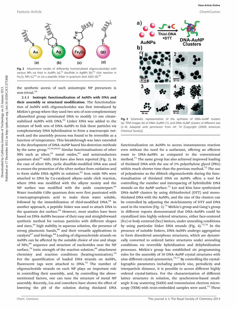
2.1.1 Isotropic functionalization of AuNPs with DNA andtheir assembly or structural modification. The functionaliza-tion of AuNPs with oligonucleotides was first introduced byMirkin’s group where they used two sets of non-complementaryalkanethiol group terminated DNA to modify 13 nm citrate-stabilized AuNPs with DNA.22 Linker DNA was added to themixture of both sets of DNA–AuNPs to link these particles viacomplementary DNA hybridization to form a macroscopic net-work and the assembly process was found to be reversible as afunction of temperature. This breakthrough was later extendedto the development of DNA–AuNP based bio-detection methodsby the same group.55,56,60,61 Similar functionalizations of otherNPs such as silver,62 metal oxides,63 and semiconductorsquantum dots64 with DNA have also been reported (Fig. 2). Inthe case of silver NPs, cyclic disulfide-modified DNA was usedto ensure the protection of the silver surface from oxidation andto form stable DNA–AgNPs in solution.62 Iron oxide NPs wereattached to DNA by Cu-catalyzed alkyne–azide click reaction,where DNA was modified with the alkyne moiety and theNP surface was modified with the azide counterpart.63
Water insoluble CdSe quantum dots were first passivated with3-mercaptopropionic acid to make them water soluble,followed by the immobilization of thiol-modified DNA.64 Inanother approach, a peptidic linker was used to attach DNA tothe quantum dot surface.65 However, most studies have beenbased on DNA–AuNPs because of their easy and straightforwardsynthetic method for various particles with different shapesand sizes,44 high stability in aqueous solution, the presence ofstrong plasmonic bands,66 and their versatile applications incatalysis67 and biology.68 Loading of oligonucleotide strands onAuNPs can be affected by the suitable choice of size and shapeof NPs,69 sequence and structure of nucleotides near the NPsurface,70 ionic strength of the reaction solution,60 attachmentchemistry and reaction conditions (heating/sonication).70
For the quantification of loaded DNA strands on AuNPs,fluorescent tags were attached to DNA.70 The number ofoligonucleotide strands on each NP plays an important rolein controlling their assembly, and, by controlling the above-mentioned factors, one can tune the structure of metal NPassembly. Recently, Liu and coworkers have shown the effect oflowering the pH of the solution during thiolated DNA
functionalization on AuNPs to access instantaneous reactioneven without the need for a surfactant, offering an efficientroute to DNA–AuNPs as compared to the conventionalmethod.71 The same group has also achieved improved loadingof thiolated DNA with the use of 2% polyethylene glycol (PEG)within much shorter time than the previous method.72 The useof polyadenine as the diblock oligonucleotide during the func-tionalization of thiolated DNA on AuNPs offers a tool forcontrolling the number and interspacing of hybridizible DNAstrands on the AuNP surface.73 Lee and Kim have synthesizedDNA–AuNP clusters by using dithiothreitol (DTT) and mono-thiolated DNA with the AuNPs, and the size of the clusters canbe controlled by adjusting the stoichiometry of DTT and DNAused in the reaction (Fig. 3).74 Mirkin’s group and Gang’s groupin different reports demonstrated that DNA–AuNPs could becrystallized into highly ordered structures, either face-centered(fcc) or body centered (bcc) lattices, under annealing conditionsby using particular linker DNA strands (Fig. 4).75,76 In thepresence of suitable linkers, DNA–AuNPs undergo aggregationto form disordered amorphous structures, which are dynami-cally converted to ordered lattice structures under annealingconditions via reversible hybridization and dehybridizationprocesses. Mirkin’s group has established six programmingrules for the assembly of 50 DNA–AuNP crystal structures withnine different crystal symmetries.75,77 By controlling the crystal-lographic parameters including particle size, periodicity andinterparticle distance, it is possible to access different highlyordered crystal-lattices. For the characterization of differentlattice structures in solution, the synchrotron-based small-angle X-ray scattering (SAXS) and transmission electron micro-scopy (TEM) with resin-embedded samples were used.75 These
Fig. 2 Attachment modes of differently functionalized oligonucleotides withvarious NPs via thiol in AuNPs (a),22 disulfide in AgNPs (b),62 click reaction inFe2O3 NPs (c),63 or via a peptidic linker in quantum dots (QD) (d).65
Fig. 3 Schematic representation of the synthesis of DNA–AuNP clusters(a). TEM images (b) of DNA–AuNPs (1), and DNA–AuNP clusters of different size(2–4). Adapted with permission from ref. 74 [Copyright (2009) AmericanChemical Society].
Feature Article ChemComm
Dow
nloa
ded
by C
alif
orni
a In
stitu
te o
f T
echn
olog
y on
25
Janu
ary
2013
Publ
ishe
d on
17
Dec
embe
r 20
12 o
n ht
tp://
pubs
.rsc
.org
| do
i:10.
1039
/C2C
C37
536B
View Article Online

This journal is c The Royal Society of Chemistry 2013 Chem. Commun.
studies demonstrated the importance of DNA in designing andsynthesizing materials of different crystal structures with thesame core metal (gold). Using a hollow three dimensional DNAnanostructure spacer within a binary superlattice, Mirkin’sgroup further showed that five distinct symmetries, includinga symmetry that has never been observed before, of DNA–AuNPcrystals can be formed.77 The crystallization pattern of sphericalDNA–AuNPs has also been studied by theoretical simulationsand obtained theoretical phase diagrams are found to be ingood agreement with experimental results.78,79
The functionalization of anisotropic metal nanostructuressuch as Au nanorods (AuNRs), Au nanoprisms and Ag nanocubeswith DNA has also been realized analogous to the spherical NPs,however, with slightly different synthetic approaches except Agnanocubes.80–83 The reason for this difference in syntheticstrategy is due to the presence of differently charged (positively)ligands, usually cetyl trimethylammonium bromide (CTAB),84,85
which electrostatically interact with oppositely charged DNA,hindering the approach of DNA towards the metal NP surfacefor functionalization. However, this problem could be overcomeby repeatedly washing the CTAB-NP to ensure the completeremoval of CTAB ligands, followed by the reaction with thiolmodified DNA.81,82 Directional assemblies and orientation-dependent properties of anisotropic NPs such as AuNRs can beexploited during their well-defined assembly processes. Mannand coworkers obtained 3D bundles of DNA-functionalizedAuNRs aligned in parallel stacks, through DNA complementarybinding.80 Recently, temperature-dependent assembly and dis-assembly of DNA–AuNR hybrids have been demonstrated for therealization of reversible plasmonic circular dichroism (CD)responses, providing a more sensitive detection method forDNA as compared to UV-Vis absorption spectroscopy.86 Thehybridization efficiency of anisotropic NPs such as DNA--AuNRsand nanoprisms is many-fold higher than spherical AuNPs withthe same amount of loaded DNA which is due to the greaterinterparticle surface contact (Fig. 5).81 The assembly of aniso-tropic NPs via DNA base pairing occurs along the directions thatmaximize parallel face-to-face interactions between particles. Forexample, nanoprisms are assembled into 1D arrays along the
prism faces while nanorods are assembled into 2D sheets withthe hexagonal close packed (hcp) ordering and the rhombicdodecahedra are assembled into 3D fcc lattices.81
Recently, Liang and coworkers have utilized the concept ofdynamic DNA-fuelled molecular machine87 for the assembly ofDNA–AuNPs, driven by a series of toehold-mediated stranddisplacement reactions of DNA and the aggregation rate ofthe AuNPs can be regulated by controlling the amount ofoligonucleotide catalyst.88 Azobenzene-modified oligonucleotide-functionalized AuNPs showed a reversible photo-switchableassembly, presenting a different stimulus than temperature inprevious cases.89 In this case, irradiation of UV light causes thetrans–cis isomerization of azobenzene, destabilizing the DNAduplex, resulting in the dissociation of the NP assemblies, whichcould be reassembled by illuminating them with blue light.Very stable Au–Ag core–shell structures have been synthesizedfrom DNA–AuNPs to generate DNA-embedded Au–Ag core–shellparticles by a silver staining method, where thickness of the Agshell is controllable based on the amount of silver stainingsolution used in the synthesis.90 Interestingly, in this case,monothiol-modified DNA functionalization is enough to stabilizeparticles because DNA strands are embedded inside the Ag shellunlike the previous cases of DNA–AgNPs that require cyclicdisulfides to ensure the stability of DNA–AgNPs.62 Core–shellmetal nanostructures, possessing the synergistically enhancedproperties, usually outperform the corresponding mixtures andalloys of the same type of metals for various applications.91
The assembly of DNA–NPs into heteropentamer clusters,consisting of a smaller Au sphere surrounded by a ring of fourlarger spheres, has been reported, and it was shown that thesestructures exhibit the magnetic and Fano-like resonances.92
Therein, DNA is not only crucial for the cluster assembly butalso functions as an insulator spacer between conductiveparticles. Further, in a quest to make highly sensitive andsignal reproducible SERS probes with high stability, Nam andcoworkers reported the synthesis of gold nanobridged nanogapparticles (Au–NNP) with an interior gap size of 1 nm (Fig. 6).35
Interestingly, DNA plays important roles in sparse nucleationsites of Au on the DNA–AuNP surface, lateral growth of the Aushell and the formation of a 1 nm interior gap inside particles,which is unusual for shell growth on a core surface.90 Duringthe synthesis, the position and number of Raman dyes can be
Fig. 4 Schematic representation of DNA-mediated NP assembly into orderedsuperlattices. Adapted with permission from ref. 54 [Copyright (2012) AmericanChemical Society].
Fig. 5 Comparison between DNA-mediated assemblies of triangular nano-prisms and spherical NPs. The corresponding TEM images are shown in the right(scale bars are 20 nm). Adapted with permission from ref. 54 [Copyright (2009)American Chemical Society].
ChemComm Feature Article
Dow
nloa
ded
by C
alif
orni
a In
stitu
te o
f T
echn
olog
y on
25
Janu
ary
2013
Publ
ishe
d on
17
Dec
embe
r 20
12 o
n ht
tp://
pubs
.rsc
.org
| do
i:10.
1039
/C2C
C37
536B
View Article Online

Chem. Commun. This journal is c The Royal Society of Chemistry 2013
readily controlled and the dyes can be precisely located in a1 nm interior gap for generating the SERS enhancement factors(EF) of B108–109 with a very narrow distribution.
2.1.2 Anisotropic functionalization of AuNPs with DNAand their assembly. It is quite challenging to anistropicallymodify metal NPs with DNA and to assemble these DNA-modified structures into a specific architecture. For the highyield synthesis of metal NP assemblies with a specific numberof metal NPs, one should be capable of synthesizing metal NPswith the desired number and position of modified DNAstrands. Such anisotropic functionalization enables linking ofa nanostructure with a desired number of NPs at the desiredpositions and orientations for building a metallic moleculefrom metal NP atoms of various structures.12 From a nano-material-design perspective, anisotropic functionalization givesa lot more opportunities to scientists for designing and synthe-sizing the unconventional nanoassembly structures, withunconventional properties. Alivisatos and coworkers reportedthe mono-DNA-modified gold nanocrystals and their assemblyto form AuNP homodimers and homotrimers.16 For the synth-esis of mono-functionalized DNA–Au nanocrystals, DNA, mod-ified with the thiol group at the 30 or 50 end, was reacted with10-fold excess of mono-maleimido-functionalized Au nanocrys-tals. However, this methodology of mono-functionalization ofmetal NPs is limited to very small metal NPs (B2 nm), not
applicable to the NPs having larger surface area. Larger(5–10 nm) mono-functionalized DNA–AuNPs could be obtainedusing the electrophoretic separation93 from the stoichiometricmixture of other components and were utilized for the assem-bly of homo- and hetero-dimeric and trimeric structures by thesame research group later (Fig. 7).94Alivisatos and coworkersfurther separated and isolated even bigger (20 nm) DNA–AuNPswith a discrete number of DNA strands by the anion exchangehigh performance liquid chromatography (AE-HPLC), whichwas not possible with the gel electrophoresis (Fig. 7).95 Afterpurification, different complementary DNA–AuNP conjugateswere assembled into discrete structures via the Watson–Crickpairing. In a further work by the Alivisatos group and others,mono-functionalized DNA–NPs were utilized to construct morecomplex chiral pyramidal plasmonic assemblies (Fig. 8).96,97 Inanother approach by Nam and coworkers, 20 or 30 nm sized
Fig. 6 Synthetic scheme for the gold nanobridged nanogap particles (Au–NNPs)using DNA–AuNPs (a). HRTEM images of intermediate (panels 1–3) and Au–NNPs(panels 4 and 5), element line mapping and the B1 nm gap in the Au–NNPstructure are shown in panel 6. Reprinted by permission from Macmillan Publish-ers Ltd: [Nat. Nanotech.] (ref. 35), copyright (2011).
Fig. 7 Panel 1: electrophoretic separation of discrete 5–10 nm DNA–AuNPs (a).Adapted with permission from ref. 93 [Copyright (2001) American ChemicalSociety]. Cartoons and TEM images of a homodimer (i), a heterodimer (ii), ahomotrimer (iii), and heterotrimers (iv–vi) assembled from 5 and 10 nmDNA–AuNPs (b). Reproduced from ref. 94 with permission from Wiley. Panel 2:AE-HPLC separation of 20 nm DNA–AuNPs (c). TEM images of a dimer (vii), atrimer (viii), a tetramer (ix), a pentamer (x), a hexamer (xi) and a heptamer (xii)assembled from 5 and 20 nm DNA–AuNPs (d). Adapted with permission fromref. 95 [Copyright (2008) American Chemical Society].
Fig. 8 Schematic representation of chiral pyramidal assembly of DNA–NPs (a).TEM image of the nanopyramid built from two AuNPs, one AgNP and onequantum dot (QD) (b). Adapted with permission from ref. 97 [Copyright (2012)American Chemical Society].
Feature Article ChemComm
Dow
nloa
ded
by C
alif
orni
a In
stitu
te o
f T
echn
olog
y on
25
Janu
ary
2013
Publ
ishe
d on
17
Dec
embe
r 20
12 o
n ht
tp://
pubs
.rsc
.org
| do
i:10.
1039
/C2C
C37
536B
View Article Online
This journal is c The Royal Society of Chemistry 2013 Chem. Commun.
AuNPs were functionalized with two kinds of DNA: a targetcapture sequence and a protecting sequence by maintaining thestoichiometry 1 : 99 for 20 nm AuNPs and 1 : 199 for 30 nmAuNPs.34 These particles were then separated using magneticmicroparticles. A very low ratio of the target capture sequencevs. the protecting sequence ensures B1 target capture DNAmodification per AuNP, and other DNA covers the AuNP surfacefor particle stabilization and prevention of nonspecific aggrega-tion. By assembling single target capture DNA-modified AuNPs viatarget DNA recognition, one can obtain dimeric nanodumbbellstructures in a high yield.34 Importantly, the gap between bothAuNPs in a dumbbell-like structure was controlled by slow silvershell growth in order to maximize the enhancement of electro-magnetic field. SERS data were collected using the AFM-correlatednano-Raman setup to ensure the analysis of individual dimericstructures for single-molecule detection.34 It was also shownthat forming o1 nm plasmonic gap between particles is key togenerating a narrow distribution of high SERS enhancementfactor values from plasmonic nanogap structures.98
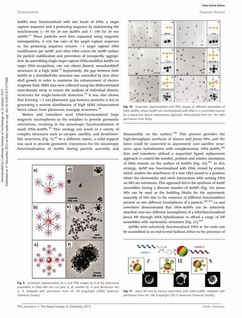
Mirkin and coworkers used DNA-functionalized largemagnetic microspheres as the template to provide geometricrestrictions, resulting in the anisotropic functionalization ofsmall DNA–AuNPs.99 This strategy can result in a variety ofcomplex structures such as cat-paw, satellite, and dendrimer-like structures (Fig. 9).99 In a different report, a solid supportwas used to provide geometric restrictions for the anisotropicfunctionalization of AuNPs during particle assembly and
disassembly on the surface.100 This process provides thehigh-throughput synthesis of dimers and Janus NPs, and thelatter could be converted to asymmetric core–satellite struc-tures upon hybridization with complementary DNA–AuNPs.100
Kim and coworkers utilized a sequential ligand replacementapproach to control the number, position and relative orientationof DNA strands on the surface of AuNPs (Fig. 10).101 In thisstrategy, AuNP was functionalized with DNA, strand by strand,which renders the attachment of a new DNA strand to a positionwhere the electrostatic and steric interactions with existing DNAon NPs are minimum. This approach led to the synthesis of AuNPassemblies having a discrete number of AuNPs (Fig. 10). JanusNPs can be used as the building blocks for the asymmetricassembly of NPs due to the existence of different functionalitiespresent on two different hemispheres of a particle.102,103 Lu andcoworkers demonstrated that DNA–AuNPs can be selectivelyattached onto two different hemispheres of a DNA-functionalizedJanus NP through DNA hybridization to afford a range of NPassemblies with asymmetric structures (Fig. 11).104
AuNRs with selectively functionalized DNA at the ends canbe assembled in an end-to-end fashion either in the presence of
Fig. 9 Schematic representation (a–c) and TEM images (d–f) of the directionalassemblies of DNA–NPs into cat paw (a, d), satellite (b, e) and dendrimer like(c, f). Adapted with permission from ref. 99 [Copyright (2006) AmericanChemical Society].
Fig. 10 Schematic representation and TEM images of different assemblies ofDNA–AuNPs, where AuNPs are functionalized with DNA in a controlled mannerby a sequential ligand replacement approach. Reproduced from ref. 101 withpermission from Wiley.
Fig. 11 Janus NP and its various assemblies with DNA–AuNPs. Adapted withpermission from ref. 104 [Copyright (2012) American Chemical Society].
ChemComm Feature Article
Dow
nloa
ded
by C
alif
orni
a In
stitu
te o
f T
echn
olog
y on
25
Janu
ary
2013
Publ
ishe
d on
17
Dec
embe
r 20
12 o
n ht
tp://
pubs
.rsc
.org
| do
i:10.
1039
/C2C
C37
536B
View Article Online
Chem. Commun. This journal is c The Royal Society of Chemistry 2013
a complementary target105,106 or via protein–aptamer recognition107
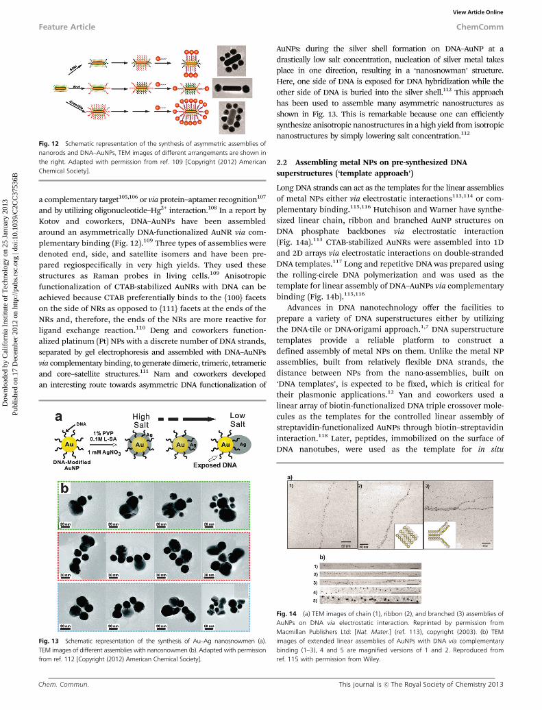
and by utilizing oligonucleotide–Hg2+ interaction.108 In a report byKotov and coworkers, DNA–AuNPs have been assembledaround an asymmetrically DNA-functionalized AuNR via com-plementary binding (Fig. 12).109 Three types of assemblies weredenoted end, side, and satellite isomers and have been pre-pared regiospecifically in very high yields. They used thesestructures as Raman probes in living cells.109 Anisotropicfunctionalization of CTAB-stabilized AuNRs with DNA can beachieved because CTAB preferentially binds to the {100} facetson the side of NRs as opposed to {111} facets at the ends of theNRs and, therefore, the ends of the NRs are more reactive forligand exchange reaction.110 Deng and coworkers function-alized platinum (Pt) NPs with a discrete number of DNA strands,separated by gel electrophoresis and assembled with DNA–AuNPsvia complementary binding, to generate dimeric, trimeric, tetramericand core–satellite structures.111 Nam and coworkers developedan interesting route towards asymmetric DNA functionalization of
AuNPs: during the silver shell formation on DNA–AuNP at adrastically low salt concentration, nucleation of silver metal takesplace in one direction, resulting in a ‘nanosnowman’ structure.Here, one side of DNA is exposed for DNA hybridization while theother side of DNA is buried into the silver shell.112 This approachhas been used to assemble many asymmetric nanostructures asshown in Fig. 13. This is remarkable because one can efficientlysynthesize anisotropic nanostructures in a high yield from isotropicnanostructures by simply lowering salt concentration.112
2.2 Assembling metal NPs on pre-synthesized DNAsuperstructures (‘template approach’)
Long DNA strands can act as the templates for the linear assembliesof metal NPs either via electrostatic interactions113,114 or com-plementary binding.115,116 Hutchison and Warner have synthe-sized linear chain, ribbon and branched AuNP structures onDNA phosphate backbones via electrostatic interaction(Fig. 14a).113 CTAB-stabilized AuNRs were assembled into 1Dand 2D arrays via electrostatic interactions on double-strandedDNA templates.117 Long and repetitive DNA was prepared usingthe rolling-circle DNA polymerization and was used as thetemplate for linear assembly of DNA–AuNPs via complementarybinding (Fig. 14b).115,116
Advances in DNA nanotechnology offer the facilities toprepare a variety of DNA superstructures either by utilizingthe DNA-tile or DNA-origami approach.1,7 DNA superstructuretemplates provide a reliable platform to construct adefined assembly of metal NPs on them. Unlike the metal NPassemblies, built from relatively flexible DNA strands, thedistance between NPs from the nano-assemblies, built on‘DNA templates’, is expected to be fixed, which is critical fortheir plasmonic applications.12 Yan and coworkers used alinear array of biotin-functionalized DNA triple crossover mole-cules as the templates for the controlled linear assembly ofstreptavidin-functionalized AuNPs through biotin–streptavidininteraction.118 Later, peptides, immobilized on the surface ofDNA nanotubes, were used as the template for in situ
Fig. 12 Schematic representation of the synthesis of asymmetric assemblies ofnanorods and DNA–AuNPs, TEM images of different arrangements are shown inthe right. Adapted with permission from ref. 109 [Copyright (2012) AmericanChemical Society].
Fig. 13 Schematic representation of the synthesis of Au–Ag nanosnowmen (a).TEM images of different assemblies with nanosnowmen (b). Adapted with permissionfrom ref. 112 [Copyright (2012) American Chemical Society].
Fig. 14 (a) TEM images of chain (1), ribbon (2), and branched (3) assemblies ofAuNPs on DNA via electrostatic interaction. Reprinted by permission fromMacmillan Publishers Ltd: [Nat. Mater.] (ref. 113), copyright (2003). (b) TEMimages of extended linear assemblies of AuNPs with DNA via complementarybinding (1–3), 4 and 5 are magnified versions of 1 and 2. Reproduced fromref. 115 with permission from Wiley.
Feature Article ChemComm
Dow
nloa
ded
by C
alif
orni
a In
stitu
te o
f T
echn
olog
y on
25
Janu
ary
2013
Publ
ishe
d on
17
Dec
embe
r 20
12 o
n ht
tp://
pubs
.rsc
.org
| do
i:10.
1039
/C2C
C37
536B
View Article Online

This journal is c The Royal Society of Chemistry 2013 Chem. Commun.
nucleation and growth of AuNPs, where peptides providenucleation sites by coordinating with precursor gold ions andstabilize the generated AuNPs.119 Biotin-functionalized DNAnanorods, synthesized using the DNA-origami approach, wereused as the template for the well-programmed assembly of quantumdot arrays via biotin–streptavidin conjugation (Fig. 15).120
In an interesting report by Sleiman and coworkers, AuNPswere encapsulated into the large capsules of nanotubes, madeof DNA, leading to ‘nanopeapod’ particle lines with theAuNPs positioned 65 nm from each other.121 Kiehl andcoworkers used pre-synthesized 2D DNA scaffolding on asurface to assemble arrays of DNA–AuNPs via complementarybase pairing (Fig. 16, panel 1).122 In a slightly different methodby Yan and coworkers, AuNP-embedded 2D DNA molecularsheets were prepared from the self-assembly of differently sizedDNA–AuNP hybridized DNA tiles (Fig. 16, panel 2).123 Later, thesame group prepared 2D DNA nanogrids having ordered stickyDNA strands to functionalize complementary DNA–AuNPs toget periodic square-like AuNP arrays.124 In another report, theyutilized lipoic acid-modified DNA–AuNPs with strong bivalentthiol–Au bonding for the assembly of size-fixed DNA tilesdecorated with a discrete number of AuNPs at the desiredpositions in high yields.125 Seeman and coworkers utilizeddouble cross-over triangles as three-space-spanning DNAmotifs to organize AuNPs in a 2D network.126 To efficientlyhandle and use the above-mentioned 2D metal NP assemblies,easy separation from the substrate, which is necessary forfabrication and to avoid substrate-induced electromagneticinterferences, is needed. Luo and coworkers prepared free-standing monolayered DNA–AuNP superlattice sheets by amicrohole-confined dewetting process without the need forthe Watson–Crick base pairing (Fig. 17).127 Inter-particlespacings among AuNPs and functional properties can berationally controlled by adjusting DNA length. Recently, Maoand coworkers developed a thermal evaporation approachtowards 2D assembly of AuNPs on DNA templates (Fig. 18).128
3D tubular structures of various conformations and spatial orien-tations, made of DNA, could be synthesized by the self-assemblyof multi-helix DNA bundles or by rolling up the 2D DNA tiles.10 Ina big step forward by Yan and coworkers, by attaching the
Fig. 15 Assembly of quantum dots on biotin-functionalized DNA nanotubes:stepwise schematic representations (a–c); corresponding AFM images atlow magnification (d–f); corresponding AFM images at high magnification(g–i). Adapted with permission from ref. 120 [Copyright (2010) AmericanChemical Society].
Fig. 16 Panel (1) Assembly of DNA–AuNPs on a 2D DNA template: a DNAtemplate without NPs (a), NPs assembled on a DNA template (b), schematicrepresentation of the assembly (c). Adapted with permission from ref. 122[Copyright (2004) American Chemical Society]. Panel (2) assembly ofDNA–AuNPs on various DNA tiles: DNA tiles without NPs (d–f), NPs assembledon DNA tiles (g–i), schematic representation of the assemblies (j–l). Reproducedfrom ref. 123 with permission from Wiley.
Fig. 17 Schematic representation of fabrication of free-standing nanoparticlesuperlattice sheets (a), 3D STEM tomography reconstruction of a folded sheet (b).Reprinted by permission from Macmillan Publishers Ltd: [Nat. Mater.] (ref. 127),copyright (2009).
ChemComm Feature Article
Dow
nloa
ded
by C
alif
orni
a In
stitu
te o
f T
echn
olog
y on
25
Janu
ary
2013
Publ
ishe
d on
17
Dec
embe
r 20
12 o
n ht
tp://
pubs
.rsc
.org
| do
i:10.
1039
/C2C
C37
536B
View Article Online
Chem. Commun. This journal is c The Royal Society of Chemistry 2013
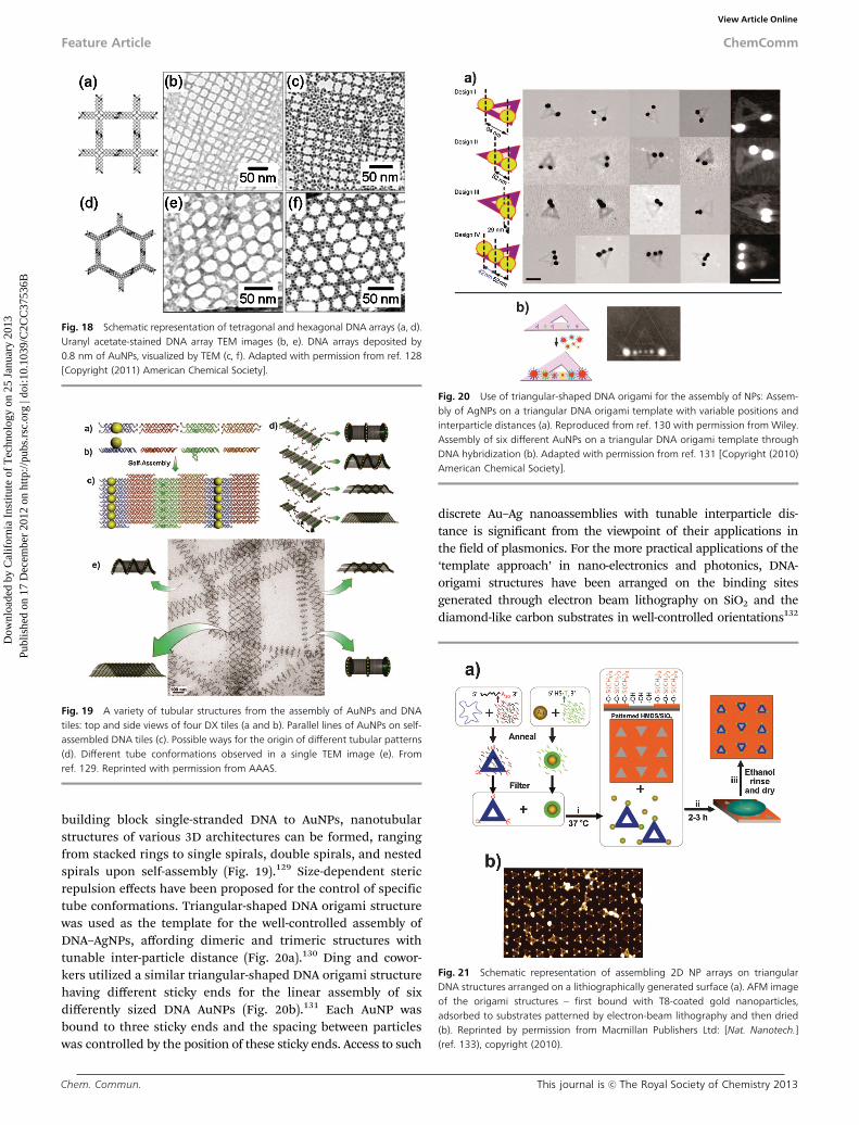
building block single-stranded DNA to AuNPs, nanotubularstructures of various 3D architectures can be formed, rangingfrom stacked rings to single spirals, double spirals, and nestedspirals upon self-assembly (Fig. 19).129 Size-dependent stericrepulsion effects have been proposed for the control of specifictube conformations. Triangular-shaped DNA origami structurewas used as the template for the well-controlled assembly ofDNA–AgNPs, affording dimeric and trimeric structures withtunable inter-particle distance (Fig. 20a).130 Ding and cowor-kers utilized a similar triangular-shaped DNA origami structurehaving different sticky ends for the linear assembly of sixdifferently sized DNA AuNPs (Fig. 20b).131 Each AuNP wasbound to three sticky ends and the spacing between particleswas controlled by the position of these sticky ends. Access to such
discrete Au–Ag nanoassemblies with tunable interparticle dis-tance is significant from the viewpoint of their applications inthe field of plasmonics. For the more practical applications of the‘template approach’ in nano-electronics and photonics, DNA-origami structures have been arranged on the binding sitesgenerated through electron beam lithography on SiO2 and thediamond-like carbon substrates in well-controlled orientations132
Fig. 18 Schematic representation of tetragonal and hexagonal DNA arrays (a, d).Uranyl acetate-stained DNA array TEM images (b, e). DNA arrays deposited by0.8 nm of AuNPs, visualized by TEM (c, f). Adapted with permission from ref. 128[Copyright (2011) American Chemical Society].
Fig. 19 A variety of tubular structures from the assembly of AuNPs and DNAtiles: top and side views of four DX tiles (a and b). Parallel lines of AuNPs on self-assembled DNA tiles (c). Possible ways for the origin of different tubular patterns(d). Different tube conformations observed in a single TEM image (e). Fromref. 129. Reprinted with permission from AAAS.
Fig. 20 Use of triangular-shaped DNA origami for the assembly of NPs: Assem-bly of AgNPs on a triangular DNA origami template with variable positions andinterparticle distances (a). Reproduced from ref. 130 with permission from Wiley.Assembly of six different AuNPs on a triangular DNA origami template throughDNA hybridization (b). Adapted with permission from ref. 131 [Copyright (2010)American Chemical Society].
Fig. 21 Schematic representation of assembling 2D NP arrays on triangularDNA structures arranged on a lithiographically generated surface (a). AFM imageof the origami structures – first bound with T8-coated gold nanoparticles,adsorbed to substrates patterned by electron-beam lithography and then dried(b). Reprinted by permission from Macmillan Publishers Ltd: [Nat. Nanotech.](ref. 133), copyright (2010).
Feature Article ChemComm
Dow
nloa
ded
by C
alif
orni
a In
stitu
te o
f T
echn
olog
y on
25
Janu
ary
2013
Publ
ishe
d on
17
Dec
embe
r 20
12 o
n ht
tp://
pubs
.rsc
.org
| do
i:10.
1039
/C2C
C37
536B
View Article Online

This journal is c The Royal Society of Chemistry 2013 Chem. Commun.
and 5 nm AuNPs were assembled on them (Fig. 21).133 Yan andcoworkers utilized triangular DNA origami structures to assembleAuNR dimers with various well-defined inter-rod angles andrelative distances.134 Recently, DNA–AuNPs have been arrangedin chiral helical arrangement on a long DNA scaffold, obtainedusing the DNA-origami approach (Fig. 22).135 Such 3D chiralassemblies of AuNPs exhibited CD response in the visibleregion analogous to the chiral proteins and DNA showing theCD in ultraviolet and infrared regions.
3. Conclusion
The roles and functions of nucleic acids in biological systemsare well known, and it is, in fact, astonishing to realize thathow a molecule with such a simple structure does its nature-assigned complicated jobs flawlessly. Recently, scientistshave explored the other uses of nucleic acids, different fromthe classic roles of them, in many fields including syntheticchemistry. This article gives an overview of the recently devel-oped synthetic strategies towards metal nanostructures andnanoassemblies based on the designed use of DNA. Shortlyafter the discovery of DNA–metal NP conjugates, the assembledstructures of DNA–AuNPs and subsequent change in theiroptical property have been used in a number of bio-detectionmethods including ‘solution-based colorimetric’ and ‘chip-based scanometric methods’.136 DNA–AuNP-based colorimetricmethods have been used for the detection of a variety ofnucleic acids,60,61 small molecules,137,138 enzymes139,140 andmetal ions.141–145 The scanometric method has been used forthe microRNA array profiling of prostate cancer markers.146
DNA–AuNP based methods such as ‘bio-barcode assay’ hold alot of potential for highly sensitive diagnostic applicationsfor both protein and DNA targets.147–149 As discussed in thepreceding parts, DNA plays versatile and crucial roles in build-ing a myriad of nanostructures with structural tunability. Sucha kind of structural tuning in a very short range has hugeimplications in nanotechnologies and optics, particularly innanoplasmonics.12 This nanostructure-based plasmonic tuningcan allow for developing the SERS probes with very strong and
tunable signals, especially for the early and multiplexed bio-diagnostic and bioimaging applications.34,35,109 It should alsobe noted that metal nanostructures can be useful in amplifyingfluorescent signals, but nanometer or subnanomter scale struc-tural control for a large number of structures is key to obtainingreliable amplified signals in a reproducible fashion.150–152 DNAtemplated well-organized and controlled assemblies will findapplications in the fields of nanoelectronics and photonics.127
Recently reported chiral helical assemblies of gold nano-particles on DNA templates have enormous scope in the devel-opment of exciting metamaterials.134 Without the nanoscalecontrol and large-scale synthesis of well-defined nanostructuresof interest, it is extremely difficult to utilize these structures forwidespread and practical applications even with many moreparadigm-shifting discoveries in nanoscience and nanotechnology.As the field of synthetic nanotechnology is growing by leaps andbounds, close attention should be paid to this field andsubsequent applications in the near future.
Acknowledgements
J.-M.N. was supported by the National Research Foundation ofKorea (NRF) grant funded by the Korean government (MEST)(No. 2011-0018198) and the Pioneer Research Center Programthrough the NRF of Korea funded by the Ministry of Education,Science and Technology (2012-0009565). The authors wouldalso like to acknowledge financial support from the IndustrialCore Technology Development Program of the Ministry ofKnowledge Economy (no. 10033183 and 10037397) and theKRICT OASIS Project from the Korea Research Institute ofChemical Technology.
Notes and references1 N. C. Seeman, Nature, 2003, 421, 427–431.2 J. D. Watson and F. H. C. Crick, Nature, 1953, 171, 737–738.3 S. Burge, G. N. Parkinson, P. Hazel, A. K. Todd and S. Neidle,
Nucleic Acids Res., 2006, 34, 5402–5415.4 N. C. Seeman, Mol. Biotechnol., 2007, 37, 246–257.5 N. C. Seeman, Annu. Rev. Biochem., 2010, 79, 65–87.6 D. Y. Zhang and G. Seelig, Nat. Chem., 2011, 3, 103–113.7 P. W. K. Rothemund, Nature, 2006, 440, 297–302.8 C. B. Reese, Org. Biomol. Chem., 2005, 3, 3851–3868.9 X. Zhang and V. K. Yadavalli, Nanoscale, 2012, 4, 2439–2446.
10 E. Stulz, Chem.–Eur. J., 2012, 18, 4456–4469.11 O. I. Wilner and I. Willner, Chem. Rev., 2012, 112, 2528–2556.12 S. J. Tan, M. J. Campolongo, D. Luo and W. Cheng, Nat. Nanotech-
nol., 2011, 6, 268–276.13 C. L. Choi and P. Alivisatos, Annu. Rev. Phys. Chem., 2010, 61,
369–389.14 G. Chen, Y. Wang, M. Yang, J. Xu, S. J. Goh, M. Pan and H. Chen,
J. Am. Chem. Soc., 2010, 132, 3644–3645.15 W. Li, P. H. C. Camargo, X. Lu and Y. Xia, Nano Lett., 2009, 9,
485–490.16 A. P. Alivisatos, K. P. Johnsson, X. Peng, T. E. Wilson, C. J. Loweth,
M. P. Bruchez Jr. and P. G. Schultz, Nature, 1996, 382, 609–611.17 K.-M. Sung, D. W. Mosley, B. R. Peelle, S. Zhang and J. M. Jacobson,
J. Am. Chem. Soc., 2004, 126, 5064–5065.18 G. A. DeVries, M. Brunnbauer, Y. Hu, A. M. Jackson, B. Long,
B. T. Neltner, O. Uzun, B. H. Wunsch and F. Stellacci, Science, 2007,315, 358–361.
19 J. H. Lee, D. P. Wernette, M. V. Yigit, J. Liu, Z. Wang and Y. Lu,Angew. Chem., Int. Ed., 2007, 46, 9006–9010.
20 T. Chen, M. Yang, X. Wang, L. H. Tan and H. Chen, J. Am. Chem.Soc., 2008, 130, 11858–11859.
Fig. 22 Cartoon of left- and right-handed helical assemblies of AuNPs on DNAtubes (a). TEM image of assembled left-handed AuNPs (b). Reprinted by permis-sion from Macmillan Publishers Ltd: [Nature] (ref. 135), copyright (2012).
ChemComm Feature Article
Dow
nloa
ded
by C
alif
orni
a In
stitu
te o
f T
echn
olog
y on
25
Janu
ary
2013
Publ
ishe
d on
17
Dec
embe
r 20
12 o
n ht
tp://
pubs
.rsc
.org
| do
i:10.
1039
/C2C
C37
536B
View Article Online
Chem. Commun. This journal is c The Royal Society of Chemistry 2013
21 L. C. Brousseau III., J. P. Novak, S. M. Marinakos andD. L. Feldheim, Adv. Mater., 1999, 11, 447–449.
22 C. A. Mirkin, R. L. Letsinger, R. C. Mucic and J. J. Storhoff, Nature,1996, 382, 607–609.
23 M. R. Jones, K. D. Osberg, R. J. Macfarlane, M. R. Langille andC. A. Mirkin, Chem. Rev., 2011, 111, 3736–3827.
24 S. Lal, S. E. Clare and N. J. Halas, Acc. Chem. Res., 2008, 41,1842–1851.
25 M. S. Yavuz, Y. Cheng, J. Chen, C. M. Cobley, Q. Zhang, M. Rycenga,J. Xie, C. Kim, K. H. Song, A. G. Schwartz, L. V. Wang and Y. Xia,Nat. Mater., 2009, 8, 935–939.
26 E. Ozbay, Science, 2006, 311, 189–193.27 A. N. Shipway, E. Katz and I. Willner, ChemPhysChem, 2000, 1,
18–52.28 J. A. Schuller, E. S. Barnard, W. Cai, Y. C. Jun, J. S. White and
M. L. Brongersma, Nat. Mater., 2010, 9, 193–204.29 A. Cao, R. Lu and G. Veser, Phys. Chem. Chem. Phys., 2010, 12,
13499–13510.30 G. Mie, Ann. Phys., 1908, 330, 377–445.31 E. Prodan, C. Radloff, N. J. Halas and P. Nordlander, Science, 2003,
302, 419–422.32 Y. Sun, F. Xu, Y. Zhang, Y. Shi, Z. Wen and Z. Li, J. Mater. Chem.,
2011, 21, 16675–16685.33 L. Guerrini, F. McKenzie, A. W. Wark, K. Faulds and D. Graham,
Chem. Sci., 2012, 3, 2262–2269.34 D.-K. Lim, K.-S. Jeon, H. M. Kim, J.-M. Nam and Y. D. Suh, Nat.
Mater., 2010, 9, 60–67.35 D.-K. Lim, K.-S. Jeon, J.-H. Hwang, H. Kim, S. Kwon, Y. D. Suh and
J.-M. Nam, Nat. Nanotechnol., 2011, 6, 452–460.36 M.-C. Daniel and D. Astruc, Chem. Rev., 2004, 104, 293–346.37 J. Perez-Juste, I. Pastoriza-Santos, L. M. Liz-Marzan and
P. Mulvaney, Coord. Chem. Rev., 2005, 249, 1870–1901.38 C. J. Murphy, A. M. Gole, S. E. Hunyadi and C. J. Orendorff, Inorg.
Chem., 2006, 45, 7544–7554.39 Z. Huo, C.-K. Tsung, W. Huang, X. Zhang and P. Yang, Nano Lett.,
2008, 8, 2041–2044.40 B. Liu, J. Xie, J. Y. Lee, Y. P. Ting and J. P. Chen, J. Phys. Chem. B,
2005, 109, 15256–15263.41 S. Porel, S. Singh and T. P. Radhakrishnan, Chem. Commun., 2005,
2387–2389.42 C. S. Ah, Y. J. Yun, H. J. Park, W. J. Kim, D. H. Ha and W. S. Yun,
Chem. Mater., 2005, 17, 5558–5561.43 Y. Xia, Y. Xiong, B. Lim and S. E. Skrabalak, Angew. Chem., Int. Ed.,
2009, 48, 60–103.44 Y. Sun and Y. Xia, Science, 2002, 298, 2176–2179.45 S. E. Skrabalak, J. Chen, Y. Sun, X. Lu, L. Au, C. M. Cobley and
Y. Xia, Acc. Chem. Res., 2008, 41, 1587–1595.46 S. Chen, Z. L. Wang, J. Ballato, S. H. Foulger and D. L. Carroll,
J. Am. Chem. Soc., 2003, 125, 16186–16187.47 X. Wang, H. Fu, A. Peng, T. Zhai, Y. Ma, F. Yuan and J. Yao, Adv.
Mater., 2009, 21, 1636–1640.48 R. Jin, Y. Cao, C. A. Mirkin, K. L. Kelly, G. C. Schatz and J. G. Zheng,
Science, 2001, 294, 1901–1903.49 W. Cheng, M. Steinhart, U. Gosele and R. B. Wehrspohn, J. Mater.
Chem., 2007, 17, 3493–3495.50 M. Grzelczak, J. Vermant, E. M. Furst and L. M. Liz-Marzan, ACS
Nano, 2010, 4, 3591–3605.51 A. Badia, S. Singh, L. Demers, L. Cuccia, G. R. Brown and
R. B. Lennox, Chem.–Eur J., 1996, 2, 359–363.52 A. C. Templeton, W. P. Wuelfing and R. W. Murray, Acc. Chem. Res.,
2000, 33, 27–36.53 Y. Min, M. Akbulut, K. Kristiansen, Y. Golan and J. Israelachvili,
Nat. Mater., 2008, 7, 527–538.54 J. I. Cutler, E. Auyeung and C. A. Mirkin, J. Am. Chem. Soc., 2012,
134, 1376–1391.55 T. A. Taton, C. A. Mirkin and R. L. Letsinger, Science, 2000, 289,
1757–1760.56 J. J. Storhoff, R. Elghanian, R. C. Mucic, C. A. Mirkin and
R. L. Letsinger, J. Am. Chem. Soc., 1998, 120, 1959–1964.57 Y. C. Cao, R. Jin and C. A. Mirkin, Science, 2002, 297, 1536–1540.58 N. L. Rosi, D. A. Giljohann, C. S. Thaxton, A. K. R. Lytton-Jean,
M. S. Han and C. A. Mirkin, Science, 2006, 312, 1027–1030.59 J. J. Storhoff and C. A. Mirkin, Chem. Rev., 1999, 99, 1849–1862.60 R. Elghanian, J. J. Storhoff, R. C. Mucic, R. L. Letsinger and
C. A. Mirkin, Science, 1997, 277, 1078–1081.
61 R. A. Reynolds III., C. A. Mirkin and R. L. Letsinger, J. Am. Chem.Soc., 2000, 122, 3795–3796.
62 J. S. Lee, A. K. R. Lytton-Jean, S. J. Hurst and C. A. Mirkin, NanoLett., 2007, 7, 2112–2115.
63 J. I. Cutler, D. Zheng, X. Xu, D. A. Giljohann and C. A. Mirkin, NanoLett., 2010, 10, 1477–1480.
64 G. P. Mitchell, C. A. Mirkin and R. L. Letsinger, J. Am. Chem. Soc.,1999, 121, 8122–8123.
65 I. L. Medintz, L. Berti, T. Pons, A. F. Grimes, D. S. English,A. Alessandrini, P. Facci and H. Mattoussi, Nano Lett., 2007, 7,1741–1748.
66 S. Eustis and M. A. El-Sayed, Chem. Soc. Rev., 2006, 35, 209–217.67 M. Stratakis and H. Garcia, Chem. Rev., 2012, 112, 4469–4506.68 R. A. Sperling, P. R. Gil, F. Zhang, M. Zanella and W. J. Parak,
Chem. Soc. Rev., 2008, 37, 1896–1908.69 H. D. Hill, J. E. Millstone, M. J. Banholzer and C. A. Mirkin, ACS
Nano, 2009, 3, 418–424.70 S. J. Hurst, A. K. R. Lytton-Jean and C. A. Mirkin, Anal. Chem., 2006,
78, 8313–8318.71 X. Zhang, M. R. Servos and J. Liu, J. Am. Chem. Soc., 2012, 134,
7266–7269.72 X. Zhang, M. R. Servos and J. Liu, J. Am. Chem. Soc., 2012, 134,
9910–9913.73 H. Pei, F. Li, Y. Wan, M. Wei, H. Liu, Y. Su, N. Chen, Q. Huang and
C. Fan, J. Am. Chem. Soc., 2012, 134, 11876–11879.74 J.-Y. Kim and J.-S. Lee, Nano Lett., 2009, 9, 4564–4569.75 S. Y. Park, A. K. R. Lytton-Jean, B. Lee, S. Weigand, G. C. Schatz and
C. A. Mirkin, Nature, 2008, 451, 553–556.76 D. Nykypanchuk, M. M. Maye, D. Van Der Lelie and O. Gang,
Nature, 2008, 451, 549–552.77 E. Auyeung, J. I. Cutler, R. J. MacFarlane, M. R. Jones, J. Wu, G. Liu,
K. Zhang, K. D. Osberg and C. A. Mirkin, Nat. Nanotechnol., 2012, 7,24–28.
78 T. I. N. G. Li, R. Sknepnek, R. J. MacFarlane, C. A. Mirkin andM. Olvera De La Cruz, Nano Lett., 2012, 12, 2509–2514.
79 B. M. Mladek, J. Fornleitner, F. J. Martinez-Veracoechea, A. Dawidand D. Frenkel, Phys. Rev. Lett., 2012, 108, 268301.
80 E. Dujardin, L.-B. Hsin, C. R. C. Wang and S. Mann, Chem.Commun., 2001, 1264–1265.
81 M. R. Jones, R. J. MacFarlane, B. Lee, J. Zhang, K. L. Young,A. J. Senesi and C. A. Mirkin, Nat. Mater., 2010, 9, 913–917.
82 J. E. Millstone, D. G. Georganopoulou, X. Xu, W. Wei, S. Li andC. A. Mirkin, Small, 2008, 4, 2176–2180.
83 H. G. Park, J. H. Joo, H. G. Kim and J. S. Lee, J. Phys. Chem. C, 2012,116, 2278–2284.
84 B. Nikoobakht and M. A. El-Sayed, Chem. Mater., 2003, 15,1957–1962.
85 J. E. Millstone, S. Park, K. L. Shuford, L. Qin, G. C. Schatz andC. A. Mirkin, J. Am. Chem. Soc., 2005, 127, 5312–5313.
86 Z. Li, Z. Zhu, W. Liu, Y. Zhou, B. Han, Y. Gao and Z. Tang, J. Am.Chem. Soc., 2012, 134, 3322–3325.
87 B. Yurke, A. J. Turberfield, A. P. Mills Jr., F. C. Simmel andJ. L. Neumann, Nature, 2000, 406, 605–608.
88 T. Song and H. Liang, J. Am. Chem. Soc., 2012, 134, 10803–10806.89 Y. Yan, J. I. L. Chen and D. S. Ginger, Nano Lett., 2012, 12,
2530–2536.90 D. K. Lim, I. J. Kim and J.-M. Nam, Chem. Commun., 2008,
5312–5314.91 S. Alayoglu and B. Eichhorn, J. Am. Chem. Soc., 2008, 130,
17479–17486.92 J. A. Fan, Y. He, K. Bao, C. Wu, J. Bao, N. B. Schade,
V. N. Manoharan, G. Shvets, P. Nordlander, D. R. Liu andF. Capasso, Nano Lett., 2011, 11, 4859–4864.
93 D. Zanchet, C. M. Micheel, W. J. Parak, D. Gerion andA. P. Alivisatos, Nano Lett., 2001, 1, 32–35.
94 C. J. Loweth, W. B. Caldwell, X. Peng, A. P. Alivisatos andP. G. Schultz, Angew. Chem., Int. Ed., 1999, 38, 1808–1812.
95 S. A. Claridge, H. W. Liang, S. R. Basu, J. M. J. Frechet andA. P. Alivisatos, Nano Lett., 2008, 8, 1202–1206.
96 A. J. Mastroianni, S. A. Claridge and A. P. Alivisatos, J. Am. Chem.Soc., 2009, 131, 8455–8459.
97 W. Yan, L. Xu, C. Xu, W. Ma, H. Kuang, L. Wang and N. A. Kotov,J. Am. Chem. Soc., 2012, 134, 15114–15121.
98 J.-H. Lee, J.-M. Nam, K.-S. Jeon, D.-K. Lim, H. Kim, S. Kwon, H. Leeand Y. D. Suh, ACS Nano, 2012, 6, 9574–9584.
Feature Article ChemComm
Dow
nloa
ded
by C
alif
orni
a In
stitu
te o
f T
echn
olog
y on
25
Janu
ary
2013
Publ
ishe
d on
17
Dec
embe
r 20
12 o
n ht
tp://
pubs
.rsc
.org
| do
i:10.
1039
/C2C
C37
536B
View Article Online
This journal is c The Royal Society of Chemistry 2013 Chem. Commun.
99 X. Xu, N. L. Rosi, Y. Wang, F. Huo and C. A. Mirkin, J. Am. Chem.Soc., 2006, 128, 9286–9287.
100 M. M. Maye, D. Nykypanchuk, M. Cuisinier, D. Van Der Lelie andO. Gang, Nat. Mater., 2009, 8, 388–391.
101 J.-W. Kim, J.-H. Kim and R. Deaton, Angew. Chem., Int. Ed., 2011,50, 9185–9190.
102 K. H. Roh, D. C. Martin and J. Lahann, Nat. Mater., 2005, 4,759–763.
103 S. Jiang, Q. Chen, M. Tripathy, E. Luijten, K. S. Schweizer andS. Granick, Adv. Mater., 2010, 22, 1060–1071.
104 H. Xing, Z. Wang, Z. Xu, N. Y. Wong, Y. Xiang, G. L. Liu and Y. Lu,ACS Nano, 2012, 6, 802–809.
105 B. Pan, L. Ao, F. Gao, H. Tian, R. He and D. Cui, Nanotechnology,2005, 16, 1776–1780.
106 Z. Zhang, Y. Wen, D. Zhao and X. Zhang, Appl. Phys. Lett., 2012,101, 213701.
107 S. J. Zhen, C. Z. Huang, J. Wang and Y. F. Li, J. Phys. Chem. C, 2009,113, 21543–21547.
108 Y. Wang, Y. F. Li, J. Wang, Y. Sang and C. Z. Huang, Chem.Commun., 2010, 46, 1332–1334.
109 L. Xu, H. Kuang, C. Xu, W. Ma, L. Wang and N. A. Kotov, J. Am.Chem. Soc., 2012, 134, 1699–1709.
110 L. Wang, Y. Zhu, L. Xu, W. Chen, H. Kuang, L. Liu, A. Agarwal,C. Xu and N. A. Kotov, Angew. Chem., Int. Ed., 2010, 49, 5472–5475.
111 Y. Li, Y. Zheng, M. Gong and Z. Deng, Chem. Commun., 2012, 48,3727–3729.
112 J. H. Lee, G. H. Kim and J. M. Nam, J. Am. Chem. Soc., 2012, 134,5456–15459.
113 M. G. Warner and J. E. Hutchison, Nat. Mater., 2003, 2, 272–277.114 G. Wang and R. W. Murray, Nano Lett., 2004, 4, 95–101.115 Z. Deng, Y. Tian, S.-H. Lee, A. E. Ribbe and C. Mao, Angew. Chem.,
Int. Ed., 2005, 44, 3582–3585.116 S. Beyer, P. Nickels and F. C. Simmel, Nano Lett., 2005, 5, 719–722.117 B. Pan, D. Cui, C. Ozkan, P. Xu, T. Huang, Q. Li, H. Chen, F. Liu,
F. Gao and R. He, J. Phys. Chem. C, 2007, 111, 12572–12576.118 H. Li, S. H. Park, J. H. Reif, T. H. LaBean and H. Yan, J. Am. Chem.
Soc., 2004, 126, 418–419.119 L. A. Stearns, R. Chhabra, J. Sharma, Y. Liu, W. T. Petuskey, H. Yan
and J. C. Chaput, Angew. Chem., Int. Ed., 2009, 48, 8494–8496.120 H. Bui, C. Onodera, C. Kidwell, Y. Tan, E. Graugnard, W. Kuang,
J. Lee, W. B. Knowlton, B. Yurke and W. L. Hughes, Nano Lett.,2010, 10, 3367–3372.
121 P. K. Lo, P. Karam, F. A. Aldaye, C. K. McLaughlin, G. D. Hamblin,G. Cosa and H. F. Sleiman, Nat. Chem., 2010, 2, 319–328.
122 J. D. Le, Y. Pinto, N. C. Seeman, K. Musier-Forsyth, T. A. Taton andR. A. Kiehl, Nano Lett., 2004, 4, 2343–2347.
123 J. Sharma, R. Chhabra, Y. Liu, Y. Ke and H. Yan, Angew. Chem., Int.Ed., 2006, 45, 730–735.
124 J. Zhang, Y. Liu, Y. Ke and H. Yan, Nano Lett., 2006, 6, 248–251.125 J. Sharma, R. Chhabra, C. S. Andersen, K. V. Gothelf, H. Yan and
Y. Liu, J. Am. Chem. Soc., 2008, 130, 7820–7821.126 J. Zheng, P. E. Constantinou, C. Micheel, A. P. Alivisatos, R. A. Kiehl
and N. C. Seeman, Nano Lett., 2006, 6, 1502–1504.
127 W. Cheng, M. J. Campolongo, J. J. Cha, S. J. Tan, C. C. Umbach,D. A. Muller and D. Luo, Nat. Mater., 2009, 8, 519–525.
128 Y. He, T. Ye, A. E. Ribbe and C. Mao, J. Am. Chem. Soc., 2011, 133,1742–1744.
129 J. Sharma, R. Chhabra, A. Cheng, J. Brownell, Y. Liu and H. Yan,Science, 2009, 323, 112–116.
130 S. Pal, Z. Deng, B. Ding, H. Yan and Y. Liu, Angew. Chem., Int. Ed.,2010, 49, 2700–2704.
131 B. Ding, Z. Deng, H. Yan, S. Cabrini, R. N. Zuckermann andJ. Bokor, J. Am. Chem. Soc., 2010, 132, 3248–3249.
132 R. J. Kershner, L. D. Bozano, C. M. Micheel, A. M. Hung,A. R. Fornof, J. N. Cha, C. T. Rettner, M. Bersani, J. Frommer,P. W. K. Rothemund and G. M. Wallraff, Nat. Nanotechnol., 2009, 4,557–561.
133 A. M. Hung, C. M. Micheel, L. D. Bozano, L. W. Osterbur,G. M. Wallraff and J. N. Cha, Nat. Nanotechnol., 2010, 5, 121–126.
134 S. Pal, Z. Deng, H. Wang, S. Zou, Y. Liu and H. Yan, J. Am. Chem.Soc., 2011, 133, 17606–17609.
135 A. Kuzyk, R. Schreiber, Z. Fan, G. Pardatscher, E. M. Roller,A. Hogele, F. C. Simmel, A. O. Govorov and T. Liedl, Nature,2012, 483, 311–314.
136 N. L. Rosi and C. A. Mirkin, Chem. Rev., 2005, 105, 1547–1562.137 J. Liu and Y. Lu, Angew. Chem., Int. Ed., 2006, 45, 90–94.138 M. S. Han, A. K. R. Lytton-Jean, B. K. Oh, J. Heo and C. A. Mirkin,
Angew. Chem., Int. Ed., 2006, 45, 1807–1810.139 X. Xu, M. S. Han and C. A. Mirkin, Angew. Chem., Int. Ed., 2007, 46,
3468–3470.140 H. Jo, S. Lee, K. Min and C. Ban, Anal. Biochem., 2012, 421, 313–320.141 J. S. Lee, M. S. Han and C. A. Mirkin, Angew. Chem., Int. Ed., 2007,
46, 4093–4096.142 J. Liu and Y. Lu, J. Am. Chem. Soc., 2003, 125, 6642–6643.143 X. Xu, W. L. Daniel, W. Wei and C. A. Mirkin, Small, 2010, 6,
623–626.144 Z. Wang, J. H. Lee and Y. Lu, Adv. Mater., 2008, 20, 3263–3267.145 J. Liu and Y. Lu, J. Am. Chem. Soc., 2004, 126, 12298–12305.146 A. H. Alhasan, D. Y. Kim, W. L. Daniel, E. Watson, J. J. Meeks,
C. S. Thaxton and C. A. Mirkin, Anal. Chem., 2012, 84, 4153–4160.147 J.-M. Nam, C. S. Thaxton and C. A. Mirkin, Science, 2003, 301,
1884–1886.148 D. G. Georganopoulou, L. Chang, J. M. Nam, C. S. Thaxton,
E. J. Mufson, W. L. Klein and C. A. Mirkin, Proc. Natl. Acad. Sci.U. S. A., 2005, 102, 2273–2276.
149 C. S. Thaxton, R. Elghanian, A. D. Thomas, S. I. Stoeva, J.-S. Lee,N. D. Smith, A. J. Schaeffer, H. Klocker, W. Horninger, G. Bartschand C. A. Mirkin, P. Natl. Acad. Sci. U. S. A., 2009, 106, 18437–18442.
150 K. Aslan, I. Gryczynski, J. Malicka, E. Matveeva, J. R. Lakowicz andC. D. Geddes, Curr. Opin. Biotechnol., 2005, 16, 55–62.
151 C. McDonagh, C. S. Burke and B. D. MacCraith, Chem. Rev., 2008,108, 400–422.
152 P. P. Pompa, L. Martiradonna, A. D. Torre, F. D. Sala, L. Manna,M. De Vittorio, F. Calabi, R. Cingolani and R. Rinaldil, Nat.Nanotechnol., 2006, 1, 126–130.
ChemComm Feature Article
Dow
nloa
ded
by C
alif
orni
a In
stitu
te o
f T
echn
olog
y on
25
Janu
ary
2013
Publ
ishe
d on
17
Dec
embe
r 20
12 o
n ht
tp://
pubs
.rsc
.org
| do
i:10.
1039
/C2C
C37
536B
View Article Online
![The Caltech DNA- nanoelectronics teamdna.caltech.edu/~pwkr/talks/ONR-DNA-nanoelectronics.pdf · The Caltech DNA-nanoelectronics team PI Paul Rothemund, ... D [n A] V g [V] I S D S](https://img.pdfslide.us/doc/110x75/5a962fc57f8b9ad96f8cc74c/the-caltech-dna-nanoelectronics-pwkrtalksonr-dna-nanoelectronicspdfthe-caltech.jpg)


![Nucleic acid-based nanoengineering: novel structures for ...pwkr/dna-nanotech...applied to improve drug and gene therapies [11–17]. To build these enabling materials or devices,](https://img.pdfslide.us/doc/110x75/5f0f89b07e708231d444a86c/nucleic-acid-based-nanoengineering-novel-structures-for-pwkrdna-nanotech.jpg)