Embed Size (px)
Citation preview

rXXXX American Chemical Society A dx.doi.org/10.1021/cr200104q | Chem. Rev. XXXX, XXX, 000–000
REVIEW
pubs.acs.org/CR
Functionalized DNA NanostructuresOfer I. Wilner and Itamar Willner*
Institute of Chemistry, Center for Nanoscience and Nanotechnology, The Hebrew University of Jerusalem, Jerusalem 91904, Israel
CONTENTS
1. Introduction A2. Biocatalytic Growth of DNA Nanostructures through
the Rolling Circle Amplification Process B3. Formation of Functionalized Nanostructures
through Directed Hybridization of DNA Tiles D4. Functional DNA Nanostructures through Directed
Origami Folding J5. Self-Assembly of DNA Nanoparticle Nanostructures
through Hybridization or DNA�Protein Interactions P6. DNA Nanostructures as Templates for the Bottom-
Up Fabrication of Nanodevices V7. Conclusions and Perspectives YAuthor Information ZBiographies ZAcknowledgment ZReferences AA
1. INTRODUCTION
DNA is one of the most promising materials for nanobiotech-nology, and the present review article aims to describe recentadvances in the self-assembly of functional DNA nanostructures.DNA exhibits nanoscale dimensions reflected by a 3.4 nm lengthfor a complete 10-base helical turn and a diameter correspondingto 2 nm for the duplex DNA.1 The base sequence in DNAencodes into the biopolymer tremendous structural and func-tional information (Figure 1). The two purine bases and the twopyrimidine bases adenine (A), guanine (G), cytosine (C), andthymine (T) constitute the building blocks of the biopolymerand provide the instructive information dictating the structureand functions of the nucleic acids. The basic Watson�Crickcomplementarity of A-T and C-G base-pairing leads to theformation of duplex DNA structures, and appropriate base-pairing leads to triplex-hybridization of DNA strands.2 Also,G-rich or C-rich single-stranded nucleic acids self-assemble intoG-quadruplexes3 or i-motifs,4 respectively, and supramolecularDNA duplexes may be cooperatively stabilized by metal ionswhile forming T�Hg2+�T or C�Ag+�C bridges.5,6 Alterna-tively, the incorporation of artificial heterocyclic bases into thenucleic acid structures may provide ligation sites (ligandosides)for the bridging of duplex structures of DNA by a variety ofmetal ions.7 The availability of enzymes that react with DNA/RNA, such as polymerase or reverse transcriptase, which repli-cate oligonucleotides, telomerase that elongates single nucleicacid strands, or sequence-specific DNA cleavage enzymes, suchas endonucleases or nicking enzymes, provide a unique “toolbox”of enzymes for manipulating DNA. This arsenal of enzymes,
together with the synthetic ability to synthesize any DNAsequence by automated chemical methods, and to replicate theproducts by the polymerase chain reaction (PCR), enable thecost-effective preparation of large amounts of any DNA. Also,ingenious organic synthesis protocols for the preparation of newnucleotide bases8 and their biopolymers, such as peptide nucleicacids (PNAs)9 or locked nucleotides (LNAs),10 introduced newman-made DNA analogues revealing unique properties. Theseartificial DNA analogues can be conjugated to native DNA toform hybrid systems exhibiting new properties and functions.11
Similarly, synthetic purine/pyrimidine-type bases,12 or chemi-cally modified nucleotides linked to redox groups,13 photoactiveunits,14 chemical functionalities (amine, thiol, azide, etc.), ormolecular labels, such as biotin, provide building blocks forincorporation into the DNA chains. Such modified nucleic acidsact as scaffolds for secondary chemical functionalization, for thecovalent or supramolecular association of proteins, or as signal-responsive biopolymers that are activated by electrical or lightstimuli.15 Nucleic acids reveal, also, sequence-specific binding ofproteins, for example, the oriC replication region in bacteria,16a,b
the TATA box in eukaryots transcription machinery,16c or theaminoacyl tRNA synthetase that recognizes different tRNAsduring the translation process.16d Also, different selection pro-cedures, such as the systematic evolution of ligands by expo-nential enrichment (SELEX),17 led to the preparation andamplification of man-made nucleic acids revealing specific bind-ing properties toward low-molecular-weight substrates, e.g.,cocaine,18 or proteins, such as thrombin19 or lysozyme20
(aptamers). Also, the different selection platforms led to catalyticDNAs (DNAzymes).21 Thus, the chemically modified DNAstructures and the specific binding of proteins to DNA enable theconstruction of hybrid systems of predesigned structures andfunctions.
The control over the stabilities of duplex DNAs is achieved bythe number of base-pairs, by the nature of base-pairs, and by thecooperative stabilization of the duplexes by ligand�metal brid-ging units. Also, external triggers, such as pH or added nucleicacid strands, may affect the duplex stabilities through the forma-tion of single-stranded C-quadruplexes22 or strand displacementof the duplex structures.23 Such processes may induce topologytransitions of DNA nanostructures.24 Indeed, the control overthe stabilities of DNA nanostructures by means of externaltriggers led to the development of DNA machines25, such asDNA “tweezers”,26 “walker”,27 “stepper”,28 or nanometronomedevices.29 Such DNA machines were further implemented tocontrol surface properties and functions30 or for the develop-ment of amplified sensing platforms.31
Received: April 3, 2011

B dx.doi.org/10.1021/cr200104q |Chem. Rev. XXXX, XXX, 000–000
Chemical Reviews REVIEW
The self-assembly of molecular32 or macromolecular com-ponents33 into nanostructures is one of themost studied researchtopics in modern science. The continuous interest in self-assembly is inspired by the living cell machinery that demon-strates a complex system operating by self-organization princi-ples. For example, the ribosome self-assembles by at least 3nucleic acids and more than 50 protein units into a functionalnanostructure, by implementing specific noncovalent interac-tions between the nucleotide bases and phosphate groups ofrRNA, as well as amino acid residues of the protein subunits (H-bonds, electrostatic interactions).34 Such self-assembly principlesdictate the specific positioning of DNA, RNA, proteins, lipids,and more, in the cellular container. The resulting order controlsthe cell machineries, such as cell replication, ion-transport, signaltransduction, metabolic enzymatic cascades and networks, fueledmolecular motors, and more.
The unique features of DNA (or RNA) or of DNA/proteinhybrids provide unique building blocks for the “bottom-up”assembly of nanoarchitectures that reveal programmed function-alities and properties that emerge from the one-, two-, or three-dimensional ordering of the systems. Such nanostructures canpartially mimic cellular functionalities and may serve as scaffoldsfor the secondary templated synthesis of nanodevices. Indeed,extensive research efforts were directed in the past decade toimplement the properties of DNA for the “bottom-up” self-assembly of nanostructures. Many review articles have addresseddifferent facets of DNA nanotechnology.35 In the present articlewe address the scientific advances in the self-assembly of one-,two-, and three-dimensional ordered DNA�proteins or DNA�nanoparticle nanostructures. We discuss the methods for theprecise nanoscale spatial positioning of proteins or nanoparticleson the DNA scaffolds and address the emerging properties of theprogrammed nanostrucutres, as well as their application for thetemplated synthesis of nanodevices.
2. BIOCATALYTIC GROWTH OF DNA NANOSTRUC-TURES THROUGH THE ROLLING CIRCLE AMPLIFICA-TION PROCESS
Rolling circle amplification (RCA) is a process where a circularsingle-stranded DNA (ssDNA) template is being replicated byDNA polymerase, with an efficient strand displacement activity,in the presence of a nucleotide mixture.36 A short DNA strand isannealed to the circular DNA template (through base-pairinghybridization) and serves as a primer for the polymeriza-tion process. In a typical RCA process the circular template isbeing replicated hundreds to thousands of times, resulting in
micrometers-long DNA strands of repetitive units that arecomplementary to the circular DNA template. These repeatunits may serve as anchoring sites for different molecules,proteins, or nanoparticles (NPs). These guest substrates maybe linked to the RCA product by their functionalization withnucleic acid tethers complementary to the RCA repeat units,followed by hybridization with the RCA chains. Alternatively,low-molecular-weight components, such as biotin labels, may beincorporated into the RCA products, and the biotinylated guestsubstrates may link to the RCA product by avidin bridges. Also,incorporation of aptamer sequences for small molecules orproteins may be used to associate these substrates into theRCA chains.
AuNPs, 5 nm, were functionalized with thiolated nucleic acidscomplementary to the repeat units of micrometer-long RCAproducts37 (Figure 2A). Hybridization of the (2)-modified AuNPs to the RCA chains yielded 1D nanostructures, where theNPs were separated by equal distances (Figure 2B). The RCAprocess was also implemented to synthesize three-dimensional(3D) Au NPs nanostructures38 (Figure 2C). Large Au NPs(15 nm) were functionalized with thiolated nucleic acid (4)(loading ca. 230 DNA strands per particle). The nucleic acidunits (4) acted as primers that hybridized with the circular DNAtemplate (6). The RCA process activated on the supramolecularstructures, in the presence of polymerase and dNTPs mixture,resulted in the “decoration” of the Au NPs with long-chain RCAproducts in a 3D configuration. The hybridization of small AuNPs (5 nm) modified with the nucleic acid (9), complementaryto the RCA repeat units, yielded 3D structures consisting of smallAu NPs (5 nm), positioned at equal distances on the RCA chainsthat surround the central large Au NPs (15 nm) (Figure 2D).
Furthermore, DNA chains were generated by the RCAprocess using a circular DNA (11) and j29 DNA polymerase inthe presence of the dNTPs mixture39 (Figure 2E). The hybridi-zation of biotinylated nucleic acids complementary to the RCArepeat units, followed by the association of streptavidin modifiedwith 5 nm Au nanoparticles (NPs) led to the formation ofmicrometer-long DNA wires on which the Au NPs-modifiedprotein units are ordered and spatially separated by distances of30�50 μm (Figure 2F).
Aptamers are nucleic acid sequences exhibiting specificrecognition properties of low-molecular-weight substrates ormacromolecules (e.g., proteins).40 The aptamers are preparedby a selection process, systematic evolution of ligands byexponential enrichment (SELEX). The selective binding ofaptamers was used to design nucleic acid chains that enabledthe programmed assembly of one or two proteins on the DNAchains.41 A circular DNA (16) that includes two domains that arecomplementary to the thrombin-binding aptamer or the lysozyme-binding aptamer was used as a template for the activation of anRCA process, in the presence of the primer, polymerase, andthe dNTPs mixture. Micrometer-long RCA products (17) weregenerated, where each revolution of the RCA process generatedtwo separated aptamer sequences against thrombin or lysozyme(Figure 3A). The selective binding of dye-labeled thrombinor dye-labeled lysozyme was monitored by AFM or confocalmicroscopy (Figure 3, parts B andC). For example, the thrombinunits were labeled with a red fluorescent dye (tetramethyl-rhodamine), and lysozyme was modified with a green dye(fluorescein). The interaction of the RCA chains with the dye-labeled proteins generated the dense deposition of the two proteins(thrombin and lysozyme) on the aptamer domains (Figure 3C).
Figure 1. Schematic structural and functional features of nucleic acids:(A) single-stranded sticky end; (B) duplex hybridization; (C) hairpinnanostructure; (D) G-quadruplex; (E) triplex hybrid; (F) DNAzymestructure; (G) metal-bridged duplex; (H) aptamer nanostructure.

C dx.doi.org/10.1021/cr200104q |Chem. Rev. XXXX, XXX, 000–000
Chemical Reviews REVIEW
Although the previous examples have addressed the useof the RCA products as templates for the organization ofDNA/metal NPs or DNA/protein hybrid nanostructures, sig-nificant advances were achieved by demonstrating emergingfunctionalities of the resulting hybrid systems. For example, theRCA chains were used as templates for the activation of abienzyme cascade42 (Figure 3D). The circular DNA (21) actedas a template for the synthesis of RCA tapes consisting ofconstant repeat sequences I and II. The enzymes glucose oxidase(GOx) and horseradish peroxidase (HRP) were modified withnucleic acid tethers (23 and 24) that are complementary to thedomains I and II of the RCA template, respectively. The twoenzymes were then ordered on the DNA template throughhybridization (Figure 3E). The spatial proximity between thetwo enzymes on the DNA template enabled the activation of theenzyme cascade, a process that was prohibited in the homo-geneous phase. In this process, the GOx-catalyzed oxidation ofglucose by O2 yielded gluconic acid and H2O2, and the latterproduct acted as a substrate for the HRP that mediated theoxidation of 2,20-azino-bis[3-ethylbenzthiazoline-6-sulfonicacid] (ABTS2‑) to ABTS.�. The resulting colored product,ABTS.�, provided a means to follow the enzyme cascade(Figure 3F). The programmed and ordered binding of enzymesto DNA templates for the activation of biocatalytic cascades maybe extended to more complex systems. By designing circularDNAs that lead to the synthesis of DNA templates with ordered
three, or more, sequence domains, the coupling of more than twoenzymes may be envisaged. The hybridization of enzymes on theRCA chains enabled also the biocatalytic growth of metallic nano-wires. The enzyme-mediated growth of metal nanoparticles onmetalNPs seeds was extensively developed in recent years.43,44 Specifically,the modification of enzymes, e.g., glucose oxidase (GOx), with AuNPs (1.4 nm) enabled the growth of metallic nanowires.45 The dip-pen nanolithographic (DPN) deposition of the biocatalyst followedby the enzyme-mediated oxidation of glucose yielded H2O2 thatacted as a reducing agent for the reduction of AuCl4
� and theenlargement of the Au NPs seeds to form Au nanowires. Theadvantage of the biocatalytic growth ofmetallic nanowires is reflectedby a self-inhibition mechanism that leads to the controlled growth ofthe nanowires. That is, as the enzyme is coated by the enlarged NPs,the accessability of glucose to the active site is hindered, and thegrowth of the nanowires is blocked. These unique features of thebiocatalytic growth of metallic nanoparticles and nanowires wereimplemented to synthesize Au nanowires on the RCA-generatedtemplate (Figure 3G). The enzyme glucose oxidase was functional-ized with the nucleic acid 23, complementary to the domain I of theRCA tape, and was further modified with Au NPs (1.4 nm). Theresulting 23/Au NPs/GOx hybrid was hybridized with the DNAtemplate, and the biocatalytic enlargement of theNPs associatedwiththe enzyme led to micrometer-long metal nanowires exhibiting aheight of ca. 70 nm (Figure 3H).
Figure 2. (A) Ordered array of Au NPs on an RCA DNA scaffold through the hybridization of nucleic acid-functionalized Au NPs on the constantrepeat units of the RCA product. (B) Transmission electronmicroscopy (TEM) image of the ordered AuNPs array on theDNA scaffold. Reprinted withpermission from ref 37. Copyright 2005 Wiley-VCH. (C) The generation of 3D Au NPs nanostructures by the primary extension of nucleic acid-functionalized Au NPs using the RCA process and by the subsequent conjugation of small Au NPs complementary to the repeat units in the RCAproducts. (D) TEM image corresponding to the ordered deposition of small (5 nm) Au NPs to the RCA synthesized scaffolds linked to large (15 nm)Au NPs. Reprinted with permission from ref 38. Copyright 2006 Wiley-VCH. (E) Preparation of micrometer-long DNA wires consisting of biotin-labeled nucleic acids hybridized to RCA-generated DNA scaffolds, and the assembly of Au NPs (5 nm)-functionalized streptavidin on the biotinylatedDNA templates. (F) Atomic force microscopy (AFM) image of the resulting hybrid consisting of Au NPs-functionalized streptavidin on the DNAtemplate. Reprinted with permission from ref 39. Copyright 2005 American Chemical Society.

D dx.doi.org/10.1021/cr200104q |Chem. Rev. XXXX, XXX, 000–000
Chemical Reviews REVIEW
3. FORMATIONOFFUNCTIONALIZEDNANOSTRUCTURESTHROUGH DIRECTED HYBRIDIZATION OF DNA TILES
DNA tiles are specific DNA constructs designed to self-assembleinto one-dimensional (1D) templates,46 two-dimensional (2D)lattices,47 and three-dimensional (3D) nanostructures.48 The uniquefeatures of DNA tiles include the existence of an immobile junctionthat rigidifies the construct and the availability of single-strand sticky-ends that allow the interhybridization of the DNA tiles and their
self-assembly into complex nanostructures. Furthermore, the helicalturns associatedwith the tiles dictate the vectorial assembly of theDNAconstructs and the dimensionality of the resulting nanostructures.Figure 4 exemplifies several prototypes of reported tiles or motifsthat consist of double-crossover tiles (DX),49 triple-crossover tiles(TX),50 12-helix tiles,51 3-helix bundle tiles,52 6-helix bundletiles,53 four 4-arm junctions,54 triangular motifs composed of4-arm junctions,55 cross-shaped tiles,56 triangular DX tiles,57
Figure 3. (A) RCA-synthesized DNA chain consisting of alternate antithrombin and antilysozyme repeat units: the selective association of TAMRA-modified thrombin and fluorescein-functionalized lysozyme on the template consisting of the two aptamers. (B) AFM images of protein�aptamer chainhybrids. (C) Confocal microscopy images corresponding to the TAMRA�thrombin and fluorescein�lysozyme proteins immobilized on the aptamerchains. (I) λex = 543 nm, λem = 570 nm; following the TAMRA emission. (II) λex = 488 nm, λem = 520; following the fluorescein emission. (III) Overlayof the CFM images shown in I and II. Reprinted with permission from ref 41. Copyright 2007 Wiley-VCH. (D) Synthesis of a single-stranded DNAnanowire by the rolling circle amplification (RCA) method, the programmed assembly of glucose oxidase (GOx) and horseradish peroxidase (HRP) onthe DNA template, and the activation of the bienzyme cascade. (E) AFM image of the DNA nanowire functionalized with the two enzymes. (F) Kineticdata following the bienzyme cascade on the RCA-generated DNA template (a) and control experiments that monitor the bienzyme cascade in theabsence of DNA or in the presence of the foreign calf-thymus DNA, (b) and (c), respectively. (G) Assembly of Au NP-functionalized glucose oxidase onthe RCA-generated DNA template, and the biocatalytic growth of Au nanowires. (H) AFM image of the biocatalytically generated Au nanowire on theDNA template. Reprinted with permission from ref 42. Copyright 2009 American Chemical Society.

E dx.doi.org/10.1021/cr200104q |Chem. Rev. XXXX, XXX, 000–000
Chemical Reviews REVIEW
and 3-point star DNA tiles.58 DX consist of two helices conjugatedalong their long axes; TX consist of three double-stranded DNAhelices lying in a plane and linked by strand exchange at fourimmobile crossover points; 4 � 4 tiles contain four 4-arm DNAbranched junctions pointing in four directions; triangular motifsare composed of three 4-arm tiles, which are fused together;3-helix bundle motifs contain three double helical DNA domainsconnected by six immobile crossover junctions. In addition to thebasic property ofDNA tiles to self-assemble into complex structures,onemay tether to the tiles ligands on nucleic acid hinges that enablethe association of NPs or proteins to the nanostructures. Alterna-tively, the incorporation of aptamer sequences into the tile unitsmayprovide specific binding sites for the association of proteins.
2D DNA nanocrystals were organized by the self-assembly oftiles, and these acted as scaffolds for the ordered positioning ofAu NPs.59 Four different DX tiles a�d were designed to self-assemble into 2D lattices by sticky-end cohesion (Figure 5A).
Tile b contained a protruding sequence, modified with a thiolfunctionality on its 50 end. Au NPs (1.4 nm) modified with amaleimide residue were then covalently linked to the thiolfunctionalities associated with the protruding units. The subse-quent hybridization of the four tiles a�d resulted in the assemblyof Au-nanoparticles-programmed arrays. Figure 5B outlines aTEM image of the resulting DNA crystal�Au conjugate; theinterparticle spacing was found to correspond to 32 and 4 nm,respectively, as dictated by the 2D DNA scaffold.
Similarly, Au NPs were assembled on a 2D DNA scaffold,consisting of four different tiles, through base-pairing hybridiza-tion60 (Figure 5C). Tile f included an extended single-strandedoverhang, designed to hybridize with its complementary strand.Au NPs, 6 nm, modified with the thiolated single stranded DNA26, complementary to the overhanging tether of tile f, weresubsequently incorporated into the 2D DNA scaffold, via hy-bridization to form an ordered Au NPs array. Topographical
Figure 4. Examples of the toolbox of DNA tiles for the assembly of DNA nanostructures: (I) a double-crossover tile; (II) a triple crossover tile; (III) a12-helix DNA tile; (IV) a 3-helix bundle tile; (V) a 6-helix bundle tile; (VI) a DNA tile consisting of four 4-arm junctions; (VII) a triangular motifconsisting of three 4-arm junctions; (VIII) a cross-shaped tile; (IX) a triangular tile composed of three double-crossover DNA units; (X) a 3-point “star”DNA tile. Reprinted with permission from ref 35 d. Copyright 2006 Wiley-VCH.

F dx.doi.org/10.1021/cr200104q |Chem. Rev. XXXX, XXX, 000–000
Chemical Reviews REVIEW
AFM image of the assembled DNA array�Au hybrid, and a TEMimage of the Au NPs hybridized with the DNA scaffolds areshown in Figure 5 parts D and E, respectively. The rows of the Aunanoparticles were separated by ca. 63 nm. Also, a DNA tile arrayfor the programmed positioning of differently sized Au NPs wasdeveloped.61 Two 3D DX triangle tiles i and j were designed toproduce, upon their self-organization, a rhombic lattice arrange-ment (Figure 5F). Two sides of the equilateral tile containedsticky-end domains that enabled the self-organization of thenanostructure. The third side included a thiolated functionalityto which Au NPs could be linked. By the mixing of the two typesof tiles, 2D arrays with ordered Au NPs arrangements wereprepared. Three different DNA scaffold�Au NP hybrids wereconstructed:
(I) By the tethering of Au NPs, 5 nm, to the thiol functionalityof tile i, particle lines with a spatial separation of∼54 nm in onedirection and ∼27 nm in the opposite direction were generated.(II) By the tethering of the Au NPs, 5 nm, to the two tiles, NParrays revealing closer proximity between the NPs were formed.(III) By the functionalization of tile iwith small NPs, 5 nm, and ofthe thiol functionality of tile jwith large NPs, a composite array ofordered, differently sized Au NPs was formed. The different
structures were visualized under TEM; the different arrange-ments of the Au NPs could be obtained (Figure 5G). 3Dnanostructures composed of DNA tiles, and modified with AuNPs, were fabricated.62 Four double-crossover (DX) DNA tiles(k�n) were designed to self-assemble into a 2D array throughsticky-end associations (Figure 5H). Tile k was modified with athiol functionality that enabled the association of a Au NP to thetile component. While the tiles were designed to self-assembleinto a two-dimensional array of Au NPs rows, due to thecomplementarity of the sticky-ends of the tiles, it was discoveredthat the tiles self-organized into tubular 3D structures consistingof tubes composed of stacked rings of AuNPs, single-spiral tubes,double-spiral tubes, and nested-spiral tubes (Figure 5H).Whereas the stacked rings were generated by the symmetricalclosure of the array, the other structures were formed bynonsymmetrical folding and hybridization of the edges. Theformation of the Au NP-functionalized 3D nanostructures wasattributed to electrostatic repulsions among the neighboringrows of Au NPs that led to a curvature of the array, leading toits closure to the 3D nanotubes. This was supported by the factthat the self-assembly of tiles l,m, and nwith tile k, lacking the AuNPs, led to the formation of only 2D arrays. Also, tile m was
Figure 5. (A) Self-assembly of a 2D DNA nanostructure consisting of four complementary double-crossover tiles that include on tile b a protrudingthiolated nucleic acid for the programmed immobilization of Au NPs. (B) TEM image corresponding to the spatially ordered Au NPs on the 4-tile 2DDNA array. Reprinted with permission from ref 59. Copyright 2002 Springer. (C) The self-assembly of four double-crossover tiles e�h, where tile fincludes a protruding nucleic acid tether, and the secondary hybridization of Au NPs (6 nm) functionalized with a nucleic acid complementary to theprotruding tether to yield a spatially ordered array of NPs. (D) AFM image of the resulting Au NPs array. (E) TEM image of the resulting Au NPs array.Reprinted with permission from ref 60. Copyright 2004 American Chemical Society. (F) Self-assembly of two different triangular tiles i and j, eachconsisting of three-dimensional double-crossover units and modified with Au NPs. (G) TEM images corresponding to (I) the 2D array formed by tiles imodified with 5 nmAuNPs and the bare tiles j. (II) The 2D array formed by tiles i and jmodified with 5 nmAuNPs. (III) The 2D array formed by tiles imodified with 5 nmAuNPs and tiles jmodified with 10 nmAuNPs. Reprinted with permission from ref 61. Copyright 2006 American Chemical Society.(H) Self-assembly of four different double-crossover tiles (k�n), where tile k includes an AuNP (of variable size) associated with a protruding thiolatednucleic acid, into nanotubes of different structures consisting of stacked ring, single spiral, double spiral, and nested spiral, and TEM images that includethe different AuNPs functionalized nanostructures (I) and (II). The TEM images of the single spiral nanotubes functionalized with 5 nmAuNPs, wherethe structure in (I) includes on tilem a stem-loop component for stabilization and in (II) the nanostructure lacks this cooperative stabilization. (III) and(IV) TEM images of stacked-ring nanotubes modified with 10 and 15 nm Au NPs, respectively. Reprinted with permission from ref 62. Copyright 2009American Association for the Advancement of Science.

G dx.doi.org/10.1021/cr200104q |Chem. Rev. XXXX, XXX, 000–000
Chemical Reviews REVIEW
functionalizedwith a stem and loop component thatwas designed tostabilize the two-dimensional tile structure. This nanoengineeringeffort turned out to be unsuccessful, and the electrostatic repulsionsbetween the AuNPs rows predominated to yield the 3D tubes. It wasfound, however, that the removal of the stabilizing stem and loopcomponents resulted in nanotubes of smaller diameter, consistentwith the enhanced electrostatic-driven closure of the edges. Figure 5H(I�IV) outlines TEM images that represent the four different tubularstructures that were formed.
In nature, the spatial organization of proteins and cofactors isan essential feature for stimulating biocatalytic processes, such asthe photosynthesis, the mitochondrial respiratory chain, and thesignal-transduction reactions. Thus, artificial systems designed tospatially order programmed structures of proteins are taking aleading role in the field of synthetic biology.63 The uniqueproperties of DNA nanostructures generated by the 2D and3D assembly of tile elements turn them into natural candidatesfor scaffolding and organizing proteins and cofactors.
Figure 6. (A) Self-assembly of an antigen-functionalized 4-arm tile into a tetragonal square array, and the ordered binding of an antibody to the antigensites. (B) AFM image of the antigen-modified array. (C) AFM image of the array functionalized with the antigen�antibody complexes. Reprinted withpermission from ref 64. Copyright 2006 American Chemical Society.
Figure 7. (A) Self-assembly of a 4-arm tile into ribbon, and 2D array nanostructures and AFM images of the resulting nanostructures: (left and middle)ribbon nanostructures and (right) two-dimensional array. (B) Self-assembly of dictated 2D arrays by the self-assembly of 4-arm tiles that constituteopposite curvatures, and AFM images of the resulting 2D arrays. (C) Self-assembly of a biotinylated four 4-arm tile into a 2D array and the association ofsteptavidin to the biotin sites, as well as the respective AFM image. (D) SEM image of the metalized four 4-arm nanoribbon deposited on amicroelectrode array and the resulting current�voltage curve of the metallic nanowire (inset). Reprinted with permission from ref 65. Copyright 2003American Association for the Advancement of Science. (E) Self-assembly of a Holliday-junction tile that includes two pairs of complementary sticky-ends into a Kagome lattice (I) or a square lattice (II). (F, G) TEM images of the Kagome-lattice and square-lattice nanostructures, respectively.Reprinted with permission from ref 67. Copyright 2005 Wiley-VCH.

H dx.doi.org/10.1021/cr200104q |Chem. Rev. XXXX, XXX, 000–000
Chemical Reviews REVIEW
The self-assembly of 4-arm tiles into tetragonal square arraysthrough sticky-ends hybridization was used for the orderedpositioning of antibodies on the array64 (Figure 6). The cross-over DNA tiles included in their center the fluorescein antigen.After the polymerization of the tiles to the array (Figure 6A), theantifluorescein antibody was associated with the antigen units.The AFM images (Figure 6, parts B and C) revealed heights of1.7�1.9 nm for the DNA arrays and 2.7�3.5 nm for the antibodymodified arrays.
Other self-assembled DNA nanostructures composed of nu-cleic acid tiles acted as scaffolds for the ordered organization ofproteins. For example, a DNA tile composed of 4 arms withopposite sticky-end complementarities was used as a subunit toassemble the nanostructure65 (Figure 7A). The resulting nano-structures consisted mostly of ribbon-like tubular objects thatoriginated from an intrinsic curvature in the tile, leading to thefolding of the array into the tube configuration and to a lowpopulation of unfolded 2D tile arrays. AFMmeasurements of theresulting array showed a center-to-center distance of 19 nm. Bythe further design of 4-arm tiles exhibiting opposite curvaturesand appropriate sticky-end complementarities, unfolded two-dimensional arrays were formed (Figure 7B). By the incorpora-tion of biotin labels into the center of the tiles, the dictatedbinding of streptavidin to these sites was accomplished(Figure 7C). The streptavidin-modified array revealed a heightof ca. 5 nm. Furthermore, the ribbon-like tubular structures,generated by the self-assembly of a single tile with intrinsiccurvature, acted as a scaffold for the synthesis of metallicnanowires. The metallization of Ag+- functionalized ribbonsand their deposition on a microelectrode array (Figure 7D)demonstrated the formation of conductive metallic nanowires.(For further DNA devices, see section 6). A protein linked to theDNA components may also dictate the structural features of theDNA nanostructure. This was exemplified with the use of theRuvA bacterial recombination protein that included the forma-tion of a two-dimensional DNA structure that differs from theDNA nanostructure formed in the absence of RuvA. RuvA is part
of the “resolvasome” protein complex that includes also RuvBand RuvC. It protects Holliday junctions from unwinding whilepromoting branch migration.66 This property of RuvA was usedto bind and position the protein to artificial Holliday junctionscomprising of DNA tiles.67 DNA tiles consisting of 4 arms of animmobile Holliday junction that include sticky-ends were used toself-assemble the nanostructures (Figure 7E). As two configura-tions of the Holliday junction are possible, their polymerizationinto different nanostructures is feasible: (I) Polymerization of thex-stacked junction, which yields a Kagome lattice and (II) theassembly of the square-planar junction, which self-assembles intoa square lattice (Figure 7E). The RuvA protein binds to thecenter of the Holliday junction and rigidifies the tile into thesquare-planar junction that leads to the self-assembly of the square-lattice system. In contrast, in the absence of RuvA the formation ofthe Kagome lattice is energetically favored. The TEM images of theresulting Kagome lattice, in the absence of RuvA, and the RuvA-induced square-lattice nanostructures are depicted in parts F and Gof Figure 7, respectively.
In a related study, a Kagome lattice of DNA was used as atemplate for the organization of proteins on the array, and the useof the nanostructures for single-molecule imaging of proteins wasdemonstrated.68a A four-component oligonucleotides Hollidayjunction tile, which includes 4 double-stranded arms with sticky-ends, was used to self-assemble the Kagome-type trigonal 2Dcrystalline DNA array (Figure 8A). One of the oligonucleotideswas labeled with either the trisnitrilotriacetic functional group(tri-NTA)68b or with the neurotensin peptide (NT). The NTA-modified array was used to selectively bind the His-6 function-alized guanine nucleotide-binding protein (Gai1) to the NTAsites through the formation of the respective NTA-Ni2+-His-tag complex. Similarly, the neurotensin receptor type 1 (NTS 1)was bound to the NT sites associated with the 2D crystallinelattice. The concentration of the proteins on the DNA templateenabled the cryo-TEM imaging of a single protein on thesurface (Figure 8B). The imaged projections of the Gai1 protein
Figure 8. (A) Self-assembly of a 4-arm Holliday-junction tile functionalized with either NTA or neurotensin peptide (NT) ligands into the Kagome-type lattices. (B) Cryo-TEM images of the Gαi1 (I) or of the neurotensin receptor type 1( NTS1) (II), linked to the respective ligand-functionalizedKagome-type lattices. (C) Single-molecule projections of the Gαi1 linked to the NTA-functionalized Kagome-type lattice in comparison to theprojection of the crystal structure of the protein available PDB: 1AS3. Reprinted with permission from ref 68a. Copyright 2011 American ChemicalSociety.
I dx.doi.org/10.1021/cr200104q |Chem. Rev. XXXX, XXX, 000–000
Chemical Reviews REVIEW
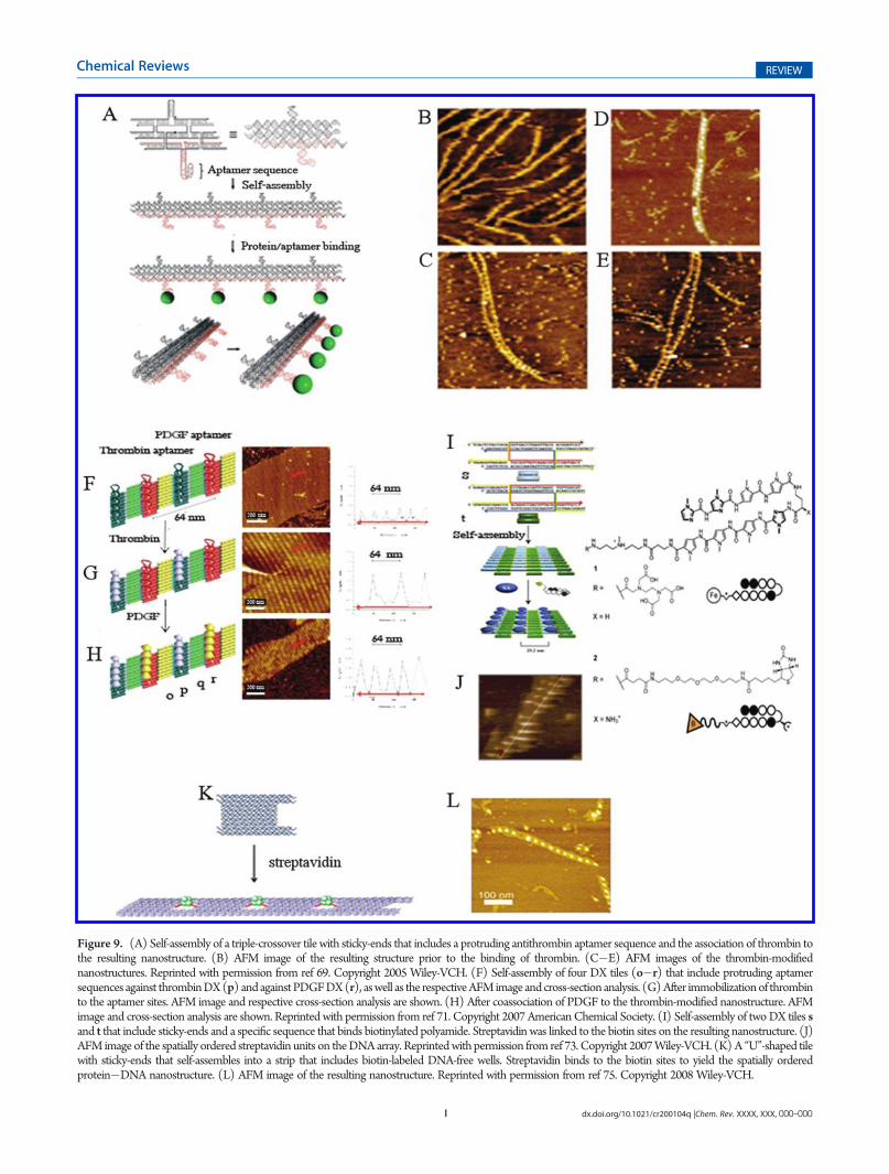
Figure 9. (A) Self-assembly of a triple-crossover tile with sticky-ends that includes a protruding antithrombin aptamer sequence and the association of thrombin tothe resulting nanostructure. (B) AFM image of the resulting structure prior to the binding of thrombin. (C�E) AFM images of the thrombin-modifiednanostructures. Reprinted with permission from ref 69. Copyright 2005 Wiley-VCH. (F) Self-assembly of four DX tiles (o�r) that include protruding aptamersequences against thrombinDX(p) andagainstPDGFDX(r), aswell as the respectiveAFMimage and cross-section analysis. (G) After immobilizationof thrombinto the aptamer sites. AFM image and respective cross-section analysis are shown. (H) After coassociation of PDGF to the thrombin-modified nanostructure. AFMimage and cross-section analysis are shown. Reprinted with permission from ref 71. Copyright 2007 AmericanChemical Society. (I) Self-assembly of twoDX tiles sand t that include sticky-ends and a specific sequence that binds biotinylated polyamide. Streptavidin was linked to the biotin sites on the resulting nanostructure. (J)AFM image of the spatially ordered streptavidin units on theDNAarray. Reprintedwith permission from ref 73. Copyright 2007Wiley-VCH. (K) A “U”-shaped tilewith sticky-ends that self-assembles into a strip that includes biotin-labeled DNA-free wells. Streptavidin binds to the biotin sites to yield the spatially orderedprotein�DNA nanostructure. (L) AFM image of the resulting nanostructure. Reprinted with permission from ref 75. Copyright 2008 Wiley-VCH.

J dx.doi.org/10.1021/cr200104q |Chem. Rev. XXXX, XXX, 000–000
Chemical Reviews REVIEW
correlated well with the projection of the high-resolution crystalstructure of this protein (Figure 8C).
Aptamer sequences were used to link a protein to orderednanoengineered sites on a self-assembled 1D DNA nano-structure.69 A triple-crossover (TX) DNA tile was used toconstruct a one-dimensional array (Figure 9A). The TX tilehad two protruding DNA loops, where one included the 15-basethrombin binding aptamer sequence and the other served as acontrol sequence. The TX tile self-assembled into a 1D nano-structure that included the thrombin-binding-aptamer units, at aperiodic distance of∼17 nm (Figure 9B). Addition of thrombinto the solution resulted in a periodic linear array of thrombinmolecules. Thrombin associated with the aptamer sites andresulted in the programmed positioning of the protein on the1D ribbon (Figure 9C�E). AFM analyses reveal a height of1.7 nm for the DNA arrays and 2.5�3 nm for the thrombin-modified DNA arrays. The lateral distance between adjacentprotein molecules was 17�19 nm. Interestingly, many of theresulting ribbons revealed double-chain nanostructures thatindicated interchain interactions. These were attributed to thedimerization of thrombin units that bind to aptamer sites onadjacent chains.70 Similarly, two different proteins were pat-terned onto a periodic 2D DNA nanoarray using two differentaptamers, one for each protein.71 A set of four double-crossover(DX) tiles was used to self-assemble the DNA nanostructure(Figure 9F). Tile p included a protruding thrombin-bindingaptamer sequence, whereas tile r was functionalized with theaptamer sequence against the platelet derived growth factor(PDGF). Figure 9(F�H) outlines the AFM images correspond-ing to the stepwise assembly of the DNA array and thesubsequent binding of the two proteins on the DNA template.The array of the tiles (Figure 9F) consists of parallel lines ofthe alternate protruding aptamer sequences separated by a distanceof 32 nm (revealing a height of ca. 0.7 nm). Interaction of theDNA array with thrombin led to the specific binding of theprotein to the thrombin-binding aptamer sequences separated bydistances corresponding to ca. 64 nm (revealing a height of ca.2 nm) (Figure 9G). The subsequent binding of PDGF to the freeanti-PDGF domains results in the densely organized nanostruc-ture where the two proteins were spatially separated one fromanother by a distance of 32 nm (Figure 9H). A related approachwas implemented for the optical detection of thrombin on a two-dimensional DNA array.72 A set of twoDX tiles, where one of thetiles included a protruding nucleic acid tether consisting of thethrombin-binding aptamer sequence that was labeled with the3-methylisoxanthopterrin dye, was used. In the presence ofthrombin, the aptamer sequence folded into a G-quadruplex,while forming the aptamer�thrombin complex. The fluores-cence of the labeling dye is enhanced in the resulting G-quad-ruplex, a property that was used to follow by confocal microscopythe binding of thrombin to the array. Further studies have appliedthe highly specific biotin�streptavidin affinity interactions toassemble the protein on an array of DNA tiles.73 Two DX tileelements, s and t, were used to generate a two-dimensional arrayof the tiles, through sticky-end hybridization. The tile s includedthe specific sequence that binds the biotin-labeled pyrrole�imidazole polyamide ligand (Figure 9I). The subsequent associa-tion of streptavidin yielded an organized nanostructure where theprotein units are periodically assembled on the respective tileunits. Figure 9J shows an AFM image of the resulting DNA�thrombin hybrid array. The average spacing between individualstreptavidin molecules was found to be ca. 24 nm, consistent
with the expected dimensions of the 2D array. This concept wasfurther extended to program the positioning of proteins on asingle DNA nanostructure.74An array consisting of four differentdouble-crossover (DX) tiles with appropriate sticky-ends wasused to assemble the 2D array. The base sequence encoded in thetiles provided the program for the specific association of threedifferent biotinylated polyamides in sequence-directed, spatiallyseparated domains on the array. The subsequent binding ofstreptavidin to the biotin units led to the generation of six differentpatterns of predesigned separation between the proteins. A parallelstudy demonstrated the assembly of two-dimensional tapes of DNAthat included periodically ordered pores, and the spatial pro-grammed assembly of tetrameric streptavidin into the pores wasachieved75 (Figure 9K). A “U”-shaped DNA structure wasdesigned to self-assemble, through sticky-end hybridization, intoa two-dimensional tape that included periodically ordered pores.The substitution of the pore sites with biotin labels resulted inthe selective binding of tetrameric streptavidin to the pores(Figure 9L). The average hole-to-hole distance was found tobe ca. 26 nm, and the streptavidin-modified array revealedheights of ca. 3 nm.
4. FUNCTIONAL DNA NANOSTRUCTURES THROUGHDIRECTED ORIGAMI FOLDING
DNA origami-based nanostructures are generated by thefolding of a viral DNA strand into two-dimensional or three-dimensional nanostructures using short complementary nucleicacid sequences acting as “staple” units for the viral DNA. By theappropriate design of the staple units, nanostructures of precisegeometries exhibiting nanometer-scale resolutions may be as-sembled. This paradigmwas pioneered by Rothemund76 with theseminal demonstrations that appropriate selection of staple unitsmay lead to predesigned nanoscale 2D or 3D77 shapes andpatterns of DNA (Figure 10). One of the challenges in the self-assembly of DNA-origami-based nanostructures involves theappropriate design of the staple strands. To date, computersoftwares are available78 to design the appropriate staple units.Not surprisingly, the design of programmed origami-based DNAassemblies provided the basis to organize ordered systems ofnanoparticles, nanotubes, and proteins on origami scaffolds.
DNA origami nanostructures were used as templates for theprecise positioning of Au NPs.79 Lipoic acid-modified Au NPsthat included a bivalent thiolate�Au linkage were prepared, and
Figure 10. (A) AFM images of two-dimensional origami nanostruc-tures. Reprinted with permission from ref 76. Copyright 2006 NaturePublishing Group. (B) Cryo-TEM images of three-dimensional DNAorigami nanostructures. Reprinted with permission from ref 77c. Copy-right 2009 Nature Publishing Group.

K dx.doi.org/10.1021/cr200104q |Chem. Rev. XXXX, XXX, 000–000
Chemical Reviews REVIEW
their assembly to the origami nanostructure was compared to theassembly of monothiolated Au NPs. The viral DNA was foldedusing ca. 200 appropriate staple nucleic acids that order theorigami DNA into a rectangular template. By the incorporation
of a single 10 nm Au NP functionalized with one of the stapleelements, or two types of 10 nm Au NPs functionalized with twodifferent staple units in the self-assembly mixture, rectangularDNA nanostructures were generated with the selective binding
Figure 11. Programmed positioning of Au NPs (10 mm) onto rectangular origami nanostructures by the incorporation of Au NPs-functionalizedstaples into the set of origami staples. (A) AFM image of the nanostructure generated using a single Au NP-functionalized staple. (B) AFM image of thenanostructures obtained by the use of two different Au NP-functionalized staples. Reprinted with permission from ref 79. Copyright 2008 AmericanChemical Society. (C) Self-assembly of an origami triangular nanostructure that includes protruding nucleic acid tethers for the specific addressing of AuNPs of variable sizes through hybridization. Right: SEM image of the resulting ordered Au NP array. Reprinted with permission from ref 80. Copyright2010 American Chemical Society. (D) Self-assembly of triangular origami nanostructures modified at their corners with Au NPs (5 nm) throughhybridization to protruding tethers. Triangles were self-assembled on a silicon wafer that was photochemically patterned with hydrophilic/hydrophobicdomains; Au NP-modified triangles were selectively associated to the hydrophilic regions. Right: the resulting AFM image. Reprinted with permissionfrom ref 81b. Copyright 2010Nature Publishing Group. (E and F) AFM images of photolithographically generated Au patches on a silicon wafer and theaddressed positioning of thiolated origami nanotubes in between the Au patches. Reprinted with permission from ref 82. Copyright 2010 AmericanChemical Society.

L dx.doi.org/10.1021/cr200104q |Chem. Rev. XXXX, XXX, 000–000
Chemical Reviews REVIEW
of one (Figure 11A) or two Au NPs (Figure 11B), respectively.AFM images revealed a spacing of∼47 nm between two adjacentAu NPs, and a height of 12 nm, consistent with the expectedheight of a 10 nmAuNP over a double-stranded DNA scaffold. Itwas also found that the yield of binding of the Au NPs to theorigami scaffold was significantly improved upon binding of thelipoic acid-modified NPs as compared to the monothiolate NPs(91% vs 45%). In a related study,80 a triangular DNA origamiscaffold was prepared, and three Au NPs of variable sizes (15, 10,and 5 nm), each modified with a nucleic acid complementary to aprotruding target sequence, were directed and ordered on the
origami nanostructures (Figure 11C). A center-to-center dis-tance of 90 nm between two 15 nm Au NPs was measured,consistent with the design. DNA/origami hybrid structures werefurther organized in microscale assemblies on surfaces.81 DNAorigami triangles were assembled by using the appropriatemixtures of staple nucleic acids that linked together the M13phage DNA to the triangle template. Au NPs (5 nm) weremodified with a nucleic acid complementary to protrudingsequences associated with the staples at the corners of thetriangles. The interaction of the functionalized Au NPs withthe origami-based template led to the positioning of the Au NPs
Figure 12. (A) AFM images of origami-folded six-helix DNA tubules that include protruding biotinylated tethers at programmed spatial distances onthe tubules. CdSe/ZnS quantum dots (15�20 nm) were functionalized with streptavidin and linked to the spatially separated tethers: (I) 71 nm, (II)43 nm, (III) 29 nm, and (IV) 14 nm. Reprinted with permission from ref 85. Copyright 2010 American Chemical Society. (B) Rectangular origamistructure that include protruding tethers above and below the origami plane. Carbon nanotubes (CNTs) were wrapped with nucleic acids that includedsticky-ends with specific complementarity to the tethers above and below the origami plane. This resulted in the orthogonal deposition of the CNTs onthe respective domains. Right: AFM height image of the CNTs of the resulting origami/CNTs hybrid. (C) Deposition of the origami�CNTsnanostructure on a four-microelectrode pattern. Left: AFM amplitude image. Right: Current�voltage curve of the resulting device demonstratingtransistor behavior. Reprinted with permission from ref 86. Copyright 2010 Nature Publishing Group.
M dx.doi.org/10.1021/cr200104q |Chem. Rev. XXXX, XXX, 000–000
Chemical Reviews REVIEW
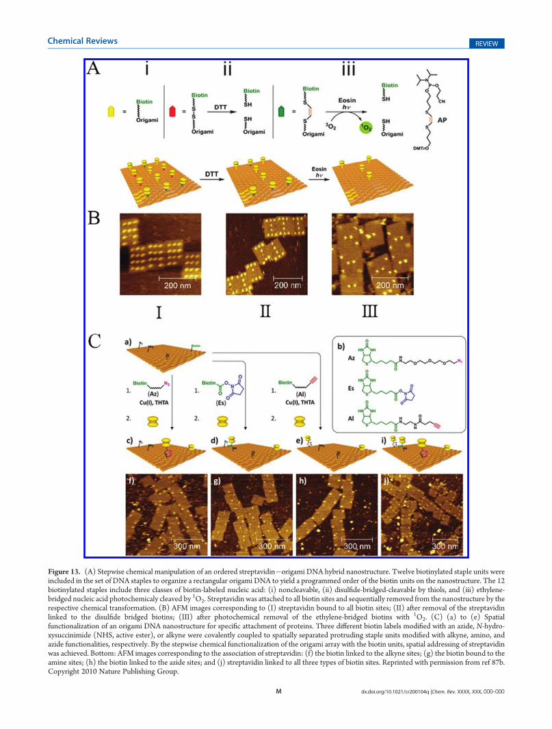
Figure 13. (A) Stepwise chemical manipulation of an ordered streptavidin�origami DNA hybrid nanostructure. Twelve biotinylated staple units wereincluded in the set of DNA staples to organize a rectangular origami DNA to yield a programmed order of the biotin units on the nanostructure. The 12biotinylated staples include three classes of biotin-labeled nucleic acid: (i) noncleavable, (ii) disulfide-bridged-cleavable by thiols, and (iii) ethylene-bridged nucleic acid photochemicaly cleaved by 1O2. Streptavidin was attached to all biotin sites and sequentially removed from the nanostructure by therespective chemical transformation. (B) AFM images corresponding to (I) streptavidin bound to all biotin sites; (II) after removal of the streptavidinlinked to the disulfide bridged biotins; (III) after photochemical removal of the ethylene-bridged biotins with 1O2. (C) (a) to (e) Spatialfunctionalization of an origami DNA nanostructure for specific attachment of proteins. Three different biotin labels modified with an azide, N-hydro-xysuccinimide (NHS, active ester), or alkyne were covalently coupled to spatially separated protruding staple units modified with alkyne, amino, andazide functionalities, respectively. By the stepwise chemical functionalization of the origami array with the biotin units, spatial addressing of streptavidinwas achieved. Bottom: AFM images corresponding to the association of streptavidin: (f) the biotin linked to the alkyne sites; (g) the biotin bound to theamine sites; (h) the biotin linked to the azide sites; and (j) streptavidin linked to all three types of biotin sites. Reprinted with permission from ref 87b.Copyright 2010 Nature Publishing Group.

N dx.doi.org/10.1021/cr200104q |Chem. Rev. XXXX, XXX, 000–000
Chemical Reviews REVIEW
at the corners of the triangle. A silicon surface was lithographi-cally patterned by e-beam lithography, leading to hydrophilicpatterned domains separated by hydrophobic regions. Theinteraction of the presynthesized DNA origami�Au NP struc-tures with the surface led to the binding of the hydrophilicorigami nanostructures on the hydrophilic patterns; the DNAAuNPs pattern dimensions corresponded to 430 nm betweencolumns of triangles and 200 nm between rows (Figure 11D).In a related work, surfaces were lithographically patterned withAu patches, and folded origami nanotubes with lengths corre-sponding to 380 nm, which included protruding thiol function-alized nucleic acids, were positioned in between the Audomains.82 Figure 11E depicts the AFM image of the origamiDNA nanotubes deposited on a hexagonal lattice of patterned Audomains. Similarly, Figure 11F reveals the AFM image of origamiDNA tubes positioned on different patterns of Au domains.These studies demonstrate the integration of DNA nanotubes ontop-down lithographically patterned circuits. In related studies,other origami nanostructures were deposited on a lithographi-cally patterned surface,83 and origami nanostructures function-alized with Au NPs were integrated with Au patterns.84
The DNA origami method was also implemented to assembleCdSe/ZnS quantum dots (QDs), 15�20 nm, with programmedspacing.85 The M13 phage DNA was folded into six-helix DNAtubules using ca. 200 staple sequences. Biotinylated staple unitswere placed at programmed spatial distances, and streptavidin-labeled QDs were linked to the biotin tethers. Using periodicseparation distances of the biotin labels that consisted of 71, 43,29, and 14 nm, the QDs were positioned at programmeddistances on the DNA tubules (Figure 12A). It was found that,although the positioning of the QDs at large separation distances(71, 43 nm) followed the original design, at shorter distancesbetween the QDs, the ordering was found to be perturbed,presumably due to electrostatic repulsion between the QDs.
Carbon nanotubes (CNTs) find growing interest as functionalunits in nanoscale electronic devices. The DNA origami scaffoldwas used to organize ordered carbon nanotubes in a predesignedconfiguration86 (Figure 12B). The M13 phage DNA was inter-acted with a nucleic acid mixture, acting as staples, to form a 2Drectangular DNA structure. A set of the staple units weredesigned to include a protruding nucleic acid chain that waspositioned vertically above the 2D origami array, whereas asecond set of staple units included the protruding nucleic acidchains in a horizontal configuration, below the origami array. Bythe modification of carbon nanotubes with nucleic acids com-plementary to the respective protruding tethers, the carbonnanotubes were positioned on the vertical/horizontal domains.Figure 12C shows an AFM image of two SWNT alignedperpendicularly, on the origami template. The CNTs assembledon the DNA origami template were then deposited on a siliconsubstrate, modified with four Pd/Au microcontacting electrodes.The resulting nanostructure revealed field effect transistor (FET)characteristics (Figure 12C). (For further applications of DNAnanostructures to assemble nanoscale devices, see vide infra).
The origami scaffold was further used for the distance-dependentmultivalent ligand�protein binding87a and for drivingspecific reactions with precise positioning of proteins at the singlemolecule level.87b The M13 phage DNA was organized into arectangular DNA origami pattern using a set of ∼200 staplestrands that included 12 biotinylated staples. The biotinylatedstaples included three types of staple units: four of the staplesincluded noncleavable biotin units, four other staples included a
disulfide bridge to a nonstapling biotinylated nucleic acid, and athird class of four staple nucleic acids was linked by an ethylene,electron-rich bridge to a nonstapling biotinylated nucleic acid.The stapled origami nanostructure resulted in the precisepositioning of streptavidin to the 12 biotin sites. Treatment ofthe biotinylated scaffold with 1,4-dithiothreitol (DTT) reducedthe disulfide bond, thus removing the respective biotinylatedunits of the second class, giving rise to the pattern of streptavidinshown in Figure 13A. The subsequent interaction of the DTT-pretreated biotinylated surface with eosin in the presence of O2
and under illumination, λ = 520 nm, resulted in the cleavage ofthe double bonds of the biotinylated units of class three, givingrise to a streptavidin-binding pattern on the noncleavablebiotin staples. Figure 13B shows AFM images of the respectivestates: (I) after incubation with streptavidin, (II) following DTTreduction, and (III) eosin-treated streptavidin-modified origamitemplate. The programmed positioning of proteins on theorigami scaffold has been extended by predesigning three dif-ferent functional tethers to the origami array. This was achievedby the linkage of an azide, an amino functionality, and an alkynefunctionality to the origami array. The subsequent specificcovalent linkage of an azide labeled with biotin to the alkyneunits by a click reaction in the presence of copper(I)�THTA(tris-(1-[3-hydroxy-propyl]triazolyl-4-methyl)amine), the covalentattachment of a biotin-labeled N-hydroxysuccinimide activatedester (NHS-ester) to the amine functionalities, or the specificclick-on reaction of a biotinylated alkyne to the azide tethers ledto the specific modification of the origami array with therespective biotin label. The subsequent attachment of streptavi-din (SA) to the biotin labels resulted in the ordered positioningof the protein on the DNA framework. Realizing that thedifferent synthetic routes may be implemented to covalently linkdifferent proteins to the origami scaffold, one may anticipate thatordered structures consisting of intercommunicating proteinsmay be generated (Figure 13C).
A related study has implemented the origami nanostructurefor the spatial positioning of proteins.88 The biotin group, thechlorohexyl unit, and the benzylguanine groups were attached tospecific stapled domains. Whereas the biotin binds streptavidin,the chlorohexyl functionalities associate specifically with haloalk-ane dehalogenase, known as the “Halo-Tag”,89 and the benzyl-guanine sites link specifically the O6-alkylguanine-DNA-alkyl-transferase (hAGT) known as the “Snap-Tag”.90 By the geneticengineering of proteins with the Halo-Tag or the Snap-Tagmoieties, and the use of streptavidin�protein conjugates, therespective proteins were addressed to the appropriate recogni-tion domains on the origami template. Another approach toconstruct one-dimensional and two-dimensional streptavidin(SA) nanoarrays on an origami scaffold has involved the con-struction of periodical nanometer-scale wells embedded in one-dimensional or two-dimensional DNA origami templates.91 TheM13 phage DNA was folded into a nanostructure using 267staple strands to yield a rectangle consisting of 76 turns-long(theoretical length 260 nm) and 10 helices-wide (theoreticalwidth of 30 nm) that include nine hollow sections (wells)exhibiting theoretical dimensions of 6.8 � 12 � 2.0 nm(Figure 14A). Each of the wells was modified with two biotinlabels that allowed the association of SA. Figure 14B shows theAFM images corresponding to the periodically separated wellsbefore and after the binding of SA (I and II, respectively). Theprecise positioning of the streptavidin at distances corresponding tothe theoretically predicted separation, 28 ( 1 nm, was visualized.

O dx.doi.org/10.1021/cr200104q |Chem. Rev. XXXX, XXX, 000–000
Chemical Reviews REVIEW
By the subsequent synthesis of two rectangles that included ontheir long edges complementary strands, the fabrication of twointerhybridized rectangles, which included two parallel rows ofwells, was achieved. The rectangles were designed, however, toinclude the biotin labels only on the even-numbered wells of onerectangle and on the odd-numbered wells of the second rectan-gle. This enabled the zigzag positioning of SA on the respectivebiotin-labeled origami composite (Figure 14C). The approach toassemble SA on periodically positioned wells constructed on anorigami template was extended to yield the switchable bindingand dissociation of SA to the wells (Figure 14D).92 The wellincluded two staple strands that hybridized with the comple-mentary biotinylated strands. The association of SA to the biotinlabels attached the protein to the well. The subsequent dis-placement of the biotinylated strand by a toehold mechanismremoved the SA�biotin structure from the wells, leavingbehind the functionalized wells for the secondary hybridiza-tion of the biotin-functionalized nucleic acid and the associa-tion of SA.
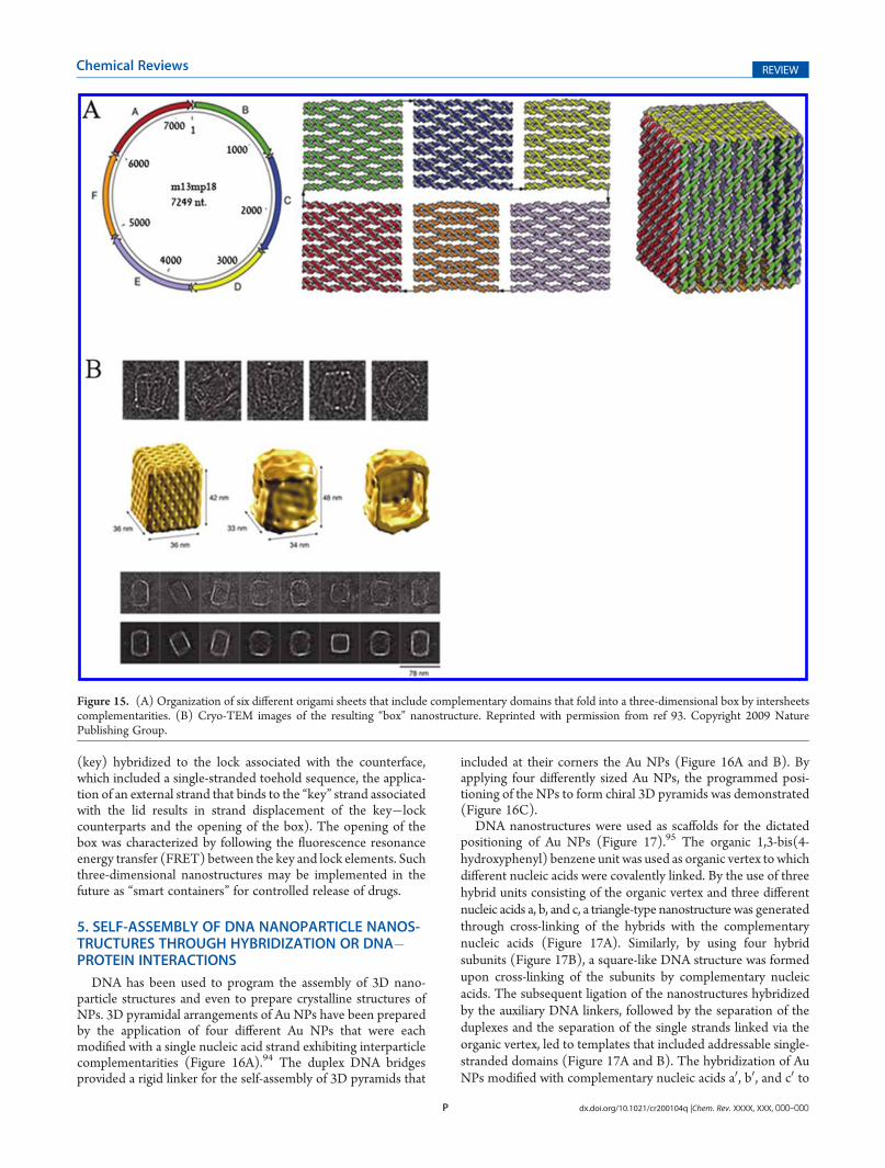
An intriguing origami-based three-dimensional nanostructurethat represents a nanobox was constructed, and the opening ofthe lid of the box was demonstrated.93 Albeit the reported boxwas not coupled to any protein, the hollow cavity inside the box,and the controllable opening of the lid, suggest the possibletrapping and release of proteins or low-molecular-weight mole-cules to and from the box, as a future perspective of thisnanostructure. M13 phage DNA was self-assembled using 220staple strands into six different origami sheets (Figure 15A). Therespective corners of each of the sheets included single toeholdstrands of appropriate complementarity that linked together theappropriate sheets. This arrangement enabled the self-assemblyof the six-face box that generated a nanostructure with dimen-sions of 42� 36� 38 nm. The cryo-TEM images of the box aredepicted in Figure 15B. The upper face of the box (the lid) wasfunctionalized with a single-stranded nucleic acid complemen-tary to a free nucleic acid strand tethered to the counterface.These two nucleic acids provided the lock�key mechanism foropening the box. (As the nucleic acid strand linked to the lid
Figure 14. (A)Origami rectangular nanostructure consisting of nine wells, eachmodified with two biotin labels. Streptavidin was then selectively linkedto the wells. (B) AFM images of the origami nanostructure (I) prior to the binding of streptavidin and (II) after the association of streptavidin to thewells. (C) AFM image of two origami subunits shown in (A) linked together by sticky-ends associated with the longitudinal domain of the nanostructure,and streptavidin bound in a zigzag configuration to the wells of the two origami nanostructures. Reprinted with permission from ref 91. Copyright 2009Wiley-VCH. (D) Origami nanostructure consisting of wells functionalized each with different biotinylated nucleic acid tethers; sreptavidin was linked tothe different biotinylated wells. By applying the strand-displacement principle, the selective removal of the streptavidin�biotin complex was achieved.Right: AFM images of (I) wells 1�8 occupied with streptavidin and (II�IV) selective removal of streptavidin from specific wells by strand displacement.Reprinted with permission from ref 92. Copyright 2010 Royal Society of Chemistry.
P dx.doi.org/10.1021/cr200104q |Chem. Rev. XXXX, XXX, 000–000
Chemical Reviews REVIEW
(key) hybridized to the lock associated with the counterface,which included a single-stranded toehold sequence, the applica-tion of an external strand that binds to the “key” strand associatedwith the lid results in strand displacement of the key�lockcounterparts and the opening of the box). The opening of thebox was characterized by following the fluorescence resonanceenergy transfer (FRET) between the key and lock elements. Suchthree-dimensional nanostructures may be implemented in thefuture as “smart containers” for controlled release of drugs.
5. SELF-ASSEMBLY OF DNA NANOPARTICLE NANOS-TRUCTURES THROUGH HYBRIDIZATION OR DNA�PROTEIN INTERACTIONS
DNA has been used to program the assembly of 3D nano-particle structures and even to prepare crystalline structures ofNPs. 3D pyramidal arrangements of Au NPs have been preparedby the application of four different Au NPs that were eachmodified with a single nucleic acid strand exhibiting interparticlecomplementarities (Figure 16A).94 The duplex DNA bridgesprovided a rigid linker for the self-assembly of 3D pyramids that
included at their corners the Au NPs (Figure 16A and B). Byapplying four differently sized Au NPs, the programmed posi-tioning of the NPs to form chiral 3D pyramids was demonstrated(Figure 16C).
DNA nanostructures were used as scaffolds for the dictatedpositioning of Au NPs (Figure 17).95 The organic 1,3-bis(4-hydroxyphenyl) benzene unit was used as organic vertex to whichdifferent nucleic acids were covalently linked. By the use of threehybrid units consisting of the organic vertex and three differentnucleic acids a, b, and c, a triangle-type nanostructure was generatedthrough cross-linking of the hybrids with the complementarynucleic acids (Figure 17A). Similarly, by using four hybridsubunits (Figure 17B), a square-like DNA structure was formedupon cross-linking of the subunits by complementary nucleicacids. The subsequent ligation of the nanostructures hybridizedby the auxiliary DNA linkers, followed by the separation of theduplexes and the separation of the single strands linked via theorganic vertex, led to templates that included addressable single-stranded domains (Figure 17A and B). The hybridization of AuNPs modified with complementary nucleic acids a0, b0, and c0 to
Figure 15. (A) Organization of six different origami sheets that include complementary domains that fold into a three-dimensional box by intersheetscomplementarities. (B) Cryo-TEM images of the resulting “box” nanostructure. Reprinted with permission from ref 93. Copyright 2009 NaturePublishing Group.

Q dx.doi.org/10.1021/cr200104q |Chem. Rev. XXXX, XXX, 000–000
Chemical Reviews REVIEW
the triangle template or Au NPs functionalized with the nucleicacids a0, b0, c0, and d0 complementary to the square-type templateled to the precise positioning of the NPs on the presynthesizedDNA scaffolds (parts C and D of Figure 17, respectively). Byusing differently sized Au NPs, the complexity of the resultingnanostructures was enhanced.
The aggregation of metallic NPs (e.g., Au NPs) through thelinking of nucleic acid-functionalized NPs by bridging DNAs hasbeen a widespread process for the development of numerousoptical sensors.96 Recently, the controlled aggregation of nucleicacid-modified Au NPs led to the formation of crystalline nano-structures consisting of the NPs.97 One face-centered cubiccrystalline structure was generated as outlined in Figure 18A.Au NPs were functionalized with the nucleic acid (27). Thecomplementary nucleic acid (28) that included palindromicsingle-stranded tethers was hybridized to the particles. Thepalindromic tethers linked to different Au NPs bridged theNPs, so that the face-centered cubic (fcc) nanostructure wasformed. Alternatively, a body-centered cubic (bcc) crystallinestructure of Au NPs was generated as outlined in Figure 18B.Two kinds of DNA-functionalized Au NPs were synthesized bythe hybridization of complementary nucleic acids 29 or 30 withthe 27-functionalized Au NPs. These NPs include differentsingle-stranded tethers of interparticle complementarities. Uponthe bridging of the two kinds of Au NPs, the bcc crystallinestructures were formed.
DNA nanotubes with longitudinal structural variations wereprepared and used to load and release a nanocargo.98 DNAtriangles of two different sizes (edge lengths of ∼7 nm for 31and ∼14 nm for 32) were used to construct triangular-based
nanotubes, by the hybridization with nine double-strandedlinking strands (Figure 19A). The resulting nanotubes werecomposed of alternate small and large triangles that were bridgedto form small and large capsules. AFM and TEM analysesdemonstrated the formation of micrometers-long DNA nano-tubes (Figure 19B). The resulting DNA nanotubes entrappedand released AuNPs. Figure 19C outlines AFM andTEM imagesof DNA nanotubes that include the encapsulated 15 nm Au NPs.Citrate-stabilized Au NPs (15 nm) were introduced into thenanotubes within the process of “glueing” together of thetriangles 31 and 32, through hybridization, and the formationof the nanotubes. One may see the linear organization of the goldNPs that is achieved with the approximate spacing of ca. 100 nmseparating the large capsules comprising the nanotubes. The AuNP-loaded nanotubes were then implemented as carriers for thedirected release of the Au NPs “cargo” units. Addition of a single-strand nucleic acid that hybridizes with the bridging duplex unitslinking the triangle elements resulted in strand displacement andthe disconnection of the rigid tubes, leading to the release of theencapsulated NPs.
The successful eliciting of DNA sequences that specificallybind to proteins (aptamers) provides a means to constructDNA�protein nanostructures. For example, two different oli-gonucleotide sequences (aptamer α and aptamer β) werereported to bind two different distinct domains of thrombin.This property was used to self-assemble linear or branched two-dimensional thrombin�DNA nanostructures99 (Figure 20A).The oligonucleotide 33 includes at its two ends the sequencescorresponding to aptamer α and aptamer β for thrombin. Thus,in the presence of thrombin, the oligonucleotide 33 acts as a
Figure 16. (A) Self-assembly of nucleic acid-functionalized Au NPs (5 nm) into a pyramidal nanostructure using complementary nucleic acids asrigidification units. (B) TEM image of the resulting pyramidal Au NPs assemblies. (C) Assembly of pyramidal DNA units consisting of differently sizedAuNPs (5, 10, 15, and 20 nm) at the pyramid corners, and the respective TEM image. Reprinted with permission from ref 94. Copyright 2009 AmericanChemical Society.

R dx.doi.org/10.1021/cr200104q |Chem. Rev. XXXX, XXX, 000–000
Chemical Reviews REVIEW
Figure 17. (A) Preparation of a DNA triangle that includes three synthetic vertices. Each of the vertices was functionalized by two nucleic acids. Thethree vertices were hybridized into the triangle configuration, and the nucleic acids associated with counter-vertices were ligated. After removal of therigidifying hybridized nucleic acid, the single-stranded triangle was purified. (B) Preparation of a square DNA using an analogous procedure outlined in(A). (C) Functionalization of the triangle DNA with Au NPs modified with nucleic acids complementary to the sides of the triangle, and the respectiveTEM image. (D) Functionalization of the square DNA with Au NPs modified with nucleic acids complementary to the side of the DNA template, andthe respective TEM image. Reprinted with permission from ref 95. Copyright 2007 American Chemical Society.

S dx.doi.org/10.1021/cr200104q |Chem. Rev. XXXX, XXX, 000–000
Chemical Reviews REVIEW
“glue” that self-assembles the thrombin-binding aptamer units/thrombin into a wire. Similarly, a mixture of the bifunctionalα/β bis-aptamer oligonucleotide 33 and the tripod oligonucleo-tide 34, which includes three aptamer α head groups led, inthe presence of thrombin, to the self-assembly of branched
thrombin/thrombin-binding aptamer-bridged nanostructures.Figure 20B depicts AFM images and cross-sectional analysis ofa linear DNA�protein wire, revealing a height of ca. 2.5 nm.
Similarly, aptamers against low-molecular-weight substrates,such as cocaine, adenosine monophosphate, and more, can begenerated by selection/amplification procedures.17,40,100 It wasdemonstrated that the specific aptamer sequences against low-molecular-weight substrates can be fragmented into subunits thatself-assemble into the aptamer subunits�substrate supramole-cular structure.101 This property was implemented to developoptical or electrochemical aptasensors for the low-molecular-weight substrates.102 The self-assembly of aptamer fragments andtheir substrates to the respective supramolecular complex wasused to self-assemble composite DNA�aptamer hybrid nanos-tructures that were subsequently used as scaffolds for theprogrammed positioning of two different enzymes103 (glucoseoxidase (GOx) and horseradish peroxidase (HRP)) (Figure 21A).The intramolecular hybridization of oligonucleotides 35 and 36by the capping nucleic acids 37 and 38 yielded the cappedcircular substrates 35/37 and 36/38. The oligonucleotides 39and 40 include domains complementary to the nucleic acid 37and 38 and specific domains corresponding to the fragmentedsubunits of the aptamer against cocaine 41. In the presence ofcocaine, the supramolecular hybrid polymeric nanostructureconsisting of the circular DNAs I and II bridged by theaptamer�cocaine complexes is formed path A Figure 21B showsan AFM image of the respective wire. The height of the wire is ca.3.5 nm. The increased height was attributed to the flexibility ofthe aptamer�DNA circle units and to the fact that the ringsmight have collapsed to compact structures of increased heights.The bridged aptamer�circular DNAs nanostructure was furtherimplemented to specifically bind two different enzymes, GOxand HRP, to the scaffold (Figure 21A, path B). In this nano-structure, the circular DNAs were capped by nucleic acids thatwere covalently tethered to the two different enzymes. Thehybrid bienzyme DNA/aptamer nanostructure enabled theeffective activation of the enzyme cascade, as previously de-scribed (see section 2).
Another method to self-assemble DNA scaffolds for thedirected positioning of enzymes or metallic nanoparticles hasinvolved the self-assembly of polycatenated DNA rings104
(Figure 22A). The single strands 42 and 43 included comple-mentary domains that upon hybridization resulted in the forma-tion of interconnected DNA strands. The ligation of the 30 and 50ends of the rings resulted in polycatenated rings of DNA.Figure 22B depicts the AFM images of wires of DNA, exhibit-ing a height of 2 nm, corresponding to double-stranded DNA.
Figure 18. Self-organization of nucleic acid-functionalized AuNPs into crystalline nanostructures: (A) a fcc structure and (B) a bcc structure. Reprintedwith permission from ref 97a. Copyright 2008 Nature Publishing Group.
Figure 19. (A) Self-assembly of DNA nanotubes with incorporated AuNPs by the cross-hybridization of two-sized DNA triangles with atethered nucleic acid and a set of nine nucleic acid linkers. (B) AFMand TEM images of the nanotubes without AuNPs. (C) AFM and TEMimages of the Au NPs encapsulated into the DNA tubes. Reprinted withpermission from ref 98. Copyright 2010 Nature Publishing Group.

T dx.doi.org/10.1021/cr200104q |Chem. Rev. XXXX, XXX, 000–000
Chemical Reviews REVIEW
One can observe the catenated DNA rings, comprising the DNAnanostructure. The polycatenated DNA included single-stranded rails that provided hybridization sites for tetheringproteins or metallic nanoparticles. By designing the rails tocontain thrombin-binding aptamer units, tetramethylrhodamine(TAMRA)-labeled thrombin was attached to the rails of thepolycatenated structure, to form protein�DNA hybrid nano-structures (Figure 22 A and C).
Single-sranded DNA chains with appropriate complementa-rities provided a means to self-assemble hexagon-like DNAstrips.105 For example, Figure 23A shows the use of two DNAchains 44 and 45 that self-assemble into a two-hexagon strip.Similarly, upon application of the four hexagons 46�49, whichinclude the appropriate complementarities, the self-assembly offour-hexagon-based strips was performed. Micrometer-longDNA strips, with an average height of ca. 2 nm, were imaged(Figure 23B). The hexagon subunits included single-strandednucleic acid tethers at the edge of the respective strips. Thesetethers provided anchoring sites for the coupling of enzymes,while dictating the distance separating the enzymes and thuscontrolling the effectiveness of communication between theenzymes on the DNA scaffolds. The enzymes glucose oxidase(GOx) and horseradish peroxidase (HRP) were modified bynucleic acids 50 and 51, which are complementary to the tethersassociated with hexagons 45 and 44 or 49 and 46, respectively(Figure 23C). The bienzyme cascade, where GOx mediated theoxidation of glucose by O2 to yield gluconic acid and hydrogen
peroxide (H2O2), and the subsequent HRP-catalyzed oxidation of2020-azino-bis[3-ethylbenzthiazoline-6-sulfonic-acid] (ABTS2‑) byH2O2 was activated on the respective DNA scaffolds. The biocata-lytic cascade generated the colored product ABTS 3 ‑, which reflectedthe effectiveness of the bienzyme biocatalytic transformation.Effective interenzyme communication, and the activation of theenzyme cascade, were observed on the DNA hexagon strips(Figure 23D, curves a and b). The enzmes, at the same concen-trations, in the absance of DNA, as in the presence of a Breign,non-organizing, DNA did not communicate with one another(Figure 23D, curves c and d), implying that their connection andordering on the DNA scaffold established the interenzymecontact. Also, the spatial separation of the two enzymes onthe four hexagon strips led to a ca. 20% less efficient activationof the bienzyme cascade, revealing the significance of proximitybetween the enzymes for an effective biocatalytic cascade. Simi-larly, the enzyme glucose dehydrogenase (GDH) and its N-(1)-(2-aminoethylnicotinamideadenine dinucleotide) (NAD+)cofactor were assembled on the two hexagon DNA scaffold(Figure 23E). Although the enzyme GDH was functionalizedwith a nucleic acid complementary to the tether unit 45, theNAD+ cofactor was functionalized with single-stranded DNAs ofvariable lengths that included a domain complementary to thetether 44. The oxidation of glucose to gluconic acid bythe NAD+ cofactor yields the reduced NADH cofactor, andthis reduces methylene blue (MB+) to the colorless reduceddye, MB, thus providing a color signal for the cofactor-mediated
Figure 20. (A) Self-assembly of linear or branched thrombin/bis-aptamer nanowires. (B) AFM images of the linear (I) and branched (II) nanowires.Reprinted with permission from ref 99. Copyright 2008 Royal Society of Chemistry.

U dx.doi.org/10.1021/cr200104q |Chem. Rev. XXXX, XXX, 000–000
Chemical Reviews REVIEW
biocatalyzed oxidation of glucose on the DNA scaffold(Figure 23F). While NAD+ linked to the DNA scaffold with a
short chain was inefficient in the activation of the cofactor-mediatedbiocatalytic transformation, elongation of the DNA tether
Figure 21. (A) Self-assembly of circular DNA units by the selective hybridization of anticocaine aptamer subunits to two different DNA circles and theprogrammed conjugation of the circles by cocaine (path A). The selective association of the enzymes glucose oxidase and horseradish peroxidase to thedifferent circles activates the bienzyme cascade (path B). (B) AFM image of the cocaine-conjugated circular DNA nanowire. Reprinted with permissionfrom ref 103. Copyright 2009 American Chemical Society.
Figure 22. (A) Synthesis of catenated DNA nanowires. (B) AFM images of the catenated DNA nanowires. (C) Association of TAMRA-labeledthrombin to the aptameric rails of the catenated DNA nanowires and the respective confocal microscopy image of the nanowires. Reprinted withpermission from ref 104. Copyright 2008 National Academy of Sciences.

V dx.doi.org/10.1021/cr200104q |Chem. Rev. XXXX, XXX, 000–000
Chemical Reviews REVIEW
improved the contact between the cofactor and the enzyme, andthe optimal biocatalytic transformation was observed upon usinga chain of 90 bases.
6. DNA NANOSTRUCTURES AS TEMPLATES FOR THEBOTTOM-UP FABRICATION OF NANODEVICES
The continuous effort to miniaturize electronic circuits is achallenging holy grail in modern science. Although the last 50years demonstrated the increase of the density of electroniccomputing elements, which is doubled every 2 years (Moore’slaw), it is anticipated that the lithographic miniaturizationmethod will reach limiting values. Accordingly, research effortsin the past decade suggested an alternative bottom-up approach,where molecules or macromolecules act as templates for thedeposition of nanocircuits. The nanodimensions of proteins orDNA, the structural information encoded in these materials, and
the available chemical means to attach metallic or semiconductornanoparticles to these biopolymers enables the growth of micro-circuits on the biomaterials acting as organizing templates. Forexample, amyloid nanotubes served as scaffolds for the deposition ofsilver and the generation of silver nanowires.106 Similarly, theenzymes glucose oxidase and alkaline phosphatase were patternedvia dip-pen nanolitography on surfaces and served as nanobioreac-tors for the electroless deposition of different metals.45
DNA provides, however, unique features for the application ofthe biopolymer as a template for the organization of nanoscaleelectronic circuitries and nanodevices. The availability of DNA ofcontrolled lengths, predesigned base sequence, and nanoengi-neered geometrical topologies enables the programming of theDNA into functional matrices, acting as scaffolds for the bottom-up organization of nanostructures. Also, the availability of speci-fic enzymes that react with nucleic acids, such as polymerase,
Figure 23. (A) Self-assembly of protruding nucleic acid-functionalized strips consisting of “two-hexagon” and “four-hexagon” building units. (B) AFMimages of the resulting two-hexagon and four-hexagon strips. (C) Programmed attachment of the enzymes glucose oxidase (GOx) and horseradishperoxidase (HRP) to the protruding nucleic acids, and the activation of the bienzyme cascade in the organized nanostructuctures. (D) Colorimetricanalysis of the bienzyme cascade in the two-hexagon/enzyme (a) and the four-hexagon/enzyme (b) nanostructures. Curves (c) and (d) correspond tocontrol experiments where the bienzyme cascade was examined in the absence of DNA (c) or in the presence of the foreign calf-thymus DNA (d). (E)Activation of theNAD+-dependent glucose dehydrogenase (GDH) on the two-hexagon DNA strips by the programmed assembly of the NAD+ cofactorand GDH on the nanostructure. The NAD+ cofactor was tethered to the DNA scaffold by nucleic acids of variable lengths. (F) Colorimetric assay of theGDH biocatalytic activity in the presence of the NAD+ linked to the scaffold by different tether lengths. Reprinted with permission from ref 105.Copyright 2009 Nature Publishing Group.

W dx.doi.org/10.1021/cr200104q |Chem. Rev. XXXX, XXX, 000–000
Chemical Reviews REVIEW
telomerase, sequence-specific endonucleases, DNA recombina-tion proteins (such as RecA), or nicking enzymes that cleaveDNA, provide unique nanoscale biological tools for manipulatingand molding of DNA structures. Also, the sequence-specificinteractions of proteins with DNA domains enable the biochem-ical “patterning” of DNA polymers with domains of predesignedchemical reactivities. Not surprisingly, DNA nanostructures findgrowing interest as templates for the assembly of nanocircuitsand nanodevices. The metallization of DNA templates, or thespecific attachment of semiconductors to DNA scaffolds, at-tracted substantial efforts directed to the fabrication of nano-electronic systems.
A silver-coated phage λ DNA metallic nanowire was used tobridge the gap between two microelectrodes and to construct aconductive nanowire107 (Figure 24A). The two electrodes weremodified with short DNA strands of 12 bases each, which werecomplementary to the 50 and 30 end of the phage λ DNA.Subsequently, the phage λ DNA was hybridized to the shortnucleic acids linked to the microelectrodes, and Ag+ ions wereassociated with the phosphate groups of the DNA scaffold. Theprimary reduction of the Ag+ ions to Ag0 nanoclusters, linked tothe DNA template, by hydroquinone under basic conditions,followed by the catalytic, electroless enlargement of the Ag0
nanoclusters by the deposition of silver on the nanoclusters, ledto the formation of continuous Ag0 nanowire (Figure 24Aand B). Figure 24C depicts the I�V curves of the resultingmetallic nanowire, revealing a nonlinear behavior and demon-strating potential regions of insulating properties of variable widths,exhibiting different on-set potentials for the generation of the
currents. The non-ohmic I�V curves were attributed todefects in the contacts between the metallic nanoclustersconsisting of the nanowires that introduced overpotentialsfor charge transport.
The concept of metallization of DNA templates and theformation of metallic nanowires was further extended to designaddressable conductive domains on DNA templates and tofabricate nanoscale transistor devices.108 To reach this goal, theRecA bacterial protein that participates in gene repair andhomologous recombination109 was implemented as an organiz-ing element of the nanodevice (Figure 25A). A single-strandedDNA was complexed with the RecA protein, and the resultingcomplex was incorporated into an addressed domain of a longduplex DNA through homologous recombination, to yield aduplex DNA patterned with RecA. The subsequent binding ofthe biotinylated anti-RecA to the protein, followed by theattachment of streptavidin-modified carbon nanotubes, ad-dressed the semiconductor carbon nanotubes onto the duplexDNA template. The further modification of the protein-freeduplex DNA domains with Ag+ ions, their reduction to nanoclus-ters, and the electroless catalytic deposition of Au on the Agnanoclusters, yielded electrical contacts that were separatedby the semiconductor carbon nanotubes, acting as a gate.Figure 25B depicts the SEM image of the resulting nanodevice.The performance of the resulting field-effect transistor is de-picted in Figure 25C, where the source-to-drain currents atvariable source-to-drain potentials (VDS) and different gatepotentials (VG) are depicted.
A related approach was applied to fabricate nanoscale metallicnanowires that bridge two microelectrodes.110 The mutatedRecA protein that included at its c-terminus a cysteine residuewas functionalized with maleimide-modified Au NPs (1.4 nm).The resulting Au NPs-functionalized RecA was complexed to asingle-stranded DNA and deposited on a gap separating twoelectrodes. The subsequent enlargement of the Au NPs with Auby an electroless deposition process yielded continuous wiresexhibiting diameters in the range of 80�500 nm. Figure 26Ashows the SEM image of the resulting nanowire that reveals anohmic-type conductivity (Figure 26B).
Single strands of DNA acted as templates for growing metallicor semiconductor nanowires and their incorporation as circuitryelements for probing the charge transport properties of thenanodevices. For example, the 48,502 base λDNAwas depositedon a gap separating two electrodes.111 The association of Pd2+
ions with the phosphate residues followed by the reduction of theions, with a mixture of sodium citrate, lactic acid, and dimethy-lamine borane, generated Pd0 nanoclusters on the DNA tem-plate. The application of repeated association of Pd2+ ions andreduction cycles led to the formation of continuous metallicnanowires (Figure 27A) that revealed ohmic conductivity(Figure 27B).
A related approach has implemented the λDNA as a templateto grow CdS semiconductor nanowires.112 The DNA wasinteracted with Cd2+ ions that associated to the phosphate units.The reaction of the Cd2+�DNA hybrid with Na2S yielded CdSnanoclusters on the DNA template. The repeated deposition ofCd2+ ions and reaction with sulfide anions generated continuousCdS nanowires (Figure 28A), and these were assembled on a gapthat separated two electrodes using a combing technique(Figure 28B). The I�V curves of the nanodevices revealednonlinear behavior with insulating regions and onset potential,exhibiting conductivity features characteristic to semiconductors.
Figure 24. (A) Bridging microelectrodes with a Ag nanowire depositedon a DNA template. (B) AFM image of the resulting nanowire. (C)Representative I�V curves of different Ag nanowires bridging themicroelectrodes. Reprinted with permission from ref 107. Copyright1998 Nature Publishing Group.
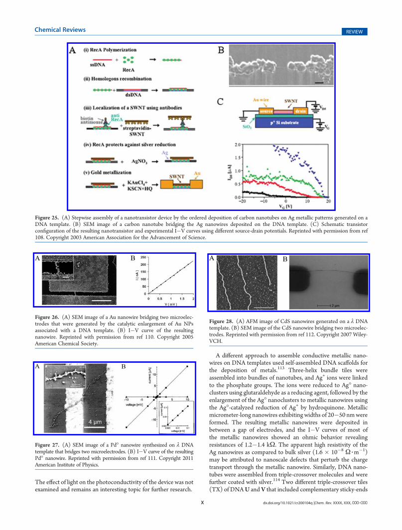
X dx.doi.org/10.1021/cr200104q |Chem. Rev. XXXX, XXX, 000–000
Chemical Reviews REVIEW
The effect of light on the photoconductivity of the device was notexamined and remains an interesting topic for further research.
A different approach to assemble conductive metallic nano-wires on DNA templates used self-assembled DNA scaffolds forthe deposition of metals.113 Three-helix bundle tiles wereassembled into bundles of nanotubes, and Ag+ ions were linkedto the phosphate groups. The ions were reduced to Ag� nano-clusters using glutaraldehyde as a reducing agent, followed by theenlargement of the Ag� nanoclusters to metallic nanowires usingthe Ag�-catalyzed reduction of Ag+ by hydroquinone. Metallicmicrometer-long nanowires exhibiting widths of 20�50 nmwereformed. The resulting metallic nanowires were deposited inbetween a gap of electrodes, and the I�V curves of most ofthe metallic nanowires showed an ohmic behavior revealingresistances of 1.2�1.4 kΩ. The apparent high resistivity of theAg nanowires as compared to bulk silver (1.6 � 10�8 Ω 3m
�1)may be attributed to nanoscale defects that perturb the chargetransport through the metallic nanowire. Similarly, DNA nano-tubes were assembled from triple-crossover molecules and werefurther coated with silver.114 Two different triple-crossover tiles(TX) of DNAU and V that included complementary sticky-ends
Figure 25. (A) Stepwise assembly of a nanotransistor device by the ordered deposition of carbon nanotubes on Ag metallic patterns generated on aDNA template. (B) SEM image of a carbon nanotube bridging the Ag nanowires deposited on the DNA template. (C) Schematic transistorconfiguration of the resulting nanotransistor and experimental I�V curves using different source-drain potentials. Reprinted with permission from ref108. Copyright 2003 American Association for the Advancement of Science.
Figure 26. (A) SEM image of a Au nanowire bridging two microelec-trodes that were generated by the catalytic enlargement of Au NPsassociated with a DNA template. (B) I�V curve of the resultingnanowire. Reprinted with permission from ref 110. Copyright 2005American Chemical Society.
Figure 27. (A) SEM image of a Pd� nanowire synthesized on λ DNAtemplate that bridges two microelectrodes. (B) I�V curve of the resultingPd� nanowire. Reprinted with permission from ref 111. Copyright 2011American Institute of Physics.
Figure 28. (A) AFM image of CdS nanowires generated on a λ DNAtemplate. (B) SEM image of the CdS nanowire bridging two microelec-trodes. Reprinted with permission from ref 112. Copyright 2007 Wiley-VCH.

Y dx.doi.org/10.1021/cr200104q |Chem. Rev. XXXX, XXX, 000–000
Chemical Reviews REVIEW
were used to self-assemble the DNA nanotubes, through theformation of sheets, that folded into nanotubes (Figure 29A).The TX nanotubes were then metalized by silver, using glutar-aldehyde as a reducing agent. The resulting metallic nanotubesexhibited dimensions of∼35 nm in height,∼40 nm in width, andup to 5 μm in length. Chromium/gold electrodes were thenpatterned onto the nanowires by electron-beam lithography.Figure 29B shows a SEM image of the DNA metallic nano-tubes-based device. I�Vmeasurements were conducted on thesedevices with various gap distances: 180, 80, and 100 nm. Themeasurements revealed mostly ohmic behavior with resistancesof 2.8, 2.35, and 2.82 kΩ, respectively, at a potential of 0.1 V atroom temperature (Figure 29C).
7. CONCLUSIONS AND PERSPECTIVES
Impressive advances were accomplished in the past 10 years inthe area of DNA nanotechnology and self-assembly of DNA�proteins or DNA�nanoparticle nanostructures. Versatile meth-ods to assemble 1D, 2D, and 3D DNA�protein or DNA�nanoparticle systems were developed. These include the 1Dorganization of hybrid nanostructures by the implementation ofthe rolling circle amplification process (RCA), the ligation ofinterhybridized quasi-circular DNA strands to form catenatedcircular DNA polymers, by the self-organization of cDNAstrands. 2D and 3D DNA�protein or DNA�nanoparticle struc-tures were assembled by the application ingenious DNA�“tile”constructs as building blocks for the formation of programmed
2D arrays that upon folding generated 3D tubes. Similarly, thedictated origami folding, where a viral DNA is folded byappropriate “staple” units, proved to be a versatile method forthe computer-aided formation of 2D or 3D nanostructures. Also,large capsules of DNA�NP nanotubes were generated throughthe directed polymerization of predesigned DNA-triangle sub-units by complementary nucleic acids acting as “glue”. Precisepositioning of proteins or NPs onto the resulting nanostructureswas realized by the programmed functionalization of the self-assembled nanostructures with protruding nucleic acids, chemi-cally reactive groups, or ligands that control the binding of thecomponents through hybridization, covalent bonds, or supra-molecular affinity interactions. Although substantial progress wasachieved in the nanoengineering of hybrid DNA nanostructures,challenging fundamental and practical issues are still ahead of us.Although the formation of the DNA nanostructures and thespatial positioning of proteins/NPs on the systems were demon-strated, the resulting systems are imperfect and include unavoid-able defects. Thus, the development of error-correction methodsis an essential future requisite. Further exciting opportunities inthe area of DNA hybrid nanostructures would involve thedevelopment of signal-triggered systems, where chemical, elec-trochemical, or photochemical stimuli reversibly interconvert thetopologies of DNA hybrid nanostructures. Such nanostructuresmay find important applications for the controlled delivery,uptake, and release of substrates.
The major challenges include, however, the identification ofpotential applications of hybrid DNA nanostructures. The use of
Figure 29. (A) Self-assembly of a DNA nanotube using two different TX tiles. (B) SEM image showing the silver-coated DNA tube deposited on anarray of microelectrodes. (C) I�V curves of three different Ag� nanowires deposited on the microelectrodes. Reprinted with permission from ref 114.Copyright 2004 National Academy of Sciences, USA.

Z dx.doi.org/10.1021/cr200104q |Chem. Rev. XXXX, XXX, 000–000
Chemical Reviews REVIEW
a DNA scaffold for the bottom-up templated synthesis ofmetallic orsemiconductor nanowires for the miniaturization of electroniccircuitry has been demonstrated. Nonetheless, the chemical growthof electronic circuitry on DNA templates is not free of limitations.Defects in the resulting nanowires were found to perturb the chargetransport through the circuitries. The improvement of the quality ofthe nano-objects and the theoretical understanding of the effectsdominating charge transport through the nanowires are desirablegoals. The fabrication of high-throughput, defect-free, nanoscaledevices based on DNA hybrid nanostructures would require,however, the development of surface-patterning methods for ad-dressing the nanocircuitries. Alternatively, one might envisage thefuture design of DNA templated metallic or semiconductor nano-wires that are integrated with motor proteins. The fueledmotility ofsuch nanowires on patterned tracks on surfaces could lead to theautonomous assembly of complex circuitries on surfaces. In fact,the ability to move metallic nanowires on surfaces by means of theactin/myosin motor protein and ATP has been demonstrated,115
and similar principles may be adapted to move hybrid DNAnanostructures on surfaces.
Hybrid DNA�proteins or DNA�NP nanostructures hold greatpromise for other applications. Such nanocomposites may finddifferent uses in nanomedicine. The encapsulation of therapeuticdrugs in DNA tubes or DNA nanoboxes may provide nanoscalecontainments for carrying drugs and their slow release. Similarly,NPs encapsulated in DNA nanostructures might be useful vehiclesfor in vivo imaging or thermal destruction of tumor cells. Althoughsuch exciting ideas float around, fundamental issues, such as crossingcell boundaries by the DNA structures, triggered release of theencapsulated substrates, and stabilization of the DNA nanostruc-tures in biological fluids or cells, need to be resolved. A furtherchallenging path to implement DNA�protein or DNA/NPs wouldinvolve the control of chemical reactions through the spatialpositioning of reactants on DNA templates. Recently, control overthe reactivity of a bienzyme cascade, or a cofactor-enzyme bio-transformation by means of DNA nanostructures, was demonst-rated.30b,42,103,105 This suggests that biocatalytic transformations,and specifically, artificial enzyme networks, might be engineered bythe spatial positioning of biocatalysts on 1D, 2D, or 3D DNAnanosystems. Furthermore, protein or nanoparticle arrangementson DNA scaffolds may act as functional nanoarchitectures for lightharvesting and solar energy conversion. The photosynthetic reac-tion centers provide a unique apparatus for the effective collection oflight and its utilization for vectorial electron transfer accompanied byefficient charge separation. The precise positioning of photosensi-tizer and electron-acceptor chains onDNAnanostructuresmay leadto such photosynthetic devices.30a Finally, the integration of DNAmachines,25�30 such as walkers or a crane on programmed “play-grounds” of DNA arrays, might be useful to assemble nanoscalemachines carrying objects in dictated directions.
The topic of DNA�protein or DNA�nanoparticle nano-structures is, certainly, at its infancy. The rapid advances in thearea promise, however, a bright future to this scientific field.Interdisciplinary research efforts of chemists, biologists, physi-cists, and materials and computer scientists are anticipated tohighlight the future perspectives of DNA nanotechnology.
AUTHOR INFORMATION
Corresponding Author*E-mail: [email protected]. Tel.: +972-2-56585272. Fax:+972-2-6527715.
BIOGRAPHIES
Ofer I. Wilner was born in 1978. He received his M.Sc. degreefrom the Tel-Aviv University in 2006 and performed his Ph.D.research at the Institute of Chemistry, The Hebrew University ofJerusalem (2006�2011). His research interests include DNA-based nanotechnology, biosensors, and artificial photosynthesis.
Itamar Willner completed his Ph.D. studies at The HebrewUniversity of Jerusalem in 1978. After postdoctoral research(1978�1981) at U.C. Berkeley, he joined the Institute ofChemistry at The Hebrew University of Jerusalem, where hewas appointed as Professor in 1986. He coauthored over 600research papers and monographs, and he serves on manyeditorial boards of journals. His research interests includebioelectronics and molecular electronics, nanobiotechnology,and, specifically, DNA-based nanotechnology, sensors and bio-sensors, application of metal and semiconductor nanoparticlesfor sensing, functional monolayer and thin film assemblies, light-induced electron-transfer processes and artificial photosynthesis,and supramolecular chemistry. He is the recipient of the KolthoffAward, The Max-Planck Research Award for International Co-operation, the Israel Chemical Society Award (2001), the IsraelPrize in Chemistry (2002), the Rothschild Prize (2008), and theEMET Prize (2008). He is a member of the Israel Academy ofSciences, the GermanNational Academy of Sciences Leopoldina,and the European Academy of Sciences and Arts.
ACKNOWLEDGMENT
Our research onDNA nanotechnology is supported by the IsraelScience Foundation and by the Volkswagen Foundation, Germany.

AA dx.doi.org/10.1021/cr200104q |Chem. Rev. XXXX, XXX, 000–000
Chemical Reviews REVIEW
REFERENCES
(1) (a) Watson, J. D.; Crick, F. H. C. Nature 1953, 171, 737. (b)Sinden, R. R.; Pearson, C. E.; Potaman, V. N.; Ussery, D. W. Adv. Gen.Biol. 1998, 5, 1.(2) Frank-Kamenetskii, M. D.; Mirkin, S. M. Annu. Rev. Biochem.
1995, 64, 65.(3) Simonsson, T. Biol. Chem. 2001, 382, 621.(4) Chen, L.; Cai, L.; Zhang, X.; Rich, A. Biochemistry 1994, 33, 13540.(5) Miyake, Y.; Togashi, H.; Tashiro, M.; Yamaguchi, H.; Oda, S.;
Kudo,M.; Tanaka, Y.; Kondo, Y.; Sawa, R.; Fujimoto, T.;Machinami, T.;Ono, A. J. Am. Chem. Soc. 2006, 128, 2172.(6) Ritchie, C. M.; Johnsen, K. R.; Kiser, J. R.; Antoku, Y.; Dickson,
R. M.; Petty, J. T. J. Phys. Chem. C 2007, 111, 175.(7) Tanaka, K.; Clever, G. H.; Takezawa, Y.; Yamada, Y.; Kaul, C.;
Shionoya, M.; Carell, T. Nature Nanotechnol. 2006, 1, 190.(8) Goodchild, J. Bioconjugate Chem. 1990, 1, 165.(9) (a) Nielsen, P. E.; Egholm, M.; Berg, R. H.; Buchardt, O. Science
1991, 254, 1497. (b) Egholm, M.; Buchardt, O.; Nielsen, P. E.; Berg,R. H. J. Am. Chem. Soc. 1992, 114, 1895. (c) Shakeel, S.; Karim, S.; Ali, A.J. Chem. Technol. Biotechnol. 2006, 81, 892.(10) (a) Singh, S. K.; Koshkin, A. A.; Wengel, J.; Nielsen, P. Chem.
Commun. 1998, 4, 455. (b) Kaur, H.; Arora, A.; Wengel, J.; Maiti, S.Biochemistry 2006, 45, 7347.(11) (a) Bergmann, F.; Bannwarth, W.; Tam, S. Tetrahedron Lett.
1995, 36, 6823. (b) Kurreck, J.; Wyszko, E.; Gillen, C.; Erdmann, V. A.Nucleic Acids Res. 2002, 30, 1911.(12) (a) Itakura, K.; Rossi, J. J.; Wallace, R. B. Annu. Rev. Biochem.
1984, 53, 323. (b) Lee, J. S.; Woodsworth, M. L.; Latimer, L. J. P.;Morgan, A. R. Nucleic Acids Res. 1984, 12, 6603. (c) Kool, E. T. Chem.Rev. 1997, 97, 1473.(13) (a) Boon, E. M.; Ceres, D. M.; Drummond, T. G.; Hill, M. G.;
Barton, J. K.Nat. Biotechnol. 2000, 18, 1096. (b) Drummond, T. G.; Hill,M. G.; Barton, J. K. Nat. Biotechnol. 2003, 21, 1192. (c) Drummond,T. G.; Hill, M. G.; Barton, J. K. J. Am. Chem. Soc. 2004, 126, 15010.(14) (a) Evans, R. K.; Johnson, J. D.; Haley, B. E. Proc. Natl. Acad. Sci.
U. S. A. 1986, 83, 5382. (b) Evans, R. K.; Haley, B. E. Biochemistry 1987,26, 269. (c) Kielkopf, C. L.; Erkkila, K. E.; Hudson, B. P.; Barton, J. K.;Rees, D. C. Nat. Struct. Biol. 2000, 7, 117. (d) Liebmann, M.; DiPasquale, F.; Marx, A. ChemBioChem 2006, 7, 1965.(15) (a) Englisch, U.; Gauss, D. H. Angew. Chem., Int. Ed. 1991,
30, 613. (b) Nielsen, P. E. J. Mol. Recognit. 1990, 3, 1. (c) Ohndorf,U.-M.; Rould, M. A.; He, Q.; Pabo, C. O.; Lippard, S. J. Nature 1999,399, 708. (d) Kelley, S. O.; Holmlin, R. E.; Stemp, E. D. A.; Barton, J. K.J. Am. Chem. Soc. 1997, 119, 9861. (e) Gierlich, J.; Burley, G. A.;Gramlich, P. M. E.; Hammond, D.M.; Carell, T.Org. Lett. 2006, 8, 3639.(16) (a) Kornberg, A. Trends Biochem. Sci. 1984, 9, 122. (b) Zyskind,
J. W.; Cleary, J. M.; Brusilow, W. S.; Harding, N. E.; Smith, D. W. Proc. Natl.Acad. Sci. U. S. A. 1983, 80, 1164. (c) Wefald, F. C.; Devlin, B. H.; Williams,R. S.Nature1990,344, 260. (d)Cusack, S.Curr.Opin. Struct. Biol.1997,7, 881.(17) (a) Tuerk, C.; Gold, L. Science 1990, 249, 505. (b) Robertson,
M. P.; Ellington, A. D. Nat. Biotechnol. 2001, 19, 650.(18) (a) Stojanovic, M. N.; de Prada, P.; Landry, D. W. J. Am. Chem.
Soc. 2001, 123, 4928. (b) Shlyahovsky, B.; Li, D.; Weizmann, Y.;Nowarski, R.; Kotler, M.; Willner, I. J. Am. Chem. Soc. 2007,129, 3814. (c) Golub, E.; Pelossof, G.; Freeman, R.; Zhang, H.; Willner,I. Anal. Chem. 2009, 81, 9291. (d) Freeman, R.; Sharon, E.; Tel-Vered,R.; Willner, I. J. Am. Chem. Soc. 2009, 131, 5028.(19) (a) Bock, L. C.; Griffin, L. C.; Latham, J. A.; Vermaas, E.H.; Toole,
J. J. Nature 1992, 355, 564. (b) Macaya, R. F.; Schultze, P.; Smith, F. W.;Roe, J. A.; Feigon, J. Proc. Natl. Acad. Sci. U. S. A. 1993, 90, 3745. (c) Pavlov,V.; Xiao, Y.; Shlyahovsky, B.; Willner, I. J. Am. Chem. Soc. 2004, 126, 11768.(20) (a) Cox, J. C.; Ellington, A. D.Bioorg. Med. Chem. 2001, 9, 2525.
(b) Willner, I.; Zayats, M. Angew. Chem., Int. Ed. 2007, 46, 6408.(21) (a) Breaker, R. R.; Joyce, G. F. Chem. Biol. 1994, 1, 223. (b)
Joyce, G. F. Annu. Rev. Biochem. 2004, 73, 791. (c) Achenbach, J. C.;Chiuman, W.; Cruz, R. P. G; Li, Y. Curr. Pharm. Biotechnol. 2004, 5, 321.(d) Willner, I.; Shlyahovsky, B.; Zayats, M.; Willner, B. Chem. Soc. Rev.
2008, 37, 1153. (e) Sando, S.; Sasaki, T.; Kanatani, K.; Aoyama, Y. J. Am.Chem. Soc. 2003, 125, 15720.
(22) Hardin, C. C.; Corregan, M.; Brown, B. A., II; Frederick, L. N.Biochemistry 1993, 32, 5870.
(23) (a) Zhang, D. Y.; Winfree, E. J. Am. Chem. Soc. 2009,131, 17303. (b) Seelig, G.; Soloveichik, D.; Zhang, D. Y.; Winfree, E.Science 2006, 314, 1585.
(24) (a) Tian, Y.; Mao, C. J. Am. Chem. Soc. 2004, 126, 11410. (b)Lin, C.; Liu, Y.; Yan, H. Biochemistry 2009, 48, 1663.
(25) (a) Yan, H.; Zhang, X.; Shen, Z.; Seeman, N. C. Nature 2002,415, 62. (b) Benenson, Y.; Adar, R.; Paz-Elizur, T.; Livneh, Z.; Shapiro,E. Proc. Natl. Acad. Sci. U. S. A. 2003, 100, 2191. (c) Dittmer, W. U.;Reuter, A.; Simmel, F. C. Angew. Chem., Int. Ed. 2004, 43, 3550. (d)Beissenhirtz, M. K.; Willner, I. Org. Biomol. Chem. 2006, 4, 3392. (e)Bath, J.; Turberfield, A. J. Nat. Biotechnol. 2007, 2, 275.
(26) (a) Yurke, B.; Turberfield, A. J.; Mills, A. P., Jr.; Simmel, F. C.;Neumann, J. L. Nature 2000, 406, 605. (b) Niemeyer, C. M.; Adler, M.Angew. Chem., Int. Ed. 2002, 41, 3779. (c) Muller, B. K.; Reuter, A.;Simmel, F. C.; Lamb, D. C. Nano Lett. 2006, 6, 2814. (d) Elbaz, J.;Moshe, M.; Willner, I. Angew. Chem., Int. Ed. 2009, 48, 3834. (e) Elbaz,J.; Wang, Z.-G.; Orbach, R.; Willner, I. Nano Lett. 2009, 9, 4510.
(27) (a) Shin, J.-S.; Pierce, N. C. J. Am. Chem. Soc. 2004, 126, 10834.(b) Yin, P.; Yan, H.; Daniell, X. G.; Turberfield, A. J.; Reif, J. H. Angew.Chem., Int. Ed. 2004, 43, 4906. (c) Ding, B.; Seeman, N. C. Science 2006,314, 1583. (d) Simmel, F. C. ChemPhysChem 2009, 10, 2593. (e)Omabegho, T.; Sha, R.; Seeman, N. C. Science 2009, 324, 67. (f)Wickham, S. F. J.; Endo, M.; Katsuda, Y.; Hidaka, K.; Bath, J.; Sugiyama,H.; Turberfield, A. J. Nature Nanotechnol. 2011, 6, 166.
(28) Wang, Z.-G.; Elbaz, J.; Willner, I. Nano Lett. 2011, 11, 304.(29) Buranachai, C.;McKinney, S. A.; Ha, T.Nano Lett. 2006, 6, 496.(30) (a) Tel-Vered, R.; Yehezkeli, O.; Yildiz, H. B.; Wilner, O. I.;
Willner, I. Angew. Chem., Int. Ed. 2008, 47, 8272. (b) Piperberg, G.;Wilner, O. I.; Yehezkeli, O.; Tel-Vered, R.; Willner, I. J. Am. Chem. Soc.2009, 131, 8724. (c) Elbaz, J.; Tel-Vered, R.; Freeman, R.; Yildiz, H. B.;Willner, I. Angew. Chem., Int. Ed. 2008, 48, 133. (d) Lund, K.; Manzo,A. J.; Dabby, N.; Michelotti, N.; Johnson-Buck, A.; Nangreave, J.; Taylor,S.; Pei, R.; Stojanovic, M. N.; Walter, N. G.; Winfree, E.; Yan, H. Nature2010, 465, 206. (e) Gu, H.; Chao, J.; Xiao, S.-J.; Seeman, N. C. Nature2010, 465, 202.
(31) (a) Weizmann, Y.; Beissenhirtz, M. K.; Cheglakov, Z.; Nowarski,R.; Kotler, M.; Willner, I. Angew. Chem., Int. Ed. 2006, 45, 7384. (b)Weizmann, Y.; Cheglakov, Z.;Willner, I. J. Am.Chem. Soc.2008, 130, 17224.(c) Simmel, F. C. Nanomedicine 2007, 2, 817. (d) Moshe, M.; Elbaz, J.;Willner, I. Nano Lett. 2009, 9, 1196. (e) Teller, C.; Shimron, S.; Willner, I.Anal. Chem. 2009, 81, 9114. (f) Freeman, R.; Sharon, E.; Teller, C.;Willner,I. Chem.—Eur. J. 2010, 16, 3690.
(32) (a) Whitesides, G. M.; Mathias, J. P.; Seto, C. T. Science 1991,254, 1312. (b) Lopinski, G. P.; Wayner, D. D. M.; Wolkow, R. A.Nature2000, 406, 48. (c) Barth, J. V.; Costantini, G.; Kern, K. Nature 2005,437, 671. (d) Gomar-Nadal, E.; Puigmarti-Luis, J.; Amabilino, D. B.Chem. Soc. Rev. 2008, 37, 490. (e) Li, D.; Wieckowska, A.; Willner,I. Angew. Chem., Int. Ed. 2008, 47, 3927. (f) Zhu, C.; Wen, Y.; Li, D.;Wang, L.; Song, S.; Fan, C.; Willner, I. Chem.—Eur. J. 2009, 15, 11898.
(33) (a) Lehn, J.-M. Angew. Chem., Int. Ed. 1988, 27, 89. (b) Stupp,S. I.; LeBonheur, V.; Walker, K.; Li, L. S.; Huggins, K. E.; Keser, M.;Amstutz, A. Science 1997, 276, 384. (c) Lehn, J.-M. Science 2002,295, 2400. (d) Ikkala, O.; ten Brinke, G. Science 2002, 295, 2407. (e)Perham, R. N. Philos. Trans. R. Soc., B 1975, 272, 123.
(34) Ramakrishnan, V. Cell 2002, 108, 557.(35) (a) Niemeyer, C. M. Curr. Opin. Chem. Biol. 2000, 4, 609. (b)
Gothelf, K. V.; LaBean, T. H. Org. Biomol. Chem. 2005, 3, 4023. (c)Niemeyer, C. M.; Simon, U. Eur. J. Inorg. Chem. 2005, 18, 3641. (d) Lin,C.; Liu, Y.; Rinker, S.; Yan, H. ChemPhysChem 2006, 7, 1641. (e)LaBean, T. H.; Li, H. Nano Today 2007, 2, 26. (f) Niemeyer, C. M.Angew. Chem., Int. Ed. 2010, 49, 1200. (g) Simmel, F. C. Angew. Chem.,Int. Ed. 2008, 47, 5884. (h) Aldaye, F. A.; Palmer, A. L.; Sleiman, H. F.Science 2008, 321, 1795. (i) Seeman, N. C. Nano Lett. 2010, 10, 1971.

AB dx.doi.org/10.1021/cr200104q |Chem. Rev. XXXX, XXX, 000–000
Chemical Reviews REVIEW
(36) Liu, D.; Daubendiek, S. L.; Zillman, M. A.; Ryan, K.; Kool, E. T.J. Am. Chem. Soc. 1996, 118, 1587.(37) Deng, Z.; Tian, Y.; Lee, S.-H.; Ribbe, A. E.; Mao, C. Angew.
Chem., Int. Ed. 2005, 44, 3582.(38) Zhao, W.; Gao, Y.; Kandadai, S. A.; Brook, M. A.; Li, Y. Angew.
Chem., Int. Ed. 2006, 45, 2409.(39) Beyer, S.; Nickels, P.; Simmel, F. C. Nano Lett. 2005, 5, 719.(40) Hermann, T.; Patel, D. J. Science 2000, 287, 820.(41) Cheglakov, Z.; Weizmann, Y.; Braunschweig, A. B.; Wilner,
O. I.; Willner, I Angew. Chem., Int. Ed. 2007, 47, 126.(42) Wilner, O. I.; Shimron, S.; Weizmann, Y.; Wang, Z.-G.; Willner,
I. Nano Lett. 2009, 9, 2040.(43) (a) Zayats, M.; Baron, R.; Popov, I.; Willner, I.Nano Lett. 2005,
5, 21. (b) Willner, I.; Baron, R.; Willner, B. Adv. Mater. 2006, 18, 1109.(c) Xiao, Y.; Pavlov, V.; Levine, S.; Niazov, T.;Markovitch, G.;Willner, I.Angew. Chem., Int. Ed. 2004, 43, 4519.(44) (a) Baron, R.; Zayats, M.; Willner, I. Anal. Chem. 2005,
77, 1566. (b) Pavlov, V.; Xiao, Y.; Willner, I. Nano Lett. 2005, 5, 649.(45) Basnar, B.; Weizmann, Y.; Cheglakov, Z.; Willner, I. Adv. Mater.
2006, 18, 713.(46) (a) Mitra, D.; Di Cesare, N.; Sleiman, H. F. Angew. Chem., Int.
Ed. 2004, 43, 5804. (b) Park, S. H.; Yin, P.; Liu, Y.; Reif, J. H.; LaBean,T. H.; Yan, H. Nano Lett. 2005, 5, 729.(47) (a) Winfree, E.; Liu, F.; Wenzler, L. A.; Seeman, N. C. Nature
1998, 394, 539. (b) Mao, C.; Sun, W.; Seeman, N. C. J. Am. Chem. Soc.1999, 121, 5437. (c) Chelyapov, N.; Brun, Y.; Gopalkrishnan, M.;Reishus, D.; Shaw, B.; Adleman, L. J. Am. Chem. Soc. 2004, 126, 13924.(d) Sharma, J.; Chhabra, R.; Liu, Y.; Ke, Y.; Yan, H.Angew. Chem., Int. Ed.2006, 45, 730.(48) (a) Chen, J.; Seeman, N. C. Nature 1991, 350, 631. (b)
Mitchell, J. C.; Harris, J. R.; Malo, J.; Bath, J.; Turberfield, A. J. J. Am.Chem. Soc. 2004, 126, 16342. (c) Aldaye, F. A.; Sleiman, H. F. J. Am.Chem. Soc. 2007, 129, 4130.(49) Fu, T. J.; Seeman, N. C. Biochemistry 1993, 32, 3211.(50) LaBean, T. H.; Yan, H.; Kopatsch, J.; Liu, F.; Winfree, E.; Reif,
J. H.; Seeman, N. C. J. Am. Chem. Soc. 2000, 122, 1848.(51) Ke, Y.; Liu, Y.; Zhang, J.; Yan, H. J. Am. Chem. Soc. 2006,
128, 4414.(52) Park, S. H.; Barish, R.; Li, H.; Reif, J. H.; Finkelstein, G.; Yan,
H.; LaBean, T. H. Nano Lett. 2005, 5, 693.(53) Mathieu, F.; Liao, S.; Kopatsch, J.; Wang, T.; Mao, C.; Seeman,
N. C. Nano Lett. 2005, 5, 661.(54) Petrillo, M. L.; Newton, C. J.; Chunningham, R. P.; Ma, R.-I.;
Kallenbach, N. R.; Seeman, N. C. Biopolymers 1988, 27, 1337.(55) Liu, D.; Wang, M.; Deng, Z.; Walulu, R.; Mao, C. J. Am. Chem.
Soc. 2004, 126, 2324.(56) Liu, Y.; Ke, Y.; Yan, H. J. Am. Chem. Soc. 2005, 127, 17140.(57) Ding, B.; Sha, R.; Seeman,N.C. J. Am. Chem. Soc. 2004, 126, 10230.(58) He, Y.; Chen, Y.; Liu, H.; Ribbe, A. E.; Mao, C. J. Am. Chem. Soc.
2005, 127, 12202.(59) Xiao, S. J.; Liu, F. R.; Rosen, A. E.; Hainfeld, J. F.; Seeman,N. C.;
Musier-Forsyth, K.; Kiehl, R. A. J. Nanopart. Res. 2002, 4, 313.(60) Le, J. D.; Pinto, Y.; Seeman, N. C.; Musier-Forsyth, K.; Taton,
T. A.; Kiehl, R. A. Nano Lett. 2004, 4, 2343.(61) Zheng, J.; Constantinou, P. E.; Micheel, C.; Alivisatos, A. P.;
Kiehl, R. A.; Seeman, N. C. Nano Lett. 2006, 6, 1502.(62) Sharma, J.; Chhabra, R.; Cheng, A.; Brownell, J.; Liu, Y.; Yan, H.
Science 2009, 323, 112.(63) (a) Seeman, N. C.; Belcher, A. M. Proc. Natl. Acad. Sci. U. S. A.
2002, 99, 6451. (b) Endy, D.Nature 2005, 438, 449. (c)Heinemann,M.;Panke, S. Bioinformatics 2006, 22, 2790. (d) McDaniel, R.; Weiss, R.Curr. Opin. Biotechnol. 2005, 16, 476.(64) He, Y.; Tian, Y.; Ribbe, A. E.;Mao, C. D. J. Am. Chem. Soc. 2006,
128, 15978.(65) Yan, H.; Park, S. H.; Finkelstein, G.; Reif, J. H.; LaBean, T. H.
Science 2003, 301, 1882.(66) Ariyoshi, M.; Nishino, T.; Iwasaki, H.; Shinagawa, H.; Morikawa,
K. Proc. Natl. Acad. Sci. U. S. A. 2000, 97, 8257.
(67) Malo, J.; Mitchell, J. C.; Venien-Bryan, C.; Harris, J. R.; Wille,H.; Sherratt, D. J.; Turberfield, A. J. Angew. Chem., Int. Ed. 2005,44, 3057.
(68) (a) Selmi, D. N.; Adamson, R. J.; Attrill, H.; Goddard, A. D.;Gilbert, R. J.; Watts, A.; Turberfield, A. J. Nano Lett. 2011, 11, 657. (b)Goodman, R. P.; Erben, C. M.; Malo, J.; Ho, W. M.; McKee, M. L.;Kapanidis, A. N.; Turberfield, A. J. ChemBioChem 2009, 10, 1551.
(69) Liu, Y.; Lin, C.; Li, H.; Yan, H. Angew. Chem., Int. Ed. 2005,44, 4333.
(70) Anderson, P. J. Biochem. J. 1998, 336, 631.(71) Chhabra, R.; Sharma, J.; Ke, Y.; Liu, Y.; Rinker, S.; Lindsay, S.;
Yan, H. J. Am. Chem. Soc. 2007, 129, 10304.(72) Lin, C.; Katilius, E.; Liu, Y.; Zhang, J.; Yan, H.Angew. Chem., Int.
Ed. 2006, 45, 5296.(73) Cohen, J. D.; Sadowski, J. P.; Dervan, P. B. Angew. Chem., Int.
Ed. 2007, 46, 7956.(74) Cohen, J. D.; Sadowski, J. P.; Dervan, P. B. J. Am. Chem. Soc.
2008, 130, 402.(75) Kuzuya, A.; Numajiri, K.; Komiyama, M. Angew. Chem., Int. Ed.
2008, 47, 3400.(76) Rothemund, P. W. K. Nature 2006, 440, 297.(77) (a) Jungmann, R.; Liedl, T.; Sobey, T. L.; Shih, W.; Simmel,
F. C. J. Am. Chem. Soc. 2008, 130, 10062. (b) Ke, Y.; Sharma, J.; Liu, M.;Jahn, K.; Liu, Y.; Yan, H. Nano Lett. 2009, 9, 2445. (c) Douglas, S. M.;Dietz, H.; Liedl, T.; Hogberg, B.; Graf, F.; Shih, W. M. Nature 2009,459, 414. (d) Dietz, H.; Douglas, S. M.; Shih, S. M. Science 2009,325, 725. (e) Shih, W. M.; Lin, C. Curr. Opin. Struct. Biol. 2010, 20, 276.(f) Nangreave, J.; Han, D.; Liu, Y.; Yan, H. Curr. Opin. Chem. Biol. 2010,14, 608.
(78) (a) www.cdna.dk/origami (accessed 2011). (b) http://cadnano.org/ (accessed 2011).
(79) Sharma, J.; Chhabra, R.; Andersen, C. S.; Gothelf, K. V.; Yan,H.; Liu, Y. J. Am. Chem. Soc. 2008, 130, 7820.
(80) Ding, B.; Deng, Z.; Yan, H.; Cabrini, S.; Zuckermann, R. N.;Bokor, J. J. Am. Chem. Soc. 2010, 132, 3248.
(81) (a) Kershner, R. J.; Bozano, L. D.; Micheel, C. M.; Hung, A. M.;Fornof, A. R.; Cha, J. N.; Rettner, C. T.; Bersani, M.; Frommer, J.;Rothemund, P. W. K.; Wallraff, G. M.Nature Nanotechnol. 2009, 4, 557.(b) Hung, A. M.; Micheel, C. M.; Bozano, L. D.; Osterbur, L. W.;Wallraff, G. M.; Cha, J. N. Nature Nanotechnol. 2010, 5, 121.
(82) Ding, B.; Wu, H.; Xu, W.; Zhao, Z.; Liu, Y.; Yu, H.; Yan, H.Nano Lett. 2010, 10, 5065.
(83) Lin, C.; Ke, Y.; Liu, Y.; Mertig, M.; Gu, J.; Yan, H.Angew. Chem.,Int. Ed. 2007, 46, 6089.
(84) Gerdon, A. E.; Oh, S. S.; Hsieh, K.; Ke, Y.; Yan, H.; Soh, H. T.Small 2009, 5, 1942.
(85) Bui, H.; Onodera, C.; Kidwell, C.; Tan, Y.; Graugnard, E.;Kuang, W.; Lee, J.; Knowlton,W. B.; Yurke, B.; Hughes,W. L.Nano Lett.2010, 10, 3367.
(86) Maune, H. T.; Han, S.-P.; Barish, R. D.; Bockrath, M.; Goddard,W. A., III; Rothemund, P. W. K.; Winfree, E. Nature Nanotechnol. 2010,5, 61.
(87) (a) Rinker, S.; Ke, Y.; Liu, Y.; Chhabra, R.; Yan, H. NatureNanotechnol. 2008, 3, 418. (b) Voigt, N. V.; Torring, T.; Rotaru, A.;Jacobsen,M. F.; Ravensbaek, J. B.; Subramani, R.;Mamdouh,W.; Kjems,J.; Mokhir, A.; Besenbacher, F.; Gothelf, K. V.Nature Nanotechnol. 2010,5, 200.
(88) Sacca, B.; Meyer, R.; Erkelenz, M.; Kiko, K.; Arndt, A.;Schroeder, H.; Rabe, K. S.; Niemeyer, C. M. Angew. Chem., Int. Ed.2010, 49, 9378.
(89) Los, G. V.; Wood, K. Methods Mol. Biol. 2007, 356, 195.(90) Regoes, A.; Hehl, A. B. BioTechniques 2005, 39, 809.(91) Kuzuya, A.; Kimura, M.; Numajiri, K.; Koshi, N.; Ohnishi, T.;
Okada, F.; Komiyama, M. ChemBioChem 2009, 10, 1811.(92) Numajiri, K.; Kimura, M.; Kuzuya, A.; Komiyama, M. Chem.
Commun. 2010, 46, 5127.(93) Andersen, E. S.; Dong,M.; Nielsen,M.M.; Jahn, K.; Subramani,
R.; Mamdouh, W.; Golas, M. M.; Sander, B.; Stark, H.; Oliveira, C. L. P.;

AC dx.doi.org/10.1021/cr200104q |Chem. Rev. XXXX, XXX, 000–000
Chemical Reviews REVIEW
Pedersen, J. S.; Birkedal, V.; Besenbacher, F.; Gothelf, K. V.; Kjems, J.Nature 2009, 459, 73.(94) Mastroianni, A. J.; Claridge, S. A.; Alivisatos, A. P. J. Am. Chem.
Soc. 2009, 131, 8455.(95) Aldaye, F. A.; Sleiman, H. F. J. Am. Chem. Soc. 2007, 129, 4130.(96) (a) Lazarides, A. A.; Schatz, G. C. J. Phys. Chem. B 2000,
104, 460. (b) Mirkin, C. A.; Letsinger, R. L.; Mucic, R. C.; Storhoff, J. J.Nature 1996, 382, 607. (c) Nam, J.-M.; Park, S.-J.; Mirkin, C. A. J. Am.Chem. Soc. 2002, 124, 3820.(97) (a) Park, S. Y.; Lytton-Jean, A. K. R.; Lee, B.; Weigand, S.;
Schatz, G. C.; Mirkin, C. A. Nature 2008, 451, 553. (b) Nykypanchuk,D.; Maye, M. M.; van der Lelie, D.; Gang, O. Nature 2008, 451, 549.(98) Lo, P. K.; Karam, P.; Aldaye, F. A.; McLaughlin, C. K.; Hamblin,
G. D.; Cosa, G.; Sleiman, H. F. Nature Chem. 2010, 2, 319.(99) Weizmann., Y.; Braunschweig, A. B.; Wilner, O. I.; Cheglakov,
Z.; Willner, I. Chem. Commun. 2008, 40, 4888.(100) (a) Wilson, D. S.; Szostak, J. W. Annu. Rev. Biochem. 1999,
68, 611. (b) Famulok, M.; Mayer, G.; Blind, M. Acc. Chem. Res. 2000,33, 591.(101) Stojanovic, M. N.; de Prada, P.; Landry, D. W. J. Am. Chem.
Soc. 2000, 122, 11547.(102) (a) Jayasena, S. D. Clin. Chem. 1999, 45, 1628. (b) Liu, J.; Lu,
Y. Angew. Chem., Int. Ed. 2006, 45, 90. (c) Tombelli, S.; Minunni, M.;Mascini, M. Biosens. Bioelectron. 2005, 20, 2424. (d) Yoshida, W.; Sode,K.; Ikebukuro, K. Anal. Chem. 2006, 78, 3296. (e) Freeman, R.; Sharon,E.; Tel-Vered, R.; Willner, I. J. Am. Chem. Soc. 2009, 131, 5028.(f) Freeman, R.; Li, Y.; Tel-Vered, R.; Sharon, E.; Elbaz, J.; Willner, I.Analyst 2009, 134, 653. (g) Baker, B. R.; Lai, R. Y.; Wood, M. S.; Doctor,E. H.; Heeger, A. J.; Plaxco, K. W. J. Am. Chem. Soc. 2006, 128, 3138.(h) Gronewold, T. M. A.; Glass, S.; Quandt, E.; Famulok, M. Biosens.Bioelectron. 2005, 20, 2044. (i) Famulok, M.; Hartig, J. S.; Mayer, G.Chem. Rev. 2007, 107, 3715.(103) Wang, Z.-G.;Wilner, O. I.;Willner, I.Nano Lett. 2009, 9, 4098.(104) Weizmann, Y.; Braunschweig, A. B.; Wilner, O. I.; Cheglakov,
Z.; Willner, I. Proc. Natl. Acad. Sci. U. S. A. 2008, 105, 5289.(105) Wilner, O. I.; Weizmann, Y.; Gill, R.; Lioubashevski, O.;
Freeman, R.; Willner, I. Nature Nanotechnol. 2009, 4, 249.(106) Reches, M.; Gazit, E. Science 2003, 300, 625.(107) Braun, E.; Eichen, Y.; Sivan, U.; Ben-Yoseph, G. Nature 1998,
391, 775.(108) Keren, K.; Berman, R. S.; Buchstab, E.; Sivan, U.; Braun, E.
Science 2003, 302, 1380.(109) (a) Miller, R. V.; Kokjohn, T. A. Annu. Rev. Microbiol. 1990,
44, 365. (b) Lusetti, S. L.; Cox, M. M. Annu. Rev. Biochem. 2002, 71, 71.(110) Nishinaka, T.; Takano, A.; Doi, Y.; Hashimoto, M.; Naka-
mura, A.; Matsushita, Y.; Kumaki, J.; Yashima, E. J. Am. Chem. Soc. 2005,127, 8120.(111) Richter, J.; Mertig,M.; Pompe,W.;Monch, I.; Schackert, H. K.
Appl. Phys. Lett. 2001, 78, 536.(112) Dong, L.; Hollis, T.; Connolly, B. A.; Wright, N. G.; Horrocks,
B. R.; Houlton, A. Adv. Mater. 2007, 19, 1748.(113) Park, S. H.; Barish, R.; Li, H.; Reif, J. H.; Finkelstein, G.; Yan,
H.; LaBean, T. H. Nano Lett. 2005, 5, 693.(114) Liu, D.; Park, S. H.; Reif, J. H.; LaBean, T. H. Proc. Natl. Acad.
Sci. U. S. A. 2004, 101, 717.(115) Patolsky, F.; Weizmann, Y.; Willner, I. Nature Mater. 2004, 3,
692.