Embed Size (px)
Citation preview
4332 J . Am. Chem. SOC. 1992, 114,4332-4335
Rotational Resonance NMR Measurements of Internuclear Distances in an a-Helical Peptide
Olve B. Peersen,+ Shoko Yoshimura,t Hironobu Hojo,' Saburo Aimoto,f and Steven 0. Smith*,+
Contribution from the Department of Molecular Biophysics and Biochemistry, Yale University, New Haven, Connecticut 0651 I , and the Institute for Protein Research, Osaka University, Osaka, Japan. Received October 21, 1991
Abstract: Magnetization transfer rates have been measured between specific I3C sites of an a-helical undecapeptide in crystals and in membrane bilayers by rotational resonance NMR methods. A comparison of the rates of magnetization transfer for five peptides bearing pairs of I3C labels separated by 3.7-6.8 8, with the internuclear distances obtained from the X-ray crystal structure of the peptide is used to define the resolution and limits of rotational resonance NMR for distance determinations. These studies demonstrate that 13C...'3C distances up to 6.8 8, can be measured with an accuracy of -0.5 8,. Comparison of the NMR data from peptide crystals with that from peptides reconstituted into lipid bilayers shows that the peptide maintains a helical structure and that these measurements can be extended to membrane systems.
Introduction There are few high-resolution structural studies of integral
membrane proteins using well-established diffraction and solution N M R methods. It has generally been a challenge to grow well-ordered crystals of large hydrophobic proteins for diffraction studies, while solution N M R approaches rely on rapid isotropic molecular motion and encounter problems with the large line widths characteristic of protein-lipid complexes. Solid-state NMR approaches, in comparison, are well-suited for studying large heterogeneous systems with restricted motion and have been ef- fective in structural studies of membrane proteins, such as gramicidins,1,2 rhodopsin^,^ and phage coat protein^.^^^ Recent advances in magic angle spinning (MAS) methods for measuring both h o m o n ~ c l e a r ~ ~ ~ and heteronuclear8 distances extend the potential of solid-state N M R for obtaining high-resolution structural data in these systems. Previous rotational resonance (RR) NMR studies of zinc acetate6 and tyrosine ethyl ester9 have shown that this techni ue can be used to investigate 13C- - -13C
simulations, of -0.5 A. In this study, we use a hydrophobic a-helical peptide to further characterize the resolution and limits of the rotational resonance N M R approach and to explore its utility in determining the local secondary structure of mem- brane-bound peptides in phospholipid bilayers.
The undecapeptide selected for the R R NMR studies has the sequence Boc-~-Ala-(Aib-Ala)~-Glu(OBzl)-Ala-(Aib-Ala)~-OMe and contains several helix-forming aminoisobutyric acid (Aib) residues. It has previously been used to model the N-terminus of alamethicin, a 20 amino acid peptide that forms voltage-gated pores in membrane The X-ray crystal structure of the undecapeptide has been solved and refined to 0.9-8, resolution by Jung and co-workersl2 and also as a part of this study. In both structures the first nine residues form an a-helix and the atomic coordinates are well-defined. Furthermore, the peptide is hy- drophobic and can be reconstituted into lipid bilayers. Rotational resonance measurements on the undecapeptide extend the previous R R studies by providing a means of comparing magnetization transfer rates between several I3C pairs that are chemically identical and differ only in their internuclear separations and molecular orientations. These measurements can be made both in crystals, where the I3C distances and orientations are known from the X-ray structure, and in membranes. Five different I3C-labele4i peptides have been synthesized, each containing two
ific labels with internuclear distances ranging from 3.7 to 6.8 The I3C labels have been placed on alanine methyl and
backbone carbonyl carbons in the first five residues of the peptide. Several labels are separated by three to four residues in the amino
distances as long as 5 91 with a resolution, based on numerical
+Yale University. 'Osaka University.
0002-7863 1921 15 14-4332%03.00/0
acid sequence but lie close together in space because of the helical geometry of the peptide. Magnetization transfer rates between these sites report directly on the peptide secondary structure.
Theory
Rotational resonance N M R uses the rotational frequency of the sample in a MAS NMR experiment to selectively restore the dipolar coupling between two dilute nuclear spins, such as the I3C labels in the experiments described below. This is achieved by spinning the sample at a rate w, such that Au = nu,, where n is a small integer and A@ is the difference in the isotropic chemical shifts of the two I3C resonances. In order to measure the dipolar coupling and thereby determine the distance between the two spins, one of the I3C resonances is selectively inverted and the intensity of one or both of the resonances is monitored as a function of a mixing time during which nuclear spin magnetization is exchanged. When spinning at the n = 1 resonance condition, the initial rate of magnetization transfer is dominated by the dipolar coupling, which is proportional to the inverse cube of the internuclear distance. Levitt et al.I3 have presented a detailed discussion of R R phenomena and show in simulations that only minor con- tributions to the n = 1 transfer rate come from the relative ge- ometry of the I3C-labeled functional groups. Increasing the value of n decreases the rate of transfer and causes the dependence on molecular orientation to become more pronounced, making in- terpretation of the transfer rate more difficult. Simulations of strongly coupled sites predict line-shape effects and oscillations in the transfer rate due to the relative orientations of the chemical shift tensors of the exchanging nuclei, but in practice these are
(1) Nicholson, L. K.; Cross, T. A. Biochemistry 1989, 28, 9379-9385. (2) Teng, Q.; Nicholson, L. K.; Cross, T. A. J . Mol. Biol. 1991, 218,
(3) Smith, S. 0.; Griffin, R. G. Ann. Rev. Phys. Chem. 1988.39, 51 1-535. (4) Opella, S. J.; Stewart, P. L.; Valentine, K. G. Quart. Rev. Biophys.
(5) Shon, K. J.; Kim, Y.; Colnago, L. A,; Opella, S. J. Science 1991, 252,
(6) Raleigh, D. P.; Levitt, M. H.; Griffin, R. G. Chem. Phys. Lett. 1988,
( 7 ) Colombo, M. G.; Meier, B. H.; Emst, R. R. Chem. Phys. Lett. 1988,
( 8 ) Gullion, T.; Schaefer, J . In Advances in Magnetic Resonance, 13;
(9) Raleigh, D. P.; Creuzet, F.; Gupta, S . K . D.; Levitt, M. H.; Griffin,
(10) Fox, R. 0.; Richards, F. M. Nature 1982, 300, 325-330. (1 1) Cascio, M.; Wallace, B. A. Proteins 1988, 4 , 89-98. (12) Bosch, R.; Jung, G.; Schmitt, H.; Winter, W. Biopolymers 1985, 24,
(13) Levitt, M. H.; Raleigh, D. P.; Creuzet, F.; Griffin, R. G. J . Chem.
607-61 9.
1981, 19,l-49.
1303-1305.
146,71-76.
146, 189-196.
Warren, W. S., Ed.; Academic Press: New York, 1989; pp 57-83.
R. G. J . Am. Chem. SOC. 1989, Ill, 4502-4503.
961-978.
Phys. 1990, 92, 6347-6364.
Q 1992 American Chemical Societv
Internuclear Distances in an a-Helical Peptide
4.5A [ r 6 . B A - &
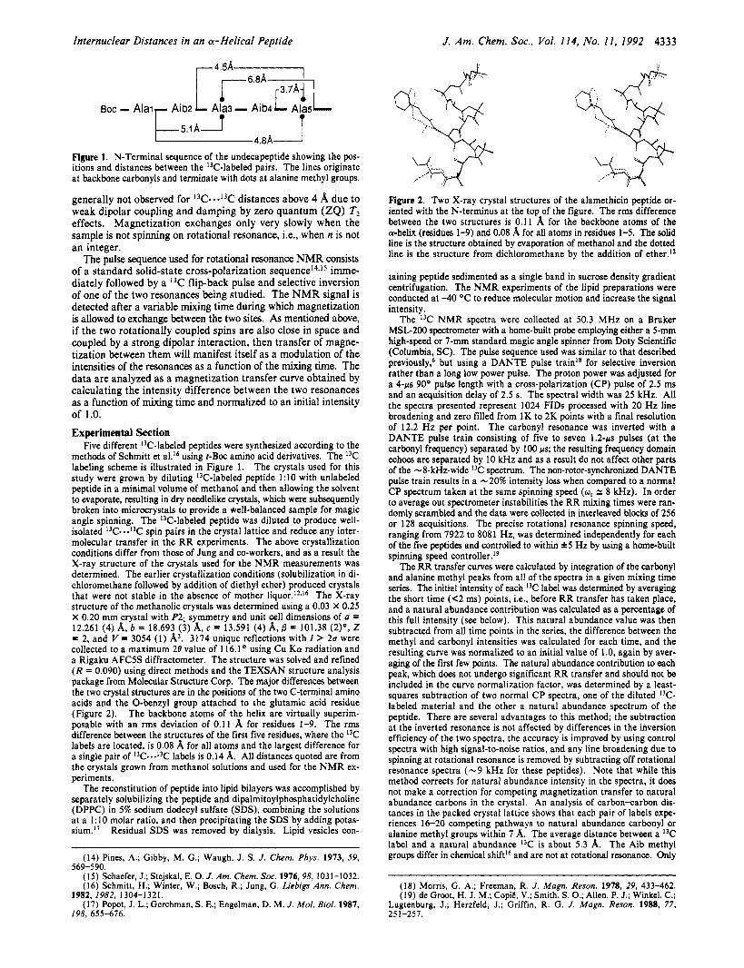
BOC - A i a i p E A J - :iriJ Figure 1. N-Terminal sequence of the undecapeptide showing the pos- itions and distances between the "C-labeled pairs. The lines originate a t backbone carbonyls and terminate with dots at alanine methyl groups.
generally not observed for 13C--.13C distances above 4 A due to weak dipolar coupling and damping by zero quantum (ZQ) T2 effects. Magnetization exchanges only very slowly when the sample is not spinning on rotational resonance, Le., when n is not an integer.
The pulse sequence used for rotational resonance NMR consists of a standard solid-state cross-polarization s e q ~ e n c e l ~ * ' ~ imme- diately followed by a I3C flip-back pulse and selective inversion of one of the two resonances being studied. The N M R signal is detected after a variable mixing time during which magnetization is allowed to exchange between the two sites. As mentioned above, if the two rotationally coupled spins are also close in space and coupled by a strong dipolar interaction, then transfer of magne- tization between them will manifest itself as a modulation of the intensities of the resonances as a function of the mixing time. The data are analyzed as a magnetization transfer curve obtained by calculating the intensity difference between the two resonances as a function of mixing time and normalized to an initial intensity of 1.0. Experimental Section
Five different "C-labeled peptides were synthesized according to the methods of Schmitt et a l l 6 using r-Boc amino acid derivatives. The 13C labeling scheme is illustrated in Figure 1. The crystals used for this study were grown by diluting '3C-labeled peptide 1:lO with unlabeled peptide in a minimal volume of methanol and then allowing the solvent to evaporate, resulting in dry needlelike crystals, which were subsequently broken into microcrystals to provide a well-balanced sample for magic angle spinning. The "C-labeled peptide was diluted to produce well- isolated 13C-.-13C spin pairs in the crystal lattice and reduce any inter- molecular transfer in the R R experiments. The above crystallization conditions differ from those of Jung and co-workers, and as a result the X-ray structure of the crystals used for the N M R measurements was determined. The earlier crystallization conditions (solubilization in di- chloromethane followed by addition of diethyl ether) produced crystals that were not stable in the absence of mother The X-ray structure of the methanolic crystals was determined using a 0.03 X 0.25 X 0.20 mm crystal with P21 symmetry and unit cell dimensions of u = 12.261 (4) A, 6 = 18.693 (3) A, c = 13.591 (4) A, 6 = 101.38 ( 2 ) O , 2 = 2, and V = 3054 (1) 8,'. 3174 unique reflections with I > 2a were collected to a maximum 28 value of 116.1' using Cu K a radiation and a Rigaku AFCSS diffractometer. The structure was solved and refined (R = 0.090) using direct methods and the TEXSAN structure analysis package from Molecular Structure Corp. The major differences between the two crystal structures are in the positions of the two C-terminal amino acids and the 0-benzyl group attached to the glutamic acid residue (Figure 2). The backbone atoms of the helix are virtually superim- posable with an rms deviation of 0.1 1 8, for residues 1-9. The rms difference between the structures of the first five residues, where the 13C labels are located, is 0.08 8, for all atoms and the largest difference for a single pair of 13C-*-13C labels is 0.14 8,. All distances quoted are from the crystals grown from methanol solutions and used for the N M R ex- periments.
The reconstitution of peptide into lipid bilayers was accomplished by separately solubilizing the peptide and dipalmitoylphosphatidylcholine (DPPC) in 5% sodium dodecyl sulfate (SDS), combining the solutions at a 1:lO molar ratio, and then precipitating the SDS by adding potas- sium." Residual SDS was removed by dialysis. Lipid vesicles con-
(14) Pines, A.; Gibby, M. G.; Waugh, J. S. J . Chem. Phys. 1973, 59,
(15) Schaefer, J.;Stejskal, E. 0. J. Am. ChemSoc. 1976.98, 1031-1032. (16) Schmitt, H.; Winter, W.; Bosch, R.; Jung, G. Liebigs Ann. Chem.
(17) Popot, J. L.; Gerchman, S. E.; Engelman, D. M. J . Mol. Biol. 1987,
569-590.
1982, 1982, 1304-1321.
198. 655-616.
J. Am. Chem. Soc., Vol. 114. No. 11, 1992 4333
:..
Figure 2. Two X-ray crystal structures of the alamethicin peptide or- iented with the N-terminus at the top of the figure. The rms difference between the two structures is 0.1 1 8, for the backbone atoms of the a-helix (residues 1-9) and 0.08 8, for all atoms in residues 1-5. The solid line is the structure obtained by evaporation of methanol and the dotted line is the structure from dichloromethane by the addition of ether.I*
taining peptide sedimented as a single band in sucrose density gradient centrifugation. The N M R experiments of the lipid preparations were conducted at -40 OC to reduce molecular motion and increase the signal intensity.
The 13C N M R spectra were collected at 50.3 MHz on a Bruker MSL-200 spectrometer with a home-built probe employing either a 5-mm high-speed or 7-mm standard magic angle spinner from Doty Scientific (Columbia, SC). The pulse sequence used was similar to that described previously,6 but using a DANTE pulse train1' for selective inversion rather than a long low power pulse. The proton power was adjusted for a 4-ps 90° pulse length with a cross-polarization (CP) pulse of 2.5 ms and an acquisition delay of 2.5 s. The spectral width was 25 kHz. All the spectra presented represent 1024 FIDs processed with 20 Hz line broadening and zero filled from 1K to 2K points with a final resolution of 12.2 Hz per point. The carbonyl resonance was inverted with a DANTE pulse train consisting of five to seven 1.2-ps pulses (at the carbonyl frequency) separated by 100 ps; the resulting frequency domain echoes are separated by 10 kHz and as a result do not affect other parts of the -8-kHz-wide 13C spectrum. The non-rotor-synchronized DANTE pulse train results in a -20% intensity loss when compared to a normal C P spectrum taken at the same spinning speed (w , = 8 kHz). In order to average out spectrometer instabilities the R R mixing times were ran- domly scrambled and the data were collected in interleaved blocks of 256 or 128 acquisitions. The precise rotational resonance spinning speed, ranging from 7922 to 8081 Hz, was determined independently for each of the five peptides and controlled to within h5 Hz by using a home-built spinning speed c~nt ro l le r . '~
The RR transfer curves were calculated by integration of the carbonyl and alanine methyl peaks from all of the spectra in a given mixing time series. The initial intensity of each "C label was determined by averaging the short time (<2 ms) points, i.e., before R R transfer has taken place, and a natural abundance contribution was calculated as a percentage of this full intensity (see below). This natural abundance value was then subtracted from all time points in the series, the difference between the methyl and carbonyl intensities was calculated for each time, and the resulting curve was normalized to an initial value of 1 .O, again by aver- aging of the first few points. The natural abundance contribution to each peak, which does not undergo significant RR transfer and should not be included in the curve normalization factor, was determined by a least- squares subtraction of two normal C P spectra, one of the diluted "C- labeled material and the other a natural abundance spectrum of the peptide. There are several advantages to this method; the subtraction at the inverted resonance is not affected by differences in the inversion efficiency of the two spectra, the accuracy is improved by using control spectra with high signal-to-noise ratios, and any line broadening due to spinning at rotational resonance is removed by subtracting off rotational resonance spectra (-9 kHz for these peptides). Note that while this method corrects for natural abundance intensity in the spectra, it does not make a correction for competing magnetization transfer to natural abundance carbons in the crystal. An analysis of carbon-carbon dis- tances in the packed crystal lattice shows that each pair of labels expe- riences 16-20 competing pathways to natural abundance carbonyl or alanine methyl groups within 7 8,. The average distance between a I3C label and a natural abundance "C is about 5.3 8,. The Aib methyl groups differ in chemical shiftI6 and are not at rotational resonance. Only
(18) Morris, G. A,; Freeman, R. J. Magn. Reson. 1978, 29, 433-462. (19) de Groot, H. J . M.; CopiE, V.; Smith, S. 0.; Allen, P. J.; Winkel, C.;
Lugtenburg, J.; Herzfeld, J.; Griffin, R. G. J . Magn. Reson. 1988, 77, 251-257.

4334 J. Am. Chem. SOC., Vol. 11 4, No. 11, 1992 Peersen et al.
--$-'e --I----+-+- _I___r zomsec 1 4-
1- - 30msec J ' p e - I
1 2w 1w 0
w m Figure 3. NMR transfer spectra of crystals of the undecapeptide at the n = 1 rotational resonance. The top spectrum corresponds to zero mixing time and the subsequent spectra are the differences obtained by subtraction of the zero time spectrum from those with mixing times of 4, 10, 20, and 30 ms. The line widths in the difference spectra of the 3.7-A peptide are 140 and 75 Hz for the carbonyl and methyl resonances, respectively. Each spectrum corresponds to 1024 scans of -10 mg of doubly I3C-labeled peptide diluted with - 100 mg of unlabeled material.
one of the five peptides has a significant intermolecular pathway between the pair of labeled carbons; in this case the intermolecular distance is 5.09 A, while the intramolecular distance is 5.12 A. Accounting for 1.1% natural abundance ')C and a 5.3-A average distance, the competing natural abundance pathways are estimated to account for 8-10% transfer at 40 ms when n = 1, 2-3% of which is due to strong intraresidue (Le., Alanc4 to AlanCH] at 2.5 A) and adjacent residue (Le., Aibnc4 to Alan+lCH3 at 3.7 A) couplings.
Results and Discussion The undecapeptide serva as a model system for characterizing
local peptide structure by RR NMR. The Aib and Ala residues strongly favor a helical conformation20,21 and the high-resolution crystal structures accurately define the atomic positions. RR measurements are described for a series of peptide crystals bearing pairs of 13C labels with internuclear distances ranging from 3.7 to 6.8 A that illustrate the range and resolution of the approach. The hydrophobic nature of the peptide favors its association with phospholipid bilayers and preliminary RR measurements on membrane-bound peptides are presented that show that this ap- proach can be extended to membrane systems.
RR Measurements of Peptide Crystals. The spectra in Figure 3 illustrate magnetization transfer at the n = 1 rotational resonance for distances of 3.7,4.8, and 6.8 1( in peptide crystals. In the top spectrum the "C-labeled carbonyl line (175 ppm) has been se- lectively inverted while spinning at rotational resonance with the I3C-labeled methyl resonance (20 ppm). Exchange has not yet taken place in this zero mixing time spectrum. The subsequent spectra are difference spectra obtained by subtraction of this initial spectrum from those obtained by using longer mixing times, clearly illustrating an increase of the carbonyl signal coupled to a re- duction of the methyl signal. Furthermore, the exchange is limited to the two rotationally coupled lines, with little or no transfer to the natural abundance a-carbons (50 ppm) or Aib methyl groups (25 ppm). The data have been summarized in Figure 4, which shows the normalized transfer curves for all five 13C-labeled peptides a t both the n = 1 and n = 2 rotational resonance con- ditions. The off-rotational rwnance data corresponds to the 6.8-A peptide spinning at 8500 Hz, which is 543 Hz faster than the n = 1 rotational resonance.
In the n = 1 transfer curves (Figure 4) the steepest slope and greatest magnetization transfer occurs between I3C labels a t AibbC4 and AlaSCH3, which are only 3.7 1( apart in the crystal structure, and the transfer is rapid enough to just reveal the predicted oscillations in the transfer curve. The extent of damping of these oscillations, in conjunction with the known dipolar coupling from the X-ray distance, can provide an estimate of the TzzQ in
(20) Karle, 1. L.; Balaram, P. Biochemistry 1990, 29, 6747-6756. (21) Karle, I. L.; Flippen-Anderson, J . L.; Uma, K.; Balaram, P. Proteim
1990, 7, 62-73.
e.. . . . . . . . . . . . . . . .o off RR
6.8A
4.8A
4.5A
5 . l A
-0.1 I I I I I I I I 1 0 5 10 15 20 25 30 35 40
Time (msec)
n = 2 1.1 1
4.8A
5 . rA 4.5A 3.7A
6.8A
-0.1 O ' l 1 0 5 10 15 20 25 30
Time (msec)
Figure 4. n = 1 and n = 2 I3C* - J3C rotational resonance magnetization transfer curves for the five undecapeptides. The distances indicated are between backbone carbonyl and alanine methyl groups and correspond to I3C-.-l3C dipolar couplings of 151,84, 68, 57, and 25 Hz. On the basis of the observed variation of peptide a-carbon intensities (50 ppm) during the rotational resonance experiments, the error in the normalized curves is 0.05 or less for all five peptides. The lines drawn are polynomial fits to illustrate the trend of the data; they are not theoretical simulations of transfer based on the molecular structure.
the peptide crystals. An accurate value of the T2zQ is crucial when simulating data where oscillations are not present.13 The next two curves are from I3C pairs that directly test the secondary structure of the peptide via transfer between adjacent turns of the helix. Both pairs use one label a t AlaSCH3 with a second label a t either AlaIC*, yielding a 4.8-A distance parallel to the helix axis, or a t AibzC4, resulting in a 4.5-1( distance across the face of the helix. The helicity of the peptide is further explored by looking across the helix diameter, from AlaIC* to AlajCH3, where the crystal structure distance is 5.1 A. The last peptide tests the limit of rotational resonance using Ala3CH~ and Alasc4 labels, which are 6.8 A apart in the crystal structure, the longest distance measured in these experiments. As shown in Figures 3 and 4, t h e transfer between these sites when n = 1 amounts to -30% after 40 ms, and even with 10% transfer to natural abundance carbons the remaining 20% is easily resolved from the off-rota- tional resonance data. Taken together, the set of five transfer curves shown in Figure 4 illustrate the potential of the rotational resonance NMR approach for determining accurate distances in protein microcrystals and provide a set of standard curves that can be used for calibration of rotational resonance experiments between carbonyl and methyl groups. Importantly, these data show experimentally that the n = 1 transfer rates are dominated by the dipolar coupling and are not significantly influenced by the relative orientations of the methyl and carbonyl groups. The accuracy of the RR measurements clearly depends on the actual distance being measured because of the $ dependence of the dipolar coupling, as is easily seen by comparing the 3.7- and 4.5-A

Internuclear Distances in an a-Helical Peptide J. Am. Chem. Soc., Vol. 114, No. 11, 1992 4335
simulations of the transfer rates based on distances and orientations available from the crystal structure. RR Measurements of MembmBound Peptides. The structure
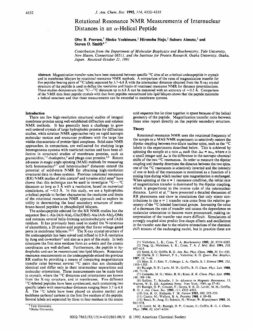
of the peptide when bound to a membrane has been addressed by reconstituting I3C-labeled peptides into lipid bilayers. RR measurements in membranes are complicated by the increased motion and structural flexibility of the peptide, since it is no longer held in a crystal lattice. As pointed out earlier, the presence of the conformationally restrictive Aib residues in the peptide favors a helical conformation, and potential problems with motion have been countered by lowering the sample temperature to -40 OC. Figure 5 shows a comparison of transfer rates in the crystals and in lipid for the 4.8- ( n = 2) and 5.1-A ( n = 1) peptides, the two pairs most sensitive to changes in secondary structure. The close correlation of these two data sets with the crystal data provides the first indication that the RR NMR methods will work at low temperature for small peptides incorporated into membranes. Further experiments to explore the temperature dependence of magnetization transfer are in progress.
These results set the stage for extending the RR methods to membrane systems in order to study the local secondary structure and possibly tertiary fold of membrane proteins. Two challenges for larger systems involve specifically targeting 13C pairs, possibly by using heteronuclear spectral editing approaches to target unique 15N-13C peptide bond^:^-*^ and accurate subtractions of protein background signals. The effective 1 0-kDa protein background in the crystal experiments discussed above (due to dilution with unlabeled material) and studies of I3C-labeled retinal in the 26-kDa protein bacteri~rhodopsin~~ illustrate that moderately large systems are tractable.
In conclusion, we show that the distance range of rotational resonance magnetization transfer is sufficient to investigate local peptide secondary structure, and when the experiment is performed at the n = 1 rotational resonance the interpretation of the initial transfer rate is straightforward. Furthermore, a set of control curves for the system, such as the carbonyl-methyl transfer curves presented in this paper, can be used to estimate distances up to 7 A, and with the proper use of backbone I3C labels this is suf- ficient to discriminate between a-helical and @-sheet structures.
Acknowledgment. We thank Gayle Schulte for the X-ray crystallography and Shy Arkin for assistance with lipid sample preparation. This work was supported by N I H (GM 41212) and the Searle Scholars Program/Chicago Community Trust.
Supplementary Material Available: Listing of crystallographic experimental details and X-ray coordinates with isotropic tem- perature factors (7 pages). Ordering information is given on any current masthead page.
1.05 7.
yo. .. 0.65
0.65 1 0.45
A
0 ..... o... ..,
“.Q.
0.25 I I I I I I 1 0 10 20 30 40 50 60
Time (mrec)
0.95 1 0.65 4 0.65 O . ’ 1
B
0.55 I I I I I I 0 5 10 15 20 25 30
Time (mrec)
Figure 5. Transfer curves for peptide-DPPC complexes. Shown are the 5.1-A peptide spinning at n = 1 (A) and the 4.8-A peptide at n = 2 (B). The lines correspond to the crystal data for each peptide and the symbols are the data from DPPC-reconstituted peptides. The lipid data were collected at a temperature of -40 ‘C to reduce peptide motion.
curves (0.8-A difference) and the 5.1- and 6.8-A curves (1.7-A difference). Even though the initial transfer rate a t 4.8 A is faster than that at 5.1 A, the transfer curves eventually merge, implying that 0.3 A is the approximate resolution limit of the method at distances in the vicinity of 5 A.
When the magic angle spinning rate is decreased so that n = 2, Le., w, = -4 kHz in these experiments, the transfer rates decrease (Figure 4), but there continues to be significant transfer. Experimentally this is quite important because the n = 2 ex- periments are generally more feasible, requiring slower spinning speeds and allowing larger sample volumes to be used. The most s i g ” t observation from the n = 2 data is the apparent reversal of the 4.8- and 5.1-8, transfer curves; in this case the I3C labels separated by the longer distance appear to be exchanging more rapidly. As shown by the n = 1 data, the 0.3-A difference between these distances appears to be the limit of resolution, raising the possibility that orientation effects play a significant role in the n = 2 data. This is currently being investigated with theoretical
(22) Schneider, D. M.; Tycko, R.; Opella, S. J. J . Magn. Reson. 1987, 73,
(23) Oas, T. G.; Hartzell, C. J.; Drobny, G. P.; Dahlquist, F. W. J . Magn.
(24) Stejskal, E. 0.; Schaefer, J.; McKay, R. A. J . Magn. Reson. 1984,
(25) Creuzet, F.; McDermott, A.; Gebhard, R.; van der Hoef, K.; Spijker-Assink, M. B.; Herzfeld, J.; Lugtenburg, J.; Levitt, M. H.; Griffin, R. G. Science 1991, 251, 183-786.
568-573.
Reson. 1989, 81, 395-399.
57, 471-485.