Embed Size (px)
Citation preview
764
PUESTA AL DÍA EN ANESTESIA REGIONAL Y TRATAMIENTO DEL DOLOR 2018 • VOL XXI
INTRANEURAL / PARANEURAL, DENTRO / FUERA ¿QUÉ MÁS?
ROLE OF ULTRASOUND IN DIAGNOSIS AND ASSESSMENT OF NERVE INJURY IN REGIONAL ANAESTHESIA, TRAUMA AND
CHRONIC PAIN *
Andrzej KrolDepartment of Anaesthesia and Chronic Pain Service
St George’s University Hospital
Peripheral nerves injury are not life threatening but may have debilitating consequences for the patients involved,changing the quality of life, limited function and causing chronic neuropathic pain. Trauma and surgery have been most common cause of nerve damage. The prevalence of posttraumatic sciatic nerve injury after pelvic trauma has been reported as high as 30 %, peroneal nerve injury after knee luxation up to 25%, brachial plexus branches injury after shoulder surgery around 5%. Nerve injury associated with peripheral nerve block have been very rare and estimated as more than 1/1.000 but less than < 1/1000 and recent metanalysis by Liu and colleagues did not show that the numbers has changed in the era of ultrasound 1. It has been further calculated that 70 000 patients per group would be required to show 50% reduction in nerve injury since ultrasound guided block has been implemented.
The commonest injury to the nerves have been due to compression eg swelling hema-toma, stretching and direct surgical insult. It may be also cause by direct needle injury violat-ing the epineurium with fascicular damage.
Nerve injury had been classified long time ago by Seddon in 1943 and has little changed since.
* This chapter should be referenced as: krol a. Role of ultrasound in diagnosis and assessment of nerve injury in regional anaesthesia, trauma and chronic pain. In J. De Andrés (editor). Update on Regional Anes-thesia and Pain management. Vol XXI. ISSN 1578-5580. Barcelona. MRA Editorial; 2018. p. 764- 781.
Este capítulo debe referenciarse como: krol a. Role of ultrasound in diagnosis and assessment of ner-ve injury in regional anaesthesia, trauma and chronic pain. En: J. De Andrés (editor). Puesta al día en Anestesia Regional y Tratamiento del Dolor. Vol XXI. ISSN 1578-5580. Barcelona. Editorial MRA; 2018. p. 764- 781.
765
PUESTA AL DÍA EN ANESTESIA REGIONAL Y TRATAMIENTO DEL DOLOR 2018 • VOL XXI
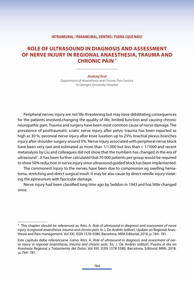
Table 1.
Neuropraxia (Class 1)
• Temporary interruption of conduction without loss of axonal continuity
• Intact structure
• Full recovery within days /weeks
• EMG shows lack of fibrillation potentials (FP) and positive sharp waves.
Axonotmesis (Class 2)
• Wallerian degeneration occurs below the site of injury.
• There are sensory and motor deficits distal to the site of lesion.
• There is not nerve conduction distal to the site of injury (3 to 4 days after injury).
• EMG shows fibrillation potentials (FP),and positive sharp waves (2 to 3 weeks pos-tinjury).
Neurotmesis (Class 3). Total severance or disruption of the entire nerve fiber
• Wallerian degeneration occurs below the site of injury.
• There is connective tissue lesion that may be partial or complete.
• Sensory-motor problems and autonomic function defect are severe.
• There is not nerve conduction distal to the site of injury (3 to 4 days after lesion).
• EMG and NCV findings are as axonotmesis.
• Because of lack of nerve repair, surgical intervention is necessary
Traditionally nerve trauma has been detected by clinical examination- sensory and mo-tor deficits. Further investigations employ electrophysiologic studies electromyography (EMG) and nerve conduction velocity (NCV) which are very sensitive indicators of peripheral nerve trauma but are not able, however,precisely detect the level or extraneural cause of signal transmission interruption such compression by hematoma, bone, scar tissue or for-eign body.
MRI provides high resolution pictures but only in limited sections and with nerves running in different planes makes planning of the adequate scanning very difficult.
Introduction of high resolution ultrasonography has provided another valuable tool for assessment of nerve integrity. Ultrasound scanning of peripheral nerves provide real time images of the nerve itself and surrounding structures. It can be useful to assess the nerve before and after regional block to exclude intraneural injection, to recognize primary nerve pathology : nerve enlargement, swelling, fascicle discontinuity and to identify external causes of nerve dysfunction caused by trauma, hematoma, scarring and connective tissue compression. Further implications of ultrasound might help to make a therapeutic decision e.g. surgical vs. non-surgical treatment or to be a tool of evaluation after primary peripheral nerve repair. Therapeutic interventions such as stump hydrodissection with local anaesthet-
766
PUESTA AL DÍA EN ANESTESIA REGIONAL Y TRATAMIENTO DEL DOLOR 2018 • VOL XXI
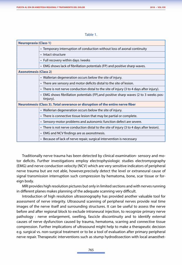
ics, neurolytic agents or neuromodulation procedures can be guided by ultrasound. Basic understanding of the nerve structure is essential in assessment.
Figure 1. Nerve Structure.
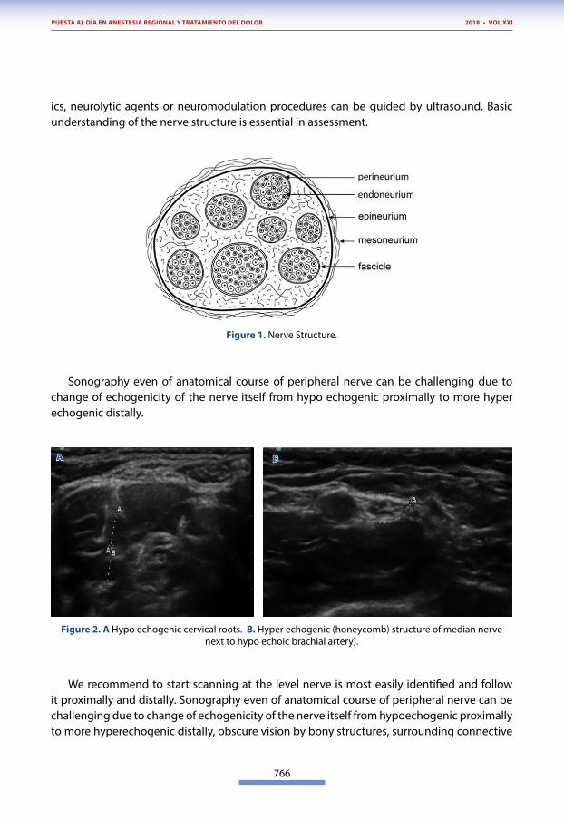
Sonography even of anatomical course of peripheral nerve can be challenging due to change of echogenicity of the nerve itself from hypo echogenic proximally to more hyper echogenic distally.
Figure 2. A Hypo echogenic cervical roots. B. Hyper echogenic (honeycomb) structure of median nerve next to hypo echoic brachial artery).
We recommend to start scanning at the level nerve is most easily identified and follow it proximally and distally. Sonography even of anatomical course of peripheral nerve can be challenging due to change of echogenicity of the nerve itself from hypoechogenic proximally to more hyperechogenic distally, obscure vision by bony structures, surrounding connective
A B
767
PUESTA AL DÍA EN ANESTESIA REGIONAL Y TRATAMIENTO DEL DOLOR 2018 • VOL XXI
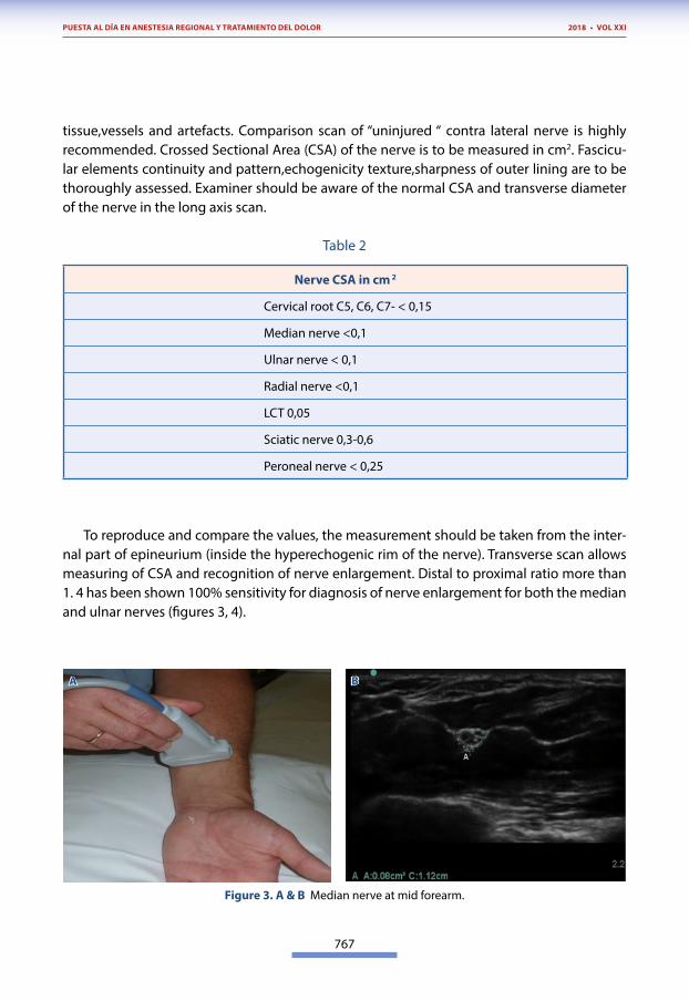
tissue,vessels and artefacts. Comparison scan of “uninjured “ contra lateral nerve is highly recommended. Crossed Sectional Area (CSA) of the nerve is to be measured in cm2. Fascicu-lar elements continuity and pattern,echogenicity texture,sharpness of outer lining are to be thoroughly assessed. Examiner should be aware of the normal CSA and transverse diameter of the nerve in the long axis scan.
Table 2
Nerve CSA in cm 2
Cervical root C5, C6, C7- < 0,15
Median nerve <0,1
Ulnar nerve < 0,1
Radial nerve <0,1
LCT 0,05
Sciatic nerve 0,3-0,6
Peroneal nerve < 0,25
To reproduce and compare the values, the measurement should be taken from the inter-nal part of epineurium (inside the hyperechogenic rim of the nerve). Transverse scan allows measuring of CSA and recognition of nerve enlargement. Distal to proximal ratio more than 1. 4 has been shown 100% sensitivity for diagnosis of nerve enlargement for both the median and ulnar nerves (figures 3, 4).
Figure 3. A & B Median nerve at mid forearm.
A B
768
PUESTA AL DÍA EN ANESTESIA REGIONAL Y TRATAMIENTO DEL DOLOR 2018 • VOL XXI
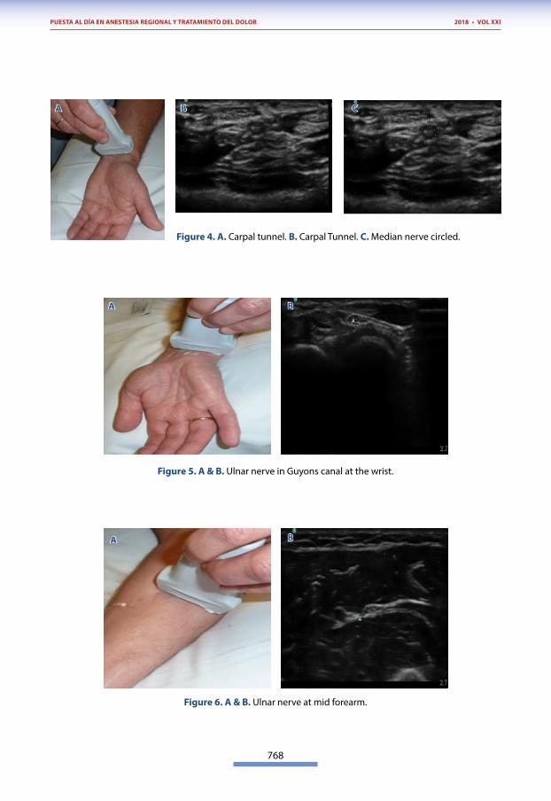
Figure 4. A. Carpal tunnel. B. Carpal Tunnel. C. Median nerve circled.
Figure 5. A & B. Ulnar nerve in Guyons canal at the wrist.
Figure 6. A & B. Ulnar nerve at mid forearm.
A
A
A
B C
B
B
769
PUESTA AL DÍA EN ANESTESIA REGIONAL Y TRATAMIENTO DEL DOLOR 2018 • VOL XXI
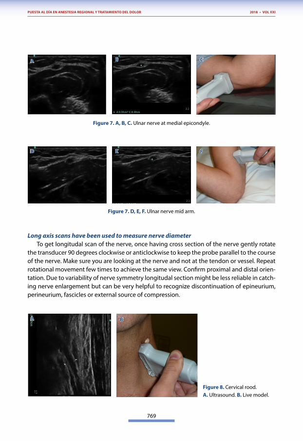
Figure 7. A, B, C. Ulnar nerve at medial epicondyle.
Figure 7. D, E, F. Ulnar nerve mid arm.
Long axis scans have been used to measure nerve diameterTo get longitudal scan of the nerve, once having cross section of the nerve gently rotate
the transducer 90 degrees clockwise or anticlockwise to keep the probe parallel to the course of the nerve. Make sure you are looking at the nerve and not at the tendon or vessel. Repeat rotational movement few times to achieve the same view. Confirm proximal and distal orien-tation. Due to variability of nerve symmetry longitudal section might be less reliable in catch-ing nerve enlargement but can be very helpful to recognize discontinuation of epineurium, perineurium, fascicles or external source of compression.
Figure 8. Cervical rood. A. Ultrasound. B. Live model.
A
D
A
B
E
C
F
B
770
PUESTA AL DÍA EN ANESTESIA REGIONAL Y TRATAMIENTO DEL DOLOR 2018 • VOL XXI
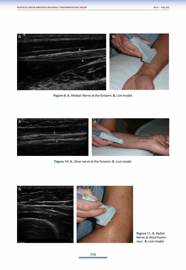
Figure 9. A. Median Nerve at the forearm. B. Live model.
Figure 10. A. Ulnar nerve at the forearm. B. Live model.
Figure 11. A. Radial Nerve at distal hume-rous. B. Live model.
A
A
A
B
B
B
771
PUESTA AL DÍA EN ANESTESIA REGIONAL Y TRATAMIENTO DEL DOLOR 2018 • VOL XXI
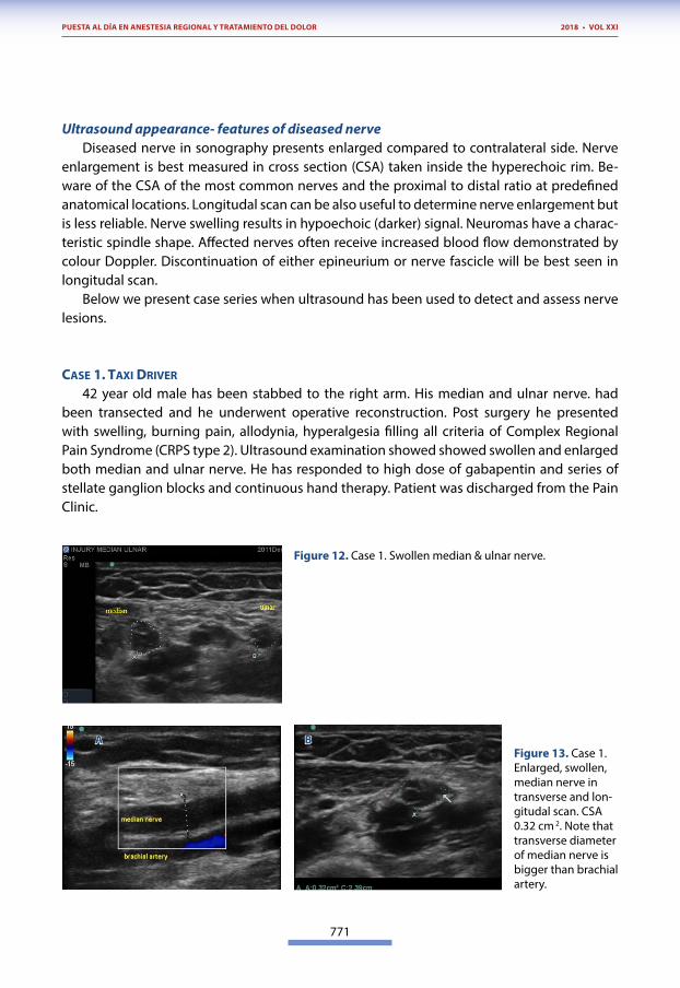
Ultrasound appearance- features of diseased nerveDiseased nerve in sonography presents enlarged compared to contralateral side. Nerve
enlargement is best measured in cross section (CSA) taken inside the hyperechoic rim. Be-ware of the CSA of the most common nerves and the proximal to distal ratio at predefined anatomical locations. Longitudal scan can be also useful to determine nerve enlargement but is less reliable. Nerve swelling results in hypoechoic (darker) signal. Neuromas have a charac-teristic spindle shape. Affected nerves often receive increased blood flow demonstrated by colour Doppler. Discontinuation of either epineurium or nerve fascicle will be best seen in longitudal scan.
Below we present case series when ultrasound has been used to detect and assess nerve lesions.
CASE 1. TAxI DRIVER
42 year old male has been stabbed to the right arm. His median and ulnar nerve. had been transected and he underwent operative reconstruction. Post surgery he presented with swelling, burning pain, allodynia, hyperalgesia filling all criteria of Complex Regional Pain Syndrome (CRPS type 2). Ultrasound examination showed showed swollen and enlarged both median and ulnar nerve. He has responded to high dose of gabapentin and series of stellate ganglion blocks and continuous hand therapy. Patient was discharged from the Pain Clinic.
Figure 12. Case 1. Swollen median & ulnar nerve.
Figure 13. Case 1. Enlarged, swollen, median nerve in transverse and lon-gitudal scan. CSA 0.32 cm 2. Note that transverse diameter of median nerve is bigger than brachial artery.
A B
772
PUESTA AL DÍA EN ANESTESIA REGIONAL Y TRATAMIENTO DEL DOLOR 2018 • VOL XXI
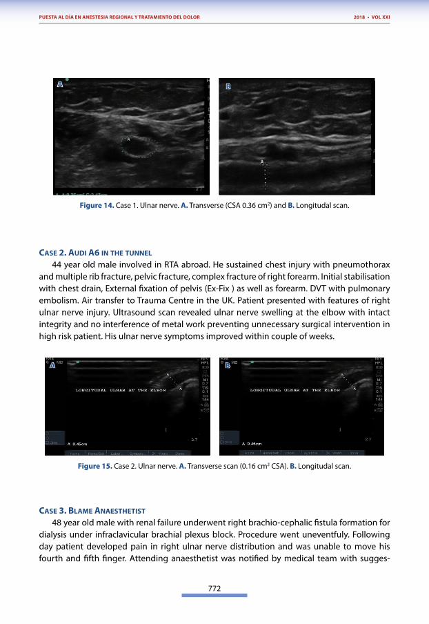
Figure 14. Case 1. Ulnar nerve. A. Transverse (CSA 0.36 cm2) and B. Longitudal scan.
CASE 2. AUDI A6 IN THE TUNNEL
44 year old male involved in RTA abroad. He sustained chest injury with pneumothorax and multiple rib fracture, pelvic fracture, complex fracture of right forearm. Initial stabilisation with chest drain, External fixation of pelvis (Ex-Fix ) as well as forearm. DVT with pulmonary embolism. Air transfer to Trauma Centre in the UK. Patient presented with features of right ulnar nerve injury. Ultrasound scan revealed ulnar nerve swelling at the elbow with intact integrity and no interference of metal work preventing unnecessary surgical intervention in high risk patient. His ulnar nerve symptoms improved within couple of weeks.
Figure 15. Case 2. Ulnar nerve. A. Transverse scan (0.16 cm2 CSA). B. Longitudal scan.
CASE 3. BLAME ANAESTHETIST
48 year old male with renal failure underwent right brachio-cephalic fistula formation for dialysis under infraclavicular brachial plexus block. Procedure went uneventfuly. Following day patient developed pain in right ulnar nerve distribution and was unable to move his fourth and fifth finger. Attending anaesthetist was notified by medical team with sugges-
A
A
B
B
773
PUESTA AL DÍA EN ANESTESIA REGIONAL Y TRATAMIENTO DEL DOLOR 2018 • VOL XXI
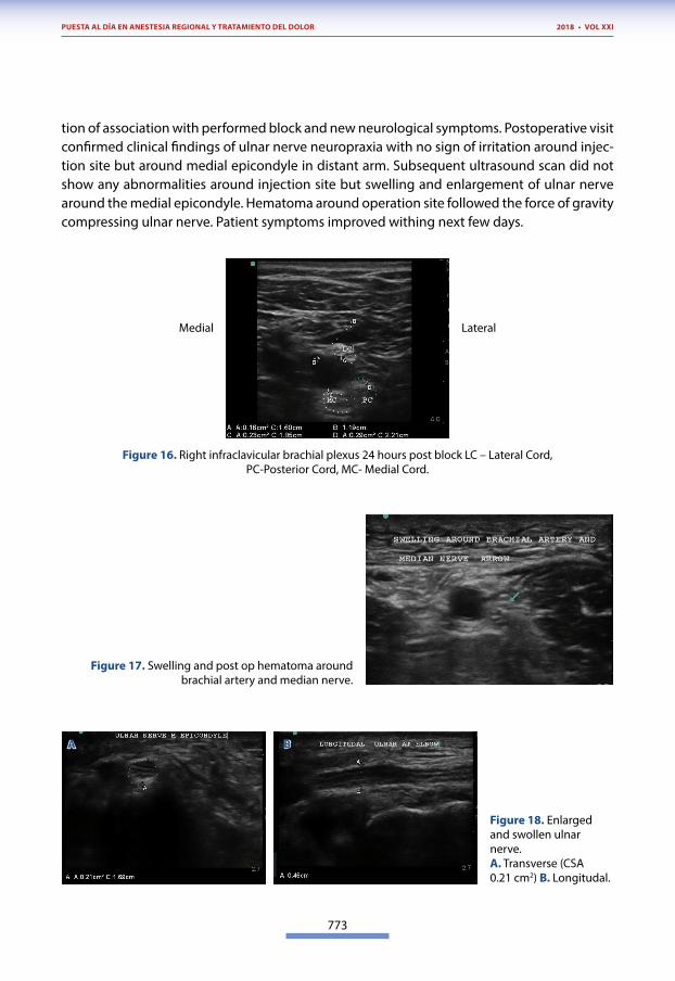
tion of association with performed block and new neurological symptoms. Postoperative visit confirmed clinical findings of ulnar nerve neuropraxia with no sign of irritation around injec-tion site but around medial epicondyle in distant arm. Subsequent ultrasound scan did not show any abnormalities around injection site but swelling and enlargement of ulnar nerve around the medial epicondyle. Hematoma around operation site followed the force of gravity compressing ulnar nerve. Patient symptoms improved withing next few days.
Medial Lateral
Figure 16. Right infraclavicular brachial plexus 24 hours post block LC – Lateral Cord,PC-Posterior Cord, MC- Medial Cord.
Figure 17. Swelling and post op hematoma around brachial artery and median nerve.
Figure 18. Enlarged and swollen ulnar nerve.A. Transverse (CSA 0.21 cm2) B. Longitudal.
A B
774
PUESTA AL DÍA EN ANESTESIA REGIONAL Y TRATAMIENTO DEL DOLOR 2018 • VOL XXI
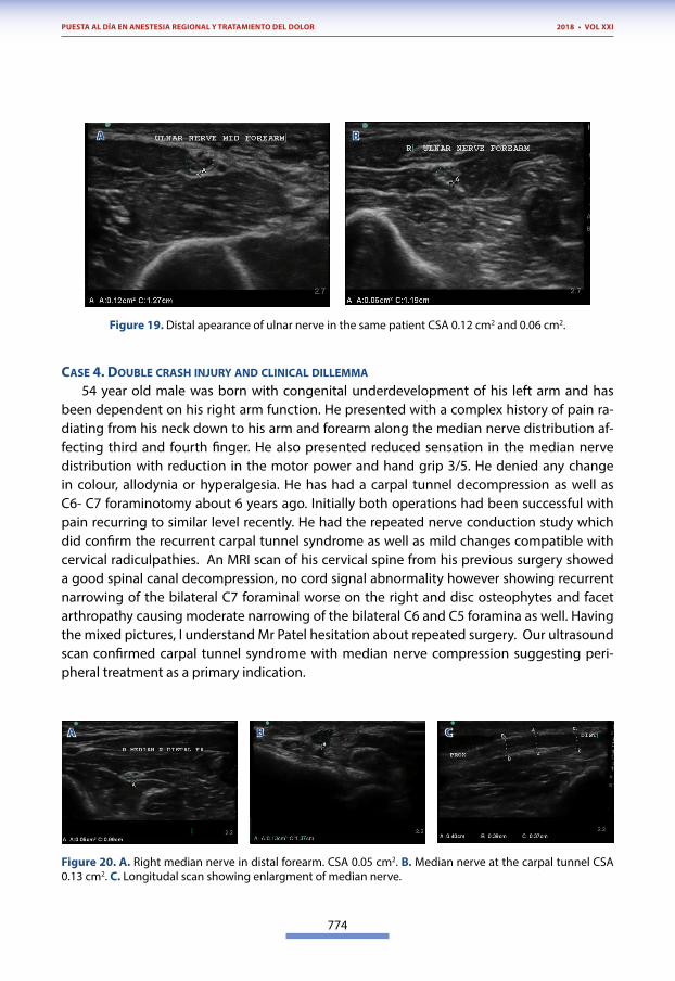
Figure 19. Distal apearance of ulnar nerve in the same patient CSA 0.12 cm2 and 0.06 cm2.
CASE 4. DOUBLE CRASH INjURY AND CLINICAL DILLEMMA
54 year old male was born with congenital underdevelopment of his left arm and has been dependent on his right arm function. He presented with a complex history of pain ra-diating from his neck down to his arm and forearm along the median nerve distribution af-fecting third and fourth finger. He also presented reduced sensation in the median nerve distribution with reduction in the motor power and hand grip 3/5. He denied any change in colour, allodynia or hyperalgesia. He has had a carpal tunnel decompression as well as C6- C7 foraminotomy about 6 years ago. Initially both operations had been successful with pain recurring to similar level recently. He had the repeated nerve conduction study which did confirm the recurrent carpal tunnel syndrome as well as mild changes compatible with cervical radiculpathies. An MRI scan of his cervical spine from his previous surgery showed a good spinal canal decompression, no cord signal abnormality however showing recurrent narrowing of the bilateral C7 foraminal worse on the right and disc osteophytes and facet arthropathy causing moderate narrowing of the bilateral C6 and C5 foramina as well. Having the mixed pictures, I understand Mr Patel hesitation about repeated surgery. Our ultrasound scan confirmed carpal tunnel syndrome with median nerve compression suggesting peri-pheral treatment as a primary indication.
Figure 20. A. Right median nerve in distal forearm. CSA 0.05 cm2. B. Median nerve at the carpal tunnel CSA 0.13 cm2. C. Longitudal scan showing enlargment of median nerve.
A
A
B
B C
775
PUESTA AL DÍA EN ANESTESIA REGIONAL Y TRATAMIENTO DEL DOLOR 2018 • VOL XXI
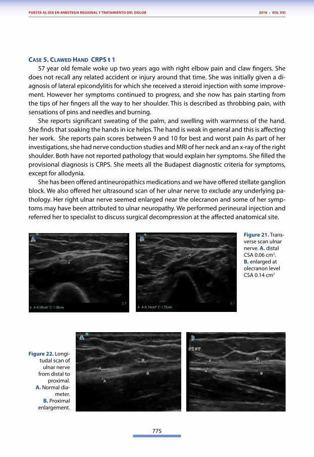
CASE 5. CLAwED HAND CRpS t 157 year old female woke up two years ago with right elbow pain and claw fingers. She
does not recall any related accident or injury around that time. She was initially given a di-agnosis of lateral epicondylitis for which she received a steroid injection with some improve-ment. However her symptoms continued to progress, and she now has pain starting from the tips of her fingers all the way to her shoulder. This is described as throbbing pain, with sensations of pins and needles and burning.
She reports significant sweating of the palm, and swelling with warmness of the hand. She finds that soaking the hands in ice helps. The hand is weak in general and this is affecting her work. She reports pain scores between 9 and 10 for best and worst pain As part of her investigations, she had nerve conduction studies and MRI of her neck and an x-ray of the right shoulder. Both have not reported pathology that would explain her symptoms. She filled the provisional diagnosis is CRPS. She meets all the Budapest diagnostic criteria for symptoms, except for allodynia.
She has been offered antineuropathics medications and we have offered stellate ganglion block. We also offered her ultrasound scan of her ulnar nerve to exclude any underlying pa-thology. Her right ulnar nerve seemed enlarged near the olecranon and some of her symp-toms may have been attributed to ulnar neuropathy. We performed perineural injection and referred her to specialist to discuss surgical decompression at the affected anatomical site.
Figure 21. Trans-verse scan ulnar nerve. A. distal CSA 0.06 cm2. B. enlarged at olecranon level CSA 0.14 cm2
Figure 22. Longi-tudal scan of
ulnar nerve from distal to
proximal. A. Normal dia-
meter. B. Proximal
enlargement.
A
A
B
B
776
PUESTA AL DÍA EN ANESTESIA REGIONAL Y TRATAMIENTO DEL DOLOR 2018 • VOL XXI
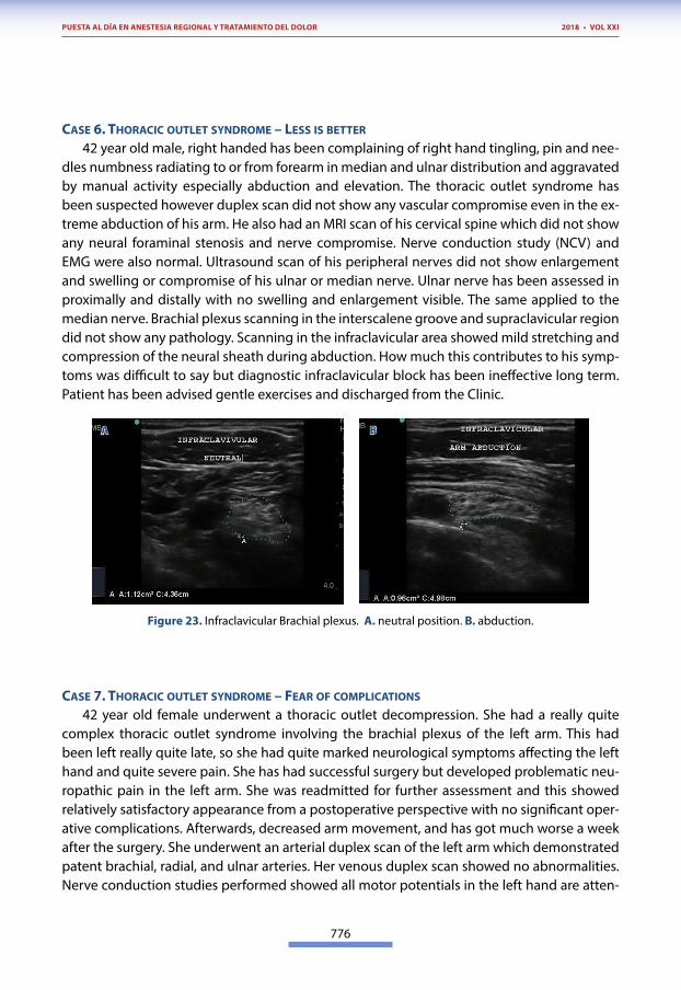
CASE 6. THORACIC OUTLET SYNDROME – LESS IS BETTER
42 year old male, right handed has been complaining of right hand tingling, pin and nee-dles numbness radiating to or from forearm in median and ulnar distribution and aggravated by manual activity especially abduction and elevation. The thoracic outlet syndrome has been suspected however duplex scan did not show any vascular compromise even in the ex-treme abduction of his arm. He also had an MRI scan of his cervical spine which did not show any neural foraminal stenosis and nerve compromise. Nerve conduction study (NCV) and EMG were also normal. Ultrasound scan of his peripheral nerves did not show enlargement and swelling or compromise of his ulnar or median nerve. Ulnar nerve has been assessed in proximally and distally with no swelling and enlargement visible. The same applied to the median nerve. Brachial plexus scanning in the interscalene groove and supraclavicular region did not show any pathology. Scanning in the infraclavicular area showed mild stretching and compression of the neural sheath during abduction. How much this contributes to his symp-toms was difficult to say but diagnostic infraclavicular block has been ineffective long term. Patient has been advised gentle exercises and discharged from the Clinic.
Figure 23. Infraclavicular Brachial plexus. A. neutral position. B. abduction.
CASE 7. THORACIC OUTLET SYNDROME – FEAR OF COMpLICATIONS
42 year old female underwent a thoracic outlet decompression. She had a really quite complex thoracic outlet syndrome involving the brachial plexus of the left arm. This had been left really quite late, so she had quite marked neurological symptoms affecting the left hand and quite severe pain. She has had successful surgery but developed problematic neu-ropathic pain in the left arm. She was readmitted for further assessment and this showed relatively satisfactory appearance from a postoperative perspective with no significant oper-ative complications. Afterwards, decreased arm movement, and has got much worse a week after the surgery. She underwent an arterial duplex scan of the left arm which demonstrated patent brachial, radial, and ulnar arteries. Her venous duplex scan showed no abnormalities. Nerve conduction studies performed showed all motor potentials in the left hand are atten-
A B
777
PUESTA AL DÍA EN ANESTESIA REGIONAL Y TRATAMIENTO DEL DOLOR 2018 • VOL XXI
uated, but there is no evidence of ulnar nerve motor slowing along the course of the nerve to suggest an ulnar neuropathy. EMG of the small muscles of the left hand shows clear neu-rogenic abnormality. These findings indicate denervation in the C8/T1 myotomes in keeping with thoracic outlet syndrome.
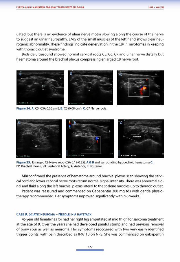
Bedside ultrasound showed normal cervical roots C5, C6, C7 and ulnar nerve distally but haematoma around the brachial plexus compressing enlarged C8 nerve root.
Figure 24. A. C5 (CSA 0.06 cm2), B. C6 (0.08 cm2), C. C7 Nerve roots.
A B C
Figure 25. Enlarged C8 Nerve root (CSA 0.19-0.25). A & B and surrounding hypoechoic hematoma C.BP: Brachial Plexus; VA: Vertebral Artery; A: Anterior; P: Posterior.
MRI confirmed the presence of hematoma around brachial plexus scan showing the cervi-cal cord and lower cervical nerve roots return normal signal intensity. There was abnormal sig-nal and fluid along the left brachial plexus lateral to the scalene muscles up to thoracic outlet.
Patient was reassured and commenced on Gabapentin 300 mg tds with gentle physio-therapy recommended. Her symptoms improved significantly within 6 weeks.
CASE 8. SCIATIC NEUROMA – NEEDLE IN A HAYSTACk
45 year old female has for had her right leg amputated at mid thigh for sarcoma treatment at the age of 9. Over the years she had developed painful stump and had previous removal of bony spur as well as neuroma. Her symptoms reoccurred with two very easily identified trigger points. with pain described as 8-9/ 10 on NRS. She was commenced on gabapentin
A
A
B
B C
C
778
PUESTA AL DÍA EN ANESTESIA REGIONAL Y TRATAMIENTO DEL DOLOR 2018 • VOL XXI
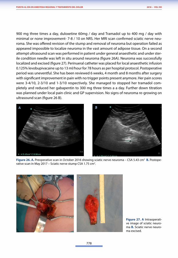
900 mg three times a day, duloxetine 60mg / day and Tramadol up to 400 mg / day with minimal or none improvement- 7-8 / 10 on NRS. Her MRI scan confirmed sciatic nerve neu-roma. She was offered revision of the stump and removal of neuroma but operation failed as appeared impossible to localize neuroma in the vast amount of adipose tissue. On a second attempt ultrasound scan was performed in patient under general anaesthetic and under ster-ile condition needle was left in situ around neuroma (figure 26A). Neuroma was successfully localized and excised (figure 27). Perinueral catheter was placed for local anaesthetic infusion 0.125% levobupivacaine up to 13 ml/hour for 78 hours as per hospital protocol. Postoperative period was uneventful. She has been reviewed 6 weeks, 4 month and 8 months after surgery with significant improvement in pain with no trigger points present anymore. Her pain scores were 3-4/10, 2-3/10 and 1-3/10 respectively. She managed to stopped her tramadol com-pletely and reduced her gabapentin to 300 mg three times a a day. Further down titration was planned under local pain clinic and GP supervision. No signs of neuroma re-growing on ultrasound scan (figure 26 B).
Figure 26. A. Preoperative scan in October 2016 showing sciatic nerve neuroma – CSA 5.43 cm2 B. Postope-rative scan in May 2017 – Sciatic nerve stump CSA 1.75 cm2.
Figure 27. A Intraoperati-ve image of sciatic neuro-ma B. Sciatic nerve neuro-ma excised.
A
A
B
B
779
PUESTA AL DÍA EN ANESTESIA REGIONAL Y TRATAMIENTO DEL DOLOR 2018 • VOL XXI
CASE 9. jUMpER CASE
48 year old female sustained a complex right lower limb fracture in 2013, with knee dis-location and vascular compromise with greater saphenous vein graft, external fixation and multiple osteotomies. Altogether, she had had about six reconstructive surgeries. She had done remarkably well in terms of functional restoration. However, she has been left with some neuropathic pain around the front of her knee, loss of sensation in the front of her shin and shooting pain from the middle of her ankle along superficial nerve distribution to the dorsum of her foot and her toes. She describes the pain as constant, up to 8/10 and average 4-6 /10 on NRS. There was no allodynia but clearly hyperalgesia especially in the superficial peroneal nerve distribution. She had tried various medications including Gabapentin, Am-itriptyline and simple painkillers like Paracetamol, Codeine, non-steroidal and Tramadol, none of them has been effective. We commenced local treatment like Capsaicin to de- sensitise the skin and local anaesthetic patch with 5% lignocaine and she found it effective. Her EMG/ NVC results confirmed our diagnosis of the partial common peroneal nerve lesion and the partial denervation affecting more superficial than deep branch.
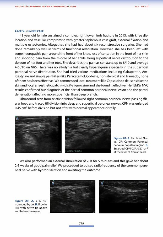
Ultrasound scan from sciatic division followed right common peroneal nerve passing fib-ular head and traced till division into deep and superficial peroneal nerves. CPN was enlarged 0.45 cm2 before division but not after with normal appearance distally.
Figure 28. A. TN: Tibial Ner-ve, CP: Common Peroneal nerve in popliteal region. B. Enlarged CPN CSA 0.27 cm2 at the level of fibular head.
We also performed an external stimulation of 2Hz for 5 minutes and this gave her about 2-3 weeks of good pain relief. We proceeded to pulsed radiofrequency of the common pero-neal nerve with hydrodissection and awaiting the outcome.
Figure 29. A. CPN su-rrounded by LA. B. Bipolar PRF with active tip above and below the nerve.
A
A
B
B
780
PUESTA AL DÍA EN ANESTESIA REGIONAL Y TRATAMIENTO DEL DOLOR 2018 • VOL XXI
CASE 10. SHOT IN THE FOOT
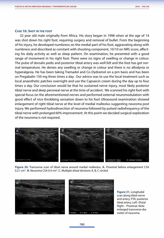
32 year old male originally from Africa. His story began in 1998 when at the age of 14 was shot down his right foot, requiring surgery and removal of bullet. From the beginning of his injury, he developed numbness on the medial part of his foot, aggravating along with numbness and described as constant with shooting component, 10/10 on NRS score, affect-ing his daily activity as well as sleep pattern. On examination, he presented with a good range of movement in his right foot. There were no signs of swelling or change in colour. The pulse of dorsalis pedis and posterior tibial artery was well felt and the foot has got nor-mal temperature. He denies any swelling or change in colour and there is no allodynia or hyperalgesia. He has been taking Tramadol and Co-Dydramol on a prn basis and has been on Pregabalin 150 mg three times a day. Our advice was to use the local treatment such as local anaesthetic patches overnight and use the Capsaicin cream during the day up to four times a day. Our conclusion would be that he sustained nerve injury, most likely posterior tibial nerve and deep peroneal nerve at the time of accident. We scanned his right foot with special focus on the aforementioned nerves and performed external neuromodulation with good effect of nice throbbing sensation down to his foot Ultrasound examination showed enlargement of right tibial nerve at the level of medial malleolus suggesting neuroma post injury. We performed hydrodissection of neuroma followed by pulsed radiofrequency of the tibial nerve with prolonged 60% improvement. At this point we decided surgical exploration of the neuroma is not required.
Figure 30. Transverse scan of tibial nerve around medial malleolus. A. Proximal before enlargement CSA 0.21 cm2. B. Neuroma CSA 0.4 cm2. C. Multiple distal divisions A, B, C circled.
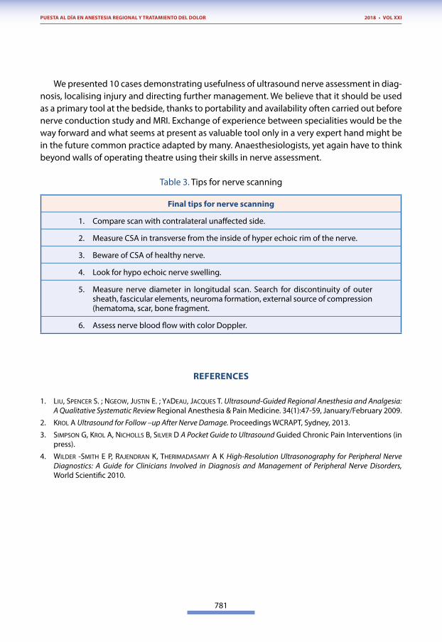
Figure 31. Longitudal scan along tibial nerve and artery. PTA: posterior tibial artery. Left -Distal Right – Proximal. Note enlarged transverse dia-meter of neuroma.
A
A
B
B
C
781
PUESTA AL DÍA EN ANESTESIA REGIONAL Y TRATAMIENTO DEL DOLOR 2018 • VOL XXI
We presented 10 cases demonstrating usefulness of ultrasound nerve assessment in diag-nosis, localising injury and directing further management. We believe that it should be used as a primary tool at the bedside, thanks to portability and availability often carried out before nerve conduction study and MRI. Exchange of experience between specialities would be the way forward and what seems at present as valuable tool only in a very expert hand might be in the future common practice adapted by many. Anaesthesiologists, yet again have to think beyond walls of operating theatre using their skills in nerve assessment.
Table 3. Tips for nerve scanning
Final tips for nerve scanning
1. Compare scan with contralateral unaffected side.
2. Measure CSA in transverse from the inside of hyper echoic rim of the nerve.
3. Beware of CSA of healthy nerve.
4. Look for hypo echoic nerve swelling.
5. Measure nerve diameter in longitudal scan. Search for discontinuity of outer sheath, fascicular elements, neuroma formation, external source of compression (hematoma, scar, bone fragment.
6. Assess nerve blood flow with color Doppler.
REFERENCES
1. liu, spencer s. ; nGeoW, jusTin e. ; yaDeau, jacques T. Ultrasound-Guided Regional Anesthesia and Analgesia: A Qualitative Systematic Review Regional Anesthesia & Pain Medicine. 34(1):47-59, January/February 2009.
2. krol a Ultrasound for Follow –up After Nerve Damage. Proceedings WCRAPT, Sydney, 2013.
3. simpson G, krol a, nicholls b, silver D A Pocket Guide to Ultrasound Guided Chronic Pain Interventions (in press).
4. WilDer -smiTh e p, rajenDran k, TherimaDasamy a k High-Resolution Ultrasonography for Peripheral Nerve Diagnostics: A Guide for Clinicians Involved in Diagnosis and Management of Peripheral Nerve Disorders, World Scientific 2010.