Embed Size (px)
Citation preview
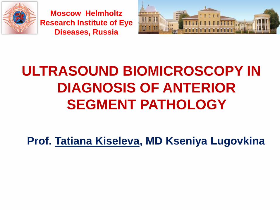
ULTRASOUND BIOMICROSCOPY IN
DIAGNOSIS OF ANTERIOR
SEGMENT PATHOLOGY
Prof. Tatiana Kiseleva, MD Kseniya Lugovkina
Moscow Helmholtz
Research Institute of Eye
Diseases, Russia
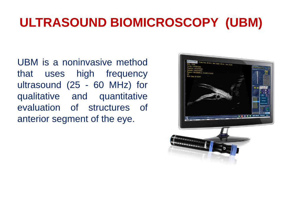
ULTRASOUND BIOMICROSCOPY (UBM)
UBM is a noninvasive method
that uses high frequency
ultrasound (25 - 60 MHz) for
qualitative and quantitative
evaluation of structures of
anterior segment of the eye.
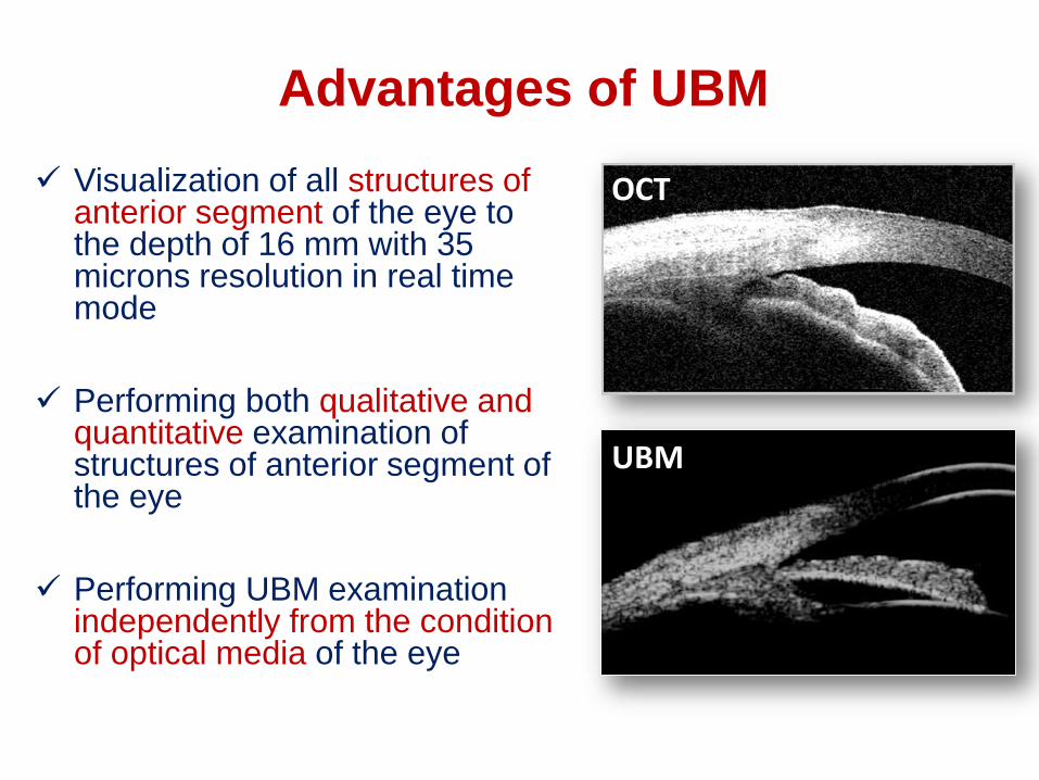
Advantages of UBM
Visualization of all structures of anterior segment of the eye to the depth of 16 mm with 35 microns resolution in real time mode
Performing both qualitative and quantitative examination of structures of anterior segment of the eye
Performing UBM examination independently from the condition of optical media of the eye
ОCТ
UBM

UBM imaging
• Cornea
• Anterior chamber
• Lens and zonule
• Iridociliary complex: iris,
ciliary body, the anterior
chamber angle (ACA)
• Posterior chamber
• The peripheries of vitreous
and retina

Conventional B-scan “window”
30-40mmTransducer
Lateral resolution = 600 microns
Axial resolution = 187 microns

Tra
nsd
ucer
40
-60 М
Hz
Diagnostic window:
15 × 16 mm
Scanning angle: 30°
Resolution: 15 - 35 microns
Technique of UBM

UBM Technique
• Patient in supine position
with topical anesthesia
• Eye cup between eyelids
filled with normal saline
• Probe placed into eye cup
• Real-time image is
displayed on a video monitor

UBM of anterior segment Imaging
Axial (panoramic) scans
Longitudinal scans
Transverse scans
Ba
sic
po
sit
ion
ing
of
sc
an
s
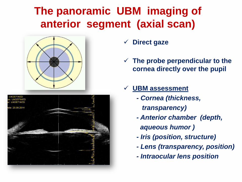
The panoramic UBM imaging of
anterior segment (axial scan)
Direct gaze
The probe perpendicular to the
cornea directly over the pupil
UBM assessment
- Cornea (thickness,
transparency)
- Anterior chamber (depth,
aqueous humor )
- Iris (position, structure)
- Lens (transparency, position)
- Intraocular lens position

Longitudinal (meridional) sections
The probe perpendicular to the
limbus with the marker towards
the pupil according to meridian
clock
UBM assessment
- Anterior chamber angle (ACA)
- Iris (thickness, convexity,
insertion)
- Ciliary body (thickness,
structure)
- Lens (zonule, capsule)
- Intraocular lens haptic
- Peripheries of vitreous and
retina
ЦТ
CB
Zonule
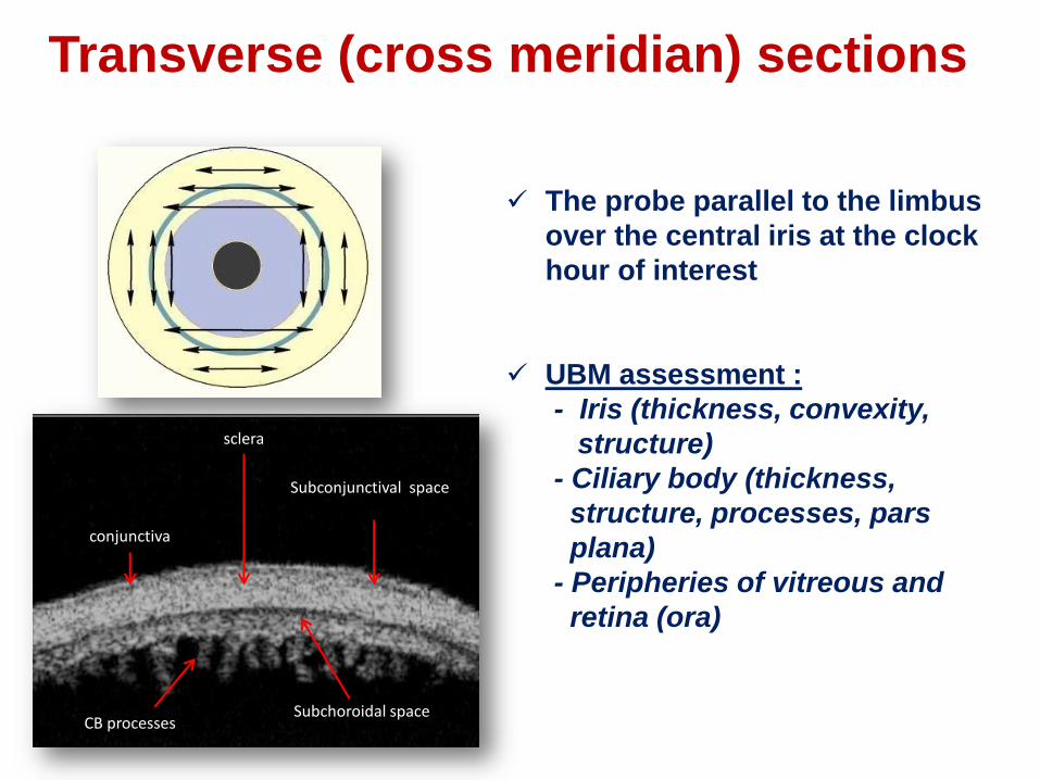
Transverse (cross meridian) sections
The probe parallel to the limbus
over the central iris at the clock
hour of interest
UBM assessment :
- Iris (thickness, convexity,
structure)
- Ciliary body (thickness,
structure, processes, pars
plana)
- Peripheries of vitreous and
retina (ora)
sclera
Subconjunctival space
CB processesSubchoroidal space
conjunctiva

Transverse Section ciliary processes
Sclera
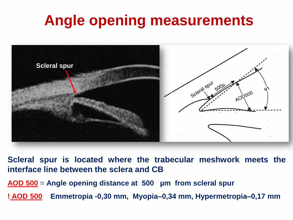
Scleral spur
Scleral spur is located where the trabecular meshwork meets the
interface line between the sclera and CB
AOD 500 = Angle opening distance at 500 µm from scleral spur
! AOD 500 Emmetropia -0,30 mm, Myopia–0,34 mm, Hypermetropia–0,17 mm
Angle opening measurements

Echographic parameters of anterior
segment structures in healthy subjects
Reflectivity Structure Size
Cornea Low Regular 0,55 – 0,59 mm
AC Anechoic - 3,0 – 3,6 mm
Iris Medium Irregular 0,2 – 0,4 mm
CB Medium Regular 0,7 – 0,73 mm
Lens Low Regular 3,5 – 4,7 mm
Zonule Medium Regular 1,0 – 1,3 mm
ACA - - 20° - 40°
Sclera High Regular 0,6 – 0,8 mm

UBM
GLAUCOMA
CATARACT & REFRACTIVE SURGERY
ONCOLOGY
GENERAL EXAMINATION
TRAUMA
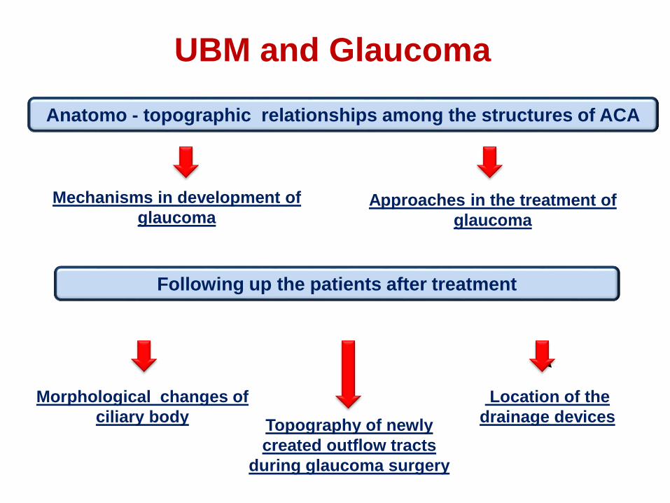
UBM and Glaucoma
Anatomo - topographic relationships among the structures of ACA
Mechanisms in development of
glaucoma Approaches in the treatment of
glaucoma
Following up the patients after treatment
Morphological changes of
ciliary bodyTopography of newly
created outflow tracts
during glaucoma surgery
Location of the
drainage devices

Pigmentary glaucoma (pigment dispersion
syndrome)
Widely open angle
Iris configuration (concave)
Reverse pupillary block
Amount of iridozonular contact
Mechanism : dissemination of pigment granules from the posterior iris

Pigment dispersion syndrome
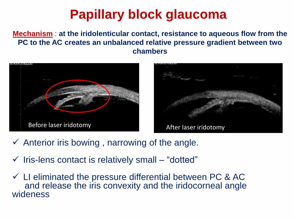
Papillary block glaucoma
Mechanism : at the iridolenticular contact, resistance to aqueous flow from the
PC to the AC creates an unbalanced relative pressure gradient between two
chambers
Before laser iridotomy After laser iridotomy
Anterior iris bowing , narrowing of the angle.
Iris-lens contact is relatively small – “dotted”
LI eliminated the pressure differential between PC & AC and release the iris convexity and the iridocorneal angle
wideness

Malignant glaucoma (ciliary block)
IrisIris
• Angle closure is caused by pressure differential between the
vitreous and aqueous compartment
• Swelling or anterior rotation of the ciliary body with formal
rotation of the lens-iris diaphragm and relation of the zonular
apparatus may cause anterior lens displacement.
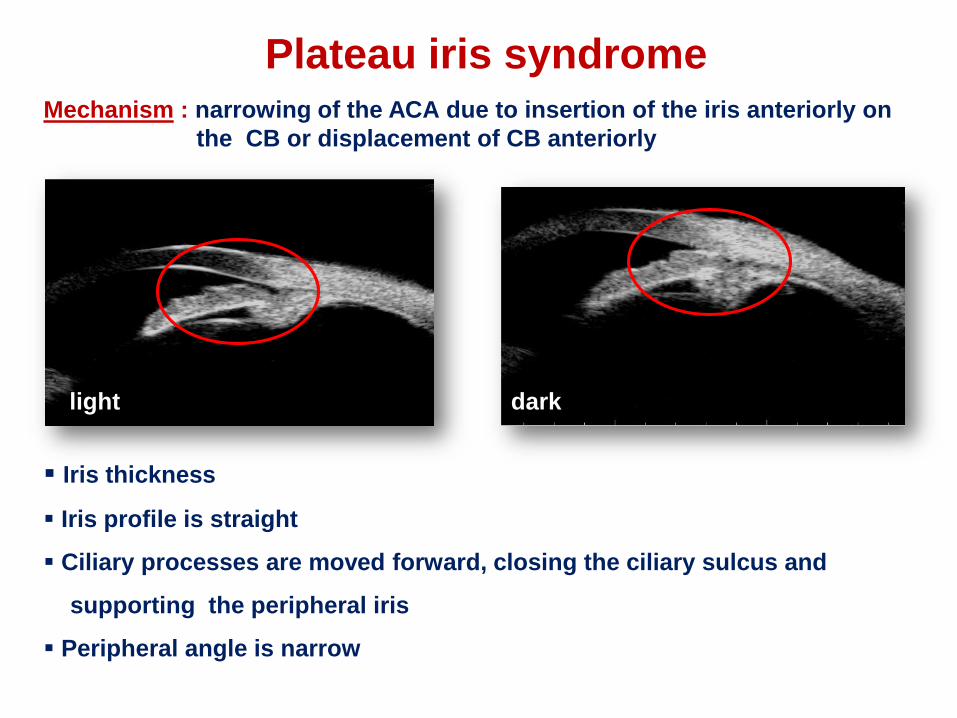
Plateau iris syndromeMechanism : narrowing of the ACA due to insertion of the iris anteriorly on
the CB or displacement of CB anteriorly
Iris thickness
Iris profile is straight
Ciliary processes are moved forward, closing the ciliary sulcus and
supporting the peripheral iris
Peripheral angle is narrow
light dark
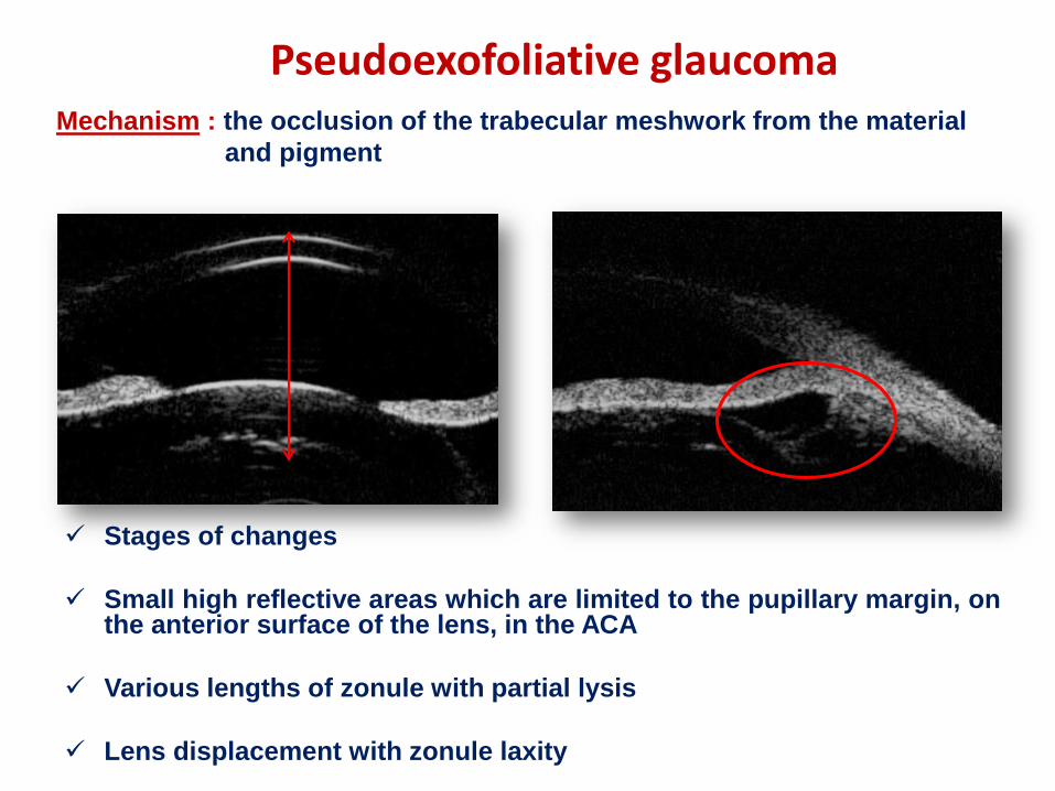
Pseudoexofoliative glaucoma
Stages of changes
Small high reflective areas which are limited to the pupillary margin, onthe anterior surface of the lens, in the ACA
Various lengths of zonule with partial lysis
Lens displacement with zonule laxity
Mechanism : the occlusion of the trabecular meshwork from the material
and pigment
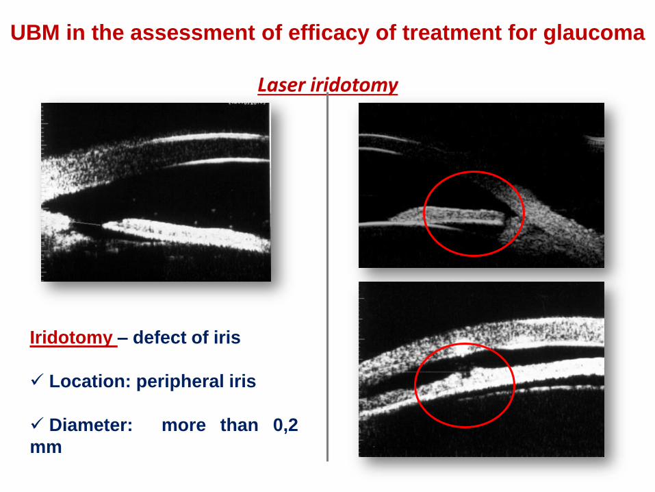
UBM in the assessment of efficacy of treatment for glaucoma
Laser iridotomy
Iridotomy – defect of iris
Location: peripheral iris
Diameter: more than 0,2
mm

Normal filtering bleb –subconjunctival fluid collection
and low to moderate intrableb
reflectivity
UBM in the assessment of efficacy of glaucoma surgery
Filtering cystic bleb –hyporeflective areas filled with
multiple fluid collections of
varying size and intensity

ммм
Glaucoma drainage devices
Drainage device in AC,
tube lumen is free
Drainage device in PC,tube lumen is free
Drainage device in sclera and doesn’t reach the AC
AC

UBM in cataract surgery
Anatomo - topographic relationships among the structures of
iridolenticular diaphragm
Location of lens, the condition of
lens substance and zonule
Approaches to cataract surgery
Following up the patients after cataract surgery
Position of IOL, haptics and
optical elements
Capsular bag status

Lens anomalies
Peters anomaly
Central corneal lenticular adhesion
Spherophakia , cataract, ectopic lens
Thinning of iris (dystrophy)
Iridocorneal adhesion
Microphakia
Abnormally small lens
Thinning of iris and CB(dystrophy)

Cataract
Post-traumatic cataract
X-ray induced cataract
Hyperechoic areas of lens, their
shape, number and placement
depend on the type of the cataract

Post-traumatic changes of lens
Immature cataract:
Thickness and high reflectivity of
cornea
Shallow AC
Iris bombe
Enlargement and “vacuoles type”
high
reflectivity of lens
anterior chamber angle closed
Subluxation (3rd degree)
Displacement of lens into
vitreous
High reflectivity of lens -
”layered type”
Slit-like ciliary body
detachment

Zonular rupture
Displacement of lens
Equator lens-ciliary process
distance ˃ 1, 3 mm
Hernia of vitreous
body
Zonular rupture
Cyst-like hernia of vitreous body
with low reflectivity of its contents

Intraocular lens position
Anterior chamber IOL
Posterior chamber IOL
IOL «RSP – 3» (mushroom)Assessment:
Location of optical part of IOL
according to optical axis
Position of haptic elements of IOL
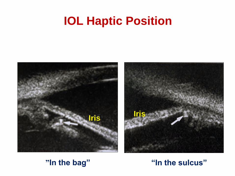
IOL Haptic Position
”In the bag” “In the sulcus”
IrisIris

Intraocular lens dislocation
1 2
3 4

The visualization of tumors
Conjunctiva
Limbus
Iris
Ciliary body
Periphery of choroid
Purpose: to determine size, structure, interaction with surrounding
tissues, degree of invasion
Development of treatment and assessment of efficacy of treatment
UBM in ocular oncology
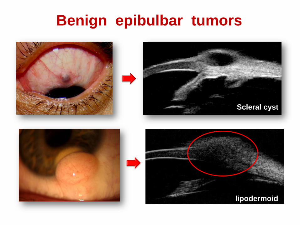
Benign epibulbar tumors
Scleral cyst
lipodermoid
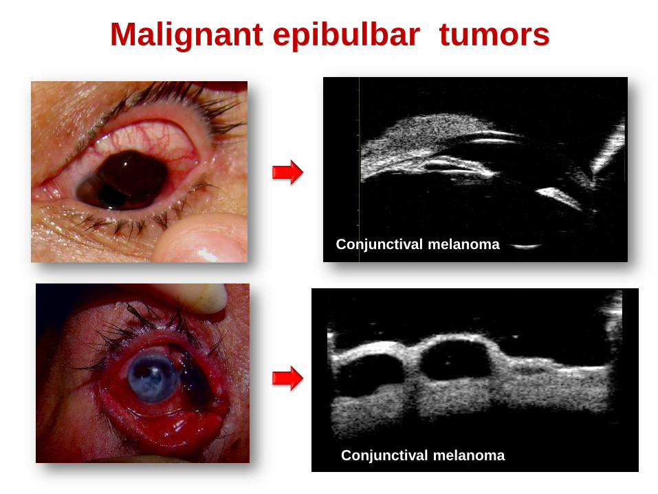
Malignant epibulbar tumors
Conjunctival melanoma
Conjunctival melanoma

Benign iris tumors. Iris nevus
UBM: hypoechoic, hyperechoic or uniform reflectivity of local thickness
of iris
Progressive nevus

Iris melanoma
Local thickness of iris with changes of anterior and/or posterior surface,
low reflectivity in comparison to intact tissues

Ciliary body melanoma
Low reflectivity of local thickness
of CB in comparison to intact
tissues
During the interaction with equator
of lens local cataract can be
formed
Local cataract in zone of contact of
tumor with lens

Invasion of the ACA, contact with the cornea
Iridociliary melanoma
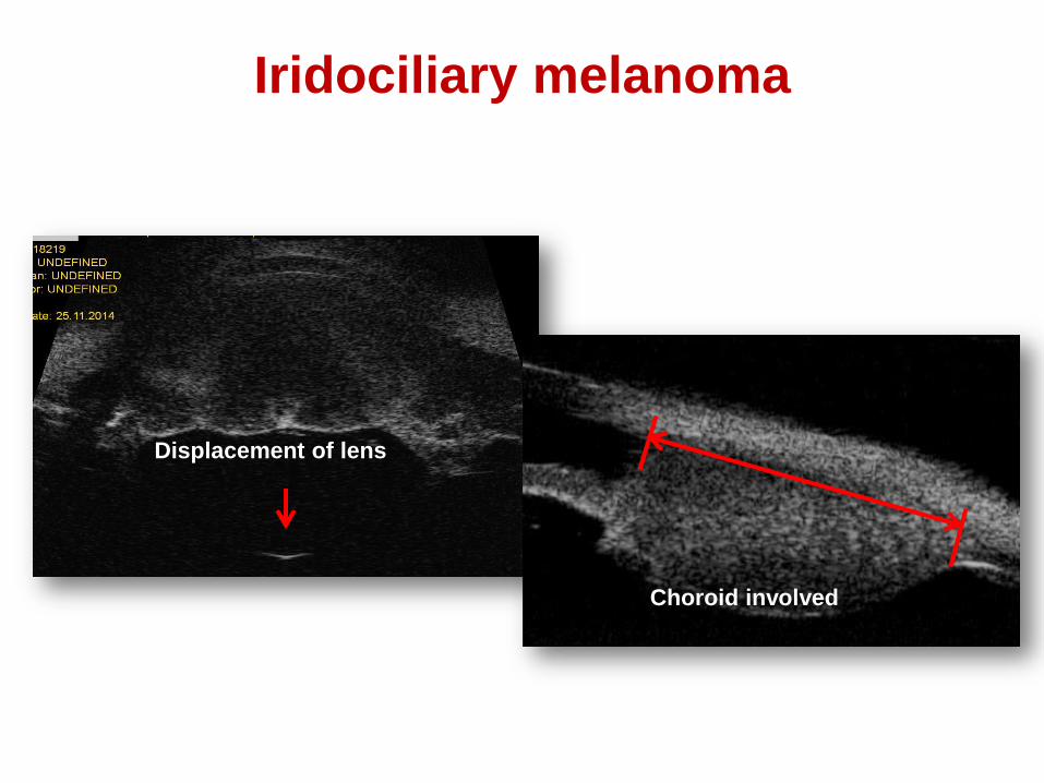
Iridociliary melanoma
Displacement of lens
Choroid involved

Iris cysts
Iris stromal cysts
Cysts of the iris pigment
epithelium
UBM appearance: thin-walled cysts with no internal reflectivity

Ciliary body cysts

UBM and ocular trauma
Anatomo - topographic relationships among the structures of
anterior segment
Cornea
Anterior chamber
Iridociliary zone (ICZ)
Posterior chamber
Lens
Zonule
Following up the patients after treatment
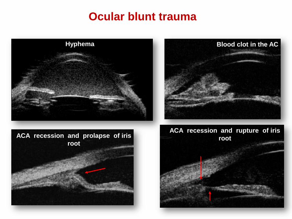
Ocular blunt trauma
ACA recession and prolapse of iris
root
Blood clot in the AC
ACA recession and rupture of iris
root
Hyphema

Ocular blunt trauma. Changes of ICZ
1- 2 – slit-like fistula between AC and suprachoroidal space (cyclodialysis cleft)
3 – 4 – shallow , uneven AC + exposure of the scleral spur + displacement of iris and
CB + CB detachment
Iridociclodialysis, displacement of CBназад
Displacement of iris and CB anteriorly
1 2
3 4

Ocular blunt trauma. Changes of ICZ
reverse profile of IR
ACA recession
ACA recession + tear of IR
Tear of the inner layers of sclera
ACA recession + reverse profile of IR

Complications of penetrating trauma
.
Conglomerate of cornea, iris
and IOL
Iridocorneal synechiae
12
3
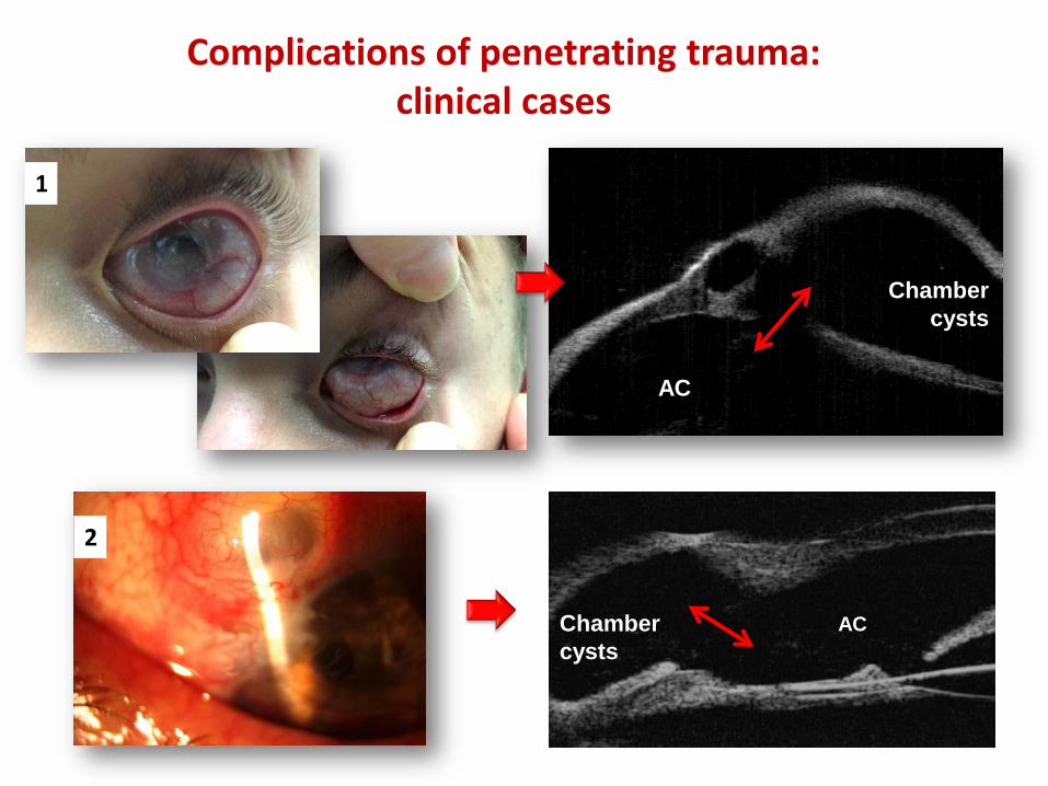
Complications of penetrating trauma:clinical cases
AC
1
Chamber
cysts
Chamber
cysts
AC
2

Foreign bodies in the anterior chamber
Glass
MetalMetal
ИТ в хрусталике
Glass
wound channel
Metal

Outcomes of ocular burns
Retrocorneal
membrane
Iridocorneal synechiae
Keratoprosthesis
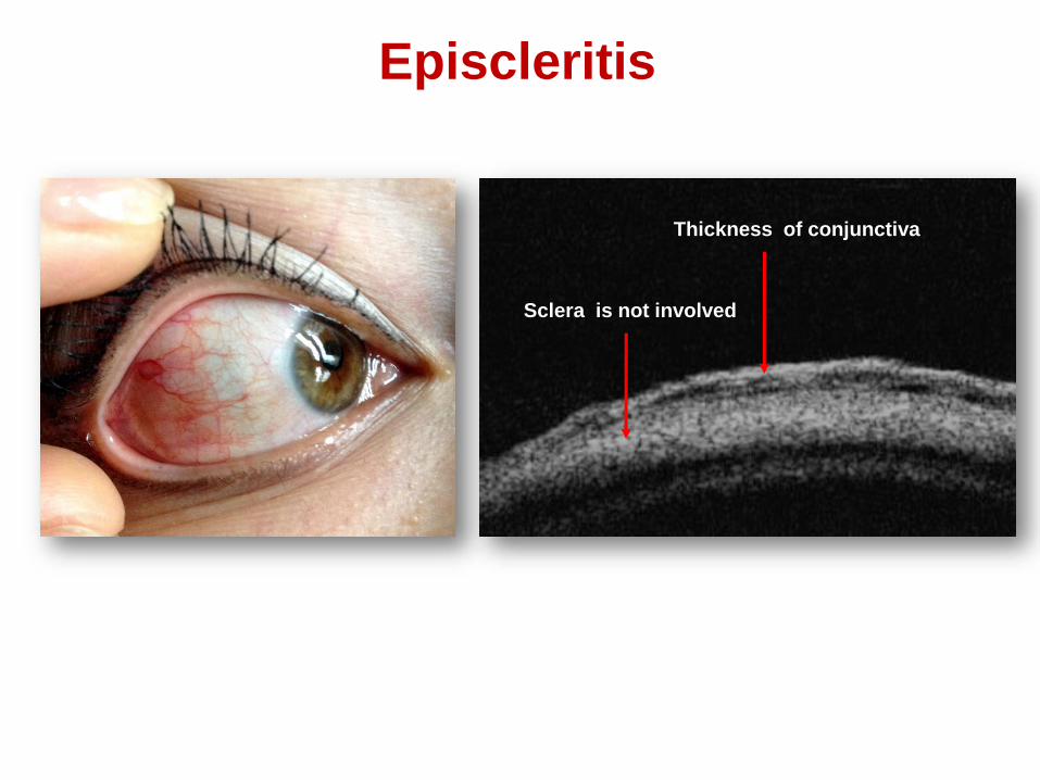
Episcleritis
Thickness of conjunctiva
Sclera is not involved
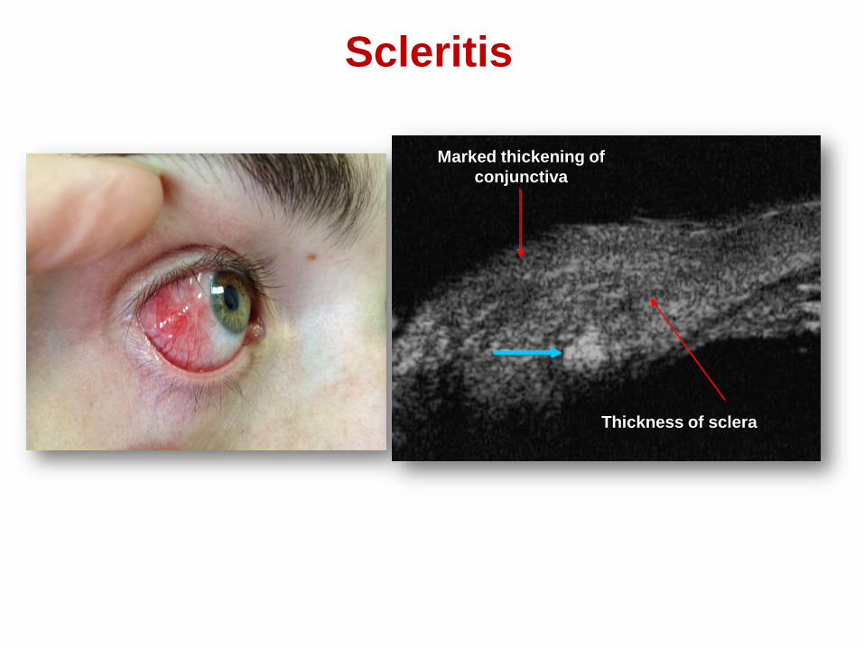
Scleritis
Marked thickening of
conjunctiva
Thickness of sclera

Outcome of scleritis in rheumatoid arthritis
Penetrating defect of sclera, with iris
tamponade effect

Outcome of fungal keratitis
Transparent
lens
Cornea
AC
“Wrong” AC
Autologous coveringПрПр
ПрПр

Outcome of pars planitis
fibrosis of zonule and capsule lens

Acute anterior uveitis
Marked thickening of CB
High reflectivity floaters in the AC and vitreous
CB detachment
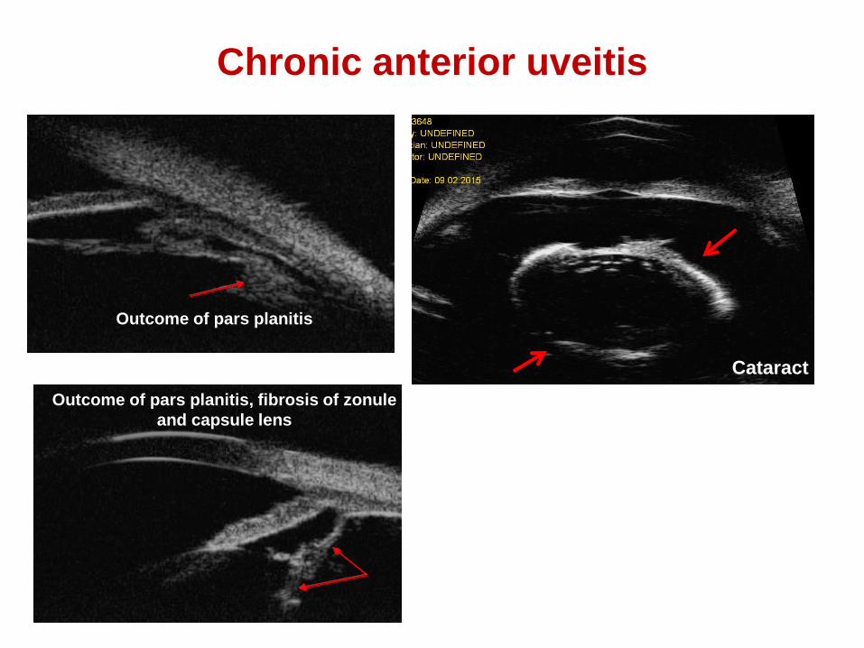
Outcome of pars planitis
Outcome of pars planitis, fibrosis of zonule
and capsule lens
Chronic anterior uveitis
Cataract

•Immersion “water bath” technique
•Cost & Availability
•Limited penetration
•Narrow field
•Resolution ?
•No “tissue diagnosis”
Current Limitation

• Open eye injury
• Recent eye surgery
• Corneal ulcer
• Infective surface eye disease
• Uncooperative patient
Contraindication to UBM
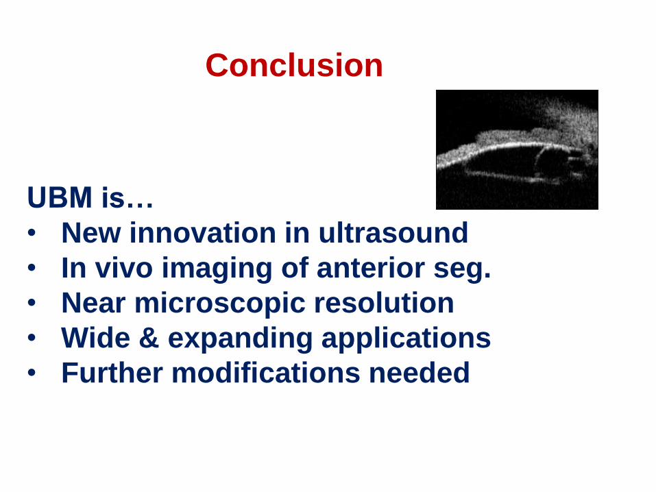
Conclusion
UBM is…
• New innovation in ultrasound
• In vivo imaging of anterior seg.
• Near microscopic resolution
• Wide & expanding applications
• Further modifications needed

Thank you for attention!

BIOMETRIC PARAMETERS OF
ANTERIOR SEGMENT
• а – trabecular meshwork
• б – scleral spur (SS)
• 1 – central anterior chamber depth (CACD; mm);
• 2 – iris root (IR, mm);
• 3–4 – angle opening distance at 250 µm and 500 µm
from scleral spur (AOD 250, 500; mm);
• 5–6– trabecular–ciliary process distance at 250 µm
and 500 µm from scleral spur (TCPD 250, 500; mm);
• 7–8 – iris-ciliary process distance at 250 µm and
500 µm from scleral spur (ICPD 250; 500; mm);
• 9 – posterior chamber depth (PCD, mm);
• 10 – central corneal thickness (CCT; mm);
• 11 – paracentral anterior chamber depth (PaACD; mm);
• 12 – maximum ciliary body thickness (CBTmax; mm);
• 13 – anterior chamber angle (ACA; °).
lens
b
a