Embed Size (px)
Citation preview

From the Division of Biochemistry
Department of Medical Biochemistry and Biophysics
Karolinska Institutet, Stockholm, Sweden
Role of Thioredoxin System in Cell Death
Caused by Toxic Compounds
Xu Zhang
Stockholm 2014

All previously published papers were reproduced with permission from the publisher.
Published by Karolinska Institutet. Printed by [ÅTTA.45 TRYCKERI AB]
© Xu Zhang, 2014
ISBN 978-91-7549-463-0

To my family
献给我的父亲,母亲,先生和即将到来的宝宝


ABSTRACT
Thioredoxin systems, comprising Trx, TrxR and NADPH, are one of the major
disulfide reductase systems, which is crucial in maintaining cellular redox balance in
mammalian cells. TrxR is a selenoprotein with a Sec residue in its C-terminal active
site. The low pKa value and the easily accessible property of the Sec residue make TrxR
a target of many electrophilic compounds, including some clinically approved drugs.
TrxR exert most of its cellular function by reducing Trx. Through the substrates of Trx,
or its interacting proteins, Trx plays important roles in DNA synthesis, cellular defense
against oxidative stress, regulation of transcription factors and cell death pathways.
There are two distinct Trx systems in mammalian cells, Trx1 system located in cytosol
and Trx2 system located in mitochondria. In Paper I we found that treatment with
brilliant green (BG) can cause a dramatic decrease of Trx2 in the mitochondria and
subsequent cell death. The natural amount of Trx2 in Hela cells are much higher
compared to that in fibroblast cells. Down-regulation of the amount of Trx2 by using an
siRNA method in both cell lines can greatly sensitize Hela cells towards BG toxicity,
but not fibroblast cells, suggesting the importance of Trx2 for some cancer cells.
Different from Trx2, which only have two Cys residues in the active site; Trx1 has
three additional Cys residues, Cys62, Cys69 and Cys73. Previous studies about the
function of Trx1 are mainly focused on the active site cysteines. However,
accumulating evidence showed that the three so called structural Cys residues also play
important roles in regulating Trx1´s activities and functions. In paper II and IV, we
focused on studying the impact of the second disulfide (Cys62-Cys69) on Trx1 activity.
We show that Trx1 with two disulfides can be found in cells under high oxidative
stress, and although it is not a substrate of TrxR, but it can be reduced by the
glutaredoxin (Grx) system at the expense of GSH. In addition the formation of the
second disulfide or only the disulfide between Cys62 and Cys69 disturbed the ability of
Trx1 to reduce oxidized Prx1, and sensitized SH-SH5Y cells towards arsenic
compounds inducing cell death.
In Paper III we characterized that GSH plus Grx2 can be a backup of TrxR and can
reduce both Trx1 and Trx2 when TrxR was inhibited. Overexpression of Grx2 in Hela
cells can protect cells from cell death induced by the inhibitors of TrxR.
Apart from Trxs, we also explored the role of TrxR as a target of the clinically applied
anti-cancer drug mitomycin C and mercury. In paper V, we proposed that targeting
TrxR as a new mechanism of mitomycin C´s action. In Paper VI, TrxR was shown to
be a target of mercury, and selenium can reactivated the TrxR treated with mercury by
a substitution mechanism.
In summary, in the thesis we stressed the role of Trx and TrxR in the cell death induced
by the toxic compounds which are targeting the Trx system.
LIST OF PUBLICATIONS
I. Xu Zhang, Yujuan Zheng, Levi E. Fried, Yatao Du, Sergio J. Montano, Allie
Sohn, Benjamin Lefkove, Lars Holmgren, Jack L. Arbiser, Arne Holmgren,
Jun Lu. Disruption of the mitochondrial thioredoxin system as a cell death
mechanism of cationic triphenylmethanes. Free Radical Biology & Medicine
50 (2011) 811-820
II. Yatao Du, Huihui Zhang, Xu Zhang, Jun Lu, Arne Holmgren. Thioredoxin 1
Is Inactivated Due to Oxidation Induced by Peroxiredoxin under Oxidative
Stress and Reactivated by the Glutaredoxin System J. Biol. Chem. 2013,
288:32241-32247
III. Huihui Zhang, Yatao Du, Xu Zhang, Jun Lu, and Arne Holmgren.
Glutaredoxin 2 Reduces Both Thioredoxin 2 and Thioredoxin 1 and Protects
Cells from Apoptosis Induced by Auranofin and 4-Hydroxynonenal.
Antioxidant & Redox Signaling, 2014 Feb. 4. Epub ahead of print.
IV. Xu Zhang, Jun Lu, Yatao Du, Panayiotis V. Ioannou and Arne Holmgren.
Besides Inhibition of Thioredoxin Reductase, Oxidation of the Structural
Cysteine residues in Thioredoxin by Certain Arsenicals Enhance Cytotoxicity
to Cancer Cells. Manuscript
V. Manuel M. Paz, Xu Zhang, Jun Lu, and Arne Holmgren. A New Mechanism
of Action for the Anticancer Drug Mitomycin C: Mechanism-Based Inhibition
of Thioredoxin Reductase. Chemical Research in Toxicology. 2012 Jul 16;
25(7):1502-11
VI. Cristina M. L. Carvahlo, Jun Lu, Xu Zhang, Elias S. J. Arner, and Arne
Holmgren. Effects of selenite and chelating agents on Mammalian thioredoxin
reductase inhibited by mercury: implications for treatment of mercury
poisoning. The FASEB Journal 2011 Jan:25(1):370-81

CONTENTS
1 INTRODUCTION -------------------------------------------------------------------------- 1
1.1 Cell Death Pathways -------------------------------------------------------------------------------------------------------- 1
1.2 Reactive oxygen species --------------------------------------------------------------------------------------------------- 2
1.3 Thioredoxin System --------------------------------------------------------------------------------------------------------- 3 1.3.1 Thioredoxin ------------------------------------------------------------------------------------------------------------ 3
1.3.1.1 Thioredoxin Substrates ------------------------------------------------------------------------------------ 5 1.3.1.2 Transcription Factors regulated by Trx --------------------------------------------------------------------------- 7 1.3.1.3 Proteins binding to Trx ------------------------------------------------------------------------------------- 8 1.3.1.4 Regulation of Trx activity/function in cells ----------------------------------------------------------- 9
1.3.2 Thioredoxin Reductase ------------------------------------------------------------------------------------------- 12 1.3.2.1 Classification ------------------------------------------------------------------------------------------------ 12 1.3.2.2 Catalytic properties of mammalian TrxR ------------------------------------------------------------ 13 1.3.2.3 Isoforms of TrxR -------------------------------------------------------------------------------------------- 14 1.3.2.4 Selenoproteins --------------------------------------------------------------------------------------------- 14 1.3.2.5 Selenocysteine v.s. cysteine ---------------------------------------------------------------------------- 15
1.3.3 Thioredoxin system in cancer ---------------------------------------------------------------------------------- 15
1.4 Glutaredoxin System ----------------------------------------------------------------------------------------------------- 17 1.4.1 Glutaredoxin--------------------------------------------------------------------------------------------------------- 18 1.4.2 Glutathione Reductase ------------------------------------------------------------------------------------------- 19 1.4.3 Glutathione ---------------------------------------------------------------------------------------------------------- 19 1.4.4 Cross-talk between Trx and Grx System --------------------------------------------------------------------- 21
2 AIM OF THE THESIS ------------------------------------------------------------------- 23
3 PRESENT INVESTIGATIONS --------------------------------------------------------- 24
3.1 Methodology ---------------------------------------------------------------------------------------------------------------- 24 3.1.1 Cell Culture----------------------------------------------------------------------------------------------------------- 24 3.1.2 RNA Silencing-------------------------------------------------------------------------------------------------------- 24 3.1.3 Cell Proliferation and Viability Assays ------------------------------------------------------------------------ 24 3.1.4 Measuring TrxR activity using fluorescent method ------------------------------------------------------ 26 3.1.5 Redox Western Blot ----------------------------------------------------------------------------------------------- 26
3.2 Results and Discussions -------------------------------------------------------------------------------------------------- 28 3.2.1 Paper I ----------------------------------------------------------------------------------------------------------------- 28 3.2.2 Paper II ---------------------------------------------------------------------------------------------------------------- 31 3.2.3 Paper III --------------------------------------------------------------------------------------------------------------- 33 3.2.4 Paper IV --------------------------------------------------------------------------------------------------------------- 35 3.2.5 Paper V ---------------------------------------------------------------------------------------------------------------- 38 3.2.6 Paper VI --------------------------------------------------------------------------------------------------------------- 40
3.3 Conclusion and Future Perspectives --------------------------------------------------------------------------------- 44
4 ACKNOWLEDGEMENTS -------------------------------------------------------------- 46
5 REFERENCES ---------------------------------------------------------------------------- 48

LIST OF ABBREVIATIONS
AF Auranofin
AIF Apoptosis inducing factor
AP-1 Activator protein-1
ARE Antioxidant responsive element
ASK1 Apoptosis signaling-regulating kinase 1
BG Brilliant green
BSO Buthionine sulfoximine
Cys Cysteine
dNDPs Deoxyribonucleoside diphosphates
FAD Flavin adenine dinucleotide
GCL Glutamate cysteine Ligase
GPx Glutathione peroxidase
GR Glutathione reductase
Grx Glutaredoxin
GSH Glutathione
NHE 4-hydroxynonenal
Met Methionine
MetSO Methionine sulfoxide
MMC Mitomycin C
Msr Methionine sulfoxide reductase
NADPH Nicotinamide adenine dinucleotide phosphate
NDPs Ribonucleoside diphosphates
NF-κB Nuclear factor-κB
Nox NADPH oxidase
Prx Peroxiredoxin
Ref-1 Redox factor-1
RNR Ribonucleotide reductase
ROS Reactive oxygen species
SOD Superoxide dismutase
TBP2 Thioredoxin binding protein-2 (also known as Txnip)
Trx Thioredoxin
TrxR Thioredoxin reductase
TNF Tumor necrosis factor
Txnip Thioredoxin interacting protein
VDUP1 Vitamin D3 upregulated protein-1 (also known as Txnip)
1
1 INTRODUCTION
1.1 CELL DEATH PATHWAYS
Cells are considered to be dead after they pass the first irreversible phase or the so
called "point-of-no-return"1. There are two major pathways of cell death to be
clarified: apoptosis and necrosis. These two types of cell death pathways are
classified according to the difference in the cell’s morphological and biochemical
properties. Generally from the morphological point of view, apoptotic cells maintain
a functioning membrane throughout the whole process, and the cell shrinks by losing
water; necrotic cells, lose their membrane integrity at a very early stage and then the
cell swells due to the influx of water, sodium and calcium1.
Apoptosis or programmed cell death needs stimulating signals from either inside or
outside of the cells. When the signals are from inside of the cells (the intrinsic
pathway), mitochondria play an important role in this kind of apoptosis. The intrinsic
signal, which can be DNA damage, reactive oxygen species (ROS), as well as
growth-factor depletion, first reaches mitochondria. Then, the proapoptotic members
of the Bcl-2 family proteins are activated and start to form pores in the mitochondrial
outer membrane2, which then can facilitate the release of proapoptotic proteins from
mitochondria, such as cytochrome c and apoptosis inducing factor (AIF). Cytochrome
c can promote the formation of the apoptosome, and subsequently activate the
caspase pathway3.
The apoptotic signal can also come from outside of the cells, in a manner called
extrinsic receptor mediated pathway. The receptors which can receive such stimuli
are belonging to the tumor necrosis factor (TNF) receptor superfamily4. Upon the
stimuli, a complex called Death Inducing Signalling Complex (DISC) is formed in
the membrane and subsequently actives caspase-85,6
. the activated caspase-8 can
either directly active caspase-3 or activate Bid, a proapoptotic member of Bcl-2
family, and initiate the mitochondrial signalling pathways7.
Apoptosis is considered as a natural process which could occur in both physiological
and pathological conditions. It is also a part of the normal maintenance process for
the multicellular lives to remove “unhealthy” cells, for example, cells undergoing
carcinogenesis8. In contrast of apoptosis, necrosis is considered to be an

2
unprogrammed, energy independent and toxic process of cell death. It is worth to
mention, new findings suggest that necrosis, even not the whole process, is also
regulated in some way9,10
.
1.2 REACTIVE OXYGEN SPECIES
Reactive oxygen species (ROS) are the inevitable byproducts of aerobic metabolism,
including a variety of molecules and free radicals derived from molecular oxygen11
. It
was reported that about 2% of electrons leak from the respiratory chain and form
reactive oxygen species11
. There are three major types of ROS: superoxide (O2˙-),
hydrogen peroxide (H2O2) and hydroxyl radical (HO˙). Superoxide is the precursor of
other ROS, and is formed when oxygen gets one electron leaking from respiratory
chain. Superoxide cannot pass through the lipid bilayer membrane of mitochondria.
However, superoxide can be easily converted into hydrogen peroxide by the catalyzing
of superoxide dismutase (SOD)12
. Hydrogen peroxide is not a radical molecule, but it
can pass through membranes and diffuse freely in the cell. Hydrogen peroxide can be
easily converted into highly reactive hydroxyl radical through a Fenton reaction
(Reaction 1); at the same time the metal (Cu+ or Fe
2+) is oxidized. The oxidized metal
can also react with superoxide and convert it into an oxygen molecule (Reaction 2).
The net reaction is called Haber-Weiss reaction (Reaction3). Hydroxyl radical is high
reactive and can react with DNA, lipids, amino acids and carbohydrates13
.
Fe2+
(Cu+) +H2O2 Fe
3+ (Cu
2+) +OH
- + HO˙ (1)
Fe3+
(Cu2+
) + O2˙- Fe
2+ (Cu
+) +O2 (2)
O2˙-+H2O2 HO˙+OH
- +O2 (3)
Besides the respiratory chain, the other sources of ROS are NADPH oxidase (Nox),
glucose oxidase, lipoxygenases, nitric oxide synthase, xanthine oxidase and
flavoprotein reductases14
.
3
1.3 THIOREDOXIN SYSTEM
Thioredoxin system, composing thioredoxin (Trx), thioredoxin reductase (TrxR) and
NADPH, plays an important role in maintaining cellular redox homeostasis and
protecting cells from oxidative stress15
. Trx executes its function by reducing the
disulfide in its target proteins (Reaction 4), or binding to its substrate as a regulator.
Oxidized Trx can then be reduced by TrxR, and NADPH is the ultimate electron donor
for the system16
(Reaction 5). There are two distinct thioredoxin systems in mammalian
cells: Trx1 system mainly in cytosol and nucleus, and Trx2 system in mitochondrial
matrix17
.
Trx-(SH)2 + Protein-S2 Trx-S2 +Protein-(SH) 2 (4)
Trx-S2+NADPH+H+
TrxR Trx-(SH) 2 + NADP+
(5)
1.3.1 Thioredoxin
Thioredoxin was first isolated and characterized by Peter Reichard and co-workers
from E. coli as the electron donor for ribonucleotide reductase (RNR)18
. Trx1 is a 12
kDa globular protein ubiquitously expressed in various species. Besides being an
electron donor for RNR, which is the rate limiting enzyme in the DNA synthesis by
reducing ribonucleotide diphosphates (NDPs) to deoxyribonucleotide
diphosphates(dNDPs)19
, Trx1 can also reduce methionine sulfoxide reductase and
peroxiredoxins in cytosol, thus plays important roles in cellular defense against
oxidative stress and regulating H2O2 signaling20,21
. Trx1 were also found to interact
with several transcription factors such as NF-κB, Ref-1, and p5317,22–24
. The regulation
of its substrates and the transcription factors will be discussed in detail later.
Mitochondrial Trx2 was first cloned and expressed as a 18 kDa mitochondrial protein
with an N-terminal extension of 60 amino acids as a translocation signal25
. Prx3 is the
primary substrate of Trx2 in mitochondrial matrix, which is at the forefront of
defending oxidative stress by eliminating excess hydrogen peroxide (H2O2). In
addition, Trx2 can bind to ASK1 and regulate its function in mitochondria-dependent
apoptosis26,27
. Trx2 is also important in keeping mitochondrial permeability
4
transition2,28
. Both Trx1 and Trx2 possess critical role in cell survival, and the mice
with homozygous knockout of Trx1 or Trx2 both showed early embryonic lethality29,30
.
Both Trx1 and Trx2 belong to thioredoxin fold protein family31
. In both structure, there
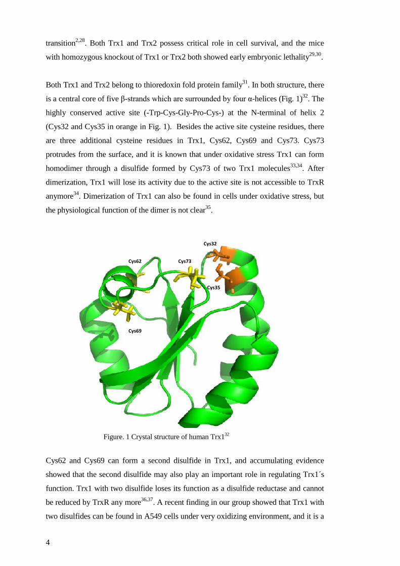
is a central core of five β-strands which are surrounded by four α-helices (Fig. 1)32
. The
highly conserved active site (-Trp-Cys-Gly-Pro-Cys-) at the N-terminal of helix 2
(Cys32 and Cys35 in orange in Fig. 1). Besides the active site cysteine residues, there
are three additional cysteine residues in Trx1, Cys62, Cys69 and Cys73. Cys73
protrudes from the surface, and it is known that under oxidative stress Trx1 can form
homodimer through a disulfide formed by Cys73 of two Trx1 molecules33,34
. After
dimerization, Trx1 will lose its activity due to the active site is not accessible to TrxR
anymore34
. Dimerization of Trx1 can also be found in cells under oxidative stress, but
the physiological function of the dimer is not clear35
.
Cys62 and Cys69 can form a second disulfide in Trx1, and accumulating evidence
showed that the second disulfide may also play an important role in regulating Trx1´s
function. Trx1 with two disulfide loses its function as a disulfide reductase and cannot
be reduced by TrxR any more36,37
. A recent finding in our group showed that Trx1 with
two disulfides can be found in A549 cells under very oxidizing environment, and it is a
Cys73
Cys69
Cys62
Cys32
Cys35
Figure. 1 Crystal structure of human Trx132
5
substrate of Grx system, both in vitro and in cells38
. In our ongoing study (Paper IV),
we found that the Trx1 with two disulfides or with only the disulfide between Cys62
and Cys69 is weakened the efficiency of reducing oxidized Prx1. All these findings
suggested the biological function of the second disulfide in Trx1, although the exact
mechanism is still unclear.
1.3.1.1 Thioredoxin Substrates
1.3.1.1.1 Ribonucleotide Reductase (RNR)
RNR is the rate-limiting enzyme in DNA synthesis and repair by reduction of
ribonucleotides (NTPs) to deoxyribonucleotides (dNTPs)19
. Mammalian RNR is
composed of two subunits: R1 and R2. R1 expresses constantly in the cells, while R2
only present during the S phase, which make it to be the rate limiting factor for enzyme
activity. Each cycle of reduction of NTPs to dNTPs results in a disulfide between the
active site Cys residues in R1 subunit. However, the narrow structure of the active site
in R1 does not allow the access of Trx1 to reduce it directly39
. But two Cys residues
located in the mobile tail of the C-terminal of R1 can be reduced by Trx1 and then
transfer the reducing equivalent to the active site disulfide40
. Evidence from
mutagenesis of E. coli R1 subunit suggest the critical role of C-terminal cysteines in
reducing R1 active site41,42
. Our result using the peptide containing 25 amino acid
residues from C-terminal of R1 subunits also proved that it is the substrate of Trx1
(unpublished data).
Additionally, both Trx and Grx can provide electrons for RNR in mammalian cells, but
through different mechanisms. By using recombinant mouse RNR, Trx1 and Grx2
showed very similar catalytic efficiency, but the Grx activity largely depended on GSH
concentration, which suggest a GSH-mixed disulfide mechanism for Grx instead of the
disulfide exchange mechanism for Trx43
.
1.3.1.1.2 Peroxiredoxin
Peroxiredoxin (Prx) was first identified as a substrate of Trx by Dr. Sue-Goo Rhee and
his group in 199444
. Besides its ability of removing access H2O2 to protect cells from
oxidative stress, Prx is also proposed to serve the function to control the redox signaling

6
through sensing and regulating H2O245–47
, which was justified as a messenger molecule
more than just evil oxidant. There are six Prxs in mammalian cells (Prx1 to Prx6), and
they are classified into three groups: Prx1 to Prx4, which all possess an active site
comprising conserved N-terminal cysteine and C-terminal cysteine, are classified into
the 2-Cys subfamily; Prx5, also known as atypical 2-Cys Prx, which have very similar
N-terminal cysteine sequence compare to the 2-Cys Prxs, but lack the C-terminal
conserved sequence containing cysteine residue48
. The Cys151 serves the role as the C-
terminal cysteine in Prx5. Prx6, which only have one cysteine residue, is termed as 1-
Cys Prx49
.
The typical 2-Cys Prxs are present as homodimers in mammalian cells. The Cys-SH in
the N-terminal of one subunit is very sensitive to H2O2 and can be easily oxidized into
Cys-SOH, which then reacts with the C-terminal Cys-SH of another subunit to generate
an intermolecular disulfide. Then the disulfide can be reduced by Trxs. All four
members of 2-Cys Prxs are substrates of Trxs50
. In Prx5, which also exists in a dimer
form, the N-terminal Cys residue is oxidized into Cys-SOH, which then react with the
Cys151 in the same subunit and forms an intramolecular disulfide48,51
. Prx5 can also be
specifically reduced by Trx. Prx6, however, is not a substrate of Trx, because it cannot
form disulfide upon oxidation52
.
1.3.1.1.3 Methionine Sulfoxide reductase
Both free methionine (Met) and methionine in proteins are easy to be oxidized into
methionine sulfoxide (MetSO) under mild oxidative condition. According to different
asymmetric forms, the oxidation of Met can result in either Met-(S)-SO or Met-(R)-SO.
The oxidation of Met into MetSO affects the protein functions53
. The methionine
sulfoxide reductase (Msr) can reduce both the free and protein-bound MetSO residues.
According to their different substrate specificities, there are two types of Msrs: MsrA
which can reduce the Met-(S)-SO; and MsrB which reduces Met-(R)-SO54,55
.
The catalytic mechanism of Msrs is quite similar to atypical 2-Cys Prx. First, the
“catalytic” Cys residues interact with the sulfoxide group of Met and form a sulfenic
acid intermediate. Then the second Cys residue referred as “recycling” Cys residue
comes into play and results in the formation of an intramolecular disulfide between the
two Cys residues, which can then be reduced by Trx54
.

7
1.3.1.2 Transcription Factors regulated by Trx
The regulation of many transcription factors by Trx mainly depends on the activity of
Trx to reduce the cysteine residues in the DNA binding domain of these transcription
factors, directly or indirectly.
1.3.1.2.1 Nuclear Factor –κB (NF-κB)
Various oxidative stresses, such as UV and H2O2, and some toxic compounds such as
cigarette smoke and lysophosphatidic acid can induce the activation of NF-κB56,57
,
which controls several inflammation genes. Under normal condition, NF-κB binds with
IκB as an inactive complex in cytosol. Under oxidative stress or other type of stimuli,
the IκB is phosphorylated by IκB kinase. Upon the phosphorylation, IκB is degraded
and NF-κB is released to be free. The free NF-κB can then translocate from cytosol to
nucleus to exert its function as a transcription factor58–60
.
In nucleus, binding of DNA requires the reduced Cys62 in its p50 subunit. The
reduction of Cys62 requires reduced Trx, on the other hand, oxidized Trx inhibits the
binding of NF-κB to DNA23,61
. In the cytoplasm, however, Trx plays distinct roles in
regulation of NF-κB activation by preventing the dissociation and degradation of IκB
from NF-κB62
.
1.3.1.2.2 Redox Factor -1 (Ref-1) and Activator Protein – 1(AP-1)
AP-1 controls the transcript of many gene involving in cell growth. The DNA binding
activity of AP-1 is dependent on Trx’s activity; however, Trx1 does not directly interact
with the Cys residues in Ref-1, but exert its reducing power through a redox factor
called Ref-1. In vitro experiments showed that Trx1 can form a heterodimer with Ref-1
through its active site Cys-32. Upon the dissociation of Ref-1 and Trx1, Ref-1 can
reduce Cys residues in the DNA binding domain of AP-1 (Fos and Jun subunits)63,64
.
8
1.3.1.2.3 Tumor Suppressor p53
After activated by series of stress signals, such as oxidative stress, hypoxia, DNA
damage agents, etc., p53 controls the transcription of proteins related to cell cycle
arrest, DNA repair, and apoptosis. The mutation of p53 will result in the loss of control
of above mentioned cellular process, and leads to tumor genesis. P53 mutations are
observed frequently in various types of human cancers65,66
. The sequence-specific
DNA binding ability relies on the reduction of the Cys residues in the DNA binding
domain of p5367
. Although there is no evidence showing the direct binding of p53 with
Trx, many studies showed that Trx can enhance the DNA biding activity of p53 by
itself, or through the activation of Ref-122,68,69
. In addition, deletion of TrxR in yeast
strongly impaired p53 activity70
. In mammalian cells, several electrophilic compounds
which can damage TrxR activity also showed disruption of the function of p5371
.
1.3.1.3 Proteins binding to Trx
Besides above mentioned enzymatic activities, there are several proteins are known to
bind to Trx. Two of the well-studied ones are Apoptosis Signal-regulating Kinase 1
(ASK1) and thioredoxin interacting protein (Txnip).
1.3.1.3.1 Apoptosis Signaling Kinase -1 (ASK1)
ASK1 is a mitogen-activated protein kinase kinase kinase (MAPKKK). ASK1 first can
activate MAP kinase kinase, which then activates two apoptosis pathways: c-Jun N-
terminal kinase (JNK) and p38 MAP kinases pathways72
. Trx inhibits the activation of
ASK1 by directly binding to it and disturbing its homo-oligomerization73
. The binding
of Trx and ASK1 requires the involvement of at least one active site Cys residue
(Cys32 or Cys35). A double mutant Trx1(Cys32S, Cys35S) cannot bind to ASK174
.
The binding of Trx also promotes the ubiquitination and degradation of ASK174
.
Besides Trx1, several studies have shown that Trx2 in mitochondria can also interact
with ASK1, and inhibits its translocation and activation26,75,76
.

9
1.3.1.3.2 Thioredoxin interacting protein (Txnip)
Txnip, which was first found in HL-60 leukemia cells as a vitamin D3 up-regulated
protein-1 (VDUP1)in 199577
. In 1999, Txnip was rediscovered as thioredoxin binding
protein in a yeast two-hybrid system and termed as thioredoxin binding protein-2
(TBP2)78
. The binding of Txnip with Trx is through formation of an intermolecular
disulfide bond between Cys32 from Trx and Cys247 from Txnip79,80
. The formation of
this disulfide is redox-dependent, in order to archiving the binding, a reduced Trx and
an oxidized Txnip are needed79
. The binding of Txnip cause reduced Trx activity as a
disulfide reductase, which results in elevated level of ROS in cells81,82
. Txnip can also
alter Trx's function as a competitive inhibitor and disturbs the interaction between Trx
and its targeting proteins, such ASK127,82
. Recently, the structure of the complex of Trx
and Txnip has been determined. The structure confirmed the disulfide formation
between Txnip Cys247 and Trx Cys32, in addition, a disulfide bond switching
mechanism was proposed to explain the structural rearrangement in Txnip80
.
1.3.1.4 Regulation of Trx activity/function in cells
The regulation of Trx activity/function can happen in different levels, including:
expression, post-translational modifications and protein-protein interaction. Txnip is a
well-known thioredoxin binding protein has its implications in regulating Trx activity
and interaction with other proteins (see above).
1.3.1.4.1 Expression
Various stress, such as H2O2, O2, hypoxia, UV, X-ray, etc.83–86
and treatments of certain
drugs such as arsenic trioxide and suberoylanilide hydroxamic acid (SAHA) 35,87
can
increase the expression of Trx1 in mammalian cells. After analyzing the promoter
region of human TXN1 gene, there are three types of stress-response elements were
found in the promoter region, including: antioxidant responsive elements (ARE),
oxidative response element and heat shock responsive element88–90
. These findings can
explain the induction of Trx1 expression under multiple stresses.
Under some conditions, such as hypertension, and the treatment of certain compounds
such as cathepsin D, the amount of Trx is reported to be decreased may be due to the
10
impaired induction of Trx1 and the degradation of damaged protein during oxidative
stress91,92
. It is interesting to point out, for the treatment with the same drug, SAHA,
which is the histone deacetylase inhibitor, contradictory results were given by different
studies. In some study, expression of Trx1 was found to be positively regulated87
, while
in another study, expression of Trx1 was shown to be negatively regulated93
. These
difference may due to the differences between cell types, the amount of the treatment,
and maybe some other factors.
1.3.1.4.2 Post-translational modifications
Oxidative post-translational modifications of the Cys residues in Trx1 were
investigated extensively, three types of modifications are the center of interest:
oxidative modification of the Cys residues (through disulfide formation), S-
glutathionylation and S-nitrosylation. The modification of each Cys residue is briefly
summarized in Table 1.
There are five Cys residues in human Trx1, two of them are involved in the active site
(Cys32 and Cys35) which are required for the activity of Trx and the interaction of
Trx1 with its interacting proteins74,79
. The rest three Cys residues, as known as
structural cysteines, first caught the attention because they cause the aggregation and
loss of activity in Trx194
. Although until now their physiological functions are still not
clear, but accumulating evidence suggesting these cysteine residues may also involve in
the redox signaling. Homodimerization of Trx1 is through the disulfide formed
between two Cys73 residues from each molecule33
. Trx1 dimer does not have any
reduce activity because the active site was not accessible for TrxR37,95
. Under oxidative
environment, an extra disulfide can form between Cys62 and Cys69. Trx1 with only the
disulfide between Cys62 and Cys69 can be reduced by TrxR because the disulfide can
be transferred to the active site. Trx1 with two disulfide is inactive and cannot be
reduced by TrxR16,96
, but is a substrate of Grx system97
. Nevertheless, the formation of
the second disulfide in Trx1 may provide a redox mechanism regulating its function
and earns more time for the redox signaling transduction38,96
.
S-glutathionylation is a reversible oxidative post-modification. Both in vitro and in cells
study showed that Cys73 can be glutahionylated under oxidative stress. The
glutathionylated protein is inactivated, but the activity can be regained automatically

11
when incubated with TrxR and NADPH, suggesting the glutathionylation process is
reversible98
. Although glutathionylation can happen during physiological conditions,
Trx1 can only be glutathionylated in oxidative stress environment. Thus, the
glutathionylation may play an protection role against irreversible oxidative
modifications, such as dimer formation which leads to loss of activity99
.
Table1. Post-translational modifications of Cys residues in Trx1
Haendeler et al found that Trx1 can also be S-nitrosylation in 2002. They claimed that
under physiological condition, Cys69 can be nitrosylated, and the nitrosylation of
Cys69 stimulates Trx1’s function in redox regulation and anti-apoptosis100
. However,
several following studies gave controversial results. In one study, the S-nitrosylation of
Cys69 was detected and no S-nitrosylation of Cys73 was detected, in addition a
disulfide between the active site Cys residues was observed101
. In a structure study,
Cys
residue Post-Translational modifications Change of Trx1's activity / function
Cys32 disulfide formation Loss of activity but can be reduced
by TrxR Cys35 disulfide formation
Cys62 extra-disulfide formation Loss of activity and cannot be
reduced by TrxR
S-nitrosylation
Cys69 extra-disulfide formation Loss of activity and cannot be
reduced by TrxR
S-nitrosylation Stimulating Trx’s activity and anti-
apoptotic function
Cys73 homodimerization loss of activity
S-glutathionylation loss of activity, but reversible
S-nitrosylation Trx can act as a trans-nitrosylation
agent

12
after treated with GSNO, both Cys69 and Cys62 were found to be nitrosylated. There is
no nitrosylation on Cys73, and a disulfide bond was formed in the active site102
. A
study from our group suggested that the different results obtained from these studies
maybe because the redox state of Trx1 was difference when the experiments were
performed. Treatment of GSNO resulted in nitrosylation of Cys69 and cys73 and an
additional disulfide in the active site in fully reduced Trx1. Treating Trx1 with two
disulfides under the same condition resulted in the nitrosylation of Cys73, which can
act as a trans-nitrosylation agents, and cause the nitrosylation of caspase-3 and
subsequently inhibit apoptosis101,103
.
1.3.2 Thioredoxin Reductase
1.3.2.1 Classification
Thioredoxin reductase (TrxR) belongs to the family of dimeric flavoenzymes that
catalyze the electron transfer from pyridine nucleotides to their substrates through
flavin adenine dinucleotide (FAD) and the active site with redox active cysteine
residues 104
. It is important to mention that there are two types of TrxRs which are
classified according to the difference in the molecular weight of the subunit. The small
TrxR or the low Mr type TrxR, comprising of two identical 35 kD subunits, which
mainly exist in lower organisms such as bacteria, fungi, yeast and plants. The large
TrxR or the high Mr type TrxR, comprising of two identical 55-60 kD subunits, which
can be found in higher organism such as mammalians104,105
. Besides the difference in
the molecular weight, their catalytic mechanisms are also different.
E. coli TrxR is a well-studied small TrxR. Here we use it as an example to discuss the
catalytic mechanism of small TrxR. In each cycle of the reaction, there is an unique
rotation of the pyridine nucleotide-binding domain by 67 degree106
. The structure
before rotation facilitate the reduction of FAD by NADPH107
, while the structure after
the rotation allows the transfer of electrons from reduced FAD to the active site
disulfide108
.
Different from the small TrxR, the large TrxR comprises three functional domains in
each subunit: FAD domain, NADPH domain and interface domain (Fig. 2). In each

13
subunit, there are also two redox active sites: one located at the C-terminal active
mobile tail, the other one is close to the FAD.
The redox active site of the C-terminal tail is different from species to species. In some
species, such as plasmodium falciparum (the malaria parasite) and Drosophila
Melanogaster (fruit fly), the C-terminal redox active site is composed of two Cys
residues; from C.elegans, the selenocysteine (Sec) replaces the last cysteine in the C-
terminal tail. Compared to Cys, Sec has a lower pKa value which makes it more active.
The C-terminal tail is highly flexible and exposed on the surface of the enzyme upon
reduction109
.
1.3.2.2 Catalytic properties of mammalian TrxR
As shown in Fig. 2, the two TrxR monomers were arranged into a head-to-tail dimer.
The two redox active sites are close to each other in space, although one of them is
located in the FAD binding domain (Cys59 and Cys64), while the other is located in
the C-terminal of the interface domain (Cys497 and Sec498). In each catalytic cycle,
the fully oxidized TrxR is first reduced by NADPH and the FAD gains two electrons
upon the reduction; then the reduced FAD transfers one electron to Cys59 and shares
the other one with Cys64; after the stable charge transfer complex is formed in N-
terminal active site, it can reduce the C-terminal of the other subunits; this reaction
results in the reduced C-terminal selenothiol and a disulfide in the N-terminal active
site; FAD can again reduce the N-terminal disulfide through transferring electrons from
NADPH109,110
.
N-terminal CVNVGC
active site
FAD domain
NADPH domain
Interface domain
C-terminal GCUG tail
Figure 2. Schematic overview of TrxR structure.

14
1.3.2.3 Isoforms of TrxR
In mammalian cells, there are three separate genes encoding three isoforms of TrxR:
TrxR1, which is mainly present in cytosol; TrxR2, which is only present in the matrix
of mitochondria; and thioredoxin glutathione reductase (TGR), which has a Grx
domain in the N-terminal sequence, and mainly expresses in testes. TrxR1 and TrxR2
have similar kinetic properties111
, whereas TGR can reduce both Trx and GSSG, and
also has a low Grx activity. But the Grx activity of TGR is not GSH-dependent since
the C-terminal Sec residue may be able to transfer electrons to the Grx domain112,113
.
TGR can catalyze the formation of intermolecular disulfide bonds and protein
isomerization, indicating its role in protein folding. In testes, TGR and its target protein
GPx4 can serve as a disulfide bond formation system, including proteins that form the
structural components of the sperm, suggesting its role in sperm maturation114
.
Besides these three isoforms of TrxR, there are many splicing variants of TrxR1 and
TrxR2. For example, five different cDNA isoform of TrxR1 have been discovered
which all have alternative N-terminal domains115,116
. The biological functions of these
variants are still under investigation.
1.3.2.4 Selenoproteins
Selenium is an essential trace element for human, and deficiency of selenium in dietary
uptake can result in loss of immune function, weakened reproduction, depression and
cardiovascular diseases117
. One important application of selenium by mammals is the
synthesis of selenoproteins. Until today, there are about 25 selenoproteins that have
been identified in human by searching the mammalian genomes118
. Most of these
selenoproteins are found to be antioxidant enzymes, such as TrxR1 and TrxR2,
methionine-R-sulfoxide reductase B1, GPx1 to 4 and 6, SelK, SelW and SelR. Other
important selenoproteins are selenoprotein P (SelP) which functions in the Sec residue
transport and storage due to the highly toxicity of free Sec residue; selenophosphate
synthetase 2 (SPS2) functions in Sec synthesis, etc. The functions of some
selenoproteins are still unknown.

15
The Sec is encoded by a UGA codon, which is in most cases recognized as a stop
codon. In order to correctly recognize the codon and insert the Sec into the right
position, a complex regulatory mechanism has been developed in mammalian cells.
First of all, a secondary RNA structure called the SECIS element is used in the 3'UTR
of the RNA message. In addition, a special translation machinery is required as well,
including: a Sec specific tRNA, the specific elongation factor EFSec, SECIS binding
protein 2 (SBP2), ribosomal L30 protein, and many other factors including some may
be undiscovered factors119
.
1.3.2.5 Selenocysteine v.s. cysteine
Compared to Cys residues, the only difference of a Sec residue is the replacement of -
SH by -SeH (Fig. 3). However, the insertion of one Sec is very expensive for cells,
which suggests the irreplaceable advantages of Sec over Cys residue. Upon the
replacement of sulfur by selenium, the pKa value of the residue is changed from 8.3 in
Cys to 5.2 in Sec. This change in the pKa value result in an almost completely
deprotonated selenolate at the physiological pH, which is more reactive comparing to
thiol120
. Actually under selenium deficiency condition, the Sec residue in TrxR was
found to be replaced by a Cys residue in rat liver, resulted in a 10 fold decrease in TrxR
activity121
.
Figure 3. Chemical structure of Selenocysteine and cysteine.
1.3.3 Thioredoxin system in cancer
The ROS level in cancer cells was shown to be elevated compared to normal cells,
which may be due to defects in the respiratory chain and disturbed redox balance122
. A
moderate elevated ROS is preferred by cancer cells because it can promote tumor
16
growth and survival by stimulating the expression of enzymes, such as mitogen-
activated protein kinase (MAPK) and extracellular signal-regulated kinase (ERK) and
cyclin D123
. Trx and TrxR were also found to be overexpressed in many cancer cells,
and they play important roles in cancer cell death24,124
. Targeting Trx and/or TrxR can
induce a massive increase in ROS level, and induces cell death through multiple
pathways, such as activation of ASK1, translocation of cytochrome c from
mitochondria to cytosol and induction of p53 expression, etc27,29,125
.These studies
suggest the cellular redox pathways, such as Trx system, can be a promising target in
anticancer therapy.
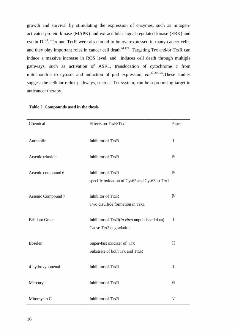
Table 2. Compounds used in the thesis
Chemical Effects on TrxR/Trx Paper
Auranofin Inhibitor of TrxR Ⅲ
Arsenic trioxide Inhibitor of TrxR Ⅳ
Arsenic compound 6 Inhibitor of TrxR Ⅳ
specific oxidation of Cys62 and Cys63 in Trx1
Arsenic Compound 7 Inhibitor of TrxR Ⅳ
Two disulfide formation in Trx1
Brilliant Green Inhibitor of TrxR(in vitro unpublished data) Ⅰ
Cause Trx2 degradation
Ebselen Super-fast oxidizer of Trx Ⅱ
Substrate of both Trx and TrxR
4-hydroxynonenal Inhibitor of TrxR Ⅲ
Mercury Inhibitor of TrxR Ⅵ
Mitomycin C Inhibitor of TrxR Ⅴ

17
TrxR emerges as a new anti-cancer therapeutic target, and has a long list of inhibitors.
The selenocysteine in the C-terminal active site is found to be a primary target of
several electrophilic compounds, such as are anticancer drugs such as cisplatin,
mitomycin C, doxorubicin, etc.126–128
Several anticancer compounds, or clinically
applied drugs have also shown the ability of interfere the expression level of
thioredoxin or change its redox state87,91,129
. Table 2 exhibits the compounds studied in
this thesis.
1.4 GLUTAREDOXIN SYSTEM
Besides Trx system, glutaredoxin system composed of glutaredoxin (Grx), glutathione
reductase (GR), glutathione (GSH) and NADPH is another major cellular protein
disulfide reductase system. GSH is the most abundant small thiol-containing molecules
in cells, which concentration can reach up to 10-15 millimolar levels. The reaction of
protein-disulfide reduction catalyzed by Grx system has two distinct mechanisms. One
is the thiol/disulfide exchange mechanism, which is similar to Trx system, called dithiol
mechanism. The other one is the so called mono-thiol mechanism, in which only the N-
terminal active site Cys residue is involved.
In a dithiol mechanism, Grx with two free thiols in the active site first reduces the
disulfide in the substrate through a thiol-disulfide exchange mechanism, and results in a
disulfide formation in the active site (Reaction 6). The disulfide in Grx's active site can
then be reduced by gaining electrons from two molecule of GSH, results in the
formation of one GSSG (Reaction 7). Finally, GSSH is reduced to two GSH by GR
using electrons from NADPH (Reaction 8).
Grx-(SH)2 + Protein-S2 Grx-S2 + Protein-(SH)2 (6)
Grx-S2 + 2 GSH Grx-(SH)2 + GSSG (7)
GSSG + NADPH + H+
GR 2GSH + NADP+ (8)

18
The monothiol mechanism is actually a deglutathionylation reaction130
. First, GSH can
form a mix-disulfide with protein thiol, and then reduced Grx can form a new mixed-
disulfide between its N-terminal active site Cys residue and the GSH, and transfer the
electron the protein thiol, which is reduced (Reaction 9). The resulting mixed-disulfide
is subsequently reduced by another molecule of GSH, results in the fully reduced Grx
and a GSSG (Reaction 10); this is the rate limiting step of the whole reaction131
.
Finally, GSSG is reduced in the same way as reaction (8).
Grx-(SH)2 + Protein-S-SG (N-terminal)GS-S-Grx-SH (c-terminal) + Protein-SH (9)
GS-S-Grx-SH +GSH Grx-(SH)2 +GSSG (10)
1.4.1 Glutaredoxin
Grx was first discovered in 1976 by Arne Holmgren as a GSH dependent electron
donor for RNR in E. coli lacking Trx system132
. Grx is a Trx fold family protein with
conserved active site sequence -Cys-X-X-Cys-133
. There are three different isoforms of
Grxs in mammalian cell: Grx1 is located in cytosol, Grx2 is located in mitochondrial,
and Grx5, which is a monothiol isoform and may be also target mitochondria134
.
Cytosolic Grx1 is about the same size of Trx1 (~12 kDa), which structure comprises a
Trx fold with a central core of four β-sheets surrounding with five α-helixes. The
conserved active site (-Cys-Pro-Tyr-Cys-) locates in the N-terminal part of helix 2 in
mammalian Grx1135
. The structure of reduced and oxidized Grx2 are very similar, only
slightly changes around the N-terminal active site area136
. The structure of E. coli Grx1
revealed a GSH binding site and a mixed-disulfide with GSH which can explain the
high specificity and affinity of Grx to GSH137
. In addition, Grx also has a hydrophobic
surface area around the active site, which facilitates the interaction of Grx with its
substrate136
. The same as Trx1, Grx1 can also translocate into nucleus upon oxidative
stress, where it may exert its role in regulating transcription factors.
Human Grx2 has two isoforms, Grx2a and Grx2c. Grx2a is located in mitochondria,
while Grx2c is localized in nucleus, and cytosol of some tumor cells138
. Under reducing
condition, two molecule of Grx2 and two GSH form heterodimer through iron-sulfur

19
cluster139
. The dimeric Grx2 is enzymatically inactive. But upon oxidative stress, Grx2
is dissociated and activated. This property of Grx2 suggests its role as a redox sensor,
and the importance of it as a backup reductase under oxidative stress140
. Compared to
Grx1, Grx2 has a higher affinity to the S-glutathionylated protein substrates with lower
turnover rates141
. Grx2 can catalyze the reversible oxidation and glutathionylation of
mitochondrial membrane thiol proteins, and plays important role in mitochondrial
redox signalling and oxidative stress defense.142
.
Grx5 is named because it is a homologue of yeast Grx5. Grx5 is also located in
mitochondrial but with only one Cys residue in its active site, and so far no redox
activity has been reported134
. Its role in mammalian cells is not clear yet , but in yeast
Grx5 is found to be involved in the synthesis of iron-sulfur clusters and regulation of
the activity of iron-sulfur enzymes143
.
1.4.2 Glutathione Reductase
GR is a flavoenzyme responsible for maintaining the reduced GSH pool in cells. The
same as TrxR, GR is also belonged to the pyridine nucleotide disulfide oxidoreductase
family. The active form of enzyme is a dimer with two identical subunit arranged into a
"head to tail" pattern. Each subunit of GR also contains three domains: FAD binding
domain, NADPH domain and interface domain144
. The redox active site in the N-
terminal (-Cys-Val-Asn-Val-Gly-Cys-) is homology to TrxR and is conserved in many
species. When reducing GSSG, two electrons from NADPH was first transferred to
FAD, and then transferred to the active site. Finally, GR reduces GSSG in a
thiol/disulfide exchange manner. There is only one gene in mammalian cells encoding
GR, although there are GR presenting both in cytosol and in mitochondrial. GR from
different subcellular compartments has the same biological and chemistry properties145
1.4.3 Glutathione
GSH is a tripeptide (L-γ-glutamyl-L-cysteinylglycine), and is the most abundant thiol-
based antioxidant in mammalian cells. The ratio between reduced GSH and oxidized
GSSG (GSH/GSSG) is used as the indicator of cellular redox state. In a physiological
state, the ratio can be above ten in the cytoplasm. The synthesis of GSH is a two steps
ATP dependent reactions happens in cytosol: first is the rate limiting step of the
20
synthesis, γ-glutamyl-cysteine is synthesized from L-glutamate and cysteine through
the catalysis of γ-glutamyl-cysteine synthetase (also called glutamate cysteine ligase,
GCL)146
. Then a glycine was added into the dipeptide by the catalysis of glutathione
synthetase. The synthesis of GSH is regulated by many factors, such as oxidative stress
which can enhance the activity of GCL by increase the holoenzyme formation; the
availability of L-cysteine; and regulation of GCL and glutathione synthetase at
transcriptional level147
.
After GSH is produced in cytosol, it can be transported into different subcellular
compartments, providing different redox environment in different organelles in a cell.
For example, GSH can be transported into mitochondria through both a carrier
mediated pathway and diffusion148,149
. Mitochondria contain a higher amount of GSH
compare to cytosol or nucleus, and the pool of GSH in mitochondria has a longer half-
time. When using buthionine sulfoximine (BSO), which is an inhibitor of GCL, to
deplete cellular GSH, the GSH in mitochondria is much more resistant compared to
cytosol150,151
. These results suggest that GSH in mitochondria protects cells from
oxidative stress when the pool in cytosol is low or oxidized. Another example is that in
endoplasmic reticulum (ER), where the environment is much more oxidized than other
parts of the cell, the ratio of GSH/GSSG is about 3:1. The oxidizing environment in ER
can facilitate protein folding152
. Besides the transportation within cell, GSH can also be
transported outside cells, but mostly in oxidized form (GSSG). The extracellular GSH
is much lower than intracellular, for example in plasma there is only about 2-20µM
GSH, and the ratio between reduced GSH and oxidized GSSG in plasma decreases by
aging, especially after 45 years of age153,154
.
Besides its role in protecting cells from oxidative stress, GSH participates in many
cellular processes through S-glutathionylation, an important post-translational
modification of proteins. Via S-glutathionylation, the target protein can either be
activated or inactivated155
. A large group of proteins can be regulated through
glutathionylation, and involve in many important physiological pathways such as cell
metabolism, growth and differentiation. Some transcription factors, such as AP-1 and
NF-κB can also be glutathionylated and loss their ability of binding to DNA156–158
. As
mentioned before, Cys73 in Trx1 can also be glutathionylated, this modification may
be able to protect protein being irreversibly oxidized, in the case of Trx1, forming
inactivated dimer through Cys7398,159
.

21
In addition, GSH is implicated in the metabolism and detoxification of several toxic
compounds. For example, after entering cells, pentavalent arsenic needs to be reduced
in to trivalent arsenic in a GSH-dependent pathway, and then conjugated with GSH in
order to be exported out of cells160
.
1.4.4 Cross-talk between Trx and Grx System
In mammalian cells, glutaredoxin system has some functions overlapping with
thioredoxin systems. For example, both systems are electron donors for RNR with
similar catalytic efficiency. With the present of 4 mM GSH, Grx1 showed a higher
affinity compare to Trx1, but the later has a higher turnover number in vitro.
Meanwhile, the catalytic activity of Grx system was strongly dependent on the
concentration of GSH, and the catalytic reaction is more likely to follow the monothiol
mechanism, which can be distinguished from the dithiol mechanism of Trx43
. Other
overlapping functions including their role in protecting cells from apoptosis, Grx1 can
also negatively regulate ASK1’s activity, though the binding site is different from
Trx1161
. Grxs are also reported to be able to regulate some transcription factors, such as
NF-κB and nuclear factor I (NFI)162,163
.
Although there is much overlap in their functions, they cannot fully substitute for each
other due to their difference in the selection of substrate groups and different reaction
mechanism. Generally, Trxs reduce the disulfide in its substrates through a
thiol/disulfide exchange mechanism, which is similar to the dithiol mechanism of Grxs.
Prxs are specific substrates of Trx system, which is in the forefront of defending
oxidative stress by elimination excess H2O245
. Grx system can exert its anti-oxidant
function by providing electrons to glutathione peroxidases (GPxs), a family of enzymes
which are also involved in balancing H2O2 homeostasis and multiple cellular signaling
pathways including carcinogenesis, apoptosis, etc.164
. In addition, Grxs can catalyze the
reversible deglutathionylation reaction of its substrates through the so called monothiol
mechanism as described previously in “Glutaredoxin” section.
Apart from the similarity and difference of their substrates and functions, Trx and Grx
system can back up each other when the enzyme in one system does not function. Grx2
can be reduced by both GR and TrxR141
. Trx can reduce GSSG in GR-deficient cells165
.
22
A study from our group showed that Grx system could work as a backup for TrxR and
keep Trx1 in the reduced condition when TrxR1 was inhibited166
. Moreover, the
heavily oxidized Trx1 with two disulfides cannot be reduced by TrxR but can be
reduced by Grx system at the expense of GSH97
. Besides the finding in the cytosol, we
also found that Grx2 is a very good back up for TrxR2 in mitochondria, and keep Trx2
in the reducing state when TrxR2 was inhibited. In Hela cells, which also express
Grx2c in the cytosol, Grx2c can also protect Trx1 from being oxidized when TrxR1 is
inhibited167
. Under reducing environment, Grx2 is inactive and stored in dimer form
together with two GSH through iron-sulfur cluster139
. Upon oxidative stress, Grx2 will
be released and activated to exert its anti-oxidant function. This fact together with its
property of less sensitive to oxidative stress damages103
, make Grx2 very fit for being a
backup for TrxRs.

23
2 AIM OF THE THESIS
As indicated in the title, our primary goal was to explore the role of Trx and TrxR in
cell death caused by toxic compounds, as well as to investigate the potential possibility
of Trx and TrxR as targets for anticancer therapy.
Specifically, in Paper I - IV we focused on studying the role Trxs in cell death and the
properties of the protein.
To investigate the role of Trx2 in some cationic triphenylmethanes causing
cancer cell death.
To characterize if Trx1 with two disulfides is a substrate of the Grx system and
its possible role in redox signaling
To characterize Grx2 as a backup system for both Trx1 and Trx2.
To investigate if the oxidation of Trx1 can enhance the cytotoxicity of some
arsenic compounds, and the role of Cys62 and Cys69 in regulating the function
of Trx1
In Paper V and Paper VI, we focused on the inhibition of TrxR by toxic compounds or
clinically applied anticancer drug.
To investigate targeting TrxR as a new anticancer mechanism of mitomycin C.
To study targeting TrxR as a new mechanism of mercury toxicity, and the role
of selenium in recovery of the activity of TrxR inhibited by mercury.

24
3 PRESENT INVESTIGATIONS
3.1 METHODOLOGY
3.1.1 Cell Culture
For most cell based experiments described in papers I-VI, commercially available
human cell lines from the American Type Culture Collection were used. Fibroblast
cells in paper I was a gift from Dr. Laura Papp, Queensland Institute of Medical
Research, Australian. Hela cell (human cervical carcinoma), A549 (human alveolar
adenocarcinoma epithelial cell), SH-SH5Y (human neuroblastoma) and Du145 (human
prostate cancer) were cultured in Dulbecco's modified Eagle's medium (DMEM) with 1
g/l glucose (VWR). HEK293t (human embryonic kidney cell) and Fibroblasts were
cultured in RPMI 1640 medium with 1 g/l glucose (VWR).
3.1.2 RNA Silencing
The transfection of siRNA in Paper I and Paper III was performed according to the
Dharmacon transfection protocol. Briefly, siRNA transfection reagent (Dharmacon,
Lafayette, Co, USA) and siRNAs (Qiagen, Valencia, CA, US) were diluted in serum
and antibiotic-free medium and left at room temperature for 5min. Then the siRNA
transfection reagent and siRNA were mixed and incubated for 20 min. Complete
medium were added into the mixture to achieve a final concentration of 50 nM siRNA
in the culture medium. After 48 hours incubation, the medium was removed, and the
cells were harvest for analysis or continue incubated with the desired compounds.
3.1.3 Cell Proliferation and Viability Assays
The most commonly used assay for cell proliferation and viability measurement in all
six papers is the MTT assay. Because MTT assay can be affected by the alteration of
mitochondrial metabolism, in order to confirm the results of MTT assay, we also used
trypan blue exclusion and neutral red up take assay to investigate the effects of
compounds we used on cell viability in some of our studies. Observations of the

25
morphology of cells by microscopy were always performed and recorded prior to each
assay.
For MTT assay, cells were plated at a density of 1×10⁴ cells/well in 96-microwell plate
and allowed to grow about 24 hours to get confluence. Then the cells were treated with
appropriate concentrations of compounds in 100 µl fresh medium for the appropriate
time. After treatment, medium containing compounds were replaced by 100 µl of fresh
medium, and 50 µl of MTT solution (1 mg/ml in PBS) was added to each well and
incubated for 3h. Then the medium was carefully removed, and 100 µl of DMSO was
added to each well. Plates were then put on a shaker for about 1 hour until all crystals
were dissolved. Then the cell viabilities were determined by measuring the absorbance
at 550 nm.
Neutral red up take assay was carried out following the previous described protocol168
;
cells were seeded and treated in the same way as in MTT assay. Neutral red working
solution (40 µg/ml in culture medium) was incubated overnight at the same temperature
as the cells, and then centrifuged to remove any precipitated dye crystals. The treatment
medium was then removed and 100 µl of neutral red medium were added into each
well. The plate was incubated for 2 hours at the appropriate culture conditions. Then
the neutral red medium was removed and the cells were washed with 150 µl PBS
carefully. Then 150 µl neutral red destaining solution (50% ethanol 96%, 49%
deionized water, 1% glacial acetic acid) was added into each well. The plate was then
placed on a plate shaker until the neural red has been extracted from the cells. Then the
cell viabilities were determined by measuring the absorbance at 540 nm.
For trypan blue exclusion assay, after the treated cells were collected, cells were
centrifuged and resuspended in PBS. The density of the cells was determined using a
hemocytometer. Then in every 1 ml cell suspension, 0.1 ml of trypan blue stock
solution (0.4% trypan blue in PBS) was added. The numbers of the blue staining cells
were counted right away, and the cells which took up trypan blue were considered non-
viable.
26
3.1.4 Measuring TrxR activity using fluorescent method
Insulin reduction assay and DTNB reduction assay are two most commonly used
method to measure TrxR activity in purified protein or in cell lysates. But in order to
detect TrxR activity in precious biological materials, such as cell lysates from different
subcellular organelles, or cell samples containing a low amount of TrxR, a newly
developed fluorescent method was applied in the studies169
. In the fluorescent method,
insulin was replaced by isothiocyanate-labeled insulin (FiTC-insulin), which emits
fluorescence at 520 nm after excitation at 480 nm. Upon the reduction of the disulfide
by Trx, the fluorescence is increased. Compared to the conventional methods,
fluorescence method is highly sensitive and stable169
.
Generally, in a 96-well black micro titer plates, appropriate amount of cell lysates were
incubated with 20 µM of Trx and 0.25 mM NADPH in assay buffer (0.2mg/ml bovine
serum albumin in 50 mM Tris-Cl and 1 mM EDTA, pH 7.5), the total volume was 90
µl in each well. After incubation 30 min at 37°C, 10 µl of FiTC-insulin was added into
each well. The final concentration of FiTC-insulin in each well is 10 µM. Then the
emission at 520 nm after 480 nm excitation was recorded for 60 min in room
temperature. The rate of the reaction was calculated as the changes of fluorescence
following time. In paper IV, due to the very low amount of TrxR in SH-SH5Y cells, an
improved fluorescent method was used, in which FiTC-insulin was replaced by a new
fluorescent substrate from the latest developed kit (FkTRXR-03-Star) by IMCO
(www.imcocorp.se).
3.1.5 Redox Western Blot
To determine the redox state of Trx1 in cells, the experiment was performed based on
the method described previously38,166
. Cells were washed three times in cold PBS and
then lysed in urea lysis buffer containing iodoacetamide (IAM) (10 mM IAM, 50 mM
Tris-HCL, 1mM EDTA, 8 M urea, and pH 8.3). Then the proteins were precipitated
and washed three times to remove the excess IAM with 1.5 ml of ice-cold acetone/HCl
(98/2, v/v). The precipitate was then resuspended in urea lysis buffer containing 3.5
mM DTT to reduce the disulfides in Trx1. The free thiols of Trx were then alkylated
with 30 mM iodoacetic acid (IAA) in urea lysis buffer. The samples were then

27
separated by PAGE containing 8 M urea and transferred with an Invitrogen transfer
system.
Figure 4. Principle of modified redox western blot (from paper II )
As exhibited in Fig. 4, in 8 M urea, proteins were all denatured and all the thiols were
exposed. When first incubated with IAM, the free thiols in Trx1 were labeled with
IAM, which will not give any extra charge to the protein. After removing excess IAM,
the thiols which are oxidized into disulfide or bind to other proteins were reduced by
3.5 mM DTT, and were labeled by IAA. IAA is negatively charged, so the protein
labeled with more IAA will migrate faster in the urea gel. In the end, Trx1 can be
separated according to the free thiols they contained initially.

28
3.2 RESULTS AND DISCUSSIONS
3.2.1 Paper I
Xu Zhang, Yujuan Zheng, Levi E. Fried, Yatao Du, Sergio J. Montano, Allie Sohn,
Benjamin Lefkove, Lars Holmgren, Jack L. Arbiser, Arne Holmgren, Jun Lu.
Disruption of the mitochondrial thioredoxin system as a cell death mechanism of
cationic triphenylmethanes. Free Radical Biology & Medicine 50 (2011) 811-820.
Trx2 system, comprising Trx2, TrxR2 and NADPH, catalyzes the disulfide reduction in
mitochondrial matrix25
. Prx3 is the main substrate of Trx2 in mitochondria, which can
scavenge H2O2. In addition, Trx2 plays a key role in regulating mitochondria-
dependent apoptosis by binding to ASK1 and Txnip, as well as regulating the
mitochondrial permeability transition2,26–28,170
. However, compared to Trx1 system,
which is found to be over-expressed in many cancer cells and merging as an anticancer
therapeutic target24,129
, the role of Trx2 system in cancer is less clear.
Cationic triphenylmenthane dyes, have been used by human beings for more than a
century as antifungal and antibacterial agents171–173
. Recently, some of them have also
shown potent anticancer activity in mice and humans174
. However the exact mechanism
of these dyes remains unclear.
In this study, we found that Hela cells over expressing Trx2 in mitochondria compared
to normal Fibroblast cells. Hela cells also showed to be more vulnerable upon BG treat
compare to Fibroblasts. Down-regulation of Trx2 in Hela cells by siRNA technique
resulted in a massive increase of cytotoxicity upon BG treatment, whereas, the same
siRNA treatment in Fibroblast cells exhibited no significant change compared to
untreated cells. These results suggest that the viability of Hela cells is more dependent
on Trx2 than that of Fibroblasts.
We then performed fluorescence staining to detect the subcellular location of BG. The
treatment of BG resulted in green fluorescence, which was overlapping with the
MitoTracker red mitochondrial staining. Furthermore, we investigated the cellular
response upon BG treatment in Hela cells and in Fibroblasts. Both Hela and Fibroblast
cells were treated with various concentrations of BG (0.25 to 2μM) for 24 hours. There
was only a marginal increase in Trx1 and TrxR1 protein levels, but no significant
effects on TrxR activity in cell lysates. Interestingly, Trx2 protein levels were
29
dramatically decreased upon the treatment with low concentrations of BG (0.25 to 1.0
μM), both in Hela and in Fibroblast cells. Upon the treatment with 2 μM BG, the
degradation of Trx2 was attenuated. RNR R1 protein level was not affected by BG
treatment, whereas, RNR R2 protein, which is only expressed in S phase175
, almost
disappeared upon 1.0 μM and 2.0 μM BG treatment. We applied a redox western blot
method, which can detect the redox state of all the thiols in Trx1 or Trx2, to analyze the
redox state of Trx1 and Trx2 as a result of BG treatment. Without any treatment, most
Trx1 and Trx2 were in the fully reduced form. Upon 24 hours BG treatment, the
reduced form of Trx1 or Trx2 decreased dramatically. By isolation of different cell
organelles, we detected that the loss of Trx2 resulted in translocation of cytochrome c
from mitochondrial to cytosol and shuffling of Trx1 from cytosol to nucleus.
In order to study how Trx2 is degraded upon treatment with BG, we used MG132,
which is an inhibitor of both proteome and mitochondrial Lon protease, to treat Hela
cells together with BG. The presence of MG132 can prevent the Trx2 degradation to
some degree. We also found that upon the BG treatment, Lon protease mRNA was
elevated. Thus we propose that mitochondrial Lon protease may mediate the Trx2
degradation process.
Discussion
The Trx1 and Trx2 system locate and exert their specific functions in different
subcellular compartments. Trx1 is mainly located in cytosol. Trx1 is an electron donor
for RNR, which is the rate limiting enzyme in DNA synthesis19
. In mitochondria,
where Trx1 only locates in the inter-membrane space, Trx2 is the most important
enzyme to keep the redox balance in the matrix. Trx2 can provide electrons to Prx3,
which can in turn remove the excess of H2O2 produced by oxidative respiration. In our
study, under oxidative stress conditions, Trx1 and Trx2 system responded differently.
Upon oxidative stress, Trx1 was found to translocate from cytosol into nucleus, where
it can exert its function as a regulator of some transcription factors, such as Nrf2124
.
Upon the treatment of BG, the mRNA level of Trx1 and TrxR1 was significantly
increased, where as, the mRNA level of TrxR2 was changed moderately and the
mRNA level of Trx2 was almost unchanged.
30
The cationic triphenylmethane used in this study dyes belong to the lipophilic cations,
which can easily pass through the lipid bilayers. The mitochondrial membrane potential
(150-180 mV) is much higher than the plasma membrane potential (30-60 mV). This
difference between membrane potentials drives the dyes to move into and accumulate
in mitochondrial matrix176
. This property of the dyes allowed them to interact with the
Trx2 system in mitochondria, whereas the Trx1 system in cytosol was not affected
directly. A remarkable founding in this study is that Trx2 was oxidized and degraded
after BG treatment. Though we did not find any inhibition on TrxR both in cytosol and
in in mitochondria, a purified TrxR protein can be inhibited by BG in vitro by reacting
with the active-site cysteine/selenocysteine.
As a quality control enzyme, Lon protease has been found to be up-regulated under
oxidative stress, and can specifically degrade oxidized proteins in mitochondria, such
as aconitase177,178
. Under the low concentration of BG treatment (below 1 μM),
oxidized Trx2 will be degraded by Lon protease, however, when the concentration of
BG treatment increased to above 2 μM, the degradation was diminished. This maybe
because when Trx2 was highly oxidized, then it was unable to be degraded by Lon
protease. The same property was also observed in the previous study, when aconitase
was highly oxidized then it is difficult to be removed by Lon protease177
. The existence
of highly oxidized Trx2 in mitochondrial matrix will cause more dramatic cell death
(above 2 μM treatment), compare to lower concentration treatment. This is also in line
with the previous finding about the role of Lon protease in cell survival179
. In addition,
when over-oxidized Trx2 was not removed by Lon protease, the induction of HO-1 and
NQO1 was also abolished.
In summary, the results in this paper demonstrate that the viability of some tumor cells
is more dependent on the Trx2 system than normal fibroblast cells. When
mitochondrial Trx system was disrupted by triphenylmethane dyes such as brilliant
green, Trx2 was oxidized and degraded by Lon protease. However, over-oxidized Trx2
was not able to be removed by the cells. The results also shed light on the fact that
mitochondrial Trx system can be a novel anticancer therapeutic target.

31
3.2.2 Paper II
Yatao Du, Huihui Zhang, Xu Zhang, Jun Lu, Arne Holmgren. Thioredoxin 1 Is
Inactivated Due to Oxidation Induced by Peroxiredoxin under Oxidative Stress and
Reactivated by the Glutaredoxin System J. Biol. Chem. 2013, 288:32241-32247
Hydrogen peroxide was long considered as only the by-product of respiratory chain and
a major source of oxidative stress in cells. However, more and more studies have
already proved the role of H2O2 as a second messenger in redox signaling45,180
. Prxs can
remove excess amount of H2O2 in living cells to protect cells from oxidative stress. On
the other hand, Prxs may also be able to control the concentration of H2O2; the latter
can exert its function as a signaling molecule45
. By providing electrons to oxidized
Prxs, Trx also plays important role in regulation of redox signaling. Trx1 with two
disulfide (Cys32-Cys35, Cys62-Cys69) cannot be reduced by TrxR together with
NADPH, and cannot provide electrons to Prxs anymore96,181
.
In our study, we found that in A549 cells, two-disulfide formed Trx1, with or without a
thiol, can be found when cell were treated with a high dose of H2O2 (15 mM). In order
to further investigate the formation of second disulfide (Cys62-Cys69), we prepared
two types of Trx1: the active site double mutant Trx1 (SGPS) and Trx-S2, in which the
active site cysteine residues was oxidized into disulfide by incubation with insulin. In
both case, upon H2O2 treatment we can observe the formation of the second disulfide,
but the present of Prx1 can strongly enhance the formation of the second disulfide.
Ebselen is a substrate for both TrxR1 and Trx1, and it is a superfast oxidant for Trx1182
.
In Hela cells, the treatment of ebselen itself did not change the redox state of Trx1,
however, upon the addition of BSO, Trx1 shifted into oxidized form dramatically.
These results suggested that GSH system protected Trx1 from oxidation in cells. By
using purified protein, we have found that GSH system can provide electrons and
reduce two-disulfide form Trx1 (Trx1-S4) or Trx1 (SGPS)-S2. We also used E. coli
Grx1C14S mutant to explore the mechanism of Grx system in reducing the non-active
site disulfide of Trx1. Grx1C14S mutant also showed the capacity to reduce Trx
(SGPS)-S2, which indicated that the reduction of non-active site disulfide by Grx1
followed the monothiol mechanism.

32
Discussion
Mammalian Trx1 has five cysteine residues, two of which locate in the active site
(Cys32 and Cys35), and can reduce the disulfide in the target proteins. There are three
so-called structural cysteines: Cys62, Cys69 and Cys73. Cys73 was known to be
involved in the dimer formation, and Cys62 and Cys69 can form an extra disulfide, or
so-called the second disulfide in Trx1. The formation of the second disulfide will affect
its ability of reducing the disulfide in its substrates, as well as binding to its substrate
such as ASK-1103,183,184
. Trx1 with two disulfides was not a substrate of TrxR anymore,
so it is important to investigate the regulation of the formation and reduction of the
second disulfide. In our study, we found that GSH and Grx system can efficiently
reduce Trx1 with two disulfide, which made the cycle of Trx1-(SH)5 to Trx-S4 is
possible in living cells. Upon oxidative stress, Trx1 was oxidized into Trx-S4 form and
the inactivation of Trx1 shuts down the electron flux from Trx1 to Prx1 to reduce H2O2,
which may cause a transient increase of H2O2 amount, which in turn can exert its
function as a secondary molecule. We also found that H2O2 alone was not very
effective in terms of oxidizing Trx1. H2O2 was much more affinitive to Prx1 than to
Trx1. Therefore, when the amount of H2O2 is enough, Prx1 will be oxidized and then it
can oxidize Trx1 and give rise to formation of the second disulfide in Trx1. Similar
mechanism was observed in endoplasmic reticulum (ER), where Prx4 can be oxidized
by H2O2 and form a disulfide. Oxidized Prx4 can then transfer the disulfide to protein-
disulfide isomerase thiols for the folding of newly synthesized proteins45,185,186
.
As a signaling messenger, H2O2 exerts its function by oxidizing and temporally
inhibiting the effectors, such as protein-tyrosine phosphatases and tumor suppressor
PTEN45,187
. Prxs can remove excess H2O2 and get oxidized. There is a hypothesis called
“floodgate hypothesis” that oxidized Prxs act as a peroxide floodgate, keeping
peroxides away from its targets until the floodgate has to be open180,188
. The
inactivation of Trx1 by forming the second disulfide provides the possibility to
inactivate Prxs, thus plays an important role in the regulation of oxidative signals.
33
3.2.3 Paper III
Huihui Zhang, Yatao Du, Xu Zhang, Jun Lu, and Arne Holmgren. Glutaredoxin 2
Reduces Both Thioredoxin 2 and Thioredoxin 1 and Protects Cells from Apoptosis
Induced by Auranofin and 4-Hydroxynonenal. Antioxidant & Redox Signaling. 2014
Feb 4. Epub ahead of print
The Trx system, composed of Trx, TrxR and NADPH, and the Grx system, composed
of GSH, Grx, GR and NADPH, are two major thiol-disulfide oxidoreductases systems
in mammalian cells. They play critical roles in maintaining cellular redox homeostasis,
DNA synthesis and regulating redox signaling of cell survival/apoptosis189–191
. In
resting cells, the two system work parallel due to their different substrate groups. In
general, Trxs are more active in catalyzing the disulfide exchange reaction with its
protein substrates. For example, Trxs will provide electron for peroxiredoxins, which is
the major player in scavenging H2O245
. Trxs can also regulate several transcript factors,
such as NF-κB, Ref-1; and apoptosis signaling factor such as ASK-1124
. Grxs, on the
other hand, are specifically active in catalyzing the deglutathionylation of their protein
substrates at the expense of GSH189
. Grx2, mainly located in mitochondrial matrix, but
was also found in the cytosol of testis and some specific cancer cell lines138
. Grx2
shares 34% sequence identity with Grx1, and has a higher affinity toward the S-
glutathionylated substrates, but with a lower turn over141
. Moreover, Grx2 is not
inactivated by oxidation, thus can be a great back up for Trxs or Grx1 under oxidative
stress condition192
.
Previous studies have proved the existence of cross talks between Trx and Grx systems.
For example, Grx2 can receive electrons from both GR and TrxR141
, and Grx system
can work as a backup for TrxR in cytosol and keep Trx1 in the reduced form even
when TrxR1 was inhibited166
. However, the backup system for Trx2 in mitochondrial
matrix is still unclear.
In this study, we found that GSH alone could reduce oxidized Trx1 and Trx2 in the
present of NADPH and GR, by adding Grx2, can greatly accelerate the reaction in
vitro. In order to prove that Grx2 can also reduce Trx1 and Trx2 in cells, we used Hela
cells which can stably overexpressing Grx2 to do further experiments. Both WT and
Grx2 overexpressing Hela cells were treated with various concentrations of auronofin

34
(AF) and 4-hydroxynonenal (HNE), the Grx2 overexpressing cells were much more
resistant to both treatments. By using redox western blot, we found that the treatment of
HNE in WT Hela cells resulted in almost fully oxidize of Trx2, whereas, in the Hela
cells overexpress mitochondrial Grx2 (M-Grx2) or cytosolic Grx2(C-Grx2), most Trx2
were still in the reduced form. The overexpression of M-Grx2 exhibited a better
protection comparing to the overexpression of C-Grx2. Similarly, the overexpression of
M-Grx2 and C-Grx2 produced the same positive effect in the protection of Trx1 from
being oxidized by HNE treatment. On the contrary, the knockdown of Grx2 in Hela
cells resulted in weakened cell viability upon AF or HNE treatment compare to WT
Hela cells. The redox state of Trx1 and Trx2 showed an enhanced oxidation in Grx2
knockdown cells after treatment by HNE. The activity of Grx2 is not affect by HNE.
Discussion
Both Trx1 and Trx2 are crucial for cell survival. The homozygous knockout of Trx1 or
Trx2 mouse showed embryonic lethality29,30
. Meanwhile, certain cell lines with
homozygous knockout of TrxR1 or TrxR2 can still survival193,194
. These finding
suggest that there must be a backup system for Trx1 and Trx2 besides TrxRs. The
depletion of GSH would cause cell death in TrxR2-/-
cells, implied the importance of
GSH and Grx system as a backup system. In our study, we found that enzymatically,
GSH can reduce the oxidized Trx1 or Trx2, but only when Grx2 was present, the
reaction rate of Grx system as a backup is comparable to TrxR.
Grx2, but not Grx1 can form dimers through an iron-sulfur cluster together with two
molecules of noncovalently bound GSH between the active sites of Grx2
monomer139,140
. The Grx2 in dimmer form is not active as a reductase, however, upon
oxidative stress, when the GSH pool became oxidized, the Grx2 dimer can be
dissociated and catalytically active Grx2 will be released. Moreover, Grx2 is very
resistant to oxidative stress compare to other thiol-based proteins103
. Thus, under
oxidative stress, when TrxR were inhibited, Grx2 can be a suitable backup.
In summary, our study provides a new insight into the new role of Grx2 in
antioxidative and anti-apoptotic functions, by catalyzing the reduction of oxidized Trx1
and Trx2. In mammalian cells, both Trx system and Grx system are crucial for
maintaining a reducing environment in cells and cell survival. In resting cells, these two
systems work parallel, but under some circumstances, when TrxR was inhibited, Grx2
35
can catalyze the reduction of Trx1 or Trx2, thus, strengthens the cellular antioxidant
capacity.
3.2.4 Paper IV
Xu Zhang, Jun Lu, Yatao Du, Panayiotis V. Ioannou and Arne Holmgren. Besides
Inhibition of Thioredoxin Reductase, Oxidation of the Structural Cysteine residues in
Thioredoxin by Certain Arsenicals Enhance Cytotoxicity to Cancer Cells. Manuscript
Arsenic is a widely existing metalloid in soil, water and air. Acute exposure to arsenic
may cause acute poisoning, and chronic exposure may cause several diseases including
cancer195
. Paradoxically, arsenic trioxide (ATO) was discovered as a treatment for
acute promyelocytic leukemia in 1990s, and now many studies showed the possibility
of using ATO to treat solid tumors196,197
. ATO treatment will cause depletion of GSH
and inhibition of TrxR in cancer cells129,160,198
, and thus can damage the redox balance
in cancer cells.
Trx1 has two cysteine residues in its active site (WCGPC), which can reduce the
disulfide of target proteins by a thiol-disulfide exchange reaction. Besides, Trx1 also
has three so called structural cysteines (Cys62, Cys69 and Cys73), and Cys62 and
Cys69 can form an extra disulfide in Trx1. These additional cysteines caused more and
more attentions, studies have shown that the disulfide between Cys62 and Cys69 is
important for the regulation of Trx1 activity and its function in redox signaling97,199
.
In this study, we found that the structures of the arsenical compounds are closely linked
to their ability to oxidize Trx1. Three out of eight compounds, which all have a
resonance structure, have the ability of oxidizing Trx into dimer or higher oligomers.
However, As5, which can oxidize Trx1 but lacks the ability of inhibit TrxR, cannot
induce cytotoxicity in SH-SH5Y cells. Thus, the inhibition of TrxR is the basis of
arsenic induced cell death. As6 and As7, on the other hand, can both inhibit TrxR and
oxidize Trx, and exhibited the strongest cytotoxicity among all the arsenical
compounds.
To further investigate why As6 and As7 had greater cytotoxicity, a redox western blot,
which can identify the redox state of each individual cysteine in Trxs, was applied.

36
Combining with the results from DTNB titration, we confirmed treatment with As7
showed a gradually decreasing in the number of free thiols in Trx1 along the ratio
between As compounds and TRx1 was increasing. The treatment of As6, on the other
hand, can only cause two specific cysteine residues oxidized in Trx1. ATO showed no
change of the number of free thiols in Trx1, even using as high as ten times of the
concentration of Trx1. In order to determine the interaction between As6 and Trx
thiols, we analyzed the redox state changes of Trx1, Trx2 and Cys62S/Cys73S double
mutant after As6 treatment. The results showed that only the wtTrx1 can form a
disulfide upon As6 treatment, which suggest the disulfide maybe formed between
Cys62 and Cys69.
We also looked into the cellular response upon As6 or As7 treatment. SH-SH5Y cells
were treated with various concentration of As7 for 24 hours, there were no significant
changes of Trx2 or TrxR1 amount in protein level, however, the amount of Trx1 was
dramatically increased at 5 µM and above treatment. The redox western revealed that
before any treatment, a large part of Trx1 was in fully reduced form, after 8 hours of 10
µM As6 treatment and after 4 hours of 10 µM As7 treatment, the oxidized form of Trx1
became obvious. There was also Trx1 with loss of four thiols present in As7 treatment,
especially after 24 hours treatment. Our in vitro experiment using Prx1 as the substrate
of Trx system showed the fact that only As6 and As7 treatment affect the ability of Trx
system to reduce Prx1, whereas ATO treatment, which can inhibit the activity of TrxR,
can only partially affected the capacity of Trx to reduce Prx1.
Discussion
TrxR is by far one of the most well understood selenoproteins. The selenocysteine is
located in the open C-terminal active site of TrxR which makes it easy to be accessed;
and its low pKa value makes it is highly reactive200,201
. These characters make TrxR an
easy target of many electrophilic compounds, such as mitomycin C, doxorubicin, and
ATO as well126,127,202
. In a previous study of our group, we found that ATO inhibits the
C-terminal activity of TrxR, most likely by binding to the selenocysteine of TrxR.
Studies have also shown that targeting the selenocysteine of TrxR can not only inhibit
the activity of TrxR but can also turn TrxR into a NADPH oxidase and enhance ROS
production in cells128,203,204
.
37
There is accumulating evident showing that the structural cysteine residues, especially
Cys62 and Cys69 which can form an extra-disulfide in Trx1, may play important roles
in regulating Trx1’s function. Trx1 with 2-disulfide is not a substrate of TrxR anymore.
Our recent study showed that the Trx1 with 2-disulfide can be reduced by Grx system,
and we also found the existence of Trx1 with 2 disulfide in A549 cells treated with high
dose of H2O2. In line with these studies, we have found that when Trx was oxidized
into two-disulfide form, cell death happened massively. We have also discovered that
oxidation of C62 and C69 into disulfide can affect Trx1’s ability of reducing Prx1.
H2O2 is not only an oxidative stress molecule as the by-product of respiratory chain, but
also an important signal molecule in redox signaling46,180
. Thus, the inactivation of Prx1
can cause a transient increase of H2O2, which make it is possible to induce oxidative
stress dependent signals. Besides the ability of removing H2O2, Prx1 also plays
important role in regulating cell death by interacting with the life span regulator protein
p66shc 205
.
Cancer cells are found to be less tolerance to oxidative stress, most likely because they
already have elevated ROS levels due to the impaired respiratory chain and defects in
regulating of redox balance122,123
. Cancer cells may prefer a moderate elevated ROS,
because it can promote cancer cell growth and survival by activating a series of
enzymes, such as MAPK, ERK and cyclin D127
. However, when extra ROS is induced
in cancer cells, it can induce cell death through multiple pathways, such as activation of
ASK1, release of cytochrome c from mitochondria and p53 induction, etc27,124,125,206
.
Trx1 and TrxR as anti-oxidative stress enzymes were found to be overexpressed in
many cancer cell lines. But SH-SH5Y cells are one of the a few cancer cells which
have lower expression of Trx1 and TrxR compare to other cancer cells and normal
fibroblast cells. In line with its nature, the cellular environment of SH-SH5Y cells is
more oxidized compare to Hela or A549 cells97,206
, because even in untreated cells, the
Trx1 is not fully reduced. This property may make Trx system in SH-SH5Y cells is a
more effective target for anticancer therapeutic treatment.

38
3.2.5 Paper V
Manuel M. Paz, Xu Zhang, Jun Lu, and Arne Holmgren. A New Mechanism of Action
for the Anticancer Drug Mitomycin C: Mechanism-Based Inhibition of Thioredoxin
Reductase. Chemical Research in Toxicology. 2012 Jul 16; 25(7):1502-11
Mitomycin C (MMC) has been used in combination chemotherapy since 1974, and has
been used as an antibiotic for several surgical procedures127,207,208
. The anticancer
mechanism of MMC has been focused on its ability to bind covalently to DNA207
.
Recently, the hypothesis that MMC can induce redox cycling and increase ROS level in
tumor cells has arose209
. The protype of MMC is inert toward nucleophiles, but upon
bioreduction, it became very reactive bis-electrophiles210
. However, the exact
mechanism of how MMC is reduced in cells is not clear. In recent studies, GRP58, a
protein that containing thioredoxin-like domains, was proposed to be able to reduce
MMC into its active form211
. Studies have also shown that simple dithiols, such as
DTT, could activate MMC by reduction. The activated MMC would then bind to the
two sulfur atoms of the dithiol212
. So we proposed that MMC could also act as an
inhibitor of enzymes containing a dithiol active site.
In our study, we have observed that TrxR was irreversibly inactivated by MMC, and
the inhibition is both time and concentration dependent, but not pH-dependent.
NADPH is required for the inhibition, which suggested that the inhibition involved the
reduced form of the C-terminal active site of TrxR. But incubating MMC, TrxR and
NADPH together would not cause consumption of NADPH, and no metabolites of
MMC were found after the incubation, so MMC is not a substrate of TrxR. Addition of
ethyl xanthante as external nucleophile did not influence the inhibition, suggested that
MMC inactivated TrxR by binding to the active site. By using UV spectroscopy we can
also characterize the complex of TrxR-MMC. By comparing the difference between
unmodified TrxR and MMC inactivated TrxR, the spectrum of the mitosene derived
from reductively activated MMC was shown.
We have also studied the toxic effect of MMC in different cell lines: Du145, Hela,
A549 and MCF7, among all the cell lines, human prostate cancer cell Du146 cells were
the most sensitive to the MMC treatment. MMC could inhibit TrxR in cells, and the
loss of TrxR activity was consistent with the loss of cell viability. The cellular response
of Trx system was also analyzed upon MMC treatment, and there was only a slight

39
decrease in the level of TrxR, so the loss of the TrxR activity was most likely due to the
inhibition of TrxR by MMC but not the loss of TrxR at protein level.
Discussion
In the end, we proposed that MMC is a mechanism based inhibitor of TrxR because the
inhibition fulfilled the requirements by which an inhibitor can be classified as a
mechanism based inhibitor, as known as suicide inhibitor. The requirements are: a.)
Time and concentration dependent; b.) irreversible; and c.) active-site directed, and d.)
the conversion of enzyme-bound inhibitor to an active form. The exact mechanism was
presented in scheme 1.
Scheme 1. Proposed mechanism for the mechanism based inhibition of TrxR by MMC
(Paper V)
First, the selenolate from the Sec-498 of TrxR can form a conjugate to the quinone ring
of the MMC to form a covalent complex intermediate (8). Then the intermediate
undergoes an internal redox reaction involving the transfer of electron from Cys-497 to
MMC to form a noncovalent complex 9. Then the hydroquinone of MMC (9) will
undergoes a series of reactions (9,10,11,12) to generate the active intermediate. The

40
activated MMC can then react with the reduced C-terminal active site seleoate of TrxR
when NADPH is present. (13 – 19)
The biological mechanism of action of MMC has mainly focused on its ability of
crosslinking complementary strands of DNA213
ever since it was first investigated in
the 1960s. Considering the fact that bioactivitied MMC is highly reactive, other type of
targets may exist. Recently, ribosome RNA was also found as a target of MMC214
. In
our study, we found that TrxR is strongly inhibited by MMC both in vitro and in cells
suggest TrxR is a cellular target for MMC as well. TrxR is known as an emerging
anticancer therapeutic target, and plays important roles in cancer development and
progression21,24,215,216
. TrxR was already known as a target of some other DNA
alkylating agents such as: cisplatin, doxorubicin and ifosfamide217–219
. Our finding
support the ability of TrxR as a target for anticancer drugs, and MMC may function as
a multi-target drugs.
3.2.6 Paper VI
Cristina M. L. Carvahlo, Jun Lu, Xu Zhang, Elias S. J. Arner, and Arne Holmgren.
Effects of selenite and chelating agents on Mammalian thioredoxin reductase inhibited
by mercury: implications for treatment of mercury poisoning. The FASEB Journal 2011
Jan: 25(1):370-81
Mercury is the only metal which is in liquid form and can evaporate in room
temperature. Mercury toxicity induces damages in brain, kidney and immune system.
Nowadays, people exposure to mercury mainly through the consumption of fish, the
use of dental fillings containing mercury, and some vaccines with mercury compound
as preservative, and acute exposure to mercury due to occupational reasons still
happens220,221
. Although the toxicity of mercury has been a highly interesting topic with
a long history, the mechanisms of its toxicity and treatment were still not clear.
Selenium is an essential trace element for human, and one important use of selenium is
the synthesis of selenoproteins. Deficiency of selenium in dietary up take can result in
loss of immune function, weakened reproduction, depression and cardiovascular
diseases117
. Although the interaction of selenium with mercury has been extensively
reported since it was first reported by Parizek and Ostadalova in 1967222,223
. The exact
mechanism is still unclear.

41
In our study, we have found that TrxR is a target of mercury, both organic MeHg and
inorganic HgCl2 can inhibit TrxR activity, but the inorganic form of mercury is more
efficient. The presence of NADPH can largely enhance the inhibitory effect of both
MeHg and HgCl2, which suggested that the active site thiol and selenothiol of TrxR are
involved in the binding. GSH, on the other hand, showed a protection effect in both the
treatments with MeHg and HgCl2, which is consistent with the previous finding that
GSH can form complexes with both organic and inorganic mercury.
In order to further invest the interaction of mercury with TrxR, we used mass
spectrometry method to compare the mass change of treated TrxR with untreated TrxR.
Table 3 showed the interpretation of the mas spectrum result. Both the treatment of
MeHg and HgCl2 caused congregation of Hg with TrxR with maximum 8 Hg detected
in 1 TrxR. Monovalent and bivalent mercury are both potent electrophiles, in our study,
Hg2+
can bind to TrxR and damage its activity more effectively, whereas, the binding of
MeHg showed less tendency of damaging its activity. Furthermore, in order to verify if
selenocysteins in the C terminal active site of TrxR was involved in the binding of
mercury, we used BIAM labeling method to detect the remaining Sec residue in TrxR
after incubation with HgCl2 and MeHg. Both the treatment of HgCl2 and MeHg can
cause the loss of detectable Sec residue in TrxR, but for HgCl2, a ratio to TrxR of 4:1
can completely shelter the Sec residue; while for MeHg, the ratio needed to increase to
8:1 in order to get the same effect.
Table 3. MS analysis of HgCl2 and MeHg binding with TrxR. ( from Paper VI)

42
Because selenite is also the substrate of TrxR, so we tried to use selenite to measure the
activity of TrxR after mercury treatment. To our surprise, with the selenite reduction
assay, the activity of TrxR after MeHg treatment gave similar result as we observed in
insulin reduction assay, while the activity of TrxR after HgCl2 treatment was 100%
recovered even when NADPH was in presence. Then we confirmed this result by using
insulin reduction assay to make sure that selenite can recovery the activity of TrxR
inhibited by HgCl2. To further investigate the mechanism of the recovery, we
performed mass spectrometry method to analyze the change of mercury biding to TrxR
upon selenite addition. The result showed that selenite treatment resulted in new peaks,
which coexist with the peaks appeared upon mercury treatment. These results
suggested that the product of selenite reduction by TrxR, selenide, can remove Hg from
the active site of TrxR. We have also compared the ability of reactivating TrxR by
selenite with some clinical used chelating agents, such as BAL, DMPS and ALA.
Selenite was as effective as any above mentioned chelating agents.
We have also examined the recovery effect of selenite in cultured HEK293t cells,
consistent with in vitro data; selenite was able to remove both the inhibition of TrxR
activity and the cytotoxicity caused by HgCl2, but only at a relatively lower
concentration (5 µM) and a shorter treatment time (9h). When the concentration of
HgCl2 was higher than 10 µM, or the treatment was longer than 24h, then the
protection effect of selenite was abolished.
Discussion
There are in total 14 cysteine residues and 1 selenocysteine in human TrxR: Cys59 and
Cys64 locate in the N-terminal active site; Cys497 and Sec498 locate in the C-terminal
active site, and the rest 11 cysteine residue which are not directly involved in the
activity of TrxR, but some of which expose in the surface of the enzyme and are also
able to bind with mercury. This may explain in the MS results we can observe as many
as 8 mercury molecules binding to one TrxR, while titration with HgCl2, when NADPH
was presence, a ratio between HgCl2:TrxR was 2, was enough to block almost 100% of
the activity.
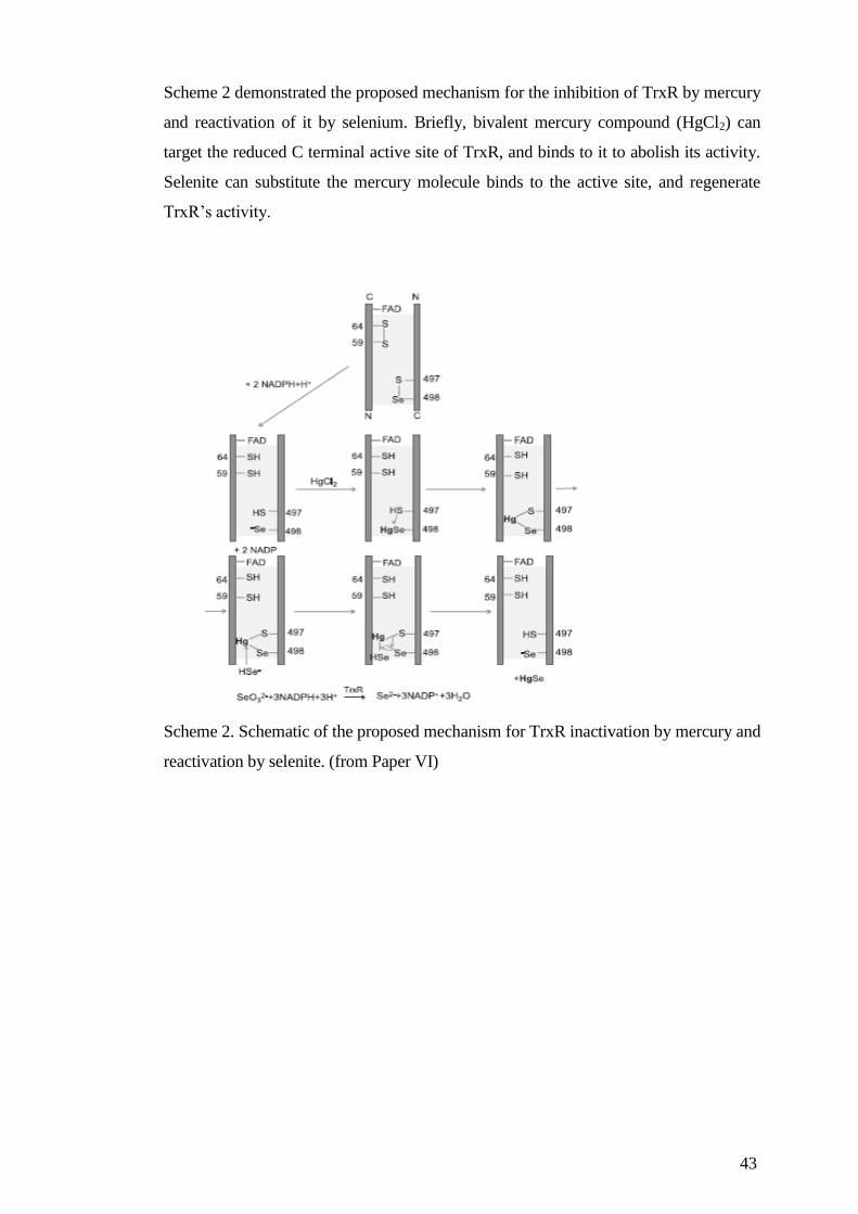
43
Scheme 2 demonstrated the proposed mechanism for the inhibition of TrxR by mercury
and reactivation of it by selenium. Briefly, bivalent mercury compound (HgCl2) can
target the reduced C terminal active site of TrxR, and binds to it to abolish its activity.
Selenite can substitute the mercury molecule binds to the active site, and regenerate
TrxR’s activity.
Scheme 2. Schematic of the proposed mechanism for TrxR inactivation by mercury and
reactivation by selenite. (from Paper VI)

44
3.3 CONCLUSION AND FUTURE PERSPECTIVES
In our first paper, we showed that the degradation of Trx2 upon BG treatment. Through
comparing with fibroblast cells, we demonstrate the importance of Trx2 in cancer cells,
such as Hela cells. However, the exact interaction of BG and Trx2 is not clear.
Although we have in vitro data showed that BG can binds to thiols in small molecule
such as dithiothreitol (DTT), but we did not find direct evidence of BG binding to Trx1
or Trx2 (unpublished data). In addition, we also found that BG is a strong inhibitor of
TrxR1 in vitro, although we cannot find the inhibition effect on TrxR in cell lysates,
this maybe because BG specifically accumulated in mitochondrial matrix, where it can
only inhibit TrxR2. But even only considering mitochondria, where TrxR2 locates in
the matrix and TrxR1 locates in the inter membrane space; the amount of TrxR2 is still
much lower than that of TrxR1. So it is very difficult to obtain the data on TrxR2 in
cells. However, the interaction of BG with Trx2 system should be further studied
because it is very important for the investigation of the mechanism of degradation of
Trx2. We have proposed mitochondrial Lon protease to be responsible for the
degradation of Trx2, but this need to be further confirmed as well.
In paper II and IV, we investigated the importance of the second disulfide in regulating
Trx1’s activity and functions. In paper II, we proposed that when Trx1 with two
disulfides cannot be reduced by TrxR, Grx system can reduce it through a monothiol
mechanism. In paper IV, we further demonstrated that the formation of the second
disulfide in Trx1 can sensitize SH-SH5Y cells towards cytotoxicity induced by some
arsenical compounds. In addition, in vitro data showed that the formation of the second
compounds can weaken Trx1’s ability to reduce oxidized Prx1. However, because
previous studies about Trx1’s activity and functions were almost all focused on the
active site Cys residues of Trx1, the data about the functions of the second disulfide in
Trx1 is very limited. Although we have found that Trx1 with two disulfides can be
generated in A549 cells under high dose of H2O2 treatment, there is no data showing its
existence under physiological conditions. In my opinion, physiological process
involving a high dose of oxidative stress should be paid more attention to, such as
during inflammation. In addition, since Trx1 with two disulfides is not a substrate of
TrxR, then the delay of reducing it may give a chance of transient inhibition of Prx1,
and causes an increase of H2O2, which can subsequently exerts its function as a
signaling molecule. Although there is no doubt that H2O2 is a messenger molecule more

45
than just an evil oxidant. Considering that it can diffuse freely in the cells, the
mechanism how it can exert its function locally without spread the signal all over the
cells is still not clear. In our Paper III, we investigated the role of Grx2 as a backup of
TrxR, which can reduce both the oxidized Trx1 and Trx2 in vitro and in cells when
TrxR is inhibited. It is interesting to think about the fact that Grx2 is more resistant to
oxidative stress compare to Grx1, and it actually can be activated by ROS. This finding
proposed a possibility that under some oxidative conditions, Grx2, not Grx1 reduces
Trx1 with two disulfides. Considering the fact that the amount of Grx2 is low and its
subcellular locations, maybe it plays a role in regulating the amount of H2O2 locally.
Another factor that needs to be considered is GSH, because both Grx1 and Grx2 need
the expense of GSH to reduce Trx, and the amount of GSH various in different cell
organelles. On all accounts, the regulation and mechanism of the whole signaling
pathway (Grx1/Grx2Trx1Prx1H2O2) is not clear, more efforts should be spent
on it, especially in a cellular model.
In our last two papers, we demonstrated TrxR as a cellular target of anticancer drug
mitomycin C and the toxic compounds of mercury, and that selenium can act as a
detoxification compound of mercury toxicity with a comparable recovery capacity of
clinically used chelating agents. It is not surprising that the exposed highly reactive Sec
residue in TrxR is a target of several electrophilic compounds and metals. However, by
collaborating with experts who works in the chemical field and toxicology field,
brought us new tools and different angels to study the interactions of compounds with
Trx system, which can be applied in future studies.
In the end, it is worth to mention two new techniques which are developed and / or
optimized by my colleagues during my PhD study, one is measuring TrxR/Trx activity
by using fluorescent substrate, and another one is redox western blot. The new
fluorescent method, which is very sensitive and stable, made it possible to measure the
activity of TrxR/Trx in a very limited amount of cell samples, such as mitochondrial
lysate; or cells with very low amount of TrxR/Trx, such as SH-SH5Y cells. The redox
western blot made it is possible and reliable to analyze the redox state of each Cys
residues in Trx1, thus it is a great tool to study the function of the structural Cys
residues in Trx1. Moreover, it is possible to optimize the method and analyze the redox
state of bigger molecules, such as peroxiredoxins. These two powerful tools will be
applied more often in my future studies.

46
4 ACKNOWLEDGEMENTS
I would like to take this opportunity to express my sincere gratitude to all of you, who
helped me in different ways, during my whole Ph.D. study.
My deepest gratitude goes first and foremost to my main supervisor Professor Arne
Holmgren. Without your generous support, I would not have the opportunity to study
in such a world leading laboratory in Karolinska Institutet. You always helped me to be
on the right track, as well as gave me enough freedom to realize my own ideas and
projects. Your advices and encouragements have been priceless for me.
Secondly, I would like to express my heartfelt gratitude to my co-supervisor Dr. Jun
Lu, who has been a tremendous mentor for me. You have played multiple roles during
my life as a Ph.D. student; you are a wise supervisor, a trustful friend and a caring
brother. I am really grateful for all the knowledge you taught me and our friendship we
have built.
To Professor Elias Arnér, the head of our division, I would like to thank you for all
the constructive suggestions you gave for my research, and all the great activities you
have organized during these years.
Lena Ringdén, you are always warm hearted and cheerful, whenever talking with you
can make me smile and felt being cared for; and Jack Andrzejewski, thank you for
always being friendly and helpful.
To Dr. Yatao Du and Dr. Huihui Zhang, thank you for the collaborations we had,
especially all the interesting discussions about the cysteine residues in thioredoxin; and
for sharing the knowledge and tips about redox western blot with me.
To Dr. Tomas Gustafsson, thank you for all the help you gave me during my Ph.D
study, especially for teaching me how to express and purify recombinant proteins. To
Dr. Sergio Montano, thank you for your help, especially for teaching me about your
powerful fluorescent method. To both of you, I am very happy that you were the
“naughty ones” in the office, so I can be the “mature” one.
To Dr. Qing Cheng and Dr. Jianqiang Xu, thank you for the endless supply of TrxR,
and your selfless help with both my research and life.
To Deepika Nair, thank you for your enthusiasm and always having good moods and
good news sharing with me. To Dr. Lucia Coppo, it was great memory to share the
cell lab and the office with you.
To the present members of Biochemistry: Xiaoxiao Peng, Xiaoyuan Ren, Marcus
Cebula, Irina Pader, Dr. Katarina Johansson, Dr. Rajib Sengupta, Dr. Hanna-
Stina Martinsson Ahlzén, William Stafford, Lena Haffo, Dr. Fredrik Tholander,
and Dr. Alfredo Gimenez-Cassina, thank you all for the great environment you have
created here.
To the former members of Biochemistry: Dr. Eng-hui Chew, Dr. Alexios Vlamis-
Gardikas, Dr. Isaac Hashemy, Dr. Farnaz Zahedi, Dr. Sofi Eriksson, Dr. Steffanie

47
Prast-Nielsen, Dr. Christoph Hudeman, Dr. Maria Lönn, Dr. Eva-Maria
Hanschmann, Dr. Carsten Berndt, Dr. Lars Bräutigam, Dr. Victor Croitoru and
Dr. Helena Wållberg, thank you for the great time we had in Biochemistry.
To Dr. Jia Sun, Dr. Hongqian Yang, Dr. Min Wan, Dr.Ying Sun, Dr. Xun Wang,
Dr. Weiping Xu, and all the friends from KI, I am really appreciate the nice moment
we had together and all the help and support from you.
To our collaborators from all over the world, Dr. Laura Papp, Dr. Cristina Carvalho,
Dr. Manuel Paz, Professor Panayiotis Ioannou, Professor Jack Arbiser, Dr.
Greicy Michelle Marafiga Conterato, Dr. Paco Dasi, and Aida Rodrigues, I want to
thank all of you, not only because what you have provided us and what I have learned
from you, but also all the happy moments we shared together in Stockholm.
To the valuable friends I met in Sweden, Anqi Luo, Jing Tang, Ting Liu, Lei Ge,
Feifan Yu, Huan Song, and Dr. Weixing Qian’s family; to my former classmates
and friends from KTH: Suman Vodnala, Palash Sen, Sumire Honda, and Rui Yao; I
would love to thank you for being supportive and all the fun we had together.
To my dearest parents, without your support I would never be able to go this far.
我要感谢我最亲爱的父母,没有你们从小对我的教育培养,和长久以来对我的
支持,我不可能顺利的完成硕士和博士的学习。
To my beloved husband, Yaoguang Zhai, I would love to thank you for sharing
everything with me, and always be there for me whenever I need you.
In the end, I would like to thank Karolinska Institutet, where the work in this thesis
was performed, and the faculty funds for partial financing of new doctoral student
(KID-funding) for the economic support.

48
5 REFERENCES
1. Kroemer, G., Galluzzi L., Vandenabeele P., Abrams J., Alnemri ES.,
Baehrecke EH., Blagosklonny MV., EI-Deiry WS., Golstein P., Green DR.,
Hengartner M., Knight RA., Kumar S., Lipton SA., Malorni W., Nunez G.,
Peter ME., Tschopp J., Yuan J., Piacentini M., Zhivotovsky B.& Melino G.
Classification of cell death: recommendations of the Nomenclature Committee
on Cell Death 2009. Cell Death Differ. 16, 3–11 (2009).
2. Wang, D., Masutani H., Oka S., Tanaka T., Yamaguchi-Iwai Y., Nakamura H.
& Yodoi J.. Control of mitochondrial outer membrane permeabilization and
Bcl-xL levels by thioredoxin 2 in DT40 cells. J. Biol. Chem. 281, 7384–91
(2006).
3. Acehan, D., Jiang X., Morgan DG., Heuser JE., Wang X.& Akey CW. Three-
dimensional structure of the apoptosome: implications for assembly,
procaspase-9 binding, and activation. Mol. Cell 9, 423–32 (2002).
4. Ashkenazi, A. & Dixit, V. M. Death receptors: signaling and modulation.
Science 281, 1305–8 (1998).
5. Muzio, M., Chinnaiyan AM., Kischkel FC., O'Rourke K., Schevchenko A., Ni
J., Scaffidi C., Bretz JD., Zhang M., Gentz R., Mann M., Krammer PH., Peter
ME. & Dixit VM. FLICE, a novel FADD-homologous ICE/CED-3-like
protease, is recruited to the CD95 (Fas/APO-1) death--inducing signaling
complex. Cell 85, 817–27 (1996).
6. Boldin, M. P., Goncharov, T. M., Goltsev, Y. V & Wallach, D. Involvement of
MACH, a novel MORT1/FADD-interacting protease, in Fas/APO-1- and TNF
receptor-induced cell death. Cell 85, 803–15 (1996).
7. Scaffidi, C., Fulda S., Srinivasan A., Friesen C., Li F., Tomaselli KJ., Debatin
KM., Krammer PH. & Peter ME. Two CD95 (APO-1/Fas) signaling pathways.
EMBO J. 17, 1675–87 (1998).
8. Elmore, S. Apoptosis: a review of programmed cell death. Toxicol. Pathol. 35,
495–516 (2007).
9. Golstein, P. & Kroemer, G. Cell death by necrosis: towards a molecular
definition. Trends Biochem. Sci. 32, 37–43 (2007).
10. Festjens, N., Vanden Berghe, T. & Vandenabeele, P. Necrosis, a well-
orchestrated form of cell demise: signalling cascades, important mediators and
concomitant immune response. Biochim. Biophys. Acta 1757, 1371–87
11. Turrens, J. F. Mitochondrial formation of reactive oxygen species. J. Physiol.
552, 335–44 (2003).
12. McCord, J. M. & Fridovich, I. Superoxide dismutase. An enzymic function for
erythrocuprein (hemocuprein). J. Biol. Chem. 244, 6049–55 (1969).
13. Nordberg, J. & Arnér, E. S. Reactive oxygen species, antioxidants, and the
mammalian thioredoxin system. Free Radic. Biol. Med. 31, 1287–312 (2001).
49
14. Finkel, T. Signal transduction by reactive oxygen species. J. Cell Biol. 194, 7–
15 (2011).
15. Holmgren, A. Thioredoxin. Annu. Rev. Biochem. 54, 237–71 (1985).
16. Luthman, M. & Holmgren, A. Rat liver thioredoxin and thioredoxin reductase:
purification and characterization. Biochemistry 21, 6628–33 (1982).
17. Lu, J. & Holmgren, A. Thioredoxin system in cell death progression. Antioxid.
Redox Signal. 17, 1738–47 (2012).
18. Laurent, T. C., Moore, E. C. & Reichard, P. Enzymatic Synthesis of
Deoxyribonucleotides. IV. Isolation and characterization of thioredoxin, the
hydrogen donor from Escherichia Coli B. J. Biol. Chem. 239, 3436–44 (1964).
19. Nordlund, P. & Reichard, P. Ribonucleotide reductases. Annu. Rev. Biochem.
75, 681–706 (2006).
20. Arnér, E. S. & Holmgren, A. Physiological functions of thioredoxin and
thioredoxin reductase. Eur. J. Biochem. 267, 6102–9 (2000).
21. Holmgren, A. & Lu, J. Thioredoxin and thioredoxin reductase: current research
with special reference to human disease. Biochem. Biophys. Res. Commun.
396, 120–124 (2010).
22. Ueno, M., Matsutani Y., Nakamura H., Masutani H., Yaqi M., Yamashiro H.,
Kato H., Inamoto T., Yamauchi A., Takahashi R., Yamaoka Y. & Yodoi J.
Possible association of thioredoxin and p53 in breast cancer. Immunol. Lett. 75,
15–20 (2000).
23. Matthews, J. R., Wakasugi, N., Virelizier, J. L., Yodoi, J. & Hay, R. T.
Thioredoxin regulates the DNA binding activity of NF-kappa B by reduction
of a disulphide bond involving cysteine 62. Nucleic Acids Res. 20, 3821–30
(1992).
24. Arnér, E. S. J. & Holmgren, A. The thioredoxin system in cancer. Semin.
Cancer Biol. 16, 420–6 (2006).
25. Spyrou, G., Enmark, E., Miranda-Vizuete, A. & Gustafsson, J. Cloning and
expression of a novel mammalian thioredoxin. J. Biol. Chem. 272, 2936–41
(1997).
26. Zhang, R. et al. Thioredoxin-2 inhibits mitochondria-located ASK1-mediated
apoptosis in a JNK-independent manner. Circ. Res. 94, 1483–91 (2004).
27. Saxena, G., Chen, J. & Shalev, A. Intracellular shuttling and mitochondrial
function of thioredoxin-interacting protein. J. Biol. Chem. 285, 3997–4005
(2010).
28. He, M., Identification of thioredoxin-2 as a regulator of the mitochondrial
permeability transition. Toxicol. Sci. 105, 44–50 (2008).
29. Matsui, M., Oshima M., Oshima H., Takaku K., Maruyama T., Yodoj J. &
Taketo MM. Early embryonic lethality caused by targeted disruption of the
mouse thioredoxin gene. Dev. Biol. 178, 179–85 (1996).
50
30. Nonn, L., Williams, R. R., Erickson, R. P. & Powis, G. The absence of
mitochondrial thioredoxin 2 causes massive apoptosis, exencephaly, and early
embryonic lethality in homozygous mice. Mol. Cell. Biol. 23, 916–22 (2003).
31. Martin, J. L. Thioredoxin--a fold for all reasons. Structure 3, 245–50 (1995).
32. Forman-Kay, J. D., Clore, G. M., Wingfield, P. T. & Gronenborn, A. M. High-
resolution three-dimensional structure of reduced recombinant human
thioredoxin in solution. Biochemistry 30, 2685–98 (1991).
33. Ren, X., Björnstedt, M., Shen, B., Ericson, M. L. & Holmgren, A. Mutagenesis
of structural half-cystine residues in human thioredoxin and effects on the
regulation of activity by selenodiglutathione. Biochemistry 32, 9701–8 (1993).
34. Weichsel, A., Gasdaska, J. R., Powis, G. & Montfort, W. R. Crystal structures
of reduced, oxidized, and mutated human thioredoxins: evidence for a
regulatory homodimer. Structure 4, 735–51 (1996).
35. Lu, J., Chew, E.-H. & Holmgren, A. Targeting thioredoxin reductase is a basis
for cancer therapy by arsenic trioxide. Proc. Natl. Acad. Sci. U. S. A. 104,
12288–93 (2007).
36. Watson, W. H., Pohl J., Montfort WR., Stuchlik O., Reed MS., Powis
G.&Jones DP . Redox potential of human thioredoxin 1 and identification of a
second dithiol/disulfide motif. J. Biol. Chem. 278, 33408–15 (2003).
37. Holmgren, A. Bovine thioredoxin system. Purification of thioredoxin reductase
from calf liver and thymus and studies of its function in disulfide reduction. J.
Biol. Chem. 252, 4600–6 (1977).
38. Du, Y., Zhang, H., Zhang, X., Lu, J. & Holmgren, A. Thioredoxin 1 is
inactivated due to oxidation induced by peroxiredoxin under oxidative stress
and reactivated by glutaredoxin system. J. Biol. Chem. (2013).
doi:10.1074/jbc.M113.495150
39. Uhlin, U. & Eklund, H. Structure of ribonucleotide reductase protein R1.
Nature 370, 533–9 (1994).
40. Holmgren, A. & Sengupta, R. The use of thiols by ribonucleotide reductase.
Free Radic. Biol. Med. 49, 1617–1628 (2010).
41. Aberg, A., Hahne S., Karlsson M., Larsson. A., Ormö A., Ahgren A.
&Sjöberg BM. Evidence for two different classes of redox-active cysteines in
ribonucleotide reductase of Escherichia coli. J. Biol. Chem. 264, 12249–52
(1989).
42. Lin, A. N., Ashley, G. W. & Stubbe, J. Location of the redox-active thiols of
ribonucleotide reductase: sequence similarity between the Escherichia coli and
Lactobacillus leichmannii enzymes. Biochemistry 26, 6905–9 (1987).
43. Zahedi Avval, F. & Holmgren, A. Molecular mechanisms of thioredoxin and
glutaredoxin as hydrogen donors for Mammalian s phase ribonucleotide
reductase. J. Biol. Chem. 284, 8233–40 (2009).
44. Chae, H. Z., Chung, S. J. & Rhee, S. G. Thioredoxin-dependent peroxide
reductase from yeast. J. Biol. Chem. 269, 27670–8 (1994).

51
45. Rhee, S. G., Chae, H. Z. & Kim, K. Peroxiredoxins: a historical overview and
speculative preview of novel mechanisms and emerging concepts in cell
signaling. Free Radic. Biol. Med. 38, 1543–52 (2005).
46. Rhee, S. G. & Woo, H. A. Multiple functions of peroxiredoxins: peroxidases,
sensors and regulators of the intracellular messenger H₂O₂, and protein
chaperones. Antioxid. Redox Signal. 15, 781–94 (2011).
47. Rhee, S. G. Cell signaling. H2O2, a necessary evil for cell signaling. Science
312, 1882–3 (2006).
48. Seo, M. S., Kang SW., Kim K., Baines IC., Lee TH. & Rhee SG. Identification
of a new type of mammalian peroxiredoxin that forms an intramolecular
disulfide as a reaction intermediate. J. Biol. Chem. 275, 20346–54 (2000).
49. Rhee, S. G. & Woo, H. A. Multiple functions of peroxiredoxins: peroxidases,
sensors and regulators of the intracellular messenger H₂O₂, and protein
chaperones. Antioxid. Redox Signal. 15, 781–94 (2011).
50. Wood, Z. A., Schröder, E., Robin Harris, J. & Poole, L. B. Structure,
mechanism and regulation of peroxiredoxins. Trends Biochem. Sci. 28, 32–40
(2003).
51. Smeets, A., Marchand, C., Linard, D., Knoops, B. & Declercq, J.-P. The
crystal structures of oxidized forms of human peroxiredoxin 5 with an
intramolecular disulfide bond confirm the proposed enzymatic mechanism for
atypical 2-Cys peroxiredoxins. Arch. Biochem. Biophys. 477, 98–104 (2008).
52. Kang, S. W., Baines, I. C. & Rhee, S. G. Characterization of a mammalian
peroxiredoxin that contains one conserved cysteine. J. Biol. Chem. 273, 6303–
11 (1998).
53. Brot, N. & Weissbach, H. Biochemistry and physiological role of methionine
sulfoxide residues in proteins. Arch. Biochem. Biophys. 223, 271–81 (1983).
54. Boschi-Muller, S., Olry, A., Antoine, M. & Branlant, G. The enzymology and
biochemistry of methionine sulfoxide reductases. Biochim. Biophys. Acta
1703, 231–8 (2005).
55. Boschi-Muller, S., Gand, A. & Branlant, G. The methionine sulfoxide
reductases: Catalysis and substrate specificities. Arch. Biochem. Biophys. 474,
266–73 (2008).
56. Hirota, K., Murata, M., Itoh, T., Yodoi, J. & Fukuda, K. An endogenous redox
molecule, thioredoxin, regulates transactivation of epidermal growth factor
receptor and activation of NF-kappaB by lysophosphatidic acid. FEBS Lett.
489, 134–8 (2001).
57. Gebel, S. & Müller, T. The activity of NF-kappaB in Swiss 3T3 cells exposed
to aqueous extracts of cigarette smoke is dependent on thioredoxin. Toxicol.
Sci. 59, 75–81 (2001).
58. Yamaoka, S., Courtois G., Bessia C., Whiteside ST., Weil R., Agou F., Kirk
HE., Kay RJ. & Israel A. Complementation cloning of NEMO, a component

52
of the IkappaB kinase complex essential for NF-kappaB activation. Cell 93,
1231–40 (1998).
59. Okamoto, T., Sakurada, S., Yang, J. P. & Merin, J. P. Regulation of NF-kappa
B and disease control: identification of a novel serine kinase and thioredoxin as
effectors for signal transduction pathway for NF-kappa B activation. Curr.
Top. Cell. Regul. 35, 149–61 (1997).
60. DiDonato, J. A., Hayakawa, M., Rothwarf, D. M., Zandi, E. & Karin, M. A
cytokine-responsive IkappaB kinase that activates the transcription factor NF-
kappaB. Nature 388, 548–54 (1997).
61. Hayashi, T., Ueno, Y. & Okamoto, T. Oxidoreductive regulation of nuclear
factor kappa B. Involvement of a cellular reducing catalyst thioredoxin. J. Biol.
Chem. 268, 11380–8 (1993).
62. Hirota, K., Murata M., Sachi Y., Nakamura H., Takeuchi J., Mori K. & Yodoi
J. Distinct roles of thioredoxin in the cytoplasm and in the nucleus. A two-step
mechanism of redox regulation of transcription factor NF-kappaB. J. Biol.
Chem. 274, 27891–7 (1999).
63. Abate, C., Patel, L., Rauscher, F. J. & Curran, T. Redox regulation of fos and
jun DNA-binding activity in vitro. Science 249, 1157–61 (1990).
64. Hirota, K., Matsui M., Iwata S., Nishiyama A., Mori K. & Yodoi J. AP-1
transcriptional activity is regulated by a direct association between thioredoxin
and Ref-1. Proc. Natl. Acad. Sci. U. S. A. 94, 3633–8 (1997).
65. Kim, D.-H., Kundu, J. K. & Surh, Y.-J. Redox modulation of p53: mechanisms
and functional significance. Mol. Carcinog. 50, 222–34 (2011).
66. Maillet, A. & Pervaiz, S. Redox regulation of p53, redox effectors regulated by
p53: a subtle balance. Antioxid. Redox Signal. 16, 1285–94 (2012).
67. Parks, D., Bolinger, R. & Mann, K. Redox state regulates binding of p53 to
sequence-specific DNA, but not to non-specific or mismatched DNA. Nucleic
Acids Res. 25, 1289–95 (1997).
68. Jayaraman, L., Murthy KG., Zhu C., Curran T., Xanthoudakis S. & Prives C.
Identification of redox/repair protein Ref-1 as a potent activator of p53. Genes
Dev. 11, 558–70 (1997).
69. Ueno, M., Masutani H., Arai RJ., Yamauchi A., Hirota K., Sakai T., Inamoto
T., Yamaoka Y., Yodoi J. & Nikaido T. Thioredoxin-dependent redox
regulation of p53-mediated p21 activation. J. Biol. Chem. 274, 35809–15
(1999).
70. Pearson, G. D. & Merrill, G. F. Deletion of the Saccharomyces cerevisiae
TRR1 gene encoding thioredoxin reductase inhibits p53-dependent reporter
gene expression. J. Biol. Chem. 273, 5431–4 (1998).
71. Moos, P. J., Edes, K., Cassidy, P., Massuda, E. & Fitzpatrick, F. A.
Electrophilic prostaglandins and lipid aldehydes repress redox-sensitive
transcription factors p53 and hypoxia-inducible factor by impairing the
selenoprotein thioredoxin reductase. J. Biol. Chem. 278, 745–50 (2003).

53
72. Ichijo, H. et al. Induction of apoptosis by ASK1, a mammalian MAPKKK that
activates SAPK/JNK and p38 signaling pathways. Science 275, 90–4 (1997).
73. Liu, H., Nishitoh, H., Ichijo, H. & Kyriakis, J. M. Activation of apoptosis
signal-regulating kinase 1 (ASK1) by tumor necrosis factor receptor-associated
factor 2 requires prior dissociation of the ASK1 inhibitor thioredoxin. Mol.
Cell. Biol. 20, 2198–208 (2000).
74. Liu, Y. & Min, W. Thioredoxin promotes ASK1 ubiquitination and
degradation to inhibit ASK1-mediated apoptosis in a redox activity-
independent manner. Circ. Res. 90, 1259–66 (2002).
75. Yu, Y. & Richardson, D. R. Cellular iron depletion stimulates the JNK and p38
MAPK signaling transduction pathways, dissociation of ASK1-thioredoxin,
and activation of ASK1. J. Biol. Chem. 286, 15413–27 (2011).
76. Lim, P. L. K., Liu, J., Go, M. L. & Boelsterli, U. A. The mitochondrial
superoxide/thioredoxin-2/Ask1 signaling pathway is critically involved in
troglitazone-induced cell injury to human hepatocytes. Toxicol. Sci. 101, 341–9
(2008).
77. Chen, K. S. & DeLuca, H. F. Cloning of the human 1 alpha,25-
dihydroxyvitamin D-3 24-hydroxylase gene promoter and identification of two
vitamin D-responsive elements. Biochim. Biophys. Acta 1263, 1–9 (1995).
78. Nishiyama, A. et al. Identification of thioredoxin-binding protein-2/vitamin
D(3) up-regulated protein 1 as a negative regulator of thioredoxin function and
expression. J. Biol. Chem. 274, 21645–50 (1999).
79. Patwari, P., Higgins, L. J., Chutkow, W. A., Yoshioka, J. & Lee, R. T. The
interaction of thioredoxin with Txnip. Evidence for formation of a mixed
disulfide by disulfide exchange. J. Biol. Chem. 281, 21884–91 (2006).
80. Hwang, J. et al. The structural basis for the negative regulation of thioredoxin
by thioredoxin-interacting protein. Nat. Commun. 5, 2958 (2014).
81. Yu, F.-X., Goh, S.-R., Dai, R.-P. & Luo, Y. Adenosine-containing molecules
amplify glucose signaling and enhance txnip expression. Mol. Endocrinol. 23,
932–42 (2009).
82. Yamawaki, H., Pan, S., Lee, R. T. & Berk, B. C. Fluid shear stress inhibits
vascular inflammation by decreasing thioredoxin-interacting protein in
endothelial cells. J. Clin. Invest. 115, 733–8 (2005).
83. Das, K. C., Guo, X. L. & White, C. W. Induction of thioredoxin and
thioredoxin reductase gene expression in lungs of newborn primates by
oxygen. Am. J. Physiol. 276, L530–9 (1999).
84. Das, K. C., Guo, X. L. & White, C. W. Hyperoxia induces thioredoxin and
thioredoxin reductase gene expression in lungs of premature baboons with
respiratory distress and bronchopulmonary dysplasia. Chest 116, 101S (1999).
85. Higashikubo, A. et al. Increase in thioredoxin activity of intestinal epithelial
cells mediated by oxidative stress. Biol. Pharm. Bull. 22, 900–3 (1999).
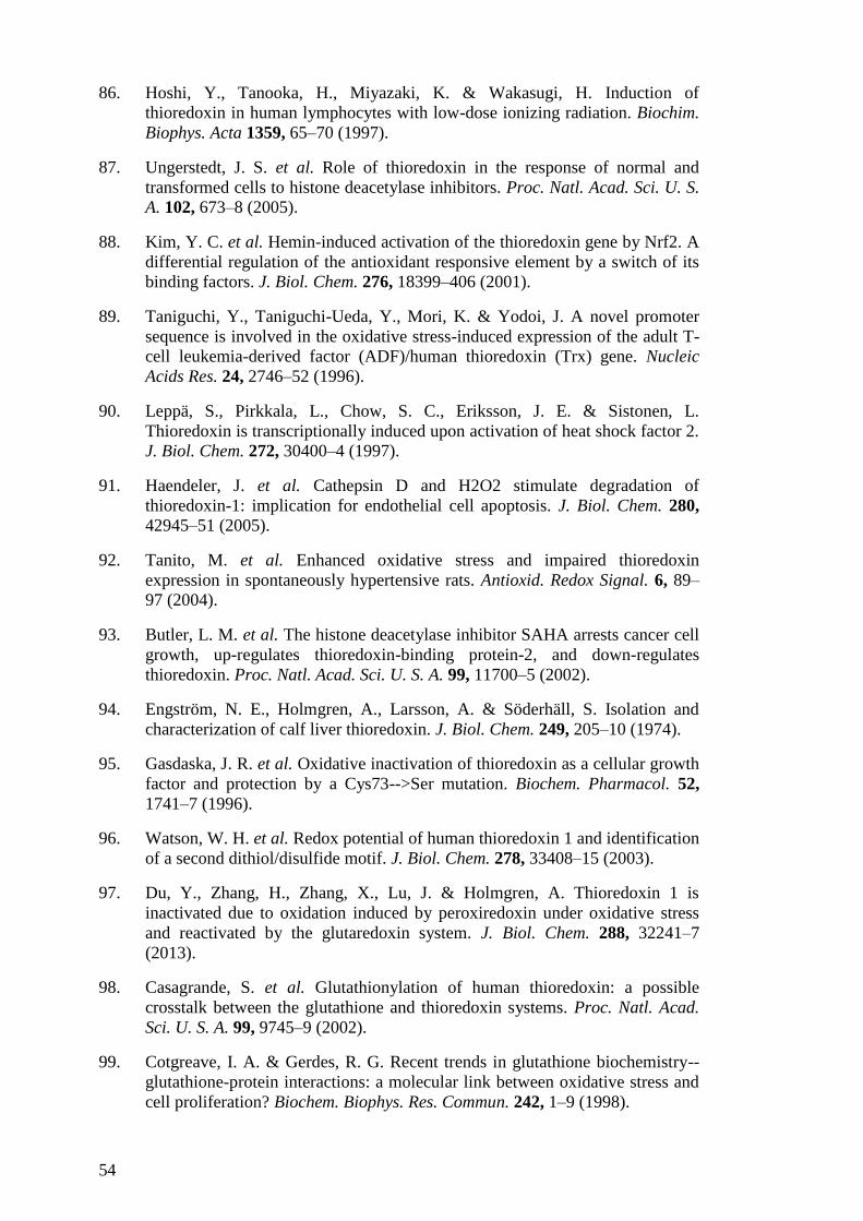
54
86. Hoshi, Y., Tanooka, H., Miyazaki, K. & Wakasugi, H. Induction of
thioredoxin in human lymphocytes with low-dose ionizing radiation. Biochim.
Biophys. Acta 1359, 65–70 (1997).
87. Ungerstedt, J. S. et al. Role of thioredoxin in the response of normal and
transformed cells to histone deacetylase inhibitors. Proc. Natl. Acad. Sci. U. S.
A. 102, 673–8 (2005).
88. Kim, Y. C. et al. Hemin-induced activation of the thioredoxin gene by Nrf2. A
differential regulation of the antioxidant responsive element by a switch of its
binding factors. J. Biol. Chem. 276, 18399–406 (2001).
89. Taniguchi, Y., Taniguchi-Ueda, Y., Mori, K. & Yodoi, J. A novel promoter
sequence is involved in the oxidative stress-induced expression of the adult T-
cell leukemia-derived factor (ADF)/human thioredoxin (Trx) gene. Nucleic
Acids Res. 24, 2746–52 (1996).
90. Leppä, S., Pirkkala, L., Chow, S. C., Eriksson, J. E. & Sistonen, L.
Thioredoxin is transcriptionally induced upon activation of heat shock factor 2.
J. Biol. Chem. 272, 30400–4 (1997).
91. Haendeler, J. et al. Cathepsin D and H2O2 stimulate degradation of
thioredoxin-1: implication for endothelial cell apoptosis. J. Biol. Chem. 280,
42945–51 (2005).
92. Tanito, M. et al. Enhanced oxidative stress and impaired thioredoxin
expression in spontaneously hypertensive rats. Antioxid. Redox Signal. 6, 89–
97 (2004).
93. Butler, L. M. et al. The histone deacetylase inhibitor SAHA arrests cancer cell
growth, up-regulates thioredoxin-binding protein-2, and down-regulates
thioredoxin. Proc. Natl. Acad. Sci. U. S. A. 99, 11700–5 (2002).
94. Engström, N. E., Holmgren, A., Larsson, A. & Söderhäll, S. Isolation and
characterization of calf liver thioredoxin. J. Biol. Chem. 249, 205–10 (1974).
95. Gasdaska, J. R. et al. Oxidative inactivation of thioredoxin as a cellular growth
factor and protection by a Cys73-->Ser mutation. Biochem. Pharmacol. 52,
1741–7 (1996).
96. Watson, W. H. et al. Redox potential of human thioredoxin 1 and identification
of a second dithiol/disulfide motif. J. Biol. Chem. 278, 33408–15 (2003).
97. Du, Y., Zhang, H., Zhang, X., Lu, J. & Holmgren, A. Thioredoxin 1 is
inactivated due to oxidation induced by peroxiredoxin under oxidative stress
and reactivated by the glutaredoxin system. J. Biol. Chem. 288, 32241–7
(2013).
98. Casagrande, S. et al. Glutathionylation of human thioredoxin: a possible
crosstalk between the glutathione and thioredoxin systems. Proc. Natl. Acad.
Sci. U. S. A. 99, 9745–9 (2002).
99. Cotgreave, I. A. & Gerdes, R. G. Recent trends in glutathione biochemistry--
glutathione-protein interactions: a molecular link between oxidative stress and
cell proliferation? Biochem. Biophys. Res. Commun. 242, 1–9 (1998).
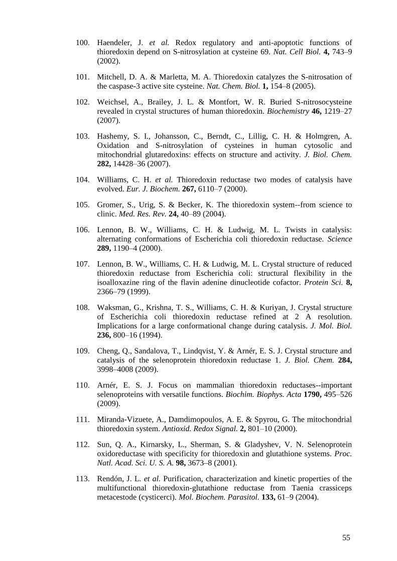
55
100. Haendeler, J. et al. Redox regulatory and anti-apoptotic functions of
thioredoxin depend on S-nitrosylation at cysteine 69. Nat. Cell Biol. 4, 743–9
(2002).
101. Mitchell, D. A. & Marletta, M. A. Thioredoxin catalyzes the S-nitrosation of
the caspase-3 active site cysteine. Nat. Chem. Biol. 1, 154–8 (2005).
102. Weichsel, A., Brailey, J. L. & Montfort, W. R. Buried S-nitrosocysteine
revealed in crystal structures of human thioredoxin. Biochemistry 46, 1219–27
(2007).
103. Hashemy, S. I., Johansson, C., Berndt, C., Lillig, C. H. & Holmgren, A.
Oxidation and S-nitrosylation of cysteines in human cytosolic and
mitochondrial glutaredoxins: effects on structure and activity. J. Biol. Chem.
282, 14428–36 (2007).
104. Williams, C. H. et al. Thioredoxin reductase two modes of catalysis have
evolved. Eur. J. Biochem. 267, 6110–7 (2000).
105. Gromer, S., Urig, S. & Becker, K. The thioredoxin system--from science to
clinic. Med. Res. Rev. 24, 40–89 (2004).
106. Lennon, B. W., Williams, C. H. & Ludwig, M. L. Twists in catalysis:
alternating conformations of Escherichia coli thioredoxin reductase. Science
289, 1190–4 (2000).
107. Lennon, B. W., Williams, C. H. & Ludwig, M. L. Crystal structure of reduced
thioredoxin reductase from Escherichia coli: structural flexibility in the
isoalloxazine ring of the flavin adenine dinucleotide cofactor. Protein Sci. 8,
2366–79 (1999).
108. Waksman, G., Krishna, T. S., Williams, C. H. & Kuriyan, J. Crystal structure
of Escherichia coli thioredoxin reductase refined at 2 A resolution.
Implications for a large conformational change during catalysis. J. Mol. Biol.
236, 800–16 (1994).
109. Cheng, Q., Sandalova, T., Lindqvist, Y. & Arnér, E. S. J. Crystal structure and
catalysis of the selenoprotein thioredoxin reductase 1. J. Biol. Chem. 284,
3998–4008 (2009).
110. Arnér, E. S. J. Focus on mammalian thioredoxin reductases--important
selenoproteins with versatile functions. Biochim. Biophys. Acta 1790, 495–526
(2009).
111. Miranda-Vizuete, A., Damdimopoulos, A. E. & Spyrou, G. The mitochondrial
thioredoxin system. Antioxid. Redox Signal. 2, 801–10 (2000).
112. Sun, Q. A., Kirnarsky, L., Sherman, S. & Gladyshev, V. N. Selenoprotein
oxidoreductase with specificity for thioredoxin and glutathione systems. Proc.
Natl. Acad. Sci. U. S. A. 98, 3673–8 (2001).
113. Rendón, J. L. et al. Purification, characterization and kinetic properties of the
multifunctional thioredoxin-glutathione reductase from Taenia crassiceps
metacestode (cysticerci). Mol. Biochem. Parasitol. 133, 61–9 (2004).

56
114. Su, D. et al. Mammalian selenoprotein thioredoxin-glutathione reductase.
Roles in disulfide bond formation and sperm maturation. J. Biol. Chem. 280,
26491–8 (2005).
115. Sun, Q. A. et al. Heterogeneity within animal thioredoxin reductases. Evidence
for alternative first exon splicing. J. Biol. Chem. 276, 3106–14 (2001).
116. Rundlöf, A.-K. & Arnér, E. S. J. Regulation of the mammalian selenoprotein
thioredoxin reductase 1 in relation to cellular phenotype, growth, and signaling
events. Antioxid. Redox Signal. 6, 41–52 (2004).
117. Rayman, M. P. The importance of selenium to human health. Lancet 356, 233–
41 (2000).
118. Kryukov, G. V et al. Characterization of mammalian selenoproteomes. Science
300, 1439–43 (2003).
119. Papp, L. V., Lu, J., Holmgren, A. & Khanna, K. K. From selenium to
selenoproteins: synthesis, identity, and their role in human health. Antioxid.
Redox Signal. 9, 775–806 (2007).
120. Huber, R. E. & Criddle, R. S. Comparison of the chemical properties of
selenocysteine and selenocystine with their sulfur analogs. Arch. Biochem.
Biophys. 122, 164–73 (1967).
121. Lu, J., Zhong, L., Lönn ME., Raymond FB., Kristina EH & Holmgren,A.
Penultimate selenocysteine residue replaced by cysteine in thioredoxin
reductase from selenium-deficient rat liver. FASEB J. 23, 2394–402 (2009).
122. Montero, A. J. & Jassem, J. Cellular redox pathways as a therapeutic target in
the treatment of cancer. Drugs 71, 1385–96 (2011).
123. Gorrini, C., Harris, I. S. & Mak, T. W. Modulation of oxidative stress as an
anticancer strategy. Nat. Rev. Drug Discov. 12, 931–47 (2013).
124. Lu, J. & Holmgren, A. Thioredoxin system in cell death progression. Antioxid.
Redox Signal. 17, 1738–47 (2012).
125. Hedström, E., Eriksson, S., Zawacka-Pankau, J., Arnér, E. S. J. & Selivanova,
G. p53-dependent inhibition of TrxR1 contributes to the tumor-specific
induction of apoptosis by RITA. Cell Cycle 8, 3576–83 (2009).
126. Prast-Nielsen, S., Cebula, M., Pader, I. & Arnér, E. S. J. Noble metal targeting
of thioredoxin reductase — covalent complexes with thioredoxin and
thioredoxin-related protein of 14kDa triggered by cisplatin. Free Radic. Biol.
Med. 49, 1765–1778 (2010).
127. Paz, M. M., Zhang, X., Lu, J. & Holmgren, A. A new mechanism of action for
the anticancer drug mitomycin C: mechanism-based inhibition of thioredoxin
reductase. Chem. Res. Toxicol. 25, 1502–11 (2012).
128. Arnér, E. S., Björnstedt, M. & Holmgren, A. 1-Chloro-2,4-dinitrobenzene is an
irreversible inhibitor of human thioredoxin reductase. Loss of thioredoxin
disulfide reductase activity is accompanied by a large increase in NADPH
oxidase activity. J. Biol. Chem. 270, 3479–82 (1995).

57
129. Lu, J., Chew, E.-H. & Holmgren, A. Targeting thioredoxin reductase is a basis
for cancer therapy by arsenic trioxide. Proc. Natl. Acad. Sci. U. S. A. 104,
12288–93 (2007).
130. Bushweller, J. H., Aslund, F., Wüthrich, K. & Holmgren, A. Structural and
functional characterization of the mutant Escherichia coli glutaredoxin (C14---
-S) and its mixed disulfide with glutathione. Biochemistry 31, 9288–93 (1992).
131. Srinivasan, U., Mieyal, P. A. & Mieyal, J. J. pH profiles indicative of rate-
limiting nucleophilic displacement in thioltransferase catalysis. Biochemistry
36, 3199–206 (1997).
132. Holmgren, A. Hydrogen donor system for Escherichia coli ribonucleoside-
diphosphate reductase dependent upon glutathione. Proc. Natl. Acad. Sci. U. S.
A. 73, 2275–9 (1976).
133. Holmgren, A. Glutathione-dependent synthesis of deoxyribonucleotides.
Purification and characterization of glutaredoxin from Escherichia coli. J. Biol.
Chem. 254, 3664–71 (1979).
134. Molina-Navarro, M. M., Casas, C., Piedrafita, L., Bellí, G. & Herrero, E.
Prokaryotic and eukaryotic monothiol glutaredoxins are able to perform the
functions of Grx5 in the biogenesis of Fe/S clusters in yeast mitochondria.
FEBS Lett. 580, 2273–80 (2006).
135. Sun, C., Berardi, M. J. & Bushweller, J. H. The NMR solution structure of
human glutaredoxin in the fully reduced form. J. Mol. Biol. 280, 687–701
(1998).
136. Xia, T. H. Bushweller, J.H., Sodano, P., Billeter, M., Björnberg, O., Holmgren,
A. & Wuthrich, K. NMR structure of oxidized Escherichia coli glutaredoxin:
comparison with reduced E. coli glutaredoxin and functionally related proteins.
Protein Sci. 1, 310–21 (1992).
137. Bushweller, J. H., Billeter, M., Holmgren, A. & Wüthrich, K. The nuclear
magnetic resonance solution structure of the mixed disulfide between
Escherichia coli glutaredoxin(C14S) and glutathione. J. Mol. Biol. 235, 1585–
97 (1994).
138. Hudemann, C., Lönn ME., Godoy JR., Zahedi Avval F., Capani F., Holmgren,
A.& Lillig CH. Identification, expression pattern, and characterization of
mouse glutaredoxin 2 isoforms. Antioxid. Redox Signal. 11, 1–14 (2009).
139. Berndt, C., Hudemann C., Hanschmann EM., Axelsson R., Holmgren,A. &
Lillig CH. How does iron-sulfur cluster coordination regulate the activity of
human glutaredoxin 2? Antioxid. Redox Signal. 9, 151–7 (2007).
140. Lillig, C. H., Berndt C., Vergnolle O., Lönn ME., Hudemann C., Bill E. &
Holmgren A. Characterization of human glutaredoxin 2 as iron-sulfur protein:
a possible role as redox sensor. Proc. Natl. Acad. Sci. U. S. A. 102, 8168–73
(2005).
141. Johansson, C., Lillig, C. H. & Holmgren, A. Human mitochondrial
glutaredoxin reduces S-glutathionylated proteins with high affinity accepting

58
electrons from either glutathione or thioredoxin reductase. J. Biol. Chem. 279,
7537–43 (2004).
142. Beer, S. M., Taylor ER., Stephanie EB., Dahm CC., Costa NJ., Runswick MJ.
& Murphy MP. Metabolism and Bioenergetics : Glutaredoxin 2 Catalyzes the
Reversible Oxidation and Glutathionylation of Mitochondrial Membrane Thiol
Proteins : IMPLICATIONS FOR REGULATION AND ANTIOXIDANT
Glutaredoxin 2 Catalyzes the Reversible Oxidation and Glutathiony. (2004).
doi:10.1074/jbc.M408011200
143. Tamarit, J., Bellı, G., Ros, J. & Herrero, E. Grx5 Is a Mitochondrial
Glutaredoxin Required for the Activity of Iron / Sulfur Enzymes ´ a Teresa
Rodrı. 13, 1109–1121 (2002).
144. Karplus, P. A. & Schulz, G. E. Refined structure of glutathione reductase at
1.54 A resolution. J. Mol. Biol. 195, 701–29 (1987).
145. Taniguchi, M., Hara, T. & Honda, H. Similarities between rat liver
mitochondrial and cytosolic glutathione reductases and their apoenzyme
accumulation in riboflavin deficiency. Biochem. Int. 13, 447–54 (1986).
146. White, C. C., Viernes, H., Krejsa, C. M., Botta, D. & Kavanagh, T. J.
Fluorescence-based microtiter plate assay for glutamate-cysteine ligase
activity. Anal. Biochem. 318, 175–80 (2003).
147. Lu, S. C. Glutathione synthesis. Biochim. Biophys. Acta 1830, 3143–53 (2013).
148. McKernan, T. B., Woods, E. B. & Lash, L. H. Uptake of glutathione by renal
cortical mitochondria. Arch. Biochem. Biophys. 288, 653–63 (1991).
149. Schnellmann, R. G. Renal mitochondrial glutathione transport. Life Sci. 49,
393–8 (1991).
150. Söderdahl, T., Enoksson M., Holmgren A., Ottersen OP., Orrenius S.,
Bolcsfoldi G., & Cotgreave I. Visualization of the compartmentalization of
glutathione and protein-glutathione mixed disulfides in cultured cells. FASEB
J. 17, 124–6 (2003).
151. Griffith, O. W. & Meister, A. Origin and turnover of mitochondrial
glutathione. Proc. Natl. Acad. Sci. U. S. A. 82, 4668–72 (1985).
152. Hwang, C., Sinskey, A. J. & Lodish, H. F. Oxidized redox state of glutathione
in the endoplasmic reticulum. Science 257, 1496–502 (1992).
153. Jones, D. P. Redox potential of GSH/GSSG couple: assay and biological
significance. Methods Enzymol. 348, 93–112 (2002).
154. Jones, D. P., Mody, V. C., Carlson, J. L., Lynn, M. J. & Sternberg, P. Redox
analysis of human plasma allows separation of pro-oxidant events of aging
from decline in antioxidant defenses. Free Radic. Biol. Med. 33, 1290–300
(2002).
155. Shelton, M. D., Chock, P. B. & Mieyal, J. J. Glutaredoxin: role in reversible
protein s-glutathionylation and regulation of redox signal transduction and
protein translocation. Antioxid. Redox Signal. 7, 348–66

59
156. Pineda-Molina, E., Klatt P., Vazquez J., Marina A., Garcia de Lacoba M.,
Perez-Sala D. & Lamas S. Glutathionylation of the p50 subunit of NF-kappaB:
a mechanism for redox-induced inhibition of DNA binding. Biochemistry 40,
14134–42 (2001).
157. Klatt, P., Molina EP., De Lacoba MG., Padila CA., Martinez-Galesteo E.,
Barcena JA. & Lamas S. Redox regulation of c-Jun DNA binding by reversible
S-glutathiolation. FASEB J. 13, 1481–90 (1999).
158. Lind, C., Gerdes R., Hamnell Y., Schuppe-Koistinen I., von Löwenhielm HB.,
Holmgren A. & Cotgreave IA. Identification of S-glutathionylated cellular
proteins during oxidative stress and constitutive metabolism by affinity
purification and proteomic analysis. Arch. Biochem. Biophys. 406, 229–40
(2002).
159. Fratelli, M., Gianazza, E. & Ghezzi, P. Redox proteomics: identification and
functional role of glutathionylated proteins. Expert Rev. Proteomics 1, 365–76
(2004).
160. Scott, N., Hatlelid, K. & MacKenzie, N. Reactions of arsenic (III) and arsenic
(V) species with glutathione. Chem. Res. … (1993). at
<http://pubs.acs.org/doi/abs/10.1021/tx00031a016\npapers2://publication/uuid/
073617A3-36B1-4F9F-B0E0-12717DD864B4>
161. Song, J. J. & Lee, Y. J. Differential role of glutaredoxin and thioredoxin in
metabolic oxidative stress-induced activation of apoptosis signal-regulating
kinase 1. Biochem. J. 373, 845–53 (2003).
162. Daily, D., Vlamis-Gardikas A., Offen D., Mittelman L., Melamed E.,
Holmgren A. & Barzilai A. Glutaredoxin protects cerebellar granule neurons
from dopamine-induced apoptosis by activating NF-kappa B via Ref-1. J. Biol.
Chem. 276, 1335–44 (2001).
163. Bandyopadhyay, S., Starke, D. W., Mieyal, J. J. & Gronostajski, R. M.
Thioltransferase (glutaredoxin) reactivates the DNA-binding activity of
oxidation-inactivated nuclear factor I. J. Biol. Chem. 273, 392–7 (1998).
164. Brigelius-Flohé, R. & Maiorino, M. Glutathione peroxidases. Biochim.
Biophys. Acta 1830, 3289–303 (2013).
165. Kanzok, S. M., Schirmer, R. H., Turbachova, I., Iozef, R. & Becker, K. The
thioredoxin system of the malaria parasite Plasmodium falciparum.
Glutathione reduction revisited. J. Biol. Chem. 275, 40180–6 (2000).
166. Du, Y., Zhang, H., Lu, J. & Holmgren, A. Glutathione and glutaredoxin act as
a backup of human thioredoxin reductase 1 to reduce thioredoxin 1 preventing
cell death by aurothioglucose. J. Biol. Chem. 287, 38210–9 (2012).
167. Zhang, H., Du, Y., Zhang, X., Lu, J. & Holmgren, A. Glutaredoxin2 Reduces
both Thioredoxin2 and Thioredoxin1 and Protects Cells from Apoptosis
Induced by Auranofin and 4-Hydroxynonenal. Antioxid. Redox Signal. (2013).
doi:10.1089/ars.2013.5499
168. Repetto, G., del Peso, A. & Zurita, J. L. Neutral red uptake assay for the
estimation of cell viability/cytotoxicity. Nat. Protoc. 3, 1125–31 (2008).

60
169. Montano, S. J., Lu, J., Gustafsson, T. N. & Holmgren, A. Activity assays of
mammalian thioredoxin and thioredoxin reductase: Fluorescent disulfide
substrates, mechanisms, and use with tissue samples. Anal. Biochem. (2013).
doi:10.1016/j.ab.2013.12.025
170. Tanaka, T., Hosoi F., Yamaquchi-Iwai Y., Nakamura H., Masutani H., Ueda
S., Nishiyama A., Takeda S., Wada H., Spyrou G. &Yodoi J. Thioredoxin-2
(TRX-2) is an essential gene regulating mitochondria-dependent apoptosis.
EMBO J. 21, 1695–703 (2002).
171. Srivastava, S., Sinha, R. & Roy, D. Toxicological effects of malachite green.
Aquat. Toxicol. 66, 319–329 (2004).
172. Balabanova, M., Popova, L. & Tchipeva, R. Dyes in dermatology. Clin.
Dermatol. 21, 2–6 (2003).
173. Docampo, R. & Moreno, S. N. The metabolism and mode of action of gentian
violet. Drug Metab. Rev. 22, 161–78 (1990).
174. Perry, B. N., Govindarajan B., Bhandarkar SS., Knaus UG., Valo M., Sturk C.,
Carrillo CO., Sohn A., Cerimele F., Dumont D., Losken A., Williams J.,
Brown LF., Tan X., Ioffe E., Yancopoulos GD. & Arbiser JL. Pharmacologic
blockade of angiopoietin-2 is efficacious against model hemangiomas in mice.
J. Invest. Dermatol. 126, 2316–22 (2006).
175. Håkansson, P., Hofer, A. & Thelander, L. Regulation of mammalian
ribonucleotide reduction and dNTP pools after DNA damage and in resting
cells. J. Biol. Chem. 281, 7834–41 (2006).
176. Murphy, M. P. Targeting lipophilic cations to mitochondria. Biochim. Biophys.
Acta 1777, 1028–31
177. Bota, D. A. & Davies, K. J. A. Lon protease preferentially degrades oxidized
mitochondrial aconitase by an ATP-stimulated mechanism. Nat. Cell Biol. 4,
674–80 (2002).
178. Ngo, J. K., Pomatto, L. C. D. & Davies, K. J. A. Upregulation of the
mitochondrial Lon Protease allows adaptation to acute oxidative stress but
dysregulation is associated with chronic stress, disease, and aging. Redox Biol.
1, 258–264 (2013).
179. Ngo, J. K. & Davies, K. J. A. Mitochondrial Lon protease is a human stress
protein. Free Radic. Biol. Med. 46, 1042–8 (2009).
180. Rhee, S. G. Cell signaling. H2O2, a necessary evil for cell signaling. Science
312, 1882–3 (2006).
181. Watson, W. H., Yang, X., Choi, Y. E., Jones, D. P. & Kehrer, J. P.
Thioredoxin and its role in toxicology. Toxicol. Sci. 78, 3–14 (2004).
182. Zhao, R., Masayasu, H. & Holmgren, A. Ebselen: a substrate for human
thioredoxin reductase strongly stimulating its hydroperoxide reductase activity
and a superfast thioredoxin oxidant. Proc. Natl. Acad. Sci. U. S. A. 99, 8579–
84 (2002).

61
183. Watson, W. H., Pohl J., Montfort WR., Stuchlik O., Reed MS., Powis G., &
Jones DP. Redox potential of human thioredoxin 1 and identification of a
second dithiol/disulfide motif. J. Biol. Chem. 278, 33408–15 (2003).
184. Cheng, Z., Zhang, J., Ballou, D. P. & Williams, C. H. Reactivity of thioredoxin
as a protein thiol-disulfide oxidoreductase. Chem. Rev. 111, 5768–83 (2011).
185. Tavender, T. J., Springate, J. J. & Bulleid, N. J. Recycling of peroxiredoxin IV
provides a novel pathway for disulphide formation in the endoplasmic
reticulum. EMBO J. 29, 4185–97 (2010).
186. Zito, E., Melo EP., Yang Y., Wahlander Å., Neubert TA. & Ron D. Oxidative
protein folding by an endoplasmic reticulum-localized peroxiredoxin. Mol.
Cell 40, 787–97 (2010).
187. Tonks, N. K. Redox redux: revisiting PTPs and the control of cell signaling.
Cell 121, 667–70 (2005).
188. Forman, H. J., Maiorino, M. & Ursini, F. Signaling functions of reactive
oxygen species. Biochemistry 49, 835–42 (2010).
189. Lillig, C. H., Berndt, C. & Holmgren, A. Glutaredoxin systems. Biochim.
Biophys. Acta 1780, 1304–17 (2008).
190. Holmgren, A., Johansson C., Berndt C., Lönn ME., Hudemann C. & Lillig CH.
Thiol redox control via thioredoxin and glutaredoxin systems. Biochem. Soc.
Trans. 33, 1375–7 (2005).
191. Berndt, C., Lillig, C. H. & Holmgren, A. Thiol-based mechanisms of the
thioredoxin and glutaredoxin systems: implications for diseases in the
cardiovascular system. Am. J. Physiol. Heart Circ. Physiol. 292, H1227–36
(2007).
192. Lundberg, M., Johansson C., Chandra J., Enoksson M., Jacobsson G., Ljung J.,
Johansson M. & Holmgren A. Cloning and expression of a novel human
glutaredoxin (Grx2) with mitochondrial and nuclear isoforms. J. Biol. Chem.
276, 26269–75 (2001).
193. Conrad, M., Jakupoglu C., Moreno SG., Lippl S., Banjac A., Schneider M.,
Beck H., Hatzopoulos AK., Just U., Sinowatz F., Schmahl W., Chien KR.,
Wurst W., Bornkamm GW. & Brielmeier M. Essential role for mitochondrial
thioredoxin reductase in hematopoiesis, heart development, and heart function.
Mol. Cell. Biol. 24, 9414–23 (2004).
194. Rollins, M. F., van der Heide DM., Weisend CM., Kundert JA., Comstock
KM., Suvorova ES., Capecchi MR., Merrill GF. & Schmidt EE. . Hepatocytes
lacking thioredoxin reductase 1 have normal replicative potential during
development and regeneration. J. Cell Sci. 123, 2402–12 (2010).
195. Li, Y., Gao Y., Zhao L., Wei Y., Feng H., Wang C., Wei W., Ding Y & Sun D.
Changes in serum thioredoxin among individuals chronically exposed to
arsenic in drinking water. Toxicol. Appl. Pharmacol. 259, 124–32 (2012).

62
196. Murgo, A. J. Clinical trials of arsenic trioxide in hematologic and solid tumors:
overview of the National Cancer Institute Cooperative Research and
Development Studies. Oncologist 6 Suppl 2, 22–8 (2001).
197. Zheng, C., Lam SK., Li YY., Fong BM., Mak JC. & Ho JC. Combination of
arsenic trioxide and chemotherapy in small cell lung cancer. Lung Cancer 82,
222–30 (2013).
198. Hu, Y., Jin, X. M., Wang, G. Q. & Snow, E. T. Alteration of GSH level, gene
expression and cell transformation in NIH3T3 cells by chronic exposure to low
dose of arsenic. Arsen. Expo. Heal. Eff. V 167–179 (2003). doi:10.1016/b978-
044451441-7/50014-2
199. Hashemy, S. I. & Holmgren, A. Regulation of the catalytic activity and
structure of human thioredoxin 1 via oxidation and S-nitrosylation of cysteine
residues. J. Biol. Chem. 283, 21890–8 (2008).
200. Zhong, L., Arnér, E. S. & Holmgren, A. Structure and mechanism of
mammalian thioredoxin reductase: the active site is a redox-active
selenolthiol/selenenylsulfide formed from the conserved cysteine-
selenocysteine sequence. Proc. Natl. Acad. Sci. U. S. A. 97, 5854–9 (2000).
201. Sandalova, T., Zhong, L., Lindqvist, Y., Holmgren, A. & Schneider, G. Three-
dimensional structure of a mammalian thioredoxin reductase: implications for
mechanism and evolution of a selenocysteine-dependent enzyme. Proc. Natl.
Acad. Sci. U. S. A. 98, 9533–8 (2001).
202. Mau, B.-L. & Powis, G. Mechanism-based inhibition of thioredoxin reductase
by antitumor quinoid compounds. Biochem. Pharmacol. 43, 1613–1620
(1992).
203. Anestål, K., Prast-Nielsen, S., Cenas, N. & Arnér, E. S. J. Cell death by
SecTRAPs: thioredoxin reductase as a prooxidant killer of cells. PLoS One 3,
e1846 (2008).
204. Peng, X., Zhang MQ., Conserva F., Hosny G., Selivanova G., Bykov VJ.,
Arner ES. & Wiman KG. APR-246/PRIMA-1MET inhibits thioredoxin
reductase 1 and converts the enzyme to a dedicated NADPH oxidase. Cell
Death Dis. 4, e881 (2013).
205. Gertz, M., Fischer, F., Leipelt, M., Wolters, D. & Steegborn, C. Identification
of Peroxiredoxin 1 as a novel interaction partner for the lifespan regulator
protein p66Shc. Aging (Albany. NY). 1, 254–65 (2009).
206. Zhang, X., Zheng Y., Fried LE., Du Y., Montano SJ., Sohn A., Lefkove B.,
Holmgren L., Arbiser JL., Holmgren A. & Lu. J. Disruption of the
mitochondrial thioredoxin system as a cell death mechanism of cationic
triphenylmethanes. Free Radic. Biol. Med. 50, 811–20 (2011).
207. Tomasz, M. Mitomycin C: small, fast and deadly (but very selective). Chem.
Biol. 2, 575–9 (1995).
208. Tabaee, A., Brown, S. M. & Anand, V. K. Mitomycin C and endoscopic sinus
surgery: where are we? Curr. Opin. Otolaryngol. Head Neck Surg. 15, 40–3
(2007).

63
209. Wang, Y., Gray JP., Mishin V., Heck DE., Laskin DL. & Laskin JD. Distinct
roles of cytochrome P450 reductase in mitomycin C redox cycling and
cytotoxicity. Mol. Cancer Ther. 9, 1852–63 (2010).
210. Hoey, B. M., Butler, J. & Swallow, A. J. Reductive activation of mitomycin C.
Biochemistry 27, 2608–14 (1988).
211. Celli, C. M. & Jaiswal, A. K. Role of GRP58 in mitomycin C-induced DNA
cross-linking. Cancer Res. 63, 6016–25 (2003).
212. Paz, M. M. Cross-linking of dithiols by mitomycin C. Chem. Res. Toxicol. 23,
1384–92 (2010).
213. IYER, V. N. & SZYBALSKI, W. A MOLECULAR MECHANISM OF
MITOMYCIN ACTION: LINKING OF COMPLEMENTARY DNA
STRANDS. Proc. Natl. Acad. Sci. U. S. A. 50, 355–62 (1963).
214. Snodgrass, R. G., Collier, A. C., Coon, A. E. & Pritsos, C. A. Mitomycin C
inhibits ribosomal RNA: a novel cytotoxic mechanism for bioreductive drugs.
J. Biol. Chem. 285, 19068–75 (2010).
215. Yoo, M.-H., Xu, X.-M., Carlson, B. A., Gladyshev, V. N. & Hatfield, D. L.
Thioredoxin reductase 1 deficiency reverses tumor phenotype and
tumorigenicity of lung carcinoma cells. J. Biol. Chem. 281, 13005–8 (2006).
216. Rigobello, M. P., Gandin V., Folda A., Rundlof AK., Fernandes AP., Bindoli
A., Marzano C & Björnstedt M. Treatment of human cancer cells with selenite
or tellurite in combination with auranofin enhances cell death due to redox
shift. Free Radic. Biol. Med. 47, 710–21 (2009).
217. Arnér, E. S., Nakumura H., Sasada T., Yodoi J., Holmgren A & Spyrou G.
Analysis of the inhibition of mammalian thioredoxin, thioredoxin reductase,
and glutaredoxin by cis-diamminedichloroplatinum (II) and its major
metabolite, the glutathione-platinum complex. Free Radic. Biol. Med. 31,
1170–8 (2001).
218. Cenas, N., Nivinskas H., Anusevicius Z., Sarlauskas J., Lederer F & Arner ES.
Interactions of quinones with thioredoxin reductase: a challenge to the
antioxidant role of the mammalian selenoprotein. J. Biol. Chem. 279, 2583–92
(2004).
219. Wang, X., Zhang, J. & Xu, T. Thioredoxin reductase inactivation as a pivotal
mechanism of ifosfamide in cancer therapy. Eur. J. Pharmacol. 579, 66–73
(2008).
220. Clarkson, T. W. & Magos, L. The toxicology of mercury and its chemical
compounds. Crit. Rev. Toxicol. 36, 609–62 (2006).
221. Clarkson, T. W., Magos, L. & Myers, G. J. Human exposure to mercury: The
three modern dilemmas. J. Trace Elem. Exp. Med. 16, 321–343 (2003).
222. Parízek, J. & Ostádalová, I. The protective effect of small amounts of selenite
in sublimate intoxication. Experientia 23, 142–3 (1967).
223. Cuvin-Aralar, M. L. & Furness, R. W. Mercury and selenium interaction: a
review. Ecotoxicol. Environ. Saf. 21, 348–64 (1991).