Embed Size (px)
Citation preview
Wayne State University
Wayne State University Dissertations
1-1-2018
Role Of Sirna Pathway In Epigenetic ModificationsOf The Drosophila Melanogaster X ChromosomeNikita DeshpandeWayne State University,
Follow this and additional works at: https://digitalcommons.wayne.edu/oa_dissertations
Part of the Genetics Commons, and the Molecular Biology Commons
This Open Access Dissertation is brought to you for free and open access by DigitalCommons@WayneState. It has been accepted for inclusion inWayne State University Dissertations by an authorized administrator of DigitalCommons@WayneState.
Recommended CitationDeshpande, Nikita, "Role Of Sirna Pathway In Epigenetic Modifications Of The Drosophila Melanogaster X Chromosome" (2018).Wayne State University Dissertations. 2019.https://digitalcommons.wayne.edu/oa_dissertations/2019
ROLE OF SIRNA PATHWAY IN EPIGENETIC MODIFICATIONS OF THE DROSOPHILA MELANOGASTER X CHROMOSOME
by
NIKITA DESHPANDE
DISSERTATION
Submitted to the Graduate School
of Wayne State University,
Detroit, Michigan
in partial fulfillment of the requirements
for the degree of
DOCTOR OF PHILOSOPHY
2018
MAJOR: BIOLOGICAL SCIENCES
Approved By:
__________________________________
Advisor Date
__________________________________
__________________________________
__________________________________

© COPYRIGHT BY
NIKITA DESHPANDE
2018
All Rights Reserved

ii
DEDICATION
I would like to dedicate this dissertation to my late grandmother, Hemalata
Bendre, my parents Nisha and Nitin Deshpande, my brother Nihir Deshpande
and my husband, Siddharth Joshi. My grandmother has inspired me with her
sheer will and open mindedness, and taught me to live my life to the fullest. My
parents have inspired me to dream big and pursue my passion. My brother has
always been there to hear me out and has been my greatest cheerleader. My
husband has been my constant motivator and has helped me face my lowest
moments. Their unconditional love and unwavering support has led to this
journey’s completion.

iii
ACKNOWLEDGEMENTS
First and foremost, I would like to thank my advisor, Dr. Victoria H. Meller
for her exceptional mentorship. She has not only helped me become a better
scientist but also taught me to be a better teacher and to be a more confident
person. The effort and time she has invested in me to realize my professional
goals, is truly recommendable. Without her intellectual, moral and financial
support, this journey would not have been possible. I am extremely grateful for
her constant guidance throughout the course of my PhD program. Her teachings
and knowledge will always guide me throughout my professional career.
I thank my committee members, Dr. Lori Pile, Dr. Athar Ansari and Dr.
Stephen Krawetz for their comments, advice, encouragement and criticism that
has helped shaped my project. Our regular committee meetings have helped me
stay focused and productive.
I have been fortunate to have the most wonderful lab mates – Dr. S. Kiran
Koya, Dr. Debashish Menon, Dr. Manasi Apte, Dr. Sonal Joshi; Alissa Bults,
John Butts, Reem Makki, and Maggie Sneideman. They have been a constant
source of encouragement and support. I have also been fortunate to be given the
opportunity to mentor undergraduates - Taania Girgla, Kassem Makki and Marah
Wahbeh. I will always cherish the wonderful memories I have, learning and
growing with each one of them.
I am grateful for all the valuable lessons and knowledge my teachers have
bestowed upon me since my early school days. They have helped mould me into
the person I am today. A special thanks to the past and present faculty and staff

iv
at the Department of Biological Sciences. I would also like to thank the Graduate
School at Wayne State University for the Summer Dissertation Fellowship, Travel
Award, Graduate Enhancement Award and various career development
activities.
Being 7920 miles away from family has been the biggest challenge in my
PhD journey. The friends I made here, made life in Detroit fun and memorable. I
have always wanted to explore the world and being in Detroit has given me a
sneak peek. I made friends from all over the world, and all of them have given
me a glimpse of the place they came from. I have enjoyed authentic home-
cooked food, celebrated various holidays, and learned about different
perspectives. This made me realize that I have taken so many things for granted,
and has helped me reflect on my life and grow as an individual. I am glad to have
chosen Wayne State University for my graduate studies and come to Detroit. I
would like to thank my friends Dr. Shyamala Jadhav, Jasmina Kulacic, Dr.
Tejeshwar Rao, Mitra Asgari, Imjoo Jang, Xiaoxiao Pu, Annisa Rochadiat and
others for being there for me.
I am thankful and indebted to my parents, Nisha and Nitin Deshpande for
their love, and support. Right from choosing what pre-school to enroll into, to
supporting my graduate studies, they have been with me every step of the way in
my professional pathway. In the process, they made innumerable sacrifices.
They have supported me even through failures. My brother, Nihir Deshpande has
often taken the brunt of my anxiety and kept me going through difficult times with

v
encouraging words and humor. I would like to acknowledge my in-laws,
Bhagyashri and Chandrashekhar Joshi for their understanding, care and love.
I am left short of words to express my gratitude to my long term partner
and now husband, Siddharth Joshi. He has been my constant source of
inspiration, comfort, strength and encouragement. He has fought through many
odds to make my life easier and has been incredibly understanding throughout
my graduate life. Thank you for always being there for me!
vi
TABLE OF CONTENTS
Dedication ............................................................................................................. ii
Acknowledgements .............................................................................................. iii
List of Tables ...................................................................................................... viii
List of Figures ...................................................................................................... ix
Chapter 1 - Introduction ........................................................................................ 1
Chapter 2 – Sex Chromosome Evolution: Life, Death and Repetitive DNA ........ 10
Chapter 3 – Chromatin at X-linked Repeats that Guide Dosage Compensation in Drosophila melanogaster is Modulated by the siRNA Pathway ............... 17
Introduction .............................................................................................. 18
Materials and Methods ............................................................................. 21
Results ..................................................................................................... 25
Discussion ................................................................................................ 55
Chapter 4 - Summary and Perspectives ............................................................. 58
Appendix A - Optimization of MNase Assay to Detect Changes in Chromatin
Accessibilty ............................................................................................. 63
Appendix B - Investigating the Role of Su(var)3-9 in X Recognition ................... 70
Appendix C - Determination of Genome Wide Alterations in H3K9me2 Level in
Flies Expressing 1.6883F siRNA ............................................................... 77
Appendix D - Determining Repeat Length and Copy Number in the Laboratory
Reference Strain ...................................................................................... 81
Appendix E - Inclusion Criteria for Genetic Screen of Ago2 Interactors .............. 83
Appendix F - Determining Role of Ago2 on H3K9me2 Enrichment at 1.688X
Repeats .................................................................................................... 87
Appendix G - Localization of Rm62 at 1.688X Repeats ...................................... 89
Appendix H - Primer List ..................................................................................... 91

vii
References ......................................................................................................... 95
Abstract ............................................................................................................. 113
Autobiographical Statement .............................................................................. 115
viii
LIST OF TABLES
Table 3.1: Panel of 1.688X Repeats Used in this Study ...................................... 29
Table A1: List of Primers Used for Quantitative RT-PCR Studies ....................... 69
Table B1: Qualitative Ranking of Gal4 Drivers Using a GFP Reporter ............... 71
Table B2: Primer Table ....................................................................................... 76
Table C1: Buffers Used for Chromatin Preparation............................................. 80
Table D1: Primer Table for Determining Repeat Length ..................................... 82
Table E1: Ago2-Interactors Ranked by Manual Curation .................................... 85
ix
LIST OF FIGURES
Figure 3.1: Mating Scheme to Express FLAG-Ago2 in Ago2 Mutants ........................... 17
Figure 3.2: FLAG-Ago2 Rescues the Ago2 Dosage Compensation Function ............... 18
Figure 3.3: FLAG-Ago2 Localizes at 1.688X Repeats .................................................... 21
Figure 3.4: Ago2-Interactors Participate in Dosage Compensation ............................... 37
Figure 3.5: Detection of Genetic Interactions Between roX1 roX2 and Candidate Genes
........................................................................................................................... 39
Figure 3.6. Elevated 1.6883F siRNA Disrupts H3K9me2 Enrichment Around 1.688X
Repeats .............................................................................................................. 41
Figure 3.7: H3K9me3 is Not Enriched Over 1.688X Repeats or Altered by Ectopic
Expression of 1.6883F siRNA .............................................................................. 42
Figure 3.8: Mating Scheme to Generate Su(var)3-9 Mutants Expressing 1.6883F siRNA
........................................................................................................................... 45
Figure 3.9: Su(var)3-9 Deposits H3K9me2 at Some 1.688X Repeats. .......................... 46
Figure 3.10: Widespread Alteration in H3K9me2 Around 1.688X Repeats is Not
Reflected in Global H3K9me2 Level ................................................................... 47
Figure 3.11: Accumulation of Transcripts from 1.688X Repeats and Surrounding
Regions is Influenced by 1.6883F siRNA ............................................................. 48
Figure 3.12: H3K9me2 Deposition on 1.6883F Chromatin Occurs 6-12 h AEL .............. 49
Figure 3.13: Mating Scheme to Generate Flies that Express 1.6883F siRNA with 1.6883F
Repeat and roX1 Autosomal Insertion ................................................................ 52
Figure 3.14: Ectopic 1.6883F siRNA Increases H3K9me2 Flanking an Autosomal 1.6883F
DNA Insertion and Elevates Expression of Nearby Genes ................................. 53
Figure A1: Comparison of Conventional and Logarithmic Representatives of MNase
Assay .................................................................................................................. 65
Figure A2: Log-log Representation of MNase Assay for Wild Type and Ago2 Mutants. 65
Figure A3: Optimization of MNase Digestion Time ........................................................ 68
x
Figure A4: Determining MNase Concentration Range .................................................. 68
Figure A5: MNase Assay in Wild Type Flies ................................................................. 69
Figure B1: Embryo Count from Different Developmental Stages of Su(var)3-9
Knockdown Embryos .......................................................................................... 74
Figure B2: Accumulation of Su(var)3-9 Transcripts is Decreased in Flies That are
Knockdown for Su(var)3-9 .................................................................................. 74
Figure C1: Reverse Crosslinked Chromatin .................................................................. 79
Figure D1: Determination of Tandem Repeat Size in Laboratory Reference Strain ...... 82
Figure F1: Loss of Ago2 Increases H3K9me2 at 1.688X Repeats ................................. 88
Figure G1: Elevated 1.6883F siRNA Increases Rm62 Localization Over and Around
1.688X repeats .................................................................................................... 90
1
CHAPTER 1 INTRODUCTION
The DNA in the nucleus of eukaryotic cells contains genes that determine
development, health and response to environmental challenges. Appropriate control of
these genes is essential for each of these processes to proceed correctly. For this
reason, the study of gene regulation is one of the most active and important areas of
genetics. Gene regulation is usually studied in the context of local regulatory elements
that control individual genes. However, eukaryotic genomes are organized into large
domains of coordinated regulation. For example, the imprinted loci of mammals are
clusters of genes that display regulatory patterns determined by marks placed in the
parental germ line (Reik and Walter, 2001). Coordinated regulation of large domains is
essential, and failure may lead to developmental abnormalities, genetic disorders, birth
defects or cancer (Culbertson, 1999; Emilsson et al., 2008; Lamb et al., 2006). An
extraordinary example of domain-wide regulation is modulation of sex chromosome
expression, a process known as dosage compensation. In many heterogametic
organisms, males have one X chromosome while females have two. In flies and
mammals the X chromosome is gene-rich, while the Y chromosome has few but very
important genes. Males are thus hemizygous for a large number of genes. The
maintenance of a similar X to autosome expression ratio in males and females is
essential for viability. Different strategies for accomplishing this have evolved
independently. In mammals, females inactivate one of their X chromosomes. X
inactivation is initiated and sustained by X-inactive specific transcript (XIST), a long non-
coding RNA (Lee, 2009). In C. elegans, gene expression from each of the two
hermaphrodite X chromosomes is reduced by half (Meyer, 2000). In Drosophila
2
melanogaster, males double transcribe almost all X-linked genes. Although these
strategies appear dramatically different, they all are achieved by modification of
chromatin on the affected chromosome (Lucchesi and Kuroda, 2015). Consequently,
each system must be able to selectively identify a single chromosome. How this is
achieved is poorly understood. The subject of my dissertation is a study of DNA
elements that contribute to this process in flies.
Dosage compensation in D. melanogaster males
In Drosophila melanogaster, dosage compensation involves the Male Specific
Lethal (MSL) complex. The MSL complex is recruited to the body of X-linked genes,
where it modifies chromatin to increase transcription of the male X chromosome
(Lucchesi and Kuroda, 2015). The MSL complex consists of one of two long non-coding
RNA on the X transcripts (roX1 or roX2) and five proteins, MSL1, -2, and -3, Maleless
(MLE), and Males Absent on the First (MOF) (Gelbart and Kuroda, 2009; Quinn et al.,
2014). Formation of the MSL complex is limited to males by the female-limited Sexlethal
protein (Sxl) (Bashaw and Baker, 1997; Beckmann et al., 2005; Kelley et al., 1995). Sxl
blocks MSL2 translation (Gebauer et al., 1998). MSL2 is the only male-limited protein in
the MSL complex, and expression of MSL2 in the male zygote at 3 h after embryo
laying (AEL) triggers formation of the intact MSL complex and X localization (Meller,
2003). MSL1 provides a scaffold for the complex through interactions with MSL2, MOF
and MSL3 (Morales et al., 2004; Scott et al., 2000). MOF is a histone acetyl transferase
(Hilfiker et al., 1997). The MSL complex acetylates histone 4 lysine 16, and this mark is
associated with increased gene expression by enhanced transcriptional elongation
(Larschan et al., 2011; Smith et al., 2000). MSL2 has been shown to have
3
ubiquitinating activity, but the role of this in dosage compensation remains unclear (Wu
et al., 2011).
While elimination of any one of the MSL proteins is lethal to males, roX1 and
roX2 appear fully redundant for compensation (Meller and Rattner, 2002). Loss of both
roX transcripts results in male lethality around the time of pupation. In these males the
MSL proteins are mislocalized to ectopic autosomal sites, and X-linked gene expression
is reduced (Deng and Meller, 2006; Meller and Rattner, 2002). roX1 and roX2 are both
transcribed from the X chromosome, and both have a limited ability to attract the MSL
complex to active genes nearby (Kelley et al., 1999). Nevertheless, when both roX
genes are mutated, roX RNA from an autosomal transgene will assemble with the MSL
proteins, localize to the X chromosome and rescue males (Meller and Rattner, 2002).
These observations implicate the roX RNAs in correct targeting of the MSL complex to
the X chromosome.
How is the X selectively identified?
The MSL complex is believed to coat the X in a two-step process. Initial MSL
recruitment is to Chromatin Entry Sites (CES; (Kageyama et al., 2001; Kelley et al.,
1999)). These are functionally identified X-linked sites with the ability to recruit residual
MSL proteins in msl3 mutants (Fagegaltier and Baker, 2004). The MSL complex
spreads from CES into nearby, active genes (Larschan et al., 2007). High resolution
binding studies reveal that the MSL complex binds in the body and 3' end of actively
transcribed genes (Alekseyenko et al., 2006). This pattern corresponds to the co-
transcriptional H3K36me3 mark, which is bound by the MSL3 chromodomain (Larschan
et al., 2011; Sural et al., 2008). Enrichment of the MSL complex and H4K16ac towards
4
the 3' end of genes suggests that transcriptional elongation could be facilitated,
irrespective of the strength of promoter, an idea supported by gene run-on sequencing
(GRO-seq) studies (Larschan et al., 2011; Smith et al., 2001). Although this model is
appealing, other studies report that a modest enrichment of MSL proteins at promoters
may contribute to activation of X-linked expression (Straub et al., 2013).
The CES are enriched for MSL Recognition Elements (MREs), 21 bp GA-rich
motif (Alekseyenko et al., 2008; Straub et al., 2008). Chromatin-Linked Adaptor for
MSL Protein (CLAMP), a zinc finger protein that binds the MRE, recruits the MSL
complex by direct interaction with at least one molecule in this complex (Soruco et al.,
2013). The MSL2 protein is reported to also directly interact with DNA at a subset of
CES (Ramirez et al., 2015; Villa et al., 2016). Cooperation by CLAMP and MSL2 is
thought to govern the properties of a subset of CES. In addition, CLAMP promotes
chromatin accessibility at a distance from sites to which it is bound, and can achieve
this in the absence of the MSL complex (Urban et al., 2017). Although CLAMP is a
central factor in MSL complex recruitment, CLAMP binding cannot identify X chromatin.
For example, CLAMP binds MREs throughout the genome, but only recruits the MSL
complex to X-linked CES (Soruco et al., 2013). Indeed, MREs are only two-fold
enriched on the X-chromosome (Alekseyenko et al., 2008). Additional factors must
therefore contribute to X recognition.
Both roX genes are located on the X chromosome, and both have a limited ability
to recruit the MSL complex in cis (reviewed in (Koya and Meller, 2011)) Additionally,
both roX genes overlap CES (Alekseyenko et al., 2008; Kelley et al., 1999; Straub et al.,
2008). However, when both roX genes are mutated, an autosomal roX transgene is able
5
to rescue male survival and restore dosage compensation on the X chromosome,
suggesting that roX RNA is capable of action in trans to the chromatin that is modified
(Meller and Rattner, 2002; Park et al., 2002). This reveals that the roX genes do not
mark the X chromosome.
Role of siRNA pathway in X identification
The signature defect of roX1 roX2 mutants is failure of exclusive X-chromosome
recognition. A series of observations in our laboratory lead us to suspect that small
RNA might cooperate with the roX RNAs in X recognition, and, in accord with this idea,
we discovered that several genes in the siRNA pathway interact genetically with roX1
roX2 mutants (Menon and Meller, 2009; Menon and Meller, 2012). These studies
utilized the partial loss of function roX1ex33roX2∆ mutant, which permits ~20% male
escapers and is thus a sensitive genetic background for identification of genetic
interactions. The initial study revealed that flies mutated for one copy of the endo-
siRNA components Dcr2, Ago2, D-elp1, or Loqs display enhanced roX1 roX2 lethality
(Menon and Meller, 2012). Lethality was accompanied by reduced MSL localization on
the X-chromosome, suggesting cooperation between siRNA and the MSL complex
during identification of X chromatin. While these findings suggested that siRNA
contributes to X-chromosome recognition during dosage compensation, extensive
proteomic analyses of siRNA proteins and MSL complex by others have failed to find
direct interactions between these pathways (Wang et al., 2013). This led us to propose
that the role of siRNA in X recognition is likely to be indirect.
In Drosophila, siRNA processing depends on the source of RNA. Endogenous
siRNAs (endo-siRNAs) are processed by Dcr-2 and R2D2, and loaded onto Ago2-
6
containing RNAi Induced Silencing Complex (RISC). RISC recognizes and degrades
complementary mRNA. A subset of endo-siRNAs originating from structured loci are
processed by Dcr-2 and the Loquacious (Loqs) isoform PD (Zhou et al., 2009). Dcr-2
associates with D-elp1, which may function in siRNA synthesis (Lipardi and Paterson,
2009). Mutations in Dcr2, Ago2, Loqs and D-elp1 all enhance roX1 roX2 male lethality,
demonstrating a role for siRNA production in dosage compensation. Loss of Ago2
further reduces X-localization of the MSL proteins in a roX1 roX2 mutant background,
suggesting that siRNA might help identify X chromatin (Menon and Meller, 2012).
In considering how the siRNA pathway might promote X recognition in the fly, it
may be helpful to consider how this pathway modulates chromatin in other organisms.
siRNA-associated heterochromatin formation in fission yeast involves the RNAi Induced
Transcriptional Silencing (RITS) complex. The RITS complex consists of Chp1 (a
chromodomain protein), Ago1 (equivalent to Drosophila Ago2) and Tas3 (Partridge et
al., 2000). siRNA bound by Ago1 recruits the RITS complex to nascent RNA, where it
acts in cis to promote RNA interference-mediated transcriptional and post-
transcriptional silencing (Sugiyama et al., 2005). Chp1 requires the methyltransferase
Clr4, which deposits the H3K9 methylation mark, for localization to chromatin (Verdel et
al., 2004). We hypothesize that a RITS-like complex could localize to and modify critical
sequences on the fly X chromosome, and that this modification could in some way
promote X recognition by the MSL complex.
Involvement of the siRNA pathway raised the question of what small RNAs were
active in dosage compensation. The euchromatin of the fly X-chromosome is enriched
for a clade of related 1.688X repeats, also known as 1.688 g/cm3 satellite repeats for
7
their density in CsCl gradients, or 359 bp repeats, the typical repeat unit length. The
1.688X repeats are A-T rich and usually present in short, tandem arrays of 1 to 5
repeats. 1.688X repeats at different cytological positions share an average 73% identity,
but individual repeats within a cluster are near-identical. Specific clusters are denoted by
a superscript denoting cytological position (Menon et al., 2014). Kuhn et al., (2012)
noted the localization of these repeats close to or within genes, and suggested that they
could play a regulatory role. The X chromosome is strikingly enriched for the 1.688X
repeats suggesting a potential role in dosage compensation (DiBartolomeis et al., 1992;
Hsieh and Brutlag, 1979; Waring and Pollack, 1987). Interestingly, many of the 1.688X
repeats are transcribed, and siRNA corresponding to them has been identified in
embryos (Menon et al., 2014; Usakin et al., 2007). To determine if this siRNA is active
in dosage compensation, Menon et al. (2014) examined the effects of long single
stranded RNA (ssRNA) and hairpin RNA (hpRNA) from 1.688X repeats on partial loss of
function roX1 roX2 mutant males. Sense or antisense long ssRNA 1.688X RNA
decreased male survival by 40-70%, but hpRNA from the 1.6883F repeat, which is
processed into short siRNA, dramatically enhanced male survival and partially restored
MSL localization on the X-chromosome (Menon et al., 2014). These findings led to the
hypothesis that the siRNA pathway and the repeats on the X-chromosome are involved
in X-recognition.
As the X chromosome is enriched with thousands of related 1.688X repeats, as
well as hundreds of CES, a level of redundancy exists that makes it impractical to study
the role of an element by deletion. To determine functionality, autosomal insertions of
1.688X DNA were created. These autosomal transgenes were able to recruit the MSL
8
complex to nearby chromatin, resulting in functional dosage compensation of nearby
autosomal genes (Joshi and Meller, 2017). Compensation was enhanced by ectopic
expression of cognate siRNA. This study demonstrated that the 1.688X repeats are cis-
acting regulatory sequences that help identify the X chromosome. How the 1.688X
repeats accomplish this remains unknown. We pursued the hypothesis that chromatin
at 1.688X repeats is modified by a siRNA-dependent mechanism, linking the 1.688X
repeats and the siRNA pathway to X-recognition.
Epigenetic modification of 1.688X repeats
The objective of my dissertation was to test whether the 1.688X repeats are
targets of siRNA-directed chromatin modification. As no RNAi components have been
found to interact directly with the MSL complex, siRNA may influence X-recognition by
an indirect and novel mechanism. For example, Ago2-containing complexes could bind
nascent RNAs from the X chromosome and recruit activities that alter chromatin
structure or biochemistry. These modifications might, in turn, facilitate MSL recruitment
and spreading into X chromatin. To explore this model, I performed a genetic screen
that revealed that mutations of numerous genes encoding proteins that physically
interact with Ago2 enhance the male lethality of roX1 roX2 mutants, and thus are likely
to participate in dosage compensation. This included the histone methyltransferase
Su(var)3-9. I hypothesize that the 1.688X repeats are enriched in H3K9me2 through a
siRNA-dependent mechanism. I tested this by chromatin immunoprecipitation (ChIP),
and found that some 1.688X repeats are indeed sites of H3K9me2 enrichment, and this
mark is disrupted by ectopic 1.6883F siRNA production. Similar disruptions are
observed in chromatin surrounding autosomal insertions of X-linked repeats. I
9
demonstrated that Su(var)3-9 is the enzyme that deposits the H3K9me2 mark on, and
near, 1.688X repeats. Finally, genes near autosomal 1.688X insertions increase in
expression in male larvae, and this increase is further elevated by ectopic 1.6883F
siRNA. These findings strongly support the hypothesis that the siRNA pathway is
responsible for modifying chromatin near 1.688X repeats, and that these modifications
contribute to recruitment of the MSL complex. These studies are included in Chapter 3,
a version of which has been submitted for publication.
Repetitive sequences have a remarkable relationship with X recognition. The
MREs themselves have arisen from a mobile element that has expanded across the X
chromosome (Ellison and Bachtrog, 2013). The X chromosomes of a number of closely
related Drosophilids are strikingly enriched for chromosome-specific repeats, and neo-X
chromosomes rapidly acquire enrichment of X-linked repeats (Gallach, 2014). In
Chapter 2, I discuss the role of repeats in speciation and development of dosage
compensation (Deshpande and Meller, 2014). In Chapter 4, I discuss the implications
of my findings, present key questions that these studies have raised and summarize
perspectives for future studies.
10
CHAPTER 2 SEX CHROMOSOME EVOLUTION: LIFE, DEATH AND REPETITIVE DNA
This chapter has been published as a review: Sex chromosome evolution: Life,
death and repetitive DNA, Deshpande N. and Meller V.H., Fly (AUSTIN). 2014; 8, 197-
199
ABSTRACT
Dimorphic sex chromosomes create problems. Males of many species, including
Drosophila, are heterogametic, with dissimilar X and Y chromosomes. The essential
process of dosage compensation modulates the expression of X-linked genes in one
sex to maintain a constant ratio of X to autosomal expression. This involves the
regulation of hundreds of dissimilar genes whose only shared property is a situation
close to each other on a chromosome. Drosophila males dosage compensate by up
regulating X-linked genes two fold. This is achieved by the Male Specific Lethal (MSL)
complex, which is recruited to genes on the X chromosome and modifies chromatin to
increase expression. How the MSL complex is restricted to X-linked genes remains
unknown. Recent studies of sex chromosome evolution have identified a central role
for two types of repetitive elements in X recognition. Helitrons carrying sites that recruit
the MSL complex have expanded across the X chromosome in at least one Drosophila
species (Ellison and Bachtrog, 2013). Our laboratory found that siRNA from an X-linked
satellite repeat promotes X recognition by a yet unknown mechanism (Menon et al.,
2014). The recurring adoption of repetitive elements as X-identify elements suggests
that the large and mysterious fraction of the genome called “junk” DNA is actually
instrumental in the evolution of sex chromosomes.
11
Many eukaryotes determine sex with dimorphic sex chromosomes, such as X
and Y. Y chromosomes have dramatically diminished coding potential, and this
produces problems for the organisms that carry them (Charlesworth, 1996).
Recombination between the X and Y produces abnormal chromosomes, and must
therefore be suppressed in the male germ line. In addition, the somatic expression of
X-linked genes must be adjusted so that males and females have equivalent levels of
most proteins encoded on the X. Mechanisms that recognize and modulate expression
from the X chromosome, termed dosage compensation, have arisen numerous times
(Lucchesi and Kuroda, 2015). The diverse epigenetic machinery that has been
recruited for this purpose is the subject of many excellent reviews (Lucchesi and
Kuroda, 2015; Samata and Akhtar, 2018). But systems of compensation share
something remarkable and less well understood: the ability to coordinate modulation of
nearly all the genes on a single chromosome. We use an evolutionary perspective to
argue that mobile elements and repetitive DNA are determinants of X chromosome
identity in flies. New studies from our laboratory and others now implicate different
types of repetitive DNA in recruitment of dosage compensation to the fly X
chromosome. Interestingly, mobile elements are also a destructive force in sex
chromosome evolution. The non-recombining Y chromosomes are havens for mobile
DNA, leading to rapid erosion of coding potential (Rice, 1996). The duality of these
roles suggests that repetitive sequences underlie the evolutionary plasticity of fly sex
chromosomes.
D. melanogaster, and related species, achieves dosage compensation by
increasing transcription from the male X chromosome approximately two-fold. This
12
occurs by selective recruitment of a ribonucleoprotein complex, the Male Specific Lethal
(MSL) complex, to transcribed X-linked genes (Alekseyenko et al., 2006). The MSL
complex acetylates H4 on lysine 16 (H4K16Ac), a modification that facilitates
transcriptional elongation, and possibly initiation (Kind et al., 2008; Larschan et al.,
2011). While the action of the MSL complex on chromatin is well studied, what limits
the complex to the X chromosome remains unclear. A group of X-linked sites termed
Chromatin Entry Sites (CES) recruits the MSL complex, which then moves into nearby
transcribed genes (Alekseyenko et al., 2008). CES contain a 21 bp MSL Recognition
Element (MRE) that binds a protein called CLAMP (Soruco et al., 2013). Knock down of
CLAMP blocks X chromosome binding of MSL proteins, demonstrating its importance
for X recognition. However, MREs are only modestly enriched on the X chromosome.
Furthermore, CLAMP binds autosomal MREs but fails to recruit MSL proteins to
autosomal sites. The question of what enables the MSL complex to selectively bind X
chromatin remains open.
Comparative studies of repetitive DNA in the Drosophila species group reveals
enrichment of different types of repetitive DNA on the X chromosome, and this occurs in
parallel to the acquisition of dosage compensation. D. miranda provides a fascinating
model as it has three X chromosomes of different ages and uses MREs to attract the
MSL complex (Alekseyenko et al., 2013). The youngest X chromosomes were produced
by fusions between autosomes and sex chromosomes (Steinemann et al., 1996).
Orthologous to the D. melanogaster X is the D. miranda XL, over 60 million years old
(Tamura et al., 2004). The D. miranda XR is 15 million years old, and the neo-X
chromosome is 1 million years old (Bachtrog and Charlesworth, 2002). The neo-X
13
chromosome of D. miranda is in the process of acquiring MREs and enrichment for
H4K16Ac in males, but this process is near-complete on the XR (Bone and Kuroda,
1996; Ellison and Bachtrog, 2013). Astonishingly, half of existing MREs on the neo-X
are found in a transposable element called ISX (Ellison and Bachtrog, 2013). ISX arose
by mutation of an existing helitron, and subsequent expansion of this element on the
neo-X. Furthermore, some MREs on the older XR originated from a different helitron,
ISXR, which also suffered a mutation that enabled MSL complex recruitment. While this
is compelling, the example of D. melanogaster suggests that MREs are not the sole
element that ensures selective recruitment of dosage compensation.
Our laboratory previously demonstrated that mutations in the siRNA (small
interfering RNA) pathway are potent enhancers of mutations that impair X recognition
during dosage compensation in D. melanogaster males (Menon and Meller, 2012). This
was exciting because many organisms modify chromatin using the siRNA pathway. In
brief, double stranded RNA from bidirectional transcription is processed into siRNA.
siRNA associates with Argonaute proteins, which in turn guide chromatin-modifying
complexes to nascent RNAs with identity to the siRNA (Ghildiyal and Zamore, 2009).
However, no physical interactions between the MSL complex and components of the
siRNA pathway have been discovered, suggesting an indirect mode of action. As many
repetitive sequences are transcribed from both strands, these became candidates for
the source of chromosome-specific siRNAs.
Our attention was attracted by a family of satellite repeats that is near-exclusive
to the D. melanogaster X chromosome and produces siRNA. The 1.688 g/cm3 repeats
(1.688X repeats) are dispersed throughout X euchromatin in short, tandem clusters
14
(Menon et al., 2014). Unusual for repetitive elements, 1.688X repeats are enriched in
active regions, often in introns (Kuhn et al., 2012). A role in directing dosage
compensation to the X chromosome would fulfill this prediction. This inspired the
suggestion that the 1.688X repeats could serve to modulate expression (Kuhn et al.,
2012). Examination of chromosome-specific repeats in several species revealed that X
chromosome enrichment for repetitive satellites is strikingly conserved in Drosophila
species, even when the precise sequence of these repeats is not (Gallach, 2014;
Menon et al., 2014). Furthermore, the neo-X chromosome of D. pseudoobscura (similar
to the XR chromosome of D. miranda) has acquired 1.688X repeats, but the autosomes
are devoid of them (Gallach, 2014).
Could the D. melanogaster 1.688X repeats produce a chromosome-specific
siRNA that helps identify X chromatin? To address this question, long single stranded
RNA and double stranded RNA was ectopically expressed in flies with moderately
reduced male survival due to impaired X recognition. Single stranded 1.688X RNA
further reduced male survival, but double stranded RNA from one 1.688X repeat
dramatically rescued males and partially restored MSL localization to the X-
chromosome (Menon et al., 2014). Based on this, we put forth a model in which siRNA
produced from 1.688X repeats serves to recruit potential chromatin modifiers to similar
X-linked regions. Rather than recruiting the MSL complex directly, we postulate that
alteration of chromatin at 1.688X repeats allows the X chromosome to assume a
characteristic interphase conformation that facilitates recognition or distribution of the
MSL complex along the chromosome. In support of this idea, the X chromosome
assumes different conformations in the interphase nuclei of males and females
15
(Grimaud and Becker, 2009). Although our studies focused on Drosophila, one of the
major classes of mammalian repetitive DNA has long been suspected to play a role in
dosage compensation. Mammals dosage compensate by inactivating a single X
chromosome in females (Disteche, 2012). Long Interspersed Nuclear Elements 1 (L1
elements) are enriched on the mammalian X and have been proposed to assist
recognition of X chromatin, or spreading of silencing, during X-inactivation in mammals
(Lyon, 2006). Interestingly, the formation of the inactive X territory during early
differentiation is coincident with a burst of siRNA production by the L1 elements (Chow
et al., 2010). We postulate that in both flies and mammals the challenge of selectively
recognizing an entire chromosome is met with a combination of collaborating epigenetic
pathways.
These findings raise several intriguing questions. Do X-enriched satellite repeats
in other Drosophila species produce siRNA that promotes X recognition? If so, the
rapid turnover of these repeats may be a factor in hybrid incompatibilities, which
preferentially effect males, sometimes disrupting dosage compensation (Barbash,
2010). Interestingly, at least 10 Mb of pericentric X heterochromatin is composed of
similar 1.688X repeats in D. melanogaster, but absent in related species (Lohe et al.,
1993). When hybrid matings introduce the D. melanogaster X chromosome into D.
simulans ooplasm, the heterochromatin of the D. melanogaster X fails to resolve during
early mitotic divisions, causing hybrid female lethality (Ferree and Barbash, 2009). One
possible explanation is that D. simulans oocytes lack the abundant 1.688X small RNAs,
which may be necessary to initiate formation of pericentromeric heterochromatin at the
1.688X repeats. Consistent with these ideas, removal of nearly all D. melanogaster X
16
heterochromatin by the Zhr1 translocation rescues mitosis in hybrid females (Ferree and
Barbash, 2009). These studies suggest that a single, rapidly evolving class of repetitive
sequences on the fly X chromosome intersects with sex chromosome biology in ways
that critically influence viability and reproduction.
Eukaryotic genomes are rich with repetitive elements, often referred to as junk
DNA, that have few known functions. Recent studies reveal that chromosome-specific
repetitive elements and small RNA based chromatin regulation have been repeatedly
adapted to guide epigenetic regulation of a chromosome. The ability to direct dosage
compensation to an entire linkage group is an essential step in the evolution of
dimorphic sex chromosomes. As repetitive sequences are also implicated in hybrid
incompatibilities, we postulate that they confer “evolvability” not only on the
predecessors of highly differentiated sex chromosomes, but also contribute to the
development of species.
17
CHAPTER 3 CHROMATIN AT X-LINKED REPEATS THAT GUIDE DOSAGE COMPENSATION IN DROSOPHILA MELANOGASTER IS MODULATED BY THE
SIRNA PATHWAY
A version of this chapter is submitted to Genetics (DESHPANDE N. and
MELLER V.H., submitted)
Abstract
Many heterogametic organisms adjust sex chromosome expression to
accommodate differences in gene dosage. This requires selective recruitment of
regulatory factors to the modulated chromosome. How these factors are localized to a
chromosome with requisite accuracy is poorly understood. Drosophila melanogaster
males increase expression from their single X chromosome. Identification of this
chromosome involves cooperation between different classes of X-identity elements.
The Chromatin Entry Sites (CES) recruit a chromatin-modifying complex that spreads
into nearby genes and increases expression. In addition, a family of satellite repeats
that is enriched on the X chromosome, the 1.688X repeats, promotes recruitment of the
complex to nearby genes. The 1.688X repeats and CES are dissimilar, and appear to
operate through different mechanisms. Interestingly, the siRNA pathway and siRNA
from a 1.688X repeat also promote X recognition. We postulate that siRNA-dependent
modification of 1.688X chromatin contributes to recognition of nearby genes. In accord
with this, we found enrichment of the siRNA effector Argonaute2 (Ago2) at some 1.688X
repeats. Mutations in several proteins that physically interact with Ago2, including the
histone methyltransferase Su(var)3-9, enhance the lethality of males with defective X
recognition. Su(var)3-9 deposits H3K9me2 on some 1.688X repeats, and this mark is
disrupted upon ectopic expression of 1.688X siRNA. Furthermore, integration of 1.688X
18
DNA on an autosome induces H3K9me2 deposition in nearby chromatin and enhances
expression of genes on either side up to 140kb away, in a siRNA-dependent manner.
Our findings are consistent with a model in which siRNA-directed modification of 1.688X
chromatin contributes to identification of the fly X chromosome.
Introduction
Males of many species carry one X chromosome and a gene-poor Y
chromosome. Hemizygosity of the male X chromosome produces a potentially lethal
imbalance in the ratio of X to autosomal gene products. This imbalance is corrected by
a process known as dosage compensation, a specialized type of gene regulation that
modulates expression of an entire chromosome. Different strategies to achieve dosage
compensation have evolved independently. In Drosophila melanogaster, males increase
X-linked gene expression by approximately two-fold (Lucchesi and Kuroda, 2015). This
involves the activity of the Male Specific Lethal (MSL) complex. The MSL complex is
recruited to active genes on the X chromosome, where it modifies chromatin to increase
expression (Lucchesi and Kuroda, 2015). The MSL complex contains five proteins,
Male-Specific Lethal 1, -2, and -3 (MSL1, -2, -3), Maleless (MLE), and Males Absent on
the First (MOF) (reviewed in (Koya and Meller, 2011)). Enhanced transcription by the
MSL complex is associated with H4K16 acetylation by MOF (Akhtar and Becker, 2000;
Smith et al., 2000). H4K16 acetylation decondenses chromatin, and this may enhance
transcriptional elongation of X-linked genes (Larschan et al., 2011; Shogren-Knaak et
al., 2006).
The MSL complex also contains one of two non-coding RNA on the X (roX1, -2)
transcripts (Quinn et al., 2014). While elimination of any one of the MSL proteins is
19
lethal to males, roX1 and roX2 are redundant for compensation. Mutation of both roX
genes leads to mislocalization of the MSL proteins to ectopic autosomal sites in male
larvae (Deng and Meller, 2006; Meller and Rattner, 2002). X-linked gene expression is
reduced in these males, as is survival to adulthood. Both roX genes are located on the
X chromosome, and both overlap Chromatin Entry Sites (CES), specialized sites with
increased affinity for the MSL complex (Alekseyenko et al., 2008; Kelley et al., 1999;
Straub et al., 2008).
Although much is known about the role of MSL complex in dosage
compensation, how this complex selectively targets the X chromosome is poorly
understood. Recognition and binding to X chromatin is believed to be a two-step
process. Initial recruitment of the MSL complex to CES is followed by spreading into
nearby transcribed genes (Gelbart and Kuroda, 2009). Contained within the CES are
motifs called MSL Recognition Elements (MREs) (Alekseyenko et al., 2008; Straub et
al., 2008). MREs are 21 bp GA-rich motifs that bind Chromatin-Linked Adaptor for MSL
Protein (CLAMP), a zinc finger protein that is essential for MSL recruitment (Soruco et
al., 2013). Spreading into nearby active genes is supported by interaction of MSL3 with
the cotranscriptional H3K36me3 mark (Kind and Akhtar, 2007; Larschan et al., 2007;
Sural et al., 2008). These mechanisms describe local recruitment of the MSL complex,
but fail to explain how the MSL complex specifically targets the X-chromosome.
H3K36me3 is enriched on active genes throughout the genome. MREs are only
modestly enriched on the X chromosome which contains 167.7 copies of MREs per Mb
compared to autosomes that contain 92.3 copies per Mb. Furthermore, CLAMP binds
MREs throughout the genome, but only recruits the MSL complex to X-linked CES
20
(Alekseyenko et al., 2008; Soruco et al., 2013). We conclude that additional
mechanisms must distinguish X and autosomal chromatin.
X-localization is disrupted in roX1 roX2 males, making them a sensitized genetic
background that can be used to identify additional factors contributing to X recognition.
Using this strategy, our laboratory demonstrated a role for the siRNA pathway in
recognition of the X-chromosome (Menon et al., 2014; Menon and Meller, 2012). A
likely source of siRNA is a family of repeats that is near exclusive to the X chromosome.
These are the AT rich, 359 bp 1.688X satellite repeats, a clade of which is found in
short, tandem arrays throughout X euchromatin (DiBartolomeis et al., 1992; Gallach,
2014; Hsieh and Brutlag, 1979; Waring and Pollack, 1987). Specific clusters are
denoted by a superscript indicating cytological position. In support of this idea, ectopic
production of siRNA from one 1.688X repeat partially rescues roX1 roX2 males (Menon
et al., 2014). 1.688X repeats are often close to or within genes, leading to the idea that
they function as “tuning knobs” for gene regulation (Kuhn et al., 2012). In accord with
these ideas, autosomal insertions of 1.688X DNA enable recruitment of functional
dosage compensation to nearby autosomal genes (Joshi and Meller, 2017).
The 1.688X repeats share no sequence identity with the CES, and appear to act
in a genetically distinct manner (Joshi and Meller, 2017). The question of how 1.688X
DNA promotes compensation of nearby genes is thus of great interest. We pursued the
idea that siRNA-directed modifications of chromatin at 1.688X repeats link the repeats
and the siRNA pathway to X recognition. Reduction of the siRNA-binding effector
protein Argonaute 2 (Ago2) enhances the lethality of partial loss of function roX1 roX2
mutations, and further reduces X-localization of MSL proteins (Menon and Meller,
21
2012). We hypothesized that an Ago2-containing complex might localize to and modify
chromatin. In accord with this idea, we find that Ago2 is enriched at 1.688X repeats.
Proteins interacting with Ago2 may also play a role in dosage compensation. To
address this, we tested high confidence Ago2-binding proteins for genetic interactions
with roX1 roX2, and found that mutations in several of these genes further reduced roX1
roX2 male survival. Of particular interest is the H3K9 methyltransferase, Su(var)3-9,
which is responsible for enrichment of H3K9me2 at a subset of 1.688X repeats.
H3K9me2 enrichment is disrupted upon ectopic expression of 1.688X siRNA.
Chromatin flanking an autosomal insertion of 1.688X DNA is enriched for H3K9me2, and
enrichment is enhanced by ectopic expression of 1.688X siRNA. Expression of
autosomal genes near the 1.688X transgene is increased in male larvae, and further
elevated by ectopic production of 1.688X siRNA. These findings support the idea that
siRNA-dependent modification of chromatin in or near 1.688X repeats contributes to X
recognition during dosage compensation. We propose that epigenetic modifications link
the siRNA pathway, 1.688X repeats on the X chromosome and X recognition.
Materials and Methods
Fly culture and Genetics
Mutations Dcr1Q1147X, Rm6201086, Fmr1Δ113m, Su(var)3-91, Su(var)3-92, smg1,
Taf111, Taf115, p535A-1-4, p5311-1B-1, foxoΔ94, PIG-Se00272, belL4740, bel6, barrL305,
SmD1EY01516, vigC274, Ago1k08121, aubQC42, piwi06843, Su(var)2-102, eggMB00702, G9aMB11975,
P{EPgy2}09821, P{EPgy2}15840, Ago2414 and FLAG.HA.Ago2 were obtained from the
Bloomington Drosophila Stock Center. Su(var)3-714 was a gift from Dr. P. Spierer
(Seum et al., 2002). ocm166 was a gift from Dr. R. Kelly. ΔDsRedΔupSET (upSET in
22
Figure 3.4) was a gift from Dr. M. Kuroda (McElroy et al., 2017). All mutations were out
crossed for five generations to minimize the effect of genetic background. Balanced
stocks were constructed with outcrossed chromosomes and a laboratory reference Y-
chromosome (Menon and Meller, 2009). All mutations were confirmed by phenotype or
PCR. Each test scored about 1000 flies and was performed in triplicate. To express
1.6883F siRNA in a Su(var)3-9-/- mutant background, we generated [hp1.6883F] [Sqh-
Gal4]/In(2LR)Gla wgGla-1; Su(var)3-91/ TM3TbSb flies and selected non-Tb third instar
males for ChIP. The [Sqh-Gal4] insertion was a gift of Dr. S. Todi.
Tissue collection and chromatin preparation
Embryo collection and chromatin preparation was as previously described (Koya
and Meller, 2015). Briefly, 0.5 g of 0 - 12 hr embryos were collected on molasses plates
with yeast. Embryos were dechorionated for 2.5 min in bleach, crosslinked in 50 mM
HEPES, 1 mM EDTA, 0.5 mM EGTA, 100 mM NaCl, 1 % formaldehyde with heptane for
20 min. Crosslinking was quenched with 125 mM glycine, 0.01 % Triton X-100, 1 X
PBS for 30 min. Embryos were washed with 10 mM HEPES, 200 mM NaCl, 1 mM
EDTA, 0.5 mM EGTA and 0.01 % Triton X-100 and suspended in 10 mM HEPES, 1 mM
EDTA, 0.5 mM EGTA, 0.1 % Na-deoxycholate and 0.02 % Na-azide for sonication in
2.5 ml buffer. Sonication was performed on ice at 35 % amplitude, 30 sec on, 59 sec off
for a total time 15 min using a Fischer Scientific Model FB505 sonicator and produced
300-600 bp fragments. Chromatin was clarified by centrifuging at 13,000 rpm for 15
min, diluted 1:1 with 2 X RIPA buffer (2 % Triton X-100, 0.2 % Na-deoxycholate, 0.2 %
SDS, 280 mM NaCl, 20 mM Tris-HCl pH 8.0, 2 mM EDTA, 0.02 % Na-azide, 2 mM
DMSF with complete protease inhibitor (Roche)). Chromatin solution (5.5 ml) was
23
preabsorbed by incubation at 4˚C for 30 min with 55 µl of blocked PierceTM Protein A
agarose beads (Catalog #20333) and aliquots stored at -80˚C.
For larval chromatin, a modified protocol from (Kuzu et al., 2016) was used. 150
larvae were frozen in liquid N2 and ground in a chilled mortar. The powder was
transferred to a cooled 15 ml Dounce and homogenized with a loose pestle (10 strokes)
and a tight pestle (15 strokes) in 10 ml PBS with protease inhibitor. Homogenate was
made to 40 ml with PBS, crosslinked with 1 % formaldehyde for 20 min and quenched
with 125 mM glycine for 30 mins. Crosslinked material was pelleted, washed once with
wash buffer A (10 mM Hepes pH 7.6, 10 mM EDTA, 0.5 mM EGTA, 0.25 % Triton X-
100, protease inhibitor and 0.2 mM PMSF), once with wash buffer B (10 mM Hepes pH
7.6, 100 mM NaCl, 1 mM EDTA, 0.5 mM EGTA, 1 % Triton X-100, protease inhibitor
and 0.2 mM PMSF), and 3 times with TE wash buffer (10 mM Tris pH 8.0, 1 mM EDTA,
0.01 % SDS, protease inhibitor and 0.2 mM PMSF). The pellet was resuspended in 2
ml pre-RIPA buffer (0.1 % SDS, 10 mM Tris-HCl, 1 mM EDTA, protease inhibitor and
0.2 mM PMSF). Sonication was performed at settings described above for 2 min.
Sonicated samples were diluted with 1 % Triton X-100, 0.1 % Na-deoxycholate, and
140 mM NaCl, centrifuged at 1500 g to clarify, aliquoted and stored at -80˚C.
Chromatin Immunoprecipitation
Seventy five micrograms of chromatin was incubated overnight with 4 µl anti-
H3K9me2 (4 µg, Abcam, ab1220) or 8 µl anti-H3K9me3 (8 µg, Abcam, ab8898) at 4˚C,
clarified by centrifugation at 14,000 rpm for 5 min and supernatants transferred to tubes
containing 40 µl blocked PierceTM Protein A agarose beads (Catalog #20333) and
incubated 4 h at 4˚C. Washing, reverse crosslinking, DNA isolation and qPCR analysis
24
was as previously described (Koya and Meller, 2015). ChIP primers are presented in
Appendix H.
Protein Isolation from embryos
Fifty mg of 0 - 12 hr embryos were homogenized in 250 µl RIPA buffer on
ice. Homogenate was passed through a 26 gauge needle 10 - 12 times to shear
DNA. Particulate matter was removed by centrifugation, and supernatant was mixed
with an equal volume of 2 X SDS Sample buffer and boiled for 5 min before separation
on a 15 % SDS polyacrylamide gel.
Protein blotting
Polyacrylamide gels were equilibrated in transfer buffer (48 mM Tris, 39 mM
glycine, 1.3 mM SDS, 20 % methanol) for 20 min. A PVDF membrane was activated in
100 % methanol for 1 min. Filter paper and activated PVDF membranes were saturated
in transfer buffer and proteins transferred using a Trans-Blot SD Semi-Dry Transfer Cell
(BIO-RAD). The membrane was washed in TBST (10 mM Tris-Cl, 200 mM NaCl, 0.1 %
Tween 20, pH 7.5), blocked in 5 % BSA, washed in TBST and probed overnight at 4˚
using 1:2000 mouse anti-H3K9me2 diluted in blocking solution (Abcam, ab1220) or
1:4000 goat anti-tubulin (Developmental Studies Hydrinoma Bank, E7). After washing
with TBST, the membrane was incubated with alkaline phosphatase conjugated
secondary antibodies (goat anti-mouse, Sigma, A3562 or rabbit anti-goat, Sigma,
A4062), washed and developed in 100 mM diethanolamine, 100 mM NaCl, 5 mM
MgCl2, pH 9.5 containing 33 µg/ml Nitroblue Tetrazolium (NBT) and 165 µg/ml 5-Bromo-
4-chloro-3-indolyl phosphate (BCIP). Signals were quantified by ImageJ.
Quantitative RT-PCR
25
Total RNA was isolated from 50 third instar male larvae or 100 mg dechorionated
embryos using Trizol reagent (Invitrogen) as previously described (Koya and Meller,
2015). One microgram of RNA was reverse-transcribed using random hexamers and
ImProm-II reverse transcriptase (Promega). Duplicate reactions were amplified using
iTaq Universal SYBR Green Supermix (Bio-Rad) with an Mx3000P Real-Time PCR
system (Stratagene). Primers are in Appendix H. Values were normalized to dmn
(DCTN2-p50) and expression calculated using the efficiency corrected comparative
quantification method (Pfaffl, 2001).
Results
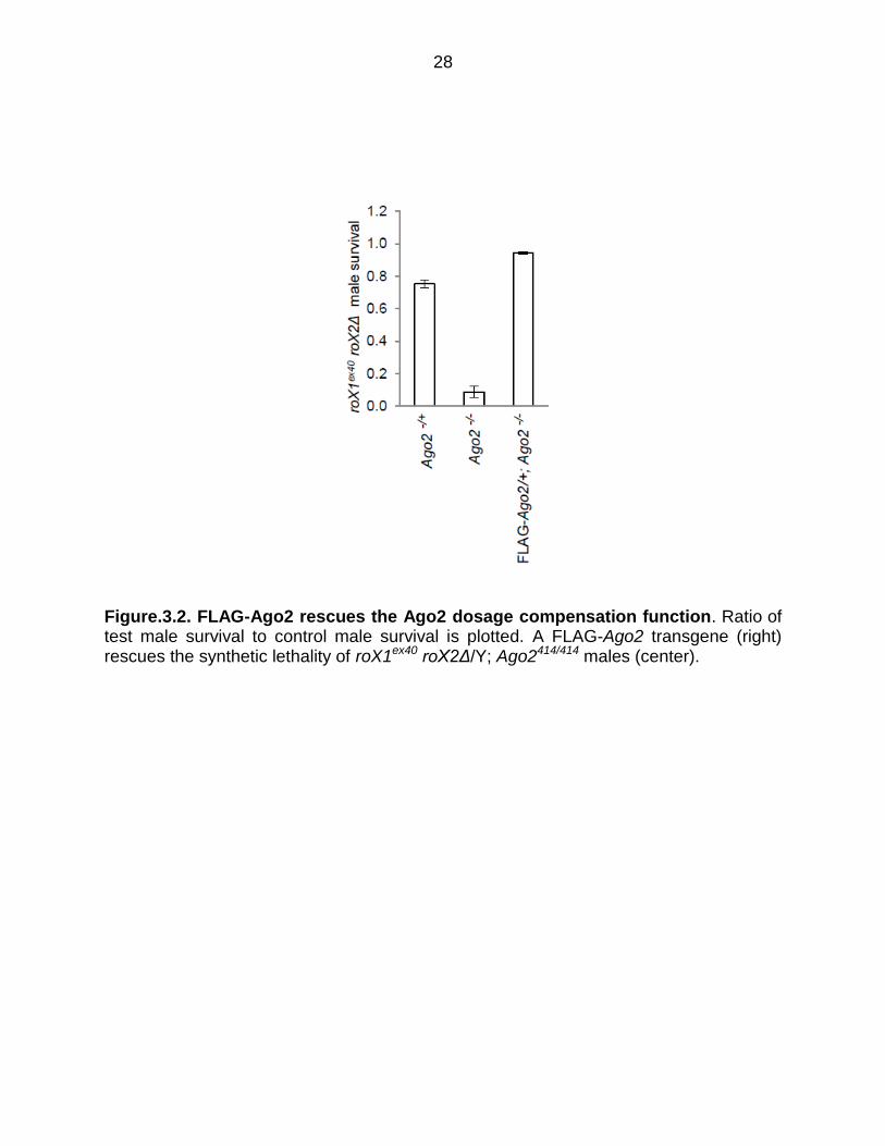
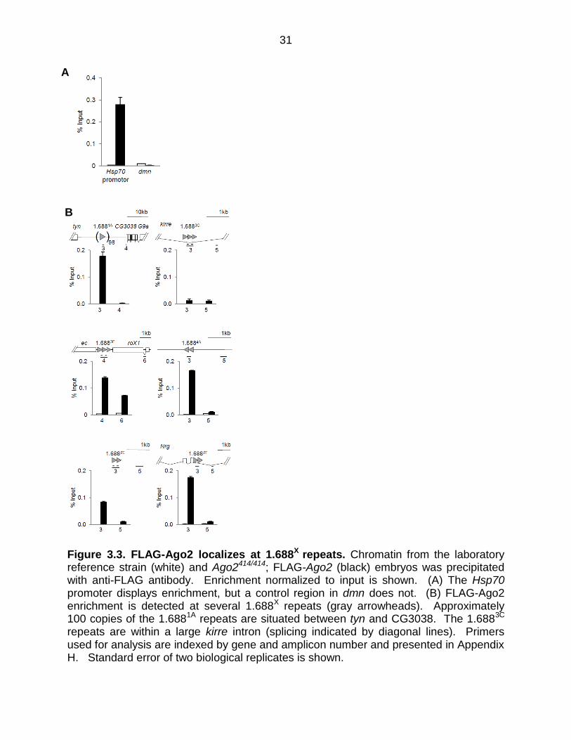
Ago2 localizes at 1.688X repeats.
Ago2 localization was determined using a FLAG-tagged Ago2 transgene that was
first tested for rescue of the dosage compensation function of Ago2. Males with the
partial loss of function roX1ex40roX2Δ chromosome have high survival, as do Ago2-/-
flies, but synthetic lethality is observed in roX1ex40roX2Δ/Y; Ago2-/- males (Menon and
Meller, 2012). One copy of a FLAG-Ago2 transgene rescues these males,
demonstrating that the FLAG tag does not disrupt the dosage compensation function of
Ago2 (Figure 3.1, 3.2). Chromatin from FLAG-Ago2; Ago-/- embryos, and from a
reference strain lacking the FLAG-Ago2 transgene, was immunoprecipitated with anti-
FLAG antibodies and enrichment determined by quantitative PCR (qPCR). FLAG-Ago2
was enriched at the Hsp70 promoter, a site known to bind Ago2 (Cernilogar et al., 2011)
(Figure 3.3 A). In contrast, a control region in the dmn gene displayed no enrichment.
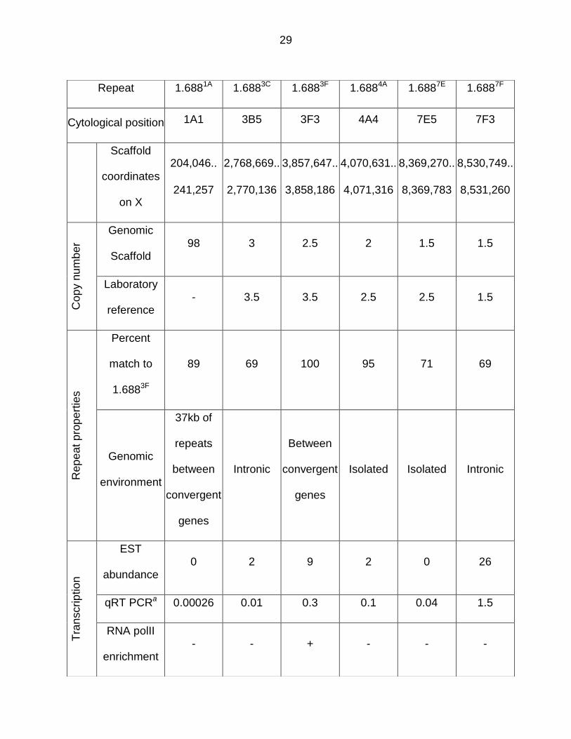
We then examined FLAG-Ago2 enrichment at a panel of six representative 1.688X
repeats that differ in location, copy number, sequence, genetic environment and
26
transcription level (Table 3.1). Interestingly, five of these show enrichment of FLAG-
Ago2 over the repeats, but little or no enrichment in flanking regions (Figure 3.3 B). We
conclude that Ago2 localizes at many 1.688X repeats, a finding that is consistent with
involvement of Ago2 in siRNA-directed recruitment of chromatin modification at or
around these regions.
27
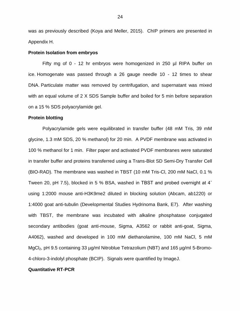
Figure 3.1. Mating scheme to express FLAG-Ago2 in Ago2 mutants. FLAG-Ago2 is marked by w+, enabling identification through the multiple crossing steps. roX2Δ is also marked by w+. Presence of both, roX2Δ and FLAG-Ago2 results in a darker red eye color.
28
Figure.3.2. FLAG-Ago2 rescues the Ago2 dosage compensation function. Ratio of test male survival to control male survival is plotted. A FLAG-Ago2 transgene (right) rescues the synthetic lethality of roX1ex40 roX2Δ/Y; Ago2414/414 males (center).
29
Repeat 1.6881A 1.6883C 1.6883F 1.6884A 1.6887E 1.6887F
Cytological position 1A1 3B5 3F3 4A4 7E5 7F3
Scaffold
coordinates
on X
204,046..
241,257
2,768,669..
2,770,136
3,857,647..
3,858,186
4,070,631..
4,071,316
8,369,270..
8,369,783
8,530,749..
8,531,260
Cop
y n
um
be
r
Genomic
Scaffold 98 3 2.5 2 1.5 1.5
Laboratory
reference - 3.5 3.5 2.5 2.5 1.5
Rep
eat
pro
pe
rtie
s
Percent
match to
1.6883F
89 69 100 95 71 69
Genomic
environment
37kb of
repeats
between
convergent
genes
Intronic
Between
convergent
genes
Isolated Isolated Intronic
Tra
nscrip
tio
n
EST
abundance 0 2 9 2 0 26
qRT PCRa 0.00026 0.01 0.3 0.1 0.04 1.5
RNA polII
enrichment - - + - - -
30
Table 3.1. Panel of 1.688X repeats used in this study. Cytological positions of 1.688X repeats and scaffold coordinates were determined from Flybase (Release 6). The size of some repeat clusters in our laboratory reference strain was found to differ from the genomic scaffold. See Appendix D for determination of copy number. Similarity to 1.6883F was determined by BLAST. EST abundance was inferred from assigned ESTs in Flybase. RNA polII enrichment is derived from ChIP-seq of 6-8 h mesoderm (Monfort and Furlong, 2015). a Quantitative RT-PCR (qRT PCR) is normalized to repeat copy number (see Figure 3.11).
31
Figure 3.3. FLAG-Ago2 localizes at 1.688X repeats. Chromatin from the laboratory reference strain (white) and Ago2414/414; FLAG-Ago2 (black) embryos was precipitated with anti-FLAG antibody. Enrichment normalized to input is shown. (A) The Hsp70 promoter displays enrichment, but a control region in dmn does not. (B) FLAG-Ago2 enrichment is detected at several 1.688X repeats (gray arrowheads). Approximately 100 copies of the 1.6881A repeats are situated between tyn and CG3038. The 1.6883C repeats are within a large kirre intron (splicing indicated by diagonal lines). Primers used for analysis are indexed by gene and amplicon number and presented in Appendix H. Standard error of two biological replicates is shown.
A
B
32
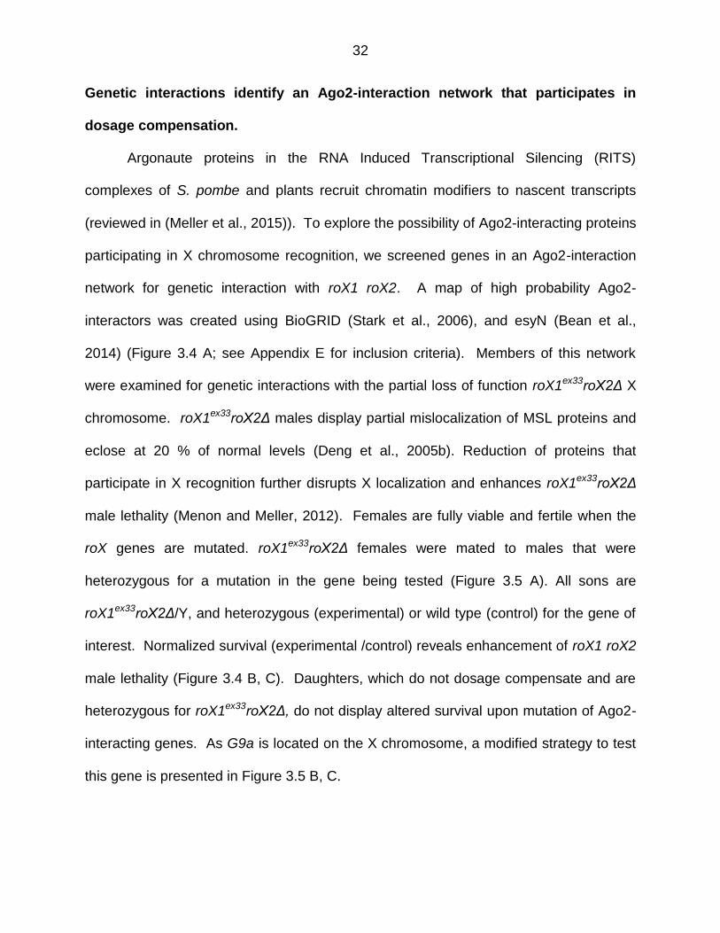
Genetic interactions identify an Ago2-interaction network that participates in
dosage compensation.
Argonaute proteins in the RNA Induced Transcriptional Silencing (RITS)
complexes of S. pombe and plants recruit chromatin modifiers to nascent transcripts
(reviewed in (Meller et al., 2015)). To explore the possibility of Ago2-interacting proteins
participating in X chromosome recognition, we screened genes in an Ago2-interaction
network for genetic interaction with roX1 roX2. A map of high probability Ago2-
interactors was created using BioGRID (Stark et al., 2006), and esyN (Bean et al.,
2014) (Figure 3.4 A; see Appendix E for inclusion criteria). Members of this network
were examined for genetic interactions with the partial loss of function roX1ex33roX2Δ X
chromosome. roX1ex33roX2Δ males display partial mislocalization of MSL proteins and
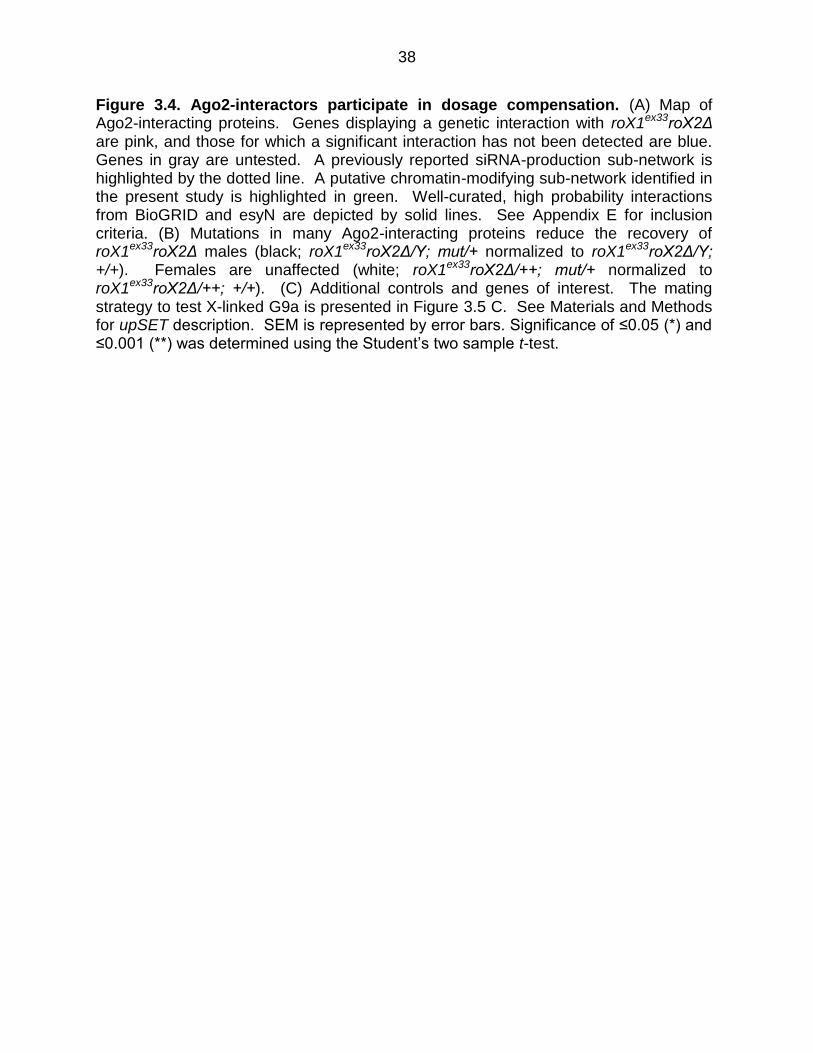
eclose at 20 % of normal levels (Deng et al., 2005b). Reduction of proteins that
participate in X recognition further disrupts X localization and enhances roX1ex33roX2Δ
male lethality (Menon and Meller, 2012). Females are fully viable and fertile when the
roX genes are mutated. roX1ex33roX2Δ females were mated to males that were
heterozygous for a mutation in the gene being tested (Figure 3.5 A). All sons are
roX1ex33roX2Δ/Y, and heterozygous (experimental) or wild type (control) for the gene of
interest. Normalized survival (experimental /control) reveals enhancement of roX1 roX2
male lethality (Figure 3.4 B, C). Daughters, which do not dosage compensate and are
heterozygous for roX1ex33roX2Δ, do not display altered survival upon mutation of Ago2-
interacting genes. As G9a is located on the X chromosome, a modified strategy to test
this gene is presented in Figure 3.5 B, C.
33
Normalized survival of roX1ex33roX2Δ males with mutations in the Ago2-
interaction network is presented in Figure 3.4 B. Genes displaying significant
interactions are noted by pink symbols, and those showing no interaction are blue in
Figure 3.4 A. We confirmed a previously identified siRNA-processing sub-network
containing Dcr2, Elp1, and loqs (Figure 3.4 A, dotted line; (Menon and Meller, 2012)).
The present study identified several additional Ago2-interactors, including a potential
chromatin-modifying sub-network containing Dcr1, Fmr1, Rm62, and the histone
methyltransferase Su(var)3-9 (green, Figure 3.4 A). Su(var)3-9 deposits H3K9me2 and
acts with Rm62 to re-silence active chromatin (Boeke et al., 2011).
Additional chromatin modifiers and genes in other small RNA pathways were
also tested (Figure 3.4 C). A previous study found no interaction between roX1ex33roX2Δ
and the piRNA pathway genes aub and piwi, or the miRNA pathway gene Ago1, a
finding replicated here. Since our findings point towards involvement of chromatin
modifiers, we tested the chromatin regulatory factor Su(var)2-10 and two additional
H3K9 methyltransferases, eggless (egg) and G9a (Figure 3.4 C). None of these
modified roX1ex33roX2Δ survival. Mutations in Su(var)3-7, important for heterochromatin
formation, and upSET, which maintains heterochromatin and H3K9me2 levels, enhance
roX1ex33roX2Δ male lethality (McElroy et al., 2017; Spierer et al., 2008). Over
compensating males (ocm) has an unusual dosage compensation phenotype as
mutations in ocm rescue males with insufficient MSL activity, suggesting that it might act
as a governor of activation (Lim and Kelley, 2013). Interestingly, mutation of ocm
significantly increased the survival of roX1ex33roX2Δ males, supporting the idea that ocm
normally restrains activation. The P{EPgy2}09821 and P{EPgy2}15840 strains, used to
34
outcross Su(var)3-9 and barr mutants, display no interaction and serve as controls for
genetic background. Taken together, these findings suggest that several genes that
deposit H3K9me2, maintain this mark or participate in heterochromatin formation also
contribute to X chromosome dosage compensation. At first glance these observations
appear to be at odds with X chromosome hypertranscription, the ultimate consequence
of X chromosome recognition.
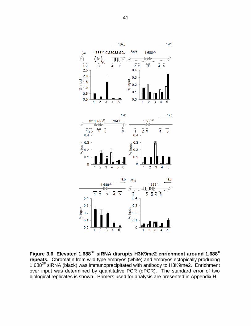
Ectopically expressed 1.6883F siRNA disrupts H3K9me2 patterns
Previous studies found that ectopically produced 1.6883F siRNA partially rescues
roX1 roX2 males and increases X localization of the MSL complex (Menon et al., 2014).
The mechanism by which siRNA promotes X recognition is unknown. The discovery
that insertion of 1.688X DNA on an autosome enables functional compensation of
nearby genes, and the enhancement of this effect by ectopic 1.6883F siRNA, suggests
siRNA action through cognate genomic regions (Joshi and Meller, 2017). In accord with
this idea, an autosomal roX1 transgene also enables compensation of nearby genes,
but is unaffected by 1.6883F siRNA. To test the idea that 1.6883F siRNA directs
epigenetic modification of 1.688X chromatin, we used ChIP to analyze chromatin around
1.688X repeats on the X chromosome. ChIP-qPCR detected H3K9me2 enrichment in 4
out of 6 repeats (white bars, Figure 3.6). As H3K9me2 enrichment was not uniform, we
considered additional factors that might determine this mark, and noted that only
repeats showing evidence of transcription were enriched for H3K9me2, consistent with
the idea of Ago2-dependent recruitment to nascent transcripts (Verdel et al., 2004).
Upon ectopic expression of 1.6883F siRNA a dramatic disruption of H3K9me2 was
observed in and around 1.688X repeats (black bars, Figure 3.6). For example, 1.6883F
35
and 1.6884A display peaks of H3K9me2 in wild type flies, but this mark was reduced
over the repeats and increased in surrounding regions by elevated 1.6883F siRNA.
Untranscribed repeat clusters at 1.6881A and 1.6887E show no H3K9me2 enrichment in
wild type flies, but gained H3K9me2 upon expression of 1.6883F siRNA. In contrast, no
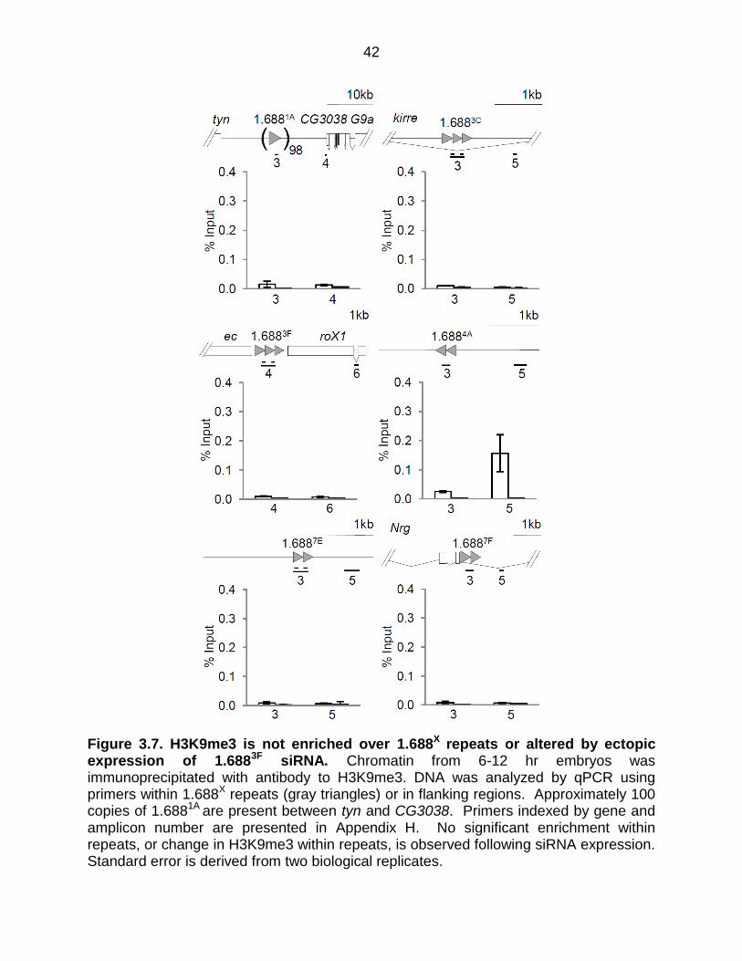
enrichment of H3K9me3 in or near 1.688X repeats was detected in wild type or 1.6883F
siRNA-expressing embryos (Figure 3.7). This is as expected as H3K9me2 is found in
facultative heterochromatin by contrast H3K9me3 is found in constitutive
heterochromatin such as at telomeres and centromeres (Becker et al., 2016). We
conclude that some 1.688X repeats are enriched for H3K9me2, and that ectopic
production of cognate siRNA broadly disrupts this mark.
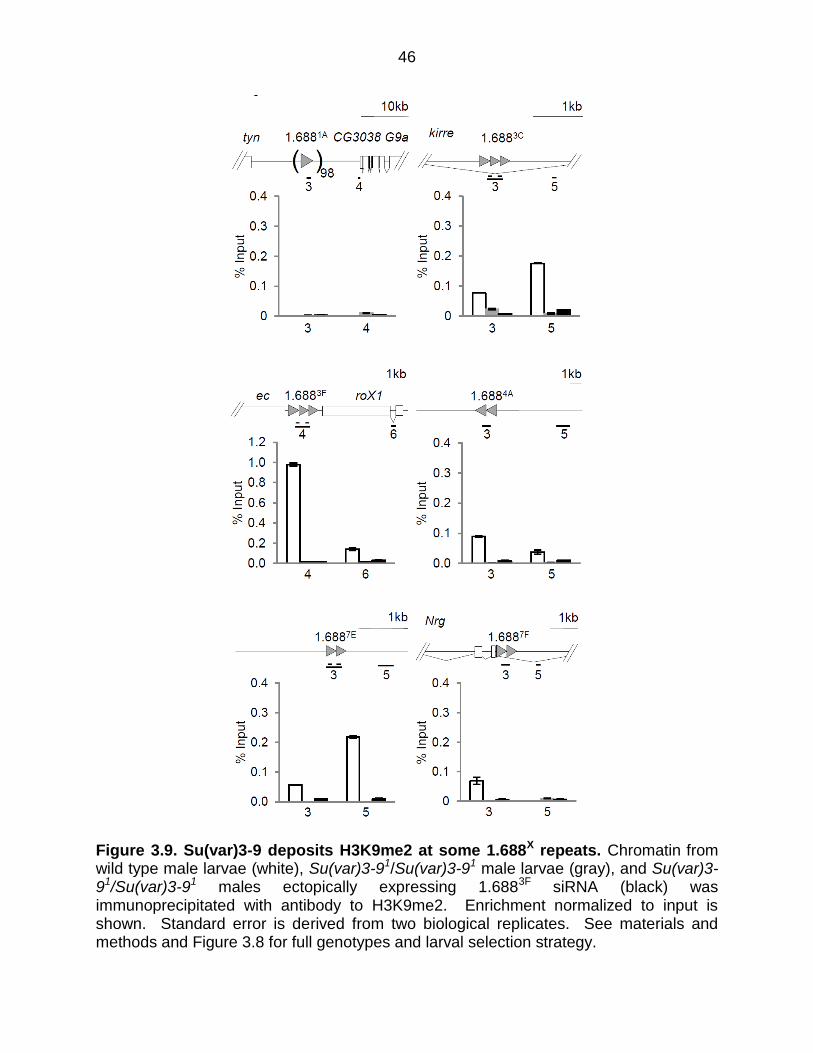
Su(var)3-9 deposits H3K9me2 at 1.688X repeats
The identification of Su(var)3-9 as an indirect binding partner of Ago2, observation of a
genetic interaction between Su(var)3-9 and roX1 roX2 and enrichment of H3K9me2 on
some 1.688X repeats suggests that Su(var)3-9 could be modifying 1.688X repeats. D.
melanogaster has three histone H3K9 methyltransferase, Su(var)3-9, eggless, and G9a,
but only Su(var)3-9 mutations enhance the male lethality of roX1 roX2 ((Swaminathan
et al., 2012); Figure 3.4). To determine if Su(var)3-9 is responsible for H3K9me2 at
1.688X chromatin, 3rd instar larvae mutated for Su(var)3-9, or mutated for Su(var)3-9
and expressing 1.6883F siRNA, were generated (Figure 3.8). H3K9me2 enrichment is
virtually eliminated over 1.688X repeats in Su(var)3-9-/- mutants (gray bars, Figure 3.9)
and remains low in Su(var)3-9-/- larvae that express 1.6883F siRNA (black bars, Figure
3.9). This reveals that Su(var)3-9 deposits H3K9me2 at 1.688X chromatin in wild type
flies, and eliminates the possibility that a different methyltransferase is recruited to these

36
regions following ectopic expression of 1.6883F siRNA. Disruption of H3K9me2 upon
expression of 1.6883F siRNA thus reflects changes in the localization or activity of
Su(var)3-9.
37
A
B
C
A
C
38
Figure 3.4. Ago2-interactors participate in dosage compensation. (A) Map of Ago2-interacting proteins. Genes displaying a genetic interaction with roX1ex33roX2Δ are pink, and those for which a significant interaction has not been detected are blue. Genes in gray are untested. A previously reported siRNA-production sub-network is highlighted by the dotted line. A putative chromatin-modifying sub-network identified in the present study is highlighted in green. Well-curated, high probability interactions from BioGRID and esyN are depicted by solid lines. See Appendix E for inclusion criteria. (B) Mutations in many Ago2-interacting proteins reduce the recovery of roX1ex33roX2Δ males (black; roX1ex33roX2Δ/Y; mut/+ normalized to roX1ex33roX2Δ/Y; +/+). Females are unaffected (white; roX1ex33roX2Δ/++; mut/+ normalized to roX1ex33roX2Δ/++; +/+). (C) Additional controls and genes of interest. The mating strategy to test X-linked G9a is presented in Figure 3.5 C. See Materials and Methods for upSET description. SEM is represented by error bars. Significance of ≤0.05 (*) and ≤0.001 (**) was determined using the Student’s two sample t-test.
39
B
A
C
40
Figure 3.5. Detection of genetic interactions between roX1 roX2 and candidate genes. (A) roX1ex33roX2Δ females were mated to males heterozygous for a mutation in the gene of interest. The survival of sons mutated for the gene of interest (bottom right) is divided by that of control brothers (bottom left) and presented in Figure 3.4 B and C. In an otherwise wild type background, roX1ex33roX2Δ allows 20 % adult male escapers. Females do not dosage compensate and serve as an internal control. (B) Mating scheme to generate G9a roX1ex33roX2Δ mutants. G9a is marked by GFP and roX2Δ is marked by w+, enabling identification of recombinants carrying both mutations. Recombinants that were also roX1ex33, predicted to be 33.5% of the lines screened, were identified by PCR. G9a roX1ex33roX2Δ recombinant 3 was used in subsequent studies. (C) Testing for genetic interaction between G9a and roX1ex33roX2Δ. Heterozygous G9aMB1197 roX1ex33roX2Δ/+ roX1ex33roX2Δ females were mated to G9a
MB1197 roX1 roX2 males. G9aMB1197 is marked with EGFP. The survival of G9a MB1197 roX1ex33roX2Δ sons (EGFP-positive, right) was divided by that of EGFP-negative + roX1ex33roX2Δ sons (left). EGFP intensity differentiates F1 females that are homozygous or heterozygous for G9a MB1197.
41
Figure 3.6. Elevated 1.6883F siRNA disrupts H3K9me2 enrichment around 1.688X repeats. Chromatin from wild type embryos (white) and embryos ectopically producing 1.6883F siRNA (black) was immunoprecipitated with antibody to H3K9me2. Enrichment over input was determined by quantitative PCR (qPCR). The standard error of two biological replicates is shown. Primers used for analysis are presented in Appendix H.
42
Figure 3.7. H3K9me3 is not enriched over 1.688X repeats or altered by ectopic expression of 1.6883F siRNA. Chromatin from 6-12 hr embryos was immunoprecipitated with antibody to H3K9me3. DNA was analyzed by qPCR using primers within 1.688X repeats (gray triangles) or in flanking regions. Approximately 100 copies of 1.6881A are present between tyn and CG3038. Primers indexed by gene and amplicon number are presented in Appendix H. No significant enrichment within repeats, or change in H3K9me3 within repeats, is observed following siRNA expression. Standard error is derived from two biological replicates.
43
To determine how far from 1.688X repeats the H3K9me2 disruption extends,
regions 10-26 kb from repeats were examined. In each case, these regions displayed
increased H3K9me2 in embryos with ectopic 1.6883F siRNA expression (Figure 3.10 A).
This suggested the possibility of a global change in H3K9me2. To address this
possibility we probed protein blots from wild type and 1.6883F siRNA-expressing
embryos to determine the levels of this modification. In spite of apparently wide-spread
elevation of H3K9me2, no evidence for a global change in H3K9me2 level is detected
(Figure 3.10 B). As most H3K9me2 is found in heterochromatic regions that comprise
~30% of the fly genome, changes in euchromatic regions may represent a negligible
portion of the nuclear pool.
H3K9me2 is generally thought to be repressive, but compensation in flies occurs
by increased expression of X-linked genes. To determine if changes in H3K9me2
enrichment correlate with changes in transcription, expression of genes near 1.688X
repeats was examined in wild type and 1.6883F siRNA-expressing embryos. Consistent
with H3K9me2 having a repressive effect, 1.6883F siRNA decreases accumulation of
RNA from non-coding intragenic or intronic regions with elevated H3K9me2 (Figure
3.11). The apparent increase in 1.6883F expression (Figure 3.11) is from the transgene
used to produce ectopic 1.6883F siRNA. We detected dramatic reductions in messages
adjacent to 1.6881A (tyn, G9a) and 1.6883F (ec, roX1). In spite of a 90% reduction in ec
transcript in embryos expressing 1.6883F siRNA, adults of this genotype do not display
the rough eye ec phenotype. It is possible that ectopic 1.6883F siRNA has a more
pronounced effect in embryos, whose undifferentiated cells may be particularly
susceptible to chromatin-based disruption. Mature patterns of chromatin organization
44
are established by late larval life, and these may be more resistant. To test this, we
examined expression in wild type and 1.6883F siRNA-expressing 3rd instar male larvae,
and found that tyn, G9a and ec regained wild type levels of transcript, and roX1 was
also largely restored (Figure 3.11). This might be due to recovery upon establishment of
mature chromatin. The precise reason for the differences between embryos and larvae
are uncertain, but restoration of normal gene expression by the 3rd instar larvae is
consistent with the lack of phenotype in otherwise wild type flies that ectopically express
1.6883F siRNA (Menon et al., 2014).
The discovery that animal age influenced the response to ectopic siRNA
prompted us to determine the time point at which H3K9me2 is established at 1.688X
repeats. A possible scenario is that this mark is placed before MSL localization, and
acts in some way to guide X recognition. X-localization of the MSL complex occurs at 3
hr after egg laying (AEL) (Meller, 2003; Rastelli et al., 1995). We determined H3K9me2
enrichment at 1.6883F in embryos before the MSL complex binds to the X (1.5-3 hr),
during initial MSL recruitment (3-4 hr), and at 4-6 hr and 6-12 hr. In contrast to our
prediction, H3K9me2 is first detected on 1.6883F between 6 and 12 h AEL, after X
localization of the MSL complex has occurred (Figure 3.12). We conclude that
H3K9me2 at 1.688X repeats is unlikely to guide initial X recognition, but may serve at a
later time point to facilitate spreading of this mark or enforce the stability of X
recognition.
45
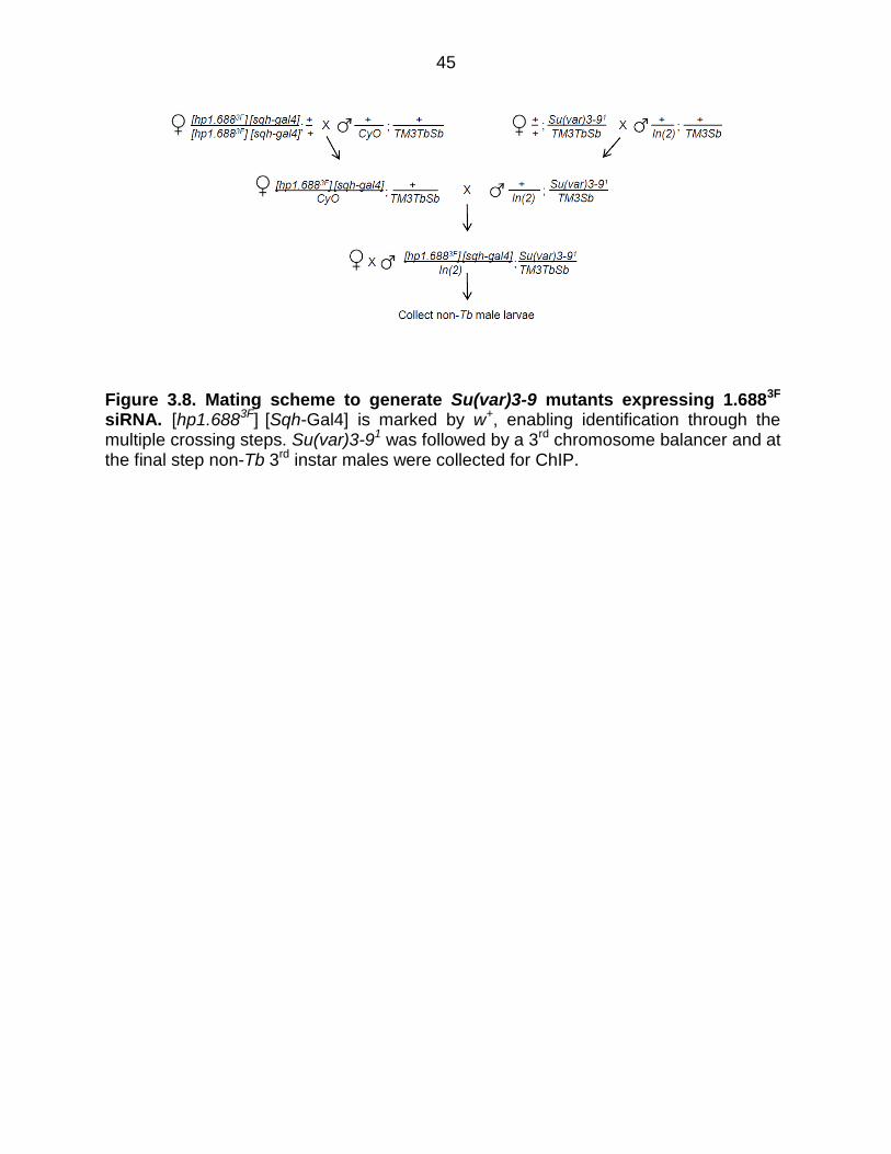
Figure 3.8. Mating scheme to generate Su(var)3-9 mutants expressing 1.6883F siRNA. [hp1.6883F] [Sqh-Gal4] is marked by w+, enabling identification through the multiple crossing steps. Su(var)3-91 was followed by a 3rd chromosome balancer and at the final step non-Tb 3rd instar males were collected for ChIP.
46
Figure 3.9. Su(var)3-9 deposits H3K9me2 at some 1.688X repeats. Chromatin from wild type male larvae (white), Su(var)3-91/Su(var)3-91 male larvae (gray), and Su(var)3-91/Su(var)3-91 males ectopically expressing 1.6883F siRNA (black) was immunoprecipitated with antibody to H3K9me2. Enrichment normalized to input is shown. Standard error is derived from two biological replicates. See materials and methods and Figure 3.8 for full genotypes and larval selection strategy.
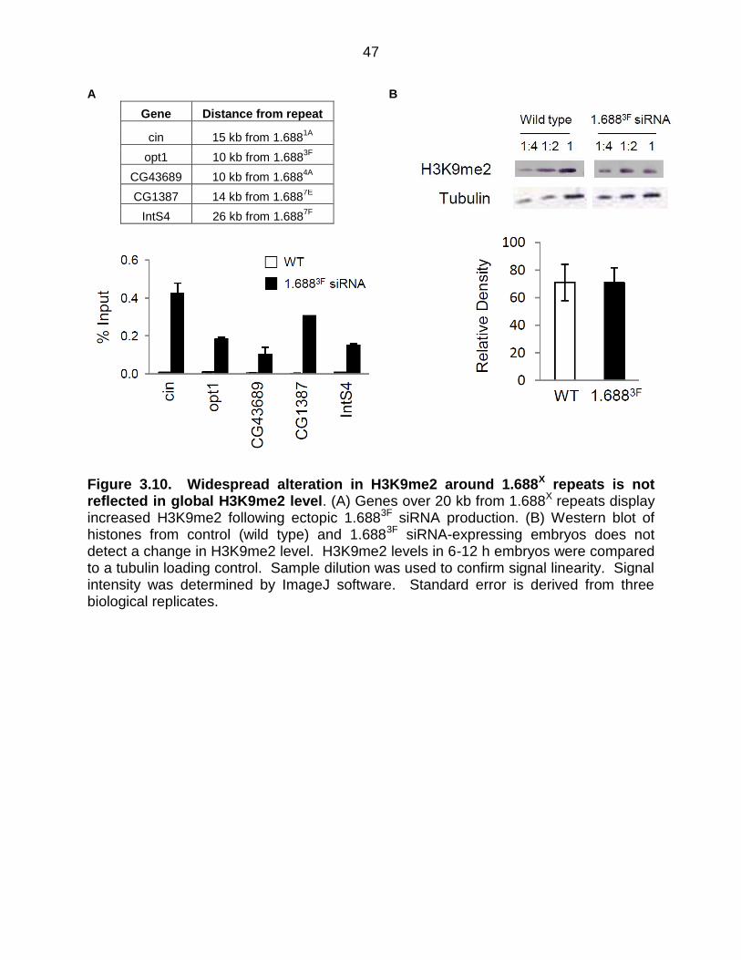
47
A B
Gene Distance from repeat
cin 15 kb from 1.6881A
opt1 10 kb from 1.6883F
CG43689 10 kb from 1.6884A
CG1387 14 kb from 1.6887E
IntS4 26 kb from 1.6887F
Figure 3.10. Widespread alteration in H3K9me2 around 1.688X repeats is not reflected in global H3K9me2 level. (A) Genes over 20 kb from 1.688X repeats display increased H3K9me2 following ectopic 1.6883F siRNA production. (B) Western blot of histones from control (wild type) and 1.6883F siRNA-expressing embryos does not detect a change in H3K9me2 level. H3K9me2 levels in 6-12 h embryos were compared to a tubulin loading control. Sample dilution was used to confirm signal linearity. Signal intensity was determined by ImageJ software. Standard error is derived from three biological replicates.
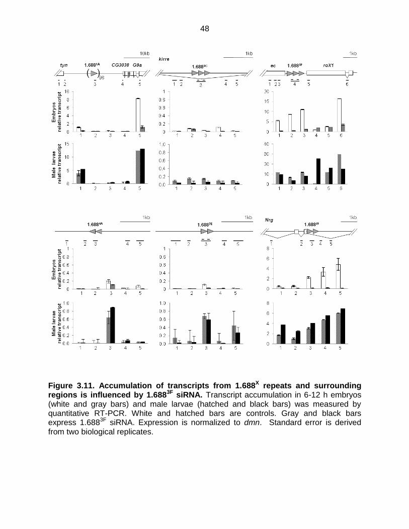
48
Figure 3.11. Accumulation of transcripts from 1.688X repeats and surrounding regions is influenced by 1.6883F siRNA. Transcript accumulation in 6-12 h embryos (white and gray bars) and male larvae (hatched and black bars) was measured by quantitative RT-PCR. White and hatched bars are controls. Gray and black bars express 1.6883F siRNA. Expression is normalized to dmn. Standard error is derived from two biological replicates.
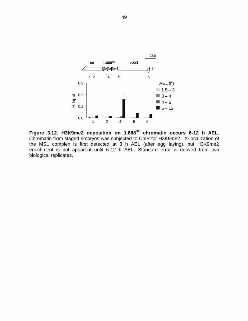
49
Figure 3.12. H3K9me2 deposition on 1.6883F chromatin occurs 6-12 h AEL. Chromatin from staged embryos was subjected to ChIP for H3K9me2. X-localization of the MSL complex is first detected at 3 h AEL (after egg laying), but H3K9me2 enrichment is not apparent until 6-12 h AEL. Standard error is derived from two biological replicates.
0.0
0.1
0.2
0.3
1.5 – 3
3 – 4
4 – 6
6 – 12
1 2 4 5 6
% In
put
AEL (h)
5
1kb
4 61 2
1.6883Fec roX1
50
H3K9me2 is enriched at regions flanking autosomal 1.6883F transgenes
One challenge of studying recruiting elements on the X chromosome is that the
redundancy and proximity of elements complicates interpretation. To overcome this we
tested integrations of 1.6883F or roX1 on 2L (Figure 3.13 A, B, 3.14 A) (Joshi and Meller,
2017). ChIP for H3K9me2 was performed on chromatin from male 3rd instar larvae with
1.6883F or roX1 integrations on 2L (gray bars, 3.14 B, C), and in the same genotypes
but with ectopic expression of 1.6883F siRNA (black bars, Figure 3.14 B, C). H3K9me2
is not strongly enriched in autosomal chromatin flanking roX1, but is striking enriched
near the 1.6883F integration. Consistent with our observations in embryos, ectopic
1.6883F siRNA expression elevated H3K9me2 near the 1.6883F integration. This
contrasts with negligible enrichment at the roX1 transgene (Figure 3.6, Figure 3.14 B,
C). For reasons that we do not understand, enrichment over the integrated 1.6883F
repeats was undetectable. We conclude that autosomal insertion of 1.688X DNA makes
nearby chromatin subject to siRNA-induced H3K9me2 deposition. Taken together,
these studies support the idea that the 1.688X repeats influence patterns of H3K9me2
nearby, but roX1, with a different class of recruiting element, does not.
To determine the influence of 1.6883F and roX1 on transcription of nearby
autosomal genes, we performed quantitative RT-PCR (qRT PCR) on total RNA from 3rd
instar male larvae with the 2L integrations described above, with and without ectopic
1.6883F siRNA. The 1.6883F and roX1 integration site is in an intron of haf, one of the
genes measured. We also examined RFeSP, CG33128, Eno (2, 58 and 114 kb from
haf, respectively), and CG31778 and Rpl37A, 2.1 and 3.5 Mb from haf (Figure 3.14 D).
The presence of 1.6883F or roX1 integrations alone had no effect on the most distant
51
genes, CG31778 and Rpl37A. A roX1 integration increased expression of haf 2.5 fold,
more than expected from full compensation. This may reflect the fact that autosomal
MSL recruitment by a roX1 transgene can overcome local, chromatin-based silencing
(Kelley and Kuroda, 2003). Addition of 1.6883F siRNA increased haf expression slightly,
and similarly increases expression of CG33128 and Eno (light gray bars, Figure 3.14 E).
A 1.6883F insertion produced a four-fold increase in haf, and a slight increase in
Eno, 114 kb distant. But upon expression of 1.6883F siRNA, haf expression increased
to 8 fold wild type levels, and CG33128 and Eno both increased to ~2 fold wild type
levels, consistent with full compensation. We conclude that an autosomal insertion of
1.688X DNA allows relatively distant genes to increase expression, an effect that is
enhanced by 1.6883F siRNA.
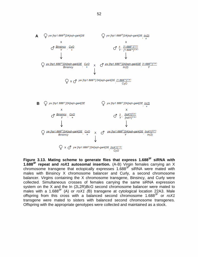
52
Figure 3.13. Mating scheme to generate flies that express 1.6883F siRNA with 1.6883F repeat and roX1 autosomal insertion. (A-B) Virgin females carrying an X chromosome transgene that ectopically expresses 1.6883F siRNA were mated with males with Binsincy X chromosome balancer and Curly, a second chromosome balancer. Virgins containing the X chromosome transgene, Binsincy, and Curly were collected. Simultaneous crosses of females carrying the same siRNA expression system on the X and the In (2L2R)BcG second chromosome balancer were mated to males with a 1.6883F (A) or roX1 (B) transgene at cytological location 22A3. Male offspring from this cross with a balanced second chromosome 1.6883F or roX1 transgene were mated to sisters with balanced second chromosome transgenes. Offspring with the appropriate genotypes were collected and maintained as a stock.
A
B
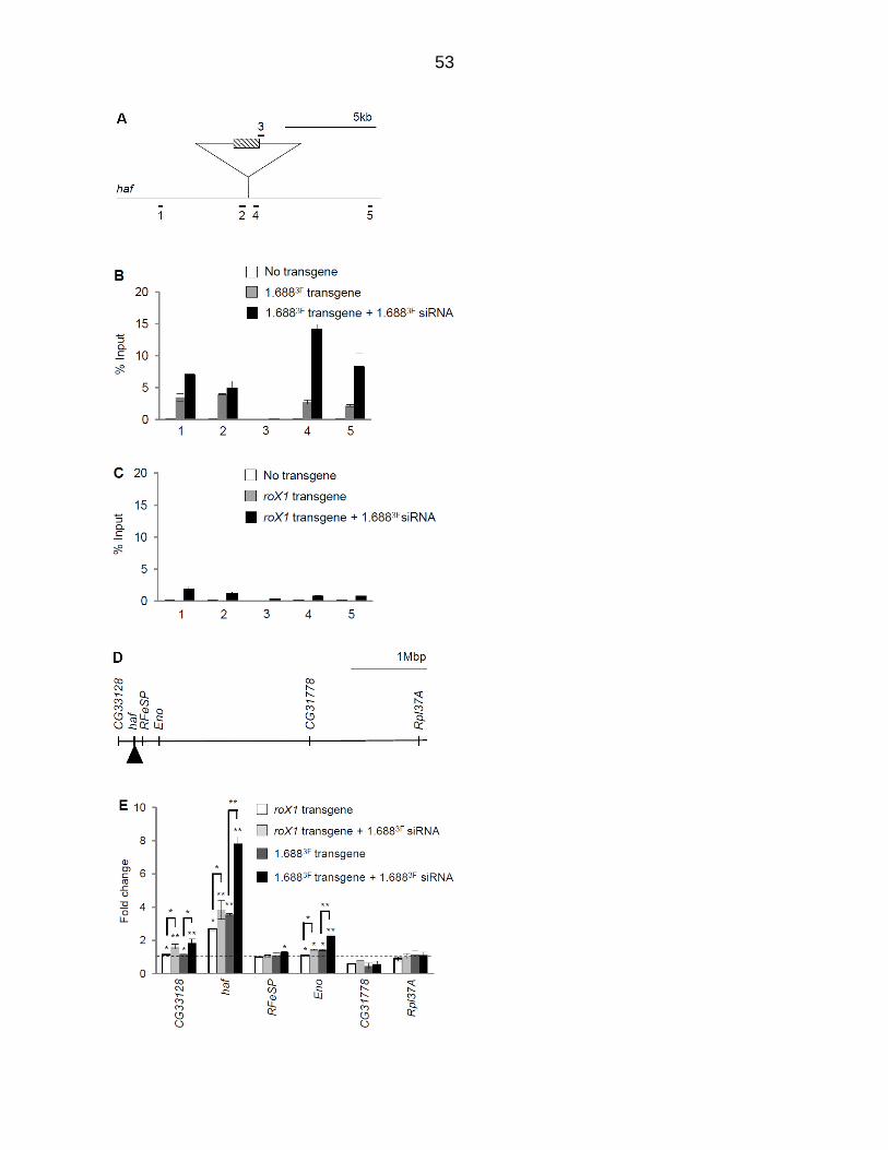
53
54
Figure 3.14. Ectopic 1.6883F siRNA increases H3K9me2 flanking an autosomal 1.6883F DNA insertion and elevates expression of nearby genes. (A) Amplicons flanking the landing site in a large haf intron at 22A3 (splicing not shown). (B) H3K9me2 enrichment surrounding the 1.6883F transgene. Chromatin from wild type third instar male larvae (white), larvae with 1.6883F DNA at the landing site (gray), and larvae with 1.6883F DNA at the landing site and ectopic 1.6883F siRNA (black) was immunoprecipitated with antibody to H3K9me2. (C) H3K9me2 enrichment surrounding a roX1 insertion. Chromatin from wild type male third instar larvae (white), larvae with the roX1 insertion (gray), and with the roX1 insertion and ectopic 1.6883F siRNA (black) was immunoprecipitated. Data is from two biological replicates and enrichment is normalized to input. (D) Portion of 2L showing relative location of CG33128, haf, RFeSP, Eno, CG31778, and Rpl37A. (E) Accumulation of transcripts in male larvae carrying roX1 (white) or 1.6883F insertions (dark gray), and in male larvae that express ectopic 1.6883F siRNA and have roX1 (light gray) or 1.6883F integrations (black). Expression is normalized to dmn. SEM is derived from three biological replicates. Significance was determined using Student’s two sample t-test, ≤0.05 (*), ≤0.001 (**) significance. Primers are presented in Appendix H.
55
Discussion
Molecularly distinct dosage compensation strategies have arisen independently
in different organisms, but a shared feature is the ability to selectively recognize and
alter an entire chromosome. How a regulatory system is directed to a single
chromosome is poorly understood. The discovery that 1.688X satellite DNA promotes
recruitment of dosage compensation to nearby genes supports the idea that these
repeats are important for selective recognition of X chromatin (Joshi and Meller, 2017).
How the 1.688X repeats accomplish this is a question of great interest. Involvement of
the siRNA pathway, and siRNA from a 1.688X repeat, in X recognition points to the
possibility that siRNA-directed modification of chromatin around 1.688X repeats plays a
role in dosage compensation. The findings of the current study support this idea.
Although numerous studies point to small RNA regulation of chromatin in flies,
this process is better understood in other organisms (reviewed in (Meller et al., 2015)).
Small RNA directed heterochromatin formation was discovered in S. pombe (reviewed
in (Moazed, 2009)). Heterochromatic regions are transcribed during S phase, and
transcripts are processed into siRNAs that guide the Ago1-containing RITS complex to
complementary, nascent transcripts (Verdel et al., 2004). In addition to several other
activities, RITS recruits the H3K9 methyltransferase Clr4 (Zhang et al., 2008). We
propose that a similar process is occurring at 1.688X chromatin in flies. Most 1.688X
repeats bind Ago2, and many are transcribed. Several of the 1.688X repeats that we
examined are enriched for H3K9me2 deposited by Su(var)3-9, an ortholog of Clr4. Our
screen identified genetic interactions between roX1 roX2 and members of a possible
RITS-like complex consisting of Ago2, Rm62 and Su(var)3-9. Finally, H3K9me2
56
enrichment in, and around, 1.688X repeats is responsive to 1.688X siRNA, and
enrichment is blocked by loss of Su(var)3-9. Taken together, these findings are
suggestive of a RITS-like complex modifying chromatin at 1.688X repeats.
The idea that repressive H3K9me2 marks participate in a process culminating in
a two-fold increase in expression is counterintuitive. However, numerous studies have
found links between the compensated X chromosome of male flies and repressive
marks. For example, the male X is enriched in HP1, a major constituent of
heterochromatin that binds H3K9me2 (de Wit et al., 2005; Liu et al., 2005). The
structure of the polytenized male X chromosome is extraordinarily sensitive to altered
levels of genes that participate in heterochromatin formation or silencing, such as HP1,
Su(var)3-7 and ISWI. Mutations in these genes produce a general disruption of
polytenization that is strikingly specific to the male X (Deuring et al., 2000; Spierer et al.,
2005; Zhang et al., 2006). JIL-1, a kinase that enforces boundaries between
heterochromatin and euchromatin, is enriched on the X chromosome and thought to
participate in compensation (Deng et al., 2005a; Ebert et al., 2004; Jin et al., 2000;
Wang et al., 2001). Upon loss of JIL-1, polytenized structure is disrupted and H3K9me2
invades euchromatic chromosome arms, but the X chromosome is most severely
affected (Zhang et al., 2006). Finally, the MSL proteins themselves have an affinity for
heterochromatin. In roX1 roX2 mutant males the MSL proteins become mislocalized to
ectopic autosomal sites (Meller and Rattner, 2002). For reasons that are still unclear,
the most prominent of these sites are the heterochromatic 4th chromosome and
chromocenter (Deng and Meller, 2006; Figueiredo et al., 2014). Taken together, these
observations suggest that recognition and spreading of the MSL complex could be
57
facilitated by repressive marks. One intriguing possibility is that 1.688X repeats guide
deposition of H3K9me2 and this mark, directly or indirectly, assists localization of the
MSL complex.
An intriguing aspect of dosage compensation is the evolutionary convergence of
mechanisms. For example, long non-coding RNA also plays a central role in X
recognition in mammals, where expression of the X inactive specific transcript (Xist)
RNA guides X inactivation (Lee, 2009). Furthermore, repetitive LINE-1 elements that
are enriched on the mammalian X chromosome are proposed to facilitate X inactivation
(Bailey et al., 2000; Lyon, 1998). Interestingly, some LINE-1 elements are transcribed
during the onset of X inactivation, producing endo-siRNAs that may guide local
spreading of heterochromatin into regions that are otherwise prone to escape (Chow et
al., 2010). These parallels are particularly striking as the outcomes, silencing of an X
chromosome in mammalian females and activation of the single X in male flies, appear
unrelated. We propose that cooperation between distinct chromatin-modifying systems
that rely on long and short non-coding RNAs is one strategy to selectively modulate an
entire chromosome.
58
CHAPTER 4 SUMMARY AND PERSPECTIVES
Accurate gene regulation is a fundamental requirement for health and proper
development. We have a sophisticated understanding of how individual genes are
controlled, but the mechanisms that coordinately control large regions of the genome
remain poorly understood. Studies of sex chromosome dosage compensation in model
organisms are a valuable contribution to a comprehensive understanding of gene
regulation. My research investigated a mechanism that contributes to whole
chromosome recognition in flies. Significantly, this mechanism is evolutionarily
conserved, suggesting a potential role in domain-wide regulation in other species.
A family of 1.688X repeats is strikingly enriched on the fly X chromosome,
suggesting that these might participate in X recognition. Some of these repeats have
been shown to produce siRNA, and siRNA from one of these repeats acts to facilitate X
recognition (Menon et al., 2014). Autosomal insertions of DNA from these repeats
recruits the MSL complex to nearby genes (Joshi and Meller, 2017). The involvement
of siRNA and DNA from these repeats in X recognition spurred me to ask if chromatin at
the repeats is modified in an siRNA-dependent manner. I discovered that the 1.688X
repeats are enriched for Ago2, Ago2 interacts genetically with roX1 roX2 mutants.
Many 1.688X repeats are also enriched for H3K9me2, deposited by the Ago2-interacting
protein Su(var)3-9, itself displaying genetic interactions with roX1 roX2 mutants. The
H3K9me2 mark is responsive to cognate siRNA. Taken together, these observations
reveal that the siRNA pathway does modulate chromatin at 1.688X repeats on the X. As
redundancy of repeats on the X chromosome makes it difficult to study the effect of a
single repeat, we used autosomal insertions of 1.688X DNA to observe chromatin
59
modifications and alterations in gene expression nearby. Autosomal insertion of 1.688X
DNA increases H3K9me2 in flanking regions and elevates expression of nearby genes.
Both are further elevated by ectopic 1.688X siRNA. These findings confirm the effect of
1.688X repeats on flanking chromatin and expression of nearby genes. But many
unanswered questions remain. The association of the repressive H3K9me2 mark with
dosage compensated chromatin remains paradoxical. The mechanism by which 1.688X
chromatin attracts dosage compensation remains a mystery. Further investigations
addressing the following questions will lead to a better understanding of mechanisms by
which these repetitive elements regulate X identification in Drosophila.
Does an Ago2-containing effector complex localize at 1.688X repeats?
We have shown that FLAG-Ago2 localizes at many 1.688X repeats. In fission
yeast, an Argonaute-containing RNA-induced transcriptional silencing (RITS) complex
recruits a histone methyltransferase to repetitive chromatin. The RITS complex
identifies these sites by base pairing between siRNA and nascent transcripts (Verdel et
al., 2004). We predict that a RITS-like Drosophila complex is recruited to the 1.688X
repeats by a similar mechanism. We also demonstrated that Su(var)3-9 places the
H3K9me2 mark at 1.688X repeats, but demonstrating that Su(var)3-9 protein localizes at
the 1.688X repeats by ChIP with anti-Su(var)3-9 would solidify this observation. My
genetic screen revealed a sub-network consisting of Ago2, Rm62, Su(var)3-9, Dcr1,
and Fmr1 that interacts genetically with roX1 roX2. It is possible that these proteins
form a RITS-like complex. In support of this hypothesis I found that the Rm62 helicase
is increased at 1.688X repeats in embryos that ectopically express 1.6883F siRNA
(Appendix G). ChIP studies and co-immunoprecipitation could be performed on the
60
other proteins in this sub-network to examine localization at 1.688X repeats and
association with each other.
What is the relationship between H3K9me2 and MSL complex localization?
It is counterintuitive that the repressive H3K9me2 contributes to dosage
compensation, a process that leads to hypertranscription of the male X chromosome.
However, many studies have found links between repressive marks and male X
chromosome. The X chromosome is enriched for HP1, a protein that binds H3K9me2
(de Wit et al., 2005; Liu et al., 2005). Jil-1, a kinase enriched on the X chromosome,
maintains euchromatin and heterochromatin boundaries is also linked to dosage
compensation (Deng et al., 2005a; Ebert et al., 2004; Jin et al., 2000). Upon JIL-1
mutation, euchromatin-heterochromatin boundaries are disrupted and the X becomes
particularly enriched for H3K9me2 (Deng et al., 2005a). Mutations in Su(var)3-7, a
heterochromatin factor, disrupt the polytenized male X chromosome and reduce survival
of roX1 roX2 males (Figure 3.4, (Spierer et al., 2005)). The repressive chromatin
remodeler ISWI is another protein whose mutations preferentially affect the polytenized
male X (Corona et al., 2007). Although the link between repressive chromatin marks
and dosage compensation is still unclear, our ability to recapitulate compensation on an
autosome may allow a systematic dissection of this process.
Do the 1.688X repeats affect interphase conformation of the X chromosome?
Interphase architecture of the X chromosome has been shown to have a male-
specific conformation, although the details of this conformation, and how it is
established, remain in dispute (Grimaud and Becker, 2009; Mendjan et al., 2006;
Ramirez et al., 2015). The general consensus is that a male-specific X conformation
61
helps to distribute the MSL complex along the entire X chromosome. It is possible that
the 1.688X repeats contribute to a chromosome-specific conformation. The clade of
1.688X repeats that I have studied is near-exclusive to the X, and the AT-rich sequence
has high similarity to Matrix Attachment or Scaffold Attachment Regions (MAR/SAR).
MAR/SAR anchors are required for the formation of chromatin loops (Heng et al., 2004).
The role of 1.688X repeats in affecting X conformation could be tested by Chromosome
Conformation Capture (3C), a technique that reveals long-range interactions.
Additionally, these repeats could be tested for association with the nuclear matrix. The
1.688X repeats also have very strong similarity to a sequence favored by topoisomerase
2 (Top2), a known component of the nuclear matrix (Adachi et al., 1989; Berrios et al.,
1985; Meller et al., 1995). Interestingly, mutations in barren (barr) a Top2 activator,
enhance the male lethality of roX1 roX2 mutants (Figure 3.4). Additionally, the proteins
D1 and HP1 interact with nuclear lamins and Top2 (Blattes et al., 2006). Interestingly,
D1 localizes to a group of closely related satellite repeats that makes up most of the
pericentric heterochromatin of the X chromosome (Aulner et al., 2002; Blattes et al.,
2006). One exciting direction for future studies is an exploration of the relationship
between X conformation, the nuclear lamin and 1.688X repeats on the X chromosome.
Does ectopic expression of 1.688X siRNA result in global alterations in H3K9me2?
In Chapter 3 I discovered a widespread increase of H3K9me2 along the X
chromosome upon expression of 1.6883F siRNA, but no apparent alteration of global
levels of H3K9me2 (Figure 3.10). H3K9me2 is also modestly increased at autosomal
sites, sparking concern that 1.688X siRNA drives a genome-wide redistribution of
H3K9me2 (Figure 3.6). For example, 10 Mb of pericentric heterochromatin on the X
62
chromosome is composed of a closely related satellite sequence. It is possible that
ectopic 1.688X siRNA disrupts H3K9me2 enrichment in this region, much as it does at
the euchromatic 1.688X repeats. To address this we propose to use ChIP-Seq to
compare the distribution of H3K9me2 in embryos from the laboratory reference strain,
and embryos ectopically expressing 1.688X siRNA. This will enable us to compare
enrichment of H3K9me2 in heterochromatin and euchromatin genome-wide, and
compare the X chromosome to the autosomes. siRNA-induced changes will be
visualized using read pileups of H3K9me2 peaks in sliding window format across the
genome. This will reveal whether a global redistribution of this mark occurs upon
1.688X siRNA production. Heterochromatin-euchromatin boundaries will be defined by
the patterns observed in the wild type embryos. Studies in a number of cell types and
developmental stages reveal a remarkably distinct boundary between the euchromatic
chromosome arms and pericentric heterochromatin (Riddle et al., 2011). The studies
presented in Chapter 3, performed on a panel of six representative 1.688X repeats,
indicate that ectopic 1.688X siRNA production leads to redistribution of H3K9me2 within
1.688X repeats and increases H3K9me2 in surrounding regions. ChIP-seq data could
be used to generate “gene models” of repeats and flanking regions to test the generality
of this observation. Repeat clusters could also be sorted by the level of transcription to
test the idea, put forth in Chapter 3, that H3K9me2 enrichment over 1.688X repeats
correlates with the level of transcription across the repeat. Although a correlation, by
itself, is not proof of mechanism, a positive relationship would be suggestive of a
cotranscriptional mechanism driving H3K9me2 enrichment.
63
APPENDIX A OPTIMIZATION OF MNASE ASSAY TO DETECT CHANGES IN CHROMATIN ACCESSIBILTY
Dosage compensation involves male-limited modification of the X-chromosome. I
hypothesize that Ago2 and siRNA recruits a RITS-like complex that modifies chromatin
at 1.688X repeats to influence chromatin accessibility. It is possible that siRNA-
dependent modification of 1.688X chromatin differs in the sexes. To address this
question I attempted to develop an MNase assay to determine chromatin accessibility at
1.688X repeats. When implemented, this assay could also be used to explore chromatin
structure at the repeats in RNAi mutants, and to determine if ectopic expression of
siRNA from the 1.6883F repeats influences chromatin accessibility.
Microccocal nuclease (MNase) cleaves DNA at sites that are not occupied by
nucleosomes or DNA binding proteins. Isolation of nuclei from intact animals is tricky
as larvae and adults have a digestive tract full of nucleases. Isolation of embryonic
nuclei is standard, but it is impractical to sex or genotype embryos. In contrast, salivary
glands can be isolated from 3rd instar larvae that have been sexed and genotyped. I
decided to isolate nuclei from the salivary glands of 3rd instar larvae, incubate them
with different concentrations of MNase, extract DNA and perform a quantitative PCR
(qPCR) with primers specific for the 1.688X repeats and control regions. Accessible
DNA will be cleaved at lower MNase concentrations, reducing the relative amount of
template from these regions.
A hypothetical plot of template remaining after digestion with different MNase
concentrations is shown in Figure A1. The y-intercept indicates the initial amount of
DNA or template. A steeper initial slope indicates greater accessibility (red line, Figure
A1 A). Accessibility could be inferred from the 50% destruction point where a shift to
64
the right would indicate less accessibility and a shift to the left would indicate more
accessible chromatin. Data could also be represented on a logarithmic scale in which
template remaining is indicated by RT PCR cycle number (Ct, Y axis) (Figure A1 B).
The log of MNase concentration is plotted on the X-axis. This graph is expected to
produce a straight line, facilitating determination of slope.
The completion of this study was intended to reveal if animal sex, Ago2 or
1.688X siRNA plays a role in establishing chromatin organization at the 1.688X repeats.
If Ago2 modifies chromatin at the 1.688X repeats then a difference in the slopes of
lines following digestion of control and Ago2 mutant nuclei is anticipated (Figure A2). I
may see a difference in their slopes if the male 1.688X repeat differs in accessibility
than the female 1.688X repeat. A difference of Ct cycle in undigested samples should
be observed, as the females have two copies of 1.688X repeats and the males have
one copy.
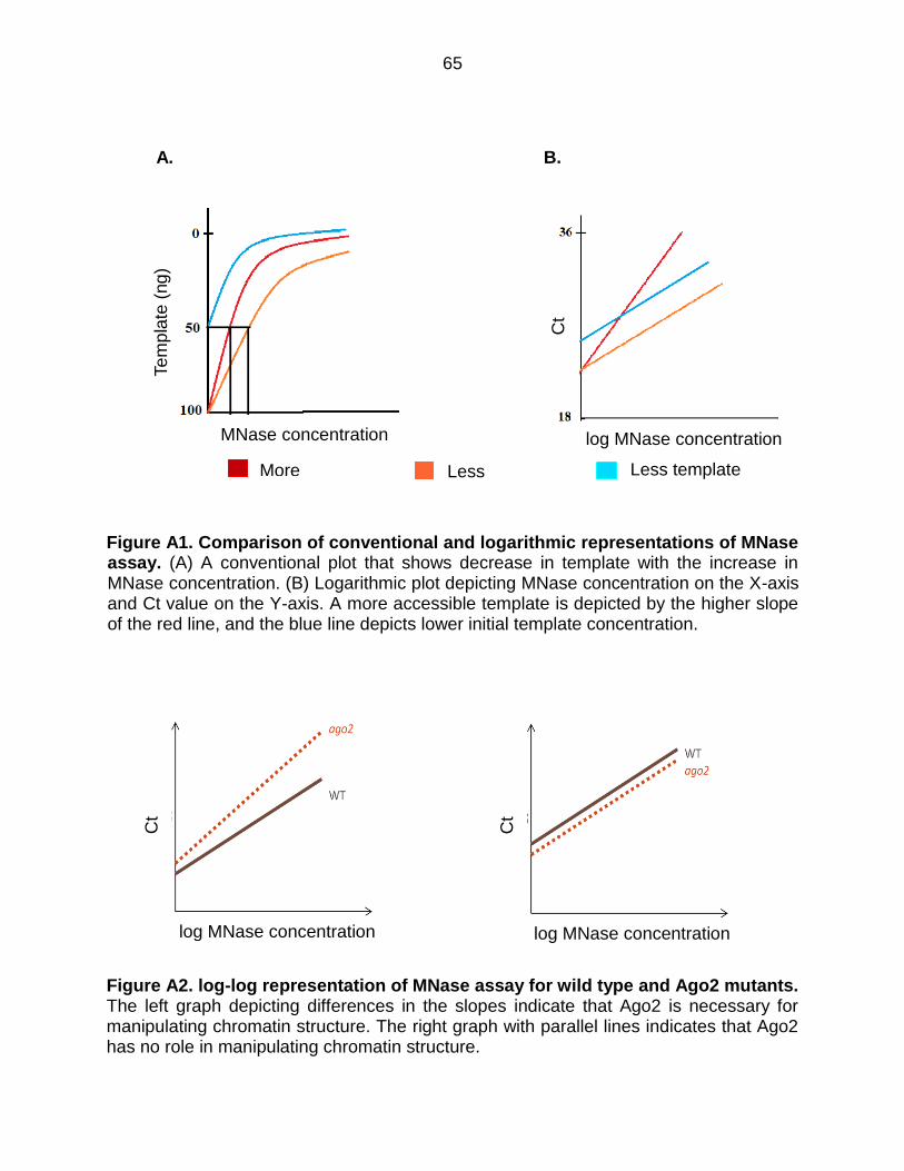
65
A. B.
Figure A1. Comparison of conventional and logarithmic representations of MNase assay. (A) A conventional plot that shows decrease in template with the increase in MNase concentration. (B) Logarithmic plot depicting MNase concentration on the X-axis and Ct value on the Y-axis. A more accessible template is depicted by the higher slope of the red line, and the blue line depicts lower initial template concentration.
Figure A2. log-log representation of MNase assay for wild type and Ago2 mutants. The left graph depicting differences in the slopes indicate that Ago2 is necessary for manipulating chromatin structure. The right graph with parallel lines indicates that Ago2 has no role in manipulating chromatin structure.
MNase Concentration
More accessible
Log MNase Concentration
Less template
Te
mp
late
(ng
)
More
accessible Less accessible
Less template
Ct
log MNase concentration MNase concentration
log MNase concentration log MNase concentration
Ct
Ct
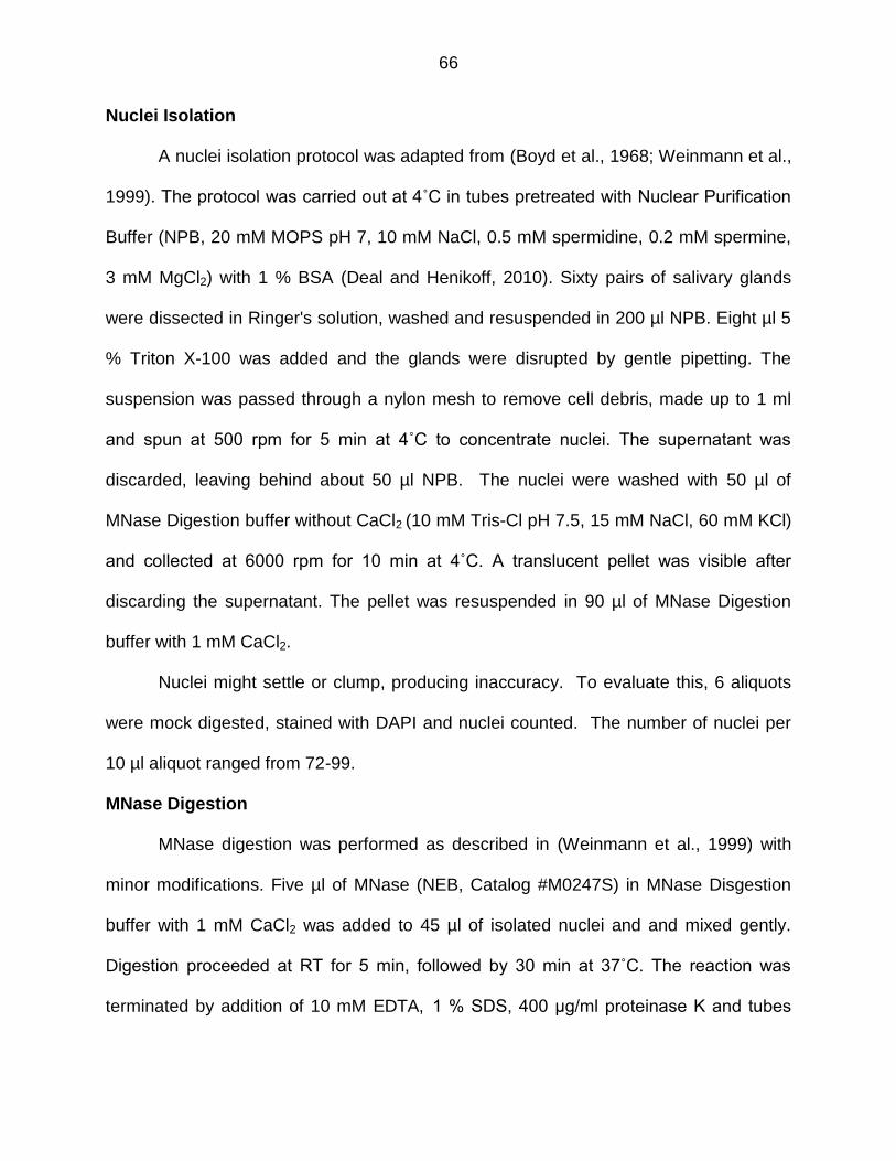
66
Nuclei Isolation
A nuclei isolation protocol was adapted from (Boyd et al., 1968; Weinmann et al.,
1999). The protocol was carried out at 4˚C in tubes pretreated with Nuclear Purification
Buffer (NPB, 20 mM MOPS pH 7, 10 mM NaCl, 0.5 mM spermidine, 0.2 mM spermine,
3 mM MgCl2) with 1 % BSA (Deal and Henikoff, 2010). Sixty pairs of salivary glands
were dissected in Ringer's solution, washed and resuspended in 200 µl NPB. Eight µl 5
% Triton X-100 was added and the glands were disrupted by gentle pipetting. The
suspension was passed through a nylon mesh to remove cell debris, made up to 1 ml
and spun at 500 rpm for 5 min at 4˚C to concentrate nuclei. The supernatant was
discarded, leaving behind about 50 µl NPB. The nuclei were washed with 50 µl of
MNase Digestion buffer without CaCl2 (10 mM Tris-Cl pH 7.5, 15 mM NaCl, 60 mM KCl)
and collected at 6000 rpm for 10 min at 4˚C. A translucent pellet was visible after
discarding the supernatant. The pellet was resuspended in 90 µl of MNase Digestion
buffer with 1 mM CaCl2.
Nuclei might settle or clump, producing inaccuracy. To evaluate this, 6 aliquots
were mock digested, stained with DAPI and nuclei counted. The number of nuclei per
10 µl aliquot ranged from 72-99.
MNase Digestion
MNase digestion was performed as described in (Weinmann et al., 1999) with
minor modifications. Five µl of MNase (NEB, Catalog #M0247S) in MNase Disgestion
buffer with 1 mM CaCl2 was added to 45 µl of isolated nuclei and and mixed gently.
Digestion proceeded at RT for 5 min, followed by 30 min at 37˚C. The reaction was
terminated by addition of 10 mM EDTA, 1 % SDS, 400 μg/ml proteinase K and tubes
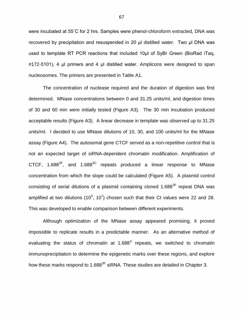
67
were incubated at 55˚C for 2 hrs. Samples were phenol-chloroform extracted, DNA was
recovered by precipitation and resuspended in 20 μl distilled water. Two μl DNA was
used to template RT PCR reactions that included 10μl of SyBr Green (BioRad iTaq,
#172-5101), 4 μl primers and 4 μl distilled water. Amplicons were designed to span
nucleosomes. The primers are presented in Table A1.
The concentration of nuclease required and the duration of digestion was first
determined. MNase concentrations between 0 and 31.25 units/ml, and digestion times
of 30 and 60 min were initially tested (Figure A3). The 30 min incubation produced
acceptable results (Figure A3). A linear decrease in template was observed up to 31.25
units/ml. I decided to use MNase dilutions of 10, 30, and 100 units/ml for the MNase
assay (Figure A4). The autosomal gene CTCF served as a non-repetitive control that is
not an expected target of siRNA-dependent chromatin modification. Amplification of
CTCF, 1.6883F, and 1.6883C repeats produced a linear response to MNase
concentration from which the slope could be calculated (Figure A5). A plasmid control
consisting of serial dilutions of a plasmid containing cloned 1.6883F repeat DNA was
amplified at two dilutions (105, 107) chosen such that their Ct values were 22 and 28.
This was developed to enable comparison between different experiments.
Although optimization of the MNase assay appeared promising, it proved
impossible to replicate results in a predictable manner. As an alternative method of
evaluating the status of chromatin at 1.688X repeats, we switched to chromatin
immunoprecipitation to determine the epigenetic marks over these regions, and explore
how these marks respond to 1.6883F siRNA. These studies are detailed in Chapter 3.
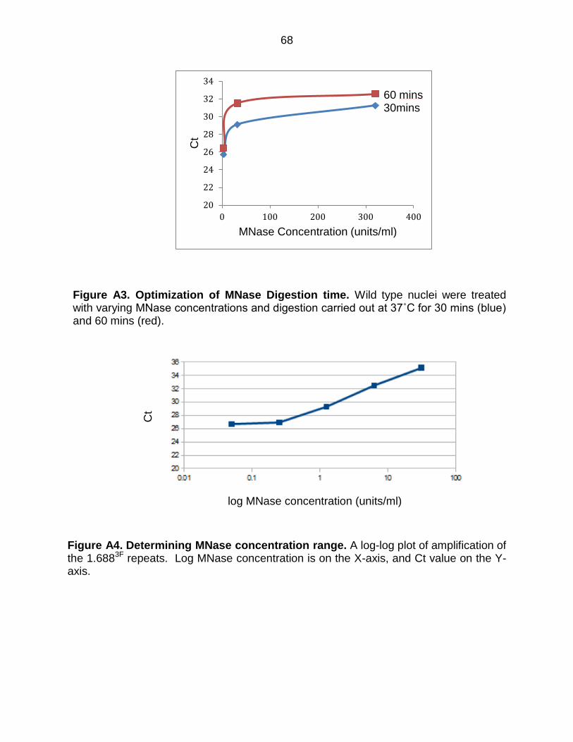
68
Figure A3. Optimization of MNase Digestion time. Wild type nuclei were treated with varying MNase concentrations and digestion carried out at 37˚C for 30 mins (blue) and 60 mins (red).
Figure A4. Determining MNase concentration range. A log-log plot of amplification of the 1.6883F repeats. Log MNase concentration is on the X-axis, and Ct value on the Y-axis.
20
22
24
26
28
30
32
34
0 100 200 300 400
Ct
MNase Concentration (units/ml)
60 mins 30mins
log MNase concentration (units/ml)
Ct
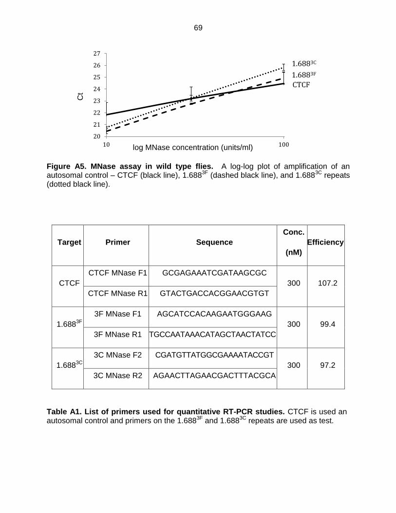
69
Figure A5. MNase assay in wild type flies. A log-log plot of amplification of an autosomal control – CTCF (black line), 1.6883F (dashed black line), and 1.6883C repeats (dotted black line).
Target Primer Sequence
Conc.
(nM)
Efficiency
CTCF
CTCF MNase F1 GCGAGAAATCGATAAGCGC
300 107.2
CTCF MNase R1 GTACTGACCACGGAACGTGT
1.6883F
3F MNase F1 AGCATCCACAAGAATGGGAAG
300 99.4
3F MNase R1 TGCCAATAAACATAGCTAACTATCC
1.6883C
3C MNase F2 CGATGTTATGGCGAAAATACCGT
300 97.2
3C MNase R2 AGAACTTAGAACGACTTTACGCA
Table A1. List of primers used for quantitative RT-PCR studies. CTCF is used an autosomal control and primers on the 1.6883F and 1.6883C repeats are used as test.
20
21
22
23
24
25
26
27
10 100
1.6883F
1.6883C
CTCF C
t
log MNase concentration (units/ml)
70
APPENDIX B INVESTIGATING THE ROLE OF SU(VAR)3-9 IN X RECOGNITION
The studies described here were performed with undergraduate students Taania
Girgla and Kassem Makki. This was a part of their summer research and Honors’
Thesis, respectively.
Determining Gal4 driver strength
The rank strength of four Gal4 drivers was evaluated to enable variable knock
down of Su(var)3-9. A stronger knockdown should more severely impact H3K9me2
enrichment, the mark deposited by the Su(var)3-9 protein. This could be used as a tool
to investigate the role of Su(var)3-9 in X recognition. The genotype, stock number, and
the location of different Gal4 drivers is presented in Table B1. All stocks, excepting w*;
P{matα4-GAL-VP16}V2H, had an intervening sequence between the Act5 promoter and
Gal4 that prevents Gal4 production. Intervening sequences, flanked by tandem Frt sites
(Flip Recombinase Target), are removed by flippase (Flp), supplied by an X-linked
transgene. The Flippase stock description is shown in Table B1. Excision of the
intervening sequence was determined by disappearance of the y+ marker. Gal4 strains
with excised intervening sequences were mated to flies containing UAS-GFP to visually
score driver strength (Table B1), and to assess the success of excision. Flippase
efficiently removed intervening sequences. Once qualitatively scored, stocks of the four
driver lines were used for Su(var)3-9 knockdown.
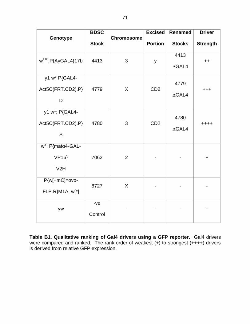
71
Table B1. Qualitative ranking of Gal4 drivers using a GFP reporter. Gal4 drivers were compared and ranked. The rank order of weakest (+) to strongest (++++) drivers is derived from relative GFP expression.
Genotype BDSC
Stock Chromosome
Excised
Portion
Renamed
Stocks
Driver
Strength
w118;P{AyGAL4}17b 4413 3 y 4413
∆GAL4 ++
y1 w* P{GAL4-
Act5C(FRT.CD2).P}
D
4779 X CD2 4779
∆GAL4 +++
y1 w*; P{GAL4-
Act5C(FRT.CD2).P}
S
4780 3 CD2 4780
∆GAL4 ++++
w*; P{matα4-GAL-
VP16}
V2H
7062 2 - - +
P{w[+mC]=ovo-
FLP.R}M1A, w[*] 8727 X - - -
yw -ve
Control - - - -
72
Knock down of Su(var)3-9
Based on the strength of the Gal4 drivers, we decided to test the weakest (w*;
P{matα4-GAL-VP16}V2H) and strongest (yw; {4780ΔGal4}) drivers to create Su(var)3-9
knockdown flies. Fifty virgins with each driver were mated to male UAS-Su(var)3-9
(w1118; UAS-Su(var)3-9 VDRC 101494/KK,) and TRiP-Su(var)3-9 (y1 sc* v1;
P{TRiP.HMS00279}attP2, BDSC 33401), or to a laboratory reference strain lacking a
knock down construct. No embryos obtained from matings with the strong {4780ΔGal4}
driver developed into larvae. Zero-12 h and 12-24 h embryos were collected from
matings using the weakest driver, P{matα4-GAL-VP16}V2H) These embryos were
washed with wash solution (7 g NaCl, 3 ml Triton X-100, 997 ml water) and
dechorionated using 50% bleach for 3 min. The dechorionated embryos were fixed
using a 1:1 ratio of 4% paraformaldehyde and n-heptane for 25 min. The lower phase
was removed, replaced with equal amount of methanol and shaken for 15 s. This step
removes the vitelline membrane, allowing embryos to sink to the bottom. The
supernatant and any embryos that did not sink were removed, and remaining embryos
washed twice with methanol. Embryos were washed three times with PBT (PBS + 0.1%
Tween 20) for 5 min each, stained with DAPI for 5 min and washed once with PBT for
15 min. PBT was removed and 10% glycerol added as the mounting medium. Embryos
in different developmental stages were counted for the three matings and the percent of
total embryos for each meeting determined (shown in Figure B1). The developmental
profiles for the three matings did not reveal any striking differences. As similar number
of females and males were used for mating, and the embryos were collected at the
same time, the total number embryos obtained from each matings were also compared.
73
The matings with UAS-Su(var)3-9 and TRiP-Su(var)3-9 reduced the total number of
embryos to 33% and 42% of that obtained for the control mating. This decrease in
number of embryos could reflect strain-specific differences in female fecundity unrelated
to knock down. Although decrease in number of embryos is observed in the matings
with UAS-Su(var)3-9 and TRiP-Su(var)3-9 with the weakest driver, all the embryos
eclosed to adult flies.
Next, I performed qRT-PCR to quantify Su(var)3-9 transcript. Su(var)3-9 has 5
exons, the first two unique to Su(var)3-9 and the last 3 shared with eIF2γ. I designed
primers spanning the 1st and the 2nd exon (Table B2). Relative transcript in 3rd instar
larvae from matings between w*; P{matα4-GAL-VP16}V2H and UAS-Su(var)3-9, and
w*; P{matα4-GAL-VP16}V2H and TRiP-Su(var)3-9 showed decrease in Su(var)3-9
transcripts (Figure B2).
74
Figure B1. Embryo count from different developmental stages of Su(var)3-9 knockdown embryos. Percent total embryos obtained from matings between laboratory reference strain (yw) and the weakest driver (w*; P{matα4-GAL-VP16}V2H) are shown in white. Gray bars show percent total embryos from matings between UAS-Su(var)3-9 and the weakest driver, and black bars show percent total embryos from matings between TRiP-Su(var)3-9 and the weakest driver.
Figure B2. Accumulation of Su(var)3-9 transcripts is decreased in flies that are knockdown for Su(var)3-9. Transcript accumulation in 3rd instar larvae was measured by quantitative RT-PCR.
0
5
10
15
20
25
30
35
40
45
3 4 5 6 7 8 9 10 11 12 13 14 15 16 17
% T
ota
l e
mb
ryo
s
Embryo developmental stage
0
0.2
0.4
0.6
0.8
1
yw UAS Su(var)3-9 TRiP Su(var)3-9
Rela
tive
Tra
nscrip
t
75
Generating Su(var)3-9 knockdown flies.
Although we were successful in achieving Su(var)3-9 knockdowns, fly stocks
used to achieve Su(var)3-9 knockdown also target an essential overlapping gene,
eIF2γ. Because of this, knockdown with a strong driver caused lethality. To limit knock
down to Su(var)3-9, I worked with Kassem Makki, an undergraduate honors student.
His study focused on generating an RNAi knockdown construct that targets an exon
exclusive to Su(var)3-9. This would allow elimination of Su(var)3-9, without disrupting
eIF2γ.
To accomplish this, an exon present only in Su(var)3-9 was cloned into a pWIZ
vector. pWIZ is designed to facilitate generation of transgenic flies for RNAi knockdown
(Bao and Cagan, 2006). The pWIZ vector allows generation of dsRNA in flies, which is
processed into siRNA. This vector will be injected in fly embryos to create strains that
knockdown Su(var)3-9 when combined with a Gal4 driver.
We amplified a 450 bp segment from the 2nd exon of Su(var)3-9 with primers that
would create AvrII restriction enzyme sites. The PCR product was purified and cloned
in a pCR4 Topo cloning vector, enabling us to sequence and amplify the insert before
cloning into pWIZ. The pWIZ vector was digested with AvrII and ligated to an AvrII-
digested insert. Colony PCR was performed using a reverse primer in the insert and a
forward primer in the hsp70 promoter present in pWIZ) (Table B2, Figure B4). A clone
with insert was identified and used for the second cloning step at Nhe1.
Cloning in NheI site proved to be challenging. After many failed attempts we
decided to use an alternative approach to show role of Su(var)3-9 in depositing
H3K9me2 at the repeats (Chapter 3, Figure 3.9).
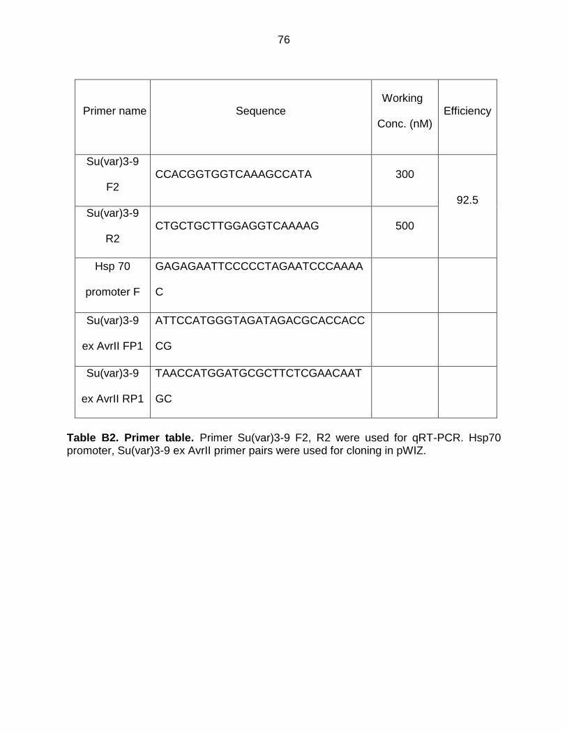
76
Primer name Sequence Working
Conc. (nM) Efficiency
Su(var)3-9
F2 CCACGGTGGTCAAAGCCATA 300
92.5 Su(var)3-9
R2 CTGCTGCTTGGAGGTCAAAAG 500
Hsp 70
promoter F
GAGAGAATTCCCCCTAGAATCCCAAAA
C
Su(var)3-9
ex AvrII FP1
ATTCCATGGGTAGATAGACGCACCACC
CG
Su(var)3-9
ex AvrII RP1
TAACCATGGATGCGCTTCTCGAACAAT
GC
Table B2. Primer table. Primer Su(var)3-9 F2, R2 were used for qRT-PCR. Hsp70 promoter, Su(var)3-9 ex AvrII primer pairs were used for cloning in pWIZ.
77
APPENDIX C DETERMINATION OF GENOME WIDE ALTERATIONS IN H3K9ME2 LEVEL IN FLIES EXPRESSING 1.6883F siRNA
The major limiting step towards carrying out ChIP-Seq is that the protocol we
were using did not yield enough double stranded DNA to make ChIP-seq libraries. Ten
ng of dsDNA is required for library generation. To get ChIP-seq compliant DNA, I
standardized the steps of chromatin generation, resulting in the protocol detailed below.
PROTOCOL FOR CHROMATIN PREPARATION
Overnight embryos (0.35 g) were collected, dechrorionated with 50 % bleach for
3 min, and crosslinked for 20 min in 50 mM HEPES, 1 mM EDTA, 0.5 mM EGTA, 100
mM NACl, 1 % formaldehyde with heptane and vigorous shaking. Crosslinking was
stopped by adding 125 mM glycine, 0.01 % Triton X-100 in 1 X PBS for 30 min.
Crosslinked embryos were washed once in PBS and collected at 2500 rpm for 5 min at
4˚C. Supernatant was discarded and embryos were resuspended in 15 ml Buffer B
(Table C1) and 1 CompleteTM protease inhibitor tablet (Roche, #11697498001), followed
by incubation on ice for 15 min. Embryos were homogenized in a precooled Dounce, 10
strokes with pestle A followed by 15 strokes with pestle B. The homogenized mixture
was centrifuged at 170 g for 10 min at 4˚C and supernatant discarded. The pellet was
washed twice with MNase Digest Buffer (Table C1), resuspended in 200 µl of MNase
Digest Buffer and transferred to a 2 ml tube. DNA was digested with 40 Units of MNase
(2.67 µl of 15 units/ µl, Wortington Stock #LS004797) 37˚C for 30 min. Digestion was
stopped by adding 12 µl of 0.5 M EDTA, and the volume made up to 1 ml using Ten140
buffer (Table C1). The sample was sonicated by a Fischer Scientific Model FB505 on
ice at 35% amplitude, total time of 2.5 min, 30 sec on, 59 sec off, producing shearing to
78
200-600 bp (Figure C1). Sonicated chromatin was centrifuged at 12,000 rpm for 15 min
at 4˚C and 200 µl aliquots stored at -80˚C.
Chromatin Immunoprecipitation
Two hundred µl of chromatin is used for each chromatin immunoprecipitation,
which was carried out as detailed in Chapter 3 with the exception of reverse
crosslinking. To reverse crosslinks of eluted samples, 4 µl RNase A (10 µg/µl, invitrogen
PureLinkTM Stock #12091-021) was added and samples were incubated at 37˚C for 15
min. Twenty µl of 5 M NaCl was added and samples were incubated at 65˚C for 6 hr.
Two µl of proteinase K (10 µg/µl, Thermo Scientific Stock #26160), 10 µl 0.5 M EDTA,
and 10 µl 1 M Tris (pH 7.6) were added and samples incubated at 45˚C for 1 hr.
Extraction with an equal volume of phenol:chloroform:isoamylalcohol (25:24:1 v/v) was
performed, and the aqueous phase transferred to another 0.5 ml tube. Ten µl glycogen,
1/10 volume of 3 M sodium acetate (pH 5.2) and 2.5 volume of chilled 100% ethanol
were added. DNA was precipitated at -20˚C for at least one hour and pelleted at 14,000
rpm for 15 min at 4˚C. The pellet was resuspended in 50 µl distilled water and stored at
-20˚C.
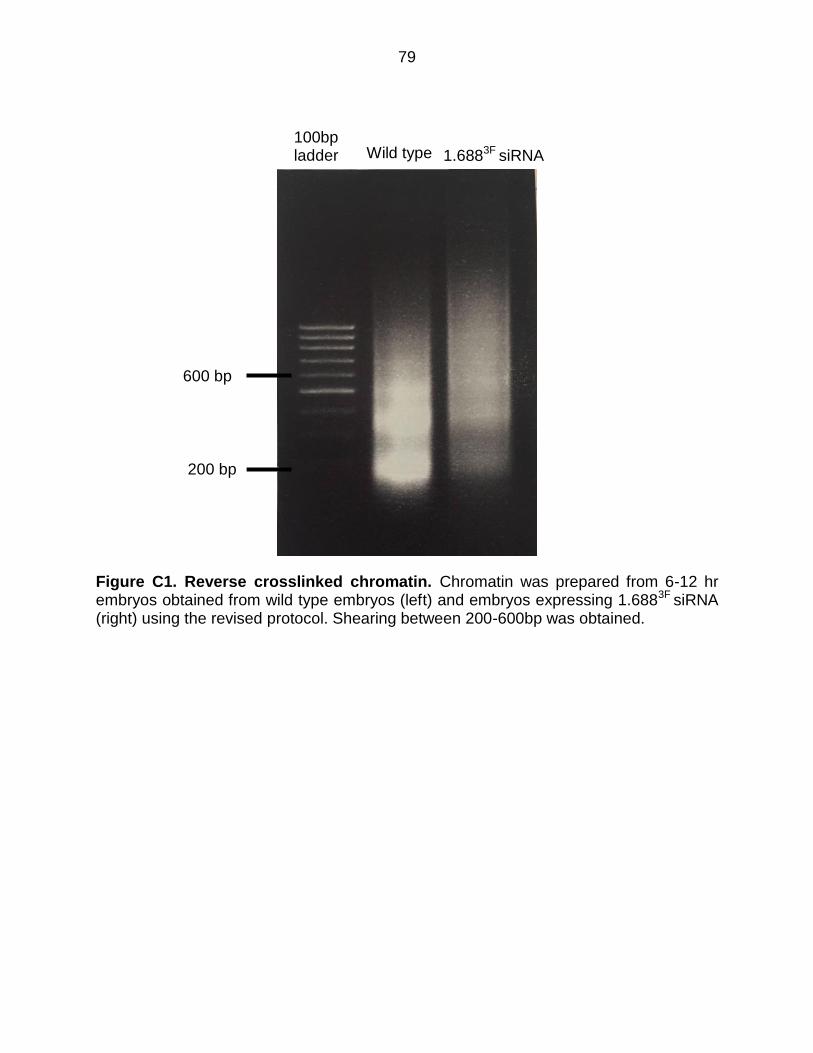
79
Figure C1. Reverse crosslinked chromatin. Chromatin was prepared from 6-12 hr embryos obtained from wild type embryos (left) and embryos expressing 1.6883F siRNA (right) using the revised protocol. Shearing between 200-600bp was obtained.
600 bp
200 bp
Wild type 1.6883F siRNA 100bp ladder
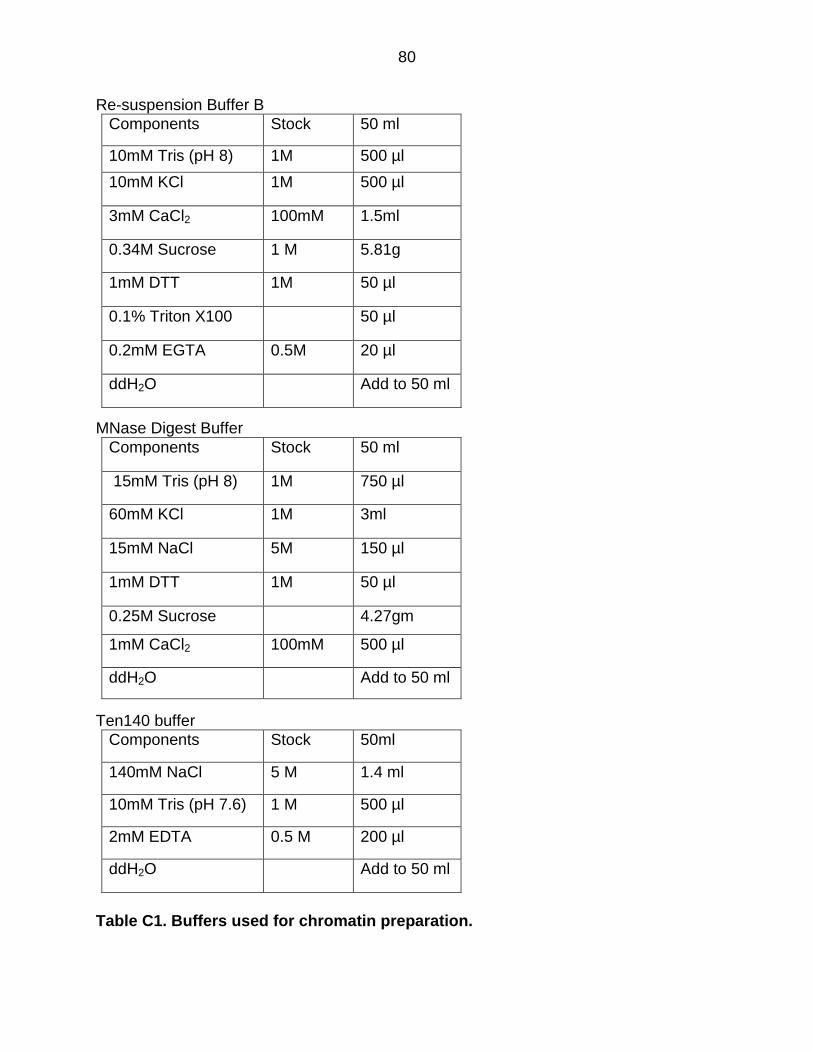
80
Re-suspension Buffer B
Components Stock 50 ml
10mM Tris (pH 8) 1M 500 µl
10mM KCl 1M 500 µl
3mM CaCl2 100mM 1.5ml
0.34M Sucrose 1 M 5.81g
1mM DTT 1M 50 µl
0.1% Triton X100 50 µl
0.2mM EGTA 0.5M 20 µl
ddH2O Add to 50 ml
MNase Digest Buffer
Components Stock 50 ml
15mM Tris (pH 8) 1M 750 µl
60mM KCl 1M 3ml
15mM NaCl 5M 150 µl
1mM DTT 1M 50 µl
0.25M Sucrose 4.27gm
1mM CaCl2 100mM 500 µl
ddH2O Add to 50 ml
Ten140 buffer
Components Stock 50ml
140mM NaCl 5 M 1.4 ml
10mM Tris (pH 7.6) 1 M 500 µl
2mM EDTA 0.5 M 200 µl
ddH2O Add to 50 ml
Table C1. Buffers used for chromatin preparation.
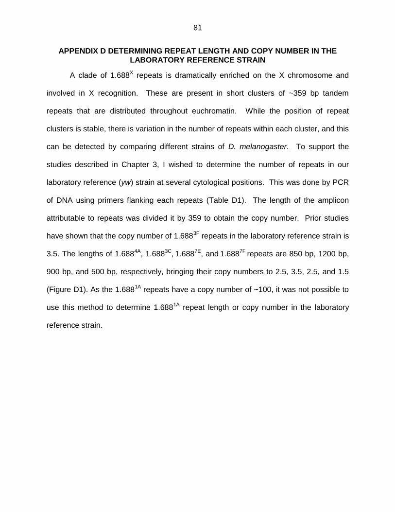
81
APPENDIX D DETERMINING REPEAT LENGTH AND COPY NUMBER IN THE LABORATORY REFERENCE STRAIN
A clade of 1.688X repeats is dramatically enriched on the X chromosome and
involved in X recognition. These are present in short clusters of ~359 bp tandem
repeats that are distributed throughout euchromatin. While the position of repeat
clusters is stable, there is variation in the number of repeats within each cluster, and this
can be detected by comparing different strains of D. melanogaster. To support the
studies described in Chapter 3, I wished to determine the number of repeats in our
laboratory reference (yw) strain at several cytological positions. This was done by PCR
of DNA using primers flanking each repeats (Table D1). The length of the amplicon
attributable to repeats was divided it by 359 to obtain the copy number. Prior studies
have shown that the copy number of 1.6883F repeats in the laboratory reference strain is
3.5. The lengths of 1.6884A, 1.6883C, 1.6887E, and 1.6887F repeats are 850 bp, 1200 bp,
900 bp, and 500 bp, respectively, bringing their copy numbers to 2.5, 3.5, 2.5, and 1.5
(Figure D1). As the 1.6881A repeats have a copy number of ~100, it was not possible to
use this method to determine 1.6881A repeat length or copy number in the laboratory
reference strain.
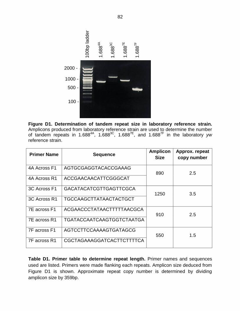
82
Figure D1. Determination of tandem repeat size in laboratory reference strain. Amplicons produced from laboratory reference strain are used to determine the number of tandem repeats in 1.6884A, 1.6883C, 1.6887E, and 1.6887F in the laboratory yw reference strain.
Primer Name Sequence Amplicon
Size
Approx. repeat
copy number
4A Across F1 AGTGCGAGGTACACCGAAAG 890 2.5
4A Across R1 ACCGAACAACATTCGGGCAT
3C Across F1 GACATACATCGTTGAGTTCGCA 1250 3.5
3C Across R1 TGCCAAGCTTATAACTACTGCT
7E across F1 ACGAACCCTATAACTTTTTAACGCA 910 2.5
7E across R1 TGATACCAATCAAGTGGTCTAATGA
7F across F1 AGTCCTTCCAAAAGTGATAGCG 550 1.5
7F across R1 CGCTAGAAAGGATCACTTCTTTTCA
Table D1. Primer table to determine repeat length. Primer names and sequences
used are listed. Primers were made flanking each repeats. Amplicon size deduced from
Figure D1 is shown. Approximate repeat copy number is determined by dividing
amplicon size by 359bp.
10
0b
p lad
de
r
1.6
88
4A
1.6
88
3C
1.6
88
7E
1.6
88
7F
1000 -
2000 -
100 -
500 -
83
APPENDIX E INCLUSION CRITERIA FOR GENETIC SCREEN OF AGO2 INTERACTORS
Physical interactions between proteins are necessary for cellular processes.
Numerous studies have experimentally deduced many protein-protein interactions.
Significant efforts have been made to develop databases that curate and archive these
experimentally discovered protein-protein interactions. Biological General Repository for
Interaction Datasets (BioGRID) (Stark et al., 2006) is one such curated database that is
widely used. Different databases use different rules and systems to compile data. In
addition, networks that integrate major databases have been developed. This allows the
user to retrieve interaction data using a simple user interface. One example of this,
esyN (Bean et al., 2014), has been adopted by Flybase (http://flybase.org). I used
BioGRID and esyN to generate a list of Ago2 interactors (Table E1). These sources
provide information on interactions from researchers with different methods and
interests, and they may or may not be relevant for our study. A manual curation of the
list of Ago2 interactors was required.
Four criteria were used to rank the Ago2 interactors identified in BioGRID (Stark
et al., 2006) and esyN (Bean et al., 2014). The means of detecting interaction was
scored 0 if high throughput and 1 if low throughput or validated. Additional criteria are
known roles in RNA interference, chromatin modification and association with
chromatin. Proteins received a score of 1 for each criterion satisfied and 0 if
unsatisfied. Values were added and presented as the Curation Score in Table E1.
Most named proteins with a curation score of 2 or more were tested for genetic
interaction with roX1 roX2. Phosphatidylinositol glycan anchor biosynthesis class S
(PIG-S, curation score 1) was also tested. No Ago2-interactors with a score of 1 or 2
84
displayed genetic interactions with roX1 roX2 mutants, but several proteins with a score
of 3 or 4 enhanced the lethality of roX1 roX2 males, suggesting a role in dosage
compensation (Table E1, Figure 3.4).
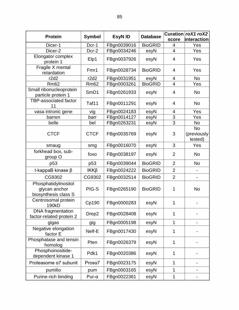
85
Protein Symbol EsyN ID Database Curation
score roX1 roX2 interaction
Dicer-1 Dcr-1 FBgn0039016 BioGRID 4 Yes
Dicer-2 Dcr-2 FBgn0034246 esyN 4 Yes
Elongator complex protein 1
Elp1 FBgn0037926 esyN 4 Yes
Fragile X mental retardation
Fmr1 FBgn0028734 BioGRID 4 Yes
r2d2 r2d2 FBgn0031951 esyN 4 No
Rm62 Rm62 FBgn0003261 BioGRID 4 Yes
Small ribonucleoprotein particle protein 1
SmD1 FBgn0261933 esyN 4 No
TBP-associated factor 11
Taf11 FBgn0011291 esyN 4 No
vasa intronic gene vig FBgn0024183 esyN 4 Yes
barren barr FBgn0014127 esyN 3 Yes
belle bel FBgn0263231 esyN 3 No
CTCF CTCF FBgn0035769 esyN 3 No
(previously tested)
smaug smg FBgn0016070 esyN 3 Yes
forkhead box, sub-group O
foxo FBgn0038197 esyN 2 No
p53 p53 FBgn0039044 BioGRID 2 No
I-kappaB kinase β IKKβ FBgn0024222 BioGRID 2 -
CG9302 CG9302 FBgn0032514 BioGRID 2 -
Phosphatidylinositol glycan anchor
biosynthesis class S PIG-S FBgn0265190 BioGRID 1 No
Centrosomal protein 190kD
Cp190 FBgn0000283 esyN 1 -
DNA fragmentation factor-related protein 2
Drep2 FBgn0028408 esyN 1 -
gigas gig FBgn0005198 esyN 1 -
Negative elongation factor E
Nelf-E FBgn0017430 esyN 1 -
Phosphatase and tensin homolog
Pten FBgn0026379 esyN 1 -
Phosphoinositide-dependent kinase 1
Pdk1 FBgn0020386 esyN 1 -
Proteasome α7 subunit Prosα7 FBgn0023175 esyN 1 -
pumilio pum FBgn0003165 esyN 1 -
Purine-rich binding Pur-α FBgn0022361 esyN 1 -
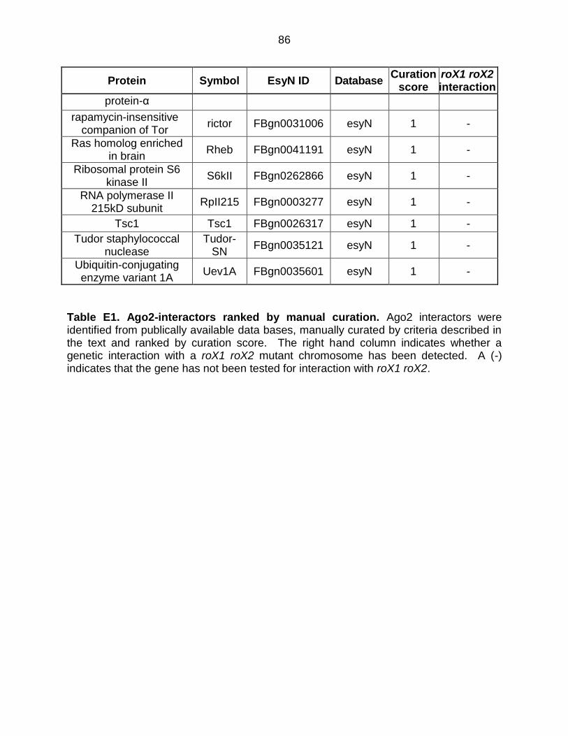
86
Protein Symbol EsyN ID Database Curation
score roX1 roX2 interaction
protein-α
rapamycin-insensitive companion of Tor
rictor FBgn0031006 esyN 1 -
Ras homolog enriched in brain
Rheb FBgn0041191 esyN 1 -
Ribosomal protein S6 kinase II
S6kII FBgn0262866 esyN 1 -
RNA polymerase II 215kD subunit
RpII215 FBgn0003277 esyN 1 -
Tsc1 Tsc1 FBgn0026317 esyN 1 -
Tudor staphylococcal nuclease
Tudor-SN
FBgn0035121 esyN 1 -
Ubiquitin-conjugating enzyme variant 1A
Uev1A FBgn0035601 esyN 1 -
Table E1. Ago2-interactors ranked by manual curation. Ago2 interactors were identified from publically available data bases, manually curated by criteria described in the text and ranked by curation score. The right hand column indicates whether a genetic interaction with a roX1 roX2 mutant chromosome has been detected. A (-) indicates that the gene has not been tested for interaction with roX1 roX2.
87
APPENDIX F DETERMINING EFFECT OF AGO2 ON H3K9ME2 ENRICHMENT AT 1.688X REPEATS
Males with the partial loss of function roX1ex40roX2Δ chromosome have normal
survival, but synthetic lethality is observed if both copies of ago2 are mutated. This is
accompanied by reduced X-localization of MSL complex (Menon and Meller, 2012). In
Chapter 3 we observed Ago2 localization at many repeats. This prompted us to ask,
what happens to H3K9me2 at the 1.688X repeats upon loss of Ago2? To test this, I
collected male third instar larvae from Ago2-/- and wild type flies and performed
chromatin immunoprecipitation using anti-H3K9me2 (ab1220). The protocol used is
described in Chapter 3. This was followed by quantitative PCR with primers listed in
Appendix H.
Contrary to expectation, these studies reveal that H3K9me2 enrichment
increases over 1.688X repeats, as well as in some flanking regions (Figure F1).
Although we do not understand why this is happening, repeat regions are typically
silenced by H3K9me2. I hypothesize that in the absence of Ago2, an alternative
silencing mechanism takes over. Future studies might be aimed at identifying the
methyltransferase responsible for placing this mark in Ago2-/- larvae.
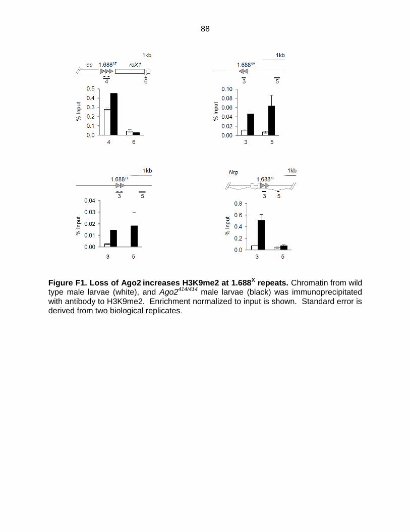
88
Figure F1. Loss of Ago2 increases H3K9me2 at 1.688X repeats. Chromatin from wild type male larvae (white), and Ago2414/414 male larvae (black) was immunoprecipitated with antibody to H3K9me2. Enrichment normalized to input is shown. Standard error is derived from two biological replicates.
89
APPENDIX G LOCALIZATION OF RM62 AT 1.688X REPEATS
The genetic screen detailed in Chapter 3 revealed a sub-network composed of
Ago2, Rm62, Su(var)3-9, Dcr1, and Fmr1 that interacts genetically with roX1 roX2
mutants. Ago2 localization at the 1.688X repeats, and deposition of H3K9me2 at these
repeats by Su(var)3-9, prompted us to ask whether Rm62 also localizes to 1.688X
chromatin. We performed ChIP on 6-12 hr wild type embryos and embryos expressing
1.6883F siRNA using 8 µl anti-Rm62 (Abcam, ab52809) antibody. The protocol used is
as described in Chapter 3. This was followed by quantitative PCR using the primers in
Appendix H. I found that Rm62 is increased at and around 1.688X repeats by increased
1.6883F siRNA.
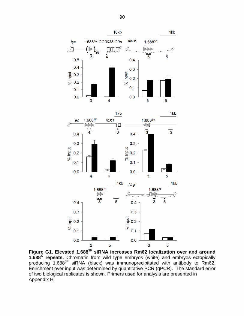
90
Figure G1. Elevated 1.6883F siRNA increases Rm62 localization over and around 1.688X repeats. Chromatin from wild type embryos (white) and embryos ectopically producing 1.6883F siRNA (black) was immunoprecipitated with antibody to Rm62. Enrichment over input was determined by quantitative PCR (qPCR). The standard error of two biological replicates is shown. Primers used for analysis are presented in Appendix H.
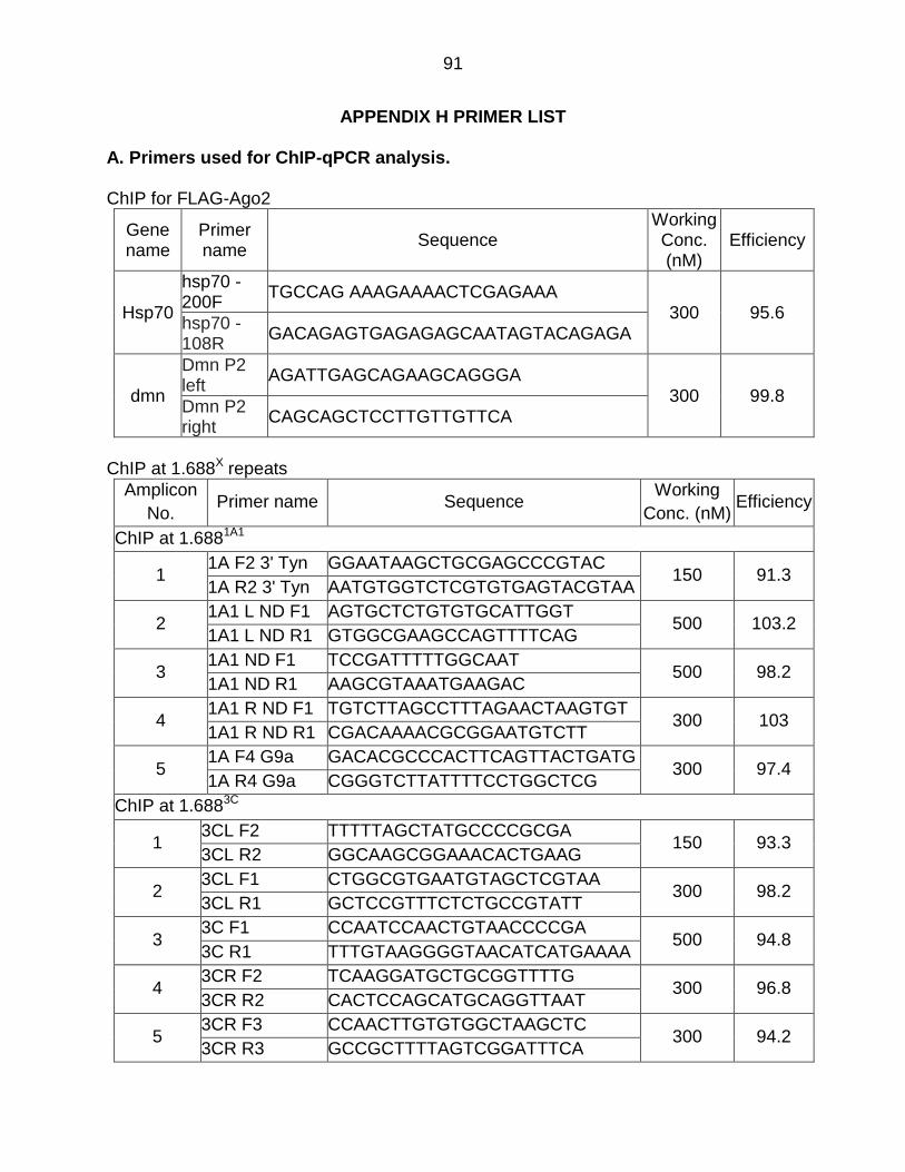
91
APPENDIX H PRIMER LIST
A. Primers used for ChIP-qPCR analysis.
ChIP for FLAG-Ago2
Gene name
Primer name
Sequence Working Conc. (nM)
Efficiency
Hsp70
hsp70 -200F
TGCCAG AAAGAAAACTCGAGAAA 300 95.6
hsp70 -108R
GACAGAGTGAGAGAGCAATAGTACAGAGA
dmn
Dmn P2 left
AGATTGAGCAGAAGCAGGGA 300 99.8
Dmn P2 right
CAGCAGCTCCTTGTTGTTCA
ChIP at 1.688X repeats
Amplicon
No. Primer name Sequence
Working
Conc. (nM) Efficiency
ChIP at 1.6881A1
1 1A F2 3' Tyn GGAATAAGCTGCGAGCCCGTAC
150 91.3 1A R2 3' Tyn AATGTGGTCTCGTGTGAGTACGTAA
2 1A1 L ND F1 AGTGCTCTGTGTGCATTGGT
500 103.2 1A1 L ND R1 GTGGCGAAGCCAGTTTTCAG
3 1A1 ND F1 TCCGATTTTTGGCAAT
500 98.2 1A1 ND R1 AAGCGTAAATGAAGAC
4 1A1 R ND F1 TGTCTTAGCCTTTAGAACTAAGTGT
300 103 1A1 R ND R1 CGACAAAACGCGGAATGTCTT
5 1A F4 G9a GACACGCCCACTTCAGTTACTGATG
300 97.4 1A R4 G9a CGGGTCTTATTTTCCTGGCTCG
ChIP at 1.6883C
1 3CL F2 TTTTTAGCTATGCCCCGCGA
150 93.3 3CL R2 GGCAAGCGGAAACACTGAAG
2 3CL F1 CTGGCGTGAATGTAGCTCGTAA
300 98.2 3CL R1 GCTCCGTTTCTCTGCCGTATT
3 3C F1 CCAATCCAACTGTAACCCCGA
500 94.8 3C R1 TTTGTAAGGGGTAACATCATGAAAA
4 3CR F2 TCAAGGATGCTGCGGTTTTG
300 96.8 3CR R2 CACTCCAGCATGCAGGTTAAT
5 3CR F3 CCAACTTGTGTGGCTAAGCTC
300 94.2 3CR R3 GCCGCTTTTAGTCGGATTTCA
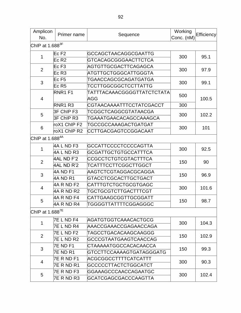
92
Amplicon
No. Primer name Sequence
Working
Conc. (nM) Efficiency
ChIP at 1.6883F
1 Ec F2 GCCAGCTAACAGGCGAATTG
300 95.1 Ec R2 GTCACAGCGGGAACTTCTCA
2 Ec F3 AGTGTTGCGACTTCAGAGCA
300 97.9 Ec R3 ATGTTGCTGGGCATTGGGTA
3 Ec F5 TGAACCAGCGCAGATGATGA
300 99.1 Ec R5 TCCTTGGCGGCTCCTTATTG
4
RNR1 F1 TATTTACAAACGGGGTTATCTCTATA
AGG 500
100.5
RNR1 R3 CGTAACAAAATTTCCTATCGACCT 300
5 3F ChIP F3 TCGGCTCAGGCGTATAACGA
300 102.2 3F ChIP R3 TGAAATGAACACAGCCAAAGCA
6 roX1 ChIP F2 TGCCGCCAAAGACTGATGAT
300 101 roX1 ChIP R2 CCTTGACGAGTCCGGACAAT
ChIP at 1.6884A
1 4A L ND F3 GCCATTCCCCTCCCCAGTTA
300 92.5 4A L ND R3 GCGATTGCTGTGCCATTTCA
2 4AL ND F’2 CCGCCTCTGTCGTACTTTCA
150 90 4AL ND R’2 TCATTTCCTTCGGCTTGGCT
3 4A ND F1 AAGTCTCGTAGGACGCAGGA
150 96.9 4A ND R1 GTACCTCGCACTTGCTGACT
4 4A R ND F2 CATTTGTCTGCTGCGTGAGC
300 101.6 4A R ND R2 TGCTGCGTCTTGACTTTCGT
5 4A R ND F4 CATTGAAGCGGTTGCGGATT
150 98.7 4A R ND R4 TGGGGTTATTTTCGGAGGGC
ChIP at 1.6887E
1 7E L ND F4 AGATGTGGTCAAACACTGCG
300 104.3 7E L ND R4 AAACCGAAACCGAGAACCAGA
2 7E L ND F2 TAGCCTGACACAAGCAAGGG
150 102.9 7E L ND R2 GCCCGTAATGAAGTCAACCAG
3 7E ND F1 CTAAAAATGGCCACACAACCA
150 99.3 7E ND R1 GTCCTTCCAAAAGTGATAGGGATG
4 7E R ND F1 ACGCGGCCTTTTCATCATTT
300 90.3 7E R ND R1 GCCCCCTTACTCTGGCATCT
5 7E R ND F3 GGAAAGCCCAACCAGAATGC
300 102.4 7E R ND R3 GCATCGAGCGACCCAAGTTA
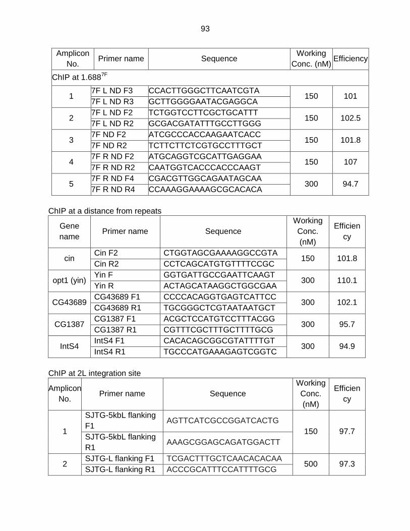
93
Amplicon
No. Primer name Sequence
Working
Conc. (nM) Efficiency
ChIP at 1.6887F
1 7F L ND F3 CCACTTGGGCTTCAATCGTA
150 101 7F L ND R3 GCTTGGGGAATACGAGGCA
2 7F L ND F2 TCTGGTCCTTCGCTGCATTT
150 102.5 7F L ND R2 GCGACGATATTTGCCTTGGG
3 7F ND F2 ATCGCCCACCAAGAATCACC
150 101.8 7F ND R2 TCTTCTTCTCGTGCCTTTGCT
4 7F R ND F2 ATGCAGGTCGCATTGAGGAA
150 107 7F R ND R2 CAATGGTCACCCACCCAAGT
5 7F R ND F4 CGACGTTGGCAGAATAGCAA
300 94.7 7F R ND R4 CCAAAGGAAAAGCGCACACA
ChIP at a distance from repeats
Gene
name Primer name Sequence
Working
Conc.
(nM)
Efficien
cy
cin Cin F2 CTGGTAGCGAAAAGGCCGTA
150 101.8 Cin R2 CCTCAGCATGTGTTTTCCGC
opt1 (yin) Yin F GGTGATTGCCGAATTCAAGT
300 110.1 Yin R ACTAGCATAAGGCTGGCGAA
CG43689 CG43689 F1 CCCCACAGGTGAGTCATTCC
300 102.1 CG43689 R1 TGCGGGCTCGTAATAATGCT
CG1387 CG1387 F1 ACGCTCCATGTCCTTTACGG
300 95.7 CG1387 R1 CGTTTCGCTTTGCTTTTGCG
IntS4 IntS4 F1 CACACAGCGGCGTATTTTGT
300 94.9 IntS4 R1 TGCCCATGAAAGAGTCGGTC
ChIP at 2L integration site
Amplicon
No. Primer name Sequence
Working
Conc.
(nM)
Efficien
cy
1
SJTG-5kbL flanking
F1 AGTTCATCGCCGGATCACTG
150 97.7 SJTG-5kbL flanking
R1 AAAGCGGAGCAGATGGACTT
2 SJTG-L flanking F1 TCGACTTTGCTCAACACACAA
500 97.3 SJTG-L flanking R1 ACCCGCATTTCCATTTTGCG
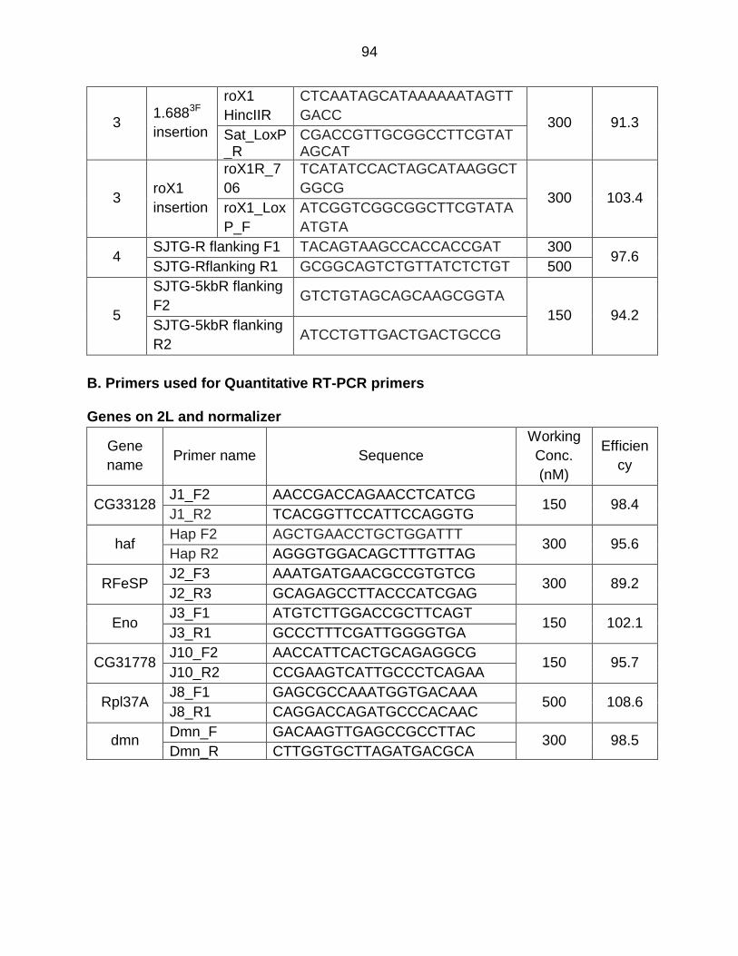
94
3 1.6883F
insertion
roX1
HincIIR
CTCAATAGCATAAAAAATAGTT
GACC 300 91.3
Sat_LoxP_R
CGACCGTTGCGGCCTTCGTATAGCAT
3 roX1
insertion
roX1R_7
06
TCATATCCACTAGCATAAGGCT
GGCG 300 103.4
roX1_Lox
P_F
ATCGGTCGGCGGCTTCGTATA
ATGTA
4 SJTG-R flanking F1 TACAGTAAGCCACCACCGAT 300
97.6 SJTG-Rflanking R1 GCGGCAGTCTGTTATCTCTGT 500
5
SJTG-5kbR flanking
F2 GTCTGTAGCAGCAAGCGGTA
150 94.2 SJTG-5kbR flanking
R2 ATCCTGTTGACTGACTGCCG
B. Primers used for Quantitative RT-PCR primers Genes on 2L and normalizer
Gene
name Primer name Sequence
Working
Conc.
(nM)
Efficien
cy
CG33128 J1_F2 AACCGACCAGAACCTCATCG
150 98.4 J1_R2 TCACGGTTCCATTCCAGGTG
haf Hap F2 AGCTGAACCTGCTGGATTT
300 95.6 Hap R2 AGGGTGGACAGCTTTGTTAG
RFeSP J2_F3 AAATGATGAACGCCGTGTCG
300 89.2 J2_R3 GCAGAGCCTTACCCATCGAG
Eno J3_F1 ATGTCTTGGACCGCTTCAGT
150 102.1 J3_R1 GCCCTTTCGATTGGGGTGA
CG31778 J10_F2 AACCATTCACTGCAGAGGCG
150 95.7 J10_R2 CCGAAGTCATTGCCCTCAGAA
Rpl37A J8_F1 GAGCGCCAAATGGTGACAAA
500 108.6 J8_R1 CAGGACCAGATGCCCACAAC
dmn Dmn_F GACAAGTTGAGCCGCCTTAC
300 98.5 Dmn_R CTTGGTGCTTAGATGACGCA
95
REFERENCES
Adachi, Y., Kas, E., and Laemmli, U.K. (1989). Preferential, cooperative binding
of DNA topoisomerase II to scaffold-associated regions. EMBO J 8, 3997-
4006.
Akhtar, A., and Becker, P.B. (2000). Activation of transcription through histone
H4 acetylation by MOF, an acetyltransferase essential for dosage
compensation in Drosophila. Mol Cell 5, 367-375.
Alekseyenko, A.A., Ellison, C.E., Gorchakov, A.A., Zhou, Q., Kaiser, V.B., Toda,
N., Walton, Z., Peng, S., Park, P.J., Bachtrog, D., et al. (2013).
Conservation and de novo acquisition of dosage compensation on newly
evolved sex chromosomes in Drosophila. Genes Dev 27, 853-858.
Alekseyenko, A.A., Larschan, E., Lai, W.R., Park, P.J., and Kuroda, M.I. (2006).
High-resolution ChIP–chip analysis reveals that the Drosophila MSL
complex selectively identifies active genes on the male X chromosome.
Genes Dev 20, 848-857.
Alekseyenko, A.A., Peng, S., Larschan, E., Gorchakov, A.A., Lee, O.K.,
Kharchenko, P., McGrath, S.D., Wang, C.I., Mardis, E.R., Park, P.J., et al.
(2008). A sequence motif within chromatin entry sites directs MSL
establishment on the Drosophila X chromosome. Cell 134, 599-609.
Aulner, N., Monod, C., Mandicourt, G., Jullien, D., Cuvier, O., Sall, A., Janssen,
S., Laemmli, U.K., and Käs, E. (2002). The AT-Hook Protein D1 Is
Essential for Drosophila melanogaster Development and Is Implicated in
Position-Effect Variegation. Molecular and Cellular Biology 22, 1218-1232.
96
Bachtrog, D., and Charlesworth, B. (2002). Reduced adaptation of a non-
recombining neo-Y chromosome. Nature 416, 323-326.
Bailey, J.A., Carrel, L., Chakravarti, A., and Eichler, E.E. (2000). Molecular
evidence for a relationship between LINE-1 elements and X chromosome
inactivation: The Lyon repeat hypothesis. PNAS 97, 6634-6639.
Bao, S., and Cagan, R. (2006). Fast cloning inverted repeats for RNA
interference. RNA 12, 2020-2024.
Barbash, D.A. (2010). Ninety years of Drosophila melanogaster hybrids.
Genetics 186, 1-8.
Bashaw, G.J., and Baker, B.S. (1997). The regulation of the Drosophila msl-2
gene reveals a function for Sex-lethal in translational control. Cell 89, 789-
798.
Bean, D.M., Heimbach, J., Ficorella, L., Micklem, G., Oliver, S.G., and Favrin, G.
(2014). esyN: network building, sharing and publishing. PLoS One 9,
e106035.
Becker, J.S., Nicetto, D., and Zaret, K.S. (2016). H3K9me3-Dependent
Heterochromatin: Barrier to Cell Fate Changes. Trends in genetics : TIG
32, 29-41.
Beckmann, K., Grskovic, M., Gebauer, F., and Hentze, M.W. (2005). A Dual
Inhibitory Mechanism Restricts msl-2 mRNA Translation for Dosage
Compensation in Drosophila. Cell 122, 529-540.

97
Berrios, M., Osheroff, N., and Fisher, P.A. (1985). In situ localization of DNA
topoisomerase II, a major polypeptide component of the Drosophila
nuclear matrix fraction. PNAS 82, 4142-4146.
Blattes, R., Monod, C., Susbielle, G., Cuvier, O., Wu, J.H., Hsieh, T.S., Laemmli,
U.K., and Kas, E. (2006). Displacement of D1, HP1 and topoisomerase II
from satellite heterochromatin by a specific polyamide. EMBO J 25, 2397-
2408.
Boeke, J., Bag, I., Ramaiah, M.J., Vetter, I., Kremmer, E., Pal-Bhadra, M.,
Bhadra, U., and Imhof, A. (2011). The RNA helicase Rm62 cooperates
with SU(VAR)3-9 to re-silence active transcription in Drosophila
melanogaster. PLoS One 6, e20761.
Bone, J.R., and Kuroda, M.I. (1996). Dosage compensation regulatory proteins
and the evolution of sex chromosomes in Drosophila. Genetics 144, 705-
713.
Boyd, J.B., Berendes, H.D., and Boyd, H. (1968). Mass Preparation of Nuclei
from the Larval Salivary Glands of Drosophila hydei. The Journal of Cell
Biology 38, 369-376.
Cernilogar, F.M., Onorati, M.C., Kothe, G.O., Burroughs, A.M., Parsi, K.M.,
Breiling, A., Lo Sardo, F., Saxena, A., Miyoshi, K., Siomi, H., et al. (2011).
Chromatin-associated RNA interference components contribute to
transcriptional regulation in Drosophila. Nature 480, 391-395.
Charlesworth, B. (1996). The evolution of chromosomal sex determination and
dosage compensation. Curr Biol 6, 149-162.
98
Chow, J.C., Ciaudo, C., Fazzari, M.J., Mise, N., Servant, N., Glass, J.L., Attreed,
M., Avner, P., Wutz, A., Barillot, E., et al. (2010). LINE-1 activity in
facultative heterochromatin formation during X chromosome inactivation.
Cell 141, 956-969.
Corona, D.F.V., Siriaco, G., Armstrong, J.A., Snarskaya, N., McClymont, S.A.,
Scott, M.P., and Tamkun, J.W. (2007). ISWI Regulates Higher-Order
Chromatin Structure and Histone H1 Assembly In Vivo. PLOS Biol 5,
e232.
Culbertson, M.R. (1999). RNA surveillance: unforeseen consequences for gene
expression, inherited genetic disorders and cancer. Trends in Genetics 15,
74-80.
de Wit, E., Greil, F., and van Steensel, B. (2005). Genome-wide HP1 binding in
Drosophila: developmental plasticity and genomic targeting signals.
Genome Res 15, 1265-1273.
Deal, R.B., and Henikoff, S. (2010). A Simple Method for Gene Expression and
Chromatin Profiling of Individual Cell Types within a Tissue.
Developmental Cell 18, 1030-1040.
Deng, H., Zhang, W., Bao, X., Martin, J.N., Girton, J., Johansen, J., and
Johansen, K.M. (2005a). The JIL-1 kinase regulates the structure of
Drosophila polytene chromosomes. Chromosoma 114, 173-182.
Deng, X., and Meller, V.H. (2006). roX RNAs are required for increased
expression of X-linked genes in Drosophila melanogaster males. Genetics
174, 1859-1866.
99
Deng, X., Rattner, B.P., Souter, S., and Meller, V.H. (2005b). The severity of
roX1 mutations is predicted by MSL localization on the X chromosome.
Mech Dev 122, 1094-1105.
Deshpande, N., and Meller, V.H. (2014). Sex chromosome evolution: life, death
and repetitive DNA. Fly (Austin) 8, 197-199.
Deuring, R., Fanti, L., Armstrong, J.A., Sarte, M., Papoulas, O., Prestel, M.,
Daubresse, G., Verardo, M., Moseley, S.L., Berloco, M., et al. (2000). The
ISWI chromatin-remodeling protein is required for gene expression and
the maintenance of higher order chromatin structure in vivo. Mol Cell 5,
355-365.
DiBartolomeis, S.M., Tartof, K.D., and Jackson, F.R. (1992). A superfamily of
Drosophila satellite related (SR) DNA repeats restricted to the X
chromosome euchromatin. Nucleic acids research 20, 1113-1116.
Disteche, C.M. (2012). Dosage compensation of the sex chromosomes. Annu
Rev Genet 46, 537-560.
Ebert, A., Schotta, G., Lein, S., Kubicek, S., Krauss, V., Jenuwein, T., and
Reuter, G. (2004). Su(var) genes regulate the balance between
euchromatin and heterochromatin in Drosophila. Genes Dev 18, 2973-
2983.
Ellison, C.E., and Bachtrog, D. (2013). Dosage compensation via transposable
element mediated rewiring of a regulatory network. Science 342, 846-850.
100
Emilsson, V., Thorleifsson, G., Zhang, B., Leonardson, A.S., Zink, F., Zhu, J.,
Carlson, S., Helgason, A., Walters, G.B., Gunnarsdottir, S., et al. (2008).
Genetics of gene expression and its effect on disease. Nature 452, 423.
Fagegaltier, D., and Baker, B.S. (2004). X chromosome sites autonomously
recruit the dosage compensation complex in Drosophila males. PLOS Biol
2, e341.
Ferree, P.M., and Barbash, D.A. (2009). Species-specific heterochromatin
prevents mitotic chromosome segregation to cause hybrid lethality in
Drosophila. PLOS Biol 7, e1000234.
Figueiredo, M.L., Kim, M., Philip, P., Allgardsson, A., Stenberg, P., and Larsson,
J. (2014). Non-coding roX RNAs prevent the binding of the MSL-complex
to heterochromatic regions. PLoS genetics 10, e1004865.
Gallach, M. (2014). Recurrent turnover of chromosome-specific satellites in
Drosophila. Genome biology and evolution 6, 1279-1286.
Gebauer, F., Merendino, L., Hentze, M.W., and Valcarcel, J. (1998). The
Drosophila splicing regulator sex-lethal directly inhibits translation of male-
specific-lethal 2 mRNA. RNA 4, 142-150.
Gelbart, M.E., and Kuroda, M.I. (2009). Drosophila dosage compensation: a
complex voyage to the X chromosome. Development 136, 1399-1410.
Ghildiyal, M., and Zamore, P.D. (2009). Small silencing RNAs: an expanding
universe. Nature reviews Genetics 10, 94-108.
101
Grimaud, C., and Becker, P.B. (2009). The dosage compensation complex
shapes the conformation of the X chromosome in Drosophila. Genes Dev
23, 2490-2495.
Heng, H.H.Q., Goetze, S., Ye, C.J., Liu, G., Stevens, J.B., Bremer, S.W., Wykes,
S.M., Bode, J., and Krawetz, S.A. (2004). Chromatin loops are selectively
anchored using scaffold/matrix-attachment regions. Journal of Cell
Science 117, 999-1008.
Hilfiker, A., Hilfiker-Kleiner, D., Pannuti, A., and Lucchesi, J.C. (1997). mof, a
putative acetyl transferase gene related to the Tip60 and MOZ human
genes and to the SAS genes of yeast, is required for dosage
compensation in Drosophila. EMBO J 16, 2054-2060.
Hsieh, T., and Brutlag, D. (1979). Sequence and sequence variation within the
1.688 g/cm3 satellite DNA of Drosophila melanogaster. Journal of
molecular biology 135, 465-481.
Jin, Y., Wang, Y., Johansen, J., and Johansen, K.M. (2000). JIL-1, a
chromosomal kinase implicated in regulation of chromatin structure,
associates with the male specific lethal (MSL) dosage compensation
complex. J Cell Biol 149, 1005-1010.
Joshi, S.S., and Meller, V.H. (2017). Satellite Repeats Identify X Chromatin for
Dosage Compensation in Drosophila melanogaster Males. Curr Biol 27,
1393-1402 e1392.
Kageyama, Y., Mengus, G., Gilfillan, G., Kennedy, H.G., Stuckenholz, C., Kelley,
R.L., Becker, P.B., and Kuroda, M.I. (2001). Association and spreading of
102
the Drosophila dosage compensation complex from a discrete roX1
chromatin entry site. EMBO J 20, 2236-2245.
Kelley, R.L., and Kuroda, M.I. (2003). The Drosophila roX1 RNA gene can
overcome silent chromatin by recruiting the male-specific lethal dosage
compensation complex. Genetics 164, 565-574.
Kelley, R.L., Meller, V.H., Gordadze, P.R., Roman, G., Davis, R.L., and Kuroda,
M.I. (1999). Epigenetic spreading of the Drosophila dosage compensation
complex from roX RNA genes into flanking chromatin. Cell 98, 513-522.
Kelley, R.L., Solovyeva, I., Lyman, L.M., Richman, R., Solovyev, V., and Kuroda,
M.I. (1995). Expression of msl-2 causes assembly of dosage
compensation regulators on the X chromosomes and female lethality in
Drosophila. Cell 81, 867-877.
Kind, J., and Akhtar, A. (2007). Cotranscriptional recruitment of the dosage
compensation complex to X-linked target genes. Genes Dev 21, 2030-
2040.
Kind, J., Vaquerizas, J.M., Gebhardt, P., Gentzel, M., Luscombe, N.M., Bertone,
P., and Akhtar, A. (2008). Genome-wide analysis reveals MOF as a key
regulator of dosage compensation and gene expression in Drosophila.
Cell 133, 813-828.
Koya, S.K., and Meller, V.H. (2011). roX RNAs and Genome Regulation in
Drosophila Melanogaster. Prog Mol Subcell Biol 51, 147-160.
103
Koya, S.K., and Meller, V.H. (2015). Modulation of Heterochromatin by Male
Specific Lethal Proteins and roX RNA in Drosophila melanogaster Males.
PLoS One 10, e0140259.
Kuhn, G.C., Kuttler, H., Moreira-Filho, O., and Heslop-Harrison, J.S. (2012). The
1.688 repetitive DNA of Drosophila: concerted evolution at different
genomic scales and association with genes. Molecular biology and
evolution 29, 7-11.
Kuzu, G., Kaye, E.G., Chery, J., Siggers, T., Yang, L., Dobson, J.R., Boor, S.,
Bliss, J., Liu, W., Jogl, G., et al. (2016). Expansion of GA Dinucleotide
Repeats Increases the Density of CLAMP Binding Sites on the X-
Chromosome to Promote Drosophila Dosage Compensation. PLoS
genetics 12, e1006120.
Lamb, J., Crawford, E.D., Peck, D., Modell, J.W., Blat, I.C., Wrobel, M.J., Lerner,
J., Brunet, J.-P., Subramanian, A., Ross, K.N., et al. (2006). The
Connectivity Map: Using Gene-Expression Signatures to Connect Small
Molecules, Genes, and Disease. Science 313, 1929-1935.
Larschan, E., Alekseyenko, A.A., Gortchakov, A.A., Peng, S., Li, B., Yang, P.,
Workman, J.L., Park, P.J., and Kuroda, M.I. (2007). MSL Complex Is
Attracted to Genes Marked by H3K36 Trimethylation Using a Sequence-
Independent Mechanism. Molecular Cell 28, 121-133.
Larschan, E., Bishop, E.P., Kharchenko, P.V., Core, L.J., Lis, J.T., Park, P.J.,
and Kuroda, M.I. (2011). X chromosome dosage compensation via
enhanced transcriptional elongation in Drosophila. Nature 471, 115-118.
104
Lee, J.T. (2009). Lessons from X-chromosome inactivation: long ncRNA as
guides and tethers to the epigenome. Genes Dev 23, 1831-1842.
Lim, C.K., and Kelley, R.L. (2013). The Drosophila over compensating males
gene genetically inhibits dosage compensation in males. PLoS One 8,
e60450.
Lipardi, C., and Paterson, B.M. (2009). Identification of an RNA-dependent RNA
polymerase in Drosophila involved in RNAi and transposon suppression.
PNAS 106, 15645-15650.
Liu, L.P., Ni, J.Q., Shi, Y.D., Oakeley, E.J., and Sun, F.L. (2005). Sex-specific
role of Drosophila melanogaster HP1 in regulating chromatin structure and
gene transcription. Nat Genet 37, 1361-1366.
Lohe, A.R., Hilliker, A.J., and Roberts, P.A. (1993). Mapping simple repeated
DNA sequences in heterochromatin of Drosophila melanogaster. Genetics
134, 1149-1174.
Lucchesi, J.C., and Kuroda, M.I. (2015). Dosage compensation in Drosophila.
Cold Spring Harb Perspect Biol 7.
Lyon, M.F. (1998). X-chromosome inactivation: a repeat hypothesis. Cytogenet
Cell Genet 80, 133-137.
Lyon, M.F. (2006). Do LINEs have a role in X-chromosome inactivation? Journal
of biomedicine & biotechnology 2006, 59746.
McElroy, K.A., Jung, Y.L., Zee, B.M., Wang, C.I., Park, P.J., and Kuroda, M.I.
(2017). upSET, the Drosophila homologue of SET3, Is Required for
105
Viability and the Proper Balance of Active and Repressive Chromatin
Marks. G3 (Bethesda) 7, 625-635.
Meller, V.H. (2003). Initiation of dosage compensation in Drosophila embryos
depends on expression of the roX RNAs. Mech Dev 120, 759-767.
Meller, V.H., Fisher, P.A., and Berrios, M. (1995). Intranuclear distribution of DNA
topoisomerase II and chromatin. Chromosome research : an international
journal on the molecular, supramolecular and evolutionary aspects of
chromosome biology 3, 255-260.
Meller, V.H., Joshi, S.S., and Deshpande, N. (2015). Modulation of Chromatin by
Noncoding RNA. Annu Rev Genet 49, 673-695.
Meller, V.H., and Rattner, B.P. (2002). The roX genes encode redundant male-
specific lethal transcripts required for targeting of the MSL complex.
EMBO J 21, 1084-1091.
Mendjan, S., Taipale, M., Kind, J., Holz, H., Gebhardt, P., Schelder, M.,
Vermeulen, M., Buscaino, A., Duncan, K., Mueller, J., et al. (2006).
Nuclear pore components are involved in the transcriptional regulation of
dosage compensation in Drosophila. Mol Cell 21, 811-823.
Menon, D.U., Coarfa, C., Xiao, W., Gunaratne, P.H., and Meller, V.H. (2014).
siRNAs from an X-linked satellite repeat promote X-chromosome
recognition in Drosophila melanogaster. PNAS 111, 16460-16465.
Menon, D.U., and Meller, V.H. (2009). Imprinting of the Y Chromosome
Influences Dosage Compensation in <em>roX1 roX2 Drosophila
melanogaster</em>. Genetics 183, 811.
106
Menon, D.U., and Meller, V.H. (2012). A role for siRNA in X-chromosome dosage
compensation in Drosophila melanogaster. Genetics 191, 1023-1028.
Meyer, B.J. (2000). Sex in the wormcounting and compensating X-chromosome
dose. Trends Genet 16, 247-253.
Moazed, D. (2009). Small RNAs in transcriptional gene silencing and genome
defence. Nature 457, 413-420.
Monfort, M., and Furlong, E.E. (2015). Furlong Lab EMBL TFBS contribution.
Morales, V., Straub, T., Neumann, M.F., Mengus, G., Akhtar, A., and Becker,
P.B. (2004). Functional integration of the histone acetyltransferase MOF
into the dosage compensation complex. EMBO J 23, 2258-2268.
Park, Y., Kelley, R.L., Oh, H., Kuroda, M.I., and Meller, V.H. (2002). Extent of
Chromatin Spreading Determined by <em>roX</em> RNA
Recruitment of MSL Proteins. Science 298, 1620.
Partridge, J.F., Borgstrom, B., and Allshire, R.C. (2000). Distinct protein
interaction domains and protein spreading in a complex centromere.
Genes Dev 14, 783-791.
Pfaffl, M.W. (2001). A new mathematical model for relative quantification in real-
time RT-PCR. Nucleic acids research 29, e45.
Quinn, J.J., Ilik, I.A., Qu, K., Georgiev, P., Chu, C., Akhtar, A., and Chang, H.Y.
(2014). Revealing long noncoding RNA architecture and functions using
domain-specific chromatin isolation by RNA purification. Nat Biotechnol
32, 933-940.
107
Ramirez, F., Lingg, T., Toscano, S., Lam, K.C., Georgiev, P., Chung, H.-R.,
Lajoie, B., de Wit, E., Zhan, Y., de Laat, W., et al. (2015). High-affinity
sites form an interaction network to facilitate spreading of the MSL
complex across the X chromosome in Drosophila. Molecular cell 60, 146-
162.
Rastelli, L., Richman, R., and Kuroda, M.I. (1995). The dosage compensation
regulators MLE, MSL-1 and MSL-2 are interdependent since early
embryogenesis in Drosophila. Mech Dev 53, 223-233.
Reik, W., and Walter, J. (2001). Genomic imprinting: parental influence on the
genome. Nature Reviews Genetics 2, 21.
Rice, W.R. (1996). Evolution of the Y sex chromosome in animals. Bioscience
Vol. 46, 331-343.
Riddle, N.C., Minoda, A., Kharchenko, P.V., Alekseyenko, A.A., Schwartz, Y.B.,
Tolstorukov, M.Y., Gorchakov, A.A., Jaffe, J.D., Kennedy, C., Linder-
Basso, D., et al. (2011). Plasticity in patterns of histone modifications and
chromosomal proteins in Drosophila heterochromatin. Genome Res 21,
147-163.
Samata, M., and Akhtar, A. (2018). Dosage Compensation of the X
Chromosome: A Complex Epigenetic Assignment Involving Chromatin
Regulators and Long Noncoding RNAs. Annual Review of Biochemistry
87, null.
108
Scott, M.J., Pan, L.L., Cleland, S.B., Knox, A.L., and Heinrich, J. (2000). MSL1
plays a central role in assembly of the MSL complex, essential for dosage
compensation in Drosophila. EMBO J 19, 144-155.
Seum, C., Pauli, D., Delattre, M., Jaquet, Y., Spierer, A., and Spierer, P. (2002).
Isolation of Su(var)3-7 mutations by homologous recombination in
Drosophila melanogaster. Genetics 161, 1125-1136.
Shogren-Knaak, M., Ishii, H., Sun, J.M., Pazin, M.J., Davie, J.R., and Peterson,
C.L. (2006). Histone H4-K16 acetylation controls chromatin structure and
protein interactions. Science 311, 844-847.
Smith, E.R., Allis, C.D., and Lucchesi, J.C. (2001). Linking global histone
acetylation to the transcription enhancement of X-chromosomal genes in
Drosophila males. J Biol Chem 276, 31483-31486.
Smith, E.R., Pannuti, A., Gu, W., Steurnagel, A., Cook, R.G., Allis, C.D., and
Lucchesi, J.C. (2000). The drosophila MSL complex acetylates histone H4
at lysine 16, a chromatin modification linked to dosage compensation. Mol
Cell Biol 20, 312-318.
Soruco, M.M., Chery, J., Bishop, E.P., Siggers, T., Tolstorukov, M.Y., Leydon,
A.R., Sugden, A.U., Goebel, K., Feng, J., Xia, P., et al. (2013). The
CLAMP protein links the MSL complex to the X chromosome during
Drosophila dosage compensation. Genes Dev 27, 1551-1556.
Spierer, A., Begeot, F., Spierer, P., and Delattre, M. (2008). SU(VAR)3-7 links
heterochromatin and dosage compensation in Drosophila. PLoS genetics
4, e1000066.
109
Spierer, A., Seum, C., Delattre, M., and Spierer, P. (2005). Loss of the modifiers
of variegation Su(var)3-7 or HP1 impacts male X polytene chromosome
morphology and dosage compensation. Journal of Cell Science 118,
5047.
Stark, C., Breitkreutz, B.J., Reguly, T., Boucher, L., Breitkreutz, A., and Tyers, M.
(2006). BioGRID: a general repository for interaction datasets. Nucleic
acids research 34, D535-539.
Steinemann, M., Steinemann, S., and Turner, B.M. (1996). Evolution of dosage
compensation. Chromosome research : an international journal on the
molecular, supramolecular and evolutionary aspects of chromosome
biology 4, 185-190.
Straub, T., Grimaud, C., Gilfillan, G.D., Mitterweger, A., and Becker, P.B. (2008).
The chromosomal high-affinity binding sites for the Drosophila dosage
compensation complex. PLoS genetics 4, e1000302.
Straub, T., Zabel, A., Gilfillan, G.D., Feller, C., and Becker, P.B. (2013). Different
chromatin interfaces of the Drosophila dosage compensation complex
revealed by high-shear ChIP-seq. Genome Res 23, 473-485.
Sugiyama, T., Cam, H., Verdel, A., Moazed, D., and Grewal, S.I. (2005). RNA-
dependent RNA polymerase is an essential component of a self-enforcing
loop coupling heterochromatin assembly to siRNA production. PNAS 102,
152-157.
Sural, T.H., Peng, S., Li, B., Workman, J.L., Park, P.J., and Kuroda, M.I. (2008).
The MSL3 chromodomain directs a key targeting step for dosage
110
compensation of the Drosophila melanogaster X chromosome. Nat Struct
Mol Biol 15, 1318-1325.
Swaminathan, A., Gajan, A., and Pile, L.A. (2012). Epigenetic regulation of
transcription in Drosophila. Front Biosci (Landmark Ed) 17, 909-937.
Tamura, K., Subramanian, S., and Kumar, S. (2004). Temporal patterns of fruit
fly (Drosophila) evolution revealed by mutation clocks. Molecular biology
and evolution 21, 36-44.
Urban, J., Kuzu, G., Bowman, S., Scruggs, B., Henriques, T., Kingston, R.,
Adelman, K., Tolstorukov, M., and Larschan, E. (2017). Enhanced
chromatin accessibility of the dosage compensated Drosophila male X-
chromosome requires the CLAMP zinc finger protein. PLoS One 12,
e0186855.
Usakin, L., Abad, J., Vagin, V.V., de Pablos, B., Villasante, A., and Gvozdev,
V.A. (2007). Transcription of the 1.688 satellite DNA family is under the
control of RNA interference machinery in Drosophila melanogaster
ovaries. Genetics 176, 1343-1349.
Verdel, A., Jia, S., Gerber, S., Sugiyama, T., Gygi, S., Grewal, S.I., and Moazed,
D. (2004). RNAi-mediated targeting of heterochromatin by the RITS
complex. Science 303, 672-676.
Villa, R., Schauer, T., Smialowski, P., Straub, T., and Becker, P.B. (2016). PionX
sites mark the X chromosome for dosage compensation. Nature 537, 244.
Wang, C.I., Alekseyenko, A.A., LeRoy, G., Elia, A.E., Gorchakov, A.A., Britton,
L.M., Elledge, S.J., Kharchenko, P.V., Garcia, B.A., and Kuroda, M.I.
111
(2013). Chromatin proteins captured by ChIP-mass spectrometry are
linked to dosage compensation in Drosophila. Nat Struct Mol Biol 20, 202-
209.
Wang, Y., Zhang, W., Jin, Y., Johansen, J., and Johansen, K.M. (2001). The JIL-
1 tandem kinase mediates histone H3 phosphorylation and is required for
maintenance of chromatin structure in Drosophila. Cell 105, 433-443.
Waring, G.L., and Pollack, J.C. (1987). Cloning and characterization of a
dispersed, multicopy, X chromosome sequence in Drosophila
melanogaster. PNAS 84, 2843-2847.
Weinmann, A.S., Plevy, S.E., and Smale, S.T. (1999). Rapid and Selective
Remodeling of a Positioned Nucleosome during the Induction of IL-12 p40
Transcription. Immunity 11, 665-675.
Wu, L., Zee, Barry M., Wang, Y., Garcia, Benjamin A., and Dou, Y. (2011). The
RING Finger Protein MSL2 in the MOF Complex Is an E3 Ubiquitin Ligase
for H2B K34 and Is Involved in Crosstalk with H3 K4 and K79 Methylation.
Molecular Cell 43, 132-144.
Zhang, K., Mosch, K., Fischle, W., and Grewal, S.I. (2008). Roles of the Clr4
methyltransferase complex in nucleation, spreading and maintenance of
heterochromatin. Nat Struct Mol Biol 15, 381-388.
Zhang, W., Deng, H., Bao, X., Lerach, S., Girton, J., Johansen, J., and
Johansen, K.M. (2006). The JIL-1 histone H3S10 kinase regulates
dimethyl H3K9 modifications and heterochromatic spreading in
Drosophila. Development 133, 229-235.
112
Zhou, R., Czech, B., Brennecke, J., Sachidanandam, R., Wohlschlegel, J.A.,
Perrimon, N., and Hannon, G.J. (2009). Processing of Drosophila endo-
siRNAs depends on a specific Loquacious isoform. RNA 15, 1886-1895.
113
ABSTRACT
ROLE OF SIRNA PATHWAY IN EPIGENETIC MODIFICATIONS OF THE DROSOPHILA MELANOGASTER X CHROMOSOME
by
NIKITA DESHPANDE
August 2018
Advisor: Dr. Victoria H. Meller
Major: Biological Sciences
Degree: Doctor of Philosophy
Eukaryotic genomes are organized into large domains of coordinated
regulation. The role of small RNAs in formation of these domains is largely
unexplored. An extraordinary example of domain-wide regulation is X
chromosome compensation in Drosophila melanogaster males. This process
occurs by hypertranscription of genes on the single male X chromosome.
Extensive research in this field has shown that the Male Specific Lethal (MSL)
complex binds X-linked genes and modifies chromatin to increase expression.
The components of this complex, and their actions on chromatin, are well
studied. In contrast, the mechanism that results in exclusive recruitment to the X
chromosome is not understood. Our research focuses on the process by which
male flies selectively modulate expression from their single X chromosome. Prior
studies in the laboratory have found that the siRNAs produced from repetitive
sequences on the X chromosome and the repeat DNA itself, participates in
dosage compensation in flies. Interestingly, the siRNA pathway contributes to X-
114
localization of the MSL complex. The basis of enhanced localization is unknown,
and no RNAi components have been found to interact directly with the MSL
complex. This suggests that siRNA influences X-recognition by an indirect and
novel mechanism. I found evidence that chromatin around these repeats is
modulated by the siRNA pathway. I demonstrated that FLAG-tagged Argonaute2
protein localizes at these repeats. We show that numerous Agonaute2-
interacting proteins show evidence of participation in compensation. One of
these, Su(var)3-9, deposits H3K9me2 in and near the repeats. When a repeat-
containing transgene is inserted on an autosome, H3K9me2 is enriched in
surrounding chromatin, an effect that is enhanced by ectopic production of
cognate siRNA. In accord with the idea that these repeats contribute to
recruitment of dosage compensation, genes as much as 100 kb from the
autosomal insertion increase in expression upon expression of ectopic siRNA.
My studies demonstrate that chromatin around a group of X-enriched sequences
is modulated by siRNA, and supports the idea that siRNA contributes to the
elevated expression that characterizes the compensated male X chromosome.
This study advances our understanding of the mechanism of X recognition by
showing a direct relationship between siRNA-directed chromatin modification and
a class of repetitive elements that helps mark the X chromosome.
115
AUTOBIOGRAPHICAL STATEMENT
NIKITA DESHPANDE
Educational Qualification:
2011-2018: Doctor of Philosophy (Biological Sciences), Wayne State University, Detroit, MI, USA
2009-2011: Master of Science (Medical Biotechnology), The Maharaja Sayajirao
University, Vadodara, India
2006-2009: Bachelor of Science (Microbiology), University of Mumbai, Mumbai,
India
Honors and Awards:
2016 WSU Graduate Student Professional Travel Award
2016 Summer Dissertation Fellowship, WSU
2015 Poster Presentation Award at the 6th Annual Graduate Research Day,
WSU
2014 Excellence in Teaching Award, WSU
2013 Graduate Enhancement Award, WSU
Publications:
Deshpande N, Meller VH (submitted, Genetics). Chromatin at X-linked repeats
that guide dosage compensation in Drosophila melanogaster is modulated by the
siRNA pathway
Meller VH, Joshi SS, Deshpande N (2015). Modulation of Chromatin by
Noncoding RNA. Annual review of genetics 2015; 49:673-95.
Deshpande N, Meller VH (2015). Sex chromosome evolution: life, death and
repetitive DNA. Fly; 8:197-9.
![Review Article Epigenetic Modifications and Diabetic …downloads.hindawi.com/journals/bmri/2013/635284.pdfthe risk of diabetic retinopathy and nephropathy in diabetic patients [ ]](https://img.pdfslide.us/doc/110x75/6005be703dc6b77d630bd463/review-article-epigenetic-modifications-and-diabetic-the-risk-of-diabetic-retinopathy.jpg)