Embed Size (px)
Citation preview
ARTICLE
Epigenetic modifications of the Zfp/ZNF423 gene control murineadipogenic commitment and are dysregulated in humanhypertrophic obesity
Michele Longo1 & Gregory A. Raciti1 & Federica Zatterale1 & Luca Parrillo1 &
Antonella Desiderio1 & Rosa Spinelli1 & Ann Hammarstedt2 & Shahram Hedjazifar2 &
Jenny M. Hoffmann2& Cecilia Nigro1 & Paola Mirra1 & Francesca Fiory1 &
Pietro Formisano1 & Claudia Miele1 & Ulf Smith2& Francesco Beguinot1
Received: 10 May 2017 /Accepted: 8 September 2017 /Published online: 24 October 2017# The Author(s) 2017. This article is an open access publication
AbstractAims/hypothesis Subcutaneous adipocyte hypertrophy is as-sociated with insulin resistance and increased risk of type 2diabetes, and predicts its future development independent ofobesity. In humans, subcutaneous adipose tissue hypertrophyis a consequence of impaired adipocyte precursor cell recruit-ment into the adipogenic pathway rather than a lack of precur-sor cells. The zinc finger transcription factor known as zincfinger protein (ZFP) 423 has been identified as a major deter-minant of pre-adipocyte commitment and maintained whiteadipose cell function. Although its levels do not change duringadipogenesis, ectopic expression of Zfp423 in non-adipogenicmurine cells is sufficient to activate expression of the geneencoding peroxisome proliferator-activated receptor γ(Pparγ; also known as Pparg) and increase the adipogenicpotential of these cells. We investigated whether the Zfp423gene is under epigenetic regulation and whether this plays a
role in the restricted adipogenesis associated with hypertro-phic obesity.Methods Murine 3T3-L1 and NIH-3T3 cells were used asfibroblasts committed and uncommitted to the adipocyte lin-eage, respectively. Human pre-adipocytes were isolated fromthe stromal vascular fraction of subcutaneous adipose tissue of20 lean non-diabetic individuals with a wide adipose cell sizerange. mRNA levels were measured by quantitative real-timePCR, while methylation levels were analysed by bisulphitesequencing. Chromatin structure was analysed bymicrococcalnuclease protection assay, and DNA-methyltransferases werechemically inhibited by 5-azacytidine. Adipocyte differentia-tion rate was evaluated by Oil Red O staining.Results Comparison of uncommitted (NIH-3T3) and commit-ted (3T3-L1) adipose precursor cells revealed that Zfp423 ex-pression increased (p < 0.01) in parallel with the ability of thecells to differentiate into mature adipocytes owing to bothdecreased promoter DNA methylation (p < 0.001) and nucle-osome occupancy (nucleosome [NUC] 1 p < 0.01; NUC2p < 0.001) in the 3T3-L1 compared with NIH-3T3 cells.Interestingly, non-adipogenic epigenetic profiles can bereverted in NIH-3T3 cells as 5-azacytidine treatment in-creased Zfp423mRNA levels (p < 0.01), reduced DNA meth-ylation at a specific CpG site (p < 0.01), decreased nucleo-some occupancy (NUC1, NUC2: p < 0.001) and induced ad-ipocyte differentiation (p < 0.05). These epigenetic modifica-tions can also be initiated in response to changes in the pre-adipose cell microenvironment, in which bone morphogeneticprotein 4 (BMP4) plays a key role. We finally showed that, inhuman adipocyte precursor cells, impaired epigenetic regula-tion of zinc nuclear factor (ZNF)423 (the human orthologue ofmurine Zfp423) was associated with inappropriate subcutane-ous adipose cell hypertrophy. As in NIH-3T3 cells, the normal
Michele Longo, Gregory A. Raciti and Federica Zatterale contributedequally to this study.
Ulf Smith and Francesco Beguinot are joint senior authors.
Electronic supplementary material The online version of this article(https://doi.org/10.1007/s00125-017-4471-4) contains peer-reviewed butunedited supplementary material, which is available to authorised users.
* Francesco [email protected]
1 URT Genomics of Diabetes-IEOS, CNR & Department ofTranslational Medicine, Federico II University of Naples,Via Pansini 5, 80131 Naples, Italy
2 Lundberg Laboratory for Diabetes Research, Department ofMolecular and Clinical Medicine, Sahlgrenska Academy, Universityof Gothenburg, Gothenburg, Sweden
Diabetologia (2018) 61:369–380https://doi.org/10.1007/s00125-017-4471-4
ZNF423 epigenetic profile was rescued by 5-azacytidineexposure.Conclusions/interpretation Our results show that epigeneticevents regulate the ability of precursor cells to commit anddifferentiate into mature adipocytes by modulating ZNF423,and indicate that dysregulation of these mechanisms accom-panies subcutaneous adipose tissue hypertrophy in humans.
Keywords Adipose tissue differentiation . Basic science .
DNAmethylation . Epigenetic regulation . Human . Insulinsensitivity and resistance . Pathogenic mechanisms .
Transcription factors .Weight regulation and obesity
AbbreviationsBMP4 Bone morphogenetic protein 4MNase Micrococcal nucleaseMSC Mesenchymal stem cellNUC1/2 Nucleosome 1/2qPCR Quantitative real-time PCRSAT Subcutaneous adipose tissueSVF Stromal vascular fractionWISP2 WNT-inducible secreted protein 2ZFP Zinc finger protein
Introduction
The worldwide increase in obesity is a major cause of thecurrent epidemic of type 2 diabetes [1, 2]. However, obesityis not a homogeneous condition. Approximately 10–30% ofobese individuals do not show metabolic complications.These individuals typically have an increased number of smalladipocytes in their subcutaneous adipose tissue (SAT) and lowvisceral and other ectopic fat depots [3–5]. In addition, a sim-ilar proportion of non-obese individuals exhibit reduced insu-lin sensitivity and altered glucose metabolism [6]. At the mo-lecular level, these human phenotypes remain incompletelycharacterised, generating uncertainties on how fat tissue ex-pansion impacts the trajectory to type 2 diabetes.
Adipose tissue expansion is usually caused by an increasein adipocyte size (hypertrophy) and/or recruitment of newadipocytes from multipotent mesenchymal stem cells(MSCs) already in the stromal vascular compartment(hyperplasia) [7, 8]. Limited expandability and recruitmentof new cells in SAT leads to prominent adipocyte hypertrophy,which is associated with ectopic accumulation of fat, function-al dysregulation of SAT, low-grade chronic inflammation, de-creased insulin sensitivity and enhanced oxidative stress[9–11]. In humans, SAT hypertrophy appears to be a conse-quence of impaired adipocyte precursor cell recruitment into
the adipogenic pathway rather than a lack of precursor cells[12–15]. Although the underlying molecular mechanismshave only been partially elucidated, current evidence indicatesthat restricted adipogenesis in SAT predicts future develop-ment of type 2 diabetes independent of obesity [16]. The pres-ent understanding of SAT expansion in human obesity anddiabetes is limited by incomplete understanding of the molec-ular basis of pre-adipocyte determination [17]. Recently, thezinc finger transcription factor known as zinc finger protein(ZFP) 423 was identified as a major determinant of pre-adipocyte commitment [17] and maintained white adipose cellfunction [18]. Zfp423 expression is enriched in a number ofadipogenic fibroblast cell lines compared with fibroblasts un-committed to the adipocyte lineage. Although Zfp423 levelsare essentially unchanged during adipogenesis, ectopic ex-pression of Zfp423 in non-adipogenicmurine cells is sufficientto activate expression of the gene encoding peroxisomeproliferator-activated receptor γ (Pparγ; also known asPparg) and increase the adipogenic potential of these cells[17, 19]. Zfp423 knockout mice feature impaired developmentof both white and brown adipose tissue [17, 19].
The activity of ZFP423 in adipose precursor cells is re-pressed by the intracellular and secreted mediator WNT-inducible secreted protein 2 (WISP2). WISP2 production issignificantly upregulated in the SAT of individuals with hy-pertrophic obesity, and is positively correlated to adipose cellsize [20]. In the cytoplasm, WISP2 protein forms a complexwith ZFP423 and prevents its translocation into the nucleus.Bonemorphogenetic protein 4 (BMP4), a secreted protein andkey regulator of the commitment of multipotent MSCs to theadipocyte lineage, dissociates this complex, allowing nuclearentry of ZFP423, thereby activating Pparγ transcription andcommitment of precursor cells into the adipocyte lineage [12,20].
Several studies have reported that epigenetic regulatorymechanisms are involved in the determination of multipotentprecursor cells to form committed pre-adipocytes and the dif-ferentiation of pre-adipocytes to mature adipocytes [21].Bioinformatic analysis of CpG islands in the promoter regionsof obesity-related genes has identified regions with a highdensity of CpGs implicated in adipogenesis and inflammation,such as Pparγ, phosphatase and tensin homologue, leptin andtumour necrosis factor-α [22, 23]. Methylation of these CpGislands influences local chromatin structure and function, andparticipates in regulation of transcriptional activation of genes[24, 25].
Elucidation of the molecular mechanisms responsible fortranscriptional regulation of Zfp423 may improve the under-standing of restricted adipogenesis in hypertrophic obesity.Here, we investigated whether Zfp423 is epigenetically regu-lated and whether these events are involved in the restrictedadipogenesis seen in humans with expanded subcutaneousadipose cells.
370 Diabetologia (2018) 61:369–380
Methods
Media, sera, insulin, TRIzol and SuperScript III were obtainedfrom Invitrogen (San Diego, CA, USA), rosiglitazone fromAlexis (Grünberg, Germany) and 5-azacytidine, 3-isobutyl-1-methylxanthine and dexamethasone from Sigma-Aldrich (StLouis, MO, USA). pCpGfree-Lucia, Escherichia coli GT115cells, and Luciferase reporter assay kit were from InvivoGen(San Diego, CA, USA), SYBR Green from Bio-Rad(Hercules, CA, USA) and the DNA Methylation Kit fromZymo Research (Orange, CA, USA). Micrococcal nuclease(MNase), Dam− /Dcm− Escherichia coli cells andHpyCH4IV, M.SssI, HhaI and HpaII enzymes were obtainedfrom New England Biolabs (Ipswich, WI, USA). The DNAPurification Kit and pGEM-T Easy Vector were fromPromega (Madison, WI, USA), the PCR Purification kit fromQiagen (Hilden, Germany), and the Big Dye Terminatorv3.1Cycle Sequencing Kit from Applied Biosystems (FosterCity, CA, USA).
Cell culture and adipocyte differentiationMouse embryon-ic fibroblasts (3T3-L1, NIH-3T3) were obtained from theAmerican Type Culture Collection (Manassas, VA, USA).These mycoplasma-free cell lines were grown in DMEMwith10% FCS. For adipocyte differentiation, see electronic supple-mentary material (ESM) Methods.
ParticipantsThis study is a secondary analysis of participantsfrom the European network on Functional Genomics of Type2 Diabetes (EUGENE2) consortium [26]. Adipose tissue-derived stromal vascular fraction (SVF) cells were obtainedfrom 20 healthy, non-obese individuals whose recruitment andclinical phenotyping has previously been described [26]. Thestudy was approved by the appropriate Institutional ReviewBoards. All participants gave informed consent.
Adipose tissue biopsies were obtained from abdominalSAT. Following careful dissection, adipose cells were digestedwith collagenase for 45 min at 37°C. After digestion, the sus-pension was centrifuged to obtain two phases: an upper (ma-ture adipocytes) and a lower (SVF cells) phase. Adipocytesize was measured according to previously described proce-dures [13, 16]. SVF cells, in which we analysed ZNF423expression, were cultured in DMEM and Ham’s F-12 supple-mented with 10% FBS and 0.002 mol/l glutamine as previ-ously reported [13], in order to remove erythrocytes and in-flammatory cells.
RNA isolation and quantitative real-time PCR RNA wasisolated by TRIzol reagent according to the manufacturer’s pro-tocol. RT-PCR of 1 μg of RNA was performed usingSuperScript III. The cDNA obtained was used as a templatefor quantitative real-time PCR (qPCR), performed in triplicateusing iQ SYBR Green Supermix on an iCycler real-time
detection system (Bio-Rad). Relative quantification of gene ex-pression was relative to the control (equal to 1) and was calcu-
lated according to the comparative 2−ΔΔCt method based on thecycle threshold (Ct) values of the target andhousekeepinggenes.
The primers used for Ppia (also known as Cypa), Pparγ2,Fabp4 (also known as Ap2), Glut4 (also known as Slc2a4),Adipoq, Zfp423 and ZNF423 gene expression in qPCR arereported in the ESM Table 1.
MNase protection assay Nuclei were isolated from bothNIH-3T3 (5 × 10⁵ cells) and 3T3-L1 (5 × 10⁵ cells) anddigested with 200 U of MNase for 20 min at 37°C. The puri-fied DNAwas subsequently amplified by qPCR. The percent-age of nucleosome occupancy across the analysed regions was
quantified by the 2−ΔCt method using the undigested input as anormalising control, using NuPoP software (available fromhttps://rdrr.io/bioc/NuPoP/; accessed November 2015).
DNA methylation assessment Genomic DNAwas extractedusing a DNA Purification Kit (Promega). Bisulphite treatmentof extracted genomic DNAwas performed using the EZ DNAMethylation Kit (Zymo Research). The bisulphite-convertedgenome was amplified by PCR using bisulphite-specificprimers for Zfp423 and ZNF423 (see ESM Table 1 for theprimers used). Bisulphite genomic sequencing was performedas previously reported [27]. DNA sequencingwas performed onan ABI 3500 Automatic Sequencer using Big Dye Terminatorv3.1 (Applied Biosystems). Bioinformatic analysis was carriedout using EMBOSS CpGplot (available from www.ebi.ac.uk/Tools/seqstats/emboss_cpgplot/; accessed November 2015).
In vitro methylation and luciferase reporter assay The 5′-flanking region of Zfp423 (−1324 to −764) was amplified byPCR and cloned into pCpGfree-Lucia (InvivoGen) luciferasereporter vector. Amplification of the reporter construct wasperformed using Dam−/Dcm− E. coli cells. These cells werepurchased from the New England Biolabs and are mycoplas-ma-free. The luciferase reporter vector was in vitro methylatedby incubation with 1 unit/μg of M.SssI enzyme (which meth-ylates all CpGs) or 1 unit/μg of HhaI (which methylates thecytosines of the sequence GCGC) and HpaII enzymes (whichmethylate the cytosines in the sequence CCGG) at 37°C for1 h. Fully methylated, unmethylated and partially methylatedZfp423 reporter vectors were transfected in NIH-3T3 cells. Tonormalise luciferase activity, a control plasmid encoding aRenilla luciferase gene was cotransfected into the cells. After48 h, firefly and Renilla luciferase activity were assayed usinga luciferase reporter assay kit (InvivoGen), according to themanufacturer’s instructions.
Site-directed mutagenesis and luciferase reporter assayZfp423 promoter (−1037/−1002) was amplified by PCR and
Diabetologia (2018) 61:369–380 371
cloned into the firefly luciferase reporter pCpGfree-promoter-Lucia vector (InvivoGen). A one-step PCR-based mutagene-sis technique was used to generate site-specific mutation [28]and produce a mutated construct. One complementary pair ofprimers was designed that contained the desired mutation,replacing the cytosine at position −1016 with adenine. Thewild-type and mutated constructs were transformed intoE. coli GT115 cells. These cells were purchased fromInvivoGen and are mycoplasma-free. In vitro methylationwas performed using M.SssI methyltransferase following themanufacturer ’s protocol (New England BioLabs).Unmethylated wild-type and mutated constructs were obtain-ed in the absence of M.SssI. Methylation was confirmed byresistance to HpyCH4IV digestion (New England Biolabs).After 48 h, firefly and Renilla luciferase activity were assayedusing a luciferase reporter assay kit, as reported in the previousparagraph.
Statistical analysis All experiments were performed threetimes for each determination, and results are shown as mean± SD. Values for p between datasets were determined by two-tailed, unpaired Student’s t test. Significant p values are indi-cated as ***p < 0.001, **p < 0.01 and *p < 0.05. Correlationanalysis was calculated using Pearson’s correlationcoefficient.
Results
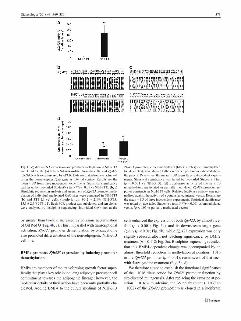
Promoter methylation reduces Zfp423 expression in NIH-3T3 cells Using qPCR, we compared Zfp423 mRNA expres-sion in NIH-3T3 and in 3T3-L1 cells and found it to be barelydetectable in the former and high in the latter (p < 0.01) (Fig.1a). Importantly, there was no sequence variation of theZfp423 promoter in NIH-3T3 and 3T3-L1 cells (data notshown), suggesting that the differential expression observedhad to be attributed to other mechanisms, including differentepigenetic profiles. Furthermore, the expression of other keyadipogenic marker genes was strongly silenced in NIH-3T3cells (ESM Fig. 1).
To explore this, we subjected the Zfp423 promoter regionto bioinformatic analysis. EMBOSS CpGplot revealed a large560 bp CpGi upstream Zfp423 transcription start site, provid-ing a potential basis for methylation control of Zfp423 expres-sion. We analysed the methylation status of the Zfp423 CpGiby bisulphite sequencing in NIH-3T3 and 3T3-L1 cells, andfound massive demethylation in the latter (methylation:15.3%vs 90.2% in NIH-3T3 cells; p < 0.001; Fig. 1b, c).
We then cloned the Zfp423 promoter into the luciferasereporter vector (pCpGfree-basic-Lucia), which was eithertreated with M.SssI methylase and fully methylated, or par-tially methylated using HhaI and HpaII methylases. Digestionwith the methylation-sensitive restriction enzyme HpyCH4IV
enabled control of methylation level in the two conditions(data not shown). Luciferase activity in the constructsharbouring the fully and the partially methylated Zfp423 pro-moters declined by 80% and 40%, respectively, comparedwith the unmethylated promoter (p < 0.001) (Fig. 1d). Theseresults demonstrate that methylation regulates Zfp423 promot-er function in vitro.
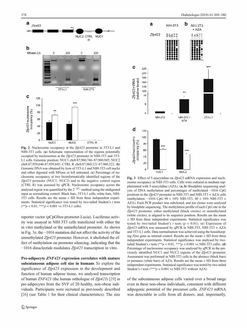
Nucleosome occupancy of Zfp423 promoter is increased inNIH-3T3 compared with 3T3-L1 cells Based on NuPoPanalysis, the Zfp423 promoter exhibited several potential re-gions where nucleosome positioning featured a high predic-tion score (Fig. 2a), suggesting that differential Zfp423 expres-sion in NIH-3T3 and 3T3-L1 cells is also accompanied by avariation in nucleosome occupancy. To validate this and as-sess nucleosome occupancy at the best predicted regions, anMNase protection assay was performed and nucleosome po-sitioning checked in mono-nucleosomal DNA by qPCR (Fig.2b). The percentage of nucleosome occupancy at two suchregions of the Zfp423 promoter was significantly higher inNIH-3T3 cells (nucleosome [NUC]1% occupancy: 72.2 vs51.5 in 3T3-L1 cells, p < 0.01; NUC2% occupancy: 94.6 vs46.4 in 3T3-L1 cells, p < 0.001; Fig. 2c). No significant dif-ference was observed in the CTRL R (negative control) re-gion, where nucleosome positioning featured a low bioinfor-matic prediction score. Thus, in these cells, nucleosome occu-pancy of the promoter inversely correlates with Zfp423 ex-pression, suggesting that dynamic chromatin remodellingmay also contribute to transcriptional regulation.
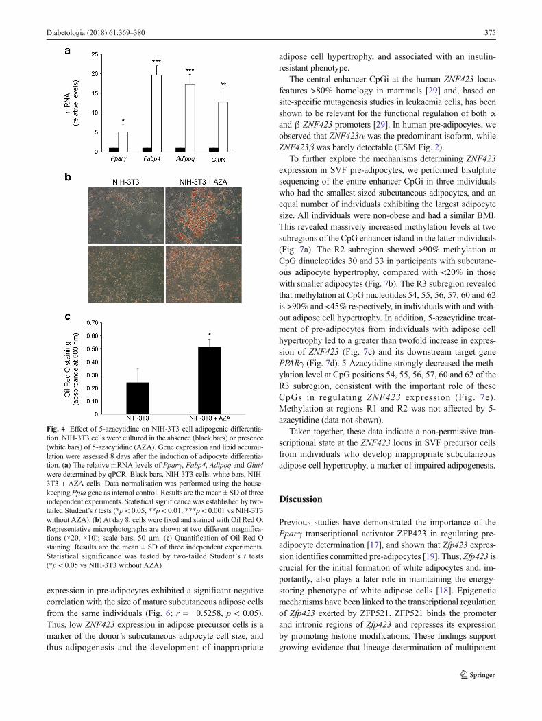
5-Azacytidine enhances Zfp423 expression and allows dif-ferentiation of non-adipogenic NIH-3T3 cells To assesswhether DNA methylation also regulates Zfp423 expressionin intact cells, we investigated the ability of the DNA methyl-transferase inhibitor 5-azacytidine to remove the transcription-al block imposed on Zfp423 in the NIH-3T3 cells. Incubatingthe cells with 5-azacytidine mainly affected methylation levelat CpG position −1016 (40% methylation in exposed vs 90%in unexposed cells, p < 0.01; Fig. 3a, b); this was associatedwith a sixfold increase in Zfp423mRNA expression (p < 0.01;Fig. 3c). However, apart from the −1016 CpG, the overallmethylation profile at the Zfp423 promoter did not change in5-azacytidine-treated cells (data not shown), providing a po-tential explanation for why mRNA expression levels are stilllower than in 3T3-L1 cells (Fig. 3c). MNase protection studiesrevealed that 5-azacytidine significantly reduced nucleosomeoccupancy at the NUC1 and NUC2 regions (p < 0.001; Fig.3d), further underlining the potential role of chromatin remod-elling of the Zfp423 regulatory region in transcriptionalregulation.
In parallel, 5-azacytidine robustly enhanced expression ofPparγ and the differentiation markers Fabp4, Adipoq andGlut4 after induction of differentiation (Fig. 4a), accompanied
372 Diabetologia (2018) 61:369–380
by greater than twofold increased cytoplasmic accumulationof Oil Red O (Fig. 4b, c). Thus, in parallel with transcriptionalactivation, Zfp423 promoter demethylation by 5-azacytidinealso promoted differentiation of the non-adipogenic NIH-3T3cell line.
BMP4 promotes Zfp423 expression by inducing promoterdemethylation
BMPs are members of the transforming growth factor super-family that play a key role in inducing adipocyte precursor cellcommitment towards the adipogenic lineage; however, themolecular details of their action have been only partially elu-cidated. Adding BMP4 to the culture medium of NIH-3T3
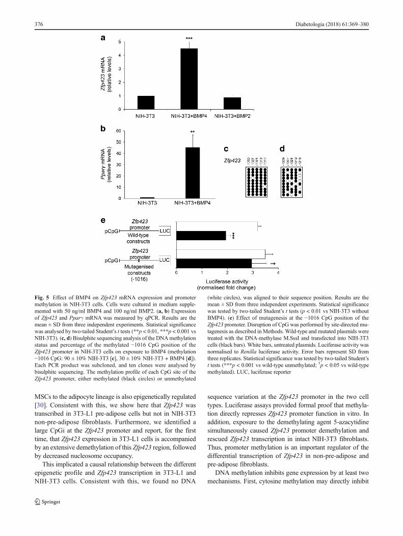
cells enhanced the expression of both Zfp423, by almost five-fold (p < 0.001; Fig. 5a), and its downstream target genePparγ (p < 0.01; Fig. 5b), while Zfp423 expression was onlyslightly reduced, albeit not reaching significance, by BMP2treatment (p = 0.118; Fig. 5a). Bisulphite sequencing revealedthat this BMP4-dependent change was accompanied by analmost threefold reduction in methylation at position −1016in the Zfp423 promoter (p < 0.01), reminiscent of that seenwith 5-azacytidine treatment (Fig. 5c, d).
We therefore aimed to establish the functional significanceof the −1016 dinucleotide for Zfp423 promoter function bysite-directed mutagenesis. After replacing the cytosine at po-sition −1016 with adenine, the 35 bp fragment (−1037 to−1002) of the Zfp423 promoter was cloned in a luciferase
Fig. 1 Zfp423mRNA expression and promoter methylation in NIH-3T3and 3T3-L1 cells. (a) Total RNAwas isolated from the cells, and Zfp423mRNA levels were assessed by qPCR. Data normalisation was achievedusing the housekeeping Ppia gene as internal control. Results are themean ± SD from three independent experiments. Statistical significancewas tested by two-tailed Student’s t test (**p < 0.01 vs NIH-3T3). (b, c)Bisulphite sequencing analysis and assessment of Zfp423 promoter meth-ylation of individual methylated CpG sites were compared in NIH-3T3(b) and 3T3-L1 (c) cells (methylation: 90.2 ± 2.1% NIH-3T3,15.3 ± 2.7% 3T3-L1). Each PCR product was subcloned, and ten cloneswere analysed by bisulphite sequencing. Individual CpG sites at the
Zfp423 promoter, either methylated (black circles) or unmethylated(white circles), were aligned to their sequence position as indicated abovethe panels. Results are the mean ± SD from three independent experi-ments. Statistical significance was tested by two-tailed Student’s t test(p < 0.001 vs NIH-3T3). (d) Luciferase activity of the in vitrounmethylated, methylated or partially methylated Zfp423 promoter re-porter constructs in NIH-3T3 cells. Relative luciferase activity was nor-malised against the activity of a cotransfected internal vector. Results arethe mean ± SD of three independent experiments. Statistical significancewas tested by two-tailed Student’s t tests (***p < 0.001 vs unmethylatedvector, †p < 0.05 vs partially methylated vector)
Diabetologia (2018) 61:369–380 373
reporter vector (pCpGfree-promoter-Lucia). Luciferase activ-ity was assayed in NIH-3T3 cells transfected with either thein vitro methylated or the unmethylated promoter. As shownin Fig. 5e, the −1016 mutation did not affect the activity of theunmethylated Zfp423 promoter. However, it abolished the ef-fect of methylation on promoter silencing, indicating that the−1016 dinucleotide modulates Zfp423 transcription in vitro.
Pre-adipocyte ZNF423 expression correlates with maturesubcutaneous adipose cell size in humans To explore thesignificance of Zfp423 expression in the development andfunction of human adipose tissue, we analysed transcriptionof human ZNF423 (the human orthologue of Zfp423) [29] inpre-adipocytes from the SVF of 20 healthy, non-obese indi-viduals. Participants were recruited as previously described[26] (see Table 1 for their clinical characteristics). The size
of the subcutaneous adipose cells varied over a broad rangeeven in these non-obese individuals, consistent with differentadipogenic potential of the precursor cells. ZNF423 mRNAwas detectable in cells from all donors, and, importantly,
Fig. 3 Effect of 5-azacytidine on Zfp423 mRNA expression and nucle-osome occupancy in NIH-3T3 cells. Cells were cultured in medium sup-plemented with 5-azacytidine (AZA). (a, b) Bisulphite sequencing anal-ysis of DNA methylation and percentages of methylated −1016 CpGpositions in the Zfp423 promoter in NIH-3T3 and NIH-3T3 + AZA cells(methylation: −1016 CpG 90 ± 10% NIH-3T3, 40 ± 10% NIH-3T3 +AZA). Each PCR product was subcloned, and ten clones were analysedby bisulphite sequencing. The methylation profile of each CpG site in theZfp423 promoter, either methylated (black circles) or unmethylated(white circles), is aligned to its sequence position. Results are the mean± SD from three independent experiments. Statistical significance wastested by two-tailed Student’s t tests (p < 0.01). (c) Expression ofZfp423 mRNA was measured by qPCR in NIH-3T3, NIH-3T3 + AZAand 3T3-L1 cells. Data normalisation was achieved using the housekeep-ing Ppia gene as internal control. Results are the mean ± SD from threeindependent experiments. Statistical significance was analysed by two-tailed Student’s t tests (**p < 0.01, ***p < 0.001 vs NIH-3T3 cells). (d)Percentage of nucleosome occupancy was analysed by qPCR in the pre-viously identified NUC1 and NUC2 regions of the Zfp423 promoter.Assessment was performed in NIH-3T3 cells in the absence (black bars)or presence (white bars) of AZA. Results are the mean ± SD from threeindependent experiments. Statistical significance was tested by two-tailedStudent’s t tests (***p < 0.001 vs NIH-3T3 without AZA)
Fig. 2 Nucleosome occupancy at the Zfp423 promoter in 3T3-L1 andNIH-3T3 cells. (a) Schematic representation of the regions potentiallyoccupied by nucleosomes at the Zfp423 promoter in NIH-3T3 and 3T3-L1 cells. Genomic position: NUC1 chr8:87,960,746–87,960,945; NUC2chr8:87,959,646-87,959,845; CTRL R chr8:87,960,121-87,960,322. (b)Genomic DNAwas obtained by lysis of 3T3-L1 and NIH-3T3 cell nucleiand either digested with MNase or left untreated. (c) Percentage of nu-cleosome occupancy at two bioinformatically identified regions of theZfp423 promoter (NUC1, NUC2) and in the negative control region(CTRL R) was assessed by qPCR. Nucleosome occupancy across theanalysed region was quantified by the 2−ΔCt method using the undigestedinput as normalising control. Black bars, 3T3-L1 cells; white bars, NIH-3T3 cells. Results are the mean ± SD from three independent experi-ments. Statistical significance was tested by two-tailed Student’s t tests(**p < 0.01, ***p < 0.001 vs 3T3-L1 cells)
374 Diabetologia (2018) 61:369–380
expression in pre-adipocytes exhibited a significant negativecorrelation with the size of mature subcutaneous adipose cellsfrom the same individuals (Fig. 6; r = −0.5258, p < 0.05).Thus, low ZNF423 expression in adipose precursor cells is amarker of the donor’s subcutaneous adipocyte cell size, andthus adipogenesis and the development of inappropriate
adipose cell hypertrophy, and associated with an insulin-resistant phenotype.
The central enhancer CpGi at the human ZNF423 locusfeatures >80% homology in mammals [29] and, based onsite-specific mutagenesis studies in leukaemia cells, has beenshown to be relevant for the functional regulation of both αand β ZNF423 promoters [29]. In human pre-adipocytes, weobserved that ZNF423α was the predominant isoform, whileZNF423β was barely detectable (ESM Fig. 2).
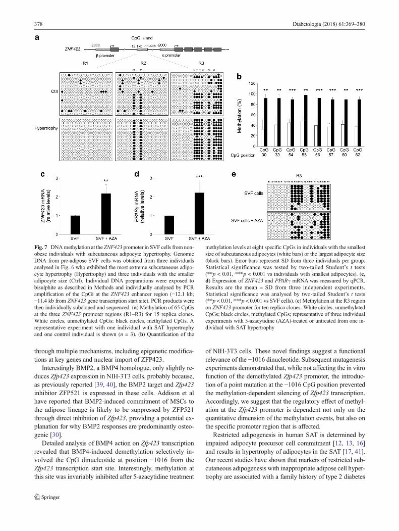
To further explore the mechanisms determining ZNF423expression in SVF pre-adipocytes, we performed bisulphitesequencing of the entire enhancer CpGi in three individualswho had the smallest sized subcutaneous adipocytes, and anequal number of individuals exhibiting the largest adipocytesize. All individuals were non-obese and had a similar BMI.This revealed massively increased methylation levels at twosubregions of the CpG enhancer island in the latter individuals(Fig. 7a). The R2 subregion showed >90% methylation atCpG dinucleotides 30 and 33 in participants with subcutane-ous adipocyte hypertrophy, compared with <20% in thosewith smaller adipocytes (Fig. 7b). The R3 subregion revealedthat methylation at CpG nucleotides 54, 55, 56, 57, 60 and 62is >90% and <45% respectively, in individuals with and with-out adipose cell hypertrophy. In addition, 5-azacytidine treat-ment of pre-adipocytes from individuals with adipose cellhypertrophy led to a greater than twofold increase in expres-sion of ZNF423 (Fig. 7c) and its downstream target genePPARγ (Fig. 7d). 5-Azacytidine strongly decreased the meth-ylation level at CpG positions 54, 55, 56, 57, 60 and 62 of theR3 subregion, consistent with the important role of theseCpGs in regulating ZNF423 expression (Fig. 7e).Methylation at regions R1 and R2 was not affected by 5-azacytidine (data not shown).
Taken together, these data indicate a non-permissive tran-scriptional state at the ZNF423 locus in SVF precursor cellsfrom individuals who develop inappropriate subcutaneousadipose cell hypertrophy, a marker of impaired adipogenesis.
Discussion
Previous studies have demonstrated the importance of thePparγ transcriptional activator ZFP423 in regulating pre-adipocyte determination [17], and shown that Zfp423 expres-sion identifies committed pre-adipocytes [19]. Thus, Zfp423 iscrucial for the initial formation of white adipocytes and, im-portantly, also plays a later role in maintaining the energy-storing phenotype of white adipose cells [18]. Epigeneticmechanisms have been linked to the transcriptional regulationof Zfp423 exerted by ZFP521. ZFP521 binds the promoterand intronic regions of Zfp423 and represses its expressionby promoting histone modifications. These findings supportgrowing evidence that lineage determination of multipotent
Fig. 4 Effect of 5-azacytidine on NIH-3T3 cell adipogenic differentia-tion. NIH-3T3 cells were cultured in the absence (black bars) or presence(white bars) of 5-azacytidine (AZA). Gene expression and lipid accumu-lation were assessed 8 days after the induction of adipocyte differentia-tion. (a) The relative mRNA levels of Pparγ, Fabp4, Adipoq and Glut4were determined by qPCR. Black bars, NIH-3T3 cells; white bars, NIH-3T3 + AZA cells. Data normalisation was performed using the house-keeping Ppia gene as internal control. Results are the mean ± SD of threeindependent experiments. Statistical significance was established by two-tailed Student’s t tests (*p < 0.05, **p < 0.01, ***p < 0.001 vs NIH-3T3without AZA). (b) At day 8, cells were fixed and stained with Oil Red O.Representative microphotographs are shown at two different magnifica-tions (×20, ×10); scale bars, 50 μm. (c) Quantification of Oil Red Ostaining. Results are the mean ± SD of three independent experiments.Statistical significance was tested by two-tailed Student’s t tests(*p < 0.05 vs NIH-3T3 without AZA)
Diabetologia (2018) 61:369–380 375
MSCs to the adipocyte lineage is also epigenetically regulated[30]. Consistent with this, we show here that Zfp423 wastranscribed in 3T3-L1 pre-adipose cells but not in NIH-3T3non-pre-adipose fibroblasts. Furthermore, we identified alarge CpGi at the Zfp423 promoter and report, for the firsttime, that Zfp423 expression in 3T3-L1 cells is accompaniedby an extensive demethylation of this Zfp423 region, followedby decreased nucleosome occupancy.
This implicated a causal relationship between the differentepigenetic profile and Zfp423 transcription in 3T3-L1 andNIH-3T3 cells. Consistent with this, we found no DNA
sequence variation at the Zfp423 promoter in the two celltypes. Luciferase assays provided formal proof that methyla-tion directly represses Zfp423 promoter function in vitro. Inaddition, exposure to the demethylating agent 5-azacytidinesimultaneously caused Zfp423 promoter demethylation andrescued Zfp423 transcription in intact NIH-3T3 fibroblasts.Thus, promoter methylation is an important regulator of thedifferential transcription of Zfp423 in non-pre-adipose andpre-adipose fibroblasts.
DNA methylation inhibits gene expression by at least twomechanisms. First, cytosine methylation may directly inhibit
Fig. 5 Effect of BMP4 on Zfp423 mRNA expression and promotermethylation in NIH-3T3 cells. Cells were cultured in medium supple-mented with 50 ng/ml BMP4 and 100 ng/ml BMP2. (a, b) Expressionof Zfp423 and Pparγ mRNA was measured by qPCR. Results are themean ± SD from three independent experiments. Statistical significancewas analysed by two-tailed Student’s t tests (**p < 0.01, ***p < 0.001 vsNIH-3T3). (c, d) Bisulphite sequencing analysis of the DNAmethylationstatus and percentage of the methylated −1016 CpG position of theZfp423 promoter in NIH-3T3 cells on exposure to BMP4 (methylation−1016 CpG: 90 ± 10% NIH-3T3 [c], 30 ± 10% NIH-3T3 + BMP4 [d]).Each PCR product was subcloned, and ten clones were analysed bybisulphite sequencing. The methylation profile of each CpG site of theZfp423 promoter, either methylated (black circles) or unmethylated
(white circles), was aligned to their sequence position. Results are themean ± SD from three independent experiments. Statistical significancewas tested by two-tailed Student’s t tests (p < 0.01 vs NIH-3T3 withoutBMP4). (e) Effect of mutagenesis at the −1016 CpG position of theZfp423 promoter. Disruption of CpG was performed by site-directed mu-tagenesis as described in Methods. Wild-type and mutated plasmids weretreated with the DNA-methylase M.SssI and transfected into NIH-3T3cells (black bars). White bars, untreated plasmids. Luciferase activity wasnormalised to Renilla luciferase activity. Error bars represent SD fromthree replicates. Statistical significance was tested by two-tailed Student’st tests (***p < 0.001 vs wild-type unmethylated; †p < 0.05 vs wild-typemethylated). LUC, luciferase reporter
376 Diabetologia (2018) 61:369–380
the association of DNA-binding factors [31, 32]. Second, pro-teins that recognise methylated CpG sites may recruit tran-scriptional corepressor molecules, including histone modifica-tion and chromatin remodelling enzymes, and cause a tran-scriptionally repressed chromatin state [31, 32]. Here, MNasedigestion assays revealed that, in the pre-adipose 3T3-L1 fi-broblasts, CpG demethylation of the Zfp423 promoter wasaccompanied by nucleosome repositioning in an open chro-matin state, which may contribute to Zfp423 active transcrip-tion. The ability of 5-azacytidine to induce this nucleosomerepositioning suggests that, in NIH-3T3 cells, demethylationof the Zfp423 promoter may trigger chromatin remodelling ina transcriptionally active conformation, thereby inducingZfp423 expression. Therefore, although we did not investigatea direct association of transcriptionally relevant DNA-bindingfactors to methylated cytosines, CpG methylation-triggered
chromatin condensation appears to be an important mecha-nism for maintaining methylated Zfp423 promoter silencing.
Previous studies have demonstrated that Zfp423 transcrip-tion is essential for pre-adipocyte commitment, enabling fur-ther adipogenic differentiation [17–19]. In line with this, weshowed that the effect of 5-azacytidine on Zfp423 promoterepigenetics and active gene transcription was followed byrescue of the differentiation capacity of the NIH-3T3 fibro-blasts, as revealed by a robust rise in Pparγ, Fabp4, Adipoqand Glut4 levels. Oil Red O accumulation in NIH-3T3 cyto-plasmwas also increased following exposure to 5-azacytidine.Therefore, in the model we now propose, commitment of anadipocyte precursor cell is accompanied by acquisition of aspecific chromatin epigenetic signature of the Zfp423 locus[17, 33]. Importantly, as shown here, these events appear tobe reversible. Indeed, exposure of the uncommitted NIH-3T3cell to an epigenetic agent, i.e. 5-azacytidine, partlyreprogrammed the epigenetic signature at the Zfp423 promot-er, favouring commitment and adipogenesis. However, 5-azacytidine does not make the NIH-3T3 cells an in vitro mod-el of spontaneous adipogenesis (data not shown). This is notsurprising because Zfp423, identified as a major determinantof pre-adipocyte commitment, is not responsible for the earlyphase of adipogenesis. At this stage, only ectopic expressionof CCAAT/enhancer-binding protein β provides a surrogatefor the requirement for 3-isobutyl-1-methylxanthine in theadipogenic differentiation of NIH-3T3 cells [34]. It is possiblethat further work will generate novel opportunities to over-come the restricted subcutaneous adipogenesis that is predic-tive of type 2 diabetes.
Studies by Bowers and co-workers [35] have demonstratedstem cell commitment to the adipocyte lineage by 5-azacytidine inhibition of DNAmethylation. They also provid-ed evidence supporting the role of BMP4 signalling in adipo-cyte lineage determination induced by 5-azacytidine [36].Additional studies have revealed that increased expressionand secretion of BMP4, a key molecule in the adipogenicmicroenvironment as it is also secreted by mature adiposecells [37], correlate with the capacity of MSCs to undergoadipogenic differentiation [36, 38]. Importantly, BMP4 hasbeen shown to enable nuclear entry of ZFP423 by dissociatingthe cytoplasmic WISP2–ZFP423 protein complex, which re-tains ZFP423 in the cytosol [20], thereby activating Pparγtranscription. Silencing of Zfp423 completely prevents the in-duction of Pparγ and other adipogenic marker genes inBMP4-treated cells, showing that ZFP423 is crucial forPparγ activation and for the ability of BMP4 to inducePparγ transcription [20].
Here, we demonstrate, for the first time, that BMP4 alsocauses demethylation of the Zfp423 promoter, which is suffi-cient to commit otherwise non-adipogenic cells to theadipogenic lineage. Thus, convergence of BMP4 signallingon Zfp423 enables its action on pre-adipocyte determination
Fig. 6 ZNF423 expression in pre-adipocytes from non-obese individualswith different subcutaneous adipocyte size. ZNF423 mRNA expressionwas quantified in pre-adipocytes obtained from the SVF of SAT from 20healthy, non-obese individuals as described in Methods. Correlation wasassessed by linear regression analysis. AU, absolute units; Subc.,subcutaneous
Table 1 Clinical characteristics of the study group
Variable Mean ± SD
N 20
Age (years) 40.8 ± 7.9
BMI (kg/m2) 25.4 ± 2.6
Fat (%) 26.0 ± 6.7
Fat-free mass (kg) 57.4 ± 10.4
Cell size (μm) 95.9 ± 9.5
GIR/bw (mg/min) 9.1 ± 3.1
f-insulin (pmol/l) 348.0 ± 179.4
fb-glucose (mmol/l) 4.6 ± 0.5
OGTT p-glucose 2 h (mmol/l) 6.1 ± 1.8
fb-glucose, fasting blood glucose; f-insulin, fasting insulin; GIR/bw, glu-cose infusion rate/body weight; OGTT p-glucose, plasma glucose duringOGTT
Diabetologia (2018) 61:369–380 377
through multiple mechanisms, including epigenetic modifica-tions at key genes and nuclear import of ZFP423.
Interestingly BMP2, a BMP4 homologue, only slightly re-duces Zfp423 expression in NIH-3T3 cells, probably because,as previously reported [39, 40], the BMP2 target and Zfp423inhibitor ZFP521 is expressed in these cells. Addison et alhave reported that BMP2-induced commitment of MSCs tothe adipose lineage is likely to be suppressed by ZFP521through direct inhibition of Zfp423, providing a potential ex-planation for why BMP2 responses are predominantly osteo-genic [30].
Detailed analysis of BMP4 action on Zfp423 transcriptionrevealed that BMP4-induced demethylation selectively in-volved the CpG dinucleotide at position −1016 from theZfp423 transcription start site. Interestingly, methylation atthis site was invariably inhibited after 5-azacytidine treatment
of NIH-3T3 cells. These novel findings suggest a functionalrelevance of the −1016 dinucleotide. Subsequent mutagenesisexperiments demonstrated that, while not affecting the in vitrofunction of the demethylated Zfp423 promoter, the introduc-tion of a point mutation at the −1016 CpG position preventedthe methylation-dependent silencing of Zfp423 transcription.Accordingly, we suggest that the regulatory effect of methyl-ation at the Zfp423 promoter is dependent not only on thequantitative dimension of the methylation events, but also onthe specific promoter region that is affected.
Restricted adipogenesis in human SAT is determined byimpaired adipocyte precursor cell commitment [12, 13, 16]and results in hypertrophy of adipocytes in the SAT [17, 41].Our recent studies have shown that markers of restricted sub-cutaneous adipogenesis with inappropriate adipose cell hyper-trophy are associated with a family history of type 2 diabetes
Fig. 7 DNAmethylation at the ZNF423 promoter in SVF cells from non-obese individuals with subcutaneous adipocyte hypertrophy. GenomicDNA from pre-adipose SVF cells was obtained from three individualsanalysed in Fig. 6 who exhibited the most extreme subcutaneous adipo-cyte hypertrophy (Hypertrophy) and three individuals with the smalleradipocyte size (Ctrl). Individual DNA preparations were exposed tobisulphite as described in Methods and individually analysed by PCRamplification of the CpGi at the ZNF423 enhancer region (−12.1 kb;−11.4 kb from ZNF423 gene transcription start site). PCR products werethen individually subcloned and sequenced. (a) Methylation of 65 CpGsat the three ZNF423 promoter regions (R1–R3) for 15 replica clones.White circles, unmethylated CpGs; black circles, methylated CpGs. Arepresentative experiment with one individual with SAT hypertrophyand one control individual is shown (n = 3). (b) Quantification of the
methylation levels at eight specific CpGs in individuals with the smallestsize of subcutaneous adipocytes (white bars) or the largest adipocyte size(black bars). Error bars represent SD from three individuals per group.Statistical significance was tested by two-tailed Student’s t tests(**p < 0.01, ***p < 0.001 vs individuals with smallest adipocytes). (c,d) Expression of ZNF423 and PPARγ mRNA was measured by qPCR.Results are the mean ± SD from three independent experiments.Statistical significance was analysed by two-tailed Student’s t tests(**p < 0.01, ***p < 0.001 vs SVF cells). (e) Methylation at the R3 regionon ZNF423 promoter for ten replica clones. White circles, unmethylatedCpGs; black circles, methylated CpGs; representative of three individualexperiments with 5-azacytidine (AZA)-treated or untreated from one in-dividual with SAT hypertrophy
378 Diabetologia (2018) 61:369–380
[16, 42] and are also present in non-obese individuals withtype 2 diabetes [43]. The relevance of our mechanistic find-ings in the NIH-3T3/3T3-L1 cell model to human adiposetissue dysfunction is underlined by our results in human adi-pocyte precursors revealing that expression of the Zfp423 hu-man orthologue ZNF423 [29] negatively correlates with thecell size of mature adipocytes. Hence, in the same individuals,low ZNF423 expression in SVF pre-adipocytes is a marker ofimpaired adipogenesis leading to inappropriate hypertrophyof mature subcutaneous adipocytes and causally contributingto it by interfering with the adipogenic commitment and dif-ferentiation of precursor cells.
Taken together, our present results suggest that the restrict-ed subcutaneous adipogenesis associated with insulin resis-tance [6] and a family history of type 2 diabetes [16, 42, 43]may be due to dysfunctional epigenetic regulation rather thanconventional DNA risk genes.
Secretion of BMP4 by mature adipose cells is positivelycorrelated with adipose cell size, and we have suggested thatthis is part of positive feedback in the tissue to enhance thecommitment and differentiation of new precursor cells to pre-vent inappropriate hypertrophy [37, 44]. Here, we provide amolecular basis for the effect of BMP4 in enhancing adipo-genesis, although secretion of BMP4 antagonists, in particularGremlin 1 in humans [37], is increased in hypertrophic obesityand prevents the expected positive effect of BMP4 onadipogenesis.
The overall structure of the regulatory regions of humanZNF423 and mouse Zfp423 is quite different [29]. However,we observed massive hypermethylation at distinct CpG dinu-cleotides in the central island serving as a promoter enhancerin human ZNF423. Hypermethylation of this island has pre-viously been shown to silence the gene in human leukaemiacells [29]. The position of the regulatory CpG dinucleotides,which are targeted by methylation events in leukaemia andadipocyte precursor cells, differ, probably reflecting tissuespecificity [45]. However, as demonstrated in the leukaemiacells, methylation at the pre-adipocyte ZNF423 central en-hancer island may also feature a repressive function as itspresence closely correlated with reduced ZNF423 expressionin the adipocyte precursor cells. We propose, therefore, thatchanges in the methylation profile at the regulatory regionaccount for the reduced ZNF423 expression observed in hy-pertrophic adipose tissue. Indeed, 5-azacytidine treatment ofpre-adipocytes isolated from individuals with adipose cell hy-pertrophy rescues both the hypomethylated and the permis-sive state at specific CpG enhancer region dinucleotides, aswell as ZNF423 expression.
Thus, based on our findings, methylation at the ZNF423regulatory region and its expression can be targeted both phar-macologically (e.g. 5-azacytidine) and by changes in the adi-pose tissue microenvironment (e.g. changes in BMP4 abun-dance or signalling). Expansion of this work may generate
attractive and novel opportunities to overcome the restrictedsubcutaneous adipogenesis and prevent inappropriate adiposetissue hypertrophy and its negative consequences on metabo-lism and risk of type 2 diabetes.
Acknowledgements We acknowledge support by the SwedishResearch Council, the Torsten Söderberg Foundation, the NovoNordiskFoundation and the Swedish Diabetes Association. The authors are grate-ful to D. Liguoro (URT Genomics of Diabetes - IEOS, CNR &Department of Translational Medicine, Federico II University ofNaples, Italy) for his continuous support, for sharing his expertise inSVF culturing and for the invaluable criticism.
Data availability Data supporting the findings of this study are avail-able upon request from the corresponding author.
Funding This study was funded by the European Foundation for theStudy of Diabetes (EFSD), by the Ministero dell’Istruzione, theUniversità e della Ricerca Scientifica (grants PRIN and FIRB MERIT,and PON 01_02460 and POR Campania Bioscience) and the SocietàItaliana di Diabetologia (SID-FO.DI.RI). ML is the recipient of theSID/FO.DI.RI.—MSD ITALIA 2017 Research Fellowship. LP is therecipient of the EFSD/Lilly Research Fellowship 2015.
Duality of interest The authors declare that there is no duality of inter-est associated with this manuscript.
Contribution statement ML and FZ designed, performed, analysed,interpreted and described the experiments; they also contributed to writ-ing the manuscript. GAR was in charge of daily supervision of the workand contributed to discussion and data analysis, along with ML. LP, AD,RS, CN, PM and FF contributed to the acquisition of data. AH, SH andJMH were in charge of the human studies, acquisition of adipose tissuebiopsies, isolation of SVF pre-adipocytes and acquisition of human data.CM and PF contributed to discussion and data analysis. US and FBconceived and designed the study, supervised the work and contributedto the writing of the paper. All authors critically revised and approved thefinal version the manuscript. FB is the guarantor of the manuscript andtakes responsibility for the integrity of the work as a whole.
Open Access This article is distributed under the terms of the CreativeCommons At t r ibut ion 4 .0 In te rna t ional License (h t tp : / /creativecommons.org/licenses/by/4.0/), which permits unrestricted use,distribution, and reproduction in any medium, provided you give appro-priate credit to the original author(s) and the source, provide a link to theCreative Commons license, and indicate if changes were made.
References
1. Eckel RH, Kahn SE, Ferrannini E et al (2011) Consensus statementobesity and type 2 diabetes: what can be unified and what needs tobe individualized? J Clin Endocrinol Metab 96:1654–1663
2. Zimmet P, Alberti KG, Shaw J (2001) Global and societal implica-tions of the diabetes epidemic. Nature 414:782–787
3. Klöting N, Fasshauer M, Dietrich A et al (2010) Insulin-sensitiveobesity. Am J Physiol Endocrinol Metab 299:E506–E515
4. Smith U, Hammarstedt A (2010) Antagonistic effects ofthiazolidinediones and cytokines in lipotoxicity. Biochim BiophysActa 1801:377–380
Diabetologia (2018) 61:369–380 379
5. Logan CY, Nusse R (2004) The WNT signaling pathway in devel-opment and disease. Annu Rev Cell Dev Biol 20:781–810
6. Scott RA, Fall T, Pasko D et al (2014) Common genetic variantshighlight the role of insulin resistance and body fat distribution intype 2 diabetes, independent of obesity. Diabetes 63:4378–4387
7. Shepherd PR, Gnudi L, Tozzo E, Yang H, Leach F, Kahn BB(1993) Adipose cell hyperplasia and enhanced glucose disposal intransgenic mice overexpressing GLUT4 selectively in adipose tis-sue. J Biol Chem 268:22243–22246
8. Rosen ED, Spiegelman BM (2014) What we talk about when wetalk about fat. Cell Rev 156:20–44
9. Klöting N, Blüher M (2014) Adipocyte dysfunction, inflammationand metabolic syndrome. Rev Endocr Metab Disord 15:277–287
10. Longo M, Spinelli R, D'Esposito V et al (2016) Pathologic endo-plasmic reticulum stress induced by glucotoxic insults inhibits ad-ipocyte differentiation and induces an inflammatory phenotype.Biochim Biophys Acta 1863:1146–1156
11. Neeland IJ, Turer AT, Ayers CR (2012) Dysfunctional adiposity andthe risk of prediabetes and type 2 diabetes in obese adults. JAMA308:1150–1159
12. Gustafson B, Hammarstedt A, Hedjazifar S, Smith U (2013)Restricted adipogenesis in hypertrophic obesity: the role ofWisp2, WNT, and Bmp4. Diabetes 62:2997–3004
13. Isakson P, Hammarstedt A, Gustafson B, Smith U (2009) Impairedpreadipocyte differentiation in human abdominal obesity: role ofWNT, tumor necrosis factor-alpha, and inflammation. Diabetes58:1550–1557
14. Talchai C, Xuan S, Lin HV, Sussel L, Accili D (2012) Pancreatic bcell dedifferentiation as a mechanism of diabetic b cell failure. Cell150:1223–1234
15. Arner E, Westermark PO, Spalding KL et al (2010) Adipocyteturnover: relevance to human adipose tissue morphology.Diabetes 59:105–109
16. Arner P, Arner E, Hammarstedt A, Smith U (2011) Genetic predis-position for type 2 diabetes, but not for overweight/obesity, is asso-ciated with a restricted adipogenesis. PLoS One 6:e18284
17. Gupta RK, Arany Z, Seale P et al (2010) Transcriptional control ofpreadipocyte determination by Zfp423. Nature 464:619–623
18. Shao M, Ishibashi J, Kusminski CM et al (2016) Zfp423 maintainswhite adipocyte identity through suppression of the beige cell ther-mogenic gene program. Cell Metab 23:1167–1184
19. Gupta RK, Mepani RJ, Kleiner S et al (2012) Zfp423 expressionidentifies committed preadipocytes and localizes to adipose endo-thelial and perivascular cells. Cell Metab 15:230–239
20. Hammarstedt A, Hedjazifar S, Jenndahl L et al (2013) Wisp2 reg-ulates preadipocyte commitment and Pparγ activation by Bmp4.Proc Natl Acad Sci U S A 110:2563–2568
21. Musri MM, Gomis R, Párrizas M (2010) A chromatin perspectiveof adipogenesis. Organ 6:15–23
22. Campión J, Milagro FI, Martínez JA (2009) Individuality and epi-genetics in obesity. Obes Rev 10:383–392
23. Martínez JA, Milagro FI, Claycombe KJ, Schalinske KL (2014)Epigenetics in adipose tissue, obesity, weight loss, and diabetes.Adv Nutr 5:71–81
24. Deaton AM, Bird A (2011) CpG islands and the regulation of tran-scription. Genes Dev 25:1010–1022
25. Raciti GA, Nigro C, Longo M et al (2014) Personalized medicineand type 2 diabetes: lesson from epigenetics. Epigenomics 6:229–238
26. Laakso M, Zilinskaite J, Hansen T et al (2008) Insulin sensitivity,insulin release and glucagon-like peptide-1 levels in persons withimpaired fasting glucose and/or impaired glucose tolerance in theEUGENE2 study. Diabetologia 51:502–511
27. Parrillo L, Costa V, Raciti GA et al (2016) Hoxa5 undergoes dy-namic DNA methylation and transcriptional repression in the adi-pose tissue of mice exposed to high-fat diet. Int J Obes 40:929–937
28. Raciti GA, Spinelli R, Desiderio A et al (2017) Specific CpG hyper-methylation leads to Ankrd26 gene downregulation in white adi-pose tissue of a mouse model of diet-induced obesity. Sci Rep 7:43526
29. Harder L, Eschenburg G, Zech A et al (2013) Aberrant ZNF423impedes B cell differentiation and is linked to adverse outcome ofETV6-RUNX1 negative B precursor acute lymphoblastic leuke-mia. J Exp Med 210:2289–2304
30. Addison WN, Fu MM, Yang HX et al (2014) Direct transcriptionalrepression of Zfp423 by Zfp521 mediates a bone morphogenicprotein-dependent osteoblast versus adipocyte lineage commitmentswitch. Mol Cell Biol 34(16):3076–3085
31. Klose RJ, Bird AP (2006) Genomic DNA methylation: the markand its mediators. Trends Biochem Sci 31:89–97
32. Fujiki K, Kano F, Shiota K, Murata M (2009) Expression of theperoxisome proliferator activated receptor gamma gene is repressedby DNAmethylation in visceral adipose tissue of mouse models ofdiabetes. BMC Biol 7:38
33. Bell O, Tiwari VK, Thomä NH, Schübeler D (2011) Determinantsand dynamics of genome accessibility. Nat Rev Genet 12:554–564
34. Wu Z, Bucher NL, Farmer SR (1996) Induction of peroxisomeproliferator-activated receptor gamma during the conversion of3T3 fibroblasts into adipocytes is mediated by C/EBPbeta,C/EBPdelta, and glucocorticoids. Mol Cell Biol 16:4128–4136
35. Bowers RR, Kim JW, Otto TC, Lane MD (2006) Stable stem cellcommitment to the adipocyte lineage by inhibition of DNA meth-ylation: role of the BMP-4 gene. Proc Natl Acad Sci U S A 103:13022–13027
36. Huang H, Song TJ, Li X et al (2009) BMP signaling pathway isrequired for commitment of C3H10T1/2 pluripotent stem cells tothe adipocyte lineage. Proc Natl Acad Sci U S A 106:12670–12675
37. Gustafson B, Hammarstedt A, Hedjazifar S et al (2015) BMP4 andBMP antagonists regulate human white and beige adipogenesis.Diabetes 64:1670–1681
38. Tang QQ, Otto TC, Lane MD (2004) Commitment of C3H10T1/2pluripotent stem cells to the adipocyte lineage. Proc Natl Acad SciU S A 101:9607–9611
39. Spina R, Filocamo G, Iaccino E et al (2013) Critical role of zincfinger protein 521 in the control of growth, clonogenicity and tu-morigenic potential of medulloblastoma cells. Oncotarget 4:1280–1292
40. Kamiya D, Banno S, Sasai N et al (2011) Intrinsic transition ofembryonic stem-cell differentiation into neural progenitors. Nature470:503–509
41. Jansson PA, Pellmé F, Hammarstedt A et al (2003) A novel cellularmarker of insulin resistance and early atherosclerosis in humans isrelated to impaired fat cell differentiation and low adiponectin.FASEB J 17:1434–1440
42. Smith U, Kahn BB (2016) Adipose tissue regulates insulin sensi-tivity: role of adipogenesis, de novo lipogenesis and novel lipids. JIntern Med 280:465–475
43. Acosta JR, Douagi I, Andersson DP et al (2016) Increased fat cellsize: a major phenotype of subcutaneous white adipose tissue innon-obese individuals with type 2 diabetes. Diabetologia 59:560–570
44. Gustafson B, Hedjazifar S, Gogg S, Hammarstedt A, Smith U(2015) Insulin resistance and impaired adipogenesis. TrendsEndocrinol Metab 26:193–200
45. Ghosh S, Yates AJ, Frühwald MC, Miecznikowski JC, Plass C,Smiraglia D (2010) Tissue specific DNA methylation of CpGislands in normal human adult somatic tissues distinguishes neuralfrom non-neural tissues. Epigenetics 5:527–538
380 Diabetologia (2018) 61:369–380


![Review Article Epigenetic Modifications and Diabetic …downloads.hindawi.com/journals/bmri/2013/635284.pdfthe risk of diabetic retinopathy and nephropathy in diabetic patients [ ]](https://img.pdfslide.us/doc/110x75/6005be703dc6b77d630bd463/review-article-epigenetic-modifications-and-diabetic-the-risk-of-diabetic-retinopathy.jpg)