Embed Size (px)
Citation preview
INTERNATIONAL JOURNAL OF HEALTH RESEARCH IN MODERN INTEGRATED MEDICAL SCIENCES (IJHRMIMS), ISSN 2394-8612 (P), ISSN 2394-8620 (O), Oct-Dec 2014
Case Report
Role of Neuro-Imaging in Dengue Encephalitis
Madhavi C1, GS Kejriwal2, Giridhar Gopal G3
Abstract : Dengue infection is endemic in many tropical countries and its incidence is increasing worldwide. The
important neurological complications are encephalopathy and encephalitis , the former being more common. Along with
serological and CSF examinations, imaging with CT or MRI is important to look for structural changes in brain and if
present, to define the pattern and extent of involvement of brain parenchyma. Here we report 2 cases of a children with
serologically proven dengue fever having features of dengue encephalitis on imaging. This report is to emphasize the
role of imaging in dengue fever with neurological manifestations.
Key Words : Bilateral thalamic hypodensity, CT scan, Dengue fever, Encephalitis, Neurological involvement
Introduction
Dengue viruses are single stranded viruses of Flaviviridae
family, which can cause dengue fever and dengue
hemorrhagic fever. A myriad of systemic manifestations
can occur and the neurological manifestations play a great
role in increasing the mortality and morbidity. The role of
imaging has become indispensable to evaluate the
structural changes in brain and thereby assessing the
prognosis of the patient.
Case Report
Case 1 A boy, 10 yrs of age came to the emergency
department with complaints of high-grade fever for 7 days,
seizures for 3 days and altered sensorium for 2 day, There
was no evidence of any muco-cutaneous rash or
haemorrhagic manifestations.
The patient was admitted in pediatric intensive care unit.
On examination, there was hypertonia involving all 4
limbs, brisk reflexes, plantar extensor & ankle clonus. His
Glasgow Coma Scale was ‘6’ at admission. Laboratory
examinations revealed a platelet count of 1.4 lakhs and
serum was positive for IgM and IgG dengue antibodies
and negative for Malarial parasite, Hepatitis A, B, C and
Japanese encephalitis virus. CSF examination showed
evidence of dengue antigen, protein 8.4mg/dl, sugar 60mg/
dl and differential count showing 100% lymphocytes.
Patient developed involuntary movements during second
week of stay in hospital and imaging of brain was
performed. MRI brain was attempted but abandoned as
sedation failed. Hence plain & contrast CT scan was taken.
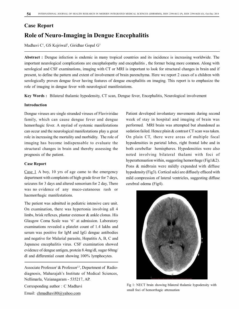
On plain CT, there were areas of multiple focal
hypodensities in parietal lobes, right frontal lobe and in
both cerebellar hemispheres. Hypodensities were also
noted involving bilateral thalami with foci of
hyperattenuation within, suggesting hemorrhage (Fig1&2).
Pons & midbrain were mildly expanded with diffuse
hypodensity (Fig3). Cortical sulci are diffusely effaced with
mild compression of lateral ventricles, suggesting diffuse
cerebral edema (Fig4).
Associate Professor1 & Professor2,3, Department of Radio-
diagnosis, Maharajah’s Institute of Medical Sciences,
Nellimarla, Vizianagaram - 535217, AP.
Corresponding author : C Madhavi
Email: [email protected]
Fig 1: NECT brain showing bilateral thalamic hypodensity with
small foci of hemorrhagic attenuation
54
INTERNATIONAL JOURNAL OF HEALTH RESEARCH IN MODERN INTEGRATED MEDICAL SCIENCES (IJHRMIMS), ISSN 2394-8612 (P), ISSN 2394-8620 (O), Oct-Dec 2014
The diagnosis of encephalitis was made and with
conservative treatment he recovered slowly. After 40 days
he was discharged. On follow-up 3weeks later, he has
regained near normal tone of limbs with only persistence
of involuntary movements to some extent.
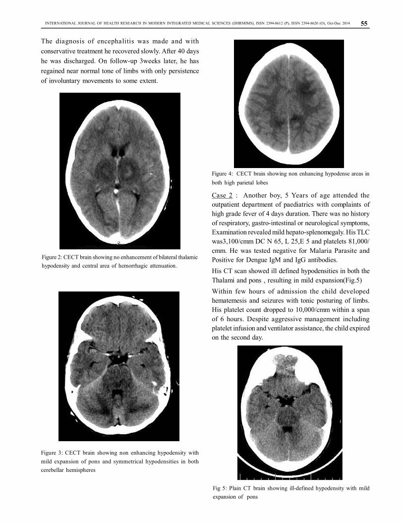
Figure 2: CECT brain showing no enhancement of bilateral thalamic
hypodensity and central area of hemorrhagic attenuation.
Figure 3: CECT brain showing non enhancing hypodensity with
mild expansion of pons and symmetrical hypodensities in both
cerebellar hemispheres
Figure 4: CECT brain showing non enhancing hypodense areas in
both high parietal lobes
Case 2 : Another boy, 5 Years of age attended the
outpatient department of paediatrics with complaints of
high grade fever of 4 days duration. There was no history
of respiratory, gastro-intestinal or neurological symptoms,
Examination revealed mild hepato-splenomegaly. His TLC
was3,100/cmm DC N 65, L 25,E 5 and platelets 81,000/
cmm. He was tested negative for Malaria Parasite and
Positive for Dengue IgM and IgG antibodies.
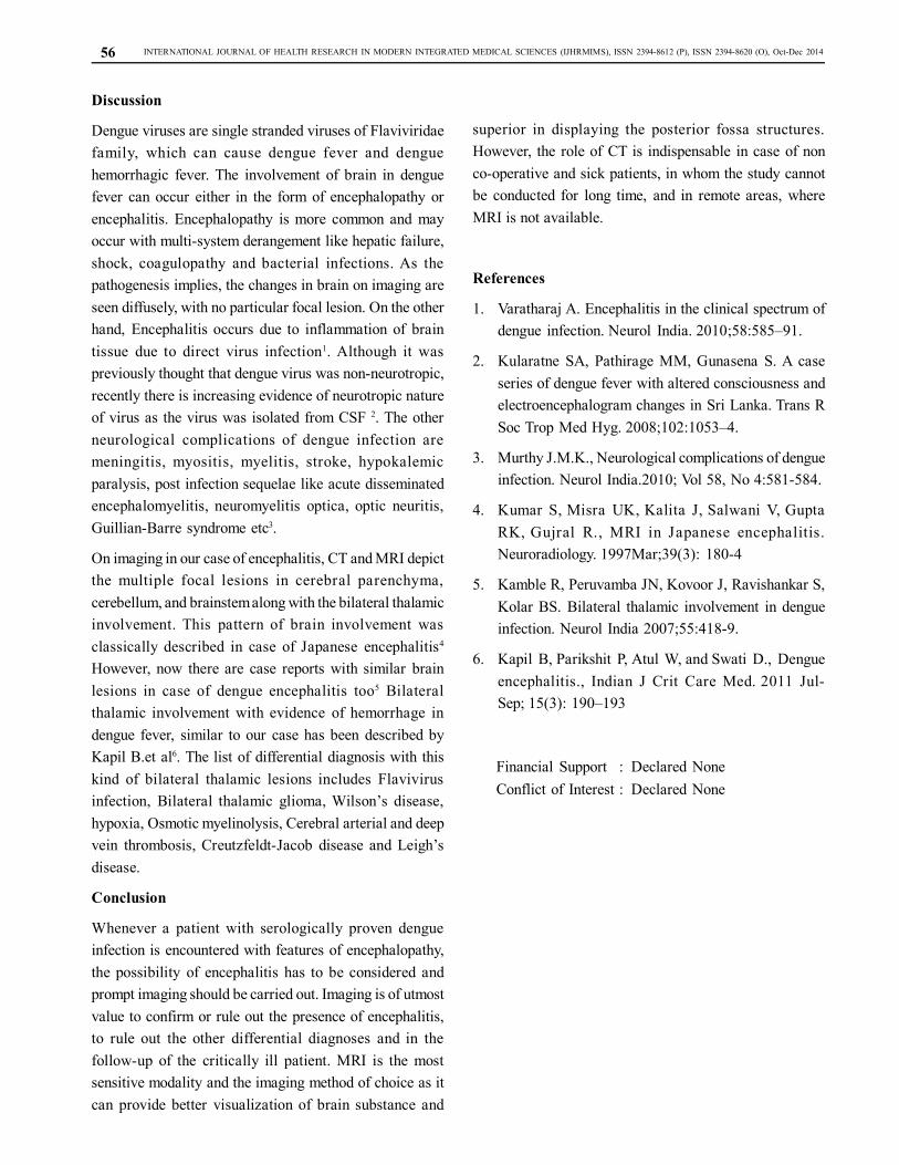
His CT scan showed ill defined hypodensities in both the
Thalami and pons , resulting in mild expansion(Fig.5)
Within few hours of admission the child developed
hematemesis and seizures with tonic posturing of limbs.
His platelet count dropped to 10,000/cmm within a span
of 6 hours. Despite aggressive management including
platelet infusion and ventilator assistance, the child expired
on the second day.
Fig 5: Plain CT brain showing ill-defined hypodensity with mild
expansion of pons
55
INTERNATIONAL JOURNAL OF HEALTH RESEARCH IN MODERN INTEGRATED MEDICAL SCIENCES (IJHRMIMS), ISSN 2394-8612 (P), ISSN 2394-8620 (O), Oct-Dec 2014
Discussion
Dengue viruses are single stranded viruses of Flaviviridae
family, which can cause dengue fever and dengue
hemorrhagic fever. The involvement of brain in dengue
fever can occur either in the form of encephalopathy or
encephalitis. Encephalopathy is more common and may
occur with multi-system derangement like hepatic failure,
shock, coagulopathy and bacterial infections. As the
pathogenesis implies, the changes in brain on imaging are
seen diffusely, with no particular focal lesion. On the other
hand, Encephalitis occurs due to inflammation of brain
tissue due to direct virus infection1. Although it was
previously thought that dengue virus was non-neurotropic,
recently there is increasing evidence of neurotropic nature
of virus as the virus was isolated from CSF 2. The other
neurological complications of dengue infection are
meningitis, myositis, myelitis, stroke, hypokalemic
paralysis, post infection sequelae like acute disseminated
encephalomyelitis, neuromyelitis optica, optic neuritis,
Guillian-Barre syndrome etc3.
On imaging in our case of encephalitis, CT and MRI depict
the multiple focal lesions in cerebral parenchyma,
cerebellum, and brainstem along with the bilateral thalamic
involvement. This pattern of brain involvement was
classically described in case of Japanese encephalitis4
However, now there are case reports with similar brain
lesions in case of dengue encephalitis too5 Bilateral
thalamic involvement with evidence of hemorrhage in
dengue fever, similar to our case has been described by
Kapil B.et al6. The list of differential diagnosis with this
kind of bilateral thalamic lesions includes Flavivirus
infection, Bilateral thalamic glioma, Wilson’s disease,
hypoxia, Osmotic myelinolysis, Cerebral arterial and deep
vein thrombosis, Creutzfeldt-Jacob disease and Leigh’s
disease.
Conclusion
Whenever a patient with serologically proven dengue
infection is encountered with features of encephalopathy,
the possibility of encephalitis has to be considered and
prompt imaging should be carried out. Imaging is of utmost
value to confirm or rule out the presence of encephalitis,
to rule out the other differential diagnoses and in the
follow-up of the critically ill patient. MRI is the most
sensitive modality and the imaging method of choice as it
can provide better visualization of brain substance and
superior in displaying the posterior fossa structures.
However, the role of CT is indispensable in case of non
co-operative and sick patients, in whom the study cannot
be conducted for long time, and in remote areas, where
MRI is not available.
References
1. Varatharaj A. Encephalitis in the clinical spectrum of
dengue infection. Neurol India. 2010;58:585–91.
2. Kularatne SA, Pathirage MM, Gunasena S. A case
series of dengue fever with altered consciousness and
electroencephalogram changes in Sri Lanka. Trans R
Soc Trop Med Hyg. 2008;102:1053–4.
3. Murthy J.M.K., Neurological complications of dengue
infection. Neurol India.2010; Vol 58, No 4:581-584.
4. Kumar S, Misra UK, Kalita J, Salwani V, Gupta
RK, Gujral R., MRI in Japanese encephalitis.Neuroradiology. 1997Mar;39(3): 180-4
5. Kamble R, Peruvamba JN, Kovoor J, Ravishankar S,
Kolar BS. Bilateral thalamic involvement in dengue
infection. Neurol India 2007;55:418-9.
6. Kapil B, Parikshit P, Atul W, and Swati D., Dengue
encephalitis., Indian J Crit Care Med. 2011 Jul-
Sep; 15(3): 190–193
Financial Support : Declared None
Conflict of Interest : Declared None
56