Embed Size (px)
Citation preview
Role of Bacteria in the Oxidation of Myoglobin" 2
D. L. ROBACH3 AND R. N. COSTILOW
Department of Microbiology and Public Health, Michigan State University, East Lansing, Michigan
Received for publication April 12, 1961
ABSTRACTROBACH, D. L. (Michigan State University, East
Lansing), and R. N. COSTILOW. Role of bacteria in theoxidation of myoglobin. Appl. Microbiol. 9:529-533.1961.-The addition to steaks of cell suspensions of anumber of aerobic bacteria and of Saccharomycescerevisiae greatly increased the rate of discoloration.Low inocula resulted in the more rapid appearance ofthe brown color of metmyoglobin, whereas high cellpopulations quickly produced the purple color ofmyoglobin. Sonically treated suspensions of Pseudo-monas geniculata produced similar changes in surfacecolor but less rapidly. No such effect was observed withLactobacillus plantarum.The visible changes in color were found to be associ-
ated with the oxygen demand of the surface tissueincluding, of course, the demand of any contaminatingmicroorganisms. Inhibitors of respiratory activityinhibited the rate of discoloration under normal at-mospheric conditions. However, when the oxygen levelin the atmosphere was reduced, the inhibitors had nosignificant effect. In an oxygen-free atmosphere, thesteak surfaces were the purple color of myoglobin; at10 mm oxygen pressure, the pigment was oxidized tometmyoglobin and the surface was brown in color. Nobacterial activity was necessary for pigment oxidationunder low oxygen pressures.
Addition of dilute solutions of glucose oxidaseresulted in rapid oxidation of the meat pigment tometmyoglobin both in extracts and on steak surfaces.More concentrated solutions resulted in further oxida-tion as evidenced by the appearance of a green color.Horseradish extract with a high peroxidase activityadded with H202 resulted in rapid oxidation of thepigment but neither were very effective alone, althoughH202 did result in a browning reaction in aged steaks.
It is concluded that the primary role of the bacteriain meat discoloration is in the reduction of the oxygentension in the surface tissue. The implications of thedata are discussed and a possible mechanism of myo-globin oxidation is proposed.
I Journal article no. 2791, Michigan Agricultural ExperimentStation.
2 This research was financed by a grant from Merck &Co., Inc., Rahway, N. J.
3 Present address: Chemical Division, Merck & Co., Inc.,Rahway, N. J.
Discoloration of fresh prepackaged meat or "loss ofbloom," as it is called by the trade, means that meatloses its bright red appearance. Meat color is due tomyoglobin, an iron porphyrin pigment very similar tohemoglobin. The chemistry of meat pigments wasreviewed by Schweigert (1956). The heme prostheticgroup is attached to a globin protein fraction by theamino acid, histidine. The heme so attached to theprotein fraction can form a dissociable compound withoxygen. In the reduced form, the iron of the heme is inthe ferrous state and the pigment is purple. Uponexposure to excess molecular oxygen the iron remainsin the ferrous state but the pigment becomes oxy-genated. This pigment called oxymyoglobin is brightred. The heme portion of myoglobin may be oxidizedby various means to metmyoglobin, a brown pigment.The iron of this compound is in the ferric state. Theproblem of discoloration involves the loss of oxygen toform reduced myoglobin (purple) and oxidation tometmyoglobin (brown).The fact that bacterial activity is a major factor in
pigment changes in fresh prepackaged meat has beenwell established (Butler, Bratzler, and Mallman, 1953;Costilow et al., 1955). Neill (1925) observed thatpneumococci, cell-free extracts of pneumococci, andsterile animal tissue extracts caused the reduction ofmethemoglobin to hemoglobin when molecular oxygenwas excluded. On introduction of small amounts ofmolecular oxygen, the reverse reaction was demon-strated with intact bacterial cells, cell extracts, andsterile extracts from potatoes. It is believed that thepneumococci bring about oxidation by the productionof peroxides or similar compounds (Neill and Hastings,1925). However, the actual role of bacteria in causingchanges in the pigment in meat surface tissue has niotbeen clearly demonstrated. This was the purpose of theinvestigation reported here.
MATERIALS AND MIETHODS
Steaks used in this study were from the longissimusdorsi muscle of U. S. Good grade beef ribs purchasedlocally. The muscle was removed from the ribs and CUtinto 12-in. thick steaks.The wrapping material employed was du Ponit
cellophane 300 MSAT-80.4Cells of all the bacteria used except Lactobacillus
4E. I. du Pont de Nemours & Co., Wilmington, Del.
529
on June 19, 2018 by guesthttp://aem
.asm.org/
Dow
nloaded from

D. L. ROBACH AND R. N. COSTILOW
plantarum were grown in nutrient broth shake culturesor on nutrient agar slants. The L. plantarum culturewas grown in a Trypticase soy broth (BBL)5 and theSaccharomyces in dextrose broth (Difco).6 All cells werewashed in saline before being inoculated on steaksurfaces by the use of sterile atomizers. Washed cellswere also used to prepare cell-free extracts. Cells werebroken in a Raytheon,7 50-w, 9-kc sonic oscillator typeR-22-3. The suspension was centrifuged to removecellular debris and the supernatant was applied tosteak surfaces by means of a small brush.The various enzyme and bacterial inhibitors, which
were used on the steak surface, were applied by sterileatomizers.Most myoglobin solutions were prepared from
longissimus dorsi muscle; however, some solutions wereprepared from beef trimmings. The myoglobin solu-tions were prepared exactly as the pigment solutionsused for spectrophotometric analysis to be describedlater.
Initial studies determining bacterial growth on steaksurfaces demonstrated that a surface-slicing techniquegave more reproducible results than did the conven-tional swabbing technique. The surface-slicing tech-nique consisted of cutting a slice approximately 3 mmthick from the surface of the steak with a Hobart8 50slicing machine. The machine was cleaned and sani-tized before each cutting by washing thoroughly withhot detergent and then rinsing with distilled water,hypochlorite (500 ppm), and finally with sterile dis-tilled water. The rotating blade was allowed to spindry. Platings made of swabbings from the machinedemonstrated that the portion which came in contactwith the meat slice was essentially free from bacteria.The thin slice from the surface was weighed and
placed in a sterile, chilled Waring Blendor. Refrig-erated sterile, distilled water was added to give a 1:5dilution. After blending for 30 sec, a sample of thehomogenate was plated with Tryptone glucose extractagar (Difco)6 (TGE) and observed for bacterial growth.The remainder of the homogenate was used for de-termining oxygen uptake by the Warburg method andfor determining the pigments present by spectrophoto-metric analysis.Two incubation temperatures for the TGE agar
plates were tested and it was noted that similar resultswere obtained from platings held at 4 C for 1 week andat 20 C for 3 days. For the sake of convenience, theshorter time and higher temperature of incubationwere used for all bacterial counts.A sample of the homogenate was prepared for pig-
6Baltimore Biological Laboratory, Inc., Baltimore, Md.6 Difco Laboratories, Inc., Detroit, Mich.Raytheon Corporation, Waltham, Mass.
8 The Hobart Mfg. Co., Troy, Ohio.
ment determination by centrifuging in a Servall' SPXcentrifuge at full speed for 10 min, and filtering throughWhatman no. 40 filter paper. Seven milliliters of filtratewere diluted with 3 ml of distilled water for spectro-photometric analysis with a Beckman'0 DU spectro-photometer. In studies conducted in optically matchedThunberg tubes, a Bausch and Lomb Spectronic 20"colorimeter was used and no dilution of the filtratewas necessary.Two methods of pigment analysis were used. The
first method was essentially that of Butler et al. (1953).The second method of analysis used was that ofBroumand, Ball, and Stier (1958). Both were found togive comparable results with respect to percentage ofmetmyoglobin. The latter method has the advantageof making it possible to estimate the relative percentageof each form of the pigment.The discoloration of steak surfaces was estimated by
the Munsell spinning disc method of Nickerson (1946)and an index of fading calculated on the basis of thestandards proposed by Butler et al. (1953). Visualobservations were also made since the fading indexdoes not reflect the type of pigment change whichoccurs, because the index values are increased oneither deoxygenation or oxidation of the muscle pig-ment.
Steaks to be incubated in atmospheres of variousoxygen pressures were placed unwrapped in vacuumdesiccators. The bottom of each desiccator was coveredwith water to maintain a high relative humidity. Theair was replaced by reducing the pressure to less than50 mm Hg and refilling with nitrogen that had beenpassed over hot copper shavings to remove contam-inating oxygen. This was repeated three times and thedesired amount of oxygen added along with the ni-trogen on the last fill. The total pressure was adjustedto 750 mm with nitrogen.
RESULTSPure cultures of Pseudomonas fluorescens, Pseudo-
monas aeruginosa, two strains of Pseudomonas geni-culata, three Pseudomonas sp., Achromobacter lique-faciens, Flavobacterium rhenanus, L. plantarum, andSaccharomyces cerevisiae were tested for their effect onthe surface color of wrapped beef steaks. At roomtemperature, the yeast and all the bacteria testedexcept L. plantarum caused rapid color changes. At4 + 1 C, the yeast had little effect on color but theaerobic bacteria tested were all active. All gave es-sentially the same results, causing the pigment tochange from red to brown and finally to purple. How-ever, when large numbers of cells were added to steak
9 Ivan Sorvall, Inc., Norwalk, Conn.10 Beckman Instruments, Inc., Fullerton, Calif."Bausch and Lomb Optical Company, Rochester, N. Y.
[VOL. I9530
on June 19, 2018 by guesthttp://aem
.asm.org/
Dow
nloaded from
OXIDATION OF MYOGLOBIN
surfaces, the purple color of myoglobin was notedwithin a few hours and no further change was observed.L. plantarum, an organism which does not utilizeoxygen to any appreciable extent, did not bring aboutthe discoloration of beef at either room temperature orat 4 C.The index of fading value for steak surfaces treated
with cell-free preparations (sonically treated) of P.geniculata increased more rapidly than that for ap-propriate controls, demonstrating that the enzymes ofP. geniculata were active in the discoloration. Steaksinoculated with intact cells discolored more rapidlythan those treated with cell extracts. This was ex-pected since the respiratory activity of the intact cellswas greater than that of the sonically treated prepara-tions.
Further evidence of the importance of the respiratory
I .-
;j
cj2tL.0
0oj
02 UPTAKE A
/4
0 2 4 6 8 10 0 2TIME - DAYS
*- * CONTROL A--A INOCULATEDo---o INOCULATED + IODOACETATE
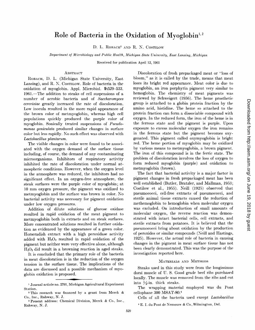
FIG. 1. Comparison of changes in fading index, metmyoglobinconcentration, oxygen demand, and bacterial count of inoculated
steaks treated with a 0.1 M solution of iodoacetate with both
inoculated and uninoculated control steaks. Groups of steakswith each treatment were prepared and one of each sampled at the
times indicated. The steaks were held at 4 4 1 C during the ex-
periment.
activity of the bacteria was obtained on comparingcontrol steaks with inoculated steaks, and inoculatedsteaks treated with inhibitors demonstrated to greatlyreduce the oxygen uptake rate of the test organismusing meat as a substrate. As may be noted in Fig. 1,the untreated inoculated steaks had a very significantoxygen demand initially and this was correlated with ahigh bacterial population and a high index of fading.The steaks were the purple color of myoglobin and nosignificant amount of metmyoglobin was detected. Theinoculated steaks treated with iodoacetate had a highbacterial load but a low oxygen demand and a lowfading index initially. These steaks never developed ashigh a bacterial population or oxygen uptake rate asthe control group. However, the metmyoglobin levelsincreased slightly faster and the change in fading indexwas comparable with the control. Treatment with a0.1 M solution of sodium malonate yielded resultssimilar to iodoacetate.The effect of various oxygen levels on the color of
beef steaks is illustrated in Fig. 2. During the first fewdays of refrigeration, the microbial activity was minor.However, the steaks held under 10 mm oxygen pressurequickly developed a brown color and those under 75 mmoxygen pressure became brown at a slower rate. Afterthe first 5 or 6 days, the microbial activity, undoubt-edly, brought about the color changes noted in thesteaks held in a normal atmosphere and in one ofoxygen. Inoculated steaks that were run for com-parison in this experiment all became purple during thefirst 6 days and remained this color for the duration ofthe experiment.The initial changes occurring under the various
oxygen pressures were not altered by the atomizing of
ATMOSPHERES: 0---O 750 mm 02o-o AIR
BRIGHTIRED I
BROWN
75 mm 02
xPURPLE L
%% \--A 10 mm 02
* * N 2
-- o- ~-o---
'*Z>sx.s '* ag
0 2 4 6 8 10 12 14 isTIME - DAYS
FIG. 2. Relative color changes observed on steak surfaces heldin various atmospheres. The atmospheres were adjusted in vacuumdesiccators by use of oxygen-free nitrogen. The total pressure ofeach atmosphere was adjusted to 750 mm. The experiment was
conducted at 4 d 1 C. The color notations used and plotted on thegraph from top to bottom were: (1) very bright red, (2) bright red,(3) red, (4) dark red, (5) brown-red, (6) brown, (7) brown-purple,(8) purple-brown, (9) purple.
1961] 531
%L
on June 19, 2018 by guesthttp://aem
.asm.org/
Dow
nloaded from
D. L. ROBACH AND R. N. COSTILOW
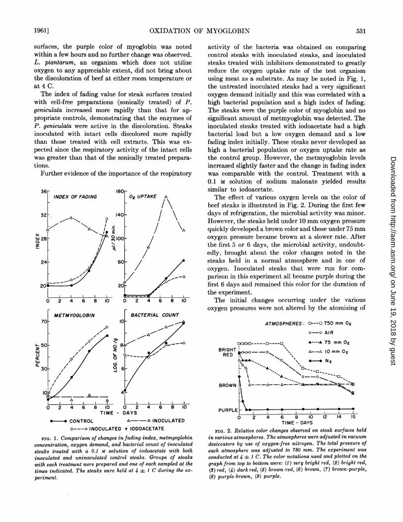
steaks with solutions of iodoacetate (0.1 M), potassiumcyanide (0.01 M), or chlortetracycline (100 ppm). Infact, there was no significant effect of these treatmentson the color changes occurring in an atmosphere with10 mm oxygen pressure throughout the test period(Fig. 3). The cyanide-treated steaks appeared some-
what more red in most instances which probablyresulted from reaction with the myoglobin. In higheratmospheres where bacterial activity became an im-portant factor, the inhibitors delayed the appearance
of color changes. In the normal atmosphere, chlortet-
BRIGHTRED
BROWN
PURPLE
o--o CONTROL
.* CHLORTETRACYCLINE
A~---A KCN
C-- ----- ODOACETATE
/0mm 02
PRESSURE
2 4 6 8 10 12 14 16
0 2 4 6 8 10 12 14 16TIME- DAYS
FIG. 3. Effect of microbial inhibitors on visual color changesof steak surfaces held in air and in an atmosphere containingonly 10 mm oxygen pressure. Steaks were treated by atomizingwith the indicated solution containing chlortetracycline (100ppm), KCN (0.01 -ii), or iodoacetate (0.1 M). The experimentalmethod and color scale used were the same as noted for Fig. 2.
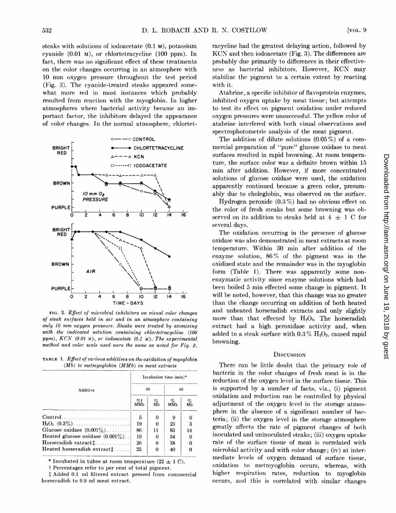
TABLE 1. Effect of various additives on the oxidation of myoglobin(Mb) to metmyoglobin (MMb) in meat extracts
Incubation time (min)$
Additive 30 60
%t % % %MMb Mb MMb Mb
Control ........................... 5 0 9 0H202 (0.3%) ...................... 19 0 23 3Glucose oxidase (0.001%) ......... 86 11 83 14Heated glucose oxidase (0.001%). 19 0 34 0Horseradish extractt .............. 26 0 38 0Heated horseradish extract$ ...... 25 0 40 0
* Incubated in tubes at room temperature (22 i 1 C).t Percentages refer to per cent of total pigment.I Added 0.1 ml filtered extract pressed from commercial
horseradish to 9.9 ml meat extract.
racycline had the greatest delaying action, followed byKCN and then iodoacetate (Fig. 3). The differences areprobably due primarily to differences in their effective-ness as bacterial inhibitors. However, KCN maystabilize the pigment to a certain extent by reactingwith it.
Atabrine, a specific inhibitor of flavoprotein enzymes,inhibited oxygen uptake by meat tissue; but attemptsto test its effect on pigment oxidation under reducedoxygen pressures were unsuccessful. The yellow color ofatabrine interfered with both visual observations andspectrophotometric analysis of the meat pigment.The addition of dilute solutions (0.05 %) of a com-
mercial preparation of "pure" glucose oxidase to meatsurfaces resulted in rapid browning. At room tempera-ture, the surface color was a definite brown within 15min after addition. However, if more concentratedsolutions of glucose oxidase were used, the oxidationapparently continued because a green color, presum-ably due to choleglobin, was observed on the surface.Hydrogen peroxide (0.3 %) had no obvious effect on
the color of fresh steaks but some browning was ob-served on its addition to steaks held at 4 + 1 C forseveral days.The oxidation occurring in the presence of glucose
oxidase was also demonstrated in meat extracts at roomtemperature. Within 30 min after addition of theenzyme solution, 86% of the pigment was in theoxidized state and the remainder was in the myoglobinform (Table 1). There was apparently some non-enzymatic activity since enzyme solutions which hadbeen boiled 5 min effected some change in pigment. Itwill be noted, however, that this change was no greaterthan the change occurring on addition of both heatedand unheated horseradish extracts and only slightlymore than that effected by H202. The horseradishextract had a high peroxidase activity and, whenadded to a steak surface with 0.3 % H202, caused rapidbrowning.
DISCUSSION
There can be little doubt that the primary role ofbacteria in the color changes of fresh meat is in thereduction of the oxygen level in the surface tissue. Thisis supported by a number of facts, viz., (i) pigmentoxidation and reduction can be controlled by physicaladjustment of the oxygen level in the storage atmos-phere in the absence of a significant number of bac-teria; (ii) the oxygen level in the storage atmospheregreatly affects the rate of pigment changes of bothinoculated and uninoculated steaks; (iii) oxygen uptakerate of the surface tissue of meat is correlated withmicrobial activity and with color change; (iv) at inter-mediate levels of oxygen demand of surface tissue,oxidation to metmyoglobin occurs, whereas, withhigher respiration rates, reduction to myoglobinoccurs, and this is correlated with similar changes
532 [VOL. 9
---a \\
on June 19, 2018 by guesthttp://aem
.asm.org/
Dow
nloaded from
OXIDATION OF MYOGLOBIN
under controlled oxygen atmospheres; and (v) agentsinhibiting the development of high oxygen uptakerates in surface tissue will result in color preservationunder normal atmospheric conditions but are ineffectiveunder low oxygen pressures.
This being true, then one must look to the meattissue per se for the mechanism of pigment changes.Grant (1955) demonstrated that the blocking of suc-cinic dehydrogenase with malonate protected theinterior of frozen ground meat from discoloration.This treatment must have eliminated practically alloxygen uptake, since the pigment remained in theoxygenated state. However, malonate did not preventthe oxidation of myoglobin in surface tissue of steaksheld at refrigerator temperatures; although it didgreatly reduce oxygen uptake of contaminating bac-teria. lodoacetate, an inhibitor of glyceraldehydephos-phate dehydrogenase (a glycolytic enzyme) and ofother sulfhydryl-containing enzymes, was more effec-tive than malonate on steak surfaces, but was withoutany measurable effect when the oxygen tension in theatmosphere was reduced to 10 mm. Since chlortetra-cycline and KCN had similar effects, it is believed thattheir primary action on meat surfaces is to inhibit thedevelopment of aerobic bacteria.The production of H202 by pneumococci leads to
methemoglobin formation (Neill, 1925), and results ofthis study demonstrate that H202 in combination withperoxidase causes rapid oxidation of myoglobin. Al-though H202 production by meat tissue was not demon-strated, the fact that atabrine inhibits oxygen uptake isindirect evidence of flavoprotein enzyme activity.Such activity would yield a certain amount of H202.Furthermore, glucose oxidase, which produces H202as a product of glucose oxidation, results in extensiveoxidation of the pigment either in surface tissue or inextracts. Keilin and Hartree (1945) demonstrated thatthe addition of glucose oxidase and glucose to eitherwashed horse erythrocytes or to laked corpusclesresulted in the oxidation of 100 % of the hemoglobin tomethemoglobin within a few minutes. Catalase was notessential for this reaction since a solution of recrystal-lized hemoglobin, free from catalase, was oxidized. Infact, the catalase may have offered some protectionbecause a portion of the partially purified pigmentunderwent oxidative destruction accompanied byprotein denaturation and formation of the greenverdohemochromogen. Bingold (1933) demonstratedthat catalase did protect oxyhemoglobin from oxida-tion by high concentration of H202.
Brooks (1935) found the rate of oxidation of hemo-globin to methemoglobin in buffered, laked, defi-brinated blood at different oxygen pressures to beproportional to the concentration of reduced hemo-globin. The decreasing rate, which occurs as oxygentension is increased, results from the increasing con-
centration of the oxygenated pigment that is not
apparently involved in the reaction. Keilin and Hartree(1945) speculate that this fact explains the protectiveaction of catalase toward hemoglobin when H202concentrations are high. There would be considerableoxygen released under such conditions and the pigmentwould probably be oxygenated and, thus, not suscepti-ble to oxidation. This would not be the case, however,with metabolic H202 since there would be a net loss ofdissolved oxygen in the over-all reaction. Data collectedin this work support this idea, since high peroxidelevels had no effect when added alone to freshly slicedsteaks, but did result in pigment oxidation in steaksheld 3 or 4 days at 4 -± 1 C. The latter steaks would beexpected to have a relatively high oxygen demand andpercentage of myoglobin, thus, providing the properstate for oxidation to occur.Based on the above, it is believed that the reduction
of oxygen tension in meat tissue, either by microbialgrowth or by physical means, results in a great increasein the reduced myoglobin, which, in turn, is oxidizedby metabolic H202 produced either by the meat tissueor by the bacteria. If the oxygen tension is reduced tolow enough a level, little or no H202 can be formed andno appreciable oxidation occurs. An oxidized myo-globin present is rapidly reduced under such conditions.
LITERATURE CITEDBINGOLD, K. 1933. Die Niere als Blutzerstorendes Organ
(zur Physiologie des Blutstoffwechsels). Klin. Wochschr.12:1201-1206.
BROOKS, J. 1935. The oxidation of haemoglobin to met-haemoglobin by oxygen. II. The relation between therate of oxidation and the partial pressure of oxygen.Proc. Roy. Soc. (Ser. B) 118:560-577.
BROUMAND, H., C. 0. BALL, AND E. F. STIER. 1958. Factorsaffecting the quality of prepackaged meat. II. E.Determining the proportions of heme derivatives in freshmeat. Food Technol. 12:65-77.
BUTLER, 0. D., L. J. BRATZLER, AND W. L. MALLMAN. 1953.The effect of bacteria on the color of prepackaged retailbeef cuts. Food Technol. 7:397-400.
COSTILOW, R. N., B. A. BATSHON, L. J. BRATZLER, AND D. L.ROBACH. 1955. Interactions between ascorbic acidand psychrophilic bacteria associated with the discolora-tion of prepackaged beef. Food Technol. 9:560-563.
GRANT, N. H. 1955. The respiratory enzymes of meat. I.Identification of the active enzymes. Food Research20:250-253.
KEILIN, D., AND E. F. HARTREE. 1945. Properties ofcatalase. Catalysis of coupled oxidation of alcohols.Biochem. J. 39:293-301.
NEILL, J. M. 1925. Studies on the oxidation-reduction ofhemoglobin and methemoglobin. I. The changes in-duced by pneumococci and by sterile animal tissue. J.Exptl. Med. 41:299-313.
NEILL, J. M., AND A. B. HASTINGS. 1925. The influence ofthe tension of molecular oxygen upon certain oxidationsof hemoglobin. J. Biol. Chem. 63:479-492.
NICKERSON, D. 1946. Color measurement and its applicationto the grading of agricultural products. U. S. Dept. Agr.,Misc. Publ. No. 580.
SCHWEIGERT, B. S. 1956. Chemistry of meat pigments.Proc. 8th Research Conf., Am. Meat Institute, Univ. ofChicago, p. 61.
1961] 533
on June 19, 2018 by guesthttp://aem
.asm.org/
Dow
nloaded from