Embed Size (px)
Citation preview
1914 CORRESPONDENCE Ann Thorac Surg2013;96:1910–7
MISCELLA
NEOUS
Forced Mechanical Sternal Elevation for Nuss RepairTo the Editor:
We thank Johnson and colleagues [1] for describing theirtechnique for sternal lift during Nuss repair for pectusexcavatum. We also have selectively performed sternalelevation for our adult minimally invasive pectus excavatumrepairs. It has greatly facilitated our past several hundred adultrepairs. In the adult patient and those with severe deformities,dissection across the mediastinum and elevation of the chestwall can be extremely difficult. Visualization is impaired andinstruments are deflected by the inward protruding deformity.Forced mechanical elevation of the sternum increases theanterior-posterior dimensions behind the sternum. The advan-tages include increased thoracoscopic visualization of thedissection and minimizing potential trauma to aorta and heart[2, 3]. Several publications have recommended variations of thistechnique [2–5]. The majority describe bilateral subcostal orsubxiphoid incisions with digital dissection for placement of re-tractors. Yoon and associates [5] have also described the use of
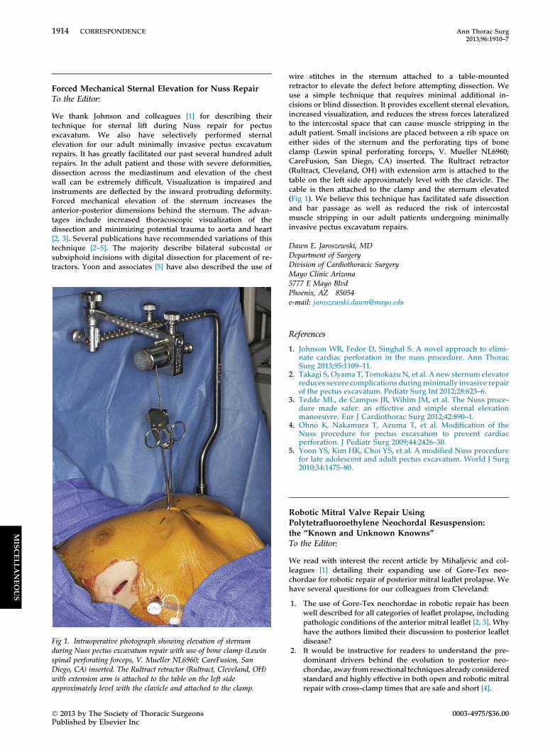
Fig 1. Intraoperative photograph showing elevation of sternumduring Nuss pectus excavatum repair with use of bone clamp (Lewinspinal perforating forceps, V. Mueller NL6960; CareFusion, SanDiego, CA) inserted. The Rultract retractor (Rultract, Cleveland, OH)with extension arm is attached to the table on the left sideapproximately level with the clavicle and attached to the clamp.
� 2013 by The Society of Thoracic SurgeonsPublished by Elsevier Inc
wire stitches in the sternum attached to a table-mountedretractor to elevate the defect before attempting dissection. Weuse a simple technique that requires minimal additional in-cisions or blind dissection. It provides excellent sternal elevation,increased visualization, and reduces the stress forces lateralizedto the intercostal space that can cause muscle stripping in theadult patient. Small incisions are placed between a rib space oneither sides of the sternum and the perforating tips of boneclamp (Lewin spinal perforating forceps, V. Mueller NL6960;CareFusion, San Diego, CA) inserted. The Rultract retractor(Rultract, Cleveland, OH) with extension arm is attached to thetable on the left side approximately level with the clavicle. Thecable is then attached to the clamp and the sternum elevated(Fig 1). We believe this technique has facilitated safe dissectionand bar passage as well as reduced the risk of intercostalmuscle stripping in our adult patients undergoing minimallyinvasive pectus excavatum repairs.
Dawn E. Jaroszewski, MDDepartment of SurgeryDivision of Cardiothoracic SurgeryMayo Clinic Arizona5777 E Mayo BlvdPhoenix, AZ 85054e-mail: [email protected]
References
1. Johnson WR, Fedor D, Singhal S. A novel approach to elimi-nate cardiac perforation in the nuss procedure. Ann ThoracSurg 2013;95:1109–11.
2. Takagi S, Oyama T, TomokazuN, et al. A new sternum elevatorreduces severe complications duringminimally invasive repairof the pectus excavatum. Pediatr Surg Int 2012;28:623–6.
3. Tedde ML, de Campos JR, Wihlm JM, et al. The Nuss proce-dure made safer: an effective and simple sternal elevationmanoeuvre. Eur J Cardiothorac Surg 2012;42:890–1.
4. Ohno K, Nakamura T, Azuma T, et al. Modification of theNuss procedure for pectus excavatum to prevent cardiacperforation. J Pediatr Surg 2009;44:2426–30.
5. Yoon YS, Kim HK, Choi YS, et al. A modified Nuss procedurefor late adolescent and adult pectus excavatum. World J Surg2010;34:1475–80.
Robotic Mitral Valve Repair UsingPolytetrafluoroethylene Neochordal Resuspension:the “Known and Unknown Knowns”To the Editor:
We read with interest the recent article by Mihaljevic and col-leagues [1] detailing their expanding use of Gore-Tex neo-chordae for robotic repair of posterior mitral leaflet prolapse. Wehave several questions for our colleagues from Cleveland:
1. The use of Gore-Tex neochordae in robotic repair has beenwell described for all categories of leaflet prolapse, includingpathologic conditions of the anterior mitral leaflet [2, 3]. Whyhave the authors limited their discussion to posterior leafletdisease?
2. It would be instructive for readers to understand the pre-dominant drivers behind the evolution to posterior neo-chordae, away from resectional techniques already consideredstandard and highly effective in both open and robotic mitralrepair with cross-clamp times that are safe and short [4].
0003-4975/$36.00
1915Ann Thorac Surg CORRESPONDENCE2013;96:1910–7
3. The proposed association of posterior leaflet neochordaewith shortened ischemic time, diminished systolic anteriormotion of the anterior mitral leaflet, and decreased reopera-tions is interesting. It remains unclear from the current article,however, how these variables were affected by (1) maturationalong the learning curve (ie, more frequent use of neochordaeby a team with greater experience and (2) differences in sur-geons’ practices: were posterior neochordae predominantlyused by one of the two surgeon-led teams? For the reader tomore completely understand whether more routine use ofposterior leaflet neochordae might be warranted in their ownpractices, further comment on these elements would beuseful.
4. When effectiveness is considered, it would be important tounderstand whether there were differences in postdismissalmitral valve regurgitation, cognizant of the fact that thefollow-up available for neochordal (a more recent trend intheir practice) and resectional groups was likely significantlydifferent. Adjudication of midterm effectiveness data iscrucial before the widespread adoption of Gore-Tex neo-chordal resuspension for the treatment of posterior leafletmitral valve prolapse can be advocated.
We appreciate the authors’ consideration of our queries.
Rakesh M. Suri, MD, DPhilHarold M. Burkhart, MD
Cardiovascular SurgeryMayo ClinicJoseph 5-200 St. Mary’s HospitalRochester, MN 55905e-mail: [email protected]
MISCELLANEOUS
References
1. Mihaljevic T, Pattakos G, Gillinov AM, et al. Robotic posteriormitral leaflet repair: neochordal versus resectional techniques.Ann Thorac Surg 2013;95:787–94.
2. Suri RM, Burkhart HM, Rehfeldt KH, et al. Robotic mitralvalve repair for all categories of leaflet prolapse: improvingpatient appeal and advancing standard of care. Mayo ClinProc 2011;86:838–44.
3. Suri RM, Burkhart HM, Schaff HV. A novel method of leafletreconstruction after triangular resection for posterior mitralvalve prolapse. Ann Thorac Surg 2010;89:e53–6.
4. Suri RM, Burkhart HM, Daly RC, et al. Robotic mitral valverepair for all prolapse subsets using techniques identicalto open valvuloplasty: establishing the benchmark againstwhich percutaneous interventions should be judged. J ThoracCardiovasc Surg 2011;142:970–9.
Cerebral Perfusion Through All Aortic Arch ArteriesTo the Editor:
Congratulations to Shimizu and colleagues [1] for this valuablestudy and excellent results. Urbanski and colleagues [2]previously reported that unilateral cerebral perfusion through asingle carotid artery sufficed to maintain neurologic functions.The development of temporary cerebral disturbances in 9.9% ofthe patients made us think that this was due to cerebral edemasecondary to selective cerebral perfusion. We believe thatmonitoring the perfusion pressure only through the leftcommon carotid artery is a pitfall of this study. Monitoring thearterial pressure of the right radial artery as well would havegiven more detailed knowledge about adequate perfusion.
� 2013 by The Society of Thoracic SurgeonsPublished by Elsevier Inc
Would the high rate of temporary neurologic disturbance bebecause of inadequate perfusion? The innominate arteryreceives almost 50% of all supraaortic arch blood flow whenperfused through a 15F cannula. Nevertheless, the left commoncarotid artery and the left subclavian artery are perfusedthrough separate 12F cannulas for each. The total cross-sectional area of both 12F cannulas is higher than that of thesingle 15F cannula. So, only measuring the flow rates may not besafe enough, although the total flow in the supraaortic vesselswas significantly lower in patients with postoperative neurologiccomplications than in those without. Monitoring the perfusionpressures of all branches should also be included in this study.
Ismail Yurekli, MDMert KestelliHabib Cakir, MD
Izmir Katip Celebi UniversityAtaturk Training and Research HospitalDepartment of Cardiovascular Surgery6436 sok. 82/3 Karsiyaka-Izmir35540 Turkeye-mail: [email protected]
References
1. Shimizu H, Matayushi T, Mohita M, Ueda T, Yozu R. Totalarch replacement under flow monitoring during selective ce-rebral perfusion using a single pump. Ann Thorac Surg2013;95:29–34.
2. Urbanski PP, Lenos A, Kolowca M, et al. Near-infrared spec-troscopy for neuromonitoring of unilateral cerebral perfusion.Eur J Cardiothorac Surg; 2012 Oct 24. [Epub ahead of print].
ReplyTo the Editor:
We appreciate the interest of Dr Yurekli and colleagues [1], andthe letter regarding our finding [2] of an 8.2% incidence oftemporary neurologic disturbances, including fairly mildrestlessness, after scheduled total arch replacement. We believethat selective cerebral perfusion (SCP) of all three supra-aorticbranches is the most reliable method of protecting the brainduring arch operations and that both flow volume and pressureare important indicators of appropriate SCP.Weused commercially available 15F or 12F cannulae for theSCP
of each branch. Side-to-side differences in hemispheric flow vol-ume were minimal overall, although the total cross-sectional areaof the inner lumen of the cannulae placed in the left carotid artery(LCA) and the left subclavian artery (LSA)was 30% larger than thatplaced in the brachiocephalic artery. The flow ratios in the LCAand LSA varied considerably among individuals (range, 0.3–1.9),although both were perfused by use of identical cannulae. Themean pressures at the top of the cannula into the LCA and at thearterial line of the right brachial artery were 38.8� 8.9mmHg and34.3� 10.0mmHg, respectively, indicating little difference. Thesefindings suggest that flow distribution among vessels waspassively and appropriately determined depending on thevascular resistance of the perfused area.The optimal flow rate for SCP remains controversial. Kazui and
colleagues [3] applied SCP at a flow rate of 10 mL/kg/min at arectal temperature of 20�C. Jacobs and colleagues [4] andMinatoya and colleagues [5] applied flow rates of 15 and 19.0mL/kg/min, respectively, at higher temperatures. We havefound that a flow rate greater than 12.5 mL/kg/min is optimal
0003-4975/$36.00