Embed Size (px)
Citation preview
Roadmaps in Biophotonics
Issue #2 In neurosciences infectious disease drug discovery & development
Strategic Road Mapping for Biophotonics:
Outcome of the 2nd International Congress on Biophotonics ICOB-2, Quebec City, Canada, September, 2010
Preamble This document is the second Roadmap generated as a result of an ICOB congress on biophotonics, namely “the application of optical science and technology to the life sciences and medicine”. ICOB-1 was held in 2008as a stand-alone meeting in Sacramento, California, USA. The resulting Roadmaps in Biophotonics, Issue #1 covered the specific topics of advanced optical microscopies, optical probes & sensors, and clinical applications. ICOB-2 was held within a series of conferences, Biophotonics Week in Quebec, in Quebec City, Canada. As a result, it was able to take advantage of the presence of experts, particularly from the end-user communities, who attended the 2nd international symposium on Frontiers of Neurophotonics and a collaborative symposium on Biophotonics in Infectious Disease. Hence, these comprised two of the topics of ICOB-2, with biophotonics in drug discovery & development being selected as the third, complementary topic. Neuroscience is among the crown jewels of biomedical research, because of the intense interest in discovering how the brain functions, as well as the major challenges of diagnosing and treating neurological disorders, both acute and chronic. High-end biophotonics technologies are frequently used in neuroscience research, so it was expected that the end-users would provide insight into their current tools and what they really need. Infectious disease was chosen as a topic because of its global prominence and the fact that it needs portable diagnostics and the development of new antibiotic approaches, in which biophotonics could have significant impact. The drug discovery and development day was chosen to assess the often veiled needs of the pharmaceutical industry sector and to see if biophotonics tools, perhaps developed in a more targeted manner, could help accelerate bringing new drugs to the marketplace.
I. Summary of Roadmapping Process The Roadmapping process consisted of bringing together biophotonics developers, life-sciences researchers and clinicians who use these methods and technologies (end-users), and industry that makes biophotonics devices or depends on them in their products. The End-Users at ICOB-2 represented a very wide range of disciplines: - basic and clinical neuroscientists at the cutting edge of research and development, - plant and animal biologists and clinicians struggling with the challenges of infectious disease in
food & agriculture and in human health, respectively, - representatives of drug companies interested to find better ways to reduce the time and cost of
getting new drugs from the idea stage through clinical trials and into the market, and - representatives of the investment community and government agencies interested in new
opportunities and in the challenges of R&D funding and health policies, respectively. This eclectic group participated in all the discussions and contributed to the roadmapping process, often making key contributions from outside as well as from within their own specialization. The primary goal was to develop a better understanding and compilation of end-user needs versus currently available biophotonic technologies. It was also expected that many existing optics-enabled technologies and methods could be re-deployed to address new end-user needs. However, it was further assumed that there would be needs identified that will require pioneering new instrumentation, and this was indeed the case.
II. Executive summary We comment first on the roadmapping process itself.
• As in ICOB-1, the meeting format was effective in bringing together the several stakeholder communities.
• The invited expert speakers, both the end users (technology adopters) and ‘biophotonicists’ (science & technology developers), identified the critical needs and potential optics-enabled approaches to address these needs.
• Panels and carefully-chosen breakout groups then served to flush out all the needs and perspectives that were not covered by individual speakers. They also provided a means to engage participation and ideas or commentary from all participants.
The roadmaps identify several generic issues relevant to biophotonics that run across all the application themes, some of which are also relevant to the topics covered in ICOB-1:
• There is a very large knowledge gap between scientists and engineers developing
biophotonics and the potential end users. It is of the highest priority to address this gap if the field is to achieve its full impact.
• There is, in some instances, a large mismatch between the goals and career/funding realities in academic labs versus those of industry, particularly where the technology is early stage and/or highly disruptive. Intellectual property (IP) issues are major barriers to co-operation: the term “irrational protectionism” was raised as applicable to both stakeholder communities. The option of open-source approaches was considered seriously, especially in the Neurosciences discussions.
• There is a need to get the word out on where Biophotonics is being deployed today across all fields of biotechnology and medicine. Many of the end-user participants would like more examples of how the technology is currently used to help understand better the potential applications in their own domains. Likewise, the biophotonics developers need to make more effort in predicting realistically where the emerging technologies could be used. It was recommended to use the www.biophotonicsworld.org website to give examples of where Biophotonics is currently deployed.
In terms of the individual applications themes, the following general points were highlighted. Neurosciences
It is important to distinguish between the needs and specifications for new biophotonics
technologies for preclinical research (neurobiology, neurophysiology) versus clinical applications (neurology, neurosurgery).
For the former, there are fundamental challenges that need to be addressed, particularly to find the optimum trade-off between spatial resolution (of optical imaging techniques), speed, brain tissue sampling volume or area, and the effective interrogation depth in brain tissue.
There are niche applications for which optical techniques provide unique capabilities, while in many applications other technologies (e.g. fMRI) are either competing or potentially complementary.
Biomarker-informed targeted optical probes will be increasingly important for both preclinical and clinical applications in order to increase sensitivity and, particularly, biomolecular specificity of the optical signals. A prime emerging example is that of probes for optogenetics, i.e. optical reports that are genetically encoded, where the need is for custom-built probes for specific neurofunctional studies.
It was noted that the National Institutes of Health (U.S.A.) have an ongoing program of blueprinting for neurosciences research, which helps provide end-user context in this domain: see http://neirosceinceblueprint.nih.gov).
Infectious Disease
Photonics could play a major role in providing point-of-care solutions to meet global challenges in infectious diseases, both in human health and in food & agriculture.
Photonic-based solutions must be cost-competitive with current standards-of-care in whatever is the local deployment setting. In addition, it is desirable that the technologies are able to be locally manufactured, so as to have direct economic impact, especially in the developing world.
The widespread applications of biophotonics in infection control will primarily be in pathogen detection, localization and identification, but important niche applications in the use of light-based techniques to destroy pathogens are also likely to emerge, particularly for treatment of existing infections and prevention of new infections in patients.
The ability to identify the pathogen species ‘on the spot’ will markedly enhance the utility of photonics-based technologies, especially if this can be done without a bio-amplification step.
It is notable that major investment is being made in research for new tools to manage infection, as part of the Gates Foundation initiative on global health. This should represent new opportunities for biophotonics development, particularly in low-cost technologies for improved sanitation, infection detection and treatments that can be easily deployed in developing countries. Drug Discovery & Development
Drug discovery is now too slow and costly, ~$1.5B/drug, so there is an urgent need to expedite the process and reduce cost, as well as avoid wasting time/money exploring drugs that fail to be efficacious or have unwanted side effects. Biophotonics technologies are already contributing to meeting these challenges but this could be greatly expanded.
Some of the pharmaceutical companies are no longer trying to develop blockbuster drugs for major diseases but instead are focusing more on “orphan diseases”, which may broaden the technology needs addressable by biophotonics.
A major part of drug discovery is to identify targets for intervention in disease progression pathways, so that this represents a potential major biophotonics application sector.
There is a need for new tools to monitor the responses to candidate drugs, both in pre-clinical testing and clinical trials. This will include responses at the genetic, molecular and functional levels.
Similarly, there is a need to determine rapidly if candidate drugs actually do what is intended, especially at the target-cell/tissue mechanism level.
Many biophotonics developers are engaged in drug efficacy studies of treatment responses using in vivo animal imaging with novel probes (nanoparticles, enzyme-activatable, bioluminescence.) Although this is important, pharma would also like effort to be put into better means to identify and validate biomarkers of disease.
It may be difficult to get the pharmaceutical industry to adopt new biophotonics tools or methods unless these are highly disruptive, since they have made large investments in current tools and methods. However, biotechnology companies, being smaller and less heavily invested in infrastructure, are potential early adopters.
Conventional SPR (surface plasmon resonance) and other biosensor methods of determining drug binding, stability and accretion/disassociation rates are primary research tools in drug

development. However, higher sensitivity, lower cost and much higher throughput are required in order to help the drug industry become more cost-effective.
The pharmaceutical industry tends not to be interested in direct investment or active participation in “tool” development, preferring to have this done by external academic laboratories and companies, since this is outside their normal core competencies. There is then not likely to be much direct financial support from individual pharma companies for biophotonics R&D.
III. Individual roadmaps
IIIA. Neurosciences Status. Challenges in the neurosciences push biophotonics technologies to the extreme of performance, in different ways for preclinical research in small animal models and in clinical neurology/neurosurgery. Thus, understanding brain function requires probing across sizes from the sub-micron scale to several centimeters, with µs-ms dynamic time resolution, and in the most fragile organ in the body. The brain is also highly compartmentalized and, viewed as an information network, has some 1016 connections, so that the “object under study” is enormously complex and dynamic. However, the low hazard and physiologic/molecular/genetic specificity of light is, in principle, highly advantageous for continuous monitoring of brain function. In particular, information can be obtained that is highly complementary to that provided by other techniques such as functional magnetic resonance imaging (fMRI). Optogenetics, using gene-based techniques targeting to enable optical modulation of brain function, has recently emerged as a potential break-through technology which provides unique capabilities and insights into brain function that are unlikely to be matched by any other (non-optical) modality. A major issue is whether and how the approach could ever be translated directly into patients: if it could, then there is high potential for applications such as symptom control in Parkinson’s disease and minimally-invasive brain stimulation In the clinic, there has been some limited penetration of optical techniques into medical practice. In diagnostics and functional monitoring, these are based primarily on near infrared (NIR) diffuse optical spectroscopy, for example, in continuous non-invasive monitoring of brain oxygen status in premature babies. Functional monitoring, based on hemodynamics, is widely used as a research tool in human subjects, but is otherwise not yet in routine clinical use: in part this is due to the fact that some critical information, in particular measures of metabolic supply and demand in the brain, is missing so that interpretation of the data is often incomplete or ambiguous. Emerging optical techniques such as diffuse correlation spectroscopy may be of value here. In neurosurgery, some biophotonic devices are in routine use, in particular the neurosurgical microscope and, increasingly, ICG (indocyanine green)-based NIR angiography. Other techniques, particularly fluorescence image guidance for defining the tumor margin in intracranial tumors, have reached the stage where adoption into clinical practice is starting: the core technology, at least in terms of hardware is essentially developed, although there is still room for significant advances, particularly in targeted optical contrast agents for increased sensitivity and/or target specificity. Here, non-label based methods like Raman, CARS, SHG and especially a combination thereof are promising. There has been minimum clinical penetration of optical techniques to date in management of neurodegenerative diseases or acutely life-threatening conditions like trauma, stroke and aneurysm. Hence, there is a need for accelerated effort in developing clinically useful biophotonic tools for these applications. A specific requirement is that these tools should ideally function without needing to turn off or modify the normal operating room lighting. Advantages of Optical Approaches. The discussions identified several major intrinsic advantages of biophotonics techniques for neurosciences applications, several of which are clearly also relevant to other biomedical fields. These advantages are substantial and have enabled approaches to studying brain function that are either complementary to or significantly better that previous methods, or have led to radically new approaches becoming possible. An
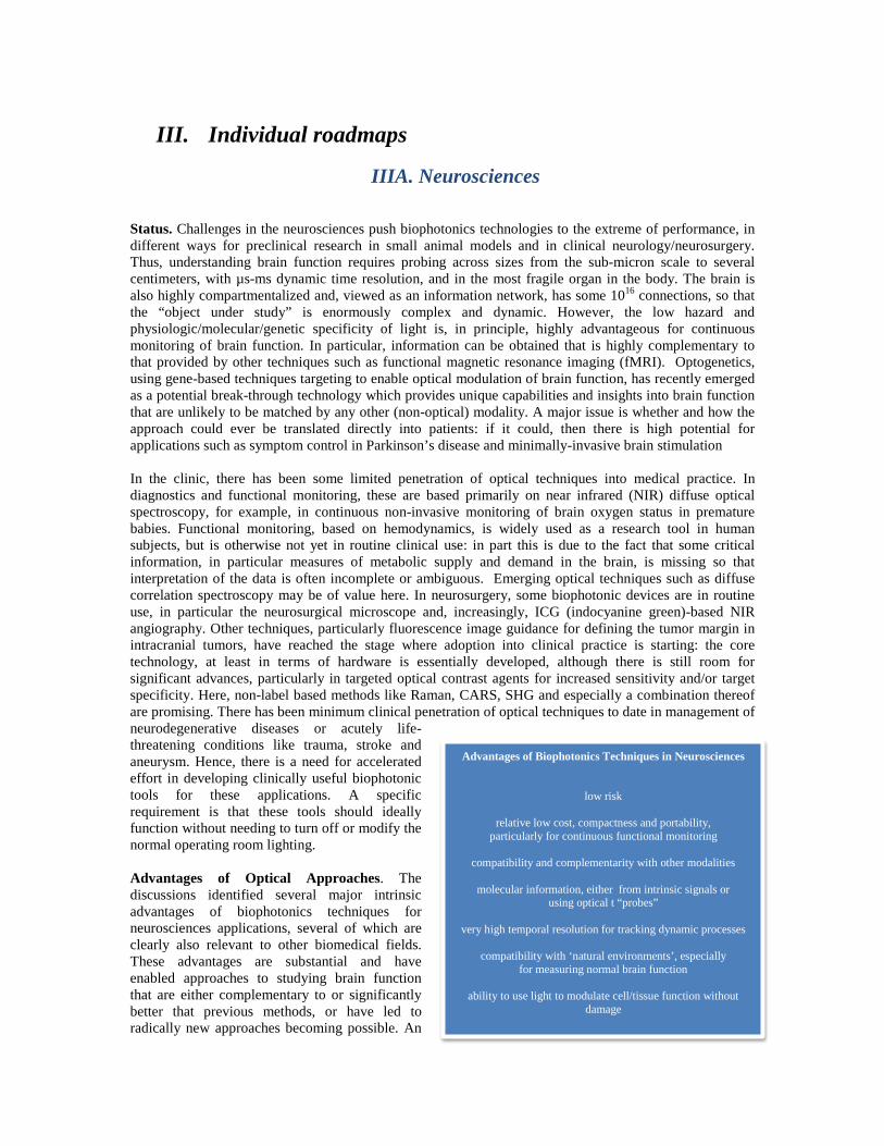
Advantages of Biophotonics Techniques in Neurosciences
low risk
relative low cost, compactness and portability, particularly for continuous functional monitoring
compatibility and complementarity with other modalities
molecular information, either from intrinsic signals or
using optical t “probes” very high temporal resolution for tracking dynamic processes
compatibility with ‘natural environments’, especially
for measuring normal brain function
ability to use light to modulate cell/tissue function without damage
example par excellence of the last is the emergence of optogenetics as a means of actively manipulating brain function and monitoring the resulting effects in a longitudinal, minimally-invasive way. End-User Needs. Restating a key point above, the needs in pre-clinical brain research are very different from those of the practicing clinician dealing with diagnoses or treatments across the spectrum of pathologies found in clinical practice (trauma, stroke, aneurysm, malignancy, chronic degenerative conditions). Thus, while there may be a common basis in optical spectroscopy and imaging, the detailed implementation is highly specific to the needs, so that there is not a generic ‘neurophotonics’ optical technology platform. In preclinical research, as in many applications of biophotonics, the trade-off between sampling depth in tissue and spatial resolution is a significant limitation in the brain. Techniques such as photoacoustic imaging may in part address this limitation by combining the molecular specificity of optics with the depth imaging capability of ultrasound. Improving the specificity of the optical signals that are accessible for monitoring brain function in health and disease is a high priority: the challenge is to find ways to convert the intrinsic chemical-electrical signals into optical signals. Even for those techniques that are already in use, such as near infrared spectroscopy (NIRS), there is incomplete understanding of the mechanisms underlying this signal transduction. In clinical diagnostics and functional monitoring, much of the work to date has been based on NIRS using external optodes that detect changes in hemoglobin content and oxygen status. However, in many cases this technique is considered to be underutilized and poorly understood, and most commercial systems do not yield the most important information from the perspective of actively managing the patient. Practical systems for bedside monitoring are needed, particularly in the critical care setting. In neurosurgery, intra-operative optical imaging is increasingly valuable and is being extended through adding functionality such as fluorescence imaging and spectroscopy, especially for accurate disease margin detection (e.g. of gliomas). Other optical signals, including label-free methods, are relatively unexplored, particularly in the routine surgical setting. The two options - to add these functions into existing neurosurgical microscopes or to develop free-standing independent systems - must be considered in developing new optical techniques for neurosurgery, from both the scientific/technical and commercial perspectives. In the case of neurosurgical microscopes, the market is dominated by a small number of large companies. Another end-user need is to increase the integration of neurosurgical guidance (e.g. intraoperative imaging) with the tools used to perform the tissue cutting, removal, coagulation and repair (including, potentially, pulsed laser-based and robotic devices). Of critical importance in these various devices is to have very close liaison with the end-users (neurosurgeons), since small details (e.g. the size or working distance of the device) can make a huge difference in the ergonomics and, hence, clinical adoption and commercial viability of the technology.
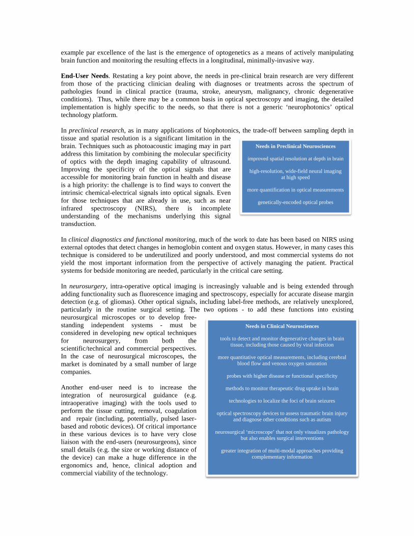
Needs in Preclinical Neurosciences
improved spatial resolution at depth in brain
high-resolution, wide-field neural imaging at high speed
more quantification in optical measurements
genetically-encoded optical probes
Needs in Clinical Neurosciences
tools to detect and monitor degenerative changes in brain tissue, including those caused by viral infection
more quantitative optical measurements, including cerebral
blood flow and venous oxygen saturation
probes with higher disease or functional specificity
methods to monitor therapeutic drug uptake in brain
technologies to localize the foci of brain seizures
optical spectroscopy devices to assess traumatic brain injury and diagnose other conditions such as autism
neurosurgical ‘microscope’ that not only visualizes pathology
but also enables surgical interventions
greater integration of multi-modal approaches providing complementary information
Major Roadblocks to Implementation and Adoption. The following were identified as among the major challenges and impediments identified in neurobiophotonics.
• For targeted probes the concentrations required may be unachievable, since for example
fluoresecence imaging with 20 nm spatial resolution requires at least one fluorophore every 10 nm, which corresponds typically to >10 mM concentration: this may force attention away from imaging and more towards gaining the information using point spectroscopy but with very high biological specificity.
• The optical barrier presented by the skull limits the optical signals, especially in adults. • The blood-brain barrier impedes access of optical probes for high sensitivity/high specificity
imaging or spectroscopy. • On-line image/data processing and analyses pose significant challenges due to the amount of data
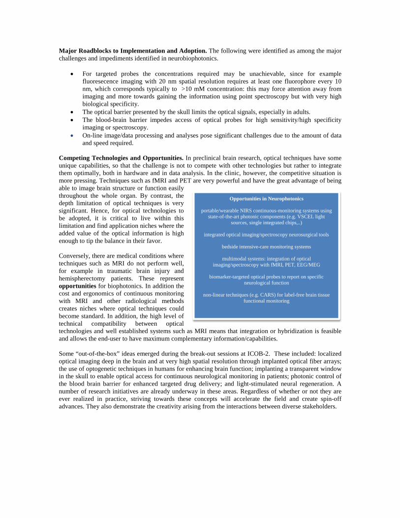
and speed required. Competing Technologies and Opportunities. In preclinical brain research, optical techniques have some unique capabilities, so that the challenge is not to compete with other technologies but rather to integrate them optimally, both in hardware and in data analysis. In the clinic, however, the competitive situation is more pressing. Techniques such as fMRI and PET are very powerful and have the great advantage of being able to image brain structure or function easily throughout the whole organ. By contrast, the depth limitation of optical techniques is very significant. Hence, for optical technologies to be adopted, it is critical to live within this limitation and find application niches where the added value of the optical information is high enough to tip the balance in their favor. Conversely, there are medical conditions where techniques such as MRI do not perform well, for example in traumatic brain injury and hemispherectomy patients. These represent opportunities for biophotonics. In addition the cost and ergonomics of continuous monitoring with MRI and other radiological methods creates niches where optical techniques could become standard. In addition, the high level of technical compatibility between optical technologies and well established systems such as MRI means that integration or hybridization is feasible and allows the end-user to have maximum complementary information/capabilities.
Some “out-of-the-box” ideas emerged during the break-out sessions at ICOB-2. These included: localized optical imaging deep in the brain and at very high spatial resolution through implanted optical fiber arrays; the use of optogenetic techniques in humans for enhancing brain function; implanting a transparent window in the skull to enable optical access for continuous neurological monitoring in patients; photonic control of the blood brain barrier for enhanced targeted drug delivery; and light-stimulated neural regeneration. A number of research initiatives are already underway in these areas. Regardless of whether or not they are ever realized in practice, striving towards these concepts will accelerate the field and create spin-off advances. They also demonstrate the creativity arising from the interactions between diverse stakeholders.
Opportunities in Neurophotonics portable/wearable NIRS continuous-monitoring systems using
state-of-the-art photonic components (e.g. VSCEL light sources, single integrated chips,..)
integrated optical imaging/spectroscopy neurosurgical tools
bedside intensive-care monitoring systems
multimodal systems: integration of optical
imaging/spectroscopy with fMRI, PET, EEG/MEG
biomarker-targeted optical probes to report on specific neurological function
non-linear techniques (e.g. CARS) for label-free brain tissue
functional monitoring
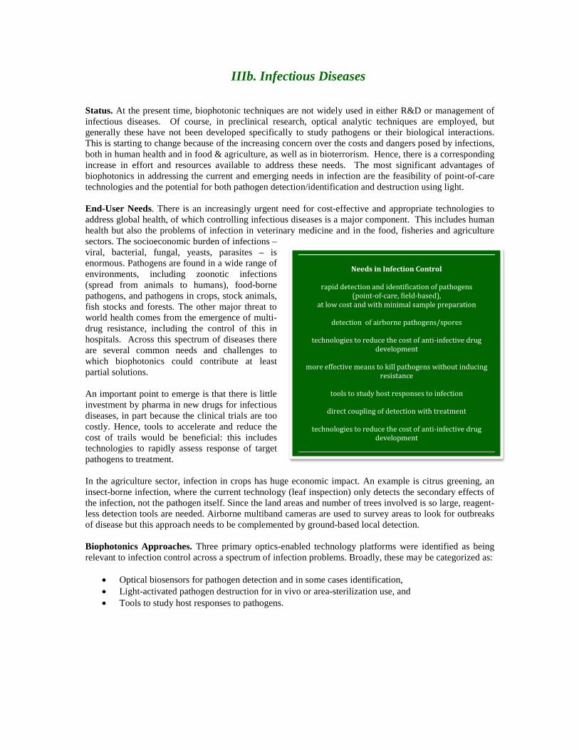
IIIb. Infectious Diseases Status. At the present time, biophotonic techniques are not widely used in either R&D or management of infectious diseases. Of course, in preclinical research, optical analytic techniques are employed, but generally these have not been developed specifically to study pathogens or their biological interactions. This is starting to change because of the increasing concern over the costs and dangers posed by infections, both in human health and in food & agriculture, as well as in bioterrorism. Hence, there is a corresponding increase in effort and resources available to address these needs. The most significant advantages of biophotonics in addressing the current and emerging needs in infection are the feasibility of point-of-care technologies and the potential for both pathogen detection/identification and destruction using light. End-User Needs. There is an increasingly urgent need for cost-effective and appropriate technologies to address global health, of which controlling infectious diseases is a major component. This includes human health but also the problems of infection in veterinary medicine and in the food, fisheries and agriculture sectors. The socioeconomic burden of infections – viral, bacterial, fungal, yeasts, parasites – is enormous. Pathogens are found in a wide range of environments, including zoonotic infections (spread from animals to humans), food-borne pathogens, and pathogens in crops, stock animals, fish stocks and forests. The other major threat to world health comes from the emergence of multi-drug resistance, including the control of this in hospitals. Across this spectrum of diseases there are several common needs and challenges to which biophotonics could contribute at least partial solutions. An important point to emerge is that there is little investment by pharma in new drugs for infectious diseases, in part because the clinical trials are too costly. Hence, tools to accelerate and reduce the cost of trails would be beneficial: this includes technologies to rapidly assess response of target pathogens to treatment. In the agriculture sector, infection in crops has huge economic impact. An example is citrus greening, an insect-borne infection, where the current technology (leaf inspection) only detects the secondary effects of the infection, not the pathogen itself. Since the land areas and number of trees involved is so large, reagent-less detection tools are needed. Airborne multiband cameras are used to survey areas to look for outbreaks of disease but this approach needs to be complemented by ground-based local detection. Biophotonics Approaches. Three primary optics-enabled technology platforms were identified as being relevant to infection control across a spectrum of infection problems. Broadly, these may be categorized as:
• Optical biosensors for pathogen detection and in some cases identification, • Light-activated pathogen destruction for in vivo or area-sterilization use, and • Tools to study host responses to pathogens.
Needs in Infection Control
rapid detection and identification of pathogens (point-of-care, field-based),
at low cost and with minimal sample preparation
detection of airborne pathogens/spores
technologies to reduce the cost of anti-infective drug development
more effective means to kill pathogens without inducing
resistance
tools to study host responses to infection
direct coupling of detection with treatment
technologies to reduce the cost of anti-infective drug development
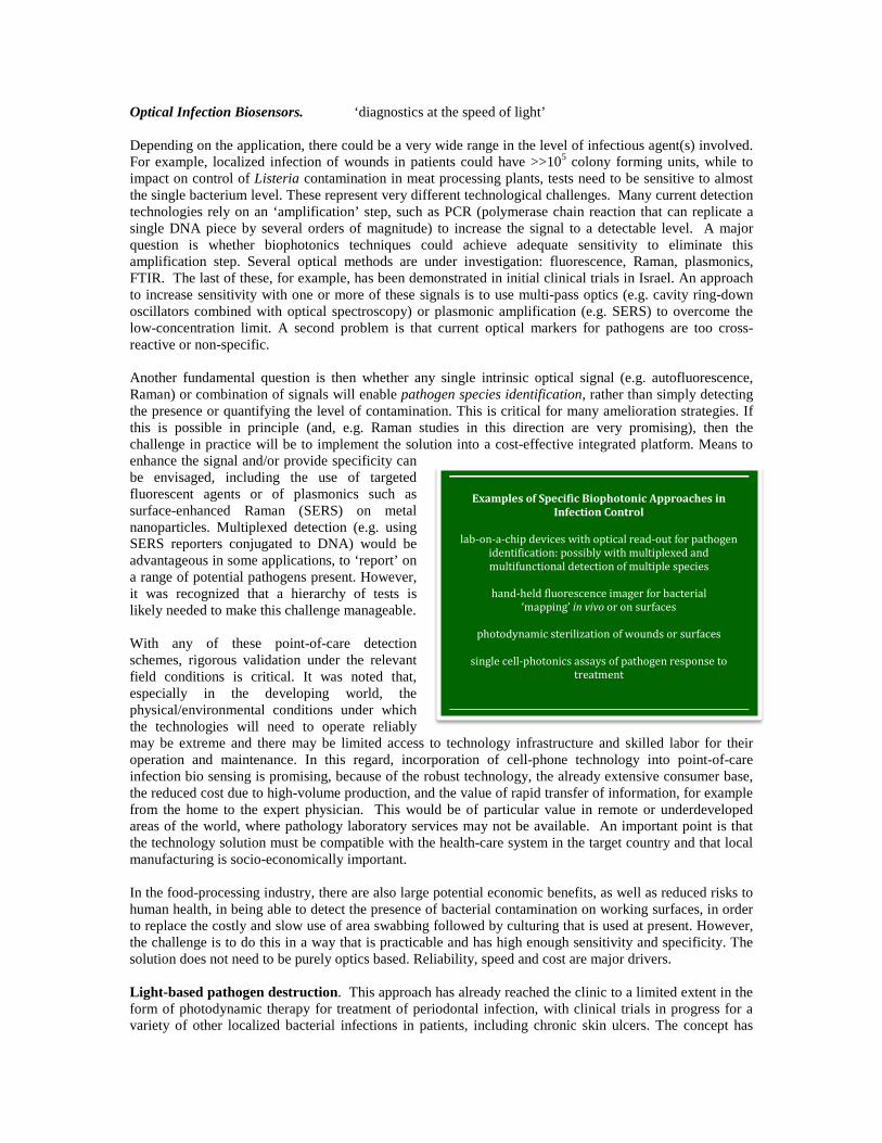
Optical Infection Biosensors. ‘diagnostics at the speed of light’ Depending on the application, there could be a very wide range in the level of infectious agent(s) involved. For example, localized infection of wounds in patients could have >>105 colony forming units, while to impact on control of Listeria contamination in meat processing plants, tests need to be sensitive to almost the single bacterium level. These represent very different technological challenges. Many current detection technologies rely on an ‘amplification’ step, such as PCR (polymerase chain reaction that can replicate a single DNA piece by several orders of magnitude) to increase the signal to a detectable level. A major question is whether biophotonics techniques could achieve adequate sensitivity to eliminate this amplification step. Several optical methods are under investigation: fluorescence, Raman, plasmonics, FTIR. The last of these, for example, has been demonstrated in initial clinical trials in Israel. An approach to increase sensitivity with one or more of these signals is to use multi-pass optics (e.g. cavity ring-down oscillators combined with optical spectroscopy) or plasmonic amplification (e.g. SERS) to overcome the low-concentration limit. A second problem is that current optical markers for pathogens are too cross-reactive or non-specific. Another fundamental question is then whether any single intrinsic optical signal (e.g. autofluorescence, Raman) or combination of signals will enable pathogen species identification, rather than simply detecting the presence or quantifying the level of contamination. This is critical for many amelioration strategies. If this is possible in principle (and, e.g. Raman studies in this direction are very promising), then the challenge in practice will be to implement the solution into a cost-effective integrated platform. Means to enhance the signal and/or provide specificity can be envisaged, including the use of targeted fluorescent agents or of plasmonics such as surface-enhanced Raman (SERS) on metal nanoparticles. Multiplexed detection (e.g. using SERS reporters conjugated to DNA) would be advantageous in some applications, to ‘report’ on a range of potential pathogens present. However, it was recognized that a hierarchy of tests is likely needed to make this challenge manageable. With any of these point-of-care detection schemes, rigorous validation under the relevant field conditions is critical. It was noted that, especially in the developing world, the physical/environmental conditions under which the technologies will need to operate reliably may be extreme and there may be limited access to technology infrastructure and skilled labor for their operation and maintenance. In this regard, incorporation of cell-phone technology into point-of-care infection bio sensing is promising, because of the robust technology, the already extensive consumer base, the reduced cost due to high-volume production, and the value of rapid transfer of information, for example from the home to the expert physician. This would be of particular value in remote or underdeveloped areas of the world, where pathology laboratory services may not be available. An important point is that the technology solution must be compatible with the health-care system in the target country and that local manufacturing is socio-economically important. In the food-processing industry, there are also large potential economic benefits, as well as reduced risks to human health, in being able to detect the presence of bacterial contamination on working surfaces, in order to replace the costly and slow use of area swabbing followed by culturing that is used at present. However, the challenge is to do this in a way that is practicable and has high enough sensitivity and specificity. The solution does not need to be purely optics based. Reliability, speed and cost are major drivers. Light-based pathogen destruction. This approach has already reached the clinic to a limited extent in the form of photodynamic therapy for treatment of periodontal infection, with clinical trials in progress for a variety of other localized bacterial infections in patients, including chronic skin ulcers. The concept has
Examples of Specific Biophotonic Approaches in Infection Control
lab-on-a-chip devices with optical read-out for pathogen
identification: possibly with multiplexed and multifunctional detection of multiple species
hand-held fluorescence imager for bacterial
‘mapping’ in vivo or on surfaces
photodynamic sterilization of wounds or surfaces
single cell-photonics assays of pathogen response to treatment
been demonstrated in the lab for other pathogens, including tuberculosis and a number of important tropical diseases. A significant advantage is the apparent effectiveness even in drug-resistant organisms (because of the different subcellular targets and mechanisms of action) and lack of induction of resistance to the treatment (since DNA damage is not a primary mechanism of photodynamic action). Development of PDT compounds that are pathogen species-specific would be an advantage, to avoid killing beneficial pathogens at time of treatment. In the hospital setting, photodynamic sterilization of the environment, of devices (e.g. implantable catheters) and of tissue surfaces during surgery are emerging ideas that address prevention rather than treatment of infections. Since post-operative infection represents a significant health-care cost that is growing because of multidrug resistance, these biophotonic approaches are likely to become increasingly relevant. The possibility of photodynamic treatment of blood-borne infection was also raised, for example, through transcutaneous illumination. Host Responses to Infection. The ability of the host (human, animal or plant) to fight infection is an important aspect of infection control. The main message to the biophotonics community is that new tools are needed to study this complex aspect in the living host. One area particularly highlighted, in part because of the specific expertise of the ICOB-2 participants, was that of the response of the human mucosal tissues, for example throughout the gastrointestinal tract, to opportunistic infections. This includes detection of latent (e.g. viral) infection and monitoring of the immune responses and the effects on mucosal structure and function, as well as the effects on the normal bacterial flora in the gut. This leads to the idea of ‘mapping the mucosa’, i.e. using optical and complementary techniques for longitudinal dynamic imaging of multiple characteristics of the GI mucosa in response to infection. This concept may be extendable to other body sites.
IIIc. Drug Discovery and Development The focus of the third and final day of ICOB-2 was to determine where biophotonics methods or technologies are currently being used and could be further developed or adapted for use in drug discovery and development. This topic evoked strong comments from most of the participants and set the tone for understanding more clearly where drug companies would like to see emphases in biophotonics research going forward. Discovering and developing new pharmaceutical therapies is of utmost importance but is fraught with major costs and technical challenges, a variety of which could be addressed using the tools and methods of biophotonics. Pharma Realities and Needs. A senior research scientist from a major drug company presented a summary perspective, key points from which are highlighted here. The challenges to biophotonics are numerous, including developing sensitive, cheap and high throughput methods and instruments/probes to measure drug targeting and efficacy, as well as assessing side effects and efficiency of targeting. Since it takes a decade and over a billion dollars on average to get a new drug to market, industry basically needs to cut time and costs. We were strongly advised to not look for a one size fits all approach, but rather to develop special photonics-based technology and methods for specific needs. It was pointed out that, in general, pharma is not interested in phototherapy or developing drugs (contrast agents) for surgical guidance, which are two mainstream R&D areas in biophotonics, so that these will require other types of corporate partners. A further clear statement was that big pharma does not buy enabling technologies from academic labs, so that the commercialization of biophotonics developments is best done in partnership with ‘trusted’ technology-supplier companies. Another approach is to collaborate with smaller biotech companies to help address their specific needs. The two major areas in which biophotonics could impact pharma are in:
• discovery and screening of novel drug targets, and • monitoring of drug toxicity/efficacy.
The particular technical needs in evaluating candidate drugs include: probes and instrumentation for dynamic and quantitative imaging to obtain pharmacokinetic and pharmacodynamic information; specific probes for monitoring molecular and cellular effects: and methods to monitor potential side effects on an individual basis Potential of Biophotonics in Drug Discovery and Development. The advantages of optical-based methods over other approaches in accelerating drug discovery and development are shared with other applications, such as those described in the other Themes, both here and in the ICOB-1 Roadmap. Many
The average cost of development and introduction of a new therapeutic drug is ~$1.5B, owing to the cost of trials and the failures of drugs to reach their efficacy
goals or avoid toxicity or other unwanted co-morbidity. The time–scale is also very long: typically
>10 years.
General Pharma Interests and Needs
drugs for orphaned diseases and for the developing world
diagnostic drugs that can detect disease and monitor patient response
to pharmaceutical therapy
use lower amounts of candidate drugs in the screening phase
detect biomarkers and use them to study drug efficacy
emerging biologic therapies
future personalized medicine
established photonics-based methods are already in widespread use in target discovery and candidate drug screening, including flow cytometry, optical microscopies, fluorescence-based analytics such as genetic microarrays, and small animal imaging (fluorescence, bioluminescence). Massively-parallel (hence fast and low cost) assessment of drug uptake, and drug localization and effects at the target-cell level (in vitro) is urgently required and is facilitated by optical techniques. An active field of biophotonics research that is highly relevant to the drug development pipeline, particularly the discovery phase, is that of optical biosensors. Label-based methods usually exploit fluorescence or other contrast agents such as functionalized nanoparticles. In contrast, label-free refers here mainly to the use of surface plasmon resonance (SPR)-based technologies, where huge signal amplification can be achieved, so increasing the sensitivity and
reducing the sample volume or mass required. Some of such implementations are already commercially available and many others are emerging with a range of different sensitivity and specificity for different applications. (Many of these platform technologies can also be applied to clinical diagnostics and treatment response monitoring, whether or not the treatment is drug-based, either for bioanalyte measurements or for evaluation of cell populations.) These sensors basically detect the presence/attachment/accretion of biomaterials for drugs that bind to a specific target or receptor, and can measure factors like the drug-target binding strength, stability, and association and disassociation dynamics. However, sensitivity, specificity, price, confounding bulk effects, non-specific binding and throughput are major challenges for these technologies. Emerging technical advances include: low-cost, miniaturized SPR biosensors; grating or polarizing ellipsometers; dual-polarization phase-sensitive SPR; tilted-fiber-grating sensors; metallic nanoparticles that resonate at a frequency that changes as
biomass is accreted; dielectric waveguides; and a host of other technologies that have an observable optical signal that is sensitive to mass accretion. To date, much of biophotonics development specifically in the pharma area has focused on using optical imaging and probes to identify lesions (primarily in animal models) and to monitor their response to drugs. This includes probes specifically for anti-cancer drugs. Such probes can either be activated inside tumor cells or targeted to them by antibodies or other receptor-specific moieties (affibodies, aptamers, peptides). Conventional tumor-specific targeting using radionuclide imaging, MRI and optical imaging all work well and in complementary ways. An advantage of optical
Using biomarkers to detect and monitor the response to pharmaceutical therapy is in line with the mounting
interest on the part of drug companies in individualizing therapy. Development of activatable
optical probes could help with detecting and monitoring the response of tumors and other
pathologies to drug therapies.
Examples of Optical Techniques to Monitor Drug Delivery
whole-body (fluorescence) imaging in (genetically-
engineered) mouse models
high-resolution microscopies to track nanoparticle-drug kinetics and monitor cellular/vascular responses in
target cells and preclinical models in vivo
label-free optical biosensors
Advantages of Optical Techniques in Drug Discovery and Development
______________________________________________________
relatively inexpensive
capable of high sensitivity, specificity and throughput
multimodality in vivo and functional animal imaging is routine
combined with molecularly targeted probes, can be used to locate and monitor response to therapy
with proper labeling could be useful to identify drug targets
can be used measure drug-target binding, stability, association and
disassociation values
using photoacoustic imaging and suitable contrast agents, can be applied to deep tissue imaging
can perform real-time in situ histological assessment (e.g. by OCT)
can determine cell viability in response to drug exposure
imaging is that it is possible to obtain a high tumor-to-background ratio using, for example, enzyme-activatable probes (molecular beacons). Dual-modality radionuclide + optical probes are increasingly investigated. Activatable probes have many uses, including imaging tumor margin development and cell death reporting (e.g. using pH-sensitive activation). In relation to infectious disease, bacteria can also be detected with activatable probes, followed by imaging of the host response to the infection and to drug (e.g. novel antibiotic) treatment. Emerging biologic therapies include vaccines, blood and blood components, allergenics, somatic cells, gene therapy, immunotherapy and the use of recombinant therapeutic proteins created by biological processes. New ideas and techniques for analytics and quality control are needed to match these developments, e.g. high-throughput and high-information flow cytometry. Since these approaches are generally developed initially by smaller, often start-up, companies, affordable solutions are needed Roles in Monitoring Drug Delivery and Therapeutic Responses. There are several different ways in which optical techniques are being developed to guide/monitor drug delivery and to monitor the pharmacological responses of either the target diseased tissues/cells (drug efficacy) or the normal organs (drug toxicity). These approaches are particularly powerful when nanoparticles are used to label &/or encapsulate the drugs of interest, since this can provide much stronger optical signals than using drug molecules alone. Complementing these optical technologies are specialized animal models, such as implanted ‘window chambers’ that allow direct optical viewing at microscopic resolution in multiple organ sites. For cancer drugs, tumors (including primary human tumors) can be grown orthotopically in these animals, i.e. in the organ of origin of the tumor, and the drug uptake kinetics and the target cell and tissue responses can be directly visualized. At the preclinical stage, the use of intravital optical microscopy to track stem cells and monitor their effectiveness (e.g. as a biologic therapy in tissue regeneration) is becoming possible using fluorescently-labeled probes. Bioluminescence imaging is also widespread in preclinical drug research, either as a constitutive signal to report on viability (e.g. of the target cells for the drug) or under control of a specific gene (e.g. a stress gene) that is up-regulated by exposure to the drug. Histopathologic assessment of drug effects, both on target and non-target tissues is a key part of drug development. Major developments that are likely to impact here include: rapid whole-slide (digital) scanning that markedly increases throughput and reduces costs; multi/hyperspectral fluorescence imaging that allows simultaneous (multiplexed) imaging of several biomarkers; and label-free microscopy (e.g. using non-linear interactions such as 2nd and 3rd harmonic generation and coherent anti-Stokes Raman scattering) that can generate images with equivalent or better information content than the common staining techniques, and with greater reproducibility and standardization. Since the #1 factor in drug discovery and development is time from discovery to market, these fast and high information-content approaches should have impact as they are adopted into pathology practice, particularly at the clinical trial phase. Optical-guided biopsy can also improve the collection of tissue samples from patients on drug trials for histopathologic evaluation, whether conventional or using emerging microscopy techniques. For response monitoring in patients, high spatial resolution may not be required: an example is the use of diffuse optical tomography in monitoring the metabolic response of breast tumors to neo-adjuvant therapy, allowing the drug regimen to be changed if the tumor is non-responsive. Barriers to Further Adoption of Biophotonic Techniques. While light-enabled techniques are already used throughout the drug discovery and development pipeline, in both academic and company research labs, there are several barriers to expanding their range and depth of applications. These are both scientific/technical and operational. For the first, considering the broad spectrum of optical ‘biosensors’ (imaging and non-imaging), key performance limitations that need to be addressed are:
- sensitivity, - specificity, - cost - speed/throughput.
The limited utility of optical techniques for deep-body imaging (except for endoscopy) is a barrier relative to other radiological modalities. The counterbalancing advantages of high molecular sensitivity and specificity need to be exploited maximally, and the impact is likely to be greatest at the earlier, pre-clinical stages of drug discovery and development. Multifunctional probes and instruments, in which optical and non-optical methods are combined to take advantage of their complementary capabilities, are particularly important. Barriers to optical image-guided drug delivery include the increased complexity for regulatory approval, the trade-off between spatial resolution and depth of imaging, and the need to develop rigorous methods for calibration and validation. The first two barriers are absent or reduced in pre-clinical studies to evaluate drug delivery in small animal models. For the operational barriers, few biophotonics developers are working directly on new tools for drug discovery and development. There is also a lack of awareness within the drug industry of the potential of biophotonics techniques, especially given that most scientists working for drug companies are chemists and biologists rather than (optical) physicists and engineers. The www.biophotonicsworld.org website was identified as a vehicle to disseminate pharma-relevant information on biophotonics, as it is also for other industry sectors. It was also noted that light can also be used for enhanced delivery of drugs or other biologics (e.g. gene therapy) through either photochemically- or photothermally-triggered release or photochemical internalization. These are currently niche methods in the overall pharma landscape but have potential to grow. As indicated, big pharma companies are not currently particularly interested in this area.
IV. Outcomes, Lessons Learned and Paths Forward ICOB-2 built on the experience gained in ICOB-1. There was greater emphasis on active participation by the end-user communities. Having other specialized conferences on neurophotonics and infection taking place just prior to ICOB-2 was tremendously valuable, since it both enabled participation by a wider group of experts and laid some of the groundwork for the ICOB-2 deliberations. From the Theme I discussions, and the preceding Frontiers in Neurophotonics conference, it is clear that neurosciences (both preclinical and clinical) is highly dependent on optical techniques, and indeed some of these (e.g. optogenetics) have become critical enablers for completely new approaches to understanding brain function in health and disease. The outlook for neurophotonics is highly promising. In the area of infection (Theme II), primary challenges and opportunities are emerging for biophotonics to have significant impact in point-of-care, including in the developing world, for detecting and monitoring infection. This is particularly the case in human health, but it was also recognized that there are major needs in food and agriculture, areas that have to date not has substantive attention in the biophotonics community. Engaging with the diverse experts, beneficiaries and development agencies in these fields will require broadening the biophotonics perspective. Pathogenic infection is a major global problem on which biophotonics community could have significant impact. Engaging with the end-user community was particularly challenging in Theme III (Drug Discovery & Development), because the key end users are the big pharmaceutical companies and it proved difficult to get senior representatives to participate in this technology-focused meeting. We are, therefore, particularly grateful to those representatives who attended and contributed in a major way to help define the needs and opportunities in this important domain. In ICOB-2, a major challenge, as in ICOB-1, was how to gather and organize the wealth of information that was generated in the 3 days of discussion into a coherent and timely document that would be a useful guide to both the biophotonics community and various end-user groups. It is clear that, if formal roadmapping is to be successful in future ICOB meetings, a greater level of professional expertise and resources will be required.
ACKNOWLEDGEMENTS. The organizers of ICOB-2 thank the following for financial support of the meeting:
We thank also all the participants at ICOB-2 for their contributions as presenters, panel members and break-out discussants that have made possible the summary presented in this Roadmap. While the authors have made every attempt to present a balanced summary of the ICOB-2 roadmapping process and to represent the opinions expressed by the participants, we take ultimate responsibility for the final content. Brian C Wilson, Toronto, Canada Dennis Mathews, California, USA Jürgen Popp, Jena, Germany June 2012