Embed Size (px)
Citation preview
Oral Presentation - UGI
November 23, 2013 63rd Congress of the Korean Society of Gastrointestinal Endoscopy 69
UGI-1
Risk Factors of Delayed Healing of Iatrogenic Gastric Ulcer after Endoscopic Resection
A Ri Shin, Jae Young Jang, Eun Jung Hwang, Jung-Wook Kim, Young Woon Chang Division of Gastroenterology, Department of Internal Medicine, College of Medicine, Kyung Hee University, Seoul, Korea
Background: Iatrogenic ulcers after Endoscopic resection(ER)
were thought to heal faster than peptic ulcers. But, about 10%
of iatrogenic gastric ulcer was still remained as incomplete
healing state at 8 week. We conducted to clarify the con-
tributing factors of delaying gastric ulcer healing after ER.
Methods: We conducted a retrospective analysis of 486 patients
who performed ER due to gastric adenoma or cancer from
January 2007 to December 2011 at Kyung Hee University
Hospital. Total 541 lesions in 486 patients were analyzed.
Follow-up endoscopy was performed at 8 week in the H. pylori
negative group and at 12 week in the H. pylori positive group.
Standard doses of PPI were administered in all patients.
Results: After ER, 56 in 541 lesions (10.4%) were healed in-
completely at the first follow-up endoscopy. In logistic re-
gression analysis, the following factors were significantly asso-
ciated with incomplete healing of iatrogenic ulcers; presence of
erosion or ulcer, removed specimen size, and use of anti-plate-
let agents (Table1).
Table 1. Logistic Regression Analysis: Predictors Associated with Delayed Healing of ER Induced Ulcer
p-value Hazard
ratio95% CI*
Lower Upper
ASA 3-4 0.959 0.971 0.315 2.991
Comorbidities 0.213 1.525 0.785 2.963
Use of anti-platelet agents 0.035 2.462 1.065 5.687
Use of NSAIDs 0.201 2.718 0.587 12.593
Presence of depression 0.242 1.544 0.746 3.197
Presence of erosion or ulcer 0.035 2.325 1.060 5.102
H. pylori infection 0.115 1.694 0.880 3.261
Treatment (ER) 0.260 1.590 0.709 3.566
Specimen size (≥ 3.0 cm) 0.006 3.452 1.422 8.380
*NSAIDs, non steroidal anti-inflammatory drugs.
Conclusion: Delayed healing of iatrogenic ulcers in ER was as-
sociated with use of antiplatelet agents, presence of ulcer or
erosion, and removed specimen size.
Key Words: Iatrogenic ulcer, Endoscopic resection (ER), Gastric
cancer
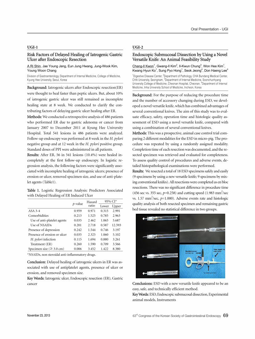
UGI-2
Endoscopic Submucosal Dissection by Using a Novel Versatile Knife: An Animal Feasibility Study
Chang-Il Kwon1, Gwang-Il Kim2, Il-Kwun Chung3, Won Hee Kim1, Kwang Hyun Ko1, Sung Pyo Hong1, Seok Jeong4, Don Haeng Lee4 1Digestive Disease Center, 2Department of Pathology, CHA Bundang Medical Center, CHA University, Seongnam, 3Department of Internal Medicine, Soonchunhyang University College of Medicine, Cheonan Hospital, Cheonan, 4Department of Internal Medicine, Inha University School of Medicine, Incheon, Korea
Background: For the purpose of reducing the procedure time
and the number of accessory changing during ESD, we devel-
oped a novel versatile knife, which has combined advantages of
several conventional knives. The aim of this study was to eval-
uate efficacy, safety, operation time and histologic quality as-
sessment of ESD using a novel versatile knife, compared with
using a combination of several conventional knives.
Methods: This was a prospective, animal case control trial com-
paring 2 different modalities for the ESD in micro-pig. The pro-
cedure was repeated by using a randomly assigned modality.
Completion time of each resection was documented, and the re-
sected specimen was retrieved and evaluated for completeness.
To assess quality control of procedures and adverse events, de-
tailed histopathological examinations were performed.
Results: We resected a total of 18 ESD specimens safely and easily
(9 specimens by using a new versatile knife; 9 specimens by mix-
ing conventional knifes). All resections were completed as en bloc
resections. There was no significant difference in procedure time
(456 sec vs. 355 sec, p=0.258) and cutting speed (1.983 mm2/sec
vs. 1.57 mm2/sec, p=1.000). Adverse events rate and histologic
quality analysis of both resected specimen and remaining gastric
bed tissue revealed no statistical difference in two groups.
Conclusions: ESD with a new versatile knife appeared to be an
easy, safe, and technically efficient method.
Key Words: ESD, Endoscopic submucosal dissection, Experimental
animal models, Instruments
Oral Presentation - UGI
70 63rd Congress of the Korean Society of Gastrointestinal Endoscopy November 23, 2013
UGI-3
Prediction of Clinical Outcome in Achalasia Patients Underwent Peroral Endoscopic Myotomy (POEM)
Jun-Hyung Cho1, Joo Young Cho1, Jin Nyoung Kim1, Byoung Moo Lee1, Mi-Young Kim1, Tae Hee Lee1, Jae Pil Han2, Su Jin Hong2 Digestive Disease Center, Soonchunhyang University Hospital, 1Seoul, 2Bucheon, Korea
Background/Aims: Peroral endoscopic myotomy (POEM) was
introduced as an alternative treatment for achalasia patients.
However, predicting factors for clinical outcome remain un-
determined in patients underwent POEM.
Methods: From November 2011 to August 2013, we reviewed clin-
ical outcome and medical records of 28 patients, retrospectively.
Clinical variables including patient's sex, age, disease duration,
achalasia subtype (sigmoid or non-sigmoid), esophageal di-
ameter, HRM finding (type 1,2,3), length of myotomy, type of
myotomy (full or partial) were analyzed. Clinical symptom
score (Eckardt score) was compared before and after POEM.
When post-POEM score was 0 or the score was decreased by 6
or more, it was defined as successful outcome.
Results: A total of 21 patients showed successful outcome. Of
these, 17 patients showed post-POEM score was 0. However,
the remaining 7 patients did not show successful outcome.
Univariate and multivariate analyses were performed for find-
ing the predictors related to successful POEM. On univariate
analyses, the short disease duration and non-sigmoid type
achalasia were related to successful POEM (p=0.092 and p=0.056,
respectively). However, there were no statistical significance.
The small esophageal diameter was significantly associated
with successful outcome (p=0.020). Multivariate analyses also
showed the significant association between the small esoph-
ageal diameter and successful outcome (p=0.027).
Conclusions: The small esophageal diameter was associated
with successful POEM outcome. In the future, large pro-
spective study is needed to confirm that POEM can be consid-
ered as standard treatment in early stage achalasia patients.
Key Words: Achalasia, POEM, Prediction, Outcome
UGI-4
Is the Edema of Interarytenoid Mucosa Related to Erosive Esophagitis?
Seung Han Kim, Hoon Jai Chun, Jae Min Lee, Jong Soo Lee, Seung Joo Nam , Hyuk Soon Choi, Eun Sun Kim, Bora Keum, Yoon Tae Jeen, Hong Sik Lee, Chang Duck Kim, Ho Sang Ryu, Jong-Jae Park, Sang Woo Lee Division of Gastroenterology and Hepatology, Department of Internal Medicine, Korea University College of Medicine, Seoul, Korea
Background/Aims: Laryngopharyngeal reflux (LPR) have been
thought to be caused by the retrograde flow of gastric acid up
to the larynx and hypopharynx. Recent study verified that the
edema of interarytenoid mucosa was independent predictor of
endoscopic confirmed esophagitis. But there have been differ-
ent perspectives about the relationship of endoscopic findings
between LPR and GERD. And there have been no data about
investigation of both findings in Korea. The aim of this study
was to evaluate the correlation between endoscopic findings of
LPR and GERD.
Methods: Between March 2013 and September 2013, 103 con-
secutive patients who were referred for endoscopic evaluation
were enrolled in this study. In the present study, Reflux symp-
tom index (RSI), Frequency scale for the symptoms of gastro-
esophageal reflux disease (FSSG) were examined and the pres-
ence of erosive esophagitis and laryngopharyngeal reflux were
evaluated. We analyzed the relationship between erosive
esophagitis and laryngopharyngeal reflux finding.
Results: About 60% patients with endoscopic laryngopharyngeal
reflux finding showed erosive esophagitis. Mann-Whitney test
presented no statistical significance on edema of the interar-
ytenoid mucosa in patients with erosive esophagitis (p value
0.54). On multivariate analysis, the existence of erosive esoph-
agitis was not significantly associated with the edema of the in-
terarytenoid mucosa (odds ration [OR]: 1.78; 95% confidence
interval [CI]: 0.35-9.0, p value 0.48). Also, RSI measures were
not correlated with the existence of erosive esophagitis (odds
ration [OR]: 0.69; 95% confidence interval [CI]: 0.035-13.74, p
value 0.81).
Conclusions: This study showed that the edema of interar-
ytenoid mucosa had no significant association with erosive
esophagitis. Also, LPR symptoms were not correlated with ero-
sive esophagitis.
Key Words: Laryngopharyngeal reflux, Erosive esophagitis
Oral Presentation - UGI
November 23, 2013 63rd Congress of the Korean Society of Gastrointestinal Endoscopy 71
UGI-5
Comparison of Accuracy of Marking Methods between Magnifying Endoscopy with NBI and Chromoendoscopy in Gastric Neoplasm
Duk Su Kim1, Yunho Jung1, Il-Kwun Chung1, Young Sin Cho1, Tae Hoon Lee1, Sang-Heum Park1, Hyun-Deuk Cho2 and Sun-Joo Kim1
Department of 1Internal Medicine, 2Pathology, Soonchunhyang University School of Medicine, Cheonan, Korea
Background/Aims: Magnifying endoscopy with narrow-band
imaging (ME-NBI) and acetic acid-indigocarmine chro-
moendoscopy (AIC) have been used for determining lateral ex-
tent of gastric tumor before endoscopic submocosal dissection
(ESD). The aim of this study was to evaluate the accuracy be-
tween ME-NBI and AIC for determining the tumor margin in
gastric neoplasm.
Methods: A total of 23 cases of gastric neoplasm (adenoma: 12,
EGC: 11) was enrolled. Two different marking methods were
performed in each case. Marking method was randomly de-
termined by drawing envelopes. Initially, the demarcation line
was carefully identified by ME-NBI or AIC. Marking dots were
placed on normal mucosa within 3 mm from the tumor by us-
ing an electrocautery with needle knife. Subsequently, the same
lesion was examined by using the other method then marking
was performed as same before. After performing ESD, the re-
section margin was carefully assessed by experienced pathologist.
If the demarcation of the lesion was clear and the lesion was
confined within the markings, it was classified as successful
delineation. If the lesion was undemarcated or cancerous tissue
was present outside the markings, it was classified as un-
successful delineation.
Results: 23 cases of ESD were performed successfully. The
mean size of resected specimen was 207 mm2 in ME-NBI
group and 269 mm2 in AIC group. The total rate of successful
delineation was significantly higher AIC group in comparison
with ME-NBI group. (95.7% (1/23) vs 60.9% (14/23), p=0.004).
In early gastric cancer, the rate of successful delineation was
not significantly difference between two groups (AIC: 100%
(11/11) vs ME-NBI: 81.8% (9/11), p=0.476). However, in the
case of gastric adenoma, the rate of successful delineation was
significantly higher AIC group than ME-NBI group (91.7%
(11/12) vs 41.7% (5/12), p=0.027).
Conclusions: AIC seems better marking method than ME-NBI
before performing ESD in gastric adenoma.
Key Words: Magnifying endoscopy with narrow-band imag-
ing, Chromoendoscopy, Gastric neoplasm, ME-NBI, ESD
UGI-6
Clinicopathologic Characteristics of Interval or Missed Early Gastric Cancer after a Negative Esophagogastroduodenoscopy
Young Sin Cho, Il-Kwun Chung, Yunho Jung, Tae Hoon Lee, Sang-Heum Park, Sun-Joo Kim Department of Internal Medicine, Soonchunhyang University College of Medicine, Cheonan Hospital, Cheonan, Korea
Background/Aims: Esophagogastroduodenoscopy (EGD) has
become the standard procedure of investigation when gastric
cancer is suspected. However, the clinicopathologic character-
istics in relation to interval early gastric cancer (EGC) is not
well known in contrast to understanding of colonoscopy
missed rate and risk factor for colorectal cancer. The aim of this
study was to evaluate the clinicopathologic and endoscopic
characteristics of interval EGC after a negative EGD.
Methods: We retrospectively analysed data on 1055 gastric ad-
enocarcinoma patients who confirmed EGC by endoscopic re-
section or operation from July 2006 to May 2013. The referrd
patients who diagnosed or suspected gastric neoplasm from
other medical clinics excluded from our study (n= 771).
Interval EGC was defined as a gastric cancer that is diagnosed
within 2 years of negative EGD result. We compared clin-
icopathologic characteristics between patients with initially di-
agnosed EGC and patients with interval EGC. The variable risk
factors of interval EGCs were investigated.
Results: We identified interval EGCs in 54 (19%) of 284
patients. Average age was 65.4 years old and average interval
time between the time of diagnosis and previous EGD was 12.6
months. Pathologic analysis showed that interval EGC group
was significantly more smaller (1.85 Vs 1.31 cm; p<0.001) and
more showed intestinal metaplasia (OR 4.85; p<0.001) than
initial diagnosed EGC group. There was no differences in loca-
tion, differentiation, gross morphology and H. pylori infection.
In factors associated with procedure, short procedure time
(7.14 Vs 5.55 min; p<0.03) and negative symptoms (OR 3.23;
p<0.02) were a predictive factors for interval or missed EGC.
Conclusions: Small sized lesion and intestinal metaplasia could
be predictive factors for the presence of interval or missed EGC
during screening EGD. Careful examination with sufficient in-
spection time is very important to detect EGC with or without
symptoms of patient.
Key Words: EGC, Interval gastric cancer, Missed gastric cancer
Oral Presentation - UGI
72 63rd Congress of the Korean Society of Gastrointestinal Endoscopy November 23, 2013
UGI-7
Deep Biopsy via Endoscopic Submucosal Dissection in Upper GI Subepithelial Tumors: A Prospective Study
Hye Jin Tae, Hang Lak Lee, Eun Young Doo, Young Woo Ahn, Ki Deok Yoo, Jin Ok Kim, Kang Nyeong Lee, Dae Won Jun, Oh Young Lee, Byung Chul Yoon, Ho Soon Choi, Joon Soo Hahm Department of Internal Medicine, Hanyang University College of Medicine, Seoul, Korea
Background: Preoperative pathologic diagnosis of subepithelial
tumors (SET) may improve clinical management decisions in
patients with upper GI SETs. The aim of this study was to eval-
uate the diagnostic yield of deep biopsy via endoscopic sub-
mucosal dissection (ESD) and its impact on management of
patients with upper GI SETs.
Patients and Methods: Eighty-seven patients with upper GI
SETs were voluntarily assigned to two groups. The specialist A
group underwent EUS and endoscopic deep biopsy using the
ESD technique. The specialist B group underwent surgical re-
section after EUS without obtaining preoperative pathological
diagnosis, in accordance with accepted clinical management
algorithms.
Results: The diagnostic yield of deep biopsy for patients with
upper GI SETs was 90% (36/40). Deep biopsy results changed
treatment plans in 14 of 40 deep biopsy patients (35%) in spe-
cialist A group. One patient with lymphoepithelial carcinoma
was scheduled for surgical resection, whereas 13 patients with
benign SETs ≥2 cm in diameter avoided unnecessary
operations. In addition, 13 of 28 patients (46.4%) in specialist
B group who underwent surgical resection without pre-
operative pathologic diagnosis had benign lesions. The proce-
dure time was relatively short (Mean 13.66±2.91 min). There
were no procedure-related complications.
Conclusions: Deep biopsy by the ESD technique is a safe,
high-yield diagnostic method in patients with upper GI SETs.
This method could improve clinical decision-making in the
management of patients with upper GI SETs.
Key Words: Subepithelial tumor, ESD, Biopsy
UGI-8
Risk Factors and Clinical Outcomes of Gastric Cancer Identified by Screening Endoscopy: A Case-Control Study
Eun Jeong Gong1, Ji Yong Ahn1, Hwoon-Yong Jung1, Hyun Lim1, Kwi-Sook Choi1, Jeong Hoon Lee1, Do Hoon Kim1, Kee Don Choi1, Ho June Song1, Gin Hyug Lee1, Jin-Ho Kim1, Son Yeong Choi2, Jae Won Choe2 Departments of 1Gastroenterology and 2Health Screening & Promotion Center, University of Ulsan College of Medicine, Asan Medical Center, Seoul, Korea
Background and Aim: A customized screening program for
gastric cancer would optimize the benefits of screening
endoscopy. This study investigated the risk factors for gastric
cancer detected during screening, and factors affecting clinical
outcomes. Methods-: From April 2000 to December 2010, sub-
jects who underwent screening endoscopy at Asan Medical
Center were included. To investigate risk factors, age and
sex-matched control group were selected. The clinical out-
comes of gastric cancer identified during screening (screening
group) were compared with age, sex and date of diag-
nosis-matched subjects who were diagnosed with gastric can-
cer in the outpatient clinic (outpatient group).
Results: Of 109,530 subjects, 327 were diagnosed with gastric
cancer. The median age of the screening group was 63.6 years
(interquartile range: 56-71 years), and the male to female ratio
was 2.4:1. When comparing with the control group, H. pylori
seropositivity (odds ratio [OR] 2.933, p<0.001), carcinoem-
bryonic antigen (OR 8.633, p=0.004), family history of gastric
cancer (OR 2.254, p=0.007), and drinking (OR 3.312, p<0.001)
were independent positive risk factors, and the use of aspirin a
negative risk factor for gastric cancer (OR 0.445, p=0.012) in
multivariate analysis. Low density lipoprotein cholesterol
(hazard ratio [HR] 0.987, p=0.005), cancer antigen 19-9 (HR
21.713, p<0.001), resectability (HR 59.833, p<0.001), and fam-
ily history (HR 0.308, p=0.009) were independent risk factors
for death. The 5-year survival rate was significantly higher in
the screening group than in the outpatient group (p<0.001).
Conclusions: Early detection of gastric cancer by screening en-
doscopy while asymptomatic enhances patient outcomes, es-
pecially in high risk groups.
Key Words: Stomach neoplasms, Risk factors, Cancer screen-
ing, Endoscopy
Oral Presentation - UGI
November 23, 2013 63rd Congress of the Korean Society of Gastrointestinal Endoscopy 73
UGI-9
What Is the Optimal Timing for Image Aanalysis of Freshly Excised Tissue for Multiphoton Microscopy?
Eun Sun Kim1, Hoon Jai Chun1, Seung Han Kim1, Jae Min Lee1, Seung Joo Nam1, Jong Soo Lee1, Hyuk Soon Choi1, Bora Keum1, Yoon Tae Jeen1, Hong Sik Lee1, Chang Duck Kim1, Ho Sang Ryu1, Jong Jae Park1, Sang Woo Lee1, Chang Su Lim2, Bong Rae Cho2 1Division of Gastroenterology and Hepatology, Department of Internal Medicine, Korea University College of Medicine, 2Department of Chemistry, Korea University, Seoul, Korea
Background and Aims: Probe based multiphoton microscopic
image analysis can provide long time and deep tissue imaging
which allow dynamic functional information of fresh live tis-
sue from patient. In contrast, confocal microscopy had limi-
tation that short observation time by photo- damage and shal-
low image depth in cellular level. Traditionally, it was consid-
ered that freshly excised tissue give a detectable fluorescence
signal if imaged within 2-3 hours from excision. However there
have been no controlled data about available time limit from
excision for fresh live tissue image analysis. The aim of our
study is to assess the change of live tissues according to time
duration using probe based multiphoton microscopy.
Patients and Methods: We obtained fresh live stomach mucosal
tissues during gastroscopic biopsy from patients in outpatient
clinic. Patients who had contraindication for biopsy were
excluded. 6 pieces of mucosal tissues from one patient were ob-
tained and merged in phosphate buffer solution immediately,
and stained with multiphoton probe. Tissues were imaged us-
ing multiphoton microscopy at 30 min, 60 min, 90min, 120
min, 150 min, and 180 min respectively. We assess changes of
mucosal structure and fluorescence from multiphoton probe.
Results: At 30 minutes, tissues were intact but fluorescence
from probe was not fully stained. At 60 minutes, all tissues were
intact and probe was fully stained. At 90 minutes some tissues
were denatured but fluorescence was consistent. At 120, 150,
and 180 minutes, almost tissues had deformation of glandular
structure and fluorescence was rapidly changed.
Conclusion: Live tissue image using probe based multiphoton
microscopy should be obtained between 60-90 minutes, optimally.
From over 120 minutes, structural deformation and functional
change occurred completely.
Key Words: Multiphoton microscopy, Image, Live tissue,
Optimal time
UGI-10
Magnifying Images by Using a Near Focus Method and a Conventional Method under Narrow Band Imaging for Gastric Epithelial Tumors
Su Jin Hong, Shin Hee Kim, Jae Pil Han, Moon Han Choi, Yun Nah Lee, Bong Min Ko, Moon Sung Lee Digestive Disease Center and Research Institute, Department of Internal Medicine, Soonchunhyang University School of Medicine, Bucheon, Korea
Background: A dual focus two-stage optical lens technology
was recently introduced. In the near focus mode, endoscopists
can perform a close examination of mucosal tissue and capil-
lary networks. The aim of this study was to investigate the mag-
nifying images on the near focus method (NFM) compared to
those on the conventional magnification method (CMM) un-
der narrow band imaging (NBI) in the patients with gastric ep-
ithelial tumors.
Methods: An experienced endoscopist performed endoscopies
by using NFM and CMM in the 20 enrolled patients with gas-
tric epithelial tumors, respectively. We selected 40 images of 40
sessions of endoscopy in the patients. Ten endoscopists asyn-
chronously reviewed for image quality. The image quality was
rated on a 5-point Likert scale (from poor, 1 to excellent, 5) for
mucosal microsurface structure, subepithelial microvascular
architecture, and demarcation line. The all of enrolled patients
received endoscopic submucosal dissection (ESD).
Results: The final diagnosis of the gastric epithelial tumors re-
vealed 10 cases of early gastric cancer, 2 cases of high grade dys-
plasia, and 8 cases of low grade dysplasia after ESD. The me-
dian number of magnification images was 11 in each method.
The average observation time (±SD) for magnification was
99.9±64.1 s in NFM and 91.5±64.6 s in CMM (p=0.54),
respectively. Judgments of image quality in mucosal microsur-
face structure were 4.09±0.39 in NFM and 3.73±0.40 in CMM
(p=0.015). Those of in subepithelial microvascular archi-
tecture were 3.53±0.45 in NFM and 4.29±0.45 in CMM
(p=0.001). Judgment of clear demarcation line were 3.91±0.41
in NFM and 3.61±0.54 in CMM (p=0.089).
Conclusion: The near focus mode which is controlled by expe-
rienced endoscopists seems to be a useful method for magnifi-
cation as the conventional method in gastric epithelial tumors.
Further evaluation of this novel technology is necessary and
awaited.
Key Words: Magnification, Gastric Epithelial Tumor, Method
Oral Presentation - UGI
74 63rd Congress of the Korean Society of Gastrointestinal Endoscopy November 23, 2013
UGI-11
Clinical Outcomes of Full Myotomy vs. Partial Myotomy in Peroral Endoscopic Myotomy for Achalasia Patients
Mi-Young Kim1, Jun-Hyung Cho1, Su Jin Hong2, Joo Young Cho1
¹Digestive Disease Center, Soonchunhyang University Hospital, Seoul, ²Department of Internal Medicine, Soonchunhyang University College of Medicine, Bucheon, Korea
Background/Aims: Peroral endoscopic myotomy (POEM) is
known to be safe and effective endoscopic surgery compared
with surgical myotomy for achalasia patients. Although full
myotomy may be more effective than partial myotomy in
symptoms resolution, the effectiveness of full myotomy is not
established. Moreover, concerns for perforation during POEM
have been an obstacle in performing full myotomy. The aim of
this study was to compare the clinical outcomes and complica-
tion rates between full myotomy and partial myotomy among
the patients underwent POEM for achalasia.
Methods: A total of 34 POEM procedures was performed from
November 2011 to September 2013 at Soonchunhyang University
Seoul and Bucheon Hospital.
Results: Among 34 procedures in 31 patients, 16 full myotomy
and 18 partial myotomy were included. The length of full and
partial myotomy were 8.4±2.6 and 7.9±1.7 cm (p=0.562),
There was no difference in pre-POEM Eckardt score, basal LES
pressure, and integrated relaxation pressure (IRP) between full
and partial myotomy group (5.8±1.7 vs. 6.6±2.6, 27.2±12.8 vs.
33.8±14.0 mmHg, and 21.7±13.6 vs. 25.8±11.8 mm Hg). All
patients showed a significant improvement in Eckardt score af-
ter full or partial myotomy during median follow-up of 8
months (1.1±1.3 vs. 0.5±0.8; p<0.001). Significant decrease in
LES pressure and IRP was found in partial myotomy group
(p=0.003, p=0.001). However, no significant difference in
Eckardt score, LES pressure and IRP score between full and
partial myotomy groups. There was no significant proce-
dure-related complication and no significant difference in the
procedure-related complication rates including pleural effu-
sion, pneumoperitoneum, retropneumoperitoneum, pneu-
momediastinum, and subcutaneous emphysema on CT or
X-ray between two groups.
Conclusion: POEM showed good clinical outcomes and safety.
Full or partial myotomy did not show significant clinical
outcomes.
Key Words: Peroral endoscopic myotomy, Outcome, Compli-
cation
UGI-12
A Novel Occluder Device for Endoscopic Closure of Gastrointestinal Leaks and Fistulas
Ji Hee Kim, Bo-In Lee, Joon Sung Kim, Byung-Wook Kim, Hwang Choi, Jae Myung Park, Sang-Woo Kim, Myung-Gyu Choi, Kyu Yong Choi Department of Internal Medicine, The Catholic University of Korea, Seoul, Korea
Background and Aims: Endoscopic treatments of gastro-
intestinal (GI) leaks and fistulas meet with only limited success.
Although successful closure of GI fistulas by using the cardiac
septal defect closure device has been reported, the cost is very
high and cannot be delivered through-the-scope.
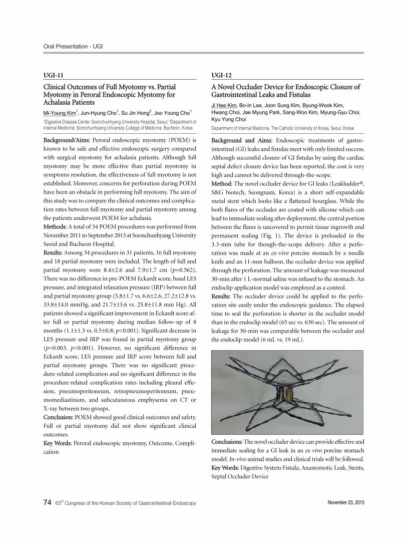
Method: The novel occluder device for GI leaks (Leakludder®,
S&G biotech, Seongnam, Korea) is a short self-expandable
metal stent which looks like a flattened hourglass. While the
both flares of the occluder are coated with silicone which can
lead to immediate sealing after deployment, the central portion
between the flares is uncovered to permit tissue ingrowth and
permanent sealing (Fig. 1). The device is preloaded in the
3.3-mm tube for though-the-scope delivery. After a perfo-
ration was made at an ex-vivo porcine stomach by a needle
knife and an 11-mm balloon, the occluder device was applied
through the perforation. The amount of leakage was measured
30-min after 1 L-normal saline was infused to the stomach. An
endoclip application model was employed as a control.
Results: The occluder device could be applied to the perfo-
ration site easily under the endoscopic guidance. The elapsed
time to seal the perforation is shorter in the occluder model
than in the endoclip model (65 sec vs. 630 sec). The amount of
leakage for 30-min was comparable between the occluder and
the endoclip model (6 mL vs. 19 mL).
Conclusions: The novel occluder device can provide effective and
immediate sealing for a GI leak in an ex vivo porcine stomach
model. In-vivo animal studies and clinical trials will be followed.
Key Words: Digestive System Fistula, Anastomotic Leak, Stents,
Septal Occluder Device
Oral Presentation - UGI
November 23, 2013 63rd Congress of the Korean Society of Gastrointestinal Endoscopy 75
UGI-13
Trucut Biopsy Using Forward-Viewing Endoscopy for Diagnosis of Upper Gastrointestinal Subepithelial Tumors
Jin Nyoung Kim, Joo Young Cho, Byoung Moo Lee, Mi-Young Kim, Jun-Hyung Cho, Tae Hee LeeDigestive Disease Center, Soonchunhyang University Hospital, Seoul, Korea
Background and Aims:For pathologic diagnosis of large upper
gastrointestinal (GI) subepithelial tumor (SET),fine needle as-
piration and trucut biopsy are usually performed under guid-
ance of endoscopic ultrasonography (EUS). However, there are
no reports about the feasibility of trucut biopsy without EUS
guidance. We conducted a pilot study to evaluate the usefulness
of trucut biopsy using forward-viewing endoscopy for the di-
agnosis of upper GI SETs.
Methods:A total of 19 patients were enrolled into this study.
Before trucut biopsy, all patients underwent EUS or Computed
tomography (CT) to confirm SETs.Inclusion criteria were size
of >2 cm, tumor location of esophagus or stomach body.
Under moderate sedation state by midazolam, this procedure
was performed by forward-viewing conventional gastroscope.
19 gauge biosy needle (EUSN-19-QC, Cook medical) was in-
serted through the working channel of endoscope and trucut
biopsies were performed three times. Biopsied specimens were
reviewed by single experienced GI pathologist. Additional im-
munohistochemical staining were performed,such as CD117,
CD34, smooth muscle actin,desmin,S-100.
Results:Tumors were located in the esophagus (73.6%, n=14)
and stomach (26.4%, n=5).Mean size of tumor was 2.73 cm.
All procedures were performed successfully. Immediate com-
plication including bleeding, perforation did not occur.
Pathologic results of trucut biopsy specimen showed GIST
(10.5%, n=2), leiomyoma (78.9%, n=15). The remaining cases
showed insufficient material (10.5%,n=2) and located in the
stomach. In two patients with inconclusive trucut biopsy re-
sult, laparoscopic wedge resection were performed and final
pathology revealed ectopic pancreas. Overall technical success
rate of this technique was 89.4% (n=17/19).
Conclusions: Trucut biopsy using forward-viewing endoscopy
is a feasible and safe for diagnosis of upper GI SETs. If SETs
more than 2 cm size are suspicious of GIST or leiomyoma on
EUS, our technique can be applied in clinical practice.
Key Words: Subepithelial tumor, Diagnosis, Trucut biopsy
UGI-14
A Novel Reinforcement Method for the Surface of Gastric Metal Stent: Gas Plasma Treatment in Vitro Study
Hyuk Soon Choi1, Hoon Jai Chun1, Eun Sun Kim1, Bora Keum1, Yeon Seok Seo1, Yoon Tae Jeen1, Hong Sik Lee1, Soon Ho Um1, Chang Duck Kim1, Ho Sang Ryu1, Jong-Jae Park1, Sang Woo Lee1, Hong Bae Kim2 1Division of Gastroenterology and Hepatology, Department of Internal Medicine, Korea University College of Medicine, Seoul, 2Institute of Interventional Medicine, M. I. Tech Co., Ltd, Pyeongtaek, Korea
Background/Aim: A gastric stent, which is made of nick-
el-titanium (NiTi) alloy coating with a silicone polymer, has
been using for the relief of obstructive symptom in both malig-
nant and benign stricture of stomach. Although coating with
the silicone polymer on the stent plays a key role in corroding,
corrosion property may differ from along statues of surface of
NiTi alloy wire as coating the silicone polymer. The surface
modification with gas plasma is a way to improve a surface ad-
hesive property to the silicon polymer. We systematically inves-
tigated a surface modification to stick the silicone employing
gas plasma treatment.
Methods: The NiTi alloy stents were treated in a few conditions
of the plasma treatment, in which vary mixture rates of Ar and
O2 gas, applied voltages and duration of exposing time. We
prepared three kinds of stents; normal stent (product by nor-
mal process), slightly etched normal stent (product by plasma
treatment) and natural oxide layer-eliminated normal stent
(product that removed natural oxide regions by plasma treat-
ment). The stents were analyzed with a TEM to examine sur-
face topographies of the stents and performed a potentiody-
namic test to compare corrosion state.
Results: The surface profile of the samples showed that some
content of oxide layer for the normal stent was formed in thick-
ness of about 100nm, while the others was less than 60nm.
Moreover, the oxide layer for normal product and slightly etch-
ed normal stent was likely to exhibit deposition of oxygen with-
out interlocking that enhances cohesion, whereas natural oxide
layer-eliminated normal stent showed behavior of strong
interlocking. It imply that an interlocking between nickel and
oxide layer plays a significant role of corrosion resistance. In a
potentiodynamic test, the sample by plasma treatment in-
dicated strong corrosion resistance.
Conclusion: These results revealed that the plasma treatment
would be employed to improve the surface property of gastric
stent.
Key Words: Stent, Gas plasma, Nickel-Titanium
Oral Presentation - UGI
76 63rd Congress of the Korean Society of Gastrointestinal Endoscopy November 23, 2013
UGI-15
Prevalence of Gastric Subepithelial Tumor in Korea: Single Center Experience
Yong Woo Ahn, Hang Lak Lee, Ki Deok Yoo, Eun Young Doo, Hye Jin Tae, Jin Ok Kim, Joon Soo Hahm, Byung Chul Yoon, Ho Soon Choi, Oh Young Lee, Dae Won Jun, Kang Nyeong Lee Department of Internal Medicine, Hanyang University College of Medicine, Seoul, Korea
Background and Aims: Incidental gastrointestinal subepithelial
tumor (SET) is increasing with national cancer screening
endoscopy. In previous report, prevalence of SET was about
0.36%. However, there is no report examining the prevalence
of SET in stomach in Korea. The aim of this study was to eval-
uate the prevalence of SET in stomach, in health examinee.
Patients and Methods: We reviewed retrospectively the endo-
scopic reports of 11,712 subjects who underwent esophagogas-
troduodenoscopy (EGD) for screening purpose at Hanyang
University Hostpital from July, 2012 to June, 2013. SET was de-
fined as "elevated lesion covered with normal-appearing mu-
cosa, and it is not lost by changes in posture". EGD was per-
formed by 11 endoscopists with endoscopic certification.
Results: Among 11,712 health examinee, 194 (1.66%) had SET
in stomach. 71 (1.33%) males had SET, and 123 (1.99%) fe-
male had SET. The average age of examinee with SET was 50.9
year old (male 50.9 year old, female 51.1 year old). When sepa-
rated by age, the prevalence of SET was as follows. Twenties
had 1.31%, thirties had 1.15%, forties had 1.63%, fifties had
2.18%, sixties had 3.62% and seventies had 3.66%. In both
male and female, there was a correlation between age and the
prevalence of SET (p=0.000). When classified according to size
of SET, 145 (74.7%) was <10 mm, 38 (19.6%) was 10-19 mm, 4
(2.1%) was 20-29 mm, and 7 (3.6%) was >30 mm. When clas-
sified according to location of SET, 36 (18.6%) was in antrum,
14 (7.2%) was in lower body, 5 (2.6%) was in mid body, 52
(26.8%) was in upper body, and 87 was in cardia or fundus.
Conclusion:- The overall prevalence of SET in stomach among
health examinee was 1.66%. There was a correlation between
age and the prevalence of SET. In the future, a community-ori-
ented well organized study should be warranted.
Key Words: SET, Subepithelial tumor, Prevalence, Korea
UGI-16
Further Diagnostic Treatments Are Necessary for Gastric Atypical Cell on Pathologic Findings of Endoscopic Biopsy
Chung Hoon Yu1, Yup Hwangbo1, Jun Young Choi1, Sun Young Ahn1, Hyun Seok Lee1, Sun Zoo Kim2, Han Ik Bae2 and Seong Woo Jeon1
Department of 1Internal Medicine and 2Pathology, Kyungpook National University School of Medicine, Daegu, Korea
Background: Gastric atypical cell (GAC) on endoscopic for-
ceps biopsy is the indefinite pathologic finding between benign
and malignant. This case often needs other diagnostic treat-
ments such as endoscopic submucosal dissection (ESD), endo-
scopic mucosal resection (EMR) or operation (OP). Aims: We
aimed to analyze the initial endoscopic and histologic findings
and to discuss the necessity of further diagnostic treatments for
cases of GAC.
Methods: This retrospective study enrolled 96 patients (M:F
61:35, mean age 59±14 years old) proven GAC on initial patho-
logic finding of endoscopic forceps biopsy (January 1999-
December 2012). ESD (n=16, 16.7%), EMR (n=5, 5.2%), OP
(n=23, 24.1%) and follow-up or other treatment after diag-
nosis by re-biopsy (n=52, 54.0%) were performed. This study
analyzed the initial endoscopic (lesion of size, location, or
type) and histologic (presence of intestinal metaplasia or H.
pylori) characteristics of GAC lesions related with the final
pathology. The univariate and multivariate logistic regression
analysis were presented as odds ratio (OR; 95% confidence in-
tervals (CI)).
Results: The final pathologic diagnoses were cancer (n=36,
37.6%), adenoma (n=9, 9.4%) and non-neoplasm (n=51,
53.0%). Sex, lesion of location, and presence of intestinal met-
aplasia did not significantly influence the result of final
outcome. With multivariate analysis, there was no independent
characteristic factor. With univariate analysis, age (p=0.020,
OR=1.04 CI 1.01-1.07), lesion of 10 mm and above (p=0.003,
OR=3.94, CI 1.61-9.61), lesion of depression (p=0.030,
OR=2.50, CI 1.09-5.72), and presence of H. pylori (p=0.030,
OR=2.83, CI 1.11-7.25) were statistically associated with the fi-
nal diagnosis of neoplasm.
Conclusions: Considering the neoplastic rate of GAC cases was
near 50%, further diagnostic treatments can be performed for
suspicious neoplastic lesions, related with the significant find-
ings (≥10 mm, depression, or H. pylori) or age of GAC
patients.
Key Words: Gatric atypical cell, Indefinite pathology, Endoscopic
biopsy, Stomach neoplasm
Oral Presentation - UGI
November 23, 2013 63rd Congress of the Korean Society of Gastrointestinal Endoscopy 77
UGI-17
Magnifying Endoscopic Findings in Adenomatous and Foveolar Gastric Dysplasia
Hong Ryeol Cheong1, Gwang Ha Kim1, Bong Eun Lee1, Geun Am Song1, Hwa Mi Kang2, Do Youn Park3 1Department of Internal Medicine, Pusan National University School of Medicine, 2Department of Internal Medicine, Busan Adeventist Hospital, 3Department of Pathology, Pusan National University School of Medicine, Busan, Korea
Background and Aims: Gastric epithelial dysplasia (GED) can
be morphologically categorized into adenomatous (or in-
testinal) and foveolar (or gastric) types. Although some stud-
ies showed the difference in the clinicopathologic features and
mucin profiles between these types, there has been no report
on the magnifying endoscopy (ME) in these types. Therefore,
the aim of this study was to investigate the difference in the
ME-NBI findings between adenomatous and foveolar GEDs.
Patients and Methods: A total of 46 patients with a final diag-
nosis of GED who underwent ME-NBI before endoscopic
submucosal dissection were included in this study. During
ME-NBI, microvascular (MV) pattern, microsurface (MS)
pattern, and the presence of light blue crest (LBC) and white
opaque substance (WOS) were investigated. GEDs were cate-
gorized into adenomatous, foveolar, and hybrid types. In ad-
dition, the expression of MUC2, MUC5AC, MUC 6 and
CD10 were evaluated.
Results: Adenomatous type was seen in 27 GEDs (58.7%), hy-
brid type in 15 (32.6%) and foveolar type in 4 (8.7%).
Irregular MV pattern was observed in 55.6% of adenomatous
type, 73.3% of hybrid type and 75.0% of foveolar type
(p=0.517). Adenomatous types showed mainly tubular
(55.6%) and mixed (37.0%) MS pattern, and hybrid type
showed mainly mixed (33.3%) and absent (33.3%) MS pat-
tern; all 4 foveolar GEDs showed MS pattern (p<0.001).
MUC5AC and MUC6 expression was associated with absent
and papillary MS pattern, and CD10 expression was asso-
ciated with round pit and/or tubular MS pattern.
Conclusions: MS pattern in ME-NBI might be helpful in pre-
dicting the morphological categorization and mucin pheno-
type in GEDs.
Key Words: Gastric epithelial dysplasia , Mgnifying endoscopy,
NBI
UGI-18
Clinical and Endoscopic Features in Metastatic Tumors to the Stomach
Ga Hee Kim, Ji Yong Ahn, Hwoon-Yong Jung, Hyun Lim, Jeong Hooon Lee, Kwi-Sook Choi, Do Hoon Kim, Kee Don Choi, Ho June Song, Gin Hyug Lee, Jin-Ho Kim Department of Gastroenterology, Asan Medical Center, Asan Digestive Disease Research Institute, Seoul, Korea
Background and Aim: Metastases to the stomach are rare and
studies relating endoscopic and pathologic findings with clinical
outcomes are lacking. The aim of this study was to describe a ser-
ies of cases of metastasis to the stomach, their primary sites, clin-
ical and endoscopic features, treatment, and prognosis. Methods
We reviewed the clinicopathological aspects of patients with gas-
tric metastases from solid organ tumors. Thirty cases were diag-
nosed between January 1995 and August 2011 at Asan Medical
Center. We evaluated histology, initial presentations, imaging find-
ings, lesion locations, treatment courses, and overall patient’s
survival.
Results: Of total 30 patients, median age was 55.5 years
(interquartile range [IQR], 48.5-60.3 years) and men were
predominent. The metastases presented as solitary (n= 17,
56.7%) or multiple lesions (n= 13, 43.3%), and were more fre-
quently located in the body of the stomach. The primary sites
were the lung (n= 6, 20%), breast (n= 2, 6.6 %), colorectum
(n=2, 6.6%), ovary (n=2, 6.6%), and kidney (n= 2, 6.6%).
Malignant melanoma was the most frequent tumor to the
metastasis to the stomach (n=10,33.3%). Only seven patients
were given some form of treatment after diagnosis of the gas-
tric metastasis. The median survival in this series, calculated
from the time of the diagnosis of the gastric metastasis, was
2.0 months (IQR, 0.9-3.1 months). The mean time lapse be-
tween the diagnosis of the primary tumor and diagnosis of the
gastric metastasis was 13.5 months (IQR, 0.75-33.5 months).
Conclusion: Gastric metastasis marks advanced disease and
the prognosis is poor. Despite the low prevalence of gastric
metastases, EGD associated with good biopsy practices and a
close study of the specimens, must be used in the follow-up of
patients with malignant neoplasia.
Key Words: Endoscopy, Gastric metastasis
Oral Presentation - UGI
78 63rd Congress of the Korean Society of Gastrointestinal Endoscopy November 23, 2013
UGI-19
A Study of Pathologic Uniformity in Gastric GISTs
Ki Deok Yoo1, Hang Lak Lee1, Young Woo Ahn1, Eun Young Doo1, Hye Jin Tae1, Jin Ok Kim1, Kang Nyeong Lee1, Dae Won Jun1, Oh Young Lee1, Byung Chul Yoon1, Ho Soon Choi1, Joon Soo Hahm1, Ki Seok Jang2
Department of 1Internal Medicine, 2Pathology, Hanyang University College of Medicine, Seoul, Korea
Background: GISTs are mesenchymal tumors originate from
the interstitial cells of Cajal. It is well recognized that all GISTs
have some degree of malignant potential. Mitotic count is im-
portant to predict malignant potential. Accurate pathological
diagnosis is mandatory for proper treatment of GISTs. In gen-
eral, EUS-FNA is used for pathologic diagnosis of gastric SMT.
The aim of this study is to evaluate the presence of pathologic
uniformity in GIST in aspect of mitotic count.
Patients and Methods: Total twenty gastric GISTs patients who
performed a wedge resection were reviewed retrospectively in
Hanyang University Hospital from 2006 to 2011. We used AFIP
criteria for classification of gastric GISTs. To identify the
pathologic uniformity of gastric GISTs, we compared GIST
risk stratification between central site and peripheral site of
GIST mass.
Results: The mean size of the GISTs was 3.27 ± 0.46 cm. Six le-
sions were located in the antrum, four in the fundus, four in the
cardia, six in the body. The mean age was 53.25 ± 8.11 years; 10
patients were male and 10 were female. Among twenty cases,
eighteen cases (90%) showed same risk stratification between
two sites (central site vs. peripheral site). Only two cases (10%)
showed a different risk between two sites. Conclusions:- In case
of GIST, EUS-FNA results can represent the surgical patho-
logic results.
Key Words: Gastric GISTs, Pathologic uniformity , EUS-FNA
UGI-20
Discrepancy between Pretreatment and Posttreatment Diagnosis of Early Gastric Cancer and Its Impact on Treatment Choice
Jun Hee Lee1, Jun Haeng Lee1, Eun Ran Kim1, Ki Joo Kang2, Byung-Hoon Min1, Jae J. Kim1
1Department of Medicine, Samsung Medical Center, Sungkyunkwan University School of Medicine, Seoul, 2Department of Medicine, Hallym University Sacred Heart Hospital, Hallym University College of Medicine, Anyang, Korea
Background and Aims: In the era of endoscopic submucosal
dissection (ESD), pretreatment diagnosis and posttreatment
diagnosis are sometimes different. However, the extent of pre-
and post-treatment discrepancy and its impact on patient care
is not known.
Patients and Methods: A total of 2,096 patients with gastric ad-
enoma or cancer underwent curative endoscopic resection or
surgery at Samsung Medical Center in 2012. Pretreatment di-
agnosis groups were low-grade dysplasia (LGD) in 162,
high-grade dysplasia (HGD) in 164, atypical cell in 15, conven-
tional indication early gastric cancer (Ix-EGC) in 396, ex-
tra-conventional indication EGC (EIx-EGC) in 824, and ad-
vanced gastric cancer (AGC) in 495. The choice of initial treat-
ment and final pathologic diagnosis in each pretreatment diag-
nosis group were analyzed retrospectively.
Results: 31.9% of EGC (407 of 1276) were treated by ESD
alone. 6.1% of the LGD, 34.2% of the HGD, and 46.6% of the
atypical group were found to be gastric cancer after ESD.
33.3% of pretreatment Ix-EGC were shifted to posttreatment
EIx-EGC due to size change, submucosal invasion, lymphovas-
cular invasion, and change of cell differentiation (53.4%,
49.6%, 19.6%, and 6.9%, respectively including duplication).
2.7% of pretreatment EIx-EGC were found to be posttreat-
ment Ix-EGC mainly due to the size change (86.4%). In 10.4%
of pretreatment Ix-EGC, surgery were initially performed. In
6.6% of pretreatment EIx-EGC, ESD were initially chosen. Of
them, 92.7% were size larger than 2 cm and their additional op-
eration rate was not significantly higher than ESD for Ix-EGC
(20.9% vs 15.8%; p=0.147).
Conclusions: About one third of pretreatment Ix-EGC were
changed to posttreatment EIx-EGC by pathologic examination.
In selected cases with differentiated EGC larger than 2 cm
without ulceration, ESD were chosen initially and had accept-
able clinical outcome.
Key Words: Early gastric cancer, Pretreatment diagnosis,
Endoscopic submucosal dissection
Oral Presentation - UGI
November 23, 2013 63rd Congress of the Korean Society of Gastrointestinal Endoscopy 79
UGI-21
CagA Antigen Is the Important Factor to the Level of Anti-Helicobacter pylori IgG and IgA and the Grade of Urease Test
Ji-Hyun Seo1, Ji Sook Park1, Hyang-Ok Woo1, Hee-Shang Youn1, Gyung-Hyuck Ko2, Seung-Chul Baik3, Woo-Kon Lee3, Myung-Je Cho3, Kwang-Ho Rhee3 Department of 1Pediatrics, 2Pathology, and 3Microbiology, Gyeongsang National University School of Medicine, Jinju, Korea
Purpose: We sought to evaluate whether the serum antibody
levels could predict the presence of gastroduodenal disease, to
identify factors that correlate with antibody levels in a multi-
variate context, and to determine the correlation of the levels of
anti-HP IgG and IgA antibody with the grade of urease test, the
presence of CagA antigen, the degree of gastritis, and age.
Methods: A total of 1,271 subjects who underwent upper gas-
troduodenoscopy at Gyeongsang National University Hospital
were enrolled. The subjects were stratified into 4 age groups:
0-4 years (n=145), 5-9 years (n=285), 10-15 years (n=111) and
20-29 years (n=730). The results of urease test were classified
into 4 grades: Grade 0 (no color change), 1 (24-48 hr), 2 (6-24
hr) and 3 (< 6 hour). Histopathologic findings were de-
termined by pathologist using Updated Sydney System.
Anti-HP IgG and IgA titers were measured by ELISA and an-
ti-cagA IgG and IgA antibody was evaluated by Western blot
using whole cell lysate of H. pylori strain 51.
Results: The positivity rate of the urease test was 50.3% for 0-4
years, 51.0% for 5-9 years, 47.2% for 10-15 years, and 67.2%
for 20-29 years (p<0.0001). The degrees of chronic and active
gastritis, and HP infiltration increased with age groups
(<0.001). The titers of anti-HP IgG were lower in 0-4 y and in
5-9 y than in 10-15 y and 20-29 y (p<0.005) and the titers of an-
ti-HP IgA were higher in 20-29 y than other 3 age groups
(p<0.005). The cagA potitive rate was 24.1% in 0-4 years,
32.7% in 5-9 years, 42.3% in 10-15 years, and 52.5% in 20-29
years for IgG and 11.7% in 0-4 years, 14.4% in 5-9 years, 16.2%
in 10-15 years, and 36.0% in 20-29 years for IgA. In the cases of
cagA positive pattern and highest grade of urease test, the titers
of IgG and IgA were higher in all age groups (p<0.0001).
Conclusion: This result showed that the presence of CagA anti-
gen is the influencing factor for high grade of urease test and
high level of anti-H. pylori IgG and IgA antibodies.
Key Words: CagA, Helicobacter pylori, Antibody, Urease test
UGI-22
Diagnostic Yields of PCR Using Tissue Sample from CLO Test Kit for the Detection of H. Pylori in Peptic Ulcer Bleeding
You Suk Oh, Woo Chul Chung, Yang Woon Lee, Hae Jung Sung, Chang Nyol Paik, Kang-Moon Lee Department of Internal Medicine, The Catholic University of Korea, College of Medicine, St. Vincent Hospital, Suwon, Korea
Background: In patients with peptic ulcer bleeding (PUB), the
prevalence rate of Helicobacter pylori infection may be
underestimated. The causes for decreased diagnostic sensi-
tivity of H. pylori test in PUB are still controversial. We aim to
investigate the diagnostic yield of Polymerase Chain Reaction
(PCR) to the detection of H. pylori in PUB.
Method: A consecutive series of patients who had PUB and ad-
mitted to the hospital between 2012 and 2013 were enrolled,
and a total of 170 patients were analyzed. During the 2nd look
endoscopy, two sets of gastric biopsy specimens were taken
from the greater curvature of the mid-antrum and corpus for
histology and CLO test. After interpretation of CLO test, the
tissue samples from the kit were used, and dual-priming oligo-
nucleotide-based multiplex PCR (DPO-PCR) was performed
to the detection of H. pylori and antibiotic resistance. If the re-
sult was H. pylori -negative, re-biopsy specimens under endos-
copy was taken after 4-8 weeks of initial examination.
Results: In PUB, the prevalence rate of H. pylori infection was
64.1% (109/170). At initial diagnostic sensitivities of histology,
CLO test, and PCR test were 65.1% (71/109), 47.7% (52/109)
and 98.2% (107/109), respectively (p<0.01). The rate of clari-
thromycin resistance by using the 23S rRNA point mutation
was 22.0% (24/109).
Conclusion: For diagnosis of H. pylori infection in PUB,
DPO-PCR test with tissue sample from CLO test kit were the
most sensitive test. Additionally, the information of clari-
thromycin resistance would be helpful for selection of the erad-
ication regimens for H. pylori.
Key Words: Peptic ulcer bleeding, Helicobacter pylori, PCR
Oral Presentation - UGI
80 63rd Congress of the Korean Society of Gastrointestinal Endoscopy November 23, 2013
UGI-23
Helicobacter Colonization and Its Inflammatory Responses in C57BL/6 Mice
Ju Yup Lee1, Nayoung Kim1, Ryoung Hee Nam1, Hyun Chang1, Yoon Jin Choi1, Kyu Keun Kang1, Hee Jin Kim1, Hye Seung Lee2, Hyuk Yoon1, Cheol Min Shin1, Young Soo Park1, Dong Ho Lee1 Departments of 1Internal Medicine and 2Pathology, Seoul National University Bundang Hospital, Seongnam, Korea
Background and Aims: The establishment of Helicobacter pylo-
ri infection in laboratory mouse contributed to understanding
of the pathogenesis of chronic gastritis and gastric carcinoma.
The aim of this study was to evaluate colonization and in-
flammatory responses by H. pylori and H. felis in C57BL/6 mice
stomach with different methods.
Materials and Methods: Total 104, 4 and 7-week-old female
mice were used. Experimental mice were gavaged with H. pylo-
ri strain Sydney-1 (SS1) or H. felis and control mice were dosed
with vehicle. In addition, they were divided into 4 groups based
on Helicobacter infection and two kinds of diet such as a basal
(0.25%) or high (7.5%) salt diet. SS1 were inoculated 3, 4 and 5
times, respectively. The infection status and inflammation
were checked by culture and histopathology after 4 weeks.
Results: The overall infection rate was 68.3% and 5 times of in-
oculation increased the infection rate (88.5%) than 4 times
(52.2%), however no differences were found in the degree of
inflammation between 2 groups. Mean colonization were 2.8 ×
105 in basal diet group and 5.1 x 10⁵ CFU/OD/ml in high-salt
diet group (p=0.087). Mean neutrophil infiltration in infected
group was 1.6 ± 0.6 (1, minimal; 2, mild; 3, moderate; 4, marked).
In uninfected high-salt diet group, neutrophil infiltration was
higher in 4-week-old mice than that of 7-week-old (p<0.05).
In infected group, high-salt diet did not elevate inflammatory
grade. In H. felis group (n = 4), mean neutrophil infiltration
was 2.0 ± 0.4 and one mouse showed high grade dysplasia with
marked inflammation.
Conclusions: H. pylori was found to be relatively well colonized
but the inflammation was not severe in spite of different trial
such as high-salt diet or increasing inoculation times or 4
weeks age of mice. High grade dysplasia in H. felis group might
be related with immunologic mechanism. To increase the gas-
tric inflammation further trials are planned in the H. pylori in-
fection mice model.
Key Words: Helicobacter pylori, Helicobacter felis, Inflammation
UGI-24
Clinical Characterstics and Outcomes of Angiodysplasia Presented as Upper Gastrointestinal Bleeding
Seok Jong Lee, Woo Chul Chung, Dae Bum Kim, Yoon Yung Chung, Ji Min Lee, You Suk Oh, Yang Woon Lee, Hae Jung Sung and Kang-Moon Lee Department of Internal Medicine, The Catholic University of Korea College of Medicine, Seoul, Korea
Background and Aims: Angiodysplasia is considered as the dif-
ferential diagnosis of upper gastrointestinal bleeding (UGIB),
but the clinical features and outcomes associated with UGIB
from angiodysplasia have not been characterized. The aim of
this study is to analyze the clinical characteristics and outcomes
of angiodysplasia presented as UGIB.
Methods: Between January 2003 and December 2012, a con-
secutive series of patients who had UGIB and admitted to St.
Vincent’s hospital, The Catholic University of Korea were ret-
rospectively analyzed. A total of 35 patients with bleeding from
angiodysplasia were enrolled in this study. We compared the
UGIB group from angiodysplasia with asymptomatic control
group (incidental findings of angiodysplasia during endo-
scopic examination in health screening center).
Results: When patients with UGIB caused by angiodysplasia
were compared with asymptomatic control group, there were
significant differences of age, hemoglobin level, hematocrit,
blood urea nitrogen, sodium, albumin, fasting blood sugar
(FBS), location (body/ fundus) and size of the lesion (>1cm) in
univariate analysis. Also, the history of diabetes (FBS >126
mg/dL or HbA1c >6.5%) and medication history (anti-platelet
agents, warfarin, NSAIDs, steroids) were different. In multi-
variate analysis, there were significant differences of the level of
albumin, sodium, FBS, location and size of the lesion. The rate
of clinical recurrence of UGIB from angiodysplasia was 14.2%
(5/35).
Conclusions: When angiodysplasia was larger than 1cm or lo-
cated in gastric body/ fundus, it was associated with UGIB. In
patients with angiodysplasia, strict control of blood sugar and
good general condition might reduce the risk of bleeding.
Key Words: Angiodysplasia, Gastrointestinal bleeding, Endoscopy
Oral Presentation - UGI
November 23, 2013 63rd Congress of the Korean Society of Gastrointestinal Endoscopy 81
UGI-25
CRP as a Screening Marker for Rebleeding in the Patients with Acute Non-Variceal Upper Gastrointestinal Bleeding
Soon-Wook Lee, Jae Myung Park, Seung Hun Kang, Jong Yul Lee, Chul-Hyun Lim, Jin Su Kim, Yu Kyung Cho, In Seok Lee, Sang Woo Kim, Myung-Gyu Choi, Kyu Yong Choi Division of Gastroenterology, Department of Internal Medicine, Seoul St. Mary’s Hospital, The Catholic University of Korea, Seoul, Korea
Background and Aims: Acute non-variceal upper GI bleeding
(NUGIB) is an important clinical issue with high mortality
rate. Rebleeding occurs in the 10% to 25% of acute NUGIB pa-
tients irrespective of the treatment methods. Identification of
patients at high risk of rebleeding is important to decide appro-
priate treatment plan. C-reactive protein (CRP) has been re-
ported as a prognostic indicator in various disorders. We were
to investigate whether the initial CRP level can provide prog-
nostic information for the risk of rebleeding in acute NUGIB
patients.
Patients and Methods: Between May 2011 and August 2013,
286 patients with acute NUGIB were investigated. The initial
CRP level in the whole blood was evaluated in all patients.
Occurrence of rebleeding was observed in these patients after
initial treatment for 30 days. The clinical characteristics, endo-
scopic features, and CRP levels were compared between the pa-
tients with and without rebleeding. Dichotomization of CRP
level was performed with ROC curve for the rebleeding.
Results: The incidence of 30-day rebleeding was 28% (n = 80).
The ROC curve area of CRP level in rebleeding was 0.649 (95%
confidence interval, 0.576-0.721; p<0.001). Univariate analysis
showed that old age (≥60 years), underlying comorbidities,
hematochezia, shock (systolic blood pressure <100 mmHg),
low hemoglobin level (<10 g/dL), high CRP level (≥0.5
mg/dL) and the presence of blood in the stomach were asso-
ciated with the high risk of rebleeding. Multivariate logistic re-
gression analysis indicated that initial CRP level of ≥0.5
mg/dL (p=0.026; OR, 2.0), initial hemoglobin level of <10
g/dL (p<0.001; odds ratio [OR], 3.4), shock (p=0.02; OR, 2.1),
and the presence of blood in the stomach (p=0.001; OR, 3.1)
were the independent risk factors for rebleeding.
Conclusions: CRP can be a useful screening indicator for pre-
dicting the risk of rebleeding in the patients with acute
NUGIB. Further validation in prospective studies is warrented.
Key Words: C-reactive protein, Gastrointestinal hemorrhage,
Risk factors
UGI-26
Difference of Acute Upper Gastrointestinal Bleeding between NSAID Induced Ulcer and Helicobacter Pylori Induced Ulcer
Jae Man Park, Il-Kwun Chung, Jong Hwa Kim, Young Sin Cho, Yunho Jung, Tae Hoon Lee, Sang-Heum Park, Sun-Joo Kim Digestive Disease Center, Department of Internal Medicine, Soonchunhyang University College of Medicine, Cheonan, Korea
Background and Aims: The main causes of acute upper gastro-
intestinal (UGI) bleeding are Helicobacter pylori (H. pylori) in-
fection and non-steroidal anti-inflammatory drugs (NSAIDs).
However, difference of two causes are not well known.
Therefore, this study aimed to investigate the difference in the
clinical characteristics and outcomes between NSAID-induced
ulcer bleeding and H. pylori-induced ulcer bleeding.
Methods: Data obtained from 320 patients who were treated
for acute UGI bleeding from June 2006 to December 2012 in
our hospital were reviewed retrospectively. Among these, 200
patients were excluded by exclusion criteria. The patients were
divided into two groups: NSAID-using patients negative for H.
pylori (NSAIDs group) and patients positive for H. pylori and
not using NSAIDs (H. pylori group). The differences between
these groups in clinical characteristics, endoscopic character-
istics (location, multiplicity, and size) and clinical outcomes
(admission days, transfusion, rebleeding within 30 days, fol-
low-up gastro-duodenal fibroscopy (GFS) number, and fol-
low-up GFS treatment) were determined.
Results: There were 45 (37.5%) and 75 (62.5%) patients in the
NSAID and H. pylori groups, respectively. The clinical features
were not significantly different between both groups. However,
the NSAIDs group had a higher incidence of multiple ulcers
(1.6 ± 1.1 vs 1.2 ± 0.43, p=0.007) and larger ulcers (1.5 ± 1 vs
1.15 ± 0.6 cm p=0.039). The H. pylori group had a higher risk
of rebleeding within 30 days (7 [9.3%] vs. 1 [2.2%], p=0.044).
Conclusions: NSAID-induced ulcer was greater in number and
size than H. pylori-induced ulcer, whereas H. pylori-induced
ulcer had a higher incidence of rebleeding. These factors
should be considered before making a plan for diagnosing and
treatment in UGI bleeding.
Key Words: UGI bleeding, NSAID, Helicobacter pylori , Ulcer
Oral Presentation - UGI
82 63rd Congress of the Korean Society of Gastrointestinal Endoscopy November 23, 2013
UGI-27
A Comparative Study of Transfusion Strategies for the Non-Variceal Upper Gastrointestinal Bleeding
Jae Min Lee, Hoon Jai Chun, Jong Soo Lee, Seung Han Kim, Seung Joo Nam, Hyuk Soon Choi, Eun Sun Kim, Bora Keum, Yoon Tae Jeen, Hong Sik Lee, Chang Duck Kim, Ho Sang Ryu, Jong Jae Park, Sang Woo Lee Division of Gastroenterology and Hepatology, Department of Internal Medicine, Korea University College of Medicine, Seoul, Korea
Background/Aims: Hb threshold is controversial in patients
with acute GI bleeding.Several studies suggested that restrictive
transfusion strategy (Hb threshold of 7 g/dL) showed accept-
able outcomes in patients with acute upper GI bleeding, in
which included variceal bleeding. We compared the two differ-
ent transfusion strategies in the patients with non-variceal up-
per GI bleeding.
Methods: Retrospective analysis was carried out in the patients
with non-variceal upper GI bleeding. We analyzed the
re-bleeding, Hb level, total consumption of packed RBC, and
clinical symptoms during follow up. We also proceed with the
prospective randomized study of transfusion strategy in upper
GI bleeding. They were randomly assigned to two groups;
Restrictive transfusion group (Target Hb of 8 g/dL) or control
group (Target Hb of 10 g/dL). Clinical data such as re-bleeding
rate, Hb level change, and symptoms were analyzed at 7 days
and 45 days after discharge.
Results: During 12 months, 112 patients satisfied the criteria in
the retrospective study. Whereas, Hb levels at 7 days after dis-
charge showed difference (9.9 g/dL vs. 10.6 g/dL; p=0.002),
there was no significant difference of clinical outcome at 45
days after discharge. Increase of Hb level was more dominant
in restrictive transfusion group than control group (after 7
days, 1.4 g/dL vs. 0.6 g/dL; p=0.001, after 45 days; 4.0 g/dL vs.
2.1 g/dL, p=0.007). In prospective study, we enrolled 50 pa-
tients with non-variceal upper GI bleeding. Similar result was
presented in prospective study. Although Hb level at 7 days af-
ter discharge shows higher level in control group, restrictive
transfusion group come close to control group after 45 days.
Clinical symptoms such as general weakness, dizziness were
showed no difference between two groups.
Conclusion: In this study, patients in restrictive group were
showed tolerable outcomes. Hb level of 8 g/dL may be consid-
ered an acceptable target in patients with non-variceal upper
GI bleeding .
Key Words: Gastrointestinal Bleeding, Transfusion, Hemoglobin
UGI-28
Usefullness of Covered Self-Expandable Metal Stent for the Treatment of Anastomotic Leaks after Upper GI Surgery
Jong-Yul Lee, Chul-Hyun Lim, Hyo Jun Ahn, Jin-Su Kim, Jae-Myung Park, In-Seok Lee, Sang-Woo Kim, Myung-Gyu Choi, Kyu-Yong Choi Department of Internal Medicine, The Catholic University of Korea, College of Medicine, Seoul, Korea
Background/Aims: Anastomotic leaks and tracheoesophageal
fistulas (TEFs) are severe complications of upper gastro-
intestinal surgery with serious morbidity and mortality.
Endoscopic placement of cSEMS is emerging as a less-invasive
alternative to surgery for the treatment of leaks and TEFs. The
aim of this study was to investigate treatment success rate of
cSEMS, removal rate of successful cSEMS and complications
of procedure.
Methods: Patients with postsurgical gastrointestinal leaks and
TEFs treated with fully cSEMS between September 2009 and
September 2012 were retrospectively reviewed. Treatment suc-
cess was defined as complete and persistent closure of leaks or
TEFs after cSEMS removal (primary closure) or after comple-
mentary endoscopic treatment (reposition or re- insertion).
Results: 19 patients were treated with covered self-expandable
metal stent (cSEMS). Included patients had anastomotic leaks
or TEFs after total gastrectomy (9), esophagectomy (6) and etc
(4, esophageal diverticulectomy, submucosal tumor enucla-
tion, transcervical mediastinal drainage, primary esophageal
closure). Overall treatment success rate of the leaks or TEFs oc-
curred in 89 % (17 of 19, including multiple procedures).
Repositioning was done in 26% (5 of 19, d/t migration) and
successful repositioning was done in 4 of 5. (additional re-in-
sertion was needed in 1 case and was successful) Re-insertion
was done in 16% (3 of 19, d/t migration) and 2 of 3 were
successful. (1 case was failed d/t stent site erosion) Stent re-
moval after successful treatment was done in 94% (16 of 17,
average 30.6 days, range 11-43 days, 1 was lost to follow-up)
There was no procedure-related complication including perfo-
ration or death.
Conclusion: cSEMSs are a minimally invasive, safe and useful
alternative for treating postsurgical leaks in the upper gastro-
intestinal tract and can be easily removed after cSEMS
insertion. Migration is a major problem, but most can be cured
by complementary endoscopic procedure.
Key Words: Covered self-expandable metal stent, Anastomotic
leak, Upper gastrointestinal surgery
Oral Presentation - UGI
November 23, 2013 63rd Congress of the Korean Society of Gastrointestinal Endoscopy 83
UGI-29
Clinical Usefulness of Self-Expandable Esophageal Metal Stents for Malignant Extrinsic Compression
Kwangwon Rhee1,2, Jie-Hyun Kim1,2, Yong Chan Lee1, Sang Kil Lee1, Jae Jun Park1,2, Young Hoon Youn1,2, Hyojin Park1,2 1Department of Internal Medicine, 2Gangnam Severance Hospital, Yonsei University College of Medicine, Seoul, Korea
Background/Aims: Self-expandable metal stents (SEMSs) are
effective palliation for malignant esophageal obstruction.
Malignant esophageal obstruction can occur from intrinsic or
other extrinsic malignant lesions, but the clinical usefulness for
SEMSs in extrinsic lesions are yet to be elucidated. The aims of
study were to evaluate clinical usefulness of SEMSs for the pal-
liation of dysphagia due to extrinsic esophageal compression
compared with intrinsic obstruction.
Method: A retrospective review was conducted for 105 patients
(intrinsic, 85; extrinsic 20) with malignant esophageal ob-
struction who underwent endoscopic SEMSs placement from
July 2006 to May 2013. Technical and clinical success rates were
evaluated and clinical outcomes were compared between ex-
trinsic and intrinsic group. Stent ingrowth, overgrowth, migra-
tion, and patency were evaluated.
Results: Most of extrinsic group were pulmonary origin.
Technical and clinical success was achieved in 100% and 90 %
without immediate complications. Clinical success rate was
not significantly different between extrinsic and intrinsic
group. The median stent patency time was 131.3±85.8 days in
intrinsic group while 54.6±45.1 days in extrinsic group due to
shorter survival after stent insertion (131.3±85.8 vs 49.9±43.7
days, p<0.001). The 4-, 8-, and 12-week patency rates were
90.5% 78.8%, and 64.9% respectively, while stents of extrinsic
group remained patent until death. Uncovered, fully covered,
or double-layered stent were used evenly in both groups and
the stent types did not influence the patency in both groups.
Conclusion: Esophageal SEMs can be safely and effectively
used for the palliation in malignant extrinsic compression as
well as intrinsic obstruction. The factor related to stent patency
was only survival in malignant extrinsic compression.
Key Words: Esophagus, SEMS, Palliation, Malignant ob-
struction
UGI-30
Follow-Up Results of Expendable Metal Stents for Malignant Esophageal Obstruction and Fistula in 210 Cases
Hye Jin Kim, Beom Yong Yoon, Se Young Park, Se Woong Hwang, Sun Hyung Kang, Hee Seok Moon, Jae Hyu Seong, Hyun Yong JeongDepartment of Internal Medicine, Chungnam National University School of Medicine, Daejeon, Korea
Background: Expendable metal stents palliate malignant
esophageal obstruction and fistulae in most cases, but compli-
cations and failures have not been well described in cardiac
cancer with esophageal invasion as well as esophageal and lung
cancer.
Methods: Over a 12-year period, 210 stents were placed in 183
patients with malignant esophageal obstruction and fistulae
from esophageal cancer, lung cancer or Cardiac cancer with
esophageal invasion. Dysphagia score, early and late complica-
tions, duration of palliation, and reintervention were evaluated.
Results: Esophageal cancer group was 109 of 210 cases
(51.9%), lung cancer group was 31 to 210 cases (14.8%), and
cardiac cancer with esophageal invasion group was 70 to 210
cases (33.3%). In most patients need a admission (198 to 210,
94.3%), and average length of admission was 21.7 days.
Median dysphagia score improved form 3.8 to 1.7. In 53 pa-
tients (25.2%) required reintervention. Complication oc-
curred in 23 (11%) patients. Procedure-relagted mortality was
2.4% (5/210). Conparison of clinical outcome of patients be-
tween esophageal, lung, and cardiac cancer, migration was a
significant difference in the cardiac cancer group but there was
no significant difference in complication.
Conclusions: Expandable metal stents offer excellent palliation
of malignant obstruction, several factors should be considered
before applying palliative therapy for malignant esophageal
obstruction. But major complications were also noted, so fac-
tors such as medical comorbidity and overall expected dura-
tion of survival are important. .
Key Words: Esophagus, Stenosis or obstruction, Fistula, Esoph-
agorespiratory, Stents and prostheses
Oral Presentation - UGI
84 63rd Congress of the Korean Society of Gastrointestinal Endoscopy November 23, 2013
UGI-31
Self-Expandable Metallic Stent for Postoperative Strictures of Laparoscopy-Assisted Gastrectomy: Outcomes for 9 Cases
Kwang Hyun Chung1, Sang Hyub Lee1, Jin Myung Park1, Jae Min Lee1, Cheol Min Shin2, Sang Hoon Ahn3, Do Joong Park3, Hyung-Ho Kim3, Ji Kon Ryu1, Yong-Tae Kim1 1Department of Internal Medicine and Liver Research Institute, Seoul National University College of Medicine, Seoul, Department of 2Internal Medicine, 3Surgery, Seoul National University Bundang Hospital, Seongnam, Korea
Background: The partially covered self-expandable metallic
stent (PCSEMS) is of proven benefit in patients with un-
resectable or inoperable malignant gastric outlet obstruction.
However, its use in patients with benign stricture of anasto-
mosis after laparoscopy-assisted gastrectomy (LAG) is not well
established.
Methods: Between April 2007 and May 2012, nine patients
who had a PCSEMS placed for benign anastomotic stricture af-
ter LAG were included in this retrospective analysis. The effi-
cacy, feasibility and safety of PCSEMS in benign anastomotic
strictures after LAG were assessed.
Results: Nine patients (6 women, 3 men, mean age 55 years,
range, 15-76 years) had PCSEMS placed successfully for be-
nign anastomotic strictures after LAG and were followed for a
mean of 1.7 years (8 months to 4.6 years). Immediate sympto-
matic improvement occurred in 7 patients. After 1week, all of
the patients were able to tolerate solid diet. Spontaneous distal
migration of stent was occurred in 2 patients (22.2%) without
recurrence of symptoms. There was no serous procedure re-
lated complication or mortality. Stent removal was successful
in remaining 7 patients after 1.1 to 3.0 months (mean 2.0
months). Recurrence of obstructive symptom was occurred in
one patient after the removal of PCSEMS but resolved after the
PCSEMS.
Conclusions: PCSEMS placement in benign anastomotic stric-
tures after LAG is a feasible and effective option which may
avoid secondary surgery. PCSEMS is relatively safe and can be
easily removed on 1 to 3 months after insertion without major
complications.
Key Words: Partially covered self-expandable metallic stent,
Laparoscopy-assisted gastrectomy, Anastomotic stricture, Stomach
neoplasms
UGI-32
The Clinical Characteristics of Local Recurrence after Argon Plasma Coagulation for Gastric Adenoma
Kyu Choi, Yang Won Min, Eun Ran Kim, Byung-Hoon Min, Jun Haeng Lee, Poong-Lyul Rhee, Jae J. Kim Department of Medicine, Samsung Medical Center, Sungkyunkwan University School of Medicine, Seoul, Korea
Background and Aims: Treatment options for gastric ad-
enomas are not established yet. The argon plasma coagulation
(APC), a method of contact-free electrocoagulation, is one of
options to treat gastric adenoma. This study aims to investigate
the clinical characteristics of local recurrence after APC for gas-
tric adenoma.
Patients and Methods: A total of 121 patients with 175 lesions
of gastric adenoma were treated by APC from Samsung
Medical Center between January 2005 and December 2011. We
analyzed the rate, and the clinical characteristics of local re-
currence, retrospectively.
Results: The median follow-up period was 15.8 months
(interquartile range (IQR) 7.0-34.8 months). The rate of local
recurrence was 25.2% (44/175). Among the 44 recurred lesions,
29 lesions were low-grade dysplasia, 12 lesions were high-grade
dysplasia, and 3 lesions were proven to be adenocarcinoma.
The median interval between APC procedure and local re-
currence was 7.5 months (IQR 6.0-18.8 months). The age
(p-value 0.041), presence of high-grade dysplasia (p-value
0.002), previous history of endoscopic mucosal resection or
ablation treatment (p-value 0.01), metaplastic background
gastric mucosa (p-value 0.035) were higher, and median size
(p-value 0.006) of the lesion was larger in recurrence group
than non-recurrence group. Multivariate analysis showed that
high-grade dysplasia (odds ratio (OR) 3.087, 95% confidence
interval (CI) 1.068-8.922) and metaplastic background gastric
mucosa (OR 4.146, 95% CI 1.424-12.066) significantly asso-
ciated with local recurrence. There were 6 cases (3.4%) of
complication. All complications were delayed bleeding, and
the median interval between APC procedure and bleeding
event was 6 days (IQR 3.0-12.0).
Conclusion: The gastric adenoma with high-grade dysplasia,
or metaplastic background gastric mucosa were risk factors for
local recurrence after APC treatment.
Key Words: Stomach, Adenoma, Argon Plasma Coagulation
Oral Presentation - UGI
November 23, 2013 63rd Congress of the Korean Society of Gastrointestinal Endoscopy 85
UGI-33
Comparison of Continuous Infusion and Bolus Injection of Proton Pump Inhibitor for the Prevention of Bleeding after ESD
Joung-Boom Hong, Cheol-Woong Choi, Dae-Hwan Kang, Hyung-Wook Kim, Su-Bum Park Department of Internal Medicine, Pusan National University Yangsan Hospital, Yangsan, Korea
Background and Aims: To prevent bleeding and healing the ar-
tificial ulcer after endoscopic submucosal dissection (ESD),
elevated intra- gastric pH is important. Intravenous bolus
loading followed by continuous infusion of proton pump in-
hibitor is the standard treatment of choice for the prevention of
gastric ulcer bleeding. The optimal dose of PPI for the pre-
vention of bleeding after ESD is unclear. Our aim is to compare
the effects of continuous infusion and bolus injection of PPI
for the prevention of delayed bleeding after gastric ESD.
Study design: Prospective randomized controlled study
Methods: From March 2012 to Feb 2013, 273 patients with gas-
tric superficial epithelial neoplasm were enrolled. All patients
who undergo ESD are randomly assigned to either the con-
tinuous-infusion or bolus group. After initial pantoprazole
80mg bolus loading for 30 min before ESD, 8mg/hr continuous
infusion for 72 hours is done after initial 80mg bolus loading
for continous infusion group. For bolus group (n=136), pan-
toprazole 40mg bolus is injected q 12 hours for 72 hours. After
72 hours, Oral pantoprazole 40 mg daily for 4 to 8 week.
Follow-up endoscopy is performed the 2 days after ESD and 4
weeks after ESD. (In case of incomplete ulcer healing, 8 week
endoscopy and pantoprazole 8 wk medication was done.)
Results: Clinical characteristics were not different between
treatment groups. Bleeding events were occurred 8.1%
(22/273) of patients. High risk of stigma during second-look
endoscopy was found 15.8% (43/2730) of patients. Pantoprazole
treatment groups were not significant different for clinical
outcomes. On multivariate analysis, submucosal invasion and
hypertension were significant risk factors for rebleeding events.
Conclusion: A continuous infusion of high-dose pantoprazole
does not show the superiority compared to pantoprazole 40mg
bolus injection in the prevention of bleeding after ESD.
Submucosal invasion and hypertension were significant risk
factors for rebleeding.
Key Words: ESD, Endoscopy, Proton pump inhibitor
UGI-34
Quality of Life and Fear of Cancer Recurrence after Endoscopic and Surgical Treatment for Early Gastric Cancer
Jae Hyuk Choi1, Eun Soo Kim1, Yoo Jin Lee1, Yu Jin Kang1, Jung Min Lee1, Sang Min Lee1, Eun Sung Choi1, Kwang Bum Cho1, Kyung Sik Park1, Woo Jin Chung1, Byoung Kuk Jang1,Seung Wan Ryu2 Department of 1Internal Medicine, 2Surgery, Keimyung University School of Medicine, Daegu, Korea
Background/Aims: As early detection and improved treatment
have led to higher survival rates and an increasing number of
long-term survivors, the importance of quality of life (QOL)
and fear of cancer recurrence have increased. However, the in-
fluence of endoscopic therapy on QOL and fear of cancer re-
currence is unknown. We explored QOL and fear of cancer re-
currence after endoscopic submucosal dissection (ESD) and
surgical treatment for early gastric cancer (EGC), using a
cross-sectional design.
Methods: Overall, 566 with EGC were enrolled in this
cross-sectional study (ESD 263, surgery 303). QOL was meas-
ured using the SF-36v2, EORTC- QLQ-C30, and the
EORTC-QLQ-STO22 questionnaires. Fear of recurrence and
anxiety were measured using the Worry of Cancer Scale
(WOCS) and the Hospital Anxiety and Depression Scale
(HADS).
Results: Questionnaires were completed by 154/263 (59 %) pa-
tients with ESD and 182/303 (60 %) patients with surgery.
Surgery group had more functional and symptomatic prob-
lems related to QOL than ESD, such as nausea, vomiting
(p=0.017), diarrhea (p<0.001), pain (p<0.001), reflux symp-
toms (p<0.001), eating restrictions (p<0.001), anxiety
(p<0.001), and lower body image (p<0.001). ESD group re-
ported significantly more fear of cancer recurrence at the time
of visiting the physicians on the WOCS questionnaire.
However, no significant differences were found between the
two groups on the overall WOCS and HADS.
Conclusions: Preservation of the stomach after endoscopy
treatment, which is preferred from a clinical perspective, im-
proves QOL, whereas it may induce fear of cancer recurrence.
In addition, it would be necessary to inform patients about
possible QOL and fear of cancer recurrence outcomes while
they are receiving information about endoscopic and surgical
treatment. (WHO ICTRP KCT0000791)
Key Words: ESD, EGC, QOL, Fear of cancer recurrence
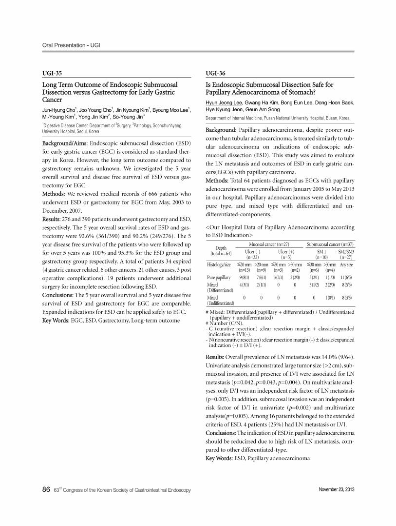
Oral Presentation - UGI
86 63rd Congress of the Korean Society of Gastrointestinal Endoscopy November 23, 2013
UGI-35
Long Term Outcome of Endoscopic Submucosal Dissection versus Gastrectomy for Early Gastric Cancer
Jun-Hyung Cho1, Joo Young Cho1, Jin Nyoung Kim1, Byoung Moo Lee1, Mi-Young Kim1, Yong Jin Kim2, So-Young Jin3
1Digestive Disease Center, Department of 2Surgery, 3Pathology, Soonchunhyang University Hospital, Seoul, Korea
Background/Aims: Endoscopic submucosal dissection (ESD)
for early gastric cancer (EGC) is considered as standard ther-
apy in Korea. However, the long term outcome compared to
gastrectomy remains unknown. We investigated the 5 year
overall survival and disease free survival of ESD versus gas-
trectomy for EGC.
Methods: We reviewed medical records of 666 patients who
underwent ESD or gastrectomy for EGC from May, 2003 to
December, 2007.
Results: 276 and 390 patients underwent gastrectomy and ESD,
respectively. The 5 year overall survival rates of ESD and gas-
trectomy were 92.6% (361/390) and 90.2% (249/276). The 5
year disease free survival of the patients who were followed up
for over 5 years was 100% and 95.3% for the ESD group and
gastrectomy group respectively. A total of patients 34 expired
(4 gastric cancer related, 6 other cancers, 21 other causes, 3 post
operative complications). 19 patients underwent additional
surgery for incomplete resection following ESD.
Conclusions: The 5 year overall survival and 5 year disease free
survival of ESD and gastrectomy for EGC are comparable.
Expanded indications for ESD can be applied safely to EGC.
Key Words: EGC, ESD, Gastrectomy, Long-term outcome
UGI-36
Is Endoscopic Submucosal Dissection Safe for Papillary Adenocarcinoma of Stomach?
Hyun Jeong Lee, Gwang Ha Kim, Bong Eun Lee, Dong Hoon Baek, Hye Kyung Jeon, Geun Am Song Department of Internal Medicine, Pusan National University Hospital, Busan, Korea
Background: Papillary adenocarcinoma, despite poorer out-
come than tubular adenocarcinoma, is treated similarly to tub-
ular adenocarcinoma on indications of endoscopic sub-
mucosal dissection (ESD). This study was aimed to evaluate
the LN metastasis and outcomes of ESD in early gastric can-
cers(EGCs) with papillary carcinoma.
Methods: Total 64 patients diagnosed as EGCs with papillary
adenocarcinoma were enrolled from January 2005 to May 2013
in our hospital. Papillary adenocarcinomas were divided into
pure type, and mixed type with differentiated and un-
differentiated-components.
<Our Hospital Data of Papillary Adenocarcinoma according to ESD Indication>
Depth(total n=64)
Mucosal cancer (n=27) Submucosal cancer (n=37)
Ulcer (-) (n=22)
Ulcer (+) (n=5)
SM 1(n=10)
SM2/SM3(n=27)
Histology/size ≤20 mm(n=13)
>20 mm(n=9)
≤30 mm(n=3)
>30 mm(n=2)
≤30 mm(n=6)
>30 mm(n=4)
Any size
Pure papillary 9 (8/1) 7 (6/1) 3 (2/1) 2 (2/0) 3 (2/1) 1 (1/0) 11 (6/5)
Mixed (Differentiated)
4 (3/1) 2 (1/1) 0 0 3 (1/2) 2 (2/0) 8 (5/3)
Mixed(Undifferentiated)
0 0 0 0 0 1 (0/1) 8 (3/5)
# Mixed: Differentiated(papillary + differentiated) / Undifferentiated (papillary + undifferentiated)
# Number (C/N).- C (curative resection) ;clear resection margin + classic/expanded
indication + LVI(-).- N(noncurative resection) ;clear resection margin (-) ± classic/expanded
indication (-) ± LVI (+).
Results: Overall prevalence of LN metastasis was 14.0% (9/64).
Univariate analysis demonstrated large tumor size (>2 cm), sub-
mucosal invasion, and presence of LVI were associated for LN
metastasis (p=0.042, p=0.043, p=0.004). On multivariate anal-
yses, only LVI was an independent risk factor of LN metastasis
(p=0.005). In addition, submucosal invasion was an independent
risk factor of LVI in univariate (p=0.002) and multivariate
analysis(p=0.005). Among 16 patients belonged to the extended
criteria of ESD, 4 patients (25%) had LN metastasis or LVI.
Conclusions: The indication of ESD in papillary adenocarcinoma
should be reducirsed due to high risk of LN metastasis, com-
pared to other differentiated-type.
Key Words: ESD, Papillary adenocarcinoma
Oral Presentation - UGI
November 23, 2013 63rd Congress of the Korean Society of Gastrointestinal Endoscopy 87
UGI-37
Is New Criteria for Mixed Histology Is Necessary for Endoscopic Resection in Early Gastric Cancer?
Hong Jin Yoon1,4, Yong Hoon Kim1,4, Jie-Hyun Kim1,4, Yong Chan Lee1, HyunKi Kim2, Hoguen Kim2, Jae Jun Park1,4, Young Hoon Youn1,4, Hyojin Park1,4, Sung Hoon Noh3, Seung Ho Choi3,4 Department of 1Internal Medicine, 2Pathology, 3Surgery, 4Gangnam Severance Hospital, Yonsei University College of Medicine, Seoul, Korea
Background/Aims: Mixed histology has been reported to show
more aggressive behavior than other histologies in early gastric
cancer (EGC). We also reported that signet ring cell (SRC)
mixed histology showed more submucosal invasion and higher
lymph node metastasis (LNM) than others. However, there are
no individual criteria for endoscopic resection (ER) in mixed
histology. The aim of study was to investigate whether new cri-
teria for mixed histology is necessary for ER in EGC.
Methods: From January 2005 to December 2012, 3,419 pa-
tients with EGC underwent surgery. Lesions were classified in-
to three histological classifications: Japanese classification,
WHO classification including SRC mixed histology, and
Lauren classification. Mixed type in Lauren classification was
reclassified into four types according to the proportion of dif-
ferentiated and undifferentiated components. Clinicopathologic
characteristics were compared according to histological classi-
fications, special reference to ER criteria.
Results: 179 (5.2%) lesions were classified as mixed type in
Lauren classification. Among them, 54 (30.2%) lesions were
differentiated- predominant mixed type. 361 (10.6%) lesions
were classified as SRC mixed histology. Mixed type lesions in
Lauren classification and SRC mixed lesions were significantly
associated with larger size and higher LNM than others.
Among the lesions meeting ER criteria, 20 and 55 lesions were
categorized into mixed type in Lauren classification and SRC
mixed histology, respectively. However, there was no LNM
among the lesions recategorized into mixed histology.
Conclusions: Mixed histology in EGC showed higher LNM
than other histologies. However, there was no LNM among the
lesions meeting the present ER criteria.
Key Words: EGC, ESD, Mixed histology
UGI-38
Retrograde Endoscopic Submucosal Dissection of Gastric Neoplasm Involving the Pyloric Channel
So Yeon Kim, Jung Wan Choe, Won woo Lee, Sung Ho Kim, Moon Kyung Joo, Beom Jae Lee, Jong-Jae Park, Jae Seon Kim, Young-Tae BakDepartment of Internal Medicine, Korea University Colleage of Medicine, Seoul, Korea
Background/Aims: Recently, successful resection of the pyloric
tumors by ESD using retroflexion in the duodenum has been
reported. However, the detailed dissection method of the pro-
cedure was not well defined yet. We also had done the retro-
flexion ESD of dissecting retrogradely from the duodenum to
the pyloric channel of duodenal part, after then antegradely
from the antrum to the pyloric channel. Since February 2012,
we introduced a new retroflexion method of retrogradely dis-
secting mucosa from duodenum to trans-pyloric antrum as ex-
tensively as possible. We studied the feasibility and effective-
ness of the new retrograde prepyloric antral dissection using
retroflexion ESD.
Methods: 61 patients with gastric neoplasm involving the py-
loric channel underwent ESD from January 2007 to March
2013. In 27 patients, the conventional anterograde ESD were
performed without using the endoscope retroflexed in the
duodenum. The other 34 patients were procedured by the ret-
roflexion method. Among that 34, the latest 16 patients under-
went the new retrograde prepyloric antral dissection method.
We retrospectively analyzed the procedure times, en bloc rates,
complete resection rates and complications associated with
each different techniques.
Results: In newly attempted retrograde prepyloric antral dis-
section method group, en bloc resection and complete re-
section rate were 100% and 94%. The average time of proce-
dure was 42min. Compared with the previously procedured
retroflexion ESD, the new technique had an advantages of sav-
ing procedure time (42min vs 48min, p=0.035). But there were
no statically significant differences at en-block resection and
complete resection (100% vs 94%, p=0.054/ 94% vs 83%,
p=0.059.
Conclusions: This new retrograde prepyloric antral dissection
method for resecting tumors involving the pyloric channel tu-
mor is feasible and effective therapeutic ESD method. So it
should become the standard treatment in such cases, based on
the favorable outcomes.
Key Words: Pyloric channel, Endoscopic submucosal dis-
section, Retroflexion, Retrograde
Oral Presentation - UGI
88 63rd Congress of the Korean Society of Gastrointestinal Endoscopy November 23, 2013
UGI-39
Multiplicity at the Initial Endoscopic Resection as a Prognostic Factor for Recurrence
Tae-Geun Gweon, Jae Myung Park, Chul-Hyun Lim, Jin Su Kim, Yu Kyung Cho, In Seok Lee, Sang Woo Kim, Myung-Gyu Choi, Kyu Yong Choi Department of Internal Medicine, The Catholic University of Korea College of Medicine, Seoul, Korea
Background and Aims: Multiple gastric cancers can be found
in up to 14% after endoscopic resection. It is not known if the
gastric remnant after the initial endoscopic resection for mul-
tiple gastric neoplasia has a greater risk for metachronous
neoplasia. The purpose of this study was to determine the in-
cidence of metachronous neoplasia in this patient subset com-
pared with that after solitary cancer in the initial gastric
neoplasia.
Patients and Methods: Data on a consecutive series of 1,281
patients who underwent endoscopic resection for early gastric
cancer or dysplasia from 2004 to 2012 were analyzed retrospectively.
Multiplicity was defined as more than two apart lesions at ini-
tial endoscopic resection. Clinicopathologic characteristics
were compared between the patients with single and multiple
neoplasia. Recurrence was evaluated with the follow-up endo-
scopic examination for 5 years after the initial treatment.
Results: Among 1,218 patients, 439 (%) had cancer and 130
(10.7%) had multiple lesions at the initial endoscopic
resection. Recurrence with adenoma or cancer was observed
in 113 patients (%) and the risk of this recurrence was higher
in the patients with multiple lesions than single lesion at the
initial treatment (p<0.01). Patients with single cancer lesion
had a significantly lower recurrence rate than those with can-
cer and the other cancer/dysplasia at the resection (p<0.01).
Recurrence risk was not significantly different between the pa-
tients with multiple and single adenoma (p=0.06). Univariate
analysis showed that age, cancer pathology, and lesion multi-
plicity were significantly associated with recurrence. Multivariate
analysis showed that age (p=0.03; odds ratio, 1.02) and multi-
plicity (p<0.01; odds ratio, 2.10) were independent risk factors
for recurrence.
Conclusions: Multiplicity can be a prognostic factor after en-
doscopic resection of gastric neoplasia, especially initial cancer.
Key Words: Stomach neoplasms , Recurrence, Endoscopic re-
section
UGI-40
Clinical Outcomes of Endoscopic Submucosal Dissection for Early Gastric Cancers in Elderly People
Yup Hwangbo, Seong Woo Jeon, Hyun Seok Lee, Yong Hwan Kwon, Jun Young Choi, Se Young Jang, Chung Hoon Yu Department of Internal Medicine, Kyungpook National University School of Medicine, Daegu, Korea
Background and Aims: Endoscopic submucosal dissection
(ESD) has been widely accepted for treatment of early gastric
cancer (EGC) in Korea. Elderly patients often have surgical op-
erative risks due to disease, and the feasibility of this treatment
for such patients will improve the quality of life. Our study in-
vestigated whether ESD was suitable of elderly people with ear-
ly gastric cancers.
Patients and Methods: We studied 845 early gastric cancers that
were treated with ESD at Kyungpook National University of
Medicine between 2005 and 2013. The patients were classified
into an elderly group (age≥75years) and a nonelderly group
(age<75years). We compared the characteristics of patients
and lesions, curative resection rate and the complications be-
tween the two groups. Also we analyzed long term outcomes
among elderly group.
Results: The elderly group included 108 patients, and the non-
elderly group included 737patients. The average age of the eld-
erly group of patients was 78.2. The incidence of comorbid dis-
ease were significantly higher in the elderly group. The curative
resection rate in the elderly group was 81.5%. Bleeding and
perforation rates were 4.6% and 0%, respectively. The curative
resection and the complication rate for the elderly patients
were not significantly different from those of nonelderly
patients. Among elderly group, 88 patients had a curative re-
section patients, 14 patients had non-curative resection with-
out surgery and 6 patients had non-curative resection with
surgery. During follow-up period, recurrence rate was 7.9%,
14.3%, 0%, respectively and mortality rate was 14.8%, 14.3%,
0% respectively.
Conclusions: This study demonstrates the efficacy of ESD in
both elderly and nonelderly patients. ESD is a safe and mini-
mally invasive treatment for elderly patients with early gastric
cancers.
Key Words: Early gastric cancer, Endoscopic submucosal dis-
section
Oral Presentation - UGI
November 23, 2013 63rd Congress of the Korean Society of Gastrointestinal Endoscopy 89
UGI-41
Therapeutic Effect of Endoscopic Enucleation for Gastric Subepithelial Tumors Originated from Muscularis Propria
Gyung Eun Kim, Hyungkil Kim, Hyun Jung Chung, Byoungwook Bang, Kyesook Kwon, Yong Woon Shin Department of Internal Medicine, Inha University School of Medicine, Incheon, Korea
Background and Aim: Most of small gastric subepithelial tu-
mors (SETs) originated from muscularispropria have clinical
importance because they have higher malignant potential, es-
pecially gastointesintal stromal tumors (GISTs).Endoscopic
enucleationfor the resection of SETs may be considered useful
treatment option to replace conventional surgery or other
treatment modality. The purpose of the present study is to
demonstrate the feasibility of endoscopic enucleation for gas-
tric SETs and to evaluate its efficacy and safety.
Patients and Methods: From January 2010 to June 2013, a total
thirty eight patients with gastric SETs underwent the endo-
scopic enucleationfor the gastric SETs. Endoscopic enucleation
was attempted by using insulted-tip knife or air- driven band
ligator. All patients performed endoscopic ultrasound (EUS)
to determine the layer of origin and the accurate size of the
SETs.The EUS finding, procedure technique and histologic
findings were analyzed in this retrospective study.
Results: A total 38 patients (15 men, 23 women; mean age
50.34 years) were eligible for inclusion in the study. All SETs
were originated from the muscularispropria layer, detected by
preperformedl EUS. The mean procedure time was 38.8 mini-
tues (range 3.0- 260) and mean size of the tumors was 15.6mm
(range 6-50). The histologic diagnosis was leiomyoma for 24
lesions and gastrointestinal stromal tumor for 14 lesions,
which showed ten very low risk , two low risk, one high risk and
one intermediate risk. The complete resection rate of leiomyo-
ma was 70.8% (17/24) and complete resection rate of GIST was
42.8% (6/14). Three small perforations occurred but was man-
aged successfully by endoscopic clip application.
Conclusion: The endoscopic enucleation is an safe and effec-
tive treatment method for gastric SETs originating in muscu-
larispropria layer.
Key Words: Gastric subepithelial tumor, Enucleation, Muscularis
propria tumor, Endoscopic resection, Endoscopic ligation
UGI-42
Clinical Factors That Should Be Considered in Patients with Gastric Hyperplastic Polyps When Treated by Polypectomy
Se Woong Hwang, Hyun Yong Jeong, Jae Kyu Sung, Hee Seok Moon, Sun Hyung Kang, Hye Jin Kim, Se Young Park, Beom Yong Yoon Department of Internal Medicine, School of Medicine, Chungnam National University, Daejeon, Korea
Background/Aims: Hyperplastic polyps are the most common
variety of gastric polyps that constitute 30-93% of all benign
epithelial gastric polyps. The overall prevalence of dysplasia in
hyperplastic polyps is believed to be <2%, and more frequent
in large polyps (>2cm). We aimed to identify the clinical fea-
tures of hyperplastic polyps that undergoing neoplastic
transformation.
Methods: Between June 2011 and June 2013, a total of 298 hy-
perplastic polyps, which had been removed by endoscopic pol-
ypectomy in 207 patients, were analyzed retrospectively.
Results: Neoplastic transformations were found in 12 cases
(3.9%) including 7 cases of adenoma (2.3%), 4 cases of ad-
enocarcinoma (1.3%) and 1 case of neuroendocrine tumor
(0.3%). Neoplastic transformation was significantly associated
with age (hyperplastic polyp group 60.1±11.8 vs. neoplastic
transformation group 68.8±10.3, p=0.032) and with sessile
shape (n=10 [83.3%] vs. n=148 [51.7%], p=0.038). However,
no significant difference was found between these groups in
terms of sex, presence of Helicobacter pylori, size, location,
number of polyps detected per patient, and endoscopic ap-
pearance such as presence of hyperemia, nodular changes and
erosions.
Conclusion: Hyperplastic polyps with or without neoplastic
transformations were virtually impossible to distinguish by en-
doscopic findings. However, compared to hyperplastic polyps
without neoplastic transformation, those with neoplastic
transformation were to be more sessile and the ages of the
group were older. Therefore, in case of endoscopic poly-
pectomy in elderly patients with hyperplastic polyps which be-
ing sessile form, it is necessary to pay detailed attentions to the
possibility of a neoplastic transformation.
Key Words: Stomach, Hyperplastic, Polyps, Neoplastic, Trans-
formation
Oral Presentation - UGI
90 63rd Congress of the Korean Society of Gastrointestinal Endoscopy November 23, 2013
UGI-43
Intermediate Follow Up Results of Endoscopic Resection for Gastric GIST
Jin Nyoung Kim, Joo Young Cho, Jun-Hyung Cho, Mi-Young Kim, Byoung Moo Lee Digestive Disease Center, Soonchunhyang University Hospital, Seoul, Korea
Backgrounds & Aim: The diagnosis of Gastrointestinal stromal
tumor (GIST) must always be investigated since every GIST is
potentially malignant. Curative treatment requires a complete
gross resection of the tumor. Both open and minimally in-
vasive operations have been shown to reduce recurrence rates
and improve long-term survival. Advances in endoscopic tech-
nology have allowed some to perform attempted excision of
these lesions endoscopically. The oncologic implications of
such an approach remain unclear. The aim of our study is to in-
vestigate the clinical outcomes of endoscopic resection of GIST
in the upper GI tract and evaluate the safety and feasibility.
Methods: To assess whether endoscopic resection is feasible ap-
proach to cure GIST, the authors performed a retrospective co-
hort study in single endoscopic center. A total 19 patients with
gastric GIST identified by endoscopic ultrasonography were
collected from November 2008 to June 2013.
Results: Of the 19 GISTs, 4 were located at the cardia, 9 at the
fundus, 5 at the body, 1 at the antrum. Endoscopic resection was
performed by hybrid natural orifice translumenal endoscopic
surgery (NOTES) (n=15), submucosal tunneling endoscopic re-
section (n=4). Mean tumor size was 23.7 mm (12-42 mm). The
13 cases had low-risk mitosis, 2 cases had intermediate-risk mi-
tosis, and 4 cases had high-risk mitosis. The average of total hos-
pital stay was 9.1 days. The average hospital stay after the proce-
dure was 5.8 days. There were no complications including severe
bleeding during the procedure. Complete resection was ob-
tained in 15 cases. All patients were followed up for 27 months
(range, 3-54 months). There was no recurrence of tumor.
Conclusion: Endoscopic resection of GIST shows promise as a
safe and feasible technique. Clinical studies with more subjects
and longer follow up are needed.
Key Words: GIST, Endoscopic resection, NOTES
UGI-44
Pneumonia after Endoscopic Resection for Gastric Neoplasm
Eun Jeong Gong, Do Hoon Kim, Hyun Lim, Ji Yong Ahn, Kwi-Sook Choi, Jeong Hoon Lee, Kee Don Choi, Ho June Song, Gin Hyug Lee, Hwoon-Yong Jung, Jin-Ho Kim Departments of Gastroenterology, University of Ulsan College of Medicine, Asan Medical Center, Seoul, Korea
Background: Pneumonia following endoscopic procedures
may affect the clinical course of the treatment and prolong hos-
pital stay. We therefore investigated the incidence and risk fac-
tors for pneumonia after endoscopic resection (ER) for gastric
neoplasm.
Methods: Subjects who underwent ER for gastric neoplasm at
the Asan Medical Center from January 1997 to March 2013
were included. To investigate risk factors, the control group
was randomly selected from these subjects.
Results: Of the 7,149 subjects who underwent ER for gastric
neoplasm, 44 (0.62%) developed pneumonia. The median age
was 68 years (range: 31-82), and the male to female ratio was
3:1. Twenty-five patients (56.8%) were smokers and 8 (18.2%)
had underlying pulmonary diseases. The median procedure
time was 23 minutes (range: 2-126). Compared with the con-
trol group, smoking status (current smoker vs. never smoker,
Odds ratio [OR] 2.031, p=0.046), total procedure time (OR
1.011, p=0.048), and hemostasis time (OR 1.026, p=0.028)
were risk factors for the development of pneumonia. In multi-
variate analysis, age >65 years (OR 2.073, p=0.031), smoking
(OR 2.324, p=0.023), and hemostasis time (OR 1.025,
p=0.038) were independent risk factors. All patients recovered
from pneumonia, and the duration of hospital stay did not dif-
fer between patients with pneumonia and the control group
(p=0.077).
Conclusions: While old age, smoking, and hemostasis time
were risk factors for pneumonia, its incidence after ER was not
associated with clinically significant adverse outcomes.
Key Words: Pneumonia, Endoscopic resection, Gastric
Neoplasm
Oral Presentation - UGI
November 23, 2013 63rd Congress of the Korean Society of Gastrointestinal Endoscopy 91
UGI-45
What Is Tailored Minimally Invasive Treatment of Early Gastric Cancer beyond Endoscopic Submucosal Dissection?
Mi-Young Kim1, Jun-Hyung Cho1, Yong Jin Kim2, So Young Jin3, Joo Young Cho1
1Digestive Disease Center, Department of 2Surgery and 3Pathology, Soonchunhyang University Hospital, Seoul, Korea
Background/Aims: The aim of this study was to evaluate the
outcomes of combined endoscopic submucosal dissection
(ESD) with sentinel node navigation surgery (ESN) or laparo-
scopic lymph node dissection (LLND) and endoscopic
full-thickness resection (EFTGR) with laparoscopic regional
lymph node dissection (hybrid natural orifice transluminal en-
doscopic surgery, hybrid NOTES) for early gastric cancer
(EGC).
Methods: This is a retrospective analysis using prospectively
collected data at a single tertiary referral center. A total of 80
patients with EGC underwent combined ESD with ESN or
LLND and hybrid NOTES between February 2007 and January
2013.
Results: The curative resection rate of all cases was 86.3% (ESN
73.9% vs. ESD with LLND 100% vs. hybrid NOTES 79.2 %, re-
spectively). Histologically, 33 cases were mucosal cancers, and
45 were submucosal cancers. There were 50 undifferentiated
cancers. The median tumor size was 2.3 cm (range, 0.6-7 cm)
in long diameter. Lymphovascular invasion was found in 27
cases with 3 lymph node metastasis. Incomplete resection was
shown in 11 (8 tumor-positive lateral margins and 3 tu-
mor-positive vertical margins). Eight patients underwent addi-
tional gastrectomy because of tumor-positive vertical margins
or treatment-related complications. During the median fol-
low-up of 23 months (range, 3-73 months), none showed local
recurrence or lymph node metastasis.
Conclusion: ESN, combined ESD with LLND and hybrid
NOTES showed favorable pathologic and clinical outcomes.
They could be utilized as a bridge between ESD and gas-
trectomy in selected patients with a risk of lymph node
metastasis.
Key Words: Sentinel node navigation surgery, ESD, Hybrid
NOTES, Laparoscopic lymph node dissection
UGI-46
Clinical Attention Points Drawn by the Cases with Non-Neoplastic Pathologic Results after ESD for GEDs or EGCs
Min Jae Yang, Sung Jae Shin, Ki Seong Lee, Eun Kyung Kim, Sun Gyo Lim, Ki Myung Lee Department of Gastroenterology, Ajou University School of Medicine, Suwon, Korea
Background: Endoscopic submucosal dissection (ESD) has
been developed as a standard treatment modality for selected
cases of gastric epithelial dysplasias (GEDs) or early gastric
cancers (EGCs). However, many endoscopists have faced para-
doxical circumstances that ESD specimens revealed non-neo-
plastic results, with reported rates ranging 3.2-7%. The aims of
this study were to find out additional evidences of complete
forceps biopsy removal of neoplastic foci, and draw clinical
points about performing endoscopic forcep biopsy (EFB) be-
fore ESD, and establishing surveillance strategy for the negative
pathology group.
Methods: Between February 2005 and December 2011, we ret-
rospectively reviewed the prospectively collected ESD results
from the endoscopic database system, and found 46 cases
(3.7% of total ESD cases) who were confirmed to have
non-neoplastic results on the final specimens.
Results: Mean tumor sizes and sampling ratios were 9.3±2.5
mm (4-16mm) and 2.98±1.56 mm/fragment, respectively. 4
cases (8.4%) showed notable morphologic changes before and
after EFB, and required reclassifications of the initial morpho-
logic diagnosis (2 : IIa->IIb, 1: IIa->IIa+IIc, 1: IIa+IIc->IIa+III).
8 cases (16.8%) showed partial non-lifting signs suggestive of
severe submucosal fibrosis. We have ever experienced 1 case of
wrong localization of ESD site at 3 month after ESD, and 2
metachronous cancerous recurrences at 12, 27 month over the
surveillance period (mean 23.8±18.9 months, range 6-75).
Conclusion: Small tumor size, low sampling ratio, and change
of morphologic type before and after EFB may be endoscopic
evidences of complete biopsy removal of neoplastic foci. The
number of biopsy fragment for suspicious dysplastic lesion
could be determined in considering reasonable sampling ratio
to prevent the adverse submucosal fibrosis. Cautious surveil-
lance for wrong localization of ESD site or metachronous re-
currence is needed in the non-neoplastic group.
Key Words: ESD, Non-neoplastic pathology, Biopsy removal,
Sampling ratio
Oral Presentation - UGI
92 63rd Congress of the Korean Society of Gastrointestinal Endoscopy November 23, 2013
UGI-47
Long-Term Clinical Outcomes of Endoscopic Submucosal Dissection for Early Gastric Cancer
Jeongmin Choi, Sang Gyun Kim, Jong Pil Im, Joo Sung Kim and Hyun Chae Jung Department of Internal Medicine and Liver Research Institute, Seoul National University College of Medicine, Seoul, Korea
Background and Aims: We aimed to evaluate the long-term
clinical outcomes for endoscopic submucosal dissection (ESD)
for early gastric cancer (EGC).
Methods: From 2005 through 2011, 961 EGC patients were
treated with ESD. Patients were grouped as standard and ex-
panded indications based on Japanese ESD guideline, while
those who did not meet expanded indication were regarded as
beyond expanded group. Curative resection rate was compared.
Patients who underwent additional surgery due to non-cura-
tive resection (n=78) or follow-up loss (n=10) were excluded.
Thereafter, 873 patients were enrolled for long-term analysis of
local tumor recurrence, development of metachronous can-
cers, overall and disease-free survival rates.
Results: Curative resection rate was significantly higher in the
standard group than in expanded and beyond expanded
group; 94.9% for standard, 87.1% for expanded, 29.1% for be-
yond expanded groups. During a median follow-up of 45
months, local tumor recurrence rate was significantly higher in
the beyond expanded group than in the standard and ex-
panded group (1.9%, 1.8%, 7.8%, respectively). Metachronous
cancer incidence did not differ significantly between the 3
groups (6.5%). Two thirds of recurred or metachronous tu-
mors were treated with ESD. Lymph node metastasis devel-
oped in 0.6%. The 5-year overall survival rate was 94.8%
(96.6%, 94.2%, 84.4%, p=0.001) and disease-free survival rate
was 99.1% (100%, 99.3%, 92.8%, p=0.001).
Conclusions: ESD was effective with favorable long-term clin-
ical outcomes for EGC only in patients who met the standard
and expanded indications. Conversely, beyond expanded
group gave less favorable clinical outcomes. Careful surveil-
lance after ESD should be taken for all patients.
Key Words: Gastroscopic surgery, Stomach neoplasm, Therapy,
Treatment outcome, Survival
UGI-48
Long-Term Outcomes of Noncurative Resection after Endoscopic Submucosal Dissection for Early Gastric Cancer
Jae Pil Han1, Su Jin Hong1, Hee Kyoung Kim2, Bong Min Ko1, Moon Sung Lee1 Digestive Disease Center and Research Institute, Department of 1Internal Medicine and 2Pathology, Soonchunhyang University School of Medicine, Bucheon, Korea
Aims: Noncurative resection after endoscopic submucosal dis-
section (ESD) for early gastric cancer (EGC) can attribute to
local recurrence or lymphatic and distant metastasis of tumor.
This study aimed to evaluate the long-term outcomes of non-
curative resection after ESD for EGC and determine the treat-
ment according to histological result from ESD.
Methods: Of 892 EGCs treated with ESD between 2001 and
2012, 152 (17.0%) were determined by histology to show non-
curative resection after ESD. Factors of noncurative resection
included tumors with margin positivity, lymphovascular in-
vasion, and beyond the absolute and expanded criteria.
Results: Among 152 EGCs, 17 were lost to follow up without
additional treatment. Of 135 noncurative resections, 45
(33.3%) were interpreted as only lateral margin positivity, 6
(4.4%) as only vertical margin positivity, and 84 (62.3%) did
not meet the absolute and expanded criteria by histology after
ESD. Additional endoscopic treatment was performed in 14
(29.2%) of 45 with only lateral margin positivity. They had no
local recurrence during follow-up period. Additional surgery
was performed in 44 (48.9%) of 90 with high risk factors of
lymph node, such as vertical margin positivity or no fulfillment
of absolute and expanded criteria. Among them, local re-
currence occurred in 1 incomplete resection by surgery during
the follow-up period after additional treatment. The histo-
logical results from 44 surgical specimens included 17 (38.6%)
residual tumor and 3 (6.8%) lymph node metastasis.
Conclusions: The risk of lymph node metastasis based on his-
tological results should be evaluated for decision of additional
treatment after ESD for EGC. Additional surgery is necessary
for the patients with high risk of lymph node metastasis, and
endoscopic treatment can be helpful in those with low risk.
Key Words: Early gastric cancer, Endoscopic submucosal dis-
section, Noncurative resection
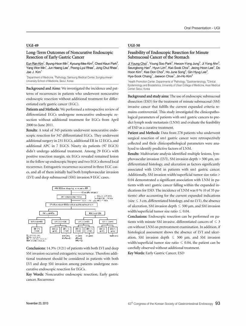
Oral Presentation - UGI
November 23, 2013 63rd Congress of the Korean Society of Gastrointestinal Endoscopy 93
UGI-49
Long-Term Outcomes of Noncurative Endoscopic Resection of Early Gastric Cancer
Eun Ran Kim1, Byung-Hoon Min1, Kyoung-Mee Kim2, Cheol Keun Park2, Yang Won Min1, Jun Haeng Lee1, Poong-Lyul Rhee1, Jong Chul Rhee1, Jae J. Kim1 1Department of Medicine, 2Pathology, Samsung Medical Center, Sungkyunkwan University School of Medicine, Seoul, Korea
Background and Aims: We investigated the incidence and pat-
terns of recurrences in patients who underwent noncurative
endoscopic resection without additional treatment for differ-
entiated early gastric cancer (EGC).
Patients and Methods: We performed a retrospective review of
differentiated EGCs undergone noncurative endoscopic re-
section without additional treatment for EGCs from April
2000 to June 2011.
Results: A total of 345 patients underwent noncurative endo-
scopic resection for 347 differentiated EGCs. They underwent
additional surgery in 231 EGCs, additional ER in 12 EGCs, and
additional APC in 7 EGCS. Ninety six patients (97 EGCS)
didn’t undergo additional treatment. Among 29 EGCs with
positive resection margin, six EGCs revealed remained lesion
in the follow up endoscopic biopsy and two EGCs showed local
recurrence. Extragastric recurrence occurred in three EGC cas-
es, and all of them initially had both lymphovascular invasion
(LVI) and deep submucosal (SM) invasion.9 EGC cases.
Conclusions: 14.3% (3/21) of patients with both LVI and deep
SM invasion occurred extragastric recurrence. Therefore addi-
tional treatment should be considered in patients with both
LVI and deep SM invasion among patients undergone non-
curative endoscopic resection for EGCs.
Key Words: Noncurative endoscopic resection, Early gastric
cancer, Recurrence
UGI-50
Feasibility of Endoscopic Resection for Minute Submucosal Cancer of the Stomach
Ji Young Choi1, Young Soo Park2, Hwoon-Yong Jung3, Ji Yong Ahn3, Seungbong Han4, Hyun Lim3, Kwi-Sook Choi3, Jeong Hoon Lee3, Do Hoon Kim3, Kee Don Choi3, Ho June Song3, Gin Hyug Lee3, Hye-Sook Chang1, Jaewon Choe1, Jin-Ho Kim3 1Health Promotion Center, Departments of 2Pathology, 3Gastroenterology, 4Clinical Epidemiology and Biostatistics, University of Ulsan College of Medicine, Asan Medical Center, Seoul, Korea
Background and study aims: The use of endoscopic submucosal
dissection (ESD) for the treatment of minute submucosal (SM)
invasive cancer that fulfills the current expanded criteria re-
mains controversial. This study investigated the clinicopatho-
logical parameters of patients with sm1 gastric cancers to pre-
dict lymph node metastasis (LNM) and evaluate the feasibility
of ESD as a curative treatment.
Patient and Methods: Data from 278 patients who underwent
surgical resection of sm1 gastric cancer were retrospectively
collected and their clinicopathological parameters were ana-
lyzed to identify predictive factors of LNM.
Results: Multivariate analysis identified multiple lesions, lym-
phovascular invasion (LVI), SM invasion depth > 500 μm, un-
differentiated histology, and ulceration as factors significantly
associated with LNM in patients with sm1 gastric cancer.
Additionally, SM invasion width/superficial tumor size ratio >
0.04 demonstrated a significant association with LNM in pa-
tients with sm1 gastric cancer falling within the expanded in-
dications for ESD. The incidence of LNM was 0 % (0 of 35 pa-
tients) after accounting for the current expanded indications
(size ≤ 3 cm, differentiated histology, and no LVI), the absence
of ulceration, SM invasion depth ≤ 500 μm, and SM invasion
width/superficial tumor size ratio ≤ 0.04.
Conclusions: Endoscopic resection can be performed on pa-
tients with minute SM invasive, differentiated cancers of ≤ 3
cm without LNM on pretreatment examination. In addition, if
histological assessment shows the absence of LVI and ulcer-
ation, SM invasion depth ≤ 500 μm, and SM invasion
width/superficial tumor size ratio ≤ 0.04, the patient can be
carefully observed without additional treatment.
Key Words: Early Gastric Cancer, ESD
Oral Presentation - UGI
94 63rd Congress of the Korean Society of Gastrointestinal Endoscopy November 23, 2013
UGI-51
Factors Associated with Successful Endoscopic Submucosal Dissection in Pyloric Neoplasms: Location and Direction
Jung Ho Bae, Gwang Ha Kim, Bong Eun Lee, Dong Yup Ryu, Geun Am Song Department of Internal Medicine, Pusan National University School of Medicine, Busan, Korea
Background and Aims: Pyloric neoplasms have been consid-
ered as technically difficult lesions for successful endoscopic
submucosal dissection (ESD). The anatomical features of pylo-
rus can adversly affect the assessment of tumor margin and
ESD procedure, consequently piecemeal resection and compli-
cation rates in pyloric neoplasms is higher compared with oth-
er sites of gastric neoplasms.
Patients and Methods: A total 158 pyloric adenomas and early
cancers resected by ESD at our hospital from January 2007 to
March 2013 were reviewed retrospectively. The location type
was defined according to distance from pyloric ring: prepyloric
ring (distal margin of the tumor was located <1cm from py-
loric ring); pyloric ring (<0.5 cm from pyloric ring); post-
pyloric ring (any invasion to duodenum side beyond pyloric
ring). A clock-face orientation with the endoscope (with the
lesser curve of the stomach in contiguity with the 12 o’clock
orientation of pylorus) was used to characterize directional
distribution.
Results: Complete resection rates differed significantly in rela-
tion to location (prepyloric ring vs pyloric ring vs postpyloric
ring, 93% vs 74% vs 62%, respectively; p=0.001) and direc-
tional distribution (upper hemisphere vs lower hemisphere,
76% vs 92%, respectively; p=0.011) and circumference extent
of pylorus mucosal resection (<50% vs >50%, 94% vs 76%, re-
spectively; p=0.001). The rates of delayed bleeding, stricture
and perforation were 7.0%, 1.3% and 0.6% after ESD, and
there was no surgery related to complications. On multivariate
analysis, close distance to pyloric ring and upper hemisphere
distribution and large tumor size were significantly relevant for
incomplete resection.
Conclusion: ESD of early pyloric neoplasms is effective and
feasible, but endoscopists require advanced technique and
more experience in the case of neoplasms located in closely to
pyloric ring, upper hemisphere and large size because of low
complete resection rates and high complications.
Key Words: Pyloric neoplasm, Pylorus, ESD
UGI-52
Preliminary Experience of Endoscopic Submucosal Tunnel Dissection for Exophytically Growing Gastric SMT
Jung Wan Choe, So Yeon Kim, Won Woo Lee, Sung Ho Kim, Moon Kyung Joo, Beom Jae Lee, Jong-Jae Park, Jae Seon Kim, Young-Tae BakDepartment of Internal Medicine, Korea University School of Medicine, Seoul, Korea
Objective: Although ESD is regarded recently as minimally in-
vasive resection methods for SMTs, it can be associated with
the major complication of perforations, especially in the exo-
phatic mass. But ESTD can be closed more easily by endoclips
compared with ESD. So, we aim to present the effectiveness
and safety of a novel ESTD method for resection of gastric
SMTs originating from the muscularis propria(MP).
Methods: In 6 patients who presented with a SMT located in
the stomach, we underwent submucosal tunnelling endoscopic
resection between August 2011 and February 2012. A sub-
mucosal tunnel was endoscopically created by starting approx-
imately 3cm distant to the lesion. After careful submucosal dis-
section of the tumor from the surrounding submucosal tissue
and the unaffected MP layer, the SMTs were completely re-
moved by the technique of endoscopic submucosal dissection.
The mucosal entrance of the tunnel was closed using endoclips
after the tumor was removed.
Results: SMTs had a mean size of 21.1mm (range 17-25mm); 4
were located in the antrum and 2 in the cardia. SMT resection
was successful in all patients with en bloc resection 100% rate
(2 lesions affected the deep MP, so complete MP resection was
performed; 4 lesions affected the superficial MP for a partial
MP resection). The mean procedure time was 64 minutes
(50-80 minutes). One patient had lost the full thickness re-
sected sample in the peritoneal space. So the endoscopic proce-
dure was converted into laparoscopic surgery. The other five
patients had no any complications. No residual tumor or tu-
mor recurrence were detected during the follow-up period.
Pathological diagnoses of these tumors were low risk gastro-
intestinal stromal tumors (4/6), a schwannoma (1/6), and a
etopic pancreas (1/6).
Conclusion: In this study, endoscopic submucosal tunnel dis-
section (ESTD) was appeared to be effective and safe endo-
scopic procedure to remove exophatically growing tumors in
the stomach.
Key Words: ESTD, Submucosal tumor, Tunnelling
Oral Presentation - UGI
November 23, 2013 63rd Congress of the Korean Society of Gastrointestinal Endoscopy 95
UGI-53
Efficacy of Additional CCRT after Non-Curative ESD in Patients with Esophageal Cancer: Single Center Experience
Byoung Moo Lee, Joo Young Cho, Jun-Hyung Cho, Mi-Young Kim, Jin Nyoung Kim Digestive Disease Center, Soonchunhyang University Hospital, Seoul, Korea
Backgrounds & Aim: Endoscopic Submucosal Dissection (ESD)
has been used conventionally to treat superficial esophageal
cancers. But, clinical outcomes with the additional treatment
such as concurrent chemoradiation therapy (CCRT) after the
non-curative resection are remained unclear. We aimed to
evaluate the outcomes of non-curative ESD with additional
CCRT in esophageal cancer in a single center experience in
Korea.
Methods: A retrograde analysis was performed with 30 patients
with superficial esophageal cancer (M and SM1 cancer) who
underwent esophageal ESD from Oct 2005 to Sep 2013 in
Soonchunhyang University Hospital. Patient characteristics,
histologic findings, curative resection rate, additional treat-
ment after the procedure, outcomes after the additional treat-
ment were analyzed. Follow up endoscopy was performed per
6 months, and computed tomography (CT) scan per 12
months.
Results: Of the 30 patients, 7 patients were excluded due to the
histologic findings of high grade dysplasia (HGD), 18 patients
who were revealed to undergo curative resection were also
excluded. A total of 5 patients had non-curative resection; 3
patients had positive resection margins (lateral margin: 2, ver-
tical margin: 1) and 2 patients had lymphatic invasions. All
non-curative patients had squamous cell carcinoma, and tu-
mor depth was to the lamina propria (2/5, 40%), to the muscu-
laris mucosa (1/5, 20%), to the SM1 (submucosal invasion
<200 mm below the muscularis mucosa) in the remaining 2
patients. All 5 patients underwent additional CCRT after ESD.
The mean follow up period after CCRT was 22.2 months
(range 12 to 33 months). And no local recurrence was noted in
any patient at the last follow up.
Conclusion: In our experience, additional CCRT in non-cura-
tive ESD of the esophageal cancer was a useful treatment mo-
dality without local recurrence. But, large scale prospective
studies with more subjects and longer follow up are needed.
Key Words: Esophageal Cancer, ESD, CCRT
UGI-54
Clinical Outcomes of the Endoscopic Treatments for the Early Esophageal Cancer and Dysplasia
Cheol Kim, Su Jin Hong, Jae Pil Han, Bong Min Ko, Moon Sung Lee Digestive Disease Center and Research Institute, Department of Internal Medicine, Soonchunhyang University School of Medicine, Bucheon, Korea
Backgrounds and Aim: Esophageal cancer and dysplasia are
relatively rare disease in Korea, compared with other Far East
countries. So, the feasibility and the safety of the endoscopic
treatments were not well established in Korea. This study
aimed to assess the clinical outcomes of endoscopic treatment
for esophageal epithelial lesions.
Methods: Between August 2001 and August 2013, 25 esoph-
ageal epithelial lesions of 22 patients were treated with endo-
scopic submucosal dissection (ESD) or endoscopic mucosal
resection (EMR).
Results: Of the 25 lesions, 11 lesions were treated with ESD and
14 lesions with EMR. All of them, 15 (60%) of the lesions were
located in lower esophagus, 9 (36%) in mid-esophagus, and 1
(4%) in upper esophagus. ESD group included 7 (64%) squ-
amous cell carcinomas, 3 (27%) high grade dysplasias, and 1
(9%) low grade dysplasia. EMR group included 8 (57%) squ-
amous cell carcinomas, 2 (14%) high grade dysplasias, and 4
(29%) low grade dysplasias. Median procedure time was 74
minutes and 35 minutes in ESD group and EMR group,
respectively. Rates of en bloc resection and curative resection
were 100% and 100% in ESD group, and 64% and 42% in
EMR group. Delayed bleeding occurred in 3 cases (1 in ESD
group, 2 in EMR group). They were managed successfully by
endoscopic procedures. Non-curative resections were found in
4 patients underwent EMR. Three of them received secondary
operation, and other received additional EMR. During fol-
low-up periods (mean, 20.0 months), there was no recurrence
in ESD group, but 3 recurrences in EMR group. Among them,
two patients received operation, and one patient received pho-
todynamic therapy due to poor comorbidities. No cancer re-
lated death occurred in both groups.
Conclusions: Endoscopic treatments for esophageal epithelial
lesions are safe and feasible therapeutic modalities. Especially,
ESD can provide high rate of en bloc and curative resection for
early esophageal cancer and dysplasia.
Key Words: Esophageal cancer, Esophageal dysplasia, Endoscopic
submucosal dissection, Endoscopic mucosal resection.
Oral Presentation - UGI
96 63rd Congress of the Korean Society of Gastrointestinal Endoscopy November 23, 2013
UGI-55
Predictors of Lymph Node Metastasis and Residual Tumor in Non-Curative Endoscopic Resection of Early Gastric Cancer
Hyo-Joon Yang1, Sang Gyun Kim1, Joo Hyun Lim1, Jeongmin Choi1, Jong Pil Im1, Joo Sung Kim1, Woo Ho Kim2, Hyun Chae Jung1
1Department of Internal Medicine and Liver Research Institute, 2Department of Pathology, Seoul National University College of Medicine, Seoul, Korea
Background/Aims: Surgical treatment is recommended after
non-curative endoscopic resection (ER) of early gastric cancer
(EGC). However, only a part of the patients is found to have
lymph node metastasis (LNM) or residual tumor. This study
aimed to identify the risk factors of LNM and residual tumor in
patients received non-curative ER.
Methods: We retrospectively evaluated surgical outcomes and
clinicopathological data of consecutive patients who received
additional surgery after non- curative ER of EGC from April
2005 to July 2013 at Seoul National University Hospital.
Non-curative ER was defined as positive deep resection mar-
gin, lymphovascular invasion, or tumor beyond the expanded
indications.
Results: A total of 1783 patients were treated with ER for EGC,
and non-curative ER was found in 256 (14.4%) patients. Of
these patients, 121 patients underwent additional gastrectomy
and lymph node dissection: 15 (12.4%) had LNM, and 23
(19.0%) had residual cancer. In patients without lymphovas-
cular invasion (LVI), LNM was observed only in sm2 cancers.
However, in those with LVI, LN metastasis was observed even
in mucosal cancer. In multiple logistic regression analysis, ve-
nous invasion (odds ratio [OR], 5.40; 95% confidence interval
[CI], 1.20-24.32; p=0.028) and lower third location (OR,
10.43; 95% CI, 1.21-90.0; p=0.033) were independent risk fac-
tors for LNM, and tumor involvement of lateral resection mar-
gin (OR, 18.20; 95% CI, 3.98-83.30; p<0.001) was independent
risk factor for residual cancer.
Conclusions: Patients with LVI or sm2 cancer might be consid-
ered to receive additional surgery due to the risk of LNM. In
particular, patients with venous invasion, lower third location,
or lateral resection margin involvement may be strongly rec-
ommended to undergo additional surgery for non-curative ER
of EGC.
Key Words: Early gastric cancer, Endoscopic resection, Lymph
node metastasis, Residual tumor, Risk factors
UGI-56
Long-Term Outcome Comparisons between Endoscopic Resection and Surgery for Expanded Indication of Early Gastric Cancer
Young-Il Kim, Il Ju Choi, Chan Gyoo Kim, Soo-Jeong Cho, Jong Yeul Lee, Keun Won Ryu, Myeong-Cheorl Kook, Young-Woo Kim Center for Gastric Cancer, National Cancer Center, Goyang, Korea
Background/Aims: Endoscopic resection (ER) for early gastric
cancer (EGC) lesions has been increasingly performed, but ER
for expanded indication is still considered as an investigational
treatment. The aim of this study was to evaluate the long-term
outcomes after ER for expanded indication in compared with
those after surgery.
Methods: We retrospectively reviewed data of gastric cancer
patients who underwent ER or surgery for EGC meeting the
expanded indication between 2001 and 2009. Multivariate
Cox-proportional hazard regression analyses after adjustments
by age, sex, and propensity score were performed for overall
survival (OS) as primary outcome. Disease-specific survival
(DSS), metachronous gastric cancer rates, and complication
rates were secondary outcomes.
Result: Among 457 patients included, 165 underwent ER and
292 underwent surgery. During a median follow-up of 58.6
months, OS and DSS rates were not different between both
treatment groups (5-year OS rate, 97.5% in ER group and
97.0% in Surgery group, p=0.425 by log-rank test; 5-year DSS
rate 100% in ER group and 99.7% in Surgery group, p=0.366
by log-rank test). A multivariate analysis showed that there was
no significant difference in OS of ER group (adjusted hazard
ratio, 0.55; 95% confidence interval, 0.19 to 1.58; p=0.270) in
compared with that of Surgery group. Although metachronous
gastric cancer developed more frequently in ER group (5.5%)
than Surgery group (0.7%) (p=0.002), all metachronous can-
cers in ER group were successfully treated by repeated ER.
Overall complication rates were not different between ER
group and Surgery group (5.5% vs. 9.9%, p=0.096), but
Surgery group had more late complication than ER group
(4.8% vs. 0%, p=0.004).
Conclusions: OS and DSS of ER for EGCs meeting the ex-
panded indication were comparable to those of surgery. e
Key Words: Endoscopic resection, Expanded indication, Surgery,
Early gastric cancer
Oral Presentation - UGI
November 23, 2013 63rd Congress of the Korean Society of Gastrointestinal Endoscopy 97
UGI-57
Surveillance Strategy after Endoscopic Resection for Early Gastric Cancer Based on the Patterns of Recurrence
Eun Ran Kim1, Byung-Hoon Min1, Kyoung-Mee Kim2, Cheol Keun Park2, Yang Won Min1, Jun Haeng Lee1, Poong-Lyul Rhee1, Jong Chul Rhee1, Jae J. Kim1
Department of 1Internal Medicine, 2Pathology, Sungkyunkwan University School of Medicine, Seoul, Korea
Background and Aims: We investigated the incidence and pat-
terns of local, metachronous, and extragastric recurrence after
ER for EGC and tried to suggest the appropriate surveillance
strategy after ER based on prospectively and consecutively col-
lected large database.
Patients and Methods: We performed a retrospective review of
prospectively and consecutively collected database of 1855
EGCs in 1803 patients (1409 lesions in the AI and 446 lesions
in EI) underwent ER for EGCs from April 2000 to June 2011.
Results: During median follow up period of 33.4 months
(range, 12-147.4 months), a total of six tumors (3.8%) were lo-
cally recurred. The median local recurrence interval was 12.5
months (range 2-36 months). In the AI group, local recurrence
was observed in five tumors (0.4%) at 2, 6, 19, 34 and 36
months after first ESD (4 en bloc and 1 piecemeal resection). In
the EI group, local recurrence was observed in one tumor
(0.3%) at 2 months after piecemeal resection by EMR.
Metachronous recurrence occurred in 49 EGC cases (3.1%)
meeting AI or EI during follow-up period. The median meta-
chronous recurrence interval was 27.0 months (range, 13-115
months). Extragastric recurrence occurred in two EGC cases
(0.13%) meeting each AI and EI. The extragastric recurrence
interval was 62 months in the AI group and 46 months in the
EI group. At that time, endoscopic examination showed no
evidence of local recurrence around ER scar area.
Conclusions: 12.2% (6/49) of metachronous recurrence and
50% (1/2) of extragastric recurrence were detected after 5 years
from initial ER for EGC. All of extragastric recurrence were not
detected by endoscopic examination. Therefore, more than 5
years of close follow up is needed and CT should be performed
together with endoscopy for surveillance of recurrence after ER
for EGC in the AI group as well as the EI group.
Key Words: Endoscopic resection, Early gastric cancer, Recurrence,
Surveillance
UGI-58
Risk Factors of Bleeding due to Dieulafoy’s Lesion and Prognostic Factors for Rebleeding due to Dieulafoy’s Lesion
Sang Hun Park, Chang Hwan Park, Sung Wook Lim, Ho Suk Ki, Chung Hwan Jun, Seon Young Park, Seong Bum Cho, Wan Sik Lee, Young Eun Joo, Hyun Soo Kim, Sung Kyu Choi, Jong Sun Rew Department of Internal Medicine, Chonnam National University Hospital, Gwangju, Korea
Background/Aims: Dieulafoy’s lesion (DL) is a rare but im-
portant cause of upper gastrointestinal bleeding (UGIB). This
study was carried out to reveal the risk factors of bleeding due
to Dieulafoy’s lesion and identify prognostic factors for re-
current bleeding (RB).
Patients and Methods: One hundred seventy two patients with
UGIB due to DL between January 2004 and June 2013 were
evaluated, retrospectively. We analyzed the demographic data,
endoscopic findings, details of endoscopic therapy and re-
currence of bleeding.
Results: The study group consists of 115 male and 57 female
patients with a mean age of 68.6 ± 14.2 years. Fifty-seven pa-
tients received antiplatelet, anticoagulant and NSAID
medication. The most common location was proximal stom-
ach (36.6%) and the most common bleeding type was oozing
(60.5%). Endoscopic therapy was successful in stopping the
bleeding in 97.7%; single endoscopic treatment in 46.2% and
combination treatment in 53.8%. Major risk factors identified
among all patients were diabetes (32.0%), hypertension (43.6%),
kidney disease (21.5%) and infection (17.4%). Rebleeding oc-
curred in 28 with mean duration of 5.9 days after initial
hemostasis. There were significant differences in the dis-
tribution of kidney disease (12/28 vs. 25/144, p=0.022,
AOR=5.329 95% CI: 1.270-22.370) and infection (9/28 vs.
21/144, p=0.016, AOR = 4.688 95% CI: 1.336-16.455) between
two groups. The mean value of APTT (52.1 sec vs. 41.8 sec,
p=0.001), creatinine (2.9 vs 1.5 mg/dl, p=0.008), and CRP (3.3
vs 2.3 mg/dl, p=0.021) were significantly higher in the RB.
Mortality rate of RB was 39.3% compared to 4.2% of NRB
(p<0.001).
Conclusion: Kidney disease and infection were the risk factors
of RB of DL. Control of underlying disease is important in the
management of DL.
Key Words: Dieulafoy
Oral Presentation - UGI
98 63rd Congress of the Korean Society of Gastrointestinal Endoscopy November 23, 2013
UGI-59
Clinical Characteristics and Risk Factors of Rebleeding or Death in Nonvariceal Upper Gastrointestinal Bleeding Patients
Hee Jung Park, Jeong Hoon Lee, Hee Kyong Na, Hyun Lim, Ji Yong Ahn, Kwi-Sook Choi, Do Hoon Kim, Kee Don Choi, Ho June Song, Gin Hyug Lee, Hwoon-Yong Jung, Jin-Ho Kim Department of Gastroenterology, University of Ulsan College of Medicine, Asan Medical Center, Asan Digestive Disease Research Institute, Seoul, Korea
Background/Aims: Prediction of patients at risk of rebleeding
or death and following prevention is important for management
of nonvariceal upper gastrointestinal bleeding (NVUGIB)
patients. We evaluated characteristics and risk factors for major
outcome in patients undergoing endoscopic therapy for
NVUGIB.
Method: Between June 2012 and May 2013, patients who pre-
sented with NVUGIB at Asan Medical center and underwent
endoscopic therapy were retrospectively enrolled. Patients who
had NVUGIB while staying in hospital for another cause or
had NVUGIB caused by iatrogenic ulcers (post EMR or post
ESD ulcer) were excluded. The clinical and endoscopic charac-
teristics of overall patients were reviewed and compared ac-
cording to major outcome; rebleeding or mortality. Rebleeding
was defined as a new bleeding episode manifested by melena,
hematemesis or hematochezia and both of early (≤30days)
and late (>30days) rebleeding were included. Mortality was re-
stricted to bleeding-related cause.
Result: Total 214 patients underwent endoscopic hemostasis
for NVUGIB. The incidence of rebleeding was 10.7% (n=23)
and mortality was 0.5% (n=1). Patients who rebleeded or died
showed no significant differences in endoscopic findings such
as bleeding site, Forrest classification, endoscopic diagnosis
and method of endoscopic hemostasis. Factors related to re-
bleeding or mortality were presence of comorbidities, lower
platelet count, number of transfused RBCs, higher GBS score
and higher pre- and post- endoscopic Rockall score in uni-
variate analysis. In multivariate analysis, there were not any
factors that affected major outcome significantly.
Conclusion: Patients with major-outcome events after endo-
scopic hemostasis for NVUGIB showed no significant differ-
ences in endoscopic characteristics. There was not a single pre-
dictive factor that could predict major clinical outcomes, thus
composite scoring systems that could be used as a decision aid
should be designed to accurately predict outcome in NVUGIB
patients.
Key Words: Gastrointestinal hemorrhage, Endoscopic hemo-
stasis, Risk factor
UGI-60
A Comparison of Risk Assessment Tools in Acute Non-Variceal Upper Gastrointestinal Bleeding (ANVUGIB) before Endoscopy
Kyung-Won Koh, Cheol-Woong Choi, Dae-Hwan Kang, Hyung-Wook Kim, Su-Bum Park Department of Internal Medicine, Pusan National University Yangsan Hospital, Yangsan, Korea
Background: Several scoring systems have been devised to
identify patients with acute nonvariceal UGI bleeding (UGIB)
who are at a high risk of poor outcomes. Glasgow-Blatchford
score(GBS)is used to estimate patients with UGIB who need
clinical intervention before endoscopy. Rockall score (RS) is
used to identify patients with UGIB who have adverse outcome.
Recently reported AIMS65 is the simple risk scoring system
that identified five factors associated with increased risk for
in-hospital mortality, length of stay and cost of admission: se-
rum albumin<3.0 g/dL, an international normalized ra-
tio>1.5, altered mental status,systolic blood pressure<90
mmHg and age>65. We retrospectively evaluated the useful-
ness of these scoring system for assessing the need for clinical
intervention, re-bleeding and death in cases of nonvariceal
UGIB.
Method: In 384 patients who underwent urgent endoscopic ex-
amination due to UGIB symptoms such as hematemesis or
melena between December 2008 and August 2013 at Pusan na-
tional university Yangsan Hospital, 223 patients were satisfied
with inclusion criteria.These patients were assessed by GBS,RS
and AIMS65. These patients were assessed by GBS, RS and
AIMS65, and the score defined as high-risk:GBS of greater
than 10,RS of greater than 2 and AIMS65 of greater than 1.The
patients who needed the clinical intervention or died in-hospi-
tal within 30 days were defined as high-risk patients.
Result: Overall in-hospital mortality was 2.2% (5/223). The re-
bleeding rate was 9.4% (21/223). In our total 223 patients,
55.1% (123/223) were categorized as those with high-risk for
clinical intervention or poor outcome. For high risk patients,
RS score was significant (121/123, 98.4%, p<0.001).For inhos-
pital mortality, AIMS65 score was significant (p=0.039). For
need for intervention, Blatchford score and CR score was sig-
nificant (p<0.001)
Conclusion: NVUGI patinets, GBS, RS and AIMS65 scoring
system can be used to manage the patients intensively.
Key Words: UGI bleeding, Endoscopy, Non-variceal bleeding,
Risk assessment tool
Oral Presentation - UGI
November 23, 2013 63rd Congress of the Korean Society of Gastrointestinal Endoscopy 99
UGI-61
Endoscopic Yield and Outcomes of Performing Urgent versus Early Endoscopy in Non-Variceal Upper GI Bleeding
Byoung-Hoon Ji, Cheol-Woong Choi, Dae-Hwan Kang, Hyung-Wook Kim, Su-Bum Park Department of Internal Medicine, Pusan National University Yangsan Hospital, Yangsan, Korea
Background:Emergency endoscopy in patients with acute GI
bleeding can determine lesion and control bleeding quickly.
Current guidelines recommend early endoscopy (within 24
hours) for suspected acute non-variceal upper GI bleeding.
According to several studies to assess the efficacy of emergency
endoscopy, it is controversial whether urgent endoscopy must
be performed.Aim: The aim of this study is to compare out-
comes of urgent (within 8hrs) with those of early Endoscopy
(8-24hrs).
Methods: We reviewed medical records of 384 patients who
visited to emergency room (ER) with hematemesis, melena,
or/and hematochezia with blood or altered blood in the naso-
gastric aspiration from December 2008 to August 2013.
Patients who presented with non-variceal upper GI bleeding
and underwent upper endoscopy within 24 hours were ana-
lyzed and received inravenous PPI. Patients were classified into
2 groups based on timing of endoscopy:urgent (<8hrs) and
early(8-24hrs). We defined positive endoscopic yield as the
presence of definite bleeding sites and high-risk stigmata of re-
cent bleeding, such as adherent clots, nonbleeding visible ves-
sels and active bleeding.
Results: A total of 223 patients met the inclusion criteria. There
were no significant differences in outcomes between the two
groups. The positive endoscopic yield for the urgent and early
endoscopy groups were similar at 100/118 (84.7%) and 81/105
(77.1%), respectively (p=0.17). There were no differences of
oucomes between urgent and early endoscopy groups with re-
gard to in-hospital mortality (2.5% vs 1.9%,p=0.75), need for
repeat endoscopy within 72hrs (6.8% vs 10.5%, p=0.40), me-
dian packed red blood cell requirements (1.73 vs 1.78 unit,
p=0.84), need for hemostatic therapy (43% vs 31%, p=0.05)
and mean length of hospital stay (6.25 ± 6.42 vs 6.43 ±
5.61days, p=0.82).
Conclusions: performance of urgent (≤ 8 hrs) as opposed to
early (9-24 hrs)endoscopy showed no differences in the yield of
yield of endoscopic findings or clinical outcomes.
Key Words: UGI bleeding, Endoscopy, Non-variceal bleeding
UGI-62
Risk Factors of Iatrogenic Mallory-Weiss Tear Which Needed Bleeding Control during Upper Endoscopy
Shin Na, Ji Yong Ahn, Hwoon-Yong Jung, Hyun Lim, Jeong Hoon Lee, Kwi- Sook Choi, Do Hoon Kim, Kee Don Choi, Ho June Song, Gin Hyug Lee and Jin-Ho Kim Departments of Gastroenterology, University of Ulsan College of Medicine, Asan Medical Center, Seoul, Korea
Background and Aims: In some cases of iatrogenic Mallory-
Weiss tear (MVT) during upper endoscopy, bleeding control is
needed due to severe mucosal tear with bleeding. In this study,
we tried to know the risk factor of severe iatrogenic MVT and
methods of endoscopic bleeding control.
Patients and Methods: Between January 2008 and December
2012, 426,085 cases of upper endoscopy were performed at
Asan Medical Center and we experienced 560 cases in 553 pa-
tients with iatrogenic MVT. We retrospectively analyzed the
risk factors for severe iatrogenic MVT which need to endo-
scopic procedure and treatment modalities of bleeding control.
Results: Iatrogenic MVT was happened in 560 cases (0.13 %) of
upper endoscopy. Median age of 553 patients was 53 years (in-
terquartile range 43-64 years) and men were 333. Bleeding con-
trol due to severe bleeding was applied in 73 cases (13.0 %) and
rebleeding after initial bleeding control was happened in 1 case
(1.4%). In bleeding controlled iatrogenic MVT patients, age
was older (p<0.001) and atrophic change in endoscopic find-
ing (p<0.001), existence of hiatal hernia (p<0.001), location of
MVT in cardia (p<0.001), and non-sedative procedure
(p=0.001) were more than non-bleeding controlled patients.
Multivariate analysis showed that atrophic change, herniation,
location in cardia, and non-sedative status were associated with
severe iatrogenic MVT which need to endoscopic procedure.
Among 73 cases of bleeding control, hemoclip was used in 58
cases (79.5%), epinephrine injection was in 9 cases (12.3%),
hemoclip with epinephrine injection was in 3 cases (4.1%),
band ligation was in 2 cases (2.7%), and hemoclip with fibrin
glue injection was in 1 case (1.4%).
Conclusions: During endoscopic examination, procedure
should be performed gently when patient is non-sedative sta-
tus, old age, and atrophic change and hiatal hernia are shown at
endoscopic findings.
Key Words: Iatrogenic, Mallory-Weiss tear, Bleeding control
Oral Presentation - UGI
100 63rd Congress of the Korean Society of Gastrointestinal Endoscopy November 23, 2013
UGI-63
Fibrin Glue Injection for the Patients with Upper Gastrointestinal Bleeding
Jeong Tae Kim, Ki Bae Kim, Soon Man Yoon, Myeong Ho Yeon, Hee Seung Lee, Seung Ho Lee, Jong Soon Jang, Joung Ho Han, Hee Bok Chae, Seon Mee Park, Sei Jin Youn Department of Internal Medicine, Chungbuk National University Hospital, Chungbuk National University College of Medicine, Cheongju, Korea
Background and Aims: Upper gastrointestinal bleeding (UGIB)
is a troublesome and life-threatening emergency. Recently, var-
ious endoscopic tools such as clip for hemostasis have been de-
veloped and clinically used. However, there are difficult cases of
bleeding control due to inaccessible location or intractable
lesion. The aim of this study was to determine the usefulness of
fibrin glue for UGIB in patients who had difficulties in other
hemostatic agents or techniques.
Patients and Methods: We investigated 67 patients who under-
went hemostasis with the injection of a fibrin glue to treat
UGIB between October 2010 and September 2012 in a tertiary
university hospital.
Results: All of 67 patients were treated with endoscopic hemo-
stasis using a local injection of the fibrin glue. The causes of
UGIB were gastric ulcer (34 cases, 50.7%), duodenal ulcer (19
cases, 28.4%), cancer bleeding (9 cases, 13.4%) and post-pro-
cedural bleeding (5 cases, 7.5%). 16 cases were treated with fi-
brin glue injection only, 19 cases were treated with combina-
tion of other hemostatic agents, and 24 cases were treated addi-
tional fibrin glue injection due to unsuccessful other hemo-
static methods. However, 8 cases (11.9%) were failed to hemo-
stasis with fibrin glue injections. The failed cases with fibrin
glue were followed by hemoclipping with or without repetitive
fibrin glue injection, or by embolization. There were no perfo-
rations or critical side effects during or after fibrin glue
injection.
Conclusions: Fibrin glue injection might be useful hemostatic
method for UGIB in patients who had difficult approach tech-
nically and fail to other hemostatic procedure such as clipping.
Key Words: Upper gastrointestinal bleeding, Fibrin glue
UGI-64
APC for the Treatment of Lateral Margin Positivity after Endoscopic Resection of Superficial Gastric Neoplasm
Yong Hun Kim, Joo Young Cho, Jun Hyung Cho, Mi-Young Kim, Byung Soo Kang Digestive Disease Center, Soonchunhyang University Hospital, Seoul, Korea
Background and Aims: Endoscopic resection is a useful treat-
ment of superficial gastric neoplasm. However, incomplete re-
moval after endoscopic resection may be associated with the
incidence of local recurrence. The aim of this study was to eval-
uate the usefulness of the argon plasma coagulation (APC) as
the treatment of lateral margin positivity after endoscopic
resection.
Patients and Methods: Among the patients who received endo-
scopic resection for the treatment of superficial gastric neo-
plasm from August 2003 to August 2008, 50 patients were
enrolled. All patients had lateral margin positivity after endo-
scopic resection. We retrospectively reviewed the medical re-
cords of the patients.
Results: Endoscopic submucosal dissection was performed in
47 patients and endoscopic mucosal resection (EMR) was per-
formed in 3 patients. Twenty one patients had differentiated
adenocarcinoma, 13 had undifferentiated adenocarcinoma,
and 16 had adenoma. The overall rate of en bloc resection was
78.0% (39/50). Median follow-up periods was 39.5 months
(range : 3-98 months). Additional treatment of lateral margin
positivity was performed in 29 patients. Among the 16 patients
who received APC, 8 patients were treated once, 4 patients were
treated twice, and 4 patients treated three times. Repeat endo-
scopic resection or surgery were performed in 13 patients.
Endoscopic surveillance without additional treatment was per-
formed in 21 patients. One patient who did not receive addi-
tional treatment experienced local recurrence, which was suc-
cessfully treated with EMR. However, there was no local re-
currence in the patients who received APC for the treatment of
lateral margin positivity.
Conclusions: After endoscopic resection of superficial gastric
neoplasm, additional treatment of lateral margin positivity
should be considered. APC may be a useful method for the
treatment of lateral margin positivity, which is thought to con-
tribute to reduce the incidence of local recurrence.
Key Words: Argon plasma coagulation, Endoscopic resection,
Superficial gastric neoplasm
Oral Presentation - UGI
November 23, 2013 63rd Congress of the Korean Society of Gastrointestinal Endoscopy 101
UGI-65
Risk Factors of Residual Lesions after Endoscopic Resection in Gastric Epithelial Neoplasia
Gak-Won Yun1,2, Jie-Hyun Kim1,2, Yong Chan Lee1, Jae Jun Park1,2, Young Hoon Youn1,2, Hyojin Park1,2 1Department of Internal Medicine, 2Gangnam Severance Hospital, Yonsei University College of Medicine, Seoul, Korea
Background/Aims: Endoscopic resection (ER) has become an
important curative option for gastric epithelial neoplasia such
as early gastric cancer (EGC) and gastric adenoma. Sometimes,
residual tumors are found at the follow-up endoscopy in spite
of complete resection after ER. Thus, the aim of study was to
investigate risk factors of residual tumors in completely re-
sected gastric epithelial neoplasia after ER.
Methods: From October 2007 to March 2013, 3,604 lesions of
gastric epithelial neoplasia were performed ER and 42 (1.1%)
residual tumors (26 EGC; 16 adenoma) were found at the fol-
low-up endoscopy. The clinicopathological characteristics
were evaluated between residual tumor group and control
group which were matched according to age and gender in 1 to
2 ratio using propensity score matching method.
Results: Among gastric adenoma, high grade dysplasia and se-
vere intestinal metaplasia (IM) in background mucosa were
significantly associated with residual tumor group. Poorly dif-
ferentiated adenocarcinoma (PD), signet ring cell carcinoma
(SRC), lateral safety margin < 3mm and upper third location
were significantly associated with residual tumor group among
EGC. In multivariate analysis, lateral safety margin < 3mm
(OR 13.8; p<0.001), PD (OR 16.3; p=0.001), and SRC (OR 9.8;
p=0.009) among EGC and severe IM in background mucosa
(OR 19.0; p=0.023) among gastric adenoma were significantly
associated with the residual tumors.
Conclusions: Undifferentiated histology (PD, SRC) and lateral
safety margin < 3mm among EGC and severe IM in back-
ground mucosa among gastric adenoma were risk factors of re-
sidual tumors after ER.
Key Words: Stomach, Epithelial neoplasia, Endoscopic re-
section
UGI-66
Clinical Outcomes of ESD for Early Gastric Cancer in Elderly Patients: Comparison with Surgery
Jun Heo, Min Kyu Jung, Sung Kook Kim, Seong Woo Jeon Department of Internal Medicine, Kyungpook National University School of Medicine, Daegu, Korea
Background and Aims: The incidence of gastric cancer has a
growing trend with increase of aging as other cancers.
Particular situations in elderly patients such as having more co-
morbidities may impede gastrectomy for the treatment of gas-
tric cancer. Although endoscopic submucosal dissection (ESD)
has been widely applied for early gastric cancer (EGC) with a
highly effective, less invasive method, there is no study about
comparing ESD with surgery for the ECC in the elderly until
now. We aimed to evaluate the efficacy and safety of ESD com-
paring with surgery for EGC in elderly patients aged 75 years or
older.
Methods: We analyzed the data on all patients who were treated
with either ESD or gastrectomy for EGC at a single institute
from July 2005 to December 2010 retrospectively. In total, 175
lesions in 169 patients with EGC were enrolled.
Results: After excluding 47 lesions which did not meet the ex-
panded ESD criteria, a total of 56 lesions were treated by ESD
and 72 lesions were treated by surgical resection. The complete
resection rate was 96.4% in ESD group and 100% in surgical
group (p=0.189). Although patients who underwent ESD had
higher risk of recurrence (including metachronous lesions)
(HR 3.13; 95% CI 0.63-15.55), most recurrent lesions after
ESD were successfully re-treated without affecting overall
survival. There was no significant difference in overall survival
and disease free survival during the medial follow up period of
49 months (range, 2-95 months), using Kaplan-Meier curves
with Log- rank test (p=0.955 and 0.132, respectively). In addi-
tion, a similar morbidity rate was observed for ESD compared
with surgery (5.4% versus 13.9%, p=0.145). A significantly
shorter hospital stay was observed in the ESD group than sur-
gery group (median three days (range, 1-19) vs. median 8 days
(range, 1-85), p<0.001).
Conclusion: In elderly patients with EGC, ESD is a promising
treatment method even when compared to surgical treatment.
Key Words: Early gastric cancer, Elderly, ESD, Surgery
Oral Presentation - UGI
102 63rd Congress of the Korean Society of Gastrointestinal Endoscopy November 23, 2013
UGI-67
Clinical Outcomes of Endoscopic Submucosal Dissection for Submucosal Invasive Gastric Cancer
Jun Young Choi, Seong Woo Jeon, Hyun Seok Lee, Yong Hwan Kwon, Yup Hwangbo, Se Young Jang, Chung Hoon Yu Department of Internal Medicine, Kyungpook National University School of Medicine, Daegu, Korea
Background: Endoscopic submucosal dissection (ESD) has
been widely conducted for treatment of early gastric cancer
(EGC). The aim of this study is to assess the therapeutic out-
comes in patients with submucosal invasive gastric cancers
(SM-GCs) treated by ESD to determine the feasibility of the
procedure across classifications.
Methods: Data for 1,246 patients who underwent ESD for
treatment of EGC at six tertiary academic hospitals, between
February 2003 and May 2010, were collected. After retro-
spective analysis of ESD databases, total 121 patients had
SM-GCs and these patients were enrolled in the study. The 121
EGC lesions were classified into three groups based on a patho-
logical review-A group of submucosal invasion of less than 500
μm-gastric cancers (SM1-GCs) that met the expanded ESD
criteria (n = 43); a SM1-GCs group that did not met the ex-
panded ESD criteria (n = 38); and a group of submucosal in-
vasion beyond 500 μm-gastric cancers (SM2-GC, n = 40).
Results: En bloc and complete resection were achieved in
92.6% and 83.5% of patients, respectively. The curative re-
section rate was significantly better for SM1-GCs group that
met the expanded ESD criteria(67.4%) than for those that did
not meet the expanded criteria(0%) or for SM2-GC(0%). The
therapeutic outcomes of ESD in relation to indication and ul-
ceration did not differ significantly among SM-GCs subgroups.
The overall survival rates were 100%, 86.8%, and 97.5%
respectively. But there were no deaths associated with gastric
cancer during follow -up.
Conclusions: Our outcome data support the clinical validity of
ESD without additional surgical resection for SM1-GCs. Also
our data suggest that it might be possible to expand the criteria
for curative ESD of SM-GCs.
Key Words: ESD, Early gastric cancer, Submucosal invasion
UGI-68
Immediate and Long-Term Outcome of ESD for Differentiated Intramucosal Gastric Cancer Compared to Surgical Resection
Ji Hee Kim1, Byung-Wook Kim1, Joon Sung Kim1, Sung Min Park1, Seok Chun Yeum1, Eun Hui Sim1, Jeong Seon Ji1, Bo-In Lee1, Hwang Choi1, Jin-Jo Kim2, Jae-Myung Park3
Department of ¹Internal Medicine, ²General Surgery, College of Medicine, The Catholic University of Korea, Incheon St. Mary's Hospital, Incheon, ³Department of Internal Medicine, College of Medicine, The Catholic University of Korea, Seoul St. Mary's Hospital, Seoul, Korea
Background/Aims: Endoscopic submucosal dissection (ESD)
is now accepted as an alternative to surgery for the treatment of
early gastric cancer (EGC). However, long-term clinical out-
come of ESD for EGC compared to surgical resection has not
been reported. The aim of this study is to evaluate the immediate
and long-term clinical outcome of ESD for differentiated intra-
mucosal gastric adenocarcinoma compared to surgical resection.
Methods: A retrospective analysis was performed in 103 pa-
tients who underwent ESD or surgical resection for differ-
entiated intramucosal gastric adenocarcinoma from 2006 and
2008 in Incheon St. Mary’s Hospital and Seoul St. Mary’s
Hospital, The Catholic University of Korea. Surveillance en-
doscopy and stomach CT was performed regularly over 5 years
in all patients. Immediate complications, overall survival and
recurrence rates were compared between the two groups.
Results: Fifty eight patients received surgical resection and 45
patients received ESD. Patients treated by surgical resection
sustained longer operative time compared to ESD (4.2 vs. 1.3
hr, p=0.026), had longer median hospital stay (14.61 vs. 7.20
days, p=0.033), and had more blood loss (drop of hemoglobin
level, 1.54 vs. 0.62 g/dl, p=0.001). Metachronous lesion was de-
tected in 6 patients (4 adenocarcinoma, 2 adenoma) among
ESD group and none in surgical resection group during the
follow up period (p=0.001). Local recurrence was detected in 1
patient among surgical resection group. Distant metastasis was
not detected in both groups. The 5-year overall and dis-
ease-specific survival rates were 100 % in both groups.
Conclusions: ESD has post-operative benefits compared to
surgical resection. ESD for differentiated intramucosal gastric
adenocarcinoma might be acceptable considering the overall
survival. However, meticulous surveillance program should be
established because metachronous recurrence is more com-
mon after ESD.
Key Words: ESD, Gastric adenocarcinoma, Surgical Resection
Oral Presentation - UGI
November 23, 2013 63rd Congress of the Korean Society of Gastrointestinal Endoscopy 103
UGI-69
How to Predict Delayed Bleeding after Endoscopic Submucosal Dissection for Gastric Neoplasms?
Ji Hyun Lee, Seong Hwan Kim, Seung Chan Kim, Young Kwan Jo, Yun Ju Jo, Sang Bong Ahn, Byoung Kwan Son, Young Sook Park Department of Internal Medicine, Eulji University School of Medicine, Seoul, Korea
Background and Aims: Recently, Endoscopic submucosal dis-
section(ESD) is a useful procedure for the treatment of early
gastric neoplasms. Sometimes, it has lead to the complications,
one of the major problem is delayed bleeding after ESD for gas-
tric neoplasms. This study aimed to clarify the risk factors for
delayed bleeding from ESD.
Patients and Methods: A total of 294 lesions diagnosed with
early gastric neoplasms and treated by ESD from October 2004
to March 2013 in Eulji Hospital. Retrospectively, patients with
or without these complications were compared on the basis of
the patient characteristics and treatment results. Post-ESD
bleeding was defined as active bleeding of the ESD ulcer lesion
by an emergency endoscopy or a planned follow-up endoscopy.
We analyzed associations between bleeding and the character-
istics of the patients and the lesions: age, sex, pathology, tumor
depth, macroscopic finding, location, size of the lesion, proce-
dure time, resection methods and fibrosis.
Results: Bleeding occurred in 5.3% lesions(16/294) and de-
layed bleeding in 3.0% lesions(9/294). En bloc resection rate
was 92.1% and piecemeal resection was 7.9% and it was risk
factor of delayed bleeding(RR 7.70). Univariate analysis re-
vealed larger resected size 0.5cm (p<0.001), the depressed mac-
roscopic findings (p=0.04), and delayed procedure time (p<0.001),
histologically adenocarcinoma (p=0.008) to be risk factors for
delayed bleeding. A third of upper stomach area, the fibrosis le-
sions and the depth of invasion are increased relative bleeding
risk (2.1, 1.1 and 1.07) but they are not significant (p=0.4,
p=0.74 and p=0.18).
Conclusions: This study demonstrated risk factors for delayed
bleeding associated with ESD. The large resected size, and de-
pressed lesion and adenocarcinoma are the significant risk fac-
tors of delayed bleeding. The procedure time and the resection
method also increase delayed bleeding risk. From these risk
factors, we can predict and prevent from post-ESD bleeding.
Key Words: ESD, Delayed bleeding, Early gastric neoplasm
UGI-70
Clinical Outcomes of Endoscopic Submucosal Dissection for Undifferentiated Early Gastric Cancer: Retrospective Analysis
Seo Hee Ryu, Sun Gyo Lim, Kee Myung Lee, Sung Jae Shin, Joon Koo Kang Department of Gastroenterology, Ajou University School of Medicine, Suwon, Korea
Background and Aims: Endoscopic submucosal dissection
(ESD) is accepted as a standard treatment for selected patients
with early gastric cancer (EGC). However, the application of
ESD for undifferentiated EGC was controversial. The aim of
this study was to evaluate the clinical outcomes in patients with
undifferentiated EGC.
Patients and Methods: Patients who had undifferentiated EGC
and received ESD as a primary treatment were collected be-
tween June 2005 and June 2013. We retrospectively analyzed
the clinical outcomes of these patients after ESD.
Results: 43 lesions from 43 patients were identified [10 poorly
differentiated adenocarcinoma (PD), 27 signet ring cell carci-
noma (SRC), 6 poorly differentiated adenocarcinoma com-
bined with signet ring cell carcinoma (PD+SRC)]. 31 have met
expanded criteria of ESD, which is undifferentiated mucosal
cancer without ulceration less than 20 mm. The rates of
en-bloc resection and complete resection were 97.7% (42/43)
and 76.7% (33/43), respectively. The rates of lymphatic in-
vasion and incomplete resection in the group of patients who
did not meet expanded criteria were significantly higher than
those in the group of patients who met expanded criteria
(p=0.001 and 0.001). In the analysis about relationship be-
tween the recurrence after ESD and pathologic types of PD,
SRC, and PD+SRC, the rate of recurrence was higher in PD
group than other groups (20% vs 0% and 0%, p=0.034). Risk
factors associated with incomplete resection were size greater
than 2 cm and submucosal invasion (p=0.04 and 0.001, re-
spectively). No local recurrence or metastasis was observed in
the group of patients who have met expanded criteria of ESD
and were completely resected.
Conclusions: ESD could be considered as a feasible treatment in
selected patients with undifferentiated mucosal gastric cancer
without ulceration less than 20 mm.
Key Words: Endoscopic submucosal dissection, Early gastric
cancer, Undifferentiated carcinoma
Oral Presentation - UGI
104 63rd Congress of the Korean Society of Gastrointestinal Endoscopy November 23, 2013
UGI-71
Synchronous and Metachronous Multiple Gastric Tumors after Endoscopic Submucosal Dissection
Yoo Jin Lee, Eun Soo Kim, Kyung Sik Park, Kwang Bum Cho, Eun Sung Choi, Jung Min Lee, Yu Jin Kang, Jae Hyuk Choi, Sang Min Lee Department of Internal Medicine, Keimyung University School of Medicine, Daegu, Korea
Background/Aims: Endoscopic submucosal dissection (ESD)
has been widely accepted as a method of treatment of gastric
tumor. The study was aimed to investigate the incidence and
characteristics of multiple gastric tumors after ESD.
Methods: Patients with gastric tumors treated by ESD from
January 2004 through June 2012 and followed up with endo-
scopic examination periodically for at least 1 year were
enrolled. All multiple gastric lesions were treated with sub-
sequent ESD method and their medical records were reviewed
retrospectively.
Results: A total of 655 patients were included. The mean period
of endoscopy follow-up was 32.89 ± 23.28 months (12-107).
Overall 126 patients (19.2%) showed multiple gastric tumors
during follow up period (synchronous 46 (7.0%) and meta-
chronous 80 (12.2%)). The annual incidence of metachronous
gastric tumor was approximately 6.8%. The number of differ-
entiated type cancer was 64 (50.8%), undifferentiated type 6
(4.8%), and adenoma 56 (44.4%). Over half of tumors devel-
oped at the same longitudinal location of the stomach, showed
the same macroscopic type, and revealed the same histological
type as the primary lesions. Old age and long endoscopic fol-
low-up period appeared to be related with the development of
metachronous gastric tumors.
Conclusions: As synchronous and/or metachronous gastric tu-
mors are not uncommon, a considerable attention should be
paid to discover multiple gastric lesions after ESD.
Key Words: Stomach, Endoscopic resection, Tumor, Metachronous,
Synchronous
UGI-72
The Therapeutic Effect of Irreversible Electroporation according to Tissue Properties in Rat Stomach
Hyuk Soon Choi, Hoon Jai Chun, Hong Bae Kim, Jae Min Lee, Seung Han Kim, Jong Soo Lee, Seung Joo Nam, Eun Sun Kim, Bora Keum, Yeon Seok Seo, Yoon Tae Jeen, Hong Sik Lee, Soon Ho Um, Chang Duck Kim, Ho Sang Ryu, Jong-Jae Park, Sang Woo Lee Division of Gastroenterology and Hepatology, Department of Internal Medicine, Korea University College of Medicine, Seoul, Korea
Background/Aims: Irreversible electroporation (IRE) is a
promising novel technique for the ablation of tumors using
short pulses of high-voltage pulse current. IRE has an advant-
age over other ablation technique in its mechanism to remove
undesired cells by affecting the cell membrane without ther-
mally destructing blood vessels, nerves and the surrounding
tissues. Recently, we have validated the effectiveness of IRE tis-
sue ablation on stomach. Previous study showed that treat-
ment effect of IRE is susceptible to local environment and tis-
sue properties, but there was no study in gastrointestinal tract.
Our purpose was to study effectiveness of IRE according to tis-
sue properties in rat stomach.
Methods: The Sprague-Dawley rats were used throughout this
study. IRE ablation was applied in upper stomach (squamous
cell epithelium) and lower stomach (columnar cell epithelium)
with same energy parameters. The energy delivered for each
ablation was 100 pulses of 2000V/cm and 3000V/cm. All sam-
ples for histologic analysis and tunnel assay were got at 0hours,
10 hours, 24 after IRE.
Results: All animals survived for their designated times. H-E
staining showed extensive cell death area, which were proved
by a pyknotic nucleus and eosinophilic cytoplasm near absence
of cell after IRE ablation. We confirmed apoptotic cell death by
Tunnel assay. But the degree of apoptosis after IRE ablation in
lower stomach tissue (columnar cell epithelium) was higher
than upper stomach tissue (squamous cell epithelium) at iden-
tical energy condition. Interestingly, squamous cells beneath
epithelial keratin in upper stomach were less affected than sub-
mucosal and muscle layers comparing columnar cell in lower
stomach.
Conclusions: This study showed that the degree of apoptosis
after IRE ablation was tissue specific in gastrointestinal tract.
This suggests strongly that gastrointestinal tissue composition
may alter clinical results including different tumor type in IRE
therapeutic setting.
Key Words: Irreversible Electroporation, Ablation, Tissue property
Oral Presentation - UGI
November 23, 2013 63rd Congress of the Korean Society of Gastrointestinal Endoscopy 105
UGI-73
Therapeutic Outcomes of Endoscopic Resection in Foregut Neuroendocrine Tumors
Hee Jae Jung, Su Jin Hong, Jae Pil Han, Bong Min Ko, Moon Sung LeeDigestive Disease Center and Research Institute, Department of Internal Medicine, Soonchunhyang University School of Medicine, Bucheon, Korea
Backgrounds and Aim: Endoscopic resection (ER) may benefit
to treat the low grade foregut neuroendocrine tumors (NETs).
This study aimed to evaluate therapeutic outcomes of ER for
foregut NETs.
Methods: From January 2003 to February 2013, a total of 40
patients were confirmed histologically as foregut NETs from
the ER (stomach=16, duodenum=13) and surgical resection
(SR, stomach=9, duodenum=2). The clinicopathological char-
acteristics and therapeutic outcomes were evaluated retrospectively.
Results: Of 29 patients underwent ER (EMR=23, ESD=6), 28
were diagnosed as NET-G1 and 1 as NEC. Of 11 patients un-
derwent SR, 9 were diagnosed as NET-G1 and 2 as NEC.
Tumor size of ER group was significantly smaller than that of
SR group (7.4 mm vs. 18.2 mm, p<0.01). Depth of invasion
was limited to mucosa and submucosa in 28 NETs of ER group.
However, all NETs of SR group invaded the submucosa or
proper muscle. Complete resections were achieved in 22 pa-
tients (75.9%) of ER group and achieved in 11 patients (100%)
of SR group. In ER group, immediate procedure-related com-
plications occurred in 2 cases (bleeding=1, perforation=1),
and they were successfully treated by endoscopic procedure.
There was no complication in SR group. There was no re-
currence in 7 NETs reported as incomplete resection in mar-
gin, but all of 3 NEC patients (ER=1, SR=2) had recurrence
during follow-up period. They were treated by additional che-
motherapy and had no more disease progression. One meta-
chronous recurrence occurred after complete ER, and it was
treated by ER.
Conclusion: ER can be used as an effective method in treat-
ment for a small-sized and low grade foregut NETs. However,
additional treatment should be performed in the patients who
were diagnosed as NEC from histological result after endo-
scopic treatment because it has high risk of recurrence rate.
Key Words: Endoscopic resection, Foregut neuroendocrine tu-
mor
UGI-74
Effectiveness of Endoscopic Management for Anastomotic Leakage after Gastrectomy in Gastric Cancer
Jong Yeul Lee, Young-Woo Kim, Il Ju Choi, Chan Gyoo Kim, Soo-Jeong Cho, Keun Won Ryu, Hong Man Yoon, Bang Wool Eom, Soo Jin Kim Center for Gastric Cancer, National Cancer Center, Goyang, Korea
Background and Aims: Anastomotic leakage after gastrectomy
is an important determinant of early and late morbidity and
mortality. Various methods for postoperative leakage are used
including conservative, endoscopic or surgical treatment. We
aimed to evaluate the effectiveness and safety of endoscopic in-
tervention for the management of leakage after gastrectomy.
Patients and Methods: We retrospectively reviewed 40 patients
with anastomotic leakage after gastrectomy for gastric cancer
that was treated with endoscopic interventions between 2007
and 2013. Clinical aspects of leakages and endoscopic manage-
ments, closure rates and treatment related complications were
evaluated.
Results: Anastomotic leakages were found at esophagojeju-
nostomy (n=22), duodenal stump (n=13), gastroduodenostomy
(n=3) or others (n=2). Median size of defect was 8mm (range,
2-25mm). The leakages were treated by endoclips (n=12), en-
doclips with detachable snare (n=20) or stent (n=8).
Simultaneously, abscess around the leak was drained by ex-
ternal drains (n=26). After endoscopic treatment, complete
closure was achieved in 27 patients (67.5%) and partial closure
in 11 patients (27.5%). In all patients with partial closure, final
closure of leak was achieved by continuing conservative
treatment. Among remaining two patients, one with failed en-
doscopic treatment went on to receive surgery and the other
died due to septic shock during endoscopic treatment.
Treatment related complication (esophageal fistula) occurred
in one patient who was treated with stent.
Conclusions: Endoscopic management using clips or stent rep-
resents an effective and safe method for anastomotic leakage
after gastrectomy in gastric cancer and it can be an easily avail-
able minimally-invasive option which may reduce leakage re-
lated mortality and morbidity.
Key Words: Gastric cancer, Gastrectomy, Anastomotic leakage,
Endoscopy
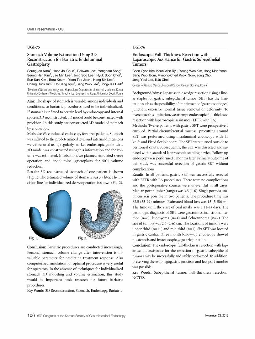
Oral Presentation - UGI
106 63rd Congress of the Korean Society of Gastrointestinal Endoscopy November 23, 2013
UGI-75
Stomach Volume Estimation Using 3D Reconstruction for Bariatric Endoluminal Gastroplasty
Seung-joo Nam1, Hoon Jai Chun1, Dokwan Lee2, Yongnam Song2, Seung Han Kim1, Jae Min Lee1, Jong Soo Lee1, Hyuk Soon Choi1, Eun Sun Kim1, Bora Keum1, Yoon Tae Jeen1, Hong Sik Lee1, Chang Duck Kim1, Ho Sang Ryu1, Sang Woo Lee1, Jong-Jae Park1
1Division of Gastroenterology and Hepatology, Department of Internal Medicine, Korea University College of Medicine, 2Mechanical Engineering, Korea University, Seoul, Korea
Aim: The shape of stomach is variable among individuals and
conditions, so bariatric procedures need to be individualized.
If stomach is inflated to certain level by endoscopy and internal
space is 3D reconstructed, 3D model could be constructed with
precision. In this study, we constructed 3D model of stomach
by endoscopy.
Methods: We conducted endoscopy for three patients. Stomach
was inflated to the predetermined level and internal dimensions
were measured using regularly marked endoscopic guide-wire.
3D model was constructed using this information and the vol-
ume was estimated. In addition, we planned simulated sleeve
operation and endoluminal gastroplasty for 50% volume
reduction.
Results: 3D reconstructed stomach of one patient is shown
(Fig. 1). The estimated volume of stomach was 3.7 liter. The in-
cision line for individualized sleeve operation is shown (Fig. 2).
Conclusion: Bariatric procedures are conducted increasingly.
Personal stomach volume change after intervention is in-
valuable parameter for predicting treatment response. Also
computerized simulation for optimal procedure is very useful
for operators. In the absence of techniques for individualized
stomach 3D modeling and volume estimation, this study
would be important basic research for future bariatric
procedures.
Key Words: 3D Reconstruction, Stomach, Endoscopy, Bariatric
UGI-76
Endoscopic Full-Thickness Resection with Laparoscopic Assistance for Gastric Subepithelial Tumors
Chan Gyoo Kim, Keun Won Ryu, Young-Woo Kim, Hong Man Yoon, Bang Wool Eom, Myeong-Cherl Kook, Soo-Jeong Cho, Jong Yeul Lee, Il Ju Choi Center for Gastric Cancer, National Cancer Center, Goyang, Korea
Background/Aims: Laparoscopic wedge resection using a line-
ar stapler for gastric subepithelial tumor (SET) has the limi-
tation such as the possibility of impairment of gastroesophageal
junction, excessive normal tissue removal or deformity. To
overcome this limitation, we attempt endoscopic full-thickness
resection with laparoscopic assistance (EFTR with LA).
Methods: Twelve patients with gastric SET were prospectively
enrolled. Partial circumferential mucosal precutting around
SET was performed using intraluminal endoscopy with IT
knife and Fixed flexible snare. The SET were turned outside to
peritoneal cavity. Subsequently, the SET was dissected and su-
tured with a standard laparoscopic stapling device. Follow-up
endoscopy was performed 3 months later. Primary outcome of
this study was successful resection of gastric SET without
complications.
Results: In all patients, gastric SET was successfully resected
with EFTR with LA procedures. There were no complications
and the postoperative courses were uneventful in all cases.
Median port number (range) was 3.5 (1-6). Single port via um-
bilicus was possible in two patients. The procedure time was
62.5 (35-99) minutes. Estimated blood loss was 15 (5-50) ml.
The time until the start of oral intake was 1 (1-4) days. The
pathologic diagnosis of SET were gastrointestinal stromal tu-
mor (n=6), leiomyoma (n=4) and Schwannoma (n=2). The
size of tumors was 2.5 (2-6) cm. The locations of tumors were
upper third (n=11) and mid third (n=1). Six SET was located
in gastric cardia. Three month follow-up endoscopy showed
no stenosis and intact esophagogastric junction.
Conclusion: The endoscopic full-thickness resection with lap-
aroscopic assistance for the resection of gastric subepithelial
tumors may be successfully and safely performed. In addition,
preserving the esophagogastric junction and less port number
was possible.
Key Words: Subepithelial tumor, Full-thickness resection,
NOTES
Fig. 2.Fig. 1.
Oral Presentation - UGI
November 23, 2013 63rd Congress of the Korean Society of Gastrointestinal Endoscopy 107
UGI-77
Accuracy of Confocal Endomicroscopy versus Conventional Biopsies for the Diagnosis of Superficial Gastric Neoplasia
Jin Nyoung Kim1, Joo Young Cho1, Jun-Hyung Cho1, Mi-Young Kim1, Byoung Moo Lee1, So Young Jin2 1Digestive Disease Center, 2Department of Pathology, Soonchunhyang University Hospital, Seoul, Korea
Background: Probe type confocal laser endomicroscopy (pCLE)
allows real- time in vivo histologic evaluation of gastrointestinal
mucosal lesions. Although pCLE has been used for various GI
disorders, the significance of pCLE for gastric lesions is largely
unknown. We compared the accuracy of conventional endo-
scopic forceps biopsy and pCLE for diagnosis of superficial
gastric neoplasia prior to endoscopic resection.
Methods: pCLE was performed prior to endoscopic resection
of superficial gastric neoplasia previously diagnosed by endo-
scopic biopsy. The overall accuracy of endoscopic, in vivo pCLE,
and offline pCLE diagnosis was compared with post-endoscopic
resection histopathology.
Results: Endoscopic resection was performed on 54 lesions. On
final histopathology, there were 3 non-neoplastic lesions, 19
gastric dysplasias, 22 differentiated adenocarcinomas and 10
undifferentiated adenocarcinomas. The overall agreement
with final histopathology was substantial for conventional bi-
opsies (κ=0.617 and excellent for in vivo pCLE (κ=0.824)
(p<0.001). The overall accuracy for diagnosis of ad-
enocarcinoma was 91.7% for pCLE and 85.2% for conven-
tional biopsies (p=0.065). The combined accuracy of conven-
tional endoscopic biopsies and pCLE was 98.1%.
Key Words: pCLE, Superficial gastric neoplasia, Conventional
endoscopic biopsies
UGI-78
ESD Using a Thulium Laser: Preliminary Results of a New Method for Treatment of Gastric Epithelial Neoplasia
Byoung Moo Lee1, Joo Young Cho1, Jin Nyoung Kim1, Mi-Young Kim1, Jun-Hyung Cho1, Soo Jin Hong2 Digestive Disease Center, Soonchunhyang University Hospital, 1Seoul, 2Bucheon, Korea
Objective: Endoscopic submucosal dissection (ESD) has been
accepted as the optimal treatment in gastric epithelial neoplasia.
This study aimed to evaluate the safety and feasibility of a novel
laser system when performing ESD of gastric epithelial neoplasia.
Methods: A total of 10 patients were enrolled into this study at
a single tertiary referral center. All patients were diagnosed as
gastric adenoma (n = 5) or adenocarcinoma (n = 5). They un-
derwent ESD by a single expert endoscopist. A thulium 2-μm
wavelength laser system was used for all ESD procedures in-
cluding marking, mucosal incision and submucosal dissection.
Instead of endoscopic knives, a prototype device (550-μm flex-
ible silica fiber attached with metallic sheath) was inserted
through the working channel of the endoscope. The main out-
come measure was technical success and complication rates.
Results: In all patients, ESD was completed using only thulium
laser without the need to change other endoscopic knives. The
median time for total procedure was 49 minutes (range, 35-203
minutes). In 80% (n = 8/10) of patients, active bleeding was
not observed during ESD. The final pathologic mapping resulted
in low-grade dysplasia (n = 4), differentiated adenocarcinoma (n
= 5), and signet ring cell carcinoma (n = 1). Curative resection
was achieved in 90% (n = 9/10) of patients. There were no sig-
nificant complications, such as delayed bleeding and perfo-
ration, in any patients.
Conclusions: The thulium laser system is safe and feasible in
ESD of gastric epithelial neoplasia.
Key Words: Thulium Laser, ESD, Gastric Epithelial Neoplasm
Oral Presentation - UGI
108 63rd Congress of the Korean Society of Gastrointestinal Endoscopy November 23, 2013
UGI-79
Fluorescein-Enhanced Autofluorescence Imaging for Exact Tumor Margin in Superficial Gastric Neoplasia
Mi-Young Kim1, Jun-Hyung Cho1, Jin Nyoung Kim1, Byoung Moo Lee1, So-Young Jin2, Joo Young Cho1
1Digestive Disease Center, 2Department of pathology, Soonchunhyang University Hospital, Seoul, Korea
Background/Aims: In spite of advances in image enhanced en-
doscopy, sometimes we encounter ill-demarcated tumors dur-
ing endoscopic submucosal dissection (ESD). Autofluorescence
imaging (AFI) has been reported to be a clinically promising
system to diagnose gastrointestinal neoplasia. However, the use
of AFI has been limited. Fluorescence-enhanced AFI is a novel
method using additional intravenous injection of fluorescein
to AFI. The aim of this study was to investigate the feasibility of
fluorescein-enhanced autofluorescence imaging (FAFI) for the
exact tumor margin in superficial gastric neoplasia.
Methods: A total of 30 patients with superficial gastric neo-
plasia was performed ESD between May 2013 and July 2013.
Prior to ESD, all of the tumors’ size and lateral margin were
evaluated with white light endoscopy (WLE), magnifying en-
doscopy with narrow band image (ME-NBI), chromoendo-
scopy (CE), AFI and FAFI. We compared the diagnostic effi-
cacy of FAFI for exact lateral margin of the tumors with that of
WLE, ME-NBI, CE, and AFI using grading system. We con-
firmed the tumor size and the margin by comparing endo-
scopic image and final pathology after ESD.
Results: Among 30 tumors, differentiated and undifferentiated
adenocarcinoma was 16 (53.3 %) and 5 (16.7%). Mucosal can-
cer made up of 43.3% and submucosal cancer 23.3 %. The tu-
mors showed 15 elevated, 5 flat, and 10 depressed types. The
median tumor size was 15.5 mm. Complete resection rate was
83.3 %. Compared to WLE, FAFI showed significantly differ-
ent diagnostic efficacy in case of the middle third location
(p=0.029), >2 cm of tumor (p=0.046), and presence of ulcer-
ation (p=0.012). The tumor histology, depth of invasion and
tumor type did not show a significant difference. Compared
with ME-NBI and CE, FAFI did not show a significant differ-
ence in diagnostic efficacy.
Conclusion: FAFI seems to be useful system for determination
of exact margin in superficial gastric neoplasia.
Key Words: Fluorescein, Autofluorescence image, Neoplasia,
gastric
UGI-80
The New Classification of Transnasal Insertion Routes
Jong Og Seo, Hak Cheon Oh, Ju Hyun Lee Stomach and Intestine Endoscope Network, Dr. Seo Internal Medicine Clinic, Suncheon, Korea
Background: Conventionally, upper gastrointestinal endos-
copy has been conducted perorally under pharyngeal anesthesia.
However,compression of the tongue root by the endoscope can
easily cause gag reflex, which makes the endoscopy uncomfortable.
In order to relieve any discomfort, an increasing number of fa-
cilities sedate the patient using sedatives before the endoscopic
procedure; however, sedation of an ambulatory patient is in-
deed fraught with risk. Under the circumstances, the transnasal
endoscopy enabled a less painful, safe procedure without
sedation. Because of this, the recent prevalence of transnasal
endoscopy is outstanding, and endoscopy became recognized
as a routine upper gastrointestinal procedure. However, appro-
priate preparation and nasal anesthesia are essential to take full
advantage of the examination. This time, in order to determine
which route is safer and more suitable,Consequently we can
classify these two routes into five new routes.
Subjects and Methods: We analysed all data for 328 cases (170
males and 158 females)underwent transnasal endoscopy in our
clinic from May 30, 2013 to August 6, 2013. After We inserted
rhinoscope through both routes in all patients, we decided
their own type of routes. The transnasal endoscope used for
this study was EG-530N, whose diameter was 5.9mm produced
by FUJINON Corporation and the rhinoscope was ENT unit
XU1 by CHAMMED Corporation. Results and
Conclusions: Among 328 cases performed in our clinic, the
numbers of right side insrtion were 188(59.3%), left side in-
sertion were 129(40.7%), 11 cases were Type 0 route mean was
none of the route can be inserted the endoscopy. By the new
classification from Japan, type 1 route accounted for 297, type
2 route accounted for 11, type 3 route accounted for 9, type 0
route accounted for 11. According to these results, type 1 route
should have been recommended in general.
Key Words: Transnasal endoscopy, Transnasal insertion routes,
Five new routes
Oral Presentation - UGI
November 23, 2013 63rd Congress of the Korean Society of Gastrointestinal Endoscopy 109
UGI-81
Endoscopic Characteristics of Gastric Cytomegalovirus Infection in Patients with Renal Transplantation
Yu Jin Kang, Eun Sung Choi, Jae Hyuk Choi, Jung Min Lee, Sang Min Lee, Yoo Jin Lee, Eun Soo Kim, Kwang Bum Cho, Kyung Sik Park, Byoung Kuk Jang, Woo Jin Chung, Jae Seok HwangDivision of Gastroenterology and Hepatology, Department of Internal Medicine, Keimyung University School of Medicine, Daegu, Korea
Backgroud and Aims: Endoscopic characteristics of gastric cy-
tomegalovirus (CMV) infection are various and mimic other
gastric diseases. The aim of this study was to figure out endo-
scopic features associated with gastric CMV infection.
Patients and Methods: Data of 301 renal transplant patients
(RTPs) who underwent endoscopic examination with mucosal
biopsies for CMV infection between March 2005 and July 2013
were reviewed retrospectively. Gastric CMV infection was con-
firmed by immunohistochemical staining and CMV PCR.
Endoscopic characteristics of ulcers and erosions such as num-
ber, size, morphology, location, and exudates were evaluated
for the potential risk factors of CMV infection. Result: Overall,
209 patients who showed ulcers (60, 28.7%) or erosions (185,
88.5%) confirmed by endoscopy were enrolled in the study.
CMV infection was confirmed in 118 patients (56.5%). For ul-
cer lesions, round shape and antral location were significantly
associated with CMV infection. For erosions, exudates attach-
ment, raised type, and antral location were significantly more
observed in CMV infection group. When patients had 2 ulcer
risk factors (round shape and antral location) or 3 erosion risk
factors (exudates, raised type, and antral location), CMV pos-
itive rate was 92.3% and 86.4%, respectively. In addition, pa-
tients with all 5 risk factors showed 100% CMV positive rate.
Conclusions: These endoscopic characteristics of ulcers and
erosions might be helpful to predict gastric CMV infection in
patients with renal transplantation. Further study is needed to
validate the results of this study.
Key Words: Cytomegalovirus, Stomach, Renal transplantation
UGI-82
Effects of Time to Endoscopic Removal for Foreign Bodies according to the Intention to Ingestion
Kyong Hee Hong, Jae Hak Kim, Hyoun Woo Kang, Jun Kyu Lee, Yun Jeong Lim, Moon-Soo Koh, Jin Ho Lee Department of Internal Medicine, Dongguk University Ilsan Hospital, Graduate School, Goyang, Korea
Introduction: There were few studies about the time effect on
endoscopic removal for ingestion of foreign bodies or im-
paction of food bolus. We aimed to analyze the factors of for-
eign body ingestion and food material impaction in upper gas-
trointestinal tract with endoscopic management.
Methods: We retrospectively collected 141 patients with a diag-
nosis of foreign body ingestion or food bolus impaction in up-
per gastrointestinal tract confirmed by endoscopy at Dongguk
University Ilsan hospital, Goyang, Korea from October 2005 to
May 2013. Patient’s demographic data included age, sex, past
medical history, intention to ingestion, symptoms at admis-
sion, and comorbidities. Clinical features of foreign bodies
were analyzed including type, size, shape, number, and
location. Collected endoscopic data were duration of foreign
body impaction, time to endoscopic removal, duration of en-
doscopy performance, endoscopic devices, the days of hospi-
talization, the complication rate, the mortality rate, and the
number of operation related to foreign body removal.
Results: Overall complication rate was 67.2%, including ero-
sions (40.5%), lacerations with minor bleeding (12.1%), ulcers
(8.6%), petechiae (4.3%), and perforation (1.7%). Hospitalization
was required in 45 cases (38.8%). In univariate analysis, acci-
dental ingestion (p<0.001) and sharpness (p=0.001) were asso-
ciated with higher complication rates. In multivariate analysis,
accidental ingestion developed more complications (p<0.001).
Cox regression analysis revealed that prolonged accidental in-
gestion and delayed endoscopic removal had a higher proba-
bility of complications than voluntary ingestion (Hazard ratio
2.25, 95% Confidence interval 1.15 to 4.40, p=0.018). There
was no death associated with the procedure.
Conclusions: In case of accidental ingestion of foreign materi-
al, more urgent endoscopic treatment could be required to pre-
vent the complications. Further prospective study would be
needed.
Key Words: Foreign body, Endoscopy, Upper gastrointestinal
tract
Oral Presentation - UGI
110 63rd Congress of the Korean Society of Gastrointestinal Endoscopy November 23, 2013
UGI-83
Endoscopic Hemostasis for Early Postoperative Anastomotic Bleeding after Gastrectomy
Shin Na, Ji Yong Ahn, Hwoon-Yong Jung, Hyun Lim, Jeong Hoon Lee, Kwi- Sook Choi, Do Hoon Kim, Kee Don Choi, Ho June Song, Gin Hyug Lee and Jin-Ho Kim Departments of Gastroenterology, University of Ulsan College of Medicine, Asan Medical Center, Seoul, Korea
Background/Aims: Postoperative anastomotic bleeding (PAB)
is a relatively rare complication, however, it is lethal if not treat-
ed immediately. The aim of our study is to investigate the effi-
cacy of the endoscopic hemostasis (EH) for early PAB.
Methods: Of 16,598 patients who underwent gastrectomy be-
tween January 2004 and May 2013 at Asan Medical Center,
Seoul, Korea, EH for early (within 2 months) PAB was per-
formed in 26 patients. The clinical outcomes, the methods of
endoscopic bleeding control, and the factors for influencing
the success of EH among these 26 patients were analyzed
retrospectively.
Results: Of the 16,598 patients, early PAB developed in 110 pa-
tients (0.66 %). Among them, EH for early PAB was performed
in 26 patients. Median age (range) of 26 patients was 64.5 years
(26.0-81.0 years) and men were 20 (76.9%). All the 26 patients
underwent R0 gastrectomy with D2 lymph node dissection for
gastric cancer and the median operation time (range) was
107.5 min (52-181). The techinal and clinical success of EH
were revealed as 69.2% (18/26) and 69.2% (18/26), respectively.
For the methods of endoscopic bleeding control, hemoclips
(14/26 [53.8%]), epinephrine injection (10/26 [38.5%]), and/
or fibrin glue (6/26 [23.1%]) were used in EH, individually
(19/26 [73.1%]) and compositively (7/26 [26.9%]). Among
EH fail group (8/26 [30.8%]), reoperation and angioemboliza-
tion were performed in 7 patients and 1 patient, respectively.
PAB related death was observed only in 2 patients of EH fail
group. There were no differences between endoscopic hemo-
stasis success and fail groups, and no factors in influencing on
success of EH.
Conclusions: PAB is an infrequent but potentially life-threat-
ening complication. Endoscopy appears to be useful for both
the confirmation of bleeding and therapeutic intervention.
Key Words: Anastomosis, Bleeding, Endoscopy, Gastrectomy,
Hemostasis
UGI-84
Diagnosis and Treatment of Esophageal Tuberculosis: A Single Center Experience
Jung Hee Kim, Min Yang Won, Byung-Hoon Min, Poong-Lyul Rhee, Jun Haeng Lee Department of Medicine, Samsung Medical Center, Sungkyunkwan University School of Medicine, Seoul, Korea
Background: The diagnosis of esophageal tuberculosis (ET) is
challenging, because its clinical and pathological features are
similar to other esophageal ulcerative lesions.
Methods: From the clinical and pathological database, we
found 9 cases of esophageal tuberculosis between 1998 and
2013. Clinical, pathological, radiologic findings and treatment
outcomes were reviewed.
Results: The presenting symptoms were dysphagia (n=6), and
epigastric pain (n=3). The location of esophageal mucosal le-
sion was mostly mid-esophagus (n=8). The most common en-
doscopic shape was an ulcerative lesion with healed-up margin
(n=6), followed by a perforating hole (n=1), an esophageal
stenosis (n=1) and a submucosal tumor-like morphology
(n=1). Endoscopic impression was mostly esophageal cancer
(n=5), followed by esophageal laceration (n=1), pilled induced
esophagitis (n=1) and esophageal submucosal tumor (n=1),
and esophageal tuberculosis (n=1). In the esophageal endo-
scopic forcep biopsy (n=7) and the endobronchial ultrasound
fine-needle aspiration (EBUS-FNA) of the subcarinal lymph
node (n=1), the pathology was chronic granulomatous in-
flammation with or without necrosis. There was no definite
histological evidence of tuberculosis. In the chest X-rays, 33%
(3/8) had pulmonary tuberculous lesion(s). In the chest CT,
88% (7/8) had enlarged lymph node(s) at subcarinal or para-
esophageal area. In the sputum AFB smear and culture, M. tu-
berculosis was detected in 25% (2/8). In the AFB smear and
culture of aspirated fluid from enlargement lymph nodes, M.
tuberculosis was detected in all tested patients (3/3). Anti-tu-
berculosis medications were given at least 6 months. The mu-
cosal lesions were improved in the follow-up endoscopies at 2
to 6 months.
Conclusion: High index of suspicion is necessary for esoph-
ageal tuberculosis in patients with symptomatic esophageal ul-
cer and enlarged mediastinal lymph node(s).
Key Words: Esophageal tuberculosis, Esophageal ulcer, Mediastinal
lymph node
Oral Presentation - UGI
November 23, 2013 63rd Congress of the Korean Society of Gastrointestinal Endoscopy 111
UGI-85
Comparison of 4 Scoring Systems for Predicting Clinical Outcomes in Non-Variceal Upper Gastrointestinal Bleeding Patient
Seok Chun Yeum, Byung-Wook Kim, Sung Min Park, Ji Hee Kim, Eun Hui Sim, Joon Sung Kim, Jeong-Seon Ji, Bo-In Lee, Hwang ChoiDivision of Gastroenterology, Department of Internal Medicine, Incheon St. Mary's Hospital, The Catholic University of Korea, Incheon, Korea
Aim: This study aimed to validate four scoring system (pre-
Rockall, full Rockall, Blatchford, AIMS 65) in Korean pop-
ulation and compare them for the prediction of re-bleeding
and death in patients with acute non-variceal upper gastro-
intestinal bleeding (UGIB).
Materials and Methods: Between 2008 and 2013, data on 161
patients who had undergone endoscopy due to non-variceal
UGIB were collected and reviewed retrospectively in Incheon
St. Mary's Hospital, The Catholic University of Korea. Admission
history, clinical and laboratory data, endoscopic findings,
treatment and clinical follow-up were recorded and inves-
tigated on the basis of three scoring systems. In subgroup anal-
ysis, we investigated patients presented with peptic ulcer bleed-
ing with three scoring system and Forrest classification. Using
ROC curves, we compared the three scores in the prediction of
death, re-bleeding, endoscopic or surgical intervention and
transfusion.
Results: Of the 161 study patients, eight patients died within 30
day, twelve patients experienced re-bleeding 7.5%), 34 patients
needed additional endoscopic management (21.1%) and 97
patients required transfusion (60.2%). Comparison of the
three scoring systems showed that the full Rockall score was su-
perior to the others in predicting re-bleeding (AUROC, 0.80 ; p
<0.0001), death (AUROC, 0.97; p <0.0001) and endoscopic in-
tervention (AUROC,0.68; p=0.001). The Blatchford score was
superior in predicting blood transfusions (AUROC, 0.86 ; p <0
.0001). AIMS 65 score predicted 30-day mortality (AUROC,
0.75 ; p <0 .26). In 47 patients with peptic ulcer bleeding, the
pre-Rockall, full Rockall score and Forrest classification were
similar in predicting mortality and re-bleeding. Forrest classi-
fication was superior in predicting endocopic intervention
(AUROC,0.96; p <0.0001).
Conclusions: Full Rockall score was the most useful scoring
system for the prediction of rebleeding and death in patients
with nonvariceal UGIB.
Key Words: Upper gastrointestinal bleeding, Rockall score,
Blatchford score, AIMS 65