Embed Size (px)
Citation preview

ICA B
est P
ractic
es
ICA Best Practices & Practice Guidelines 34
© 2013, International Chiropractors Association, Arlington VA. All Rights Reserved
Chapter 4 Risk of Chiropractic Care
Introduction While the benefits of Chiropractic care are too numerous to list here (see Tables 1-6 in Chapter 10), the risks of chiropractic spinal adjustments and spinal manipulation will be reviewed in this chapter. The first important thing to note is that the risks of spinal adjustments and spinal manipulations compared to standard medical care are minimal, which can be shown by the malpractice insurance premiums that each groups pays ($2,000/yr for DCs and $30,000-$100,000 for MDs). We will divide our discussion of risks with chiropractic care into two categories: Non-catastrophic risks and catastrophic risks.
The ICA’s malpractice insurance organization, ChiroSecure, has provided a list of the most common claims arising against chiropractors. They are in order, 1) Rib Fracture, 2) Stroke, 3) Patients who present with low back pain and acquire neck pain or vice versa, 4) exacerbation of existing complaints, 5) Sexual misconduct, 6) Board-related issues (vary by state), and 7) Insurance Audits. Interestingly, The FCLB (Federation of Chiropractic Licensing Board) has insisted that diagnosis is one of the reasons that chiropractors have claims brought against them, as this list shows, diagnosis is absent. We will discuss these complaints and provide any evidence that may be found in the biomedical literature concerning this list. Non-catastrophic Risks Criticism by other healthcare providers (MDs, PTs, DOs) tend to exaggerate, dramatize, and misreport the risks with Chiropractic spinal manipulation.1-7 For example regarding adverse effects, Ernst2 stated, “searches identified 32 case reports, four case series, two prospective series, three case-control studies and three surveys [our emphasis]. In case reports or case series, more than 200 patients were suspected [our emphasis] to have been seriously harmed. The most common serious adverse effects were due to vertebral artery dissections. The two prospective reports suggested that relatively mild adverse effects occur in 30% to 61% of all patients.”2 There were 7 negative letters to the journal Editor concerning this publication. In 2006, Bronfort et al8 stated that “Based on a critical appraisal of their review, the authors of this commentary seriously challenge the conclusions by Ernst and Canter,5 who did not adhere to standard systematic review methodology, thus threatening the validity of their conclusions. There was no systematic assessment of the literature pertaining to the hazards of manipulation, including comparison to other therapies. Hence, their claim that the risks of manipulation outweigh the benefits, and thus spinal manipulation cannot be recommended as treatment for any condition, was not supported by the data analyzed. Their conclusions are misleading and not based on evidence that allow discrediting of a large body of professionals using spinal manipulation.”8
In contrast, three recent publications by chiropractors have shown that the non-catastrophic risks are minimal.9-11 In 2007, Hawk et al9 reported on a systematic review of the evidence on the effect of chiropractic care, other than spinal manipulation only, on patients with nonmusculoskeletal conditions. Their search yielded 179 papers addressing 50 different nonmusculoskeletal conditions. There were 122 case reports or case series, 47 experimental designs, including 14 RCTs, 9 systematic reviews, and 1 a large cohort study. The 14 RCTs addressed 10 conditions. They concluded that for the few studies that did report, adverse effects of spinal manipulation for all ages and conditions were rare, transient, and not severe.9
In another 2007 study, Rubinstein et al10 reported on a prospective, multicenter, observational cohort study. Patients with neck pain of any duration were recruited in a practice-based study. Data were collected on the patients and from the chiropractors at baseline, the first 3 visits, and at 3 and 12 months. An adverse event was defined as either a new related complaint or a worsening of the presenting or existing complaint by >30% based upon an 11-point numerical rating scale. A total of 79 chiropractors participated, recruiting 529 subjects, representing 4891 treatment consultations. Adverse events after any of the first 3 treatments were reported by 56%, and 13% of the study population reported these events to
ICA B
est P
ractic
es
ICA Best Practices & Practice Guidelines 35
© 2013, International Chiropractors Association, Arlington VA. All Rights Reserved
be severe in intensity. The most common adverse events affected the musculoskeletal system or were pain related, whereas symptoms such as tiredness, dizziness, nausea, or ringing in the ears were uncommon (< 8%). Only 5 subjects (1%) reported to be much worse at 12 months. No serious adverse events were recorded during the study period. Of the patients who returned for a fourth visit, approximately half reported to be recovered, whereas approximately two thirds of the cohort were recovered at 3 and 12 months. They concluded that (a) while adverse events may be common, these are rarely severe in intensity, (b) most of the patients report recovery, particularly in the long term, and (c) the benefits of chiropractic care for neck pain seem to outweigh the potential risks.10
Also in 2007, Thiel et al11 studied treatment outcomes obtained from 19,722 patients. Spinal manipulation (SMT) was defined as the application of a high-velocity/ low-amplitude or mechanically assisted thrust to the cervical spine. Serious adverse events, defined as "referred to hospital A&E and/or severe onset/worsening of symptoms immediately after treatment and/or resulted in persistent or significant disability/incapacity," and minor adverse events reported by patients as a worsening of presenting symptoms or onset of new symptoms, were recorded immediately, and up to 7 days, after treatment. Data were obtained from 28,807 treatment consultations and 50,276 cervical spine manipulations. There were no reports of serious adverse events. This translates to an estimated risk of a serious adverse event of, at worse to be 1 per 10,000 immediately after cervical spine manipulation, 2 per 10,000 up to 7 days after treatment and 6 per 100,000 cervical spine manipulations. Minor side effects with a possible neurologic involvement were more common. The highest risk immediately after treatment was fainting/dizziness/ light-headedness in, at worst 16 per 1000 treatments. Up to 7 days after treatment, these risks were headache in, at worst 4 per 100, numbness/tingling in upper limbs in, at worst 15 per 1000 and fainting/dizziness/light-headedness in, at worst 13 per 1000 treatments. They11 concluded that minor side effects following cervical spine manipulation were relatively common and the risk of a serious adverse event, immediately or up to 7 days after treatment, was low to very low. Furthermore, the authors conclude that the risk rates described in this study compare favorably to those linked to drugs routinely prescribed for musculoskeletal condition in general (MD) practice.11
In 2006, Childs et al12 reported that low back pain subjects were at risk if they did not receive spinal manipulation. One hundred and thirty-one consecutive patients with LBP were randomly assigned to receive manipulation and an exercise intervention (n = 70) or an exercise intervention without manipulation (n = 61). Patients were classified as to whether they had experienced a worsening in disability upon follow-up. Patients who completed the exercise intervention without manipulation were eight (95% CI: 1.1, 63.5) times more likely to experience a worsening in disability than patients who received manipulation.12
As we discussed in the introduction of this chapter, rib fracture is the number one reason for claims arising against chiropractors. A medline search of “chiropractic AND rib fracture presented only two results.13,14 These cases appear to be non-consequential as one13 is merely a case study of a patient who presented with a fracture to a chiropractor and the other14 appears not to include chiropractors in the survey. A non-peer reviewed article by Laurin McElheran, DC in the Beacon, a student newspaper at Palmer Chiropractic presents a case of a 64-year-old man with a history of corticosteroid use suffering from low back pain apparently receive multiple rib fractures following a side-posture adjustment. The article reports that the case was settled out of court for a total of $5,500.00.15 The fact that there are very few references to rib fracture found in the biomedical literature indicates that either this condition is severely underreported or it is rare. Catastrophic Risks Probably the most catastrophic risk of manipulation that has been proposed is the risk of Vertebrobasilar Artery (VBA) dissection or stroke following cervical spine manipulation. Anatomically, there are several factors that one must take into consideration when analyzing the cervical spine and determining the risk of VBA dissection following manipulation. The vertebral arteries are branches of the subclavian artery. The arteries ascend up the cervical spine through the foramina in the transverse processes and, in combination with the basilar artery create the blood supply to the Circle of Willis, the

ICA B
est P
ractic
es
ICA Best Practices & Practice Guidelines 36
© 2013, International Chiropractors Association, Arlington VA. All Rights Reserved
main supply to deep brain structures such as the cerebellum and brainstem. The vertebral arteries run roughly parallel to the carotid arteries however, at the level of C1, they travel across the posterior arch of atlas and enter the foramen magnum into the skull. The vertebral arteries are an arterial redundancy to ensure blood supply to the head in the case of constriction of one of the carotid arteries.16 Redundancy is a necessary and common occurrence in the arterial supply to the head and appears to reduce neurological sequalae of ischemic events.16
Risk factors for VBA are extensively reported in the literature and include, but are not limited to, age,17-20 gender18, 19, 21, 22 hypertension,22-27 diabetes,24, 26, 27 oral contraceptives, 28-31 smoking.32, 33 and certain diseases such as fibromuscular hyperplasia23, 34-37 and Marfan’s Syndrome.38-39 Cervical manipulation40-43 is only one of the proposed causes of VBA dissection and others include, but are not limited to major trauma,44-45 rotatory head movements,25, 42, 46 sports activities47-51, and some have no known cause whatsoever.23, 51-54 Wise clinicians should educate themselves on the possible complications and underlying causes of stroke and pre-screening activities such as vertebro-basilar function tests should be performed prior to cervical spine manipulation. However, understanding the signs and symptoms of VBA strokes, taking pre-cautions, and screening will not prevent an arterial dissection if the patient is having the cerebrovascular event before entering the chiropractor’s office.
While some investigators claim that SMT can cause loss of vision, ophthalmoplegia, diplopia, and Horner's syndrome (14 case reports in 8 years),55 phrenic nerve injury (subject was symptom free after 1 year),56 spinal epidural hematoma (7 additional cases found on review),57 intracranial hypotension and abducens palsy,58 intracranial hypotension (claimed cervical spinal manipulation produced a remote lumbar dural tear),59 and stroke.60-70 Chiropractors71-75 have challenged the analyses, claims, and correlations in many of these publications. 59-69 They71-75 have indicated that (a) in one instance, SMT was correlated to stroke in 14% while in the authors’ data, alcohol use was correlated with stroke at 76%, (b) in approximately 50% of cases where the headline was “stroke with Chiropractic SMT”, in fact the maneuver was NOT even performed by a Doctor of Chiropractic, but by MDs, PTs, DOs, or American Indian faith healers, (c) incidence of stroke with SMT were greatly exaggerated, (d) in some instances, surveys instead of data were used to report that SMT is associated with stroke, and (e) stroke caused by SMT is very unlikely in healthy individuals.
It is significant that some competitive professionals (MDs, PTs, DOs) exaggerate unfortunate stroke victim incidents after spinal manipulation (often submitting these cases to the national press). Every decade, hundreds of thousands of surgery patients have strokes from surgery induced blood clots (1.2% of all surgical events), but these rarely are discussed in national newspapers.76 In fact, a PubMed search in March 2008 with “Surgery AND Strokes” yielded 7,424 papers while “Chiropractic AND Stroke” yielded 89 citations.
Two recent publications77, 78 may help to end the exaggerated claims of stroke caused by SMT made by other healthcare providers. In 2008, Boyle et al77 reported the annual incidence of hospitalized vertebrobasilar artery (VBA) stroke and chiropractic utilization in Saskatchewan and Ontario between 1993 and 2004. They77 wished to determine whether at an ecological level, the incidence of VBA stroke parallels the incidence of chiropractic utilization. They concluded that, at the ecological level, the increase in VBA stroke did not seem to be associated with an increase in the rate of chiropractic utilization. Again in 2008, Cassidy et al78 wished to investigate associations between chiropractic visits and vertebrobasilar artery (VBA) stroke and to contrast this with primary care physician (PCP) visits and VBA stroke. Cases included eligible incident VBA strokes admitted to Ontario hospitals from April 1, 1993 to March 31, 2002. There were 818 VBA strokes hospitalized in a population of more than 100 million person-years. They78 concluded that VBA stroke is a very rare event in the population. They78 found no evidence of excess risk of VBA stroke associated with chiropractic care compared to primary care MDs. Furthermore they78 conclude that the reason that the individual sought care from the chiropractor for neck pain or headache was due to the fact that the stroke had already occurred or was in the process of occurring.
It is of primary importance for the practicing chiropractor to be aware of, although the risk of VBA dissection does exist with the use of spinal manipulation, the proper history and examination should
ICA B
est P
ractic
es
ICA Best Practices & Practice Guidelines 37
© 2013, International Chiropractors Association, Arlington VA. All Rights Reserved
allow the astute clinician to recognize not only the patient who is a greatest risk but also the patient who may already be suffering from symptoms related to VBA dissection.
In the past, criticisms of cervical extension traction methods to restore lordosis and x-ray protocols have been based on the 1999 Commentary by Haas et al79 These criticisms often neglect to provide the Harrison rebuttal written in 2000,80 which reported that Haas et al.79 misrepresented references, misinterpreted references, misquoted references, and performed a selective literature review.80 In fact, this Harrison-Haas debate was a series of three publications.79-81 Additionally, the uniformed often state that the extension position in CBP® cervical traction methods are dangerous. Numerous articles from the literature lead to the conclusion that this is definitely not the case. In fact, in a 1999 thorough review of the literature on varying positions of the head associated with vertebral and basilar artery blood flow and dissection, Haldeman et al.82 concluded that “examination of the data fails to show a consistent position or movement of the neck that could be considered particularly dangerous.” Disk Rupture Cannot be Caused by Chiropractic/SMT
It has been postulated by some that chiropractic or spinal manipulative therapy (SMT) can be a cause of disc herniation. We will present current biomedical literature which demonstrates that the herniation of an intervertebral disc is impossible with spinal manipulation. The literature has demonstrated that not only is SMT not a cause of disc herniations, it is the preferred form of treatment to alleviate the symptoms associated with existing disc injuries. As is the case with VBA dissection and stroke, it is probable that the claims brought against chiropractors for causing disc herniations are actually patients who had the disc injuries prior to presenting to the chiropractor, and, unfortunately were not properly informed of their condition. In order to understand the possible implications chiropractic adjustment/SMT has in the rupture of the intervertebral disc an understanding of the biomechanics is first necessary.
The primary issue at hand is whether or not Chiropractic Manipulation/Manual Therapy applied to the cervical spine, thoracic spine, or lumbar spine is capable of causing herniation/prolapse of the cervical discs. In order to answer this question the biomechanics literature detailing the mechanical causes of disc injury, including anatomy, mechanisms of injury and magnitude of applied load(s) are required, must be reviewed. To begin, there are two main categories of intervertebral disc herniations: A) Sudden prolapse resulting from a traumatic event and B) Gradual prolapse or herniation resulting from sustained applied loads or multiple repetitive micro-traumatic events.83-85
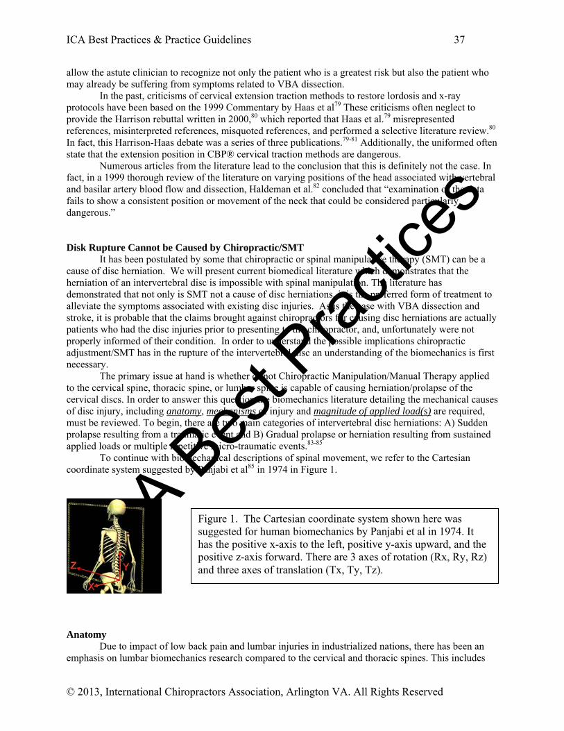
To continue with biomechanical descriptions of spinal movement, we refer to the Cartesian coordinate system suggested by Panjabi et al85 in 1974 in Figure 1.
Anatomy Due to impact of low back pain and lumbar injuries in industrialized nations, there has been an emphasis on lumbar biomechanics research compared to the cervical and thoracic spines. This includes
Figure 1. The Cartesian coordinate system shown here was suggested for human biomechanics by Panjabi et al in 1974. It has the positive x-axis to the left, positive y-axis upward, and the positive z-axis forward. There are 3 axes of rotation (Rx, Ry, Rz) and three axes of translation (Tx, Ty, Tz).

ICA B
est P
ractic
es
ICA Best Practices & Practice Guidelines 38
© 2013, International Chiropractors Association, Arlington VA. All Rights Reserved
studies on the intervertebral discs. Thus, up until the early 1990’s, it was assumed that the thoracic and cervical intervertebral discs were exactly like the lumbar discs, only smaller.86 The lumbar discs have more central nucleus and layers of a some what elliptical shaped lamellae. However, in 1999 Mercer and Bogduk87 reported that the cervical discs were quite different, and in fact, they lack a posterior annulus and are more of a flat plate with the posterior longitudinal ligament covering the posterior. The geometrical designs in a vertical view are that the thoracic discs are more circular, while the cervical and lumbar are more of an elliptical shape. Mechanisms of Disc Herniation A) Sudden Prolapse: A sudden macro-traumatic injury to the disc only occurs during significant, forceful injuries, such as that of whiplash injury, blow to head (diving, football, rugby etc.), forced neck flexion combined with compression, or sudden lifting in a stooped posture. Importantly, according to the recent biomechanical literature, the mechanism of sudden prolapse has to involve spinal hyperflexion combined with either compression along the long axis of the spine, lateral bending, and/or axial rotation 83, 85, 88,89 For example, Adams and Hutton83 state “Hyperflexion stretches and thins the posterior annulus, making it the weakest structure surrounding the nucleus. A high compressive force C then raises the hydrostatic pressure in the nucleus until it either bursts through the posterior annulus or causes it to collapse outward.” Additionally, in a 1995 review of the literature, Adams and Dolan88 state “the only loading conditions known to cause posterior disc prolapse involves a combination of compression, lateral bending and forward bending (emphasis added). This combination stretches and thins the posterolateral annulus and simultaneously raises the hydrostatic pressure in the nucleus”. Similarly, White and Panjabi85 state “clinical evidence of annular disruption implies that the disc failed because of some combination of bending (flexion), torsion, and tension”. Shea et al.89 demonstrated that cadaveric disc “specimens from the mid cervical region generally failed in flexion (flexion < 33 degrees), while specimens from the lower cervical region generally sustained 33 degrees of flexion rotation and only failed with combined flexion-compression motion”. Lastly, Ghanayem et al. state “The loading pattern that a functional spinal unit undergoes that will result in annular tearing and subsequent disc herniation is that of flexion, coupled with lateral bending and torsion”.90
Torsion, axial rotation left and right, by itself cannot cause sudden disc prolapse.83,88,85,92 According to White and Panjabi85 “torsional loading of the disc beyond its physiological limit does result in circumferential tears in the annulus but does not result in disc prolapse”. These tears are in the outer most layers of the annulus and the nucleus and the inner annulus remains intact .88 B) Gradual Prolapse of the Disc: The second type, gradual disc prolapse, is the most common type of disc prolapse 83-85. Gradual disc prolapse, is again, only caused by combined loading (e.g. flexion with compression, flexion with axial rotation, flexion with lateral flexion and rotation) 83, 85. Importantly, this type of loading is not rapid, it is sustained or repetitive episodes of normally non-damaging loads that occur in a given time frame, such as ½ hour to several hours. Gradual disc prolapse is the only case where radial fissures extending from the inner annulus to the outer annulus are formed83,85. Adams and Dolan88 state “radial fissure formation requires the redistribution of fluid within the disc, and this is a slow, time-dependent process, radial fissures are not formed when discs prolapse suddenly”. Ninomiya et al.92 have shown that axial loading (compression) combined with repetitive flexion motions will accelerate disc herniation and radial fissures. Examples of this would be the job description of a welder when flipping his/her face mask in front of the face, and sneezing forcefully with neck flexion repetitively.
Magnitude of Applied Loads and Kinematics of Cervical Spinal Manipulation as They Relate to Disc Herniation

ICA B
est P
ractic
es
ICA Best Practices & Practice Guidelines 39
© 2013, International Chiropractors Association, Arlington VA. All Rights Reserved
The magnitude of applied loads and kinematics (movement) of the vertebral segments during
cervical manipulation are critical to the understanding of disc herniation. Loads acting on the neck during cervical manipulation have been studied. In a study of 66 manipulations applied to the neck, Triano93 calculated the loads applied to the neck and the consequent loads transmitted to the neck. The mean peak transmitted loads acting on the cervical joints were found to be between 34-93 Newtons for Forces and 32-65 Newton Meters for Moments. According to Triano93, even at loads clinically considered as the maximally applied loads, the applied loads are well below injury loads and are thus, safe. For an example of this, the loads necessary to cause cervical disc herniations have been shown to be 10 times greater than the loads applied by cervical manipulation. Therefore, cervical manipulation cannot be implicated in cervical disc herniation.
Concerning the kinematics (movement) of the cervical spine, Kelsey and colleagues94 studied factors associated with acute cervical intervertebral disc prolapse. Here frequent twisting of the neck left and right on the job was not related to disc herniation. BenEliyahu91 discusses that it takes 22.6 degrees of rotation in a healthy disc and 14.3 degrees in a damaged disc to cause disc rupture. Recent 3-D in vivo (living subjects) studies on the range of motions at the cervical vertebral joints have shown that 60-70% occurs at occiput to C2, while C3-C7 only averages 4.2-7.4 degrees of rotation each.95,96 For example, Lai95 et al state the average axial rotation between C2 and C7 (combined segmental angles for 4 joints, 3-4 degrees each) was 15 degrees in the current study…” which is much smaller than the 25 degrees found in cadaveric studies. Accordingly, it is very unlikely that even the most extreme, maximum amount of cervical rotation can cause disc rupture even in cases of degeneration of the intervertebral disc.” The conclusion of BenEliyahu summarizes this point. He states “Cervical kinematic studies reveal that there is only 1-12 degrees of rotation in the cervical motion segments (depending upon the segmental level and magnitude of head movement)”.91 He continues, “Therefore, in the cervical and lumbar spine, rotational manipulation most likely cannot be implicated in disc failure or exacerbation of a disc herniation, and for rotational forces from a manipulation to be involved in disc failure, facet fracture must occur first”. 91 Standard of Chiropractic Care for Disc Herniations
It is very likely that reports of disc herniations caused by manipulation are in fact pre-existing conditions that were not fully explained to the patient prior to adjustment/SMT. Critical to the discussion of pre-existing disc herniations is the fact that chiropractic manipulation is actually the standard of care for patients who have cervical disc herniations.84, 91, 97-99 In fact, even in the Mercy Center Guidelines, it is stated that manipulation is only contraindicated in the case of “extensive disc prolapse with evidence of severe nerve damage”.100 In a prospective study of 27 individuals with MRI confirmed cervical disc herniations, BenEliyahu91 demonstrated statistically significant improvements in visual analog scales, pain intensity, return to work, and a reduced size of the disc herniation following chiropractic care. He states “Chiropractic management of disc herniation, including spinal manipulation, may be a safe and helpful modality for the treatment of cervical and lumbar disc herniation”.91 Not only does spinal manipulation not cause disc herniations, there is strong evidence that it is of benefit to these cases. Mechanics of torsion or torque
Axial rotation of the in vivo spine, y-axis rotation, is an example of torsion applied to the disc. It is helpful to note, that the term torque is used when this type of moment load is applied to cadaveric discs, motion segments, or spinal regions. Figure 1B in the last issue depicts torsional loading and the resultant elastic deformation. This simple model is applicable to our discussion. Figure 1F shows that cross-sectional shear stress is caused by torsion. The arrows increase in length moving away from the center toward the periphery. This indicates that the inner portion is under minimal stress and strain, while the outer margin is subjected to large stress and strain. Applying this to the disc, the nucleus and inner annulus will experience low magnitudes of stress and strain during torsion, while the outer annulus will experience the greatest stress and strain.

ICA B
est P
ractic
es
ICA Best Practices & Practice Guidelines 40
© 2013, International Chiropractors Association, Arlington VA. All Rights Reserved
The above analysis of stress and strain in the disc is only correct for a structure with a circular cross-sectional shape. A predominantly circular or cylindrical shape is only encountered for discs in the thoracic region. The cervical and lumbar IVD’s are elliptical in cross-sectional shape.87 Different shapes of the disc will have different stress and strain distributions. A disc which is circular in transverse section would have an even distribution of the stress arising from torsion around its circumference; the thoracic discs exhibit this type of stress. However, the elliptical cervical and lumbar discs will develop posterior and posteriolateral stress concentrations as a result of torsion.87 At first glance, this may seem to indicate a structural design flaw in the cervical and lumbar regions, however, other design features and material properties compensate for these stress concentrations. In actuality, these cross-sectional shape changes are structurally suited for the regional variations in movements and loading throughout the spine. 85,87 There are three primary mechanisms whereby a motion segment resists torsion: 1) the fiber direction and stiffness of the annular fibers, 2) bony restriction via articular process contact which depends upon the orientation of the zygapophyseal joints in the different spinal regions, 3) increased strain in other spinal ligaments, which primarily occurs in the thoracic region and in some areas of the cervical spine during larger rotation angles.83 The first two are relevant to our discussion. 1) All collagen fibers, in their long axis direction, are organized into a wavy, undulating pattern called the crimp of the material. This crimped orientation allows for longitudinal strain with minimal stress in the initial stages. As the crimp is taken out, the fibers become increasingly stiff (large increases in stress are required to deform the tissue further). Strains of 3-4 % are required to remove the crimped pattern of collagen.86 Longitudinal strains of 4% in single101 and multiple102 annular lamellae are required before the tissue begins to develop maximum stiffness. Hukins86 has suggested that this is the strain at which damage may begin, but this has yet to be confirmed. There exists regional and circumferential variations in the failure stress, failure strain, and tensile modulus for the annulus.101,102 The inner lamellae demonstrate the lowest values, while the outer lamellae have the largest with the anterior having higher values than the posterior and posteriolateral areas. It is important to realize that the increase in material properties of the anterior annulus is related to the location of the axis of rotation during torsion (i.e., the axis is posteriorly located which means the anterior annulus will have larger stress and strains compared to the posterior).83 However, Ebara and colleagues102 state, “importantly, values for tensile modulus, failure stress, and failure strain appear to vary more with position in the radial direction than between anterior and posteriolateral sites”. Therefore, it should be obvious that the mechanical properties of the outer annulus are ideally suited to resist the increasing stresses and strains moving peripherally caused by torsion. According to Hukins,86 collagen fibers can only strengthen or reinforce tissue if a given stress increases their longitudinal strain (i.e, the material experiences tensile strain). Torsion is the only load which acts parallel to the fiber direction of the annulus. Thus, the fiber direction is ideally suited for resisting the stress generated by torsion. However, only 50% of the annulus can resist a given torsional load due to the alternating direction of the adjacent lamellae.86 2) Hukins86 has shown that in order to obtain an annular fiber strain of 3.0 % (recall, 3-4 % strain is thought to be where tissue damage begins) during torsion, a rotation angle of 5 degrees must be applied. However, Adams and Hutton83 have shown that the disc will recover completely from rotations of up to 9 degrees. In intact lumbar spines in vitro and in vivo, the articular processes only allow a maximum of 3 degrees and in most cases only 1-2 degrees of axial rotation per side occurs.104-111 It appears, therefore, that torsion is incapable of inducing the rotation required to cause annular damage or failure. Farfan and Sullivan112 first proposed that asymmetric facets or facet tropism was a direct mechanism leading to disc pathology during torsional loading. Later, others suggested similar mechanisms. 113 In contrast, recent investigations have rendered this hypothesis to be invalid.104,114-116 For instance, Ahmed et al., 115 in reference to Farfan and Sullivan112 and Loback et al,113 state “the foregoing researchers have only quantified the facet geometry in the transverse cross-sectional plane,”and “these methods are limited to two-dimensional geometric features.” This prompted Ahmed et al.115 to study the three-dimensional facet geometry in 35 L2-L3 and 35 L4-L5 motion segments and its relation to axial torque. They state, “the results of the present study indicate that the axial torque-rotation response is not

ICA B
est P
ractic
es
ICA Best Practices & Practice Guidelines 41
© 2013, International Chiropractors Association, Arlington VA. All Rights Reserved
affected by asymmetry of the facet joint geometry at either the L2-L3 or the L4-L5 segmental level.” 115 Adams and Dolan116 state “Torsional loading damages the lumbar apophyseal joints long before the disc regardless of the precise orientation of the articular surfaces.” Still, many investigators continue to support the ideas of Farfan117 and claim that torsion is a damaging load for the disc.86,118 However, Adams and Dolan88 discussed that the apophyseal joints must be removed in order to obtain the magnitude of rotation (10-20 degrees) used by Farfan to damage the annulus. Krismer et al118 concluded that annular fibers restrict rotation more than facets. However, their methodology is questionable. They used functional spinal units (FSUs) that were given two separate injuries in alternating sequences: removal of the facets and complete sectioning of half of the posterior annular fibers. Pure torque moments were used without compression. This does not simulate in vivo behavior. For instance, Adams and Hutton83 showed that with compression (to simulate muscle activity and body weight) combined with axial torque, the apophyseal joints restrict more than 50% of the applied moment. Additionally, FSUs do not simulate the intact behavior of full lumbar spines which generates complex moments and forces which differ at the various segmental levels.109 In whole lumbar spine specimens, only total discectomy at one level will cause an increase in the axial rotation to occur during applied torque moments. 109,120 When all the information is pooled, it is apparent that torsional loading is an inadequate cause of damage to the annulus and most certainly does not cause or lead to nuclear herniation. 83,
85,88,103,104,110,114,115,119,120, 121 Summary While the Chiropractic adjustment has been implicated in a variety of potential side-effects, there is inadequate evidence that any perceived risk outweighs the likely benefit of the procedure. Biomechanical data demonstrates that it is unlikely that the adjustive force could damage a spinal disc. While the concept of a cervical spine manipulation causing a cerebrovascular accident has been discussed in the literature in case reports and physiological studies, more recent information demonstrates that the likelihood of such an event in lower than ever thought before. Significant adverse events from chiropractic adjustments are rare. It is likely that many patients present to the chiropractors office with the implicated side-effects as a pre-existing conditions that went undetected and/or unexplained to the patient. References
1. Ernst E. The complexity of complementary medicine: chiropractic for back pain. Clin Rheumatol. 2005 Sep;24(5):445-6. Epub 2004 Dec 1.
2. Ernst E Adverse effects of spinal manipulation: a systematic review. J R Soc Med. 2007 Jul;100(7):330-8. (7 letters to editor)
3. Ernst E. Chiropractic: A Critical Evaluation. J Pain Symptom Manage. 2008 Feb 13; [Epub ahead of print]
4. Ernst E. Spinal manipulation: are the benefits worth the risks? Expert Rev Neurother. 2007 Nov;7(11):1451-2. No abstract available.
5. Ernst E, Canter PH. A systematic review of systematic reviews of spinal manipulation. J R Soc Med. 2006 Apr;99(4):192-6. Review.
6. Ernst E. Manipulation of the cervical spine: a systematic review of case reports of serious adverse events, 1995-2001. Med J Aust. 2002 Apr 15;176(8):376-80. Review.
7. Ernst E, Harkness E. Spinal manipulation: a systematic review of sham-controlled, double-blind, randomized clinical trials. J Pain Symptom Manage. 2001 Oct;22(4):879-89. Review.
8. Bronfort G, Haas M, Moher D, Bouter L, van Tulder M, Triano J, Assendelft WJ, Evans R, Dagenais S, Rosner A. Review conclusions by Ernst and Canter regarding spinal manipulation refuted. Chiropr Osteopat. 2006 Aug 3;14:14.

ICA B
est P
ractic
es
ICA Best Practices & Practice Guidelines 42
© 2013, International Chiropractors Association, Arlington VA. All Rights Reserved
9. Hawk C, Khorsan R, Lisi AJ, Ferrance RJ, Evans MW. Chiropractic care for nonmusculoskeletal conditions: a systematic review with implications for whole systems research. J Altern Complement Med. 2007 Jun;13(5):479-80.
10. Rubinstein SM, Leboeuf-Yde C, Knol DL, de Koekkoek TE, Pfeifle CE, van Tulder MW. The benefits outweigh the risks for patients undergoing chiropractic care for neck pain: a prospective, multicenter, cohort study. J Manipulative Physiol Ther. 2007 Jul-Aug;30(6):408-18
11. Thiel HW, Bolton JE, Docherty S, Portlock JC. Safety of chiropractic manipulation of the cervical spine. Spine 2007;32(21):2375-2378.
12. Childs JD, Flynn TW, Fritz JM. A perspective for considering the risks and benefits of spinal manipulation in patients with low back pain. Man Ther. 2006 Nov;11(4):316-20.
13. Nguyen HT, Carmichael JP, Bainbridge JS, Kozak C. First rib fracture of unknown etiology: a case report. J Manipulative Physiol Ther. 2006;29(7):590-4.
14. Dupeyron A, Vautravers P, Lecocq J, Isner-Horobeti ME. Complications following vertebral manipulation-a survey of a French region physicians. Ann Readapt Med Phys. 2003;46(1):33-40.
15. Laurin McElheran, DC. Examining The Case: Rib Fracture. The Beacon, Student Newspaper of Palmer college of Chiropractic. May 2006.
16. Kwon BJ, Jung C, Im SH, Lee DH, Han MH. Double origin of the posteroinferior cerebellar artery: angiographic anatomy and endovascular treatment of concurrent vertebrobasilar dissection. Neurosurgery. 2007;61(5) Suppl 2:242-7; discussion 247-8.
17. Crawford J, Hwang B, Asselbergs P, Hickson G. Vascular ischemia of the cervical spine: A review of relationship to therapeutic manuipulation. J. Manipulative Physiol Ther 1984;4:149-55.
18. Frisony G. Anzola GP. Vertebrobasilar ischemia after neck motion. Stroke 1991;22:1452-60. 19. Hinse P, Thie A, Lachenmeyer L. Dissection of the extracranial vertebral artery: Report of four
cases and review of the literature. J Neurol Neurosurg Psychiatry 1991;54:863-9. 20. Marteinssen J, Nilsson N. Cerebrovascular accidents following upper cervical manipulation: The
importance of age, gender and technique. Am J Chiropractic Med 1989;2:160-3. 21. Mokri B, Houser W. Sandok B, Piepgras D, Spontaneous dissections of the vertebral arteries.
Neuurology 1988;38:880-5. 22. Sasaki O, Ogawa H, Koike T, Koizumi T, Tanaka R. A clinicopathological study of dissecting
aneurysms of the intracranial vertebral artery. J Neurosurgery 1991;75:874-82. 23. Chiras J, Marciano S, Vega MJ, Touboul J, Poirier B, Bories J. Spontaneous Dissecting aneurysm
of the extracranial vertebral artery (20 cases). Neuroradiology 1985;27:392-6. 24. Dickerson C. Cervical manipulation: How much of a risk for stroke? ACA J Chiropractic
1987;21:63-69. 25. Friedman A, Drake CG. Subarachnoid Hemorrhage from intracranial dissecting aneurysm. J
Neurosurg 1984;60:325-34. 26. George P, Silverstein H, Wallace H, Marshall M. Identification of the high-risk pre-stroke patient.
ACA J Chiroipractic 1981;15(Suppl):S26-8. 27. Sullivan E. Pre-stroke screening befor the cervical adjustment. Digest Chiropractic Econom
1988;30:64-69. 28. Berger M, Wilson C. Intracranial dissecting aneurysms of the posterior circulation. J Neurosurg
1984;61:882-94. 29. Bickerstaff E. Neurological complications of oral contraceptives. Oxford UK: Clarendon Press,
1975. 30. D’Angeljan-Chatillon J, Ribeiro V. Mas JL, Youl BD, Bousser MG, Migraine- A risk factor for
dissection of cervical arteries. Headache 1989;29:560-1. 31. Mas J, Goeau C, Bousser M, Chiras J, Verret J, Touboul PJ. Spontaneous dissecting aneurysms of
the internal carotid and vertebral arteries: Two case reports. Stroke 1985;16:125-9. 32. Hannaford P, Croft P, Kay C. Oral contraception and stroke: Evidence from the Royal College of
General Practitioners’ Oral Contraception Study. Stroke 1994;25:935-42.

ICA B
est P
ractic
es
ICA Best Practices & Practice Guidelines 43
© 2013, International Chiropractors Association, Arlington VA. All Rights Reserved
33. Peters M, Bohl J, Thomke F et al, Dissections on the internal carotid artery after chiropractic manipulation of the neck. Neurology 1995;45:2284-6.
34. Bostrom K, Liliequiest B. Primary dissecting aneurysm of the extracranial part of the internal carotid and vertebral arteries. Neurology 1967;17:179-86.
35. Perez-Higueras A, Alvarez-Ruiz F, Martinez-Bermejo A, Frutos R, Villar O, Diez-Tjedor E. Cerebellar infarction from fibromuscular dysplasia and dissecting aneurysm of the vertebral artery: Report of a child. Stroke 1988;19:521-4.
36. Ringle S, Harrison S, Norenberg M Austin JH. Fibromuscular dusplasia: Multiple “spontaneous” dissecting aneurysms of the major cervical arteries. Ann Neurol1977:1:301-4.
37. Vles J, Hendriks J, Lodder J, Janevski B. Multiple vertebrobasilar infarctions from fibromuscular dysplasia related dissecting aneurysm of the vertebral artery in a child. Neuropediatrics 1990;21:104-5.
38. Youl B, Coutellier A, Dubois B, Legar J, Bousser MG. Three cases of spontaneous extracranial vertebral dissection. Stroke 1990;21:618-25.
39. Austin M, Schaefer R. Marfan’s Syndrome with unusual blood vessel manifestations. Arch Pathol 1957;64:205-9.
40. Frumkin L, Baloh RW. Wallenberg’s syndrome following neck manipulation. Neurology 1990;40:611-15.
41. Terrett A, Kleynhans A. Cerebrovascular complications of manipulation. In Haldeman S. Ed. Principles and Practice of Chiropractic. 2nd Ed. Stamford, CTAppleton and Lange; 1992:579-98.
42. Sherman D, Hart R, Easton JD. Abrupt change in head position and cerebral infarction. Stroke 1981;12:2-6.
43. Sherman M, Smialek J, Zane WE. Pathogenesis of vertebral artery occlusion following cervical spine manipulation. Arch Pathol Lab Med 1987;111:851-3.
44. Katjiri MB, Reinmuth OM, Latchaw RE. Stroke due to vertebral artery injury. Arch Neurol 1985;42:242-8.
45. Hart R, Easton J. Dissection of cervical and cerebral arteries. Neurol Clin 1983;1:155-82. 46. Hoffman M, Sacco R, Chan S, Mohr J. Noninvasive detection of vertebral artery dissection.
Stroke 1993;24:815-9. 47. Caplan L, Baquis G, Pessin M, et al. Dissection of the intracranial vertebral artery. Neurology
1988;38:868-77. 48. Chang C, Ng H, Leung S, Fong K, YuYL. Fatal bilateral vertebral artery dissection in a patient
with cystic medial necrosis. Clin Neurol Neurosurg 1991;93:309-11. 49. Goldstein S. Dissecting hematoma of the cervical vertebral artery: Case Report. J Neurosurg
1982;56:451-4. 50. Hanus S, Homer T harter DH. Vertebral artery occlusion complicating yoga exercises. Arch
Neurol 1977;34:574-5. 51. Josien E. Extracranial vertebral dissection: Nine cases. J Neurol 1992;239:327-30. 52. Caplan L, Zarins C, Hemmati M. Spontaneous dissection of the extracranialvertebral arteries.
Stroke 1985;16:1030-8. 53. Fisher C, Ojemann R, Roberson GH. Spontaneous dissection of cervico-cerebral arteries. Can J.
Neurol Sci 1978;5:9-19. 54. Mas J. Bousser M, Hasboun D, Hauw JJ. Extracranial vertebral artery dissections: A review of
13 cases. Stroke 1987;18:1037-47. 55. Ernst E. Ophthalmological adverse effects of (chiropractic) upper spinal manipulation: evidence
from recent case reports. Acta Ophthalmol Scand. 2005 Oct;83(5):581-5. 56. Merino-Ramírez MA, Juan G, Ramón M, Cortijo J, Morcillo EJ. Diaphragmatic paralysis
following minor cervical trauma. Muscle Nerve. 2007 Aug;36(2):267-70 57. Whedon JM, Quebada PB, Roberts DW, Radwan TA. Spinal epidural hematoma after spinal
manipulative therapy in a patient undergoing anticoagulant therapy: a case report. J Manipulative Physiol Ther 2006 Sep;29(7):582-5.

ICA B
est P
ractic
es
ICA Best Practices & Practice Guidelines 44
© 2013, International Chiropractors Association, Arlington VA. All Rights Reserved
58. Kurbanyan K, Lessell S. Intracranial hypotension and abducens palsy following upper spinal manipulation. Br J Ophthalmol. 2008 Jan;92(1):153-5.
59. Prasad S, El-Haddad G, Zhuang H, Khella S Intracranial hypotension following chiropractic spinal manipulation. Headache. 2006 Oct;46(9):1456-58.
60. Gouveia LO, Castanho P, Ferreira JJ, Guedes MM, Falcão F, e Melo TP. Chiropractic manipulation: reasons for concern? Clin Neurol Neurosurg. 2007 Dec;109(10):922-5. Epub 2007 Sep 29.
61. Cerimagic D, Glavic J, Lovrencic-Huzjan A, Demarin V. Occlusion of vertebral artery, cerebellar infarction and obstructive hydrocephalus following cervical spine manipulation. Eur Neurol. 2007;58(4):248-50. Epub 2007 Sep 12.
62. Leon-Sanchez A, Cuetter A, Ferrer G. Cervical spine manipulation: an alternative medical procedure with potentially fatal complications. South Med J. 2007 Feb;100(2):132-3.
63. Chen WL, Chern CH, Wu YL, Lee CH. Vertebral artery dissection and cerebellar infarction following chiropractic manipulation. Emerg Med J. 2007 Feb;24(2):146.
64. Jay WM, Shah MI, Schneck MJ. Bilateral occipital-parietal hemorrhagic infarctions following chiropractic cervical manipulation. Semin Ophthalmol. 2003 Dec;18(4):205-9.
65. J Neurol. 2005 Jan;252(1):97-8; author reply 99. Dziewas R, Konrad C, Dräger B, Evers S, Besselmann M, Lüdemann P, Kuhlenbäumer G, Stögbauer F, Ringelstein EB. Cervical artery dissection--clinical features, risk factors, therapy and outcome in 126 patients. J Neurol. 2003 Oct;250(10):1179-84.
66. Jay WM, Shah MI, Schneck MJ. Bilateral occipital-parietal hemorrhagic infarctions following chiropractic cervical manipulation. Semin Ophthalmol. 2003 Dec;18(4):205-9.
67. Oehler J, Gandjour J, Fiebach J, Schwab S. [Bilateral vertebral artery dissection after chiropractic treatment [German]. Orthopade. 2003 Oct;32(10):911-3; discussion 914-5.
68. Wenban AB. [Wallenberg's syndrome secondary to dissection of the vertebral artery caused by chiropractic manipulation] [in Spanish] Rev Neurol. 2004 Sep 1-15;39(5):497; author reply 497. [Comment in Rev Neurol. 2003 Nov 1-15;37(9):837-9. ]
69. Smith WS, Johnston SC, Skalabrin EJ, Weaver M, Azari P, Albers GW, Gress DR Spinal manipulative therapy is an independent risk factor for vertebral artery dissection. Neurology. 2003 May 13;60(9):1424-8. [ 4 letters to editor]
70. Williams LS, Biller J. Vertebrobasilar dissection and cervical spine manipulation A complex pain in the neck. Neurology. 2003 May 13;60(9):1408-9. [Comment in Neurology. 2003 May 13;60(9):1424-8. ]
71. Melin T, Söderström A. [No proven connection between chiropractic neck manipulation and stroke] [Article in Swedish]. Lakartidningen. 2007 May 30-Jun 3;104(22):1713; discussion 1713-4.
72. Liu D. Cervical manipulation leading to dissection was not performed by a chiropractor. Emerg Med J. 2007 Feb;24(2):146. [Comment in Emerg Med J. 2006 Jan;23(1):e1. ]
73. Rubinstein SM, Haldeman S, van Tulder MW. An etiologic model to help explain the pathogenesis of cervical artery dissection: implications for cervical manipulation. J Manipulative Physiol Ther. 2006 May;29(4):336-8.
74. Haldeman S, Carey P, Townsend M, Papadopoulos C. Clinical perceptions of the risk of vertebral artery dissection after cervical manipulation: the effect of referral bias. Spine J. 2002 Sep-Oct;2(5):334-42.
75. Chestnut JL. The stroke issue: paucity of valid data, plethora of unsubstantiated conjecture. J Manipulative Physiol Ther. 2004 Jun;27(5):368-72.
76. Gawande AA, Thomas EJ, Zinner MJ, et al. The incidence and nature of surgical adverse events in Colorado and Utah in 1992. Surgery 1999;126(1):66-75.
77. Boyle E, Côté P, Grier AR, Cassidy JD. Examining vertebrobasilar artery stroke in two Canadian provinces. Spine. 2008 Feb 15;33(4 Suppl):S170-5..
ICA B
est P
ractic
es
ICA Best Practices & Practice Guidelines 45
© 2013, International Chiropractors Association, Arlington VA. All Rights Reserved
78. Cassidy JD, Boyle E, Côté P, He Y, Hogg-Johnson S, Silver FL, Bondy SJ. Risk of vertebrobasilar stroke and chiropractic care: results of a population-based case-control and case-crossover study. Spine. 2008 Feb 15;33(4 Suppl):S176-83.
79. Haas M, Taylor JAM, Gillette RG. The routine use of radiographic spinal displacement analysis: a dissent. J Manipulative Physiol Ther 1999;22:254-259.
80. Harrison DE, Harrison DD, Troyanovich SJ, Harmon S. A normal spinal position: It's time to accept the evidence. J Manipulative Physiol Ther 2000;23:623-644.
81. Harrison DE, Harrison DD, Troyanovich SJ. Reliability of spinal displacement analysis on plane x-rays: A review of commonly accepted facts and fallacies with implications for chiropractic education and technique. J Manipulative Physiol Ther 1998; 21(4):252-66.
82. Haldeman S, Kohlbeck FJ, Mcgregor M. Risk factors and precipitating neck movements causing vertebrobasilar artery dissection after trauma and spinal manipulation. Spine 1999; 24:785-794.
83. Adams MA, Hutton WC. Mechanics of the intervertebral disc. In: Ghosh P, editor. The Biology of the Intervertebral Disc. Volume II. CRC Press, Boca Raton, 1988, pgs. 39-71.
84. BenEliyahu DJ. Disc herniations of the cervical spine. AJCM 1989; 2(3):93-100. 85. White AA, Panjabi MM. Clinical Biomechanics of the Spine. 2nd Edition. JB Lippincott Co.,
Philadelphia, 1990. 86. Hukins DWL. Disc structure and function. In: Ghosh P, ed. The Biology of the Intervertebral
Disc. Vol I. CRC Press, Boca Raton, 1988, pgs. 2-37. 87. Mercer S, Bogduk N. The ligaments and annulus fibrosis of human adult cervical intervertebral
discs. Spine 1999;24:619-626. 88. Adams MA, Dolan P. Recent advances in lumbar spinal mechanics and their clinical
significance. Clinical Biomechanics 1995; 10(1):3-19. 89. Shea M, Edwards WT, White AA, Hayes WC. Variations of stiffness and strength along the
human cervical spine. J Biomechanics 1991; 24(2):95-107. 90. Ghanayem AJ, Zdeblic TA, Dvorak J. Functional anatomy of joints, ligaments, and discs. In: The
Cervical Spine. 3rd edition. Clark CR. Editor and The Cervical Spine Research Society Editorial Committee. Pg 48.
91. BenEliyahu DJ. Magnetic resonace imaging and clinical follow-up: Study of 27 patients receiving chiropractic care for cervical and lumbar disc herniations. J of Manipulative and Physiological Therapeutics 1996; 19(9):597-606.
92. Ninomiya M, Munro T. Pathoanatomy of lumbar disc herniation as demonstrated by CT/discography. Spine 1992; 17:1316-1322.
93. Triano JJ. Biomechanical analysis of motions and loads during spinal manipulation. 1-164. 1998 University of Michigan. 1998. Ref Type: Thesis/Dissertation.
94. Kelsey JL, Githens PB, Walter SK, Southwick WO, Weil U, Holford TR, Ostfeld AM, Calogero JA, O’Connor T, White AA. An epidemiological study of acute prolapsed cervical intervertebral disc. J Bone and Joint Surg 1984; 66A:907-920.
95. Lai H, Moriya H, Goto S, Takahashi K, Yamagata M, Tamaki T. Three-dimensional motion analysis of the upper cervical spine during axial rotation. Spine 1989; 18:2388-2392.
96. Mimura M, Moriya H, Watanabe T, Takahashi K, Yamagata M, Tamaki T. Three-dimensional motion analysis of the cervical spine with special reference to the axial rotation. Spine 1989; 14:1135-1139.
97. BenEliyahu DJ. Chiropractic management and manipulative therapy for MRI documented cervical disc herniations. J of Manipulative and Physiological Therapeutics 1994; 17:177-185.
98. Herzog J: Use of cervical spine manipulation under anesthesia for management of cervical disc herniation, cervical radiculopathy, and associated cervicogenic headache syndrome. JMPT 1999;22:166-170.
99. Senstad O, Leboeuf-Yde C, et al. Frequency and characteristics of side effects of spinal manipulative therapy. Spine 1997;22:435-441.
ICA B
est P
ractic
es
ICA Best Practices & Practice Guidelines 46
© 2013, International Chiropractors Association, Arlington VA. All Rights Reserved
100. Haldeman S, et al. Guidelines for Chiropractic Quality Assurance and Practice Parameters: Proceedings of the Mercy Center Consensus Conference. Gaithersburg, MD, Aspen, 1993, pages xxxviii, 44, 173.
101. Skaggs DL, Weidenbaum M, Iatridis JC, Ratcliffe A, Mow VC. Regional variation in tensile properties and biochemical composition of the human lumbar annulus fibrosis. Spine 1994; 19:1310-1319.
102. Ebara S, Iatridis JC, Setton LA, Foster RJ, Mow VC, Weidenbaum M. Tensile properties of nondegenerate human lumbar annulus fibrosis. Spine 1996; 21:452-461.
103. Adams MA, Hutton WC. The mechanical function of the lumbar apophyseal joints. Spine 1983; 8:327-335.
104. Shirazi-Adl. Nonlinear stress analysis of the whole lumbar spine in torsion-mechanics of facet articulation. J of Biomechanics 1994; 27:289-299.
105. Yamamoto I, Panjabi MM, Crisco T, Oxland T. Three-dimensional movements of the whole lumbar spine and lumbosacral joint. Spine 1989; 14:1256-1260.
106. Pearcy MJ, Tibrewal SB. Axial rotation and lateral bending in the normal lumbar spine measured by three-dimensional radiography. Spine 1984; 9:582-587.
107. Panjabi M, Yamamoto I, Oxland T, Crisco J. How does posture affect coupling in the lumbar spine. Spine 1989; 14:1002-1011.
108. Cholewicki J, Crisco JJ, Oxland TR, Yamamoto I, Panjabi MM. Effects of posture and structure on three-dimensional coupled rotations in the lumbar spine. A biomechanical analysis. Spine 1996; 21:2421-2428.
109. Goel Vk, Goyal S, Clark C, Nishiyama K, Nye T. Kinematics of the whole lumbar spine. Effect of discectomy. Spine 1985; 10:543-554.
110. Gunzburg R, Hutton W, Fraser R. Axial rotation of the lumbar spine and the effect of flexion. An in vitro and in vivo biomechanical study. Spine 1991; 16:22-28.
111. Panjabi MM, Oxland TR, Yamamoto I, Crisco JJ. Mechanical behavior of the human lumbar and lumbosacral spine as shown by three-dimensional load-displacement curves. J of Bone and Joint Surgery 1994; 76-A:413-423.
112. Farfan HF, Sullivan JD. The relation of facet orientation to intervertebral disc failure. Can J of Surgery 1967; 10:179-185.
113. Loback D, Yong-Hing K, Cassidy D, Tchang S. The relationship between facet orientation and lumbar disc herniation: The role of torsion in intervertebral disc failure. Orthopedic Transactions 1985; 9:560.
114. Murtagh FR, Paulsen RD, Rechtine GR. The role and incidence of facet tropism in lumbar spine degenerative disc disease. J of Spinal Disorders 1991; 4:86-89.
115. Ahmed AM, Duncan NA, Burke DL. The effect of facet geometry on the axial torque-rotation response of lumbar motion segments. Spine 1990; 15:391-401.
116. Adams MA, Dolan P. Recent advances in lumbar spinal mechanics and their clinical significance. Clinical Biomechanics 1995; 10:3-19.
117. Farfan HF, Cossette JW, Robertson GH, Wells RV, Kraus H. The effects of torsion on the lumbar intervertebral joints: The role of torsion in the production of disc degeneration. J of Bone and Joint Surgery 1970; 52-A:468.
118. Krismer M, Haid C, Rabl W. The contributions of annulus fibrosis to torque resistance. Spine 1996; 21:2551-2557
119. Adams MA, Hutton WC. The relevance of torsion to the mechanical derangement of the lumbar spine. Spine 1981; 6:241.
120. Oxland TR, Crisco JJ, Panjabi MM, Yamamoto I. The effect of injury on rotational coupling at the lumbosacral joint. A biomechanical investigation. Spine 1992; 17:74-80.
121. Goel VK, Weinstein JN. Biomechanics of the Spine: Clinical and Surgical Perspective. CRC Press Inc., Boca Raton, Florida, 1990.