Embed Size (px)
Citation preview

Right Heart Evaluation – Right Heart Evaluation – ASE Guidelines ReviewASE Guidelines Review
Chris Mann RDCS, RCS, FASEFaculty, EchocardiographyPitt Community CollegeGreenville, NC

Do you agree?
Yes.
Do I look like I have ti..
0%0%
1. Yes.2. Do I look like I
have time to readlong journalarticles…

Has your lab altered echo protocolbased on the guidelines?
Yes No
0%0%
1. Yes2. No

Objectives• Briefly review right atrial and right ventricular anatomy• Describe the conventional 2D acoustic windows required for
optimal evaluation of the right heart• Describe the measurements and technique required in routine and
directed echocardiography studies• Identify advantages and disadvantages of each measurement as
supported by available literature• Explain the clinical and prognostic significance of RA/RV
assessment

Right Atrium A&P• Smooth walled myocardium, except for
appendage• Three inlets- SVC, IVC, and coronary sinus• Visible fetal remnants-
Eustachian valve Chiari network • Normal pressures ranges from 0-5 mmHg

Right Ventricle A&P• Most anterior chamber• Crescent shaped• 3 Sections:
– Inlet– Body– Muscular outflow tract
• Moderator band• Thinner walls compared to LV• Normal systolic pressure: 15-30 mmHg

ASE guidelines• In all studies, the sonographer and physician should examine the right heart
using multiple acoustic windows, and the report should represent anassessment based on qualitative and quantitative parameters
• The parameters to be performed and reported should include:1. RV size2. RA size3. RV systolic function (at least one of the following: FAC, S’, and TAPSE;
with or without RIMP)4. SPAP with estimate of RA pressure on the basis of IVC size and collapse• In many conditions, additional measures such as PAEDP and an assessment
of RV diastolic function are indicated

Summary of reference limits for recommendedmeasures of right heart structure and function
Values obtained from:•normal individuals withoutany histories of heart disease•excluded those with historiesof congenital heart disease

Acoustic windows and echocardiographicviews of the right heart
• It is important to use all available views, because each viewadds complementary information, permitting a more completeassessment of the different segments of the right heartchambers
• Do not use off-axis views for interpretation• When there are discrepancies in structure and function between
different views, the interpreting physician must integrate allinformation contained within the echocardiographic study tosynthesize a global assessment of the right heart

Parasternal long axis view of RV anterior wall
Measurement of:•RV enlargement•RV wall thickness•RV OT dimension
Caveats:1. View is highly
variable dependingon the transducerangulation and the ribinterspace from whichit was obtained
2. It should not be thesole view to evaluateRVOT size

Parasternal long axis of RVOT and PA
Utility• Shows anterior RVOT in
its long axis view withinfundibular segment
• Assessment of PV andPA
• Measurement of PVannular dimension

Is the PLAX of the RVOT a part of your normalscanning protocol? (honestly)
Yes sir!
No… is that wrong?
0%0%
1. Yes sir!2. No… is that
wrong?

Parasternal long axis of RV inflowAssessment of:
• RV anterior andinferior walls
• Anterior and posteriorleaflets of TV
• Eustachian valve• IVC ostium• TR jet• CS ostium
(sometimes)

Parasternal short axis of basal RV
Utility:
• Assessment of basalanterior RV wall, RVOT,TV, PV and RA
• Measurement of RVOTdimension in diastole
• TR jet• Assess inter-atrial septum
for shunts, especiallyPFO posterior to theaortic root

Parasternal short axis - bifurcation of PA
2D Measurements:•PV annulus•PA size•RV infundibulumDoppler measurements:•RVOT velocity, acc. time•PA max velocity•PAEDP, MPAP
Assessment of:• PV• MPA• PA branches• Proximal RVOT• Distal RVOT

Parasternal RV short axis at MV level
Caveat: Cannot usefor assessment of RVsystolic function dueto asymmetric natureof RV contraction
Utility:
• Assessment of lateral,anterior and inferiorwalls of basal RV
• Septal flattening- bestappreciated
• Crescent shaped RV• Initial assessment of
RV size

Parasternal RV short axis at papillary musclelevel
Caveat: Cannot usefor assessment of RVsystolic function dueto asymmetric nature ofRV contraction
Utility:• Assessment of lateral,
anterior and inferiorwalls of mid-level RV
• Septal flattening- bestappreciated
• Crescent shaped RV• Initial assessment of
RV size

Apical 4 Chamber view
Assessment ofRA/RV size, shapeand function
2D Measurements:
• RV: long axis• RV: minor axis at base and mid
levels• RV: Area, FAC• RA: area, volume• RA: major and minor axis• TAPSE
Doppler Measurements
• RV inflow• RV strain• TR jet Velocity• S’, RIMP

RV focused Apical 4-Chamber view
Utility• Recommended alternative to
apical 4 chamber to measureRV minor dimension in basalsegment of RV
• Useful view for demonstratingRV/RA size, shape andfunction, with enhancedvisualization of the RV freewall
• TR jet parameters

RV modified Apical 4-Chamber view
Do not use it quantitativelyto assess RA andRV-foreshortened andoblique image angle
Utility:
• RV inflow parameters• TR jet parameters• ASD and PFO flow

RV Apical 5-Chamber view
Utility• Modified view to
visualize the antero-lateral RV wall
• Moderator band-bestvisualized
• TR jet parameters

Apical coronary sinus view
Utility• Modified view to
visualize posteriorlateral RV wall
• Coronary sinus-bestview
• TR jet parameters

RV Subcostal 4-chamber viewUtility
• RV wall thickness-bestmeasured in this view
• RA/RV wallinversion/collapse inpatients with cardiactamponade
• ASD and PFO evaluation• TR jet parametersDo not use it quantitatively
to assess RA and RV-foreshortened and obliqueimage angle

Subcostal short axis of basal RVUtility:
• Base of the RV wall includingRV inflow, RV outflow,pulmonary valve, pulmonaryartery and its branches arewell visualized
• RVOT dimensions• Doppler measurement of the
infundibulum, pulmonary valveand pulmonary artery

Ok, let’s measure some stuff…

RA Dimensions•Maximal long axis distance: Center oftricuspid annulus to the center of thesuperior right atrial wall, parallel to theinteratrial septumAbnormal: > 5.3 cm
•Mid-RA minor distance: mid-level of RAfree wall through the interatrial septum,perpendicular to the long axisAbnormal: > 4.4 cm
•RA area: traced at the end of ventricularsystole (largest volume) from the lateralaspect of the tricuspid annulus to theseptal aspect excluding the area betweenthe leaflets and annulus, following theright atrial endocardium, excluding the IVCand superior vena cava and right atrialappendageAbnormal: > 18 cm2
Primary transthoracic window: Apical 4 chamber view

RA Dimensions: Guideline statementsImages adequate for RA areaestimation should be obtainedin patients undergoingevaluation for RV or LVdysfunction.
RA dimensions should beconsidered in patients withsignificant RV dysfunctionwhen image quality does notpermit for the measurement ofRA area.

Determining RA Pressure•Subcostal view is most useful forimaging the IVC viewed in itslong axis.•Measure at end-expiration andjust proximal to the junction ofthe hepatic veins, approximately0.5 to 3.0 cm from the ostium ofthe right atrium.•To accurately asses IVCcollapse, change in diameter ofthe IVC with a sniff test and alsowith quiet respiration should bemeasured,.•Use M-mode for improvedaccuracy and ease ofmeasurement.


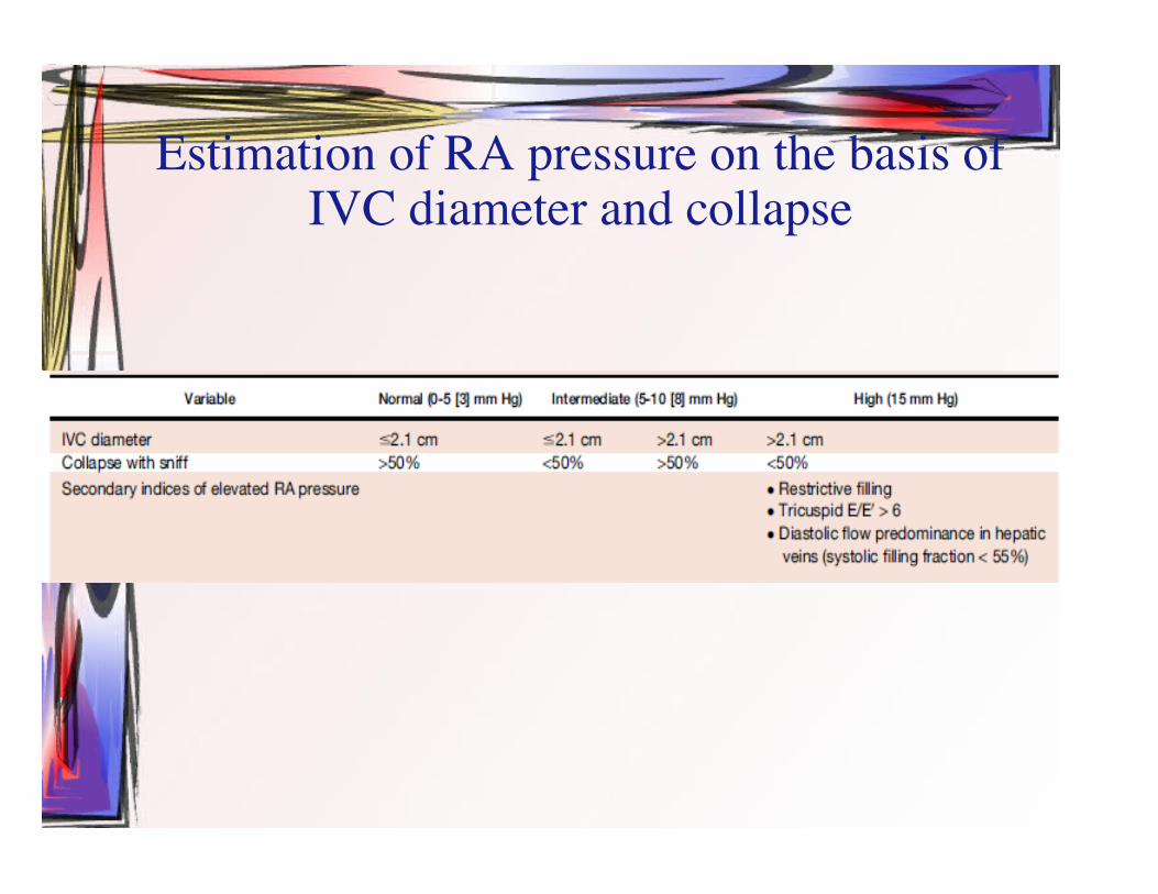
Estimation of RA pressure on the basis ofIVC diameter and collapse

RA Pressure: Guideline statements• For simplicity of reporting, specific values of RA pressure should be used in the
determination of SPAP• In indeterminate cases, if none of the secondary indices of elevated RA pressure
are present, RA pressure may be downgraded to 3 mm Hg• If there is minimal IVC collapse with a sniff (<35%) and secondary indices of
elevated RA pressure are present, RA pressure may be upgraded to 15mmHg.• If uncertainty remains, RA pressure may be left at the intermediate value of 8 mm
Hg.• In patients who are unable to adequately perform a sniff, an IVC that collapses <
20% with quiet inspiration suggests elevated RA pressure• This method of assigning an RA pressure is preferable to assuming a fixed RA
pressure value for all patients

RA pressure: Misreporting• Although a distended IVC usually denotes elevated RA
pressures, in patients with otherwise normal examresults, reassessing the IVC size and collapsibility in theleft lateral position may be useful to avoid the potentiallyerroneous inference of increased RA filling pressure
• The IVC may also be dilated in normal young athletes: itmay not reflect elevated RA pressure

RA pressure: Ventilated patients• In patients being ventilated using positive pressure, the degree of IVC
collapse cannot be used to reliably estimate RA pressure, and RApressure measured by transduction of a central line should be used ifavailable
• An IVC diameter ≤ 12 mm in these patients, appears accurate inidentifying patients with RA pressures < 10 mm Hg
• If the IVC is small and collapsed, this suggests hypovolemia.

RA pressure: secondary signs• Dilated right atrium/ coronary sinus• Interatrial septum that bulges into the left atrium
throughout the cardiac cycle

RV Wall Thickness
RVH RV wall thinning• Pulmonary Hypertension• Infiltrative cardiomyopathy• Hypertrophic cardiomyopathy• In patients with significant LV
hypertrophy, even in the absence ofPH
• Uhl’s anomaly• Arrhythmogenic RV
cardiomyopathy• RV infarct

Measurement of end-diastolic right ventricularwall thickness in 2D and M-Mode
•From the subcostal view, align the ultrasound beam perpendicular to the RV freewall•Move the focus to the RV wall region and decrease the depth to improve theendocardial border definition•Every effort must be made to exclude epicardial fat, erroneously increasingmeasurement.•When image quality permits, fundamental imaging should be used to avoid theincreased structure thickness seen with harmonic imaging.

Recommendations: RV wall thickness
• Abnormal RV wall thickness should be reported in patientssuspected of having RV and/or LV dysfunction, using the normalcutoff of 0.5 cm from either PLAX or subcostal windows
• There are no accepted echocardiographic criteria to define anabnormally thin RV wall

RV Linear Dimensions: Clinicalsignificance
• The right ventricle dilates in response to chronic volume and/orpressure overload and with RV failure
• Indexed RV end-diastolic diameter has been identified as a predictorof survival in patients with chronic pulmonary disease
• RV/LV end-diastolic diameter ratio was shown to be a predictor ofadverse clinical events and/or hospital survival in patients with acutepulmonary embolism
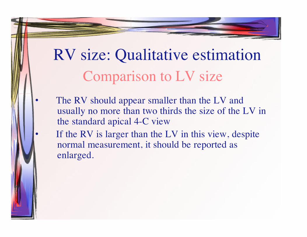
RV size: Qualitative estimation
• The RV should appear smaller than the LV andusually no more than two thirds the size of the LV inthe standard apical 4-C view
• If the RV is larger than the LV in this view, despitenormal measurement, it should be reported asenlarged.
Comparison to LV size

RV size: Qualitative estimation
• Standard TTE apical 4-C window, the LV isconsidered the ‘‘apex-forming’’ ventricle
• As the RV enlarges, it may displace the LV andoccupy the apex: significantly dilated
‘‘Apex-forming’’ ventricle
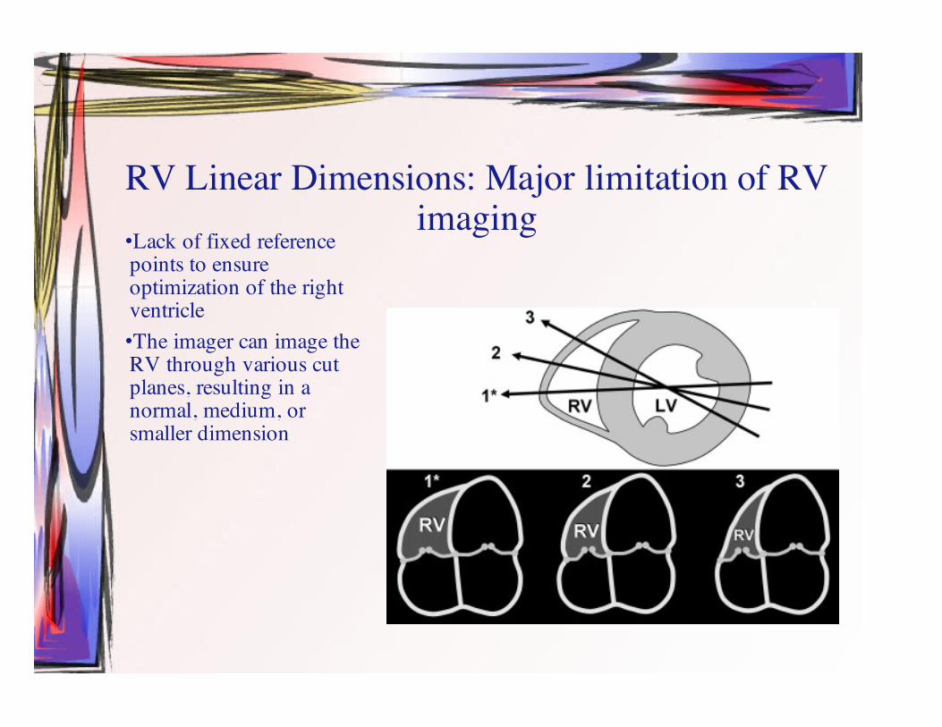
RV Linear Dimensions: Major limitation of RVimaging
•Lack of fixed referencepoints to ensureoptimization of the rightventricle•The imager can image theRV through various cutplanes, resulting in anormal, medium, orsmaller dimension

It is critical to attempt to adjust the apical 4-chamber toacquire the ‘‘right ventricle–focused view’’
Avoid underestimation• Rotate the transducer until
the maximal plane isobtained
Avoid overestimation• Ensure that the RV is not
foreshortened and that the LVOT isnot opened up
• avoid the apical 5-chamber view

Guideline Recommendations: RV linear dimensions• Patients with echocardiographic evidence of right-sided heart disease or PH should
have measurements of RV basal, mid cavity, and longitudinal dimensions on a 4-chamber view
• In all complete echocardiographic studies, the RV basal measurement should bereported, and the report should state the window from which the measurement wasperformed (ideally the right ventricle–focused view)

RVOT size
•Should be measured at end-diastole on the QRS deflection•Limited normative data are available•The window for measurement of RVOT size has not been standardized•Oblique imaging of the RVOT may underestimate or overestimate its size•The endocardial definition of the anterior wall is often suboptimal

RVOT size: GuidelineRecommendations
• Proximal and distal diameters of the RVOT should be measuredfrom the PSAX or PLAX views
• The PSAX distal RVOT diameter, just proximal to thepulmonary annulus, is the most reproducible and should begenerally used

RV systolic function assessment

Regional Assessment of RV Systolic Function: TAPSE orTricuspid Annular Motion (TAM)
Simple and reproducible
Less dependent on optimal image quality
Does not requiresophisticated equipment orprolonged image analysis
Zoom on the tricuspid annulus, place cursor across it and measure the amount of longitudinal motion at peaksystole.

TAPSE: Recommendations TAPSE should be used routinely as a simple
method of estimating RV function, with a lowerreference value for impaired RV systolic functionof 16 mm (1.6 cm)

RV systolic function assessmentTissue Doppler methods: RV S’ or systolic excursion velocity
A simple, reproducible technique with good ability to detect normal versus abnormal RV function

RV S’ or systolic excursion velocity: Technical considerations
An apical 4-chamber window is used with a tissue Doppler mode region ofinterest highlighting the RV free wall The pulsed Doppler sample volume is placed in either the tricuspid annulus orthe middle of the basal segment of the RV free wall Assessment of the mid and apical RV free wall velocities is discouraged in theroutine echocardiographic studies, because there is a lower rate of obtainingadequate signals and greater variability Because the interventricular septum does not exclusively reflect RV function,it should not be used alone to assess the RV

FRACTIONAL AREA CHANGE OF THE RIGHT VENTRICLE
•Trace the endocardial border in apical 4-chamber views•Include the trabeculations, tricuspid leaflets, and chords in thetrace
aPercentage FAC=end-diastolic area

FRACTIONAL AREA CHANGE• Correlates with RV EF by MRI• Independent predictor of heart failure,
sudden death, stroke, and/or mortality instudies of patients after pulmonaryembolism and myocardial infarction

FRACTIONAL AREA CHANGE: GuidelineRecommendations
2D FAC is one of the recommended methods ofquantitatively estimating RV function, with a lowerreference value for normal RV systolic function of 35%

vBased on the relationship between ejection and non-ejection work ofthe heartvThe measure remains accurate within a broad range of heart ratesvfeasible in a large majority of subjects both with and without TRvReproduciblevAvoids the geometric assumptions and limitations of complex RVgeometryvThe pulsed tissue Doppler method allows for measurement of MPI aswell as S’, E’, and A’, all from a single image
Global Assessment of RV Systolic Function:RIMP or Tei Index
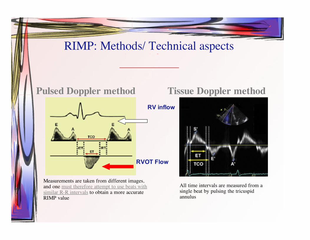
RIMP: Methods/ Technical aspects
Pulsed Doppler method Tissue Doppler method
PW of RV outflowor
CW of the TR jet
Measurements are taken from different images,and one must therefore attempt to use beats withsimilar R-R intervals to obtain a more accurateRIMP value
All time intervals are measured from asingle beat by pulsing the tricuspidannulus
IVRTIVCTET+
RV inflow
RVOT Flow

RIMP or Tei Index: Recommendations
• The MPI may be used for initial and serial measurementsas an estimate of RV function in complement with otherquantitative and non-quantitative measures.
• The upper reference limit for the right-sided MPI is 0.40using the pulsed Doppler method and 0.55 using the pulsedtissue Doppler method
• It should not be used as the sole quantitative method forevaluation of RV function and should not be used withirregular heart rates

• Interrogation of S’ by pulsed tissue Doppler is a simple andreproducible measure to assess basal RV free wall function andshould be used in the assessment of RV function.
• S’ < 10 cm/s should raise the suspicion for abnormal RV function,particularly in a younger adult patient
RV S’ Velocity Recommendations

RV Diastolic Dysfunction: Recommendationsv Measurement of RV diastolic function should be considered in patients with suspected RV
impairment as a marker of early or subtle RV dysfunction or in patients with known RVimpairment as a marker of poor prognosis
v Trans-tricuspid E/A ratio, E/E’ ratio, and RA size have been most validated and are thepreferred measures
v Grading of RV diastolic dysfunction should be done as follows:1. tricuspid E/A ratio < 0.8 suggests impaired relaxation2. tricuspid E/A ratio of 0.8 to 2.1 with an E/E’ ratio > 6 or diastolic flow predominance in the
hepatic veins suggests pseudonormal filling3. tricuspid E/A ratio > 2.1 with a deceleration time < 120 ms suggests restrictive filling (as
does late diastolic antegrade flow in the pulmonary artery)v Further studies are warranted to validate the sensitivity and specificity and the prognostic
implications of this classification

My proposed adoption to normal Protocol:
• In four chamber:– Measure basal RV dimension– Trace RA area (end-systole)– TAPSE– Tissue Doppler of TV annulus (measure S’ velocity
and E’/A’)• In subcostal:
– Measure RV wall thickness– IVC diameter from M-mode (inspiration/expiration)