Embed Size (px)
Citation preview
Journal of the American College of Cardiology Vol. 62, No. 25, Suppl D, 2013� 2013 by the American College of Cardiology Foundation ISSN 0735-1097/$36.00Published by Elsevier Inc. http://dx.doi.org/10.1016/j.jacc.2013.10.027
Downloa
Right Heart Adaptation toPulmonary Arterial Hypertension
Physiology and PathobiologyAnton Vonk-Noordegraaf, MD,* François Haddad, MD,y Kelly M. Chin, MD,z Paul R. Forfia, MD,xSteven M. Kawut, MD,k Joost Lumens, PHD,{ Robert Naeije, MD,# John Newman, MD,**
Ronald J. Oudiz, MD,yy Steve Provencher, MD,zz Adam Torbicki, MD,xxNorbert F. Voelkel, MD,kk{{ Paul M. Hassoun, MDzAmsterdam and Maastricht, the Netherlands; Stanford and Torrance, California; Dallas, Texas;
Philadelphia, Pennsylvania; Brussels, Belgium; Nashville, Tennessee; Chemin Sainte-Foy, Québec, Canada;
Otwock, Poland; Richmond, Virginia; and Baltimore, Maryland
S
From the *Departm
the Netherlands; yStanford Universi
Pulmonary Divisio
Texas; xPulmona
University Hospit
University of Pe
Cardiovascular D
#Department of P
Brussels, Belgium;
Critical Care Med
nessee; yyThe Dav
Hypertension, Div
UCLA Medical C
Group, Centre de
mologie de Québ
Pulmonary Circul
Medical Education
Care Medicine an
wealth University,
more, Maryland.
Bayer, GlaxoSmith
industry advisory
ded From: http
urvival in patients with pulmonary arterial hypertension (PAH) is closely related to right ventricular (RV) function.Although pulmonary load is an important determinant of RV systolic function in PAH, there remains a significantvariability in RV adaptation to pulmonary hypertension. In this report, the authors discuss the emerging concepts ofright heart pathobiology in PAH. More specifically, the discussion focuses on the following questions. 1) How is rightheart failure syndrome best defined? 2) What are the underlying molecular mechanisms of the failing right ventriclein PAH? 3) How are RV contractility and function and their prognostic implications best assessed? 4) What is the roleof targeted RV therapy? Throughout the report, the authors highlight differences between right and left heart failureand outline key areas of future investigation. (J Am Coll Cardiol 2013;62:D22–33) ª 2013 by the AmericanCollege of Cardiology Foundation
Pulmonary arterial hypertension (PAH) is a progressivedisorder that affects both the pulmonary vasculature and theheart (1–6). Although the initial insult in PAH involves thepulmonary vasculature, survival of patients with PAH isclosely related to right ventricular (RV) function (7–19). Theright ventricle adapts to the increased afterload by increasingits wall thickness and contractility. In the vast majority of
ent of Pneumology, VU University Medical Center, Amsterdam,
Division of Cardiovascular Medicine, Department of Medicine,
ty, Stanford, California; zDepartment of Internal Medicine,
n, University of Texas Southwestern Medical Center, Dallas,
ry Hypertension and Right Heart Failure Program, Temple
al, Philadelphia, Pennsylvania; kPerelman School of Medicine,
nnsylvania, Philadelphia, Pennsylvania; {CARIM School for
iseases, Maastricht University, Maastricht, the Netherlands;
athophysiology, Faculty of Medicine, Free University of Brussels,
**Department of Medicine, Division of Allergy, Pulmonary and
icine, Vanderbilt University School of Medicine, Nashville, Ten-
id Geffen School of Medicine at UCLA, Liu Center for Pulmonary
ision of Cardiology, Biomedical Research Institute at Harbor-
enter, Torrance, California; zzPulmonary Hypertension Research
Recherche de l’Institut Universitaire de Cardiologie et de Pneu-
ec, Chemin Sainte-Foy, Québec, Canada; xxDepartment of
ation and Thromboembolic Diseases, Centre of Postgraduate
, ECZ, Otwock, Poland; kkDivision of Pulmonary and Critical
d Victoria Johnson Lab for Lung Research, Virginia Common-
Richmond, Virginia; and the {{Johns Hopkins University, Balti-
Dr. Vonk-Noordegraaf has received lecture fees from Actelion,
Kline, United Therapeutics, Lilly, Pfizer, and Novartis; is on the
board from Actelion and Bayer; and serves on the steering
://content.onlinejacc.org/ on 01/24/2014
patients, however, these compensatory mechanisms areinsufficient, and RV dysfunction occurs. In this report, wehighlight current understanding of RV pathobiology inPAH and briefly outline the evidence underlying themanagement of right heart failure (RHF) in PAH. Futuredirections and priorities of research in RV research in PAHare also discussed. Although the majority of the report
committee for Actelion, Bayer, and Pfizer. Dr. Chin has received research grants
from Actelion, Bayer, Gilead, GlaxoSmithKline, Novartis, United Therapeutics,
GeNO, and the National Institutes of Health; and honoraria for service on advisory
boards for Actelion, Bayer, and Gilead. Dr. Forfia is a consultant for Actelion and
United Therapeutics. Dr. Lumens has received a research grant within the framework
of the Dr. E. Dekker Program of the Dutch Heart Foundation (NHS-2012T010).
Dr. Oudiz has received grants and research funding, consulting and speaking fees,
and honoraria from Actelion, AIRES, Bayer, Gilead, Ikaria, Lung LLC, Pfizer,
United Therapeutics. Dr. Provencher has received consulting fees from Actelion,
GlaxoSmithKline, Pfizer, and Unither Biotech; research grants from Actelion, Bayer,
and GlaxoSmithKline; and speaker fees from Actelion. Dr. Torbicki has received
research grants and speaker’s honoraria from Actelion, Bayer, AOP, United Thera-
peutics, and GlaxoSmithKline; and has served on advisory boards and scientific
committees for Actelion, Bayer, AOP, and GlaxoSmithKline. Dr. Voelkel has
received grant support for pre-clinical studies from Actelion and United Therapeu-
tics. Dr. Hassoun has participated on advisory boards for Gilead, Merck, Bayer, and
Novartis; has received research funding from United Therapeutics for the Registry to
Evaluate Early and Long-Term Pulmonary Arterial Hypertension Disease
Management; and his research was supported by grants P50 HL084946 and R01
HL114910 from the National Heart, Lung, and Blood Institute. All other authors
have reported that they have no relationships relevant to the contents of this paper to
disclose. Drs. Vonk-Noordegraaf and Haddad contributed equally to this manuscript.
Manuscript received October 15, 2013; accepted October 22, 2013.
Abbreviationsand Acronyms
BNP = B type natriuretic
peptide
CO = cardiac output
Ea = arterial elastance
Ees = ventricular elastance
LV = left ventricular
MHC = myosin heavy chain
MPAP = mean pulmonary
arterial pressure
PAC = pulmonary arterial
compliance
PAH = pulmonary arterial
hypertension
PH = pulmonary
hypertension
PVR = pulmonary vascular
resistance
RAP = right atrialpressure
RHF = right heart failure
RNA = ribonucleic acid
RV = right ventricular
RVEF = right ventricular
ejection fraction
JACC Vol. 62, No. 25, Suppl D, 2013 Vonk-Noordegraaf et al.December 24, 2013:D22–33 The Right Ventricle in Pulmonary Hypertension
D23
Downloa
focuses on RHF in the setting of PAH, the committee alsowants to emphasizes that RV function is also a strongpredictor of outcomes in patients with left heart failure,advanced lung disease, and congenital heart disease (20).
Definition of the Right Heart Failure Syndrome
RHF in patients with PAH can be defined as a complexclinical syndrome due to suboptimal delivery of blood and/orelevated systemic venous pressure at rest or exercise asa consequence of elevated RV afterload.
The cardinal clinical manifestations of RHF are exerciselimitation and fluid retention. Exercise limitation is theearliest symptom of RHF and is a strong predictor ofsurvival in patients with PAH (8,14,21). Exercise limitationis related to a decrease in flow reserve during exercise(decreased peak cardiac index) (22–24). In addition,a reduction in peripheral blood flow can increase lactateproduction, further contributing to muscle fatigue andexercise limitation. Supraventricular tachycardia, which canoccur in approximately 12% of patients with PAH orinoperable thromboembolic pulmonary hypertension, canalso lead to clinical deterioration and reduced exercisecapacity (25). Syncope, a less common symptom of PAH,often indicates severe limitations in flow reserve. As in left-sided heart failure, RHF can also lead to chronic kidneydisease and hyponatremia (26). Shah et al. (27) showed thatchronic kidney disease in patients with PAH is associatedwith increased right atrial pressure and a higher likelihood ofdeath or transplantation. Similarly, acute kidney injury hasalso been associated with worse outcomes after acute RHF(28). Although congestive hepatopathy is often observed inpatients with RHF and PAH, cirrhosis is a late complicationof severe RHF. In patients with worsening hypoxemia andPAH, right-to-left shunting through a patent foramen ovalemust be considered.
Patients with PAH may also present with acute heartfailure. Recent studies have shown that the short-termmortality in patients with PAH and acute RHF may be ashigh as 40% in patients who require admission to the hospital(29–32). Although the majority of patients with acute RHFare admitted with congestive symptoms requiring diuretictherapy, a smaller proportion of patients will have low–cardiacoutput syndrome requiring inotropic or vasopressor support(29,30). Although the most common cause of death inpatients with PAH is progressive RHF, sudden and unex-pected death may also occur (33). In a study by Hoeper et al.(33), sudden death explained 17% of cardiopulmonary arrestfor which resuscitation was attempted. Among all patientswho had cardiopulmonary arrest (not just those with suddendeath), the initial electrocardiogram at the time of cardio-pulmonary resuscitation showed bradycardia in 45%, elec-tromechanical dissociation in 28%, asystole in 15%,ventricular fibrillation in 8%, and other rhythms in 4% (33).
In patients with chronic left heart failure, heart failure isusually classified into 4 stages of development: at risk for heart
ded From: http://content.onlinejacc.org/ on 01/24/2014
failure (stage A), “asymptomatic”heart dysfunction (stage B), symp-tomatic heart failure (stage C), andend-stage heart failure (stage D)(34). This classification couldpotentially be applied to patientswith RHF, with the caveat thatthe majority of patients withadvanced RHF (stage D) havethe ability to reverse remodelafter lung transplantation. Alsoimportantly, although we oftenconsider the right ventricle andthe left ventricle as separate en-tities, this distinction is some-what artificial, because bothventricles are interconnected th-rough the interventricular sep-tum, shared myofibers, and thepericardium. Because of ventric-ular interdependence, patientswith RHF will often have relax-ation abnormalities of the leftventricle and, in severe cases,even left ventricular systolicdysfunction (2,20,35). Recent stu-dies have also emphasized electro-
physiological remodeling in the left ventricle in patientswith PAH (36).Pathobiology of Right Heart Failure Syndrome inPulmonary Arterial Hypertension
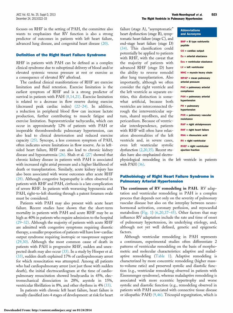
The continuum of RV remodeling in PAH. RV adap-tation and ventricular remodeling in PAH is a complexprocess that depends not only on the severity of pulmonaryvascular disease but also on the interplay between neuro-hormonal activation, coronary perfusion, and myocardialmetabolism (Fig. 1) (6,20,37–45). Other factors that mayinfluence RV adaptation include the rate and time of onsetof pulmonary hypertension, its underlying etiology, and,although not yet well defined, genetic and epigeneticfactors.
Although ventricular remodeling in PAH representsa continuum, experimental studies often differentiate 2patterns of ventricular remodeling on the basis of morpho-metric and molecular characteristics: adaptive and malad-aptive remodeling (Table 1). Adaptive remodeling ischaracterized by more concentric remodeling (higher mass-to-volume ratio) and preserved systolic and diastolic func-tion (e.g., ventricular remodeling observed in patients withEisenmenger syndrome), whereas maladaptive remodeling isassociated with more eccentric hypertrophy and worsesystolic and diastolic function (e.g., remodeling observed inpatients with PAH associated with connective tissue diseaseor idiopathic PAH) (9,46). Tricuspid regurgitation, which is
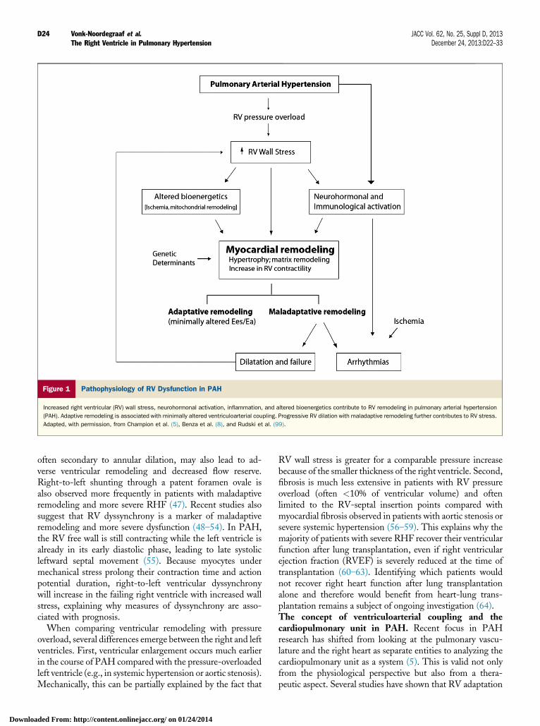
Figure 1 Pathophysiology of RV Dysfunction in PAH
Increased right ventricular (RV) wall stress, neurohormonal activation, inflammation, and altered bioenergetics contribute to RV remodeling in pulmonary arterial hypertension
(PAH). Adaptive remodeling is associated with minimally altered ventriculoarterial coupling. Progressive RV dilation with maladaptive remodeling further contributes to RV stress.
Adapted, with permission, from Champion et al. (5), Benza et al. (8), and Rudski et al. (99).
Vonk-Noordegraaf et al. JACC Vol. 62, No. 25, Suppl D, 2013The Right Ventricle in Pulmonary Hypertension December 24, 2013:D22–33
D24
Downloa
often secondary to annular dilation, may also lead to ad-verse ventricular remodeling and decreased flow reserve.Right-to-left shunting through a patent foramen ovale isalso observed more frequently in patients with maladaptiveremodeling and more severe RHF (47). Recent studies alsosuggest that RV dyssynchrony is a marker of maladaptiveremodeling and more severe dysfunction (48–54). In PAH,the RV free wall is still contracting while the left ventricle isalready in its early diastolic phase, leading to late systolicleftward septal movement (55). Because myocytes undermechanical stress prolong their contraction time and actionpotential duration, right-to-left ventricular dyssynchronywill increase in the failing right ventricle with increased wallstress, explaining why measures of dyssynchrony are asso-ciated with prognosis.
When comparing ventricular remodeling with pressureoverload, several differences emerge between the right and leftventricles. First, ventricular enlargement occurs much earlierin the course of PAH compared with the pressure-overloadedleft ventricle (e.g., in systemic hypertension or aortic stenosis).Mechanically, this can be partially explained by the fact that
ded From: http://content.onlinejacc.org/ on 01/24/2014
RV wall stress is greater for a comparable pressure increasebecause of the smaller thickness of the right ventricle. Second,fibrosis is much less extensive in patients with RV pressureoverload (often <10% of ventricular volume) and oftenlimited to the RV-septal insertion points compared withmyocardial fibrosis observed in patients with aortic stenosis orsevere systemic hypertension (56–59). This explains why themajority of patients with severe RHF recover their ventricularfunction after lung transplantation, even if right ventricularejection fraction (RVEF) is severely reduced at the time oftransplantation (60–63). Identifying which patients wouldnot recover right heart function after lung transplantationalone and therefore would benefit from heart-lung trans-plantation remains a subject of ongoing investigation (64).The concept of ventriculoarterial coupling and thecardiopulmonary unit in PAH. Recent focus in PAHresearch has shifted from looking at the pulmonary vascu-lature and the right heart as separate entities to analyzing thecardiopulmonary unit as a system (5). This is valid not onlyfrom the physiological perspective but also from a thera-peutic aspect. Several studies have shown that RV adaptation
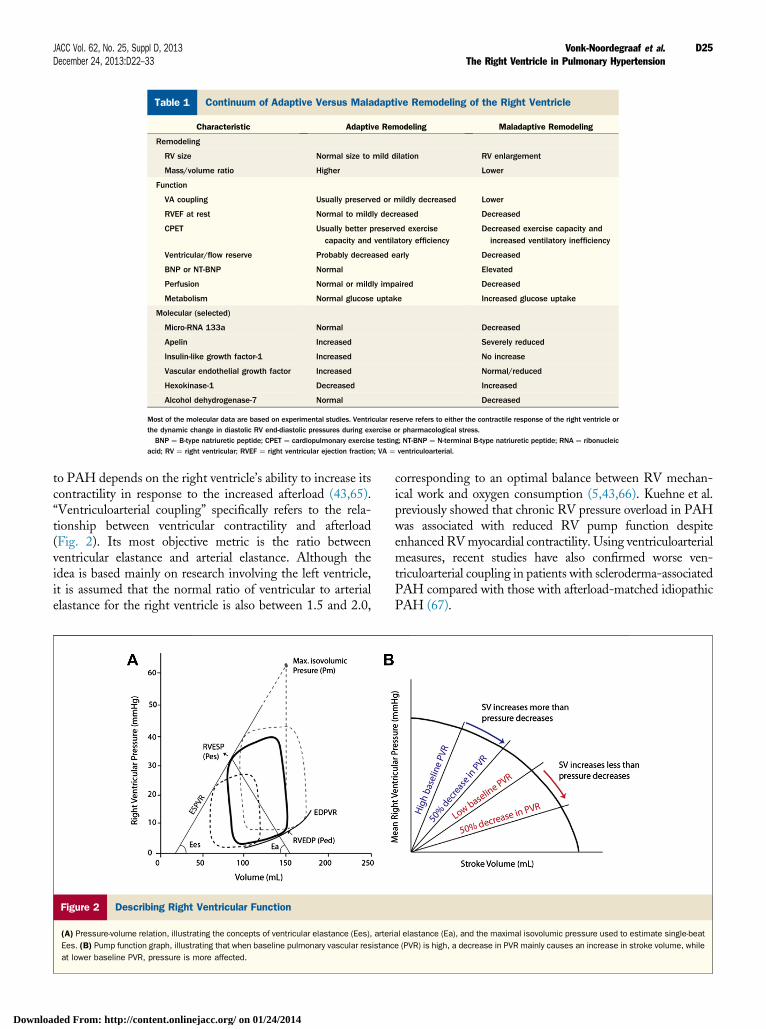
Table 1 Continuum of Adaptive Versus Maladaptive Remodeling of the Right Ventricle
Characteristic Adaptive Remodeling Maladaptive Remodeling
Remodeling
RV size Normal size to mild dilation RV enlargement
Mass/volume ratio Higher Lower
Function
VA coupling Usually preserved or mildly decreased Lower
RVEF at rest Normal to mildly decreased Decreased
CPET Usually better preserved exercisecapacity and ventilatory efficiency
Decreased exercise capacity andincreased ventilatory inefficiency
Ventricular/flow reserve Probably decreased early Decreased
BNP or NT-BNP Normal Elevated
Perfusion Normal or mildly impaired Decreased
Metabolism Normal glucose uptake Increased glucose uptake
Molecular (selected)
Micro-RNA 133a Normal Decreased
Apelin Increased Severely reduced
Insulin-like growth factor-1 Increased No increase
Vascular endothelial growth factor Increased Normal/reduced
Hexokinase-1 Decreased Increased
Alcohol dehydrogenase-7 Normal Decreased
Most of the molecular data are based on experimental studies. Ventricular reserve refers to either the contractile response of the right ventricle orthe dynamic change in diastolic RV end-diastolic pressures during exercise or pharmacological stress.BNP ¼ B-type natriuretic peptide; CPET ¼ cardiopulmonary exercise testing; NT-BNP ¼ N-terminal B-type natriuretic peptide; RNA ¼ ribonucleic
acid; RV ¼ right ventricular; RVEF ¼ right ventricular ejection fraction; VA ¼ ventriculoarterial.
JACC Vol. 62, No. 25, Suppl D, 2013 Vonk-Noordegraaf et al.December 24, 2013:D22–33 The Right Ventricle in Pulmonary Hypertension
D25
Downloa
to PAH depends on the right ventricle’s ability to increase itscontractility in response to the increased afterload (43,65).“Ventriculoarterial coupling” specifically refers to the rela-tionship between ventricular contractility and afterload(Fig. 2). Its most objective metric is the ratio betweenventricular elastance and arterial elastance. Although theidea is based mainly on research involving the left ventricle,it is assumed that the normal ratio of ventricular to arterialelastance for the right ventricle is also between 1.5 and 2.0,
Figure 2 Describing Right Ventricular Function
(A) Pressure-volume relation, illustrating the concepts of ventricular elastance (Ees), arter
Ees. (B) Pump function graph, illustrating that when baseline pulmonary vascular resistanc
at lower baseline PVR, pressure is more affected.
ded From: http://content.onlinejacc.org/ on 01/24/2014
corresponding to an optimal balance between RV mechan-ical work and oxygen consumption (5,43,66). Kuehne et al.previously showed that chronic RV pressure overload in PAHwas associated with reduced RV pump function despiteenhanced RVmyocardial contractility. Using ventriculoarterialmeasures, recent studies have also confirmed worse ven-triculoarterial coupling in patients with scleroderma-associatedPAH compared with those with afterload-matched idiopathicPAH (67).
ial elastance (Ea), and the maximal isovolumic pressure used to estimate single-beat
e (PVR) is high, a decrease in PVR mainly causes an increase in stroke volume, while
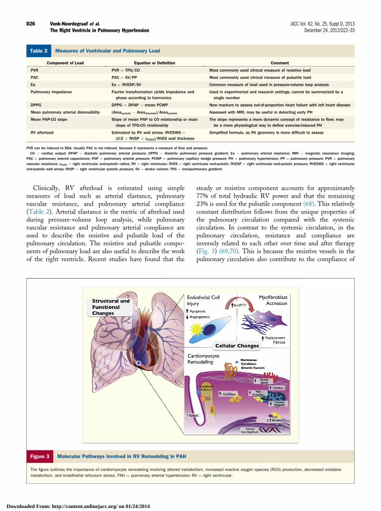
Table 2 Measures of Ventricular and Pulmonary Load
Component of Load Equation or Definition Comment
PVR PVR ¼ TPG/CO Most commonly used clinical measure of resistive load
PAC PAC ¼ SV/PP Most commonly used clinical measure of pulsatile load
Ea Ea ¼ RVESP/SV Common measure of load used in pressure-volume loop analysis
Pulmonary impedance Fourier transformation yields impedance andphase according to harmonics
Used in experimental and research settings; cannot be summarized by asingle number
DPPG DPPG ¼ DPAP � mean PCWP New markers to assess out-of-proportion heart failure with left heart disease
Mean pulmonary arterial distensibility (Areasystole � Areadiastole)/Areasystole Assessed with MRI; may be useful in detecting early PH
Mean PAP-CO slope Slope of mean PAP to CO relationship or mainslope of TPG-CO relationship
The slope represents a more dynamic concept of resistance to flow; maybe a more physiological way to define exercise-induced PH
RV afterload Estimated by RV wall stress: RVESWS ¼(0.5 � RVSP � rRVES)/RVES wall thickness
Simplified formula, as RV geometry is more difficult to assess
PVR can be indexed to BSA. Usually PAC is not indexed, because it represents a measure of flow and pressure.CO ¼ cardiac output; DPAP ¼ diastolic pulmonary arterial pressure; DPPG ¼ diastolic pulmonary pressure gradient; Ea ¼ pulmonary arterial elastance; MRI ¼ magnetic resonance imaging;
PAC ¼ pulmonary arterial capacitance; PAP ¼ pulmonary arterial pressure; PCWP ¼ pulmonary capillary wedge pressure; PH ¼ pulmonary hypertension; PP ¼ pulmonary pressure; PVR ¼ pulmonaryvascular resistance; rRVES ¼ right ventricular end-systolic radius; RV ¼ right ventricular; RVES ¼ right ventricular end-systolic; RVESP ¼ right ventricular end-systolic pressure; RVESWS ¼ right ventricularend-systolic wall stress; RVSP ¼ right ventricular systolic pressure; SV ¼ stroke volume; TPG ¼ transpulmonary gradient.
Vonk-Noordegraaf et al. JACC Vol. 62, No. 25, Suppl D, 2013The Right Ventricle in Pulmonary Hypertension December 24, 2013:D22–33
D26
Downloa
Clinically, RV afterload is estimated using simplemeasures of load such as arterial elastance, pulmonaryvascular resistance, and pulmonary arterial compliance(Table 2). Arterial elastance is the metric of afterload usedduring pressure-volume loop analysis, while pulmonaryvascular resistance and pulmonary arterial compliance areused to describe the resistive and pulsatile load of thepulmonary circulation. The resistive and pulsatile compo-nents of pulmonary load are also useful to describe the workof the right ventricle. Recent studies have found that the
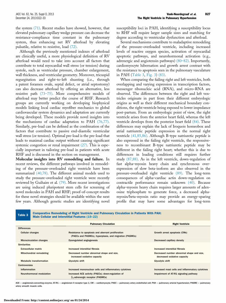
Figure 3 Molecular Pathways Involved in RV Remodeling in PAH
The figure outlines the importance of cardiomyocyte remodeling involving altered metabo
metabolism, and endothelial reticulum stress. PAH ¼ pulmonary arterial hypertension; R
ded From: http://content.onlinejacc.org/ on 01/24/2014
steady or resistive component accounts for approximately77% of total hydraulic RV power and that the remaining23% is used for the pulsatile component (68). This relativelyconstant distribution follows from the unique properties ofthe pulmonary circulation compared with the systemiccirculation. In contrast to the systemic circulation, in thepulmonary circulation, resistance and compliance areinversely related to each other over time and after therapy(Fig. 3) (69,70). This is because the resistive vessels in thepulmonary circulation also contribute to the compliance of
lism, increased reactive oxygen species (ROS) production, decreased oxidative
V ¼ right ventricular.
JACC Vol. 62, No. 25, Suppl D, 2013 Vonk-Noordegraaf et al.December 24, 2013:D22–33 The Right Ventricle in Pulmonary Hypertension
D27
Downloa
the system (71). Recent studies have showed, however, thatelevated pulmonary capillary wedge pressure can decrease theresistance-compliance time constant in the pulmonarysystem, thus enhancing net RV afterload by elevatingpulsatile, relative to resistive, load (72).
Although the previously mentioned indexes of afterloadare clinically useful, a more physiological definition of RVafterload would need to take into account all factors thatcontribute to total myocardial wall stress (or tension) duringsystole, such as ventricular pressure, chamber enlargement,wall thickness, and ventricular geometry. Moreover, tricuspidregurgitation and right-to-left shunting (i.e., througha patent foramen ovale, septal defect, or atrial septostomy)can also decrease afterload by offering an alternative, lessresistive path (73–75). More comprehensive models ofafterload may better predict the course of RHF. Researchgroups are currently working on developing biophysicalmodels linking local cardiac myofiber mechanics to globalcardiovascular system dynamics and adaptation are currentlybeing developed. These models provide novel insights intothe mechanisms of cardiac adaptation to PAH (76,77).Similarly, pre-load can be defined as the combination of thefactors that contribute to passive end-diastolic ventricularwall stress (or tension). Optimal pre-load is the pre-load thatleads to maximal cardiac output without causing significantsystemic congestion or renal impairment (27). This is espe-cially important in tailoring pre-load in patients with acuteRHF and is discussed in the section on management.Molecular insights into RV remodeling and failure. Inrecent reviews, the different pathways involved in remodel-ing of the pressure-overloaded right ventricle have beensummarized (40,78). The different animal models used tostudy the pressure-overloaded right ventricle were recentlyreviewed by Guihaire et al. (79). More recent investigationsare using induced pluripotent stem cells for screening ofnovel molecules in PAH and RHF; proof-of-concept resultsfor these novel strategies should be available within the nextfew years. Although genetic studies are identifying novel
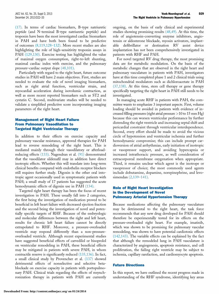
Table 3Comparative Remodeling of Right Ventricle and Pulmonary CMain Cellular and Interstitial Features (19–22)
Pulmonary Circulation
Differences
Cellular changes Resistance to apoptosis and aberrant prolif(PAECs and PASMCs), hyperplasia, and m
Microcirculation changes Dysregulated angiogenesis
Similarities
Extracellular matrix Increased interstitial fibrosis
Mitochondrial remodeling Decreased number abnormal shape and sizincreased oxidative capacity
Metabolic transformation Glycolytic shift
Controversies
Inflammation Increased mononuclear cells and inflamma
Neurohormonal modulation Increased ACE activity (PAECs); down-regulab1-adrenergic receptor (PASMCs)
ACE ¼ angiotensin-converting enzyme; AT-R1 ¼ angiotensin II receptor type 1; CM ¼ cardiomyocyte; PAECartery smooth muscle cells.
ded From: http://content.onlinejacc.org/ on 01/24/2014
susceptibility loci in PAH, identifying a susceptibility locusto RHF will require larger sample sizes and matching fordegree according to ventricular dysfunction and afterload.
Several mechanisms contribute to maladaptive remodelingof the pressure-overloaded ventricle, including increasedlevels of reactive oxygen species, activation of myocardialapoptotic pathways, and neurohormonal activation (e.g.,adrenergic and angiotensin pathways) (80–82). Importantly,cardiomyocyte hibernation and growth arrest contrast withthe resistance to apoptosis seen in the pulmonary vasculaturein PAH (Table 3, Fig. 3) (83).
When comparing the failing right and left ventricles, bothoverlapping and varying expression in transcription factors,messenger ribonucleic acid (RNA), and micro-RNA areobserved. The differences between the right and left ven-tricles originate in part from their different embryologicorigins as well as their different mechanical boundary con-ditions, the right ventricle being exposed to lower impedancepost-partum. From an embryologic point of view, the rightventricle arises from the anterior heart field, whereas the leftventricle develops from the posterior heart field (84). Thesedifferences may explain the lack of Iroquois homeobox andatrial natriuretic peptide expression in the normal rightventricle (41,85,86). Although B-type natriuretic peptide isalso expressed in the failing right ventricle, the responsive-ness to recombinant B-type natriuretic peptide may bedifferent in the failing right heart; whether this is due todifferences in loading conditions still requires furtherstudy (87,88). As in the left ventricle, down-regulation offast alpha–myosin heavy chain and synchronous over-expression of slow beta-isoform are also observed in thepressure-overloaded right ventricle (89). The long-termconsequences of alpha–cardiac actin down-regulation oncontractile performance remain unknown (40). Becausealpha–myosin heavy chain requires larger amounts of aden-osine triphosphate to generate force, a decreased alpha-myosin/beta-myosin ratio may provide an energy-sparingprofile that may have some advantages for long-term
irculation in Patients With PAH:
Right Ventricle
erationigration (PASMCs)
Growth arrest apoptosis (CMs)
Decreased capillary density
Increased interstitial fibrosis
e, Decreased number abnormal shape and size,decreased oxidative capacity
Glycolytic shift
tory cytokines Increased mast cells and inflammatory cytokines
tion of Impairment of AT-R1 signaling pathway
¼ pulmonary artery endothelial cell; PAH ¼ pulmonary arterial hypertension; PASMC ¼ pulmonary
Vonk-Noordegraaf et al. JACC Vol. 62, No. 25, Suppl D, 2013The Right Ventricle in Pulmonary Hypertension December 24, 2013:D22–33
D28
Downloa
compensation of the right ventricle. Studies using microarraygene chips have highlighted that 1 of the pathways thatappears to be more activated in the pressure-overloaded rightventricle compared with the pressure overloaded left ventricleis the Wnt pathway, which regulates glycogen synthasekinase–3b activity, a serine/threonine protein kinase involvedin cell proliferation, migration, inflammation, glucose regu-lation, and apoptosis (90). Chamber-specific expression ofphosphodiesterase type 5 in the pressure-overloaded rightventricle has also been demonstrated in experimental andhuman studies (91,92). Although most expressed micro-RNA is similar in right and left heart failure, Reddy et al.(93) showed that 4 micro-RNAs (34a, 28, 148a, and 93) areupregulated in RHF/RV hypertrophy that are down-regulated or unchanged in left heart failure/left ventricularhypertrophy.
A change in myocardial metabolism is a prominentfeature of RHF. A switch from fatty acid oxidation toglycolysis is presumably a protective response of the stressedleft heart, as carbohydrate metabolism requires less oxygenamount than fatty acid oxidation (81). Decreased mito-chondrial activity resulting in a switch from aerobic toanaerobic metabolism might also be involved in the transi-tion from compensated RV hypertrophy to maladaptiveremodeling (81). Insufficient adaptation of the capillarynetwork and myocardial ischemia may also impair vascularendothelial growth factor signaling and the hypertrophicresponse (94). Increased rate of myocardial fibrosis has beenreported in RV failure and may contribute to decreasedventricular compliance (56,95).
Recent animal and human studies support a role ofinflammation in the pressure-overloaded right ventricle(82,96). Studies by Watts et al. (97) have shown thatneutrophils are found in the RV myocardium early after anacute increase in afterload, whereas macrophages may beinvolved during progressive remodeling in the setting ofchronic pulmonary hypertension (97). Ongoing studies areinvestigating the role of macrophages, T-regulatory cells, andleukotriene in the development of RHF in patients with PAH.
Evaluation of Right Heart Size and Function:From Resting Parameters to Dynamic Evaluation
The assessment of the right heart plays an essentialpart in managing patients with PAH (98). Although echo-cardiography is the mainstay in the evaluation of the rightheart in clinical practice, magnetic resonance imaging hasemerged as the most accurate method for evaluating RVmass, RV volume, and RVEF. In addition, magnetic reso-nance imaging offers the possibility to quantify regurgitantvolumes; delayed enhancement, a marker for focal scarburden; myocardial strain; coronary perfusion; and pulmo-nary pulsatility (98). Positron emission tomography is usedexperimentally to assess RV and pulmonary metabolism and,at specialized centers, for apoptosis imaging. Finallyconductance catheterization represents the gold-standard
ded From: http://content.onlinejacc.org/ on 01/24/2014
method for evaluating ventricular elastance, arterial ela-stance, and ventriculoarterial coupling (5,43).
The American Society of Echocardiography guidelinesoffer the most comprehensive review of normative echo-cardiographic values of the right heart (99). The guidelinealso highlights the need for future scaled echocardiographicreferences for right heart dimensions as well as referencesadjusted for age, sex, and race. Using a large cohort of 4,204participants, the MESA (Multi-Ethnic Study of Athero-sclerosis) investigators were able to develop these normativeequations for RV mass and systolic function on the basis ofage, sex, and race (100). Kawut et al. (100) demonstratedthat in general, younger age, male sex, and Hispanicethnicity are associated with higher RV mass, while olderage and female sex are associated with higher RVEF.
More recently, several investigators are working on definingdynamic right heart and pulmonary circulation measures.These dynamic measures include themean pulmonary arterialpressure–cardiac output slope as well as RV reserve, usuallydefined either as peak RVEF, peak stroke volume, or peakcardiac index after exercise or pharmacological stress (101,102).In controls, mean pulmonary arterial pressure–cardiac outputslope is usually <1.5 to 2.5 mm Hg$min/l, with older healthysubjects having higher average slope values (22,103–113).
The committee wants to emphasize that the commonlyused indexes of RV systolic performance, such as RVEFand tricuspid annular plane systolic excursion, are markersof ventriculoarterial coupling rather than ventricular con-tractility, which is increased in PAH (65). Another caveat thecommitteememberswant to emphasize is that the reduction intricuspid annular plane systolic excursion after cardiac surgerydoes not reflect corresponding changes in RVEF, becauseannular plane motion is preferentially compromised (114).
Prediction of Outcomes in Patients WithPulmonary Arterial Hypertension:The Importance of the Right Heart
Outcome prediction in patients with PAH has been exten-sively studied using large-cohort designs as well as in smallerstudies incorporating imaging parameters (7,8,10,12,13,16–19,115–123). One consistent finding among studies isthat survival in PAH is closely related to RV adaptation tothe increased pressure overload. Hemodynamic studies havedemonstrated the predictive value of right atrial pressure andcardiac index (7,14,117,118,124). Echocardiographic studieshave highlighted the predictive value of tricuspid annularplane systolic excursion, RV myocardial performance in-dex, atrial size, and pericardial effusion (10,12,13,17,125).Magnetic resonance imaging studies have emphasizedthe predictive value of stroke volume index, RVEF, andindexed RV end-diastolic and end-systolic volumes(18,115,116,122,126). Although delayed enhancement hasbeen associated with the severity of PAH, its independentpredictive value has not yet been proved (127). More recentstudies have shown the potential predictive value of RV strain
JACC Vol. 62, No. 25, Suppl D, 2013 Vonk-Noordegraaf et al.December 24, 2013:D22–33 The Right Ventricle in Pulmonary Hypertension
D29
Downloa
(17). In terms of cardiac biomarkers, B-type natriureticpeptide (and N-terminal B-type natriuretic peptide) andtroponin have been the most investigated cardiac biomarkersin PAH and have both been found to be predictiveof outcomes (8,119,128–132). More recent studies are alsohighlighting the role of high-sensitivity troponin assays inPAH (129,130). Exercise testing has highlighted the valueof maximal oxygen consumption, right-to-left shunting,maximal cardiac index with exercise, and the pulmonarypressure–cardiac output slope (48,101).
Particularly with regard to the right heart, future outcomestudies in PAH will have 2 main objectives. First, studies areneeded to evaluate the role of novel imaging biomarkers,such as right atrial function, ventricular strain, andmyocardial acceleration during isovolumic contraction, aswell as more recent reported biomarkers such as ST2 andcystatin C. Second, multivariate studies will be needed tovalidate a simplified predictive score incorporating imagingparameters of the right heart.
Management of Right Heart FailureFrom Pulmonary Vasodilation toTargeted Right Ventricular Therapy
In addition to their effects on exercise capacity andpulmonary vascular resistance, approved therapies for PAHlead to reverse remodeling of the right heart. This ismediated mainly through their vasodilatory or afterload-reducing effects (133). Nagendran et al. (91) demonstratedthat the vasodilator sildenafil may in addition have directinotropic effects. Whether this will translate into long-termclinical benefits compared with endothelin receptor blockersstill requires further study. Digoxin is the other oral into-tropic agent occasionally used in symptomatic patients withPAH; a small study of 17 patients has suggested the acutehemodynamic effects of digoxin use in PAH (134).
Targeted right heart therapy has been the focus of recentinvestigation in PAH. These usually fall into 2 categories,the first being the investigation of medication proved to bebeneficial in left heart failure with decreased ejection fractionand the second being the investigation of novel and poten-tially specific targets of RHF. Because of the embryologicand molecular differences between the right and left heart,results for chronic left heart failure cannot be directlyextrapolated to RHF. Moreover, a pressure-overloadedventricle may respond differently than a non-pressure-overloaded ventricle. Although recent experimental studieshave suggested beneficial effects of carvedilol or bisoprololon ventricular remodeling in PAH, these beneficial effectsmay be mitigated in patients with severe PAH, in whomcontractile reserve is significantly reduced (135,136). In fact,a small clinical study by Provencher et al. (137) showeddetrimental effects of nonselective and selective beta-blockade on exercise capacity in patients with portopulmo-nary PAH. Clinical trials regarding the effects of resynch-ronization therapy in patients with PAH are currently
ded From: http://content.onlinejacc.org/ on 01/24/2014
ongoing, on the basis of early clinical and experimentalstudies showing promising results (48,49). At this time, therole of angiotensin-converting enzyme inhibitors, angio-tensin or aldosterone blockade, myosin activators, implant-able defibrillator or destination RV assist deviceimplantation has not been comprehensively investigated inpatients with RHF and PAH.
For novel targeted RV drug therapy, the most promisingdata are for metabolic modulation. On the basis of themetabolic changes that are observed in the right heart andpulmonary vasculature in patients with PAH, investigatorshave at this time completed phase 1 and 2 clinical trials usingmitochondrial modulators such as dichloroacetate in PAH(37,138). At this time, stem cell therapy or gene therapyspecifically targeting the right heart in PAH still needs to beinvestigated.
In managing acute RHF in patients with PAH, the com-mittee wants to emphasize 3 important aspects. First, volumeloading should be avoided in patients with evidence of in-creased filling pressures (right atrial pressure>10 to 15mmHg)because this can worsen ventricular performance by furtherdistending the right ventricle and increasing septal shift andpericardial constraint through ventricular interdependence.Second, every effort should be made to avoid the viciouscircle of hypotension and ventricular ischemia and furtherhemodynamic compromise; this can include prompt car-dioversion of atrial arrhythmias, early initiation of inotropicor vasopressor support, and avoiding hypercapnia orincreased intrathoracic pressure, as well consideration ofextracorporeal membrane oxygenation when appropriate.Third, it remains unclear which agent is the inotrope orvasopressor of choice; the most commonly used agentsinclude dobutamine, dopamine, norepinephrine, and levo-simendan (2,139–141).
Role of Right Heart Investigationin the Development of NovelPulmonary Arterial Hypertension Therapy
Because medications affecting the pulmonary vasculaturemay be detrimental to the right heart, the task forcerecommends that any new drug developed for PAH shouldtherefore be experimentally tested for its effects on thepressure-overloaded right heart. For example, imatinib,which was shown to be promising for pulmonary vascularremodeling, was shown to have potential cardiotoxic effects(142,143). The variable effects can be explained by the factthat although the remodeled lung in PAH vasculature ischaracterized by angiogenesis, apoptosis resistance, and cellproliferation, the failing right ventricle may be subject toischemia, capillary rarefaction, and cardiomyocyte apoptosis.
Future Directions
In this report, we have outlined the recent progress made inunderstanding of the RHF syndrome, identifying key areas
Vonk-Noordegraaf et al. JACC Vol. 62, No. 25, Suppl D, 2013The Right Ventricle in Pulmonary Hypertension December 24, 2013:D22–33
D30
Downloa
of future investigation. Areas of research priority are diverseand include refining the definition of normal right heartstructure and function; investigating novel genetic, epige-netic, and molecular pathways of RHF; and developingmore effective management strategies for refractory RHF.Furthermore, before proceeding to clinical trials, the effectsof any new medication should also be experimentally testedon a pressure-overloaded right ventricle. Importantly, wealso recommend that clinical trials in PAH incorporate assecondary outcome analysis parameters of right heart sizeand function.
Reprint requests and correspondence: Dr. Anton Vonk-Noordegraaf, Department of Pulmonology, VU UniversityMedical Center, De Boelelaan 1117, 1081 HV Amsterdam, theNetherlands. E-mail: [email protected].
REFERENCES
1. Chesler NC, Roldan A, Vanderpool RR, Naeije R. How to measurepulmonary vascular and right ventricular function. In: Proceedingsof the Annual International Conference of the IEEE Engineeringin Medicine and Biology Society. New York, NY: IEEE, 2009:177–80.
2. Dell’Italia LJ. Anatomy and physiology of the right ventricle. CardiolClin 2012;30:167–87.
3. Farber HW, Loscalzo J. Pulmonary arterial hypertension. N Engl JMed 2004;351:1655–65.
4. Haddad F, Hunt SA, Rosenthal DN, Murphy DJ. Right ventricularfunction in cardiovascular disease, part I: anatomy, physiology, aging,and functional assessment of the right ventricle. Circulation 2008;117:1436–48.
5. Champion HC, Michelakis ED, Hassoun PM. Comprehensiveinvasive and noninvasive approach to the right ventricle-pulmonarycirculation unit: state of the art and clinical and research implica-tions. Circulation 2009;120:992–1007.
6. Voelkel NF, Quaife RA, Leinwand LA, et al. Right ventricularfunction and failure: report of a National Heart, Lung, and BloodInstitute working group on cellular and molecular mechanisms of rightheart failure. Circulation 2006;114:1883–91.
7. D’Alonzo GE, Barst RJ, Ayres SM, et al. Survival in patients withprimary pulmonary hypertension. Results from a national prospectiveregistry. Ann Intern Med 1991;115:343–9.
8. Benza RL, Miller DP, Gomberg-Maitland M, et al. Predictingsurvival in pulmonary arterial hypertension: insights from theRegistry to Evaluate Early and Long-Term Pulmonary ArterialHypertension Disease Management (REVEAL). Circulation 2010;122:164–72.
9. Campo A, Mathai SC, Le Pavec J, et al. Hemodynamic predictors ofsurvival in scleroderma-related pulmonary arterial hypertension. Am JRespir Crit Care Med 2010;182:252–60.
10. Forfia PR, Fisher MR, Mathai SC, et al. Tricuspid annulardisplacement predicts survival in pulmonary hypertension. Am JRespir Crit Care Med 2006;174:1034–41.
11. Gan CT, Lankhaar JW, Westerhof N, et al. Noninvasively assessedpulmonary artery stiffness predicts mortality in pulmonary arterialhypertension. Chest 2007;132:1906–12.
12. Ghio S, Klersy C, Magrini G, et al. Prognostic relevance of theechocardiographic assessment of right ventricular function in patientswith idiopathic pulmonary arterial hypertension. Int J Cardiol 2010;140:272–8.
13. Ghio S, Pazzano AS, Klersy C, et al. Clinical and prognostic relevanceof echocardiographic evaluation of right ventricular geometry inpatients with idiopathic pulmonary arterial hypertension. Am J Car-diol 2011;107:628–32.
14. Humbert M, Sitbon O, Chaouat A, et al. Survival in patients withidiopathic, familial, and anorexigen-associated pulmonary arterial
ded From: http://content.onlinejacc.org/ on 01/24/2014
hypertension in the modern management era. Circulation 2010;122:156–63.
15. Mahapatra S, Nishimura RA, Sorajja P, Cha S, McGoon MD.Relationship of pulmonary arterial capacitance and mortality in idio-pathic pulmonary arterial hypertension. J Am Coll Cardiol 2006;47:799–803.
16. Raymond RJ, Hinderliter AL, Willis PW, et al. Echocardiographicpredictors of adverse outcomes in primary pulmonary hypertension.J Am Coll Cardiol 2002;39:1214–9.
17. Sachdev A, Villarraga HR, Frantz RP, et al. Right ventricular strainfor prediction of survival in patients with pulmonary arterial hyper-tension. Chest 2011;139:1299–309.
18. van Wolferen SA, Marcus JT, Boonstra A, et al. Prognostic value ofright ventricular mass, volume, and function in idiopathic pulmonaryarterial hypertension. Eur Heart J 2007;28:1250–7.
19. Yeo TC, Dujardin KS, Tei C, Mahoney DW, McGoon MD,Seward JB. Value of a Doppler-derived index combining systolic anddiastolic time intervals in predicting outcome in primary pulmonaryhypertension. Am J Cardiol 1998;81:1157–61.
20. Haddad F, Doyle R, Murphy DJ, Hunt SA. Right ventricular func-tion in cardiovascular disease, part II: pathophysiology, clinicalimportance, and management of right ventricular failure. Circulation2008;117:1717–31.
21. Miyamoto S, Nagaya N, Satoh T, et al. Clinical correlates andprognostic significance of six-minute walk test in patients withprimary pulmonary hypertension. Comparison with cardiopulmonaryexercise testing. Am J Respir Crit Care Med 2000;161:487–92.
22. Provencher S, Herve P, Sitbon O, Humbert M, Simonneau G,Chemla D. Changes in exercise haemodynamics during treatment inpulmonary arterial hypertension. Eur Respir J 2008;32:393–8.
23. NootensM,WolfkielCJ,ChomkaEV,RichS.Understanding right andleft ventricular systolic function and interactions at rest and with exercisein primary pulmonary hypertension. Am J Cardiol 1995;75:374–7.
24. Groepenhoff H, Vonk-Noordegraaf A, Boonstra A,Spreeuwenberg MD, Postmus PE, Bogaard HJ. Exercise testing toestimate survival in pulmonary hypertension. Med Sci Sports Exerc2008;40:1725–32.
25. Tongers J, Schwerdtfeger B, Klein G, et al. Incidence and clinicalrelevance of supraventricular tachyarrhythmias in pulmonary hyper-tension. Am Heart J 2007;153:127–32.
26. Forfia PR, Mathai SC, Fisher MR, et al. Hyponatremia predicts rightheart failure and poor survival in pulmonary arterial hypertension. AmJ Respir Crit Care Med 2008;177:1364–9.
27. Shah SJ, Thenappan T, Rich S, Tian L, Archer SL, Gomberg-Maitland M. Association of serum creatinine with abnormalhemodynamics and mortality in pulmonary arterial hypertension.Circulation 2008;117:2475–83.
28. Haddad F, Fuh E, Peterson T, et al. Incidence, correlates, andconsequences of acute kidney injury in patients with pulmonaryarterial hypertension hospitalized with acute right-side heart failure.J Card Fail 2011;17:533–9.
29. Campo A, Mathai SC, Le Pavec J, et al. Outcomes of hospitalisationfor right heart failure in pulmonary arterial hypertension. Eur Respir J2011;38:359–67.
30. Haddad F, Peterson T, Fuh E, et al. Characteristics and outcome afterhospitalization for acute right heart failure in patients with pulmonaryarterial hypertension. Circ Heart Fail 2011;4:692–9.
31. Sztrymf B, Souza R, Bertoletti L, et al. Prognostic factors of acuteheart failure in patients with pulmonary arterial hypertension. EurRespir J 2010;35:1286–93.
32. Kurzyna M, Zylkowska J, Fijalkowska A, et al. Characteristics andprognosis of patients with decompensated right ventricular failureduring the course of pulmonary hypertension. Kardiol Pol 2008;66:1033–9.
33. Hoeper MM, Galie N, Murali S, et al. Outcome after cardiopul-monary resuscitation in patients with pulmonary arterial hypertension.Am J Respir Crit Care Med 2002;165:341–4.
34. Hunt SA, Abraham WT, Chin MH, et al. 2009 focused updateincorporated into the ACC/AHA 2005 guidelines for the diagnosisand management of heart failure in adults: a report of the AmericanCollege of Cardiology Foundation/American Heart Association TaskForce on Practice Guidelines developed in collaboration with theInternational Society for Heart and Lung Transplantation. J Am CollCardiol 2009;53:e1–90.
JACC Vol. 62, No. 25, Suppl D, 2013 Vonk-Noordegraaf et al.December 24, 2013:D22–33 The Right Ventricle in Pulmonary Hypertension
D31
Downloa
35. Belenkie I, Smith ER, Tyberg JV. Ventricular interaction: from benchto bedside. Ann Med 2001;33:236–41.
36. Hardziyenka M, Campian ME, Verkerk AO, et al. Electrophysiologicremodeling of the left ventricle in pressure overload-induced rightventricular failure. J Am Coll Cardiol 2012;59:2193–202.
37. Nagendran J, Michelakis ED. Mitochondrial NOS is upregulatedin the hypoxic heart: implications for the function of the hypertro-phied right ventricle. Am J Physiol Heart Circ Physiol 2009;296:H1723–6.
38. Sutendra G, Bonnet S, Rochefort G, et al. Fatty acid oxidation andmalonyl-CoA decarboxylase in the vascular remodeling of pulmonaryhypertension. Sci Translat Med 2010;2:44ra58.
39. Archer SL, Gomberg-Maitland M, Maitland ML, Rich S, Garcia JG,Weir EK. Mitochondrial metabolism, redox signaling, and fusion:a mitochondria-ROS-HIF-1alpha-Kv1.5 O2-sensing pathway at theintersection of pulmonary hypertension and cancer. Am J PhysiolHeart Circ Physiol 2008;294:H570–8.
40. Bogaard HJ, Abe K, Vonk Noordegraaf A, Voelkel NF. The rightventricle under pressure: cellular and molecular mechanisms ofright-heart failure in pulmonary hypertension. Chest 2009;135:794–804.
41. Voelkel NF, Natarajan R, Drake JI, Bogaard HJ. Right ventricle inpulmonary hypertension. Compr Physiol 2011;1:525–40.
42. Dell’Italia LJ, Walsh RA. Application of a time varying elastancemodel to right ventricular performance in man. Cardiovasc Res 1988;22:864–74.
43. Kuehne T, Yilmaz S, Steendijk P, et al. Magnetic resonance imaginganalysis of right ventricular pressure-volume loops: in vivo validationand clinical application in patients with pulmonary hypertension.Circulation 2004;110:2010–6.
44. Michelakis ED, Wilkins MR, Rabinovitch M. Emerging conceptsand translational priorities in pulmonary arterial hypertension.Circulation 2008;118:1486–95.
45. Tanaka Y, Takase B, Yao T, Ishihara M. Right ventricular electricalremodeling and arrhythmogenic substrate in rat pulmonary hyper-tension. Am J Respir Cell Mol Biol 2013;49:426–36.
46. Chung L, Liu J, Parsons L, et al. Characterization of connective tissuedisease-associated pulmonary arterial hypertension from REVEAL:identifying systemic sclerosis as a unique phenotype. Chest 2010;138:1383–94.
47. Oudiz RJ, Midde R, Hovenesyan A, et al. Usefulness of right-to-leftshunting and poor exercise gas exchange for predicting prognosis inpatients with pulmonary arterial hypertension. Am J Cardiol 2010;105:1186–91.
48. Dubin AM, Feinstein JA, Reddy VM, Hanley FL, Van Hare GF,Rosenthal DN. Electrical resynchronization: a novel therapy for thefailing right ventricle. Circulation 2003;107:2287–9.
49. Handoko ML, de Man FS, Allaart CP, Paulus WJ, Westerhof N,Vonk-Noordegraaf A. Perspectives on novel therapeutic strategies forright heart failure in pulmonary arterial hypertension: lessons from theleft heart. Eur Respir Rev 2010;19:72–82.
50. Handoko ML, Lamberts RR, Redout EM, et al. Right ventricularpacing improves right heart function in experimental pulmonaryarterial hypertension: a study in the isolated heart. Am J Physiol HeartCirc Physiol 2009;297:H1752–9.
51. Marcus JT, Gan CT, Zwanenburg JJ, et al. Interventricularmechanical asynchrony in pulmonary arterial hypertension: left-to-rightdelay in peak shortening is related to right ventricular overload and leftventricular underfilling. J Am Coll Cardiol 2008;51:750–7.
52. Mauritz GJ, Vonk-Noordegraaf A, Kind T, et al. Pulmonaryendarterectomy normalizes interventricular dyssynchrony and rightventricular systolic wall stress. J Cardiovasc Magn Reson 2012;14:5.
53. Vonk-Noordegraaf A, Marcus JT, Gan CT, Boonstra A, Postmus PE.Interventricular mechanical asynchrony due to right ventricularpressure overload in pulmonary hypertension plays an important rolein impaired left ventricular filling. Chest 2005;128:628S–30S.
54. Lumens J, Arts T, Marcus JT, Vonk-Noordegraaf A, Delhaas T.Early-diastolic left ventricular lengthening implies pulmonaryhypertension-induced right ventricular decompensation. CardiovascRes 2012;96:286–95.
55. Helderman F, Mauritz GJ, Andringa KE, Vonk-Noordegraaf A,Marcus JT. Early onset of retrograde flow in the main pulmonaryartery is a characteristic of pulmonary arterial hypertension. J MagnReson Imaging 2011;33:1362–8.
ded From: http://content.onlinejacc.org/ on 01/24/2014
56. Sanz J, Dellegrottaglie S, Kariisa M, et al. Prevalence and correlates ofseptal delayed contrast enhancement in patients with pulmonaryhypertension. Am J Cardiol 2007;100:731–5.
57. McCann GP, Gan CT, Beek AM, Niessen HW, VonkNoordegraaf A, van Rossum AC. Extent of MRI delayed enhance-ment of myocardial mass is related to right ventricular dysfunction inpulmonary artery hypertension. AJR Am J Roentgenol 2007;188:349–55.
58. Bessa LG, Junqueira FP, Bandeira ML, et al. Pulmonary arterialhypertension: use of delayed contrast-enhanced cardiovascularmagnetic resonance in risk assessment. Arq Bras Cardiol 2013;101:336–43.
59. Shehata ML, Lossnitzer D, Skrok J, et al. Myocardial delayedenhancement in pulmonary hypertension: pulmonary hemodynamics,right ventricular function, and remodeling. AJR Am J Roentgenol2011;196:87–94.
60. Frist WH, Lorenz CH,Walker ES, et al. MRI complements standardassessment of right ventricular function after lung transplantation.Ann Thorac Surg 1995;60:268–71.
61. Kramer MR, Valantine HA, Marshall SE, Starnes VA, Theodore J.Recovery of the right ventricle after single-lung transplantation inpulmonary hypertension. Am J Cardiol 1994;73:494–500.
62. Kasimir MT, Seebacher G, Jaksch P, et al. Reverse cardiac remod-elling in patients with primary pulmonary hypertension after isolatedlung transplantation. Eur J Cardiothorac Surg 2004;26:776–81.
63. Conte JV, Borja MJ, Patel CB, Yang SC, Jhaveri RM, Orens JB.Lung transplantation for primary and secondary pulmonary hyper-tension. Ann Thorac Surg 2001;72:1673–9.
64. Fadel E, Mercier O, Mussot S, et al. Long-term outcome of double-lung and heart-lung transplantation for pulmonary hypertension:a comparative retrospective study of 219 patients. Eur J CardiothoracSurg 2010;38:277–84.
65. Guihaire J, Haddad F, Boulate D, et al. Non-invasive indices ofright ventricular function are markers of ventricular-arterial couplingrather than ventricular contractility: insights from a porcine modelof chronic pressure overload. Eur Heart J Cardiovasc Imaging2013;14:1140–9
66. Chesler NC, Argiento P, Vanderpool R, D’Alto M, Naeije R.How to measure peripheral pulmonary vascular mechanics. In:Proceedings of the Annual International Conference of the IEEEEngineering in Medicine and Biology Society. New York, NY:IEEE, 2009:173–6.
67. Tedford RJ, Mudd JO, Girgis RE, et al. Right ventricular dysfunctionin systemic sclerosis associated pulmonary arterial hypertension. CircHeart Fail 2013;6:953–63.
68. Saouti N, Westerhof N, Helderman F, et al. Right ventricular oscil-latory power is a constant fraction of total power irrespective ofpulmonary artery pressure. Am J Respir Crit Care Med 2010;182:1315–20.
69. Saouti N, Westerhof N, Helderman F, et al. RC time constant ofsingle lung equals that of both lungs together: a study in chronicthromboembolic pulmonary hypertension. Am J Physiol Heart CircPhysiol 2009;297:H2154–60.
70. Lankhaar JW, Westerhof N, Faes TJ, et al. Pulmonary vascularresistance and compliance stay inversely related during treatment ofpulmonary hypertension. Eur Heart J 2008;29:1688–95.
71. Saouti N, Westerhof N, Postmus PE, Vonk-Noordegraaf A. Thearterial load in pulmonary hypertension. Eur Respir Rev 2010;19:197–203.
72. Tedford RJ, Hassoun PM, Mathai SC, et al. Pulmonary capillarywedge pressure augments right ventricular pulsatile loading. Circula-tion 2012;125:289–97.
73. Luecke T, Pelosi P. Clinical review: positive end-expiratory pressureand cardiac output. Crit Care 2005;9:607–21.
74. Mitchell JR, Doig CJ, Whitelaw WA, Tyberg JV, Belenkie I. Volumeloading reduces pulmonary vascular resistance in ventilated animalswith acute lung injury: evaluation of RV afterload. Am J PhysiolRegulat Integr Compar Physiol 2011;300:R763–70.
75. Norton JM. Toward consistent definitions for pre-load and afterload.Adv Physiol Educ 2001;25:53–61.
76. Lumens J, Delhaas T. Cardiovascular modeling in pulmonaryarterial hypertension: focus on mechanisms and treatment of rightheart failure using the CircAdapt model. Am J Cardiol 2012;110:39S–48S.
Vonk-Noordegraaf et al. JACC Vol. 62, No. 25, Suppl D, 2013The Right Ventricle in Pulmonary Hypertension December 24, 2013:D22–33
D32
Downloa
77. Arts T, Lumens J, Kroon W, Delhaas T. Control of whole heartgeometry by intramyocardial mechano-feedback: a model study. PLoSComput Biol 2012;8:e1002369.
78. Voelkel NF, Gomez-Arroyo J, Abbate A, Bogaard HJ, Nicolls MR.Pathobiology of pulmonary arterial hypertension and right ventricularfailure. Eur Respir J 2012;40:1555–65.
79. Guihaire J, Bogaard HJ, Flecher E, et al. Experimental models ofright heart failure: a window for translational research in pulmonaryhypertension. Semin Respir Crit Care Med 2013;34:689–99.
80. Bogaard HJ, Natarajan R, Henderson SC, et al. Chronic pulmonaryartery pressure elevation is insufficient to explain right heart failure.Circulation 2009;120:1951–60.
81. Watts JA, Marchick MR, Kline JA. Right ventricular heart failurefrom pulmonary embolism: key distinctions from chronic pulmonaryhypertension. J Card Fail 2010;16:250–9.
82. Wrigley BJ, Lip GY, Shantsila E. The role of monocytes andinflammation in the pathophysiology of heart failure. Eur J Heart Fail2011;13:1161–71.
83. Tuder RM, Davis LA, Graham BB. Targeting energetic metabolism:a new frontier in the pathogenesis and treatment of pulmonaryhypertension. Am J Respir Crit Care Med 2012;185:260–6.
84. Buckingham M, Meilhac S, Zaffran S. Building the mammalianheart from two sources of myocardial cells. Nat Rev Genet 2005;6:826–35.
85. Drake JI, Bogaard HJ, Mizuno S, et al. Molecular signature of a rightheart failure program in chronic severe pulmonary hypertension. Am JRespir Cell Mol Biol 2011;45:1239–47.
86. Raizada V, Thakore K, Luo W, McGuire PG. Cardiac chamber-specific alterations of ANP and BNP expression with advancing ageand with systemic hypertension. Mol Cell Biochem 2001;216:137–40.
87. Michaels AD, Chatterjee K, De Marco T. Effects of intravenousnesiritide on pulmonary vascular hemodynamics in pulmonaryhypertension. J Card Fail 2005;11:425–31.
88. Klinger JR, Thaker S, Houtchens J, Preston IR, Hill NS, Farber HW.Pulmonary hemodynamic responses to brain natriuretic peptide andsildenafil in patients with pulmonary arterial hypertension. Chest2006;129:417–25.
89. Lowes BD, Minobe W, Abraham WT, et al. Changes in geneexpression in the intact human heart. Downregulation of alpha-myosin heavy chain in hypertrophied, failing ventricular myocar-dium. J Clin Invest 1997;100:2315–24.
90. Urashima T, Zhao M, Wagner R, et al. Molecular and physiologicalcharacterization of RV remodeling in a murine model of pulmonarystenosis. Am J Physiol Heart Circ Physiol 2008;295:H1351–68.
91. Nagendran J, Archer SL, Soliman D, et al. Phosphodiesterase type 5is highly expressed in the hypertrophied human right ventricle, andacute inhibition of phosphodiesterase type 5 improves contractility.Circulation 2007;116:238–48.
92. Nagendran J, Gurtu V, Fu DZ, et al. A dynamic and chamber-specificmitochondrial remodeling in right ventricular hypertrophy can betherapeutically targeted. J Thorac Cardiovasc Surg 2008;136:168–78.
93. Reddy S, Zhao M, Hu DQ, et al. Dynamic microRNA expressionduring the transition from right ventricular hypertrophy to failure.Physiol Genom 2012;44:562–75.
94. Ruiter G, Ying Wong Y, de Man FS, et al. Right ventricular oxygensupply parameters are decreased in human and experimental pulmo-nary hypertension. J Heart Lung Transplant 2013;32:231–40.
95. Rain S, Handoko ML, Trip P, et al. Right ventricular diastolicimpairment in patients with pulmonary arterial hypertension. Circu-lation 2013;128:2016–25
96. von Haehling S, von Bardeleben RS, Kramm T, et al. Inflammationin right ventricular dysfunction due to thromboembolic pulmonaryhypertension. Int J Cardiol 2010;144:206–11.
97. Watts JA, Zagorski J, Gellar MA, Stevinson BG, Kline JA. Cardiacinflammation contributes to right ventricular dysfunction followingexperimental pulmonary embolism in rats. J Mol Cell Biol 2006;41:296–307.
98. Sanz J, Conroy J, Narula J. Imaging of the right ventricle. CardiolClin 2012;30:189–203.
99. Rudski LG, Lai WW, Afilalo J, et al. Guidelines for the echocar-diographic assessment of the right heart in adults: a report from theAmerican Society of Echocardiography endorsed by the EuropeanAssociation of Echocardiography, a registered branch of the European
ded From: http://content.onlinejacc.org/ on 01/24/2014
Society of Cardiology, and the Canadian Society of Echocardiog-raphy. J Am Soc Echocardiogr 2010;23:685–713.
100. Kawut SM, Lima JA, Barr RG, et al. Sex and race differences in rightventricular structure and function: the Multi-Ethnic Study ofAtherosclerosis-Right Ventricle study. Circulation 2011;123:2542–51.
101. Blumberg FC, Arzt M, Lange T, Schroll S, Pfeifer M, Wensel R.Impact of right ventricular reserve on exercise capacity and survival inpatients with pulmonary hypertension. Eur J Heart Fail 2013;15:771–5.
102. Lewis GD, Murphy RM, Shah RV, et al. Pulmonary vascularresponse patterns during exercise in left ventricular systolic dysfunc-tion predict exercise capacity and outcomes. Circ Heart Fail 2011;4:276–85.
103. Riley RL, Himmelstein A, et al. Studies of the pulmonary circulationat rest and during exercise in normal individuals and in patients withchronic pulmonary disease. Am J Physiol 1948;152:372–82.
104. Fowler NO. The normal pulmonary arterial pressure-flow relation-ships during exercise. Am J Med 1969;47:1–6.
105. Kovacs G, Olschewski A, Berghold A, Olschewski H. Pulmonaryvascular resistances during exercise in normal subjects: a systematicreview. Eur Respir J 2012;39:319–28.
106. D’Alto M, Ghio S, D’Andrea A, et al. Inappropriate exercise-inducedincrease in pulmonary artery pressure in patients with systemic scle-rosis. Heart 2011;97:112–7.
107. Reeves JT, Linehan JH, Stenmark KR. Distensibility of the normalhuman lung circulation during exercise. Am J Physiol Lung Cell MolPhysiol 2005;288:L419–25.
108. Kovacs G, Maier R, Aberer E, et al. Assessment of pulmonary arterialpressure during exercise in collagen vascular disease: echocardiographyvs right-sided heart catheterization. Chest 2010;138:270–8.
109. Argiento P, Chesler N, Mule M, et al. Exercise stress echocardiog-raphy for the study of the pulmonary circulation. Eur Respir J 2010;35:1273–8.
110. D’Andrea A, Naeije R, D’Alto M, et al. Range in pulmonary arterysystolic pressure among highly trained athletes. Chest 2011;139:788–94.
111. Kovacs G, Maier R, Aberer E, et al. Borderline pulmonary arterialpressure is associated with decreased exercise capacity in scleroderma.Am J Respir Crit Care Med 2009;180:881–6.
112. Whyte K, Hoette S, Herve P, et al. The association between restingand mild-to-moderate exercise pulmonary artery pressure. EurRespir J 2012;39:313–8.
113. Saggar R, Lewis GD, Systrom DM, Champion HC, Naeije R.Pulmonary vascular responses to exercise: a haemodynamic observa-tion. Eur Respir J 2012;39:231–4.
114. Tamborini G, Muratori M, Brusoni D, et al. Is right ventricularsystolic function reduced after cardiac surgery? A two- and three-dimensional echocardiographic study. Eur J Echocardiogr 2009;10:630–4.
115. Hagger D, Condliffe R, Woodhouse N, et al. Ventricular mass indexcorrelates with pulmonary artery pressure and predicts survival insuspected systemic sclerosis-associated pulmonary arterial hyperten-sion. Rheumatology (Oxford) 2009;48:1137–42.
116. van de Veerdonk MC, Kind T, Marcus JT, et al. Progressive rightventricular dysfunction in patients with pulmonary arterialhypertension responding to therapy. J Am Coll Cardiol 2011;58:2511–9.
117. Thenappan T, Glassner C, Gomberg-Maitland M. Validation of thepulmonary hypertension connection equation for survival predictionin pulmonary arterial hypertension. Chest 2012;141:642–50.
118. Sandoval J, Bauerle O, Palomar A, et al. Survival in primary pulmo-nary hypertension. Validation of a prognostic equation. Circulation1994;89:1733–44.
119. Nagaya N, Nishikimi T, Uematsu M, et al. Plasma brain natriureticpeptide as a prognostic indicator in patients with primary pulmonaryhypertension. Circulation 2000;102:865–70.
120. Haeck ML, Scherptong RW, Marsan NA, et al. Prognostic value ofright ventricular longitudinal peak systolic strain in patients withpulmonary hypertension. Circ Cardiovasc Imaging 2012;5:628–36.
121. Haeck ML, Scherptong RW, Antoni ML, et al. Right ventricularlongitudinal peak systolic strain measurements from the subcostal viewin patients with suspected pulmonary hypertension: a feasibility study.J Am Soc Echocardiogr 2012;25:674–81.
JACC Vol. 62, No. 25, Suppl D, 2013 Vonk-Noordegraaf et al.December 24, 2013:D22–33 The Right Ventricle in Pulmonary Hypertension
D33
Downloa
122. Vonk Noordegraaf A, Galie N. The role of the right ventricle inpulmonary arterial hypertension. Eur Respir Rev 2011;20:243–53.
123. van Wolferen SA, van de Veerdonk MC, Mauritz GJ, et al. Clinicallysignificant change in stroke volume in pulmonary hypertension. Chest2011;139:1003–9.
124. Thenappan T, Shah SJ, Rich S, Tian L, Archer SL, Gomberg-Maitland M. Survival in pulmonary arterial hypertension: a reappraisalof the NIH risk stratification equation. Eur Respir J 2010;35:1079–87.
125. Vonk MC, Sander MH, van den Hoogen FH, van Riel PL,Verheugt FW, van Dijk AP. Right ventricle Tei-index: a tool toincrease the accuracy of non-invasive detection of pulmonary arterialhypertension in connective tissue diseases. Eur J Echocardiogr 2007;8:317–21.
126. Mauritz GJ, Kind T, Marcus JT, et al. Progressive changes in rightventricular geometric shortening and long-term survival in pulmonaryarterial hypertension. Chest 2012;141:935–43.
127. McCann GP, Beek AM, Vonk-Noordegraaf A, van Rossum AC.Delayed contrast-enhanced magnetic resonance imaging in pulmonaryarterial hypertension. Circulation 2005;112:e268.
128. Torbicki A, Kurzyna M, Kuca P, et al. Detectable serum cardiactroponin T as a marker of poor prognosis among patients withchronic precapillary pulmonary hypertension. Circulation 2003;108:844–8.
129. Velez-Martinez M, Ayers C, Mishkin JD, et al. Association of cardiactroponin I with disease severity and outcomes in patients withpulmonary hypertension. Am J Cardiol 2013;111:1812–7.
130. Filusch A, Giannitsis E, Katus HA, Meyer FJ. High-sensitivetroponin T: a novel biomarker for prognosis and disease severity inpatients with pulmonary arterial hypertension. Clin Sci 2010;119:207–13.
131. Heresi GA, Tang WH, Aytekin M, Hammel J, Hazen SL,Dweik RA. Sensitive cardiac troponin I predicts poor outcomes inpulmonary arterial hypertension. Eur Respir J 2012;39:939–44.
132. Fijalkowska A, Kurzyna M, Torbicki A, et al. Serum N-terminal brainnatriuretic peptide as a prognostic parameter in patients withpulmonary hypertension. Chest 2006;129:1313–21.
133. Kerbaul F, Brimioulle S, Rondelet B, Dewachter C, Hubloue I,Naeije R. How prostacyclin improves cardiac output in right heart
ded From: http://content.onlinejacc.org/ on 01/24/2014
failure in conjunction with pulmonary hypertension. Am J Respir CritCare Med 2007;175:846–50.
134. Rich S, Seidlitz M, Dodin E, et al. The short-term effects of digoxinin patients with right ventricular dysfunction from pulmonaryhypertension. Chest 1998;114:787–92.
135. de Man FS, Handoko ML, van Ballegoij JJ, et al. Bisoprolol delaysprogression towards right heart failure in experimental pulmonaryhypertension. Circ Heart Fail 2012;5:97–105.
136. Bogaard HJ, Natarajan R, Mizuno S, et al. Adrenergic receptorblockade reverses right heart remodeling and dysfunction in pulmo-nary hypertensive rats. Am J Respir Crit Care Med 2010;182:652–60.
137. Provencher S, Herve P, Jais X, et al. Deleterious effects of beta-blockers on exercise capacity and hemodynamics in patients withportopulmonary hypertension. Gastroenterology 2006;130:120–6.
138. Haddad F, Ashley E, Michelakis ED. New insights for the diagnosisand management of right ventricular failure, from molecular imagingto targeted right ventricular therapy. Curr Opin Cardiol 2010;25:131–40.
139. Lambermont B, D’Orio V. The role of right ventricular-pulmonaryarterial coupling to differentiate between effects of inotropicagents in experimental right heart failure. Crit Care Med 2006;34:2864–5.
140. Kerbaul F, Rondelet B, Motte S, et al. Effects of norepinephrine anddobutamine on pressure load-induced right ventricular failure. CritCare Med 2004;32:1035–40.
141. Kerbaul F, Rondelet B, Demester JP, et al. Effects of levosimendanversus dobutamine on pressure load-induced right ventricular failure.Crit Care Med 2006;34:2814–9.
142. Hoeper MM, Barst RJ, Bourge RC, et al. Imatinib mesylate as add-ontherapy for pulmonary arterial hypertension: results of the randomizedIMPRES study. Circulation 2013;127:1128–38.
143. Ghofrani HA, Morrell NW, Hoeper MM, et al. Imatinib inpulmonary arterial hypertension patients with inadequate responseto established therapy. Am J Respir Crit Care Med 2010;182:1171–7.
Key Words: echocardiography - heart failure - MRI - myocardium -
pulmonary artery hypertension - right ventricle.