Embed Size (px)
Citation preview
THE OCULAR MOTOR SYSTEM:BASIC CONCEPTS AND ORGANIZATION
Richard W. Hertle, M.D., FAAO, FACS, FAAP
The Department of Ophthalmology and Vision Science Center
Akron Children’s Hospital Vision Center and SUMMA Medical Center, Akron, OH
The Northeast Ohio Medical College, Rootstown, OH

Type of Eye
Movement
Function Stimulus Clinical Tests
Vestibular
Maintain steady
fixation during
head rotation
Head rotationFixate on object while
moving head; Calorics
Saccades
Rapid refixation
to eccentric
stimuli
Eccentric
retinal image
Voluntary movement
between two objects; Fast
phases of OKN or of
vestibular nystagmus
Smooth PursuitKeep moving
object on fovea
Retinal image
slip
Voluntarily follow a moving
target; OKN Slow phases
Vergence
Disconjugate,
slow movement
to maintain
binocular vision
Binasal or
Bitemporal
disparity;
Retinal blur
Fusional amplitudes; Near
point of convergence
Types of Eye Movements
OKN = optokinetic nystagmus

SACCADES
VERGENCEPURSUIT
VOR OKN
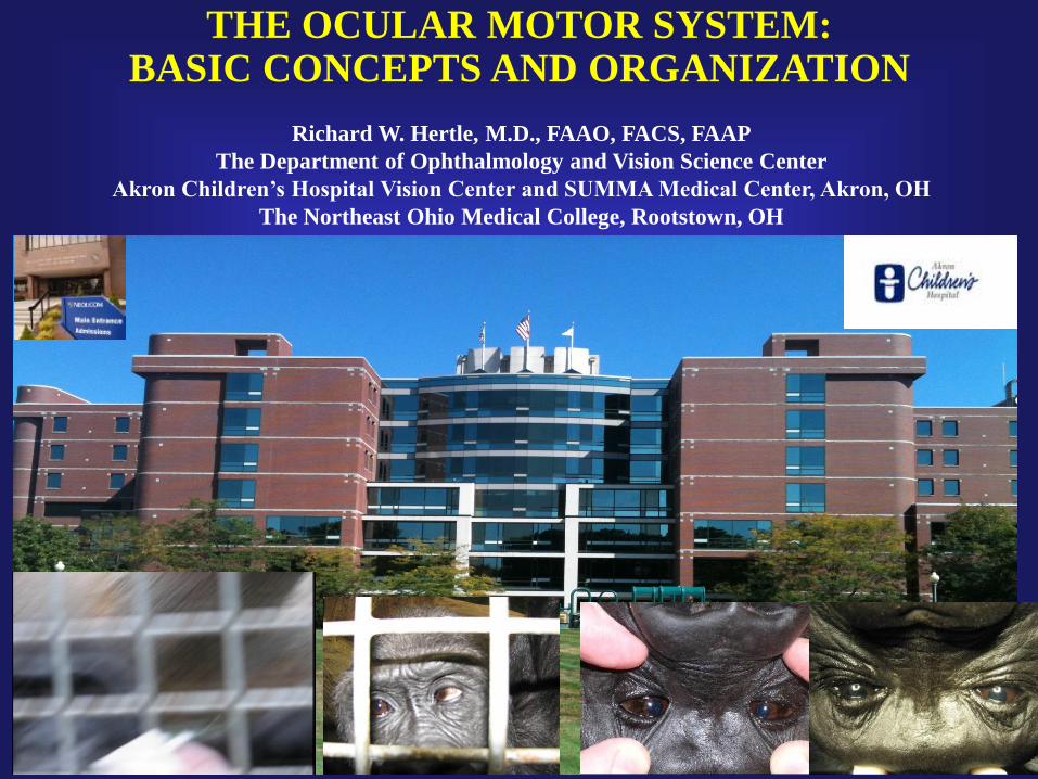
Cerebellum
Medulla
Pons
ThalamusSCPC
MLFCN II
IC
CN VI
VN
Midbrain
riMLF
CN III
PPRF
Cerebellum
Medulla
Pons
ThalamusSCPC
MLFCN II
IC
CN VI
VN
Midbrain
riMLF
CN III
PPRF
Cerebellum
Medulla
Pons
ThalamusSCPC
MLFON
IC
CN VI
VN
Midbrain
riMLF
CN III
PPRF

Supranuclear
Dysfunction
COMITANT
Neuromuscular
Dysfunction
INCOMITANT
FULL
"Paretic Disease"
FORCED DUCTION -
"Restrictive Disease"
GOOD
Combined
"Restrictive-Paretic"
POOR
FORCED GENERATIONS
FORCED DUCTION +
LIMITED
VERSIONS
Figure 4 Clinical Evaluation of Range of Eye Movements
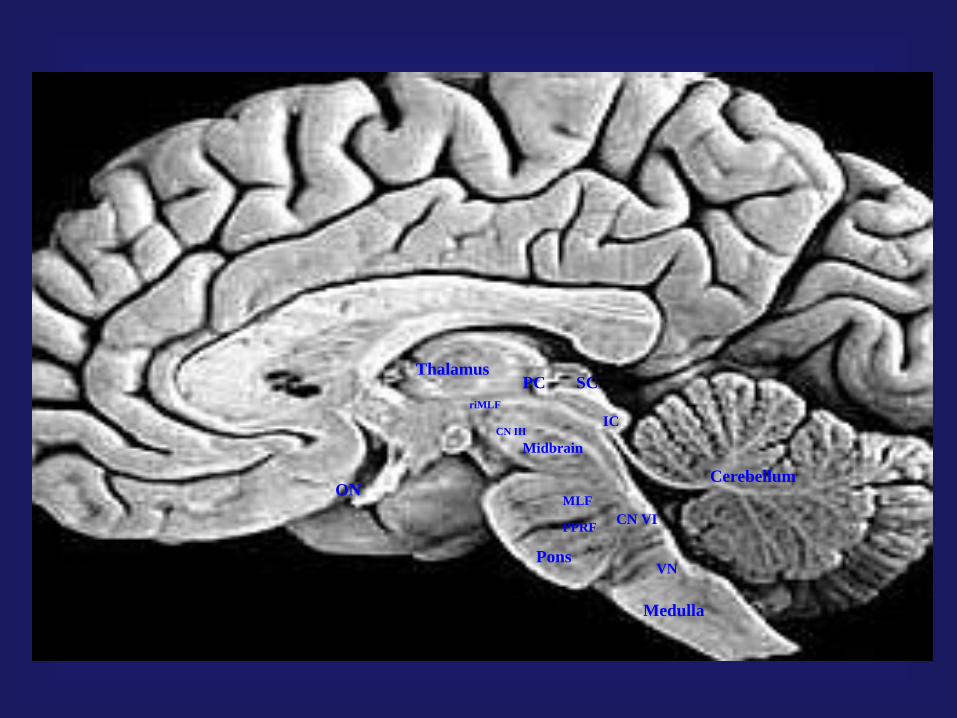
P
P
R
F
VI
Left
Lateral
Rectus
Left
Sixth
Nerve
Right
Medial
Rectus
Right
Third
NerveIII
Cranial Nerve Nuclei
Brainstem Pathway For Horizontal Saccades
“Para” Abducens
Nuclei

riMLF
P
P
R
F
riMLF riMLFriMLF
P
P
R
F
P
P
R
F
P
P
R
F
INC INC
III III
IV IV IV IV
III III
INC INC
Aqueduct Aqueduct
Posterior Commissure Posterior Commissure
Brainstem Pathways For Downward (A) and Upward (B) Saccades
A B


Testing the ability of the extra-ocular muscles to perform
smooth eye movements forms an important part of the eye
examination routine.
Testing Ocular Motility enables the practitioner to
differentiate between
COMITANT vs INCOMITANT Incomitant - varies in size with the direction of gaze.
Comitant - remains constant with gaze direction

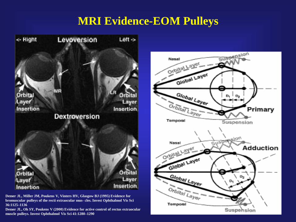
MRI Evidence-EOM Pulleys
Demer JL, Miller JM, Poukens V, Vinters HV, Glasgow BJ (1995) Evidence for
bromuscular pulleys of the recti extraocular mus- cles. Invest Ophthalmol Vis Sci
36:1125–1136
Demer JL, Oh SY, Poukens V (2000) Evidence for active control of rectus extraocular
muscle pulleys. Invest Ophthalmol Vis Sci 41:1280–1290

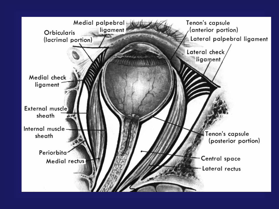
Pulley System
Complete
Ring
encircling
the EOM
Connecting Bands
between the muscle
pulleys forming a circle
around the globe

Mechanics of Movement
• Two components to neurological generation of muscle movement➢ Phasic Pulse
• Momentary force needed to overcome the relaxation of an antagonistic muscle
➢ Tonic Step
• The long-term force needed to oppose the lesser elastic load where position is maintained
• Global Layer demonstrates both kinds of neurological input
• Orbital Layer only demonstrates tonic step
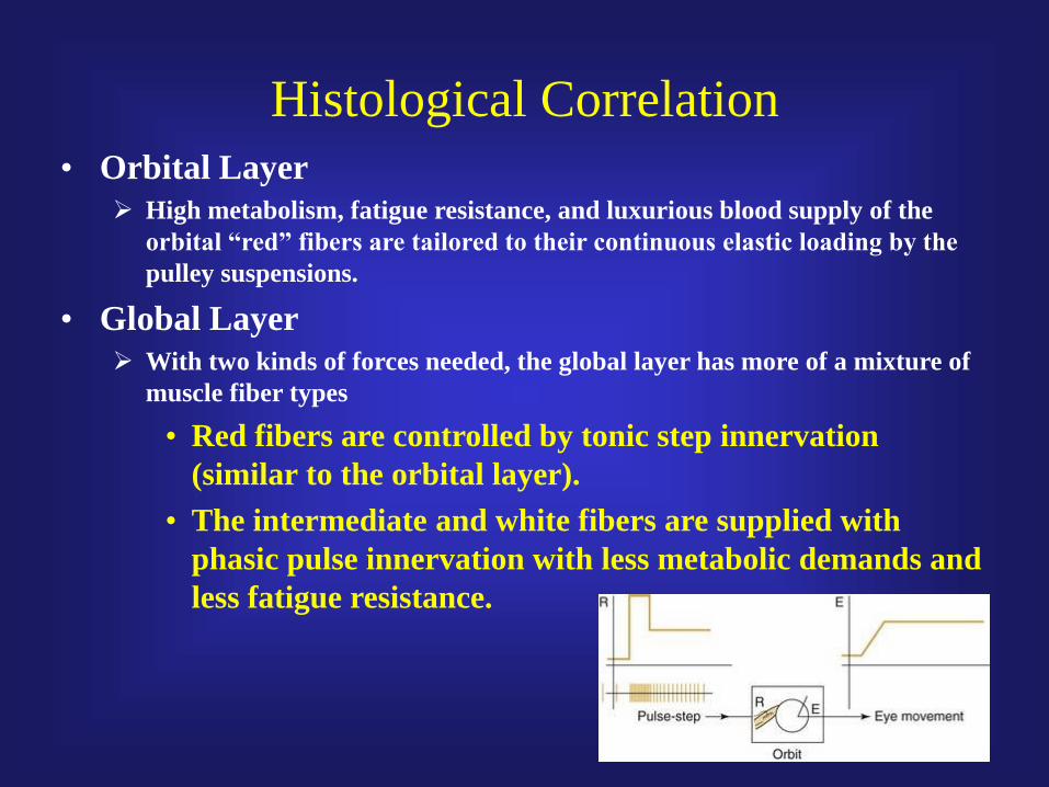
Histological Correlation
• Orbital Layer
➢ High metabolism, fatigue resistance, and luxurious blood supply of the
orbital “red” fibers are tailored to their continuous elastic loading by the
pulley suspensions.
• Global Layer
➢ With two kinds of forces needed, the global layer has more of a mixture of
muscle fiber types
• Red fibers are controlled by tonic step innervation
(similar to the orbital layer).
• The intermediate and white fibers are supplied with
phasic pulse innervation with less metabolic demands and
less fatigue resistance.
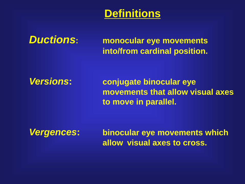
Definitions
Ductions: monocular eye movements
into/from cardinal position.
Versions: conjugate binocular eye
movements that allow visual axes
to move in parallel.
Vergences: binocular eye movements which
allow visual axes to cross.

Elevation
supraduction
Depression
infraduction
R L
Primary Position
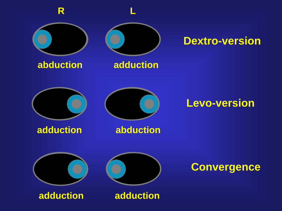
R L
Dextro-version
abduction adduction
Levo-version
adduction abduction
Convergence
adduction adduction
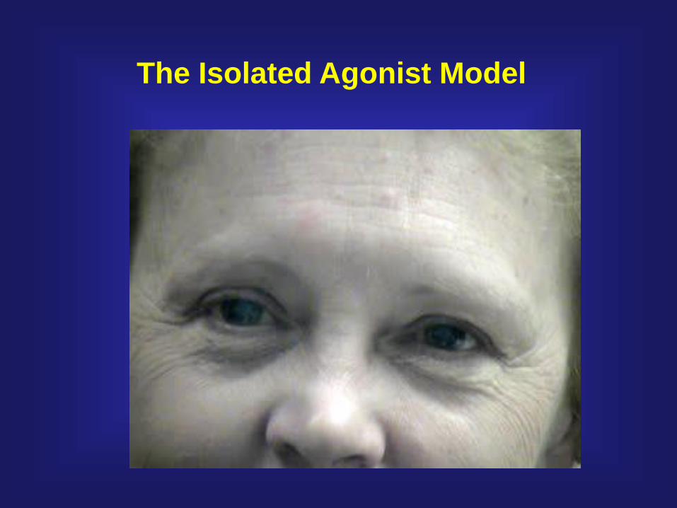
The Isolated Agonist Model

Lateral Rectus
Main Action: abduction
Innervation: Abducens
C
Temporal Nasal
C

Medial Rectus
Main Action: adduction
Innervation:
Inf. Div of Oculomotor Nerve
C
Temporal Nasal
C

Temporal Nasal
C
23°
Superior Rectus
Main Action: supraduction
Secondary: incycloduction
adduction
Innervation:
Sup. Div of Oculomotor Nerve
(i.e. III Cranial Nerve)
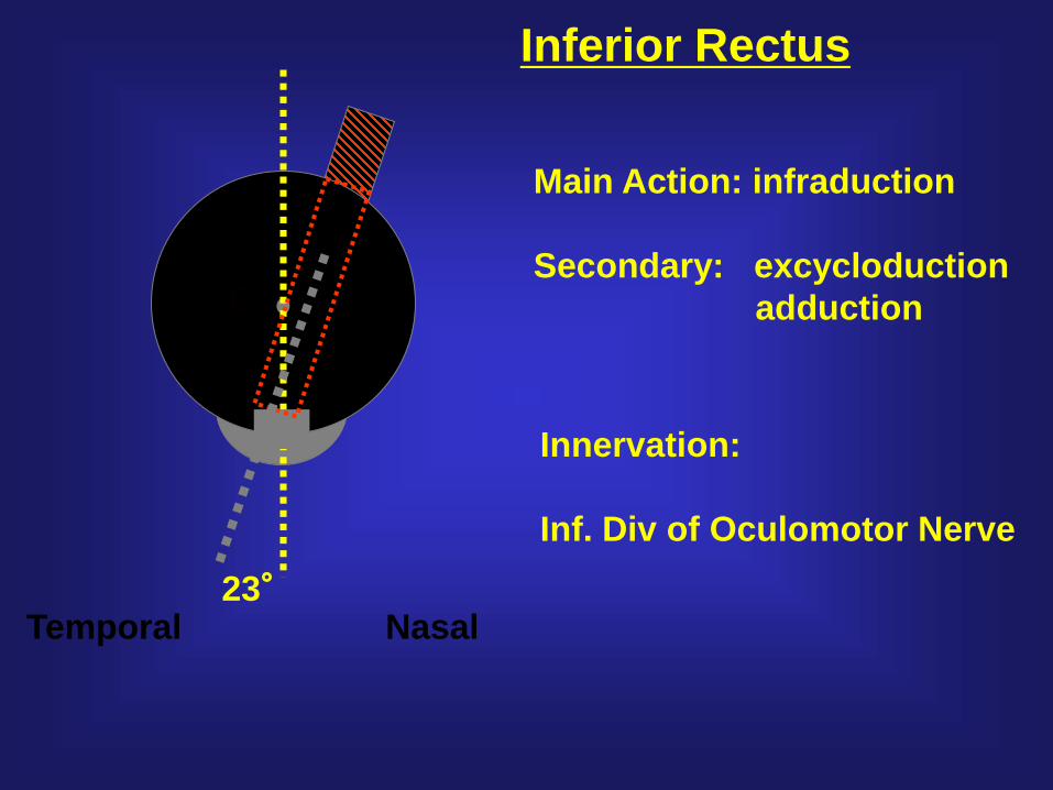
Inferior Rectus
Main Action: infraduction
Secondary: excycloduction
adduction
Innervation:
Inf. Div of Oculomotor Nerve
Temporal Nasal
C
23°

Superior Oblique
Main Action: incylcoduction
Secondary: depression
abduction
Innervation:
Trochlear Nerve
C
Temporal Nasal
C
51°

Inferior Oblique
Main Action: exycloduction
Secondary: supraduction
abduction
Innervation:
Inf. Div of Oculomotor Nerve
C
Temporal Nasal
C
51°
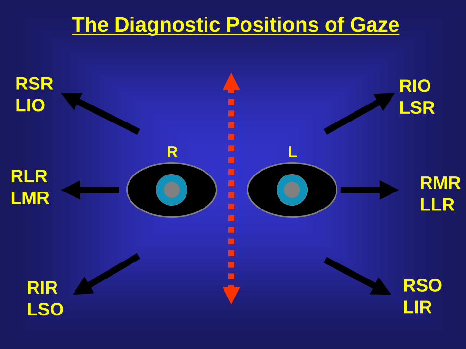
R L
RLR
LMRRMR
LLR
RSR
LIORIO
LSR
RIR
LSO
RSO
LIR
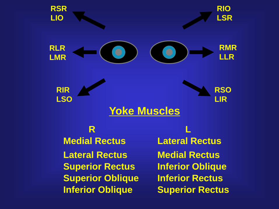
The Diagnostic Positions of Gaze

‘A’ Pattern ‘V’ Pattern
RSR
LIO
RIO
LSR
RIR
LSO
RSO
LIR
RLR
LMR
RMR
LLR
Yoke Muscles
R L
Medial Rectus Lateral Rectus
Lateral Rectus Medial Rectus
Superior Rectus Inferior Oblique
Superior Oblique Inferior Rectus
Inferior Oblique Superior Rectus
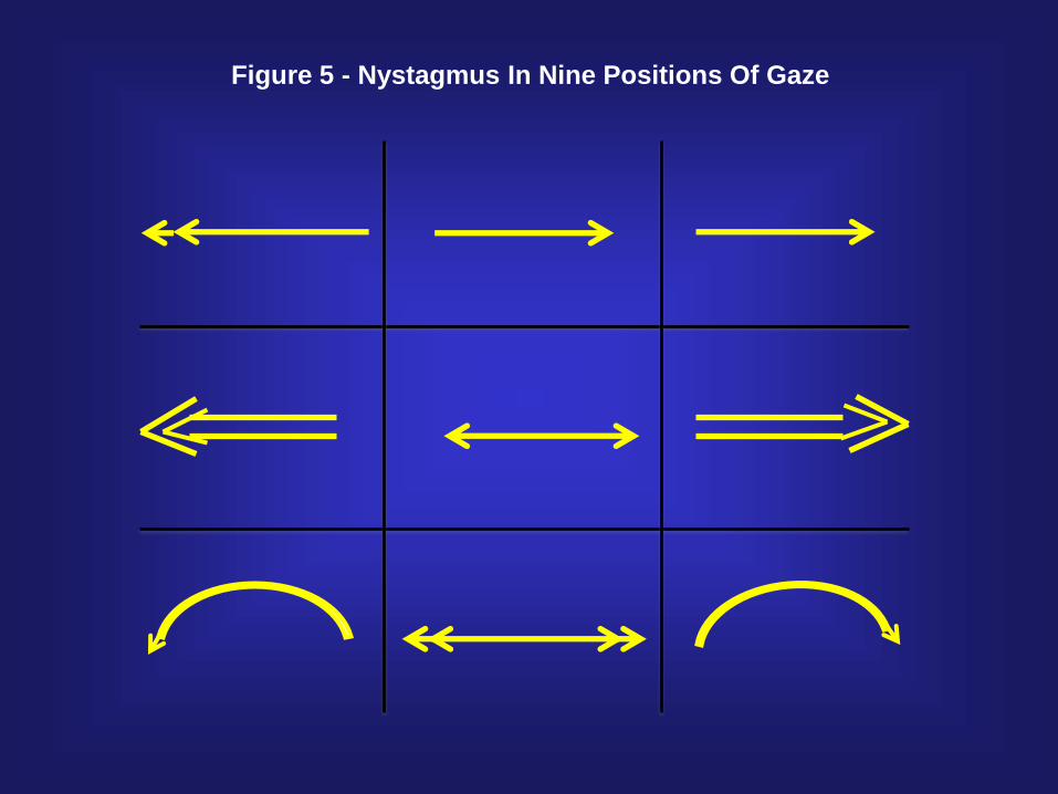
Figure 5 - Nystagmus In Nine Positions Of Gaze
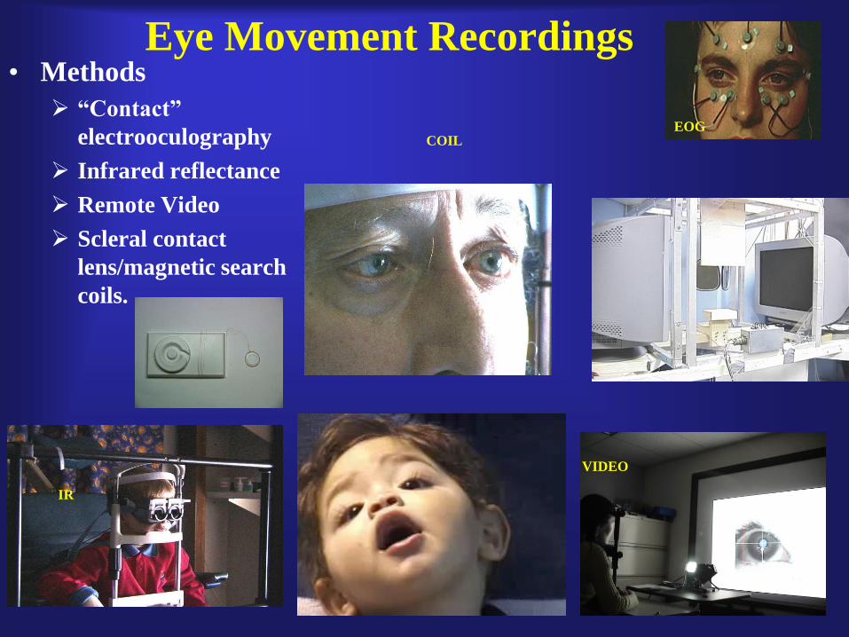
Eye Movement Recordings• Methods
➢ “Contact”
electrooculography
➢ Infrared reflectance
➢ Remote Video
➢ Scleral contact
lens/magnetic search
coils.
IR
COIL
VIDEO
EOG


Ocular Oscillations
TIME
R
L
a. Pure Jerk Left Nystagmus with Linear Slow Phases – Typical Response to
Right OKN
b. Gaze Left with Jerk Left Decreasing Velocity Slow Phases – Typical “Leaky
Integrator”
0 Position
0 Position
0 Position
c. Pure Pendular Nystagmus

Dysmetria
Eye Position
Eye Position
Eye Position
Dysmetria
Eye Position
Eye Position
Eye Position
Flutter
Flutter Dysmetria
0 T
T
T
T
0
0
0
0
Saccadic pulseSquare wave jerk Double saccadic pulse
Macro Saccadic Oscillations
T
T = Target
Saccadic Intrusions
Eye Position
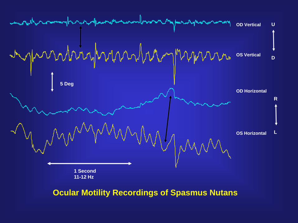
1 Second
11-12 Hz
OD Vertical
OS Vertical
OD Horizontal
OS Horizontal
Ocular Motility Recordings of Spasmus Nutans
5 Deg
L
R
D
U


VII. Imaging• CAT Scan.
• MRI (Cine, Orbital, Flare, Contrast, DWI).
• PET Scan (Positron Emission. Tomography)
• FMRI (Functional Imaging).
• OCT (Optical Coherence Tomography)

VIII. Laboratory Studies
• Infectious Inflammatory Disease.
➢TORCH Infections
• Immune Inflammatory Diseases.
➢Multiple Sclerosis
➢Leukodystrophies
• Genetic Diseases.
➢“Storage” Diseases
• Chromosomal Disorders and Systemic
Syndromes.

A Classification of Eye Movement Abnormalities
and Strabismus (CEMAS)--Report of a National Eye Institute
Sponsored Workshop
Summarizes the results from a two-day workshop in February 2001 on
the National Institutes of Health Campus, Bethesda, MD, that brought
together some of the Nation's most experienced clinical and basic
science investigators in the diagnosis, treatment, and etiology of eye
movement abnormalities and strabismus.
http://www.nei.nih.gov/news/statements/cemas.pdf

ORGANIZATION (122 Disorders)
I. Ocular Motor Aspects of Vision
II. Sensory Aspects of Binocular Vision
III. Horizontal Heterotropias
IV. Horizontal Heterophorias
V. Cyclovertical Heterotropias and Special Forms of
Strabismus
VI. Cyclovertical Heterophorias
VII. Accommodative Disorders
VIII. Nystagmus and Other Ocular Motor Oscillations
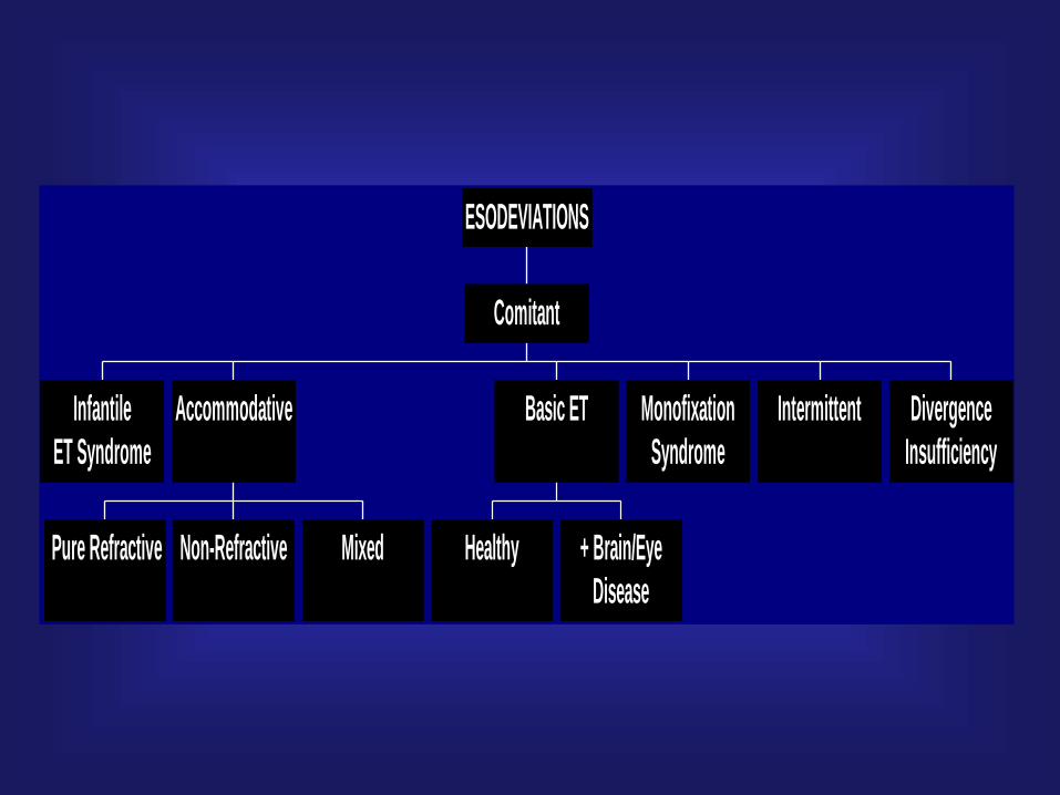
Infantile
ET Syndrome
Pure Refractive Non-Refractive Mixed
Accommodative
Healthy + Brain/Eye
Disease
Basic ET Monofixation
Syndrome
Intermittent Divergence
Insufficiency
Comitant
ESODEVIATIONS
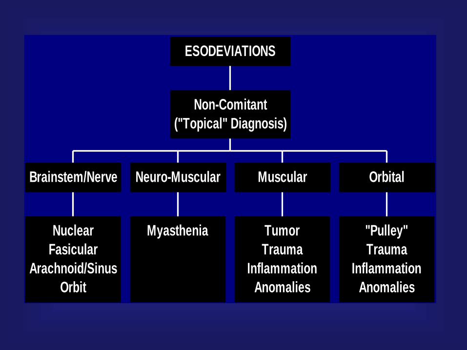
Nuclear
Fasicular
Arachnoid/Sinus
Orbit
Brainstem/Nerve
Myasthenia
Neuro-Muscular
Tumor
Trauma
Inflammation
Anomalies
Muscular
"Pulley"
Trauma
Inflammation
Anomalies
Orbital
Non-Comitant
("Topical" Diagnosis)
ESODEVIATIONS

Early Infancy Onset
Healthy Infant
Constant, Large Angle
No Response to + Lenses
Infantiile ET
Syndrome
Comitant
ESODEVIATIONS

Hyperopia
Dist ET = Near ET
Rx c Distance
Specs
"Pure" Refractive
+/- Hyperopia
0 Dist ET + Near ET
Rx c Bifocals
Only
Non-Refractive
+/- Hyperopia
Dist ET < Near ET
Rx c Dist Specs
& Bifocals
Mixed
Accommodative ET
Comitant
ESODEVIATIONS

Variable Onset
Inconsistent Devaition
No Eye or Brain Disease
Not Infantile ET
Basic ET
Variable Onset
Inconsistent Deviation
+ Eye or Brain Disease
"Sensory" ET
Comitant
ESODEVIATIONS

Disease Name MONOFIXATION ESOTROPIA SYNDROME
[Old – Microtropia]
Criteria Small angle esotropia to no tropia, macular scotoma in
non-fixing eye with anomalous retinal correspondence.
Common
Associated
Findings
Can be primary, genetic or acquired after surgical
treatment of infantile strabismus, can be associated with
anisometropia, amblyopia often present, stereopsis
present but poor, alternate cover test may reveal larger
deviation than simultaneous cover test. Good fusional
vergence amplitudes.
General
Comments
Promotes stable ocular alignment and sensory status.
Can deteriorate into constant, larger angle esotropia,
requiring surgical treatment.
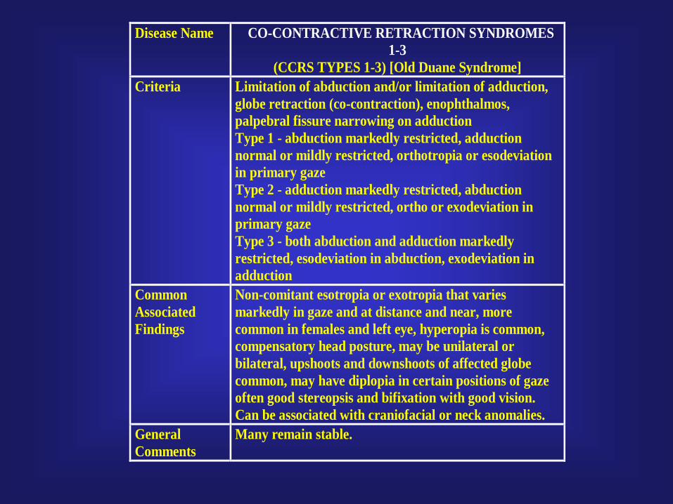
Disease Name CO-CONTRACTIVE RETRACTION SYNDROMES
1-3
(CCRS TYPES 1-3) [Old Duane Syndrome]
Criteria Limitation of abduction and/or limitation of adduction,
globe retraction (co-contraction), enophthalmos,
palpebral fissure narrowing on adduction
Type 1 - abduction markedly restricted, adduction
normal or mildly restricted, orthotropia or esodeviation
in primary gaze
Type 2 - adduction markedly restricted, abduction
normal or mildly restricted, ortho or exodeviation in
primary gaze
Type 3 - both abduction and adduction markedly
restricted, esodeviation in abduction, exodeviation in
adduction
Common
Associated
Findings
Non-comitant esotropia or exotropia that varies
markedly in gaze and at distance and near, more
common in females and left eye, hyperopia is common,
compensatory head posture, may be unilateral or
bilateral, upshoots and downshoots of affected globe
common, may have diplopia in certain positions of gaze
often good stereopsis and bifixation with good vision.
Can be associated with craniofacial or neck anomalies.
General
Comments
Many remain stable.

CEMAS Nystagmus Types
1. Peripheral Vestibular Imbalance➢ Meniere, drug toxicity
2. Central Vestibular Imbalance➢ Downbeat, Upbeat, drug toxicity
3. Instability of Vestibular Mechanisms➢ PAN
4. Disorders of Visual Fixation➢ Vision Loss, SSN, drug toxicity
5. Disorders of Gaze Holding➢ GEN, ?APN, drug toxicity
6. Acquired Pendular Nystagmus➢ central myelin, oculopalatal, Whipple,
drug toxicity
7. Saccadic Intrusions and Oscillations➢ SWJ, MSO, opsoclonus
8. Miscellaneous Eye Movements➢ SO Myokymia, OM neuromyotonia
9. Infantile Nystagmus Syndrome➢ “congenital,” “motor,” “sensory,” idiopathic, nystagmus
blockage
10. Fusion Maldevelopment Nystagmus Syndrome➢ Latent, manifest latent, nystagmus blockage
11. Spasmus Nutans Syndrome➢ Without optic pathway glioma
➢ With optic pathway glioma
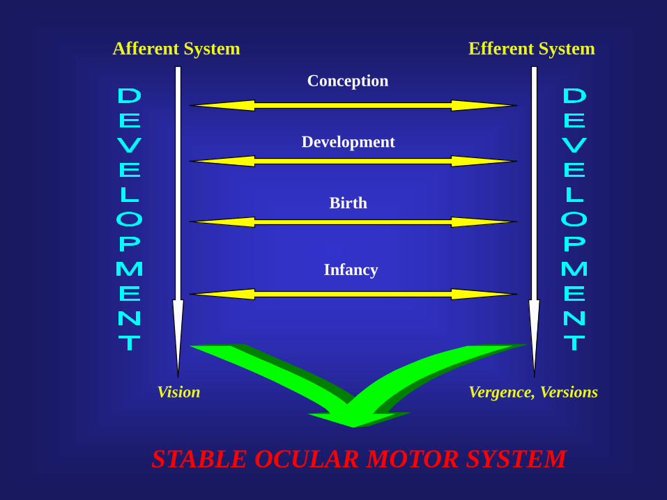
Afferent System Efferent System
Conception
Birth
Infancy
Development
STABLE OCULAR MOTOR SYSTEM
Vision Vergence, Versions

Visuo-Vestibular
Motion Sensitivity
Contrast
Visual Recognition
Time
Gaze Dependent
Vision
Time
“VISION”EFFERENT

Photosensitivity
Optic Nerve
Amblyopia
Ammetropia
Hypoaccommodation
Visual Cortex
Retina
“VISION”AFFERENT

“Myotendon”Annulus Of
Zinn
Enthesial
Area
Summary• “Value” of “Work-Up”
➢ Diagnosis.
➢ Classification.
➢ Etiology.
➢ Therapy.
➢ Research.