Embed Size (px)
Citation preview
elifesciences.org
RESEARCH ARTICLE
Ribosomes slide on lysine-encodinghomopolymeric A stretchesKristin S Koutmou1, Anthony P Schuller1, Julie L Brunelle1,2,Aditya Radhakrishnan1, Sergej Djuranovic3, Rachel Green1,2*
1Department of Molecular Biology and Genetics, Johns Hopkins School of Medicine,Baltimore, United States; 2Howard Hughes Medical Institute, Johns Hopkins School ofMedicine, Baltimore, United States; 3Department of Cell Biology and Physiology,Washington University School of Medicine, St. Louis, United States
Abstract Protein output from synonymous codons is thought to be equivalent if appropriate
tRNAs are sufficiently abundant. Here we show that mRNAs encoding iterated lysine codons, AAA or
AAG, differentially impact protein synthesis: insertion of iterated AAA codons into an ORF
diminishes protein expression more than insertion of synonymous AAG codons. Kinetic studies in
E. coli reveal that differential protein production results from pausing on consecutive AAA-lysines
followed by ribosome sliding on homopolymeric A sequence. Translation in a cell-free expression
system demonstrates that diminished output from AAA-codon-containing reporters results from
premature translation termination on out of frame stop codons following ribosome sliding. In
eukaryotes, these premature termination events target the mRNAs for Nonsense-Mediated-Decay
(NMD). The finding that ribosomes slide on homopolymeric A sequences explains bioinformatic
analyses indicating that consecutive AAA codons are under-represented in gene-coding sequences.
Ribosome ‘sliding’ represents an unexpected type of ribosome movement possible during
translation.
DOI: 10.7554/eLife.05534.001
IntroductionMessenger RNA (mRNA) transcripts can contain errors that result in the production of incorrect
protein products. Both bacterial and eukaryotic cells have evolved mechanisms to deal with such
errors which involve (1) proteolytic degradation of the aberrant protein product, (2) mRNA decay and
(3) ribosome rescue (Shoemaker and Green, 2012). One such mRNA surveillance pathway in
eukaryotes targets mRNAs that lack stop codons (Non-Stop-Decay or NSD). In these cases, actively
translating ribosomes are thought to read into the 3′ terminal poly(A) sequence of the mRNA
triggering ribosome pausing as poly(lysine) is translated, followed by the recruitment of ubiquitin
ligases, mRNA decay and ribosome recycling factors (review Klauer and van Hoof, 2012). Given the
substantial amount of premature (or alternative) polyadenylation that has been documented in
eukaryotes (Ozsolak et al., 2010), it seems that such an mRNA surveillance pathway might have
considerable biological significance. Similarly, in bacteria, while no ‘NSD-like’ response has been
characterized, it is known that poly(A) sequences are added to mRNAs in the process of being
degraded (review Dreyfus and Regnier, 2002), and so ribosomes on these mRNAs may encounter
similar challenges. The utilization in bacteria and eukaryotes of 3′ poly(A) tails as non-coding elements
may reflect a common solution to the challenges for the ribosome in translating such sequences.
Most studies investigating how NSD works have been conducted in yeast using reporter
constructs. Early studies in Saccharomyces cerevisiae revealed that mRNAs lacking stop-codons are
targeted for decay both in a reaction dependent on the exosome-associated factor Ski7 (van Hoof
et al., 2002) and in a more canonical degradation reaction involving decapping and 5′ to 3′
*For correspondence: ragreen@
jhmi.edu
Competing interests:
See page 16
Funding: See page 16
Received: 09 November 2014
Accepted: 18 February 2015
Published: 19 February 2015
Reviewing editor: Nahum
Sonenberg, McGill University,
Canada
Copyright Koutmou et al. This
article is distributed under the
terms of the Creative Commons
Attribution License, which
permits unrestricted use and
redistribution provided that the
original author and source are
credited.
Koutmou et al. eLife 2015;4:e05534. DOI: 10.7554/eLife.05534 1 of 18
exonucleolytic degradation (Frischmeyer et al., 2002). Other factors involved in NSD have since been
discovered; these include Dom34 and Hbs1 which facilitate ribosome rescue during NSD (Izawa et al.,
2012; Tsuboi et al., 2012), Ltn1 and Not4 which ubiquitinate the protein products on non-stop
mRNAs (Dimitrova et al., 2009; Bengtson and Joazeiro, 2010), and a number of other factors
genetically identified as critical for poly(basic)-mediated stalling (Kuroha et al., 2010; Brandman
et al., 2012; Chiabudini et al., 2014). Although many players in NSD have been identified and their
functions defined, there remain critical gaps in our understanding.
In this manuscript, we focus on what must be the earliest events in NSD, the translation of poly
(lysine) sequences by the ribosome. NSD is widely thought to be triggered by unfavorable
electrostatic interactions that occur in the ribosomal exit tunnel when ribosomes translate the poly
(lysine) sequences encoded by poly(A). Indeed, biochemical studies in rabbit reticulocyte lysate with
proteins interrupted by iterated poly(lysine) and poly(arginine) sequences indicate that positively
charged residues do slow translation and produce transiently arrested species (Lu and Deutsch,
2008). Other examples of peptide-mediated stalling have also been documented in bacterial and
eukaryotic systems. In some cases, such as the tnaC gene, secM, or ermCL in bacteria, the peptide
stalling motif is several amino acids in length and appears to specifically engage the contours of the
exit tunnel to elicit stalling (Gong and Yanofsky, 2002; Nakatogawa and Ito, 2002; Vazquez-Laslop
et al., 2008; Seidelt et al., 2009; Bhushan et al., 2011; Ito and Chiba, 2013; Arenz et al., 2014).
Poly(proline) sequences have recently been shown to cause stalling during translation in bacteria and
eLife digest Genes provide the instructions to assemble proteins from smaller molecules called
amino acids. When a gene is ‘switched on’, the DNA that makes up the gene is copied into
messenger ribonucleic acid (or mRNA) molecules, composed of building blocks called nucleotides.
There are four types of nucleotides in mRNA molecules—commonly referred to as A, C, G, and
U—and a set of three nucleotides is called a codon.
A molecular machine called a ribosome moves along an mRNA molecule translating the codons
into protein. Each codon instructs the ribosome to add a particular amino acid to the chain of amino
acids that will make up the protein. Some codons do not specify an amino acid but instead mark the
point on the mRNA that the ribosome should stop and release the new protein. Most mRNAs have
nucleotides beyond the ‘stop’ codon and these often contain a long stretch of A nucleotides, one
after the other, which is known as the poly(A) tail.
Some mRNA copies may contain poly(A) tails before a stop codon, which can lead to the
production of alternate and potentially harmful proteins. Cells have developed ways to identify and
dispose of these mRNAs and their protein products. For example, in yeast and other eukaryotes, if
an mRNA is missing a stop codon, the ribosomes will continue to translate along the mRNA into the
poly(A) tail where they stall and are eventually removed. When this happens, the mRNA and protein
are rapidly destroyed. However, it is not clear how this works.
Koutmou et al. studied the translation of a series of artificial mRNAs that contained different
numbers of A nucleotides in codons of either AAA or AAG. Both of these codons specify the same
amino acid, and should therefore be translated equivalently. The experiments show that the
ribosomes read the AAA and AAG codons differently. When consecutive AAA codons are found in
the mRNA, the level of protein production is significantly lower than when the mRNA contains
iterated AAGs instead.
Koutmou et al. found that when ribosomes encounter consecutive AAA codons they undergo an
unusual ‘sliding’ movement and are unable to accurately produce proteins. When a cell detects this
abnormal sliding behavior, it rapidly triggers the destruction of the mRNA molecule. In contrast,
when ribosomes encounter consecutive AAG codons, they slow down but do not slide, and therefore
produce a correct protein.
Koutmou et al.’s findings also provide an explanation for why there are relatively few AAA codons
within the regions of genes that encode proteins. The prevalence of alternative forms of mRNAs with
poly(A) sequences before their stop codons suggests that ribosome sliding may contribute to an
important pathway to control the activity of genes.
DOI: 10.7554/eLife.05534.002
Koutmou et al. eLife 2015;4:e05534. DOI: 10.7554/eLife.05534 2 of 18
Research article Biochemistry
eukaryotes in the absence of specialized bypass factors, EFP and eIF5A, respectively (Doerfel et al.,
2013; Gutierrez et al., 2013; Ude et al., 2013). In this case, proline is thought to adopt
a conformation that interferes with the ribosome active site geometry.
Here we take a high-resolution biochemical look at the molecular events that occur when the
ribosome translates poly(lysine) peptides. We find that insertion of consecutive AAA lysine codons
into reporters has a stronger negative impact on protein expression than insertion of an equivalent
number of AAG lysine codons in both eukaryotes and bacteria. Kinetic and toeprinting studies in an in
vitro reconstituted Escherichia coli translation system reveal that differential protein output is the
downstream consequence of ribosome pausing followed by an unanticipated ribosome movement on
successive AAA codons that we refer to as ‘sliding’. When sliding occurs in the middle of genuine
ORFs in a cell, frame is lost and ribosomes encounter out of frame stop codons that result in canonical
(stop-codon mediated) termination. In eukaryotes, such premature termination events target the
mRNA for non-sense mediated decay (NMD). The finding that the ribosome can robustly slide on poly
(A) sequences explains bioinformatic analyses revealing that consecutive AAA codons are under-
represented in ORFs in all genomes (unpublished data) and helps to rationalize the widespread usage
of poly(A) sequence as a regulatory rather than a coding feature.
Results
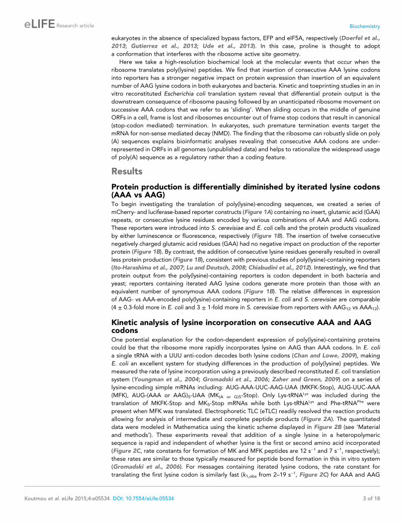
Protein production is differentially diminished by iterated lysine codons(AAA vs AAG)To begin investigating the translation of poly(lysine)-encoding sequences, we created a series of
mCherry- and luciferase-based reporter constructs (Figure 1A) containing no insert, glutamic acid (GAA)
repeats, or consecutive lysine residues encoded by various combinations of AAA and AAG codons.
These reporters were introduced into S. cerevisiae and E. coli cells and the protein products visualized
by either luminescence or fluorescence, respectively (Figure 1B). The insertion of twelve consecutive
negatively charged glutamic acid residues (GAA) had no negative impact on production of the reporter
protein (Figure 1B). By contrast, the addition of consecutive lysine residues generally resulted in overall
less protein production (Figure 1B), consistent with previous studies of poly(lysine)-containing reporters
(Ito-Harashima et al., 2007; Lu and Deutsch, 2008; Chiabudini et al., 2012). Interestingly, we find that
protein output from the poly(lysine)-containing reporters is codon dependent in both bacteria and
yeast; reporters containing iterated AAG lysine codons generate more protein than those with an
equivalent number of synonymous AAA codons (Figure 1B). The relative differences in expression
of AAG- vs AAA-encoded poly(lysine)-containing reporters in E. coli and S. cerevisiae are comparable
(4 ± 0.3-fold more in E. coli and 3 ± 1-fold more in S. cerevisiae from reporters with AAG12 vs AAA12).
Kinetic analysis of lysine incorporation on consecutive AAA and AAGcodonsOne potential explanation for the codon-dependent expression of poly(lysine)-containing proteins
could be that the ribosome more rapidly incorporates lysine on AAG than AAA codons. In E. coli
a single tRNA with a UUU anti-codon decodes both lysine codons (Chan and Lowe, 2009), making
E. coli an excellent system for studying differences in the production of poly(lysine) peptides. We
measured the rate of lysine incorporation using a previously described reconstituted E. coli translation
system (Youngman et al., 2004; Gromadski et al., 2006; Zaher and Green, 2009) on a series of
lysine-encoding simple mRNAs including: AUG-AAA-UUC-AAG-UAA (MKFK-Stop), AUG-UUC-AAA
(MFK), AUG-(AAA or AAG)5-UAA (MK(A or G)5-Stop). Only Lys-tRNALys was included during the
translation of MKFK-Stop and MK5-Stop mRNAs while both Lys-tRNALys and Phe-tRNAPhe were
present when MFK was translated. Electrophoretic TLC (eTLC) readily resolved the reaction products
allowing for analysis of intermediate and complete peptide products (Figure 2A). The quantitated
data were modeled in Mathematica using the kinetic scheme displayed in Figure 2B (see ‘Material
and methods’). These experiments reveal that addition of a single lysine in a heteropolymeric
sequence is rapid and independent of whether lysine is the first or second amino acid incorporated
(Figure 2C, rate constants for formation of MK and MFK peptides are 12 s−1 and 7 s−1, respectively);
these rates are similar to those typically measured for peptide bond formation in this in vitro system
(Gromadski et al., 2006). For messages containing iterated lysine codons, the rate constant for
translating the first lysine codon is similarly fast (k1,obs from 2–19 s−1, Figure 2C) for AAA and AAG
Koutmou et al. eLife 2015;4:e05534. DOI: 10.7554/eLife.05534 3 of 18
Research article Biochemistry
codons. However, subsequent lysines in an iter-
ated sequence are added with considerably
slower kinetics on both AAA (k2,obs = 0.0005 and
k3,obs = 0.0003 s−1) and AAG codons (k2,obs =0.009 and k3,obs = 0.015 s−1) (Figure 2C). We note
that the rate of second lysine addition during the
translation of MK5-STOP messages are somewhat
slower on AAA relative to AAG codons, potentially
partially explaining the decreased overall protein
output on these mRNAs. More importantly, how-
ever, these data show that the reactivity of the
second Lys-tRNALys on iterated lysine containing
messages (such as MK5-Stop) is substantially re-
duced (at least 130-fold) on both lysine codon-
containing mRNAs relative to normal elongation
rates. Interestingly, the addition of a second lysine
to messages with fewer sequential lysine codons
(such asMK2F-STOP) does not exhibit such a striking
kinetic defect (k2,obs is not largely affected, data not
shown). These data suggest that the identity of
the message (i.e. a long poly(A) sequence) plays
a critical role in the observed slowing of elongation.
Toeprinting assays performed using the E. coli
PURE cell-free translation system are consistent
with these observations; E. coli ribosomes stall when
the second lysine codon in iterated (AAA)- and
(AAG)-codon containing sequences is positioned
in the A site (Figure 2—figure supplement 1).
Together, these results reveal that translating con-
secutive lysines in a poly(lysine) peptide sequence,
either on iterated AAA or AAG codons, can lead to
substantial kinetic delays in vitro.
E. coli ribosomes add extra lysines on iterated AAA-containing mRNAsAs we explored the kinetics of lysine incorporation, we evaluated the ability of the ribosome to
translate a variety of MK(A or G)2 di-lysine messages (Figure 3A). Unexpectedly, we found that
messages containing iterated AAA codons generate extended peptides longer than the designed
coding sequence (Figure 3A). When E. coli initiation complexes (programmed with fMet-tRNAfMet) are
reacted with Lys-tRNALys on messages containing two consecutive lysine codons followed by a variety
of non-lysine codons (Phe (UUC), Val (GUC), or Stop (UAA)), only MKK peptide should be synthesized.
However, we see the formation of a majority population of extended peptide product containing at
least four lysines on all messages with two consecutive AAA codons (Figure 3B, lanes 2-4). In contrast,
equivalent messages with two AAG codons predominantly form the expected MKK product
(Figure 3B, compare lane 3 vs 5). We also find that a mixed sequence of lysine codons (AAA-AAG) can
form some extended peptide (Figure 3—figure supplement 1). These data suggest that 5 As in a row
are sufficient to promote the addition of extra lysines in vitro. We note that the identity of the codon
that follows the di-lysine sequence is not relevant to the observed amount of extended peptide
product (Figure 3B, Figure 3—figure supplement 2).
The production of peptide products containing more than the encoded number of lysines is
surprising, especially given that there are no nearby upstream or downstream in-frame or out-of-frame
lysine codons in these mRNAs (Figure 3A). We speculate that these extended peptides result from the
ribosome repeatedly moving backwards by at least three nucleotides to position an AAA Lys codon in
the A site, and then subsequent standard peptide bond formation. Toeprinting assays performed on
iterated AAA- and AAG-containing mRNAs provide further support for such irregular movement of
ribosomes specifically on iterated AAA codons (Figure 2—figure supplement 1); the toeprint on the
iterated AAA sequence is diffuse relative to the discrete toeprint seen on iterated AAG sequence.
Figure 1. Protein production is differentially
diminished by iterated lysine codons (AAA vs AAG)
in E. coli and S. cerevisiae. (A) Schematics of the
mCherry (top) and luciferase (middle, and bottom)
reporters used in this study. The mCherry reporter
contains an N-terminal thioredoxin (Thrdx) domain,
3HA-tag, sequence of interest (black section), fol-
lowed by the C-terminal mCherry sequence. The top
luciferase reporter includes a 2HA tag followed by
sequences of interest (used for study in Figure 1B).
The second luciferase reporter (used in Figure 6) has
sequences of interest at the N-terminal end of
Renilla. Firefly is used in this construct as an internal
control in the second luciferase reporter. (B) Relative
amounts of protein expressed from reporters
expressed in E. coli (mCherry, red) and S. cerevisiae
(luciferase, green). Error bars results from for the
standard error of at least three experiments.
DOI: 10.7554/eLife.05534.003
Koutmou et al. eLife 2015;4:e05534. DOI: 10.7554/eLife.05534 4 of 18
Research article Biochemistry
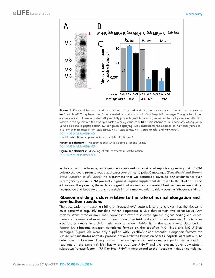
In the course of performing our experiments we carefully considered reports suggesting that T7 RNA
polymerase could promiscuously add extra adenosines to poly(A) messages (Tsuchihashi and Brown,
1992; Ratinier et al., 2008); no experiment that we performed revealed any evidence for such
heterogeneity in our mRNA products (Figure 3—figure supplement 3). Unlike better studied −1 and
+1 frameshifting events, these data suggest that ribosomes on iterated AAA sequences are making
unexpected and large excursions from their initial frame; we refer to this process as ‘ribosome sliding’.
Ribosome sliding is slow relative to the rate of normal elongation andtermination reactionsThe observation of ribosome sliding on iterated AAA codons is surprising given that the ribosome
must somewhat regularly translate mRNA sequences in vivo that contain two consecutive AAA
codons. While three or more AAA codons in a row are selected against in gene coding sequences,
there are thousands of examples of two consecutive AAA codons in S. cerevisiae and E. coli genes
(see further details in bioinformatic analysis below, Table 1). In the experiments described in
Figure 3A, ribosome initiation complexes formed on the specified MKA2-Stop and MKA2F-Stop
messages (Figure 3B) were only supplied with Lys-tRNALys and essential elongation factors; the
subsequent substrates normally present in vivo after the formation of MKK peptide were left out. To
determine if ribosome sliding occurs in more typical circumstances, we performed elongation
reactions on the same mRNAs, but where both Lys-tRNALys and the relevant other downstream
substrates (release factor 1 (RF1) or Phe-tRNAPhe) were added to the ribosome initiation complexes.
Figure 2. Kinetic defect observed on addition of second and third lysine residues in iterated lysine stretch.
(A) Example eTLC displaying the E. coli translation products of a AUG-(AAA)5-UAA message. The ± poles of the
electrophoretic TLC are indicated. MK4 and MK5 products (and those with greater numbers of lysine) are difficult to
resolve in this system but the other products are easily visualized. (B) Kinetic scheme for rate constants of sequential
lysine additions to peptide chain. (C) Bar graph displaying rate constants for the addition of individual lysines to
a variety of messages: MKFK-Stop (gray), MKA5-Stop (blue), MKG5-Stop (black), and MFK (gray).
DOI: 10.7554/eLife.05534.004
The following figure supplements are available for figure 2:
Figure supplement 1. Ribosomes stall while adding a second lysine.
DOI: 10.7554/eLife.05534.005
Figure supplement 2. Modeling of rate constants in Mathematica.
DOI: 10.7554/eLife.05534.006
Koutmou et al. eLife 2015;4:e05534. DOI: 10.7554/eLife.05534 5 of 18
Research article Biochemistry
The result is clear; in this latter case, the anticipated MKKF or MKK peptide products are
predominantly generated (Figure 3C, Figure 3—figure supplement 2). These data suggest that
ribosome sliding on iterated AAA sequences occurs more slowly than the normal rate of peptidyl
transfer with Phe-tRNAPhe or RF1-catalyzed peptide release, respectively. Moreover, these results
readily explain how the ribosome can normally translate (at least two) sequential AAA codons in vivo
without sliding. When there are more than two AAA codons in a row, each lysine after the first is
added slowly (Figure 2B), raising the possibility that sliding may become relevant on such messages.
Ribosomes slide on poly(A)-containing reporters in an E. coli cell-freetranslation systemThe initial in vivo observation that protein production is more severely impacted by iterated AAA than
AAG codons (Figure 1) was recapitulated using the PURExpress E. coli cell-free translation system
(NEB) (Figure 4A). This system contains all factors required for normal translation, but lacks cellular
Figure 3. E. coli ribosomes add extra lysines on messages containing two sequential AAA, but not AAG, lysine
codons. (A) Illustration of the ribosome on the entire MKA2-Stop message. (B) eTLCs showing the peptide products
resulting from translation of indicated messages with Lys-tRNAlys (but no other tRNAs or release factors) present.
(C) eTLC displaying the peptide products resulting from the translation of indicated messages in the presence of
Lys-tRNALys alone, or in the presence of Lys-tRNALys + factors (either RF1 or Phe-tRNAPhe) necessary for messages to
be fully translated.
DOI: 10.7554/eLife.05534.007
The following figure supplements are available for figure 3:
Figure supplement 1. E. coli ribosomes add extra lysines to peptides translated on messages containing sequential
AAA-AAG lysine codons.
DOI: 10.7554/eLife.05534.008
Figure supplement 2. Quantification of the percentage of translated peptide containing more lysine residues than
expected.
DOI: 10.7554/eLife.05534.009
Figure supplement 3. T7 transcribed messages visualized on 15% denaturing PAGE gel.
DOI: 10.7554/eLife.05534.010
Koutmou et al. eLife 2015;4:e05534. DOI: 10.7554/eLife.05534 6 of 18
Research article Biochemistry
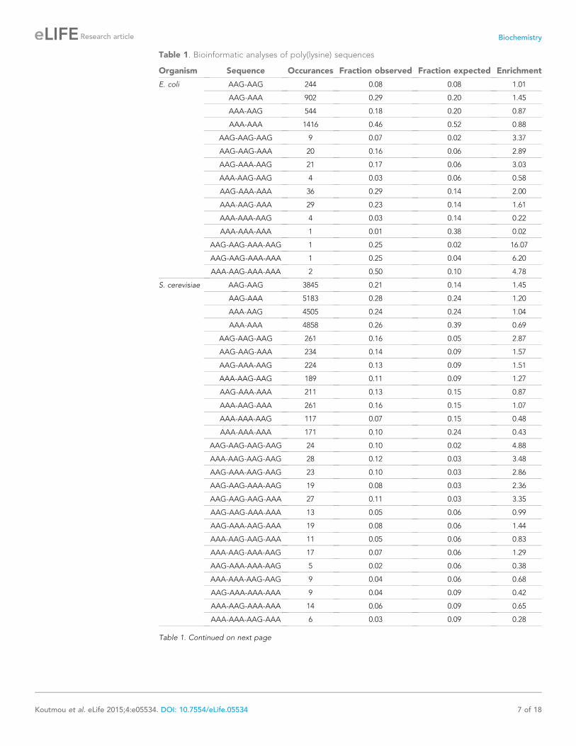
Table 1. Bioinformatic analyses of poly(lysine) sequences
Organism Sequence Occurances Fraction observed Fraction expected Enrichment
E. coli AAG-AAG 244 0.08 0.08 1.01
AAG-AAA 902 0.29 0.20 1.45
AAA-AAG 544 0.18 0.20 0.87
AAA-AAA 1416 0.46 0.52 0.88
AAG-AAG-AAG 9 0.07 0.02 3.37
AAG-AAG-AAA 20 0.16 0.06 2.89
AAG-AAA-AAG 21 0.17 0.06 3.03
AAA-AAG-AAG 4 0.03 0.06 0.58
AAG-AAA-AAA 36 0.29 0.14 2.00
AAA-AAG-AAA 29 0.23 0.14 1.61
AAA-AAA-AAG 4 0.03 0.14 0.22
AAA-AAA-AAA 1 0.01 0.38 0.02
AAG-AAG-AAA-AAG 1 0.25 0.02 16.07
AAG-AAG-AAA-AAA 1 0.25 0.04 6.20
AAA-AAG-AAA-AAA 2 0.50 0.10 4.78
S. cerevisiae AAG-AAG 3845 0.21 0.14 1.45
AAG-AAA 5183 0.28 0.24 1.20
AAA-AAG 4505 0.24 0.24 1.04
AAA-AAA 4858 0.26 0.39 0.69
AAG-AAG-AAG 261 0.16 0.05 2.87
AAG-AAG-AAA 234 0.14 0.09 1.57
AAG-AAA-AAG 224 0.13 0.09 1.51
AAA-AAG-AAG 189 0.11 0.09 1.27
AAG-AAA-AAA 211 0.13 0.15 0.87
AAA-AAG-AAA 261 0.16 0.15 1.07
AAA-AAA-AAG 117 0.07 0.15 0.48
AAA-AAA-AAA 171 0.10 0.24 0.43
AAG-AAG-AAG-AAG 24 0.10 0.02 4.88
AAA-AAG-AAG-AAG 28 0.12 0.03 3.48
AAG-AAA-AAG-AAG 23 0.10 0.03 2.86
AAG-AAG-AAA-AAG 19 0.08 0.03 2.36
AAG-AAG-AAG-AAA 27 0.11 0.03 3.35
AAG-AAG-AAA-AAA 13 0.05 0.06 0.99
AAG-AAA-AAG-AAA 19 0.08 0.06 1.44
AAA-AAG-AAG-AAA 11 0.05 0.06 0.83
AAA-AAG-AAA-AAG 17 0.07 0.06 1.29
AAG-AAA-AAA-AAG 5 0.02 0.06 0.38
AAA-AAA-AAG-AAG 9 0.04 0.06 0.68
AAG-AAA-AAA-AAA 9 0.04 0.09 0.42
AAA-AAG-AAA-AAA 14 0.06 0.09 0.65
AAA-AAA-AAG-AAA 6 0.03 0.09 0.28
Table 1. Continued on next page
Koutmou et al. eLife 2015;4:e05534. DOI: 10.7554/eLife.05534 7 of 18
Research article Biochemistry
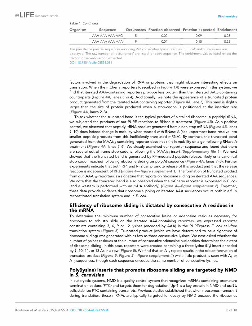
factors involved in the degradation of RNA or proteins that might obscure interesting effects on
translation. When the mCherry reporters (described in Figure 1A) were expressed in this system, we
find that iterated AAA-containing reporters produce less protein than their iterated AAG-containing
counterparts (Figure 4A, lanes 3 vs 4). Additionally, we note the appearance of a truncated protein
product generated from the iterated AAA-containing reporter (Figure 4A, lane 3). This band is slightly
larger than the size of protein produced when a stop-codon is positioned at the insertion site
(Figure 4A, lanes 2–3).
To ask whether the truncated band is the typical product of a stalled ribosome, a peptidyl-tRNA,
we subjected the products of our PURE reactions to RNase A treatment (Figure 4B). As a positive
control, we observed that peptidyl-tRNA product generated from a non-stop mRNA (Figure 4B, lanes
9–10) does indeed change in mobility when treated with RNase A (see uppermost band resolve into
smaller peptide products from this inefficiently translated mRNA). By contrast, the truncated band
generated from the (AAA)12-containing reporter does not shift in mobility on a gel following RNase A
treatment (Figure 4A, lanes 5–6). We closely examined our reporter sequence and found that there
are several out of frame stop-codons following the (AAA)12 insert (Supplementary file 1). We next
showed that the truncated band is generated by RF-mediated peptide release, likely on a canonical
stop codon reached following ribosome sliding on poly(A) sequence (Figure 4A, lanes 7–8). Further
experiments indicate that both RF1 and RF2 can promote release of this product and that the release
reaction is independent of RF3 (Figure 4—figure supplement 1). The formation of truncated product
from our (AAA)12 reporters is a signature that reports on ribosome sliding on iterated AAA sequences.
We note that the truncated band is also observed when the mCherry reporter is expressed in E. coli
(and a western is performed with an α-HA antibody) (Figure 4—figure supplement 2). Together,
these data provide evidence that ribosome slipping on iterated AAA sequences occurs both in a fully
reconstituted translation system and in E. coli.
Efficiency of ribosome sliding is dictated by consecutive A residues inthe mRNATo determine the minimum number of consecutive lysine or adenosine residues necessary for
ribosomes to robustly slide on the iterated AAA-containing reporters, we expressed reporter
constructs containing 3, 6, 9 or 12 lysines (encoded by AAA) in the PURExpress E. coli cell-free
translation system (Figure 5). Truncated product (which we have determined to be a signature of
ribosome sliding) was generated with as few as three consecutive lysines. We next asked whether the
number of lysines residues or the number of consecutive adenosine nucleotides determines the extent
of ribosome sliding. In this case, reporters were created containing a three lysine (K3) insert encoded
by 9, 10, 11, or 13 As in a row (Figure 5). We find that an A11 repeat results in the robust formation of
truncated product (Figure 5, Figure 5—figure supplement 1) while little product is seen with A9 or
A10 sequences, though each sequence encodes the same number of consecutive lysines.
Poly(lysine) inserts that promote ribosome sliding are targeted by NMDin S. cerevisiaeIn eukaryotic systems, NMD is a quality control system that recognizes mRNAs containing premature
termination codons (PTC) and targets them for degradation. Upf1 is a key protein in NMD and upf1Δcells stabilize PTC-containing transcripts. Previous studies established that when ribosomes frameshift
during translation, these mRNAs are typically targeted for decay by NMD because the ribosomes
Table 1. Continued
Organism Sequence Occurances Fraction observed Fraction expected Enrichment
AAA-AAA-AAA-AAG 5 0.02 0.09 0.23
AAA-AAA-AAA-AAA 9 0.04 0.15 0.25
The prevalence precise sequences encoding 2–3 consecutive lysine residues in E. coli and S. cerevisiae are
displayed. The raw number of ‘occurrences’ are listed for each sequence. The enrichment values listed reflect the
fraction observed/fraction expected.
DOI: 10.7554/eLife.05534.011
Koutmou et al. eLife 2015;4:e05534. DOI: 10.7554/eLife.05534 8 of 18
Research article Biochemistry
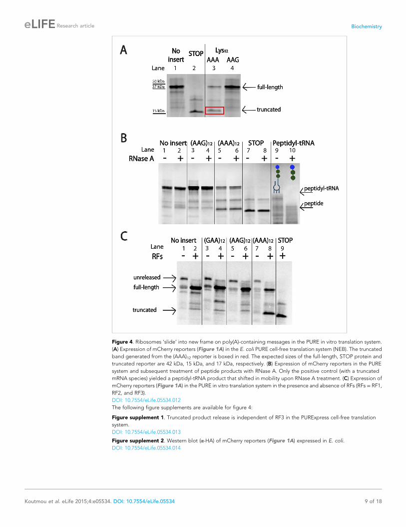
Figure 4. Ribosomes ‘slide’ into new frame on poly(A)-containing messages in the PURE in vitro translation system.
(A) Expression of mCherry reporters (Figure 1A) in the E. coli PURE cell-free translation system (NEB). The truncated
band generated from the (AAA)12 reporter is boxed in red. The expected sizes of the full-length, STOP protein and
truncated reporter are 42 kDa, 15 kDa, and 17 kDa, respectively. (B) Expression of mCherry reporters in the PURE
system and subsequent treatment of peptide products with RNase A. Only the positive control (with a truncated
mRNA species) yielded a peptidyl-tRNA product that shifted in mobility upon RNase A treatment. (C) Expression of
mCherry reporters (Figure 1A) in the PURE in vitro translation system in the presence and absence of RFs (RFs = RF1,
RF2, and RF3).
DOI: 10.7554/eLife.05534.012
The following figure supplements are available for figure 4:
Figure supplement 1. Truncated product release is independent of RF3 in the PURExpress cell-free translation
system.
DOI: 10.7554/eLife.05534.013
Figure supplement 2. Western blot (α-HA) of mCherry reporters (Figure 1A) expressed in E. coli.
DOI: 10.7554/eLife.05534.014
Koutmou et al. eLife 2015;4:e05534. DOI: 10.7554/eLife.05534 9 of 18
Research article Biochemistry
generally encounter an out of frame premature termination codon (Belew et al., 2011, 2014). We
proposed that if the ribosome slides on iterated AAA-containing mRNAs in yeast, as it does in the
bacterial system, then iterated AAA-containing mRNAs should be targeted by NMD. We addressed
this possibility by measuring the levels of (AAA)12, (AAG)12, and (AAGAAGAAA)4-containing mRNAs
in two different yeast-expressed reporter systems (Figure 1A) in wild-type and upf1Δ cells.
First, as a control, we measured the mRNA levels of luciferase reporters containing no insert, an
engineered premature stop codon (positive control), and a stem-loop known to trigger an alternative
mRNA quality control pathway, no-go decay (negative control) (Doma and Parker, 2006). We find that
the levels of mRNA for PTC and stem-loop containing reporters are lowered (PTC = 2 fold, stem-loop
= 21 fold) relative to reporters with no insert in wild-type yeast cells. Moreover, as expected, the level
of PTC, but not stem-loop-containing, mRNA is recovered when the reporters are expressed in upf1Δcells (Figure 6A). When this same experiment was performed with a luciferase reporter containing an
(AAA)12 sequence, we find that reporter mRNA levels are substantially reduced in wild-type cells
(>50-fold down), and that these levels are partially recovered in a upf1Δ strain (Figure 6A). These
results suggest that the (AAA)12-containing reporter is indeed a target of NMD in vivo.
To more directly compare our S. cerevisiae and E. coli results, we performed experiments instead
using the related mCherry reporters (Figure 1A) with no insert, or a variety of lysine inserts ((AAG)12,
(AAA)12, and (AAGAAGAAA)4). In addition to measuring the absolute levels of reporter mRNAs in
wild-type and upf1Δ cells (Figure 6), we asked whether the rates of mRNA decay for these reporters
are impacted in the upf1Δ knock-out background (Figure 6B and Figure 6—figure supplement 1).
We chose to include a mixed AAA/AAG reporter in addition to the simpler AAA and AAG repeat
reporters because this sequence is commonly used to report on the NSD phenomenon (Dimitrova
et al., 2009; Chiabudini et al., 2012, 2014). Indeed, a recent study with an (AAGAAGAAA)4-
containing reporter argued that a truncated product generated by such a construct resulted from an
unusual release factor-dependent termination event on a sense (lysine) codon (Chiabudini et al.,
2014). In an attempt to recapitulate these results, we directly looked for evidence of eRF1:eRF3-
mediated termination activity on iterated lysine mRNAs in vitro using a yeast reconstituted translation
system (Shoemaker et al., 2010); we see no evidence that such an event can occur (Figure 6—figure
supplement 2). We propose that an alternative explanation for the published data could be that the
ribosome slides out of frame on the (AAGAAGAAA)4 sequence, resulting in premature termination on
a previously out-of-frame stop codon, akin to what we observe in the PURE E. coli cell-free translation
system (Figure 4C). This possibility seemed particularly likely given that we observed sliding
activity on a AUG-AAA-AAG-UUC-STOP sequence in our in vitro reconstituted E. coli system
(Figure 3—figure supplement 1).
Figure 5. Position and length of poly(A) stretch contributes to ribosome ‘sliding’ in the PURE in vitro translation
system. Expression of mCherry reporters containing poly(A) inserts of various lengths in the presence (+) andabsence (−) of RFs.DOI: 10.7554/eLife.05534.015
The following figure supplement is available for figure 5:
Figure supplement 1. Quantification of the efficiency of ribosome sliding on mCherry reporters expressed in the
PURExpress system.
DOI: 10.7554/eLife.05534.016
Koutmou et al. eLife 2015;4:e05534. DOI: 10.7554/eLife.05534 10 of 18
Research article Biochemistry
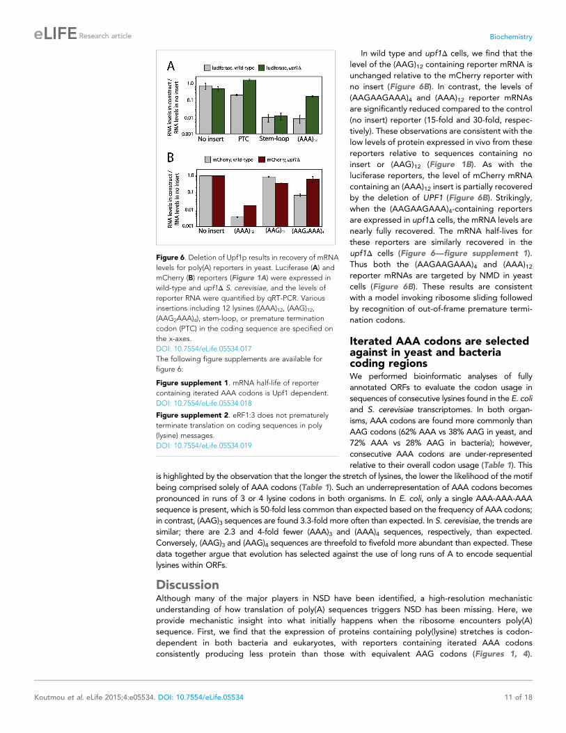
In wild type and upf1Δ cells, we find that the
level of the (AAG)12 containing reporter mRNA is
unchanged relative to the mCherry reporter with
no insert (Figure 6B). In contrast, the levels of
(AAGAAGAAA)4 and (AAA)12 reporter mRNAs
are significantly reduced compared to the control
(no insert) reporter (15-fold and 30-fold, respec-
tively). These observations are consistent with the
low levels of protein expressed in vivo from these
reporters relative to sequences containing no
insert or (AAG)12 (Figure 1B). As with the
luciferase reporters, the level of mCherry mRNA
containing an (AAA)12 insert is partially recovered
by the deletion of UPF1 (Figure 6B). Strikingly,
when the (AAGAAGAAA)4-containing reporters
are expressed in upf1Δ cells, the mRNA levels are
nearly fully recovered. The mRNA half-lives for
these reporters are similarly recovered in the
upf1Δ cells (Figure 6—figure supplement 1).
Thus both the (AAGAAGAAA)4 and (AAA)12reporter mRNAs are targeted by NMD in yeast
cells (Figure 6B). These results are consistent
with a model invoking ribosome sliding followed
by recognition of out-of-frame premature termi-
nation codons.
Iterated AAA codons are selectedagainst in yeast and bacteriacoding regionsWe performed bioinformatic analyses of fully
annotated ORFs to evaluate the codon usage in
sequences of consecutive lysines found in the E. coli
and S. cerevisiae transcriptomes. In both organ-
isms, AAA codons are found more commonly than
AAG codons (62% AAA vs 38% AAG in yeast, and
72% AAA vs 28% AAG in bacteria); however,
consecutive AAA codons are under-represented
relative to their overall codon usage (Table 1). This
is highlighted by the observation that the longer the stretch of lysines, the lower the likelihood of the motif
being comprised solely of AAA codons (Table 1). Such an underrepresentation of AAA codons becomes
pronounced in runs of 3 or 4 lysine codons in both organisms. In E. coli, only a single AAA-AAA-AAA
sequence is present, which is 50-fold less common than expected based on the frequency of AAA codons;
in contrast, (AAG)3 sequences are found 3.3-fold more often than expected. In S. cerevisiae, the trends are
similar; there are 2.3 and 4-fold fewer (AAA)3 and (AAA)4 sequences, respectively, than expected.
Conversely, (AAG)3 and (AAG)4 sequences are threefold to fivefold more abundant than expected. These
data together argue that evolution has selected against the use of long runs of A to encode sequential
lysines within ORFs.
DiscussionAlthough many of the major players in NSD have been identified, a high-resolution mechanistic
understanding of how translation of poly(A) sequences triggers NSD has been missing. Here, we
provide mechanistic insight into what initially happens when the ribosome encounters poly(A)
sequence. First, we find that the expression of proteins containing poly(lysine) stretches is codon-
dependent in both bacteria and eukaryotes, with reporters containing iterated AAA codons
consistently producing less protein than those with equivalent AAG codons (Figures 1, 4).
Figure 6. Deletion of Upf1p results in recovery of mRNA
levels for poly(A) reporters in yeast. Luciferase (A) and
mCherry (B) reporters (Figure 1A) were expressed in
wild-type and upf1Δ S. cerevisiae, and the levels of
reporter RNA were quantified by qRT-PCR. Various
insertions including 12 lysines ((AAA)12, (AAG)12,
(AAG2AAA)4), stem-loop, or premature termination
codon (PTC) in the coding sequence are specified on
the x-axes.
DOI: 10.7554/eLife.05534.017
The following figure supplements are available for
figure 6:
Figure supplement 1. mRNA half-life of reporter
containing iterated AAA codons is Upf1 dependent.
DOI: 10.7554/eLife.05534.018
Figure supplement 2. eRF1:3 does not prematurely
terminate translation on coding sequences in poly
(lysine) messages.
DOI: 10.7554/eLife.05534.019
Koutmou et al. eLife 2015;4:e05534. DOI: 10.7554/eLife.05534 11 of 18
Research article Biochemistry
This differential protein output is not the result of imprecise RNA polymerase action (Figure 3—figure
supplement 3) nor likely of disparities in the rate of adding lysine codons (Figure 2); lysines are slowly
incorporated on iterated AAA and AAG codons. Instead, the codon-dependent disparity primarily
stems from an unusual sliding event that occurs when ribosomes encounter consecutive AAA
codons (Figures 3, 4). Our observation that ribosomes can slide in multiple frames on iterated AAA
sequences provides a rationale for consecutive AAA codons being substantially under-represented
in open reading frames in most genomes (see Bioinformatic discussion below, Table 1 and
(unpublished data).
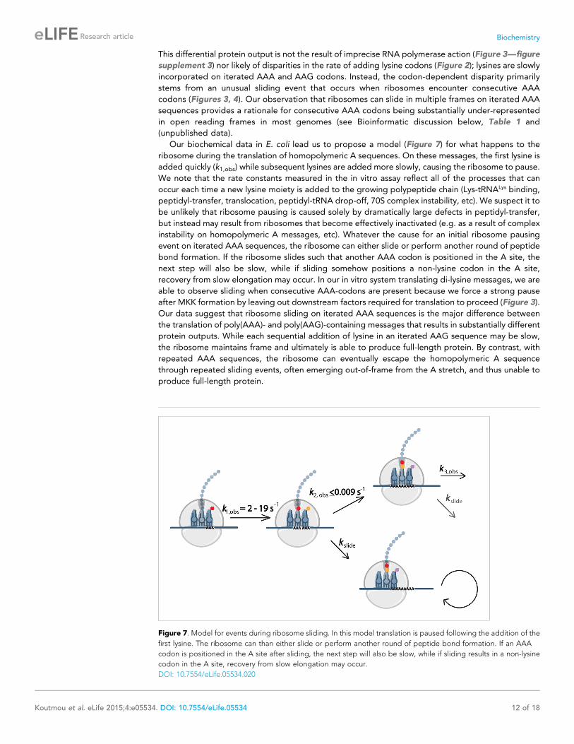
Our biochemical data in E. coli lead us to propose a model (Figure 7) for what happens to the
ribosome during the translation of homopolymeric A sequences. On these messages, the first lysine is
added quickly (k1,obs) while subsequent lysines are added more slowly, causing the ribosome to pause.
We note that the rate constants measured in the in vitro assay reflect all of the processes that can
occur each time a new lysine moiety is added to the growing polypeptide chain (Lys-tRNALys binding,
peptidyl-transfer, translocation, peptidyl-tRNA drop-off, 70S complex instability, etc). We suspect it to
be unlikely that ribosome pausing is caused solely by dramatically large defects in peptidyl-transfer,
but instead may result from ribosomes that become effectively inactivated (e.g. as a result of complex
instability on homopolymeric A messages, etc). Whatever the cause for an initial ribosome pausing
event on iterated AAA sequences, the ribosome can either slide or perform another round of peptide
bond formation. If the ribosome slides such that another AAA codon is positioned in the A site, the
next step will also be slow, while if sliding somehow positions a non-lysine codon in the A site,
recovery from slow elongation may occur. In our in vitro system translating di-lysine messages, we are
able to observe sliding when consecutive AAA-codons are present because we force a strong pause
after MKK formation by leaving out downstream factors required for translation to proceed (Figure 3).
Our data suggest that ribosome sliding on iterated AAA sequences is the major difference between
the translation of poly(AAA)- and poly(AAG)-containing messages that results in substantially different
protein outputs. While each sequential addition of lysine in an iterated AAG sequence may be slow,
the ribosome maintains frame and ultimately is able to produce full-length protein. By contrast, with
repeated AAA sequences, the ribosome can eventually escape the homopolymeric A sequence
through repeated sliding events, often emerging out-of-frame from the A stretch, and thus unable to
produce full-length protein.
Figure 7. Model for events during ribosome sliding. In this model translation is paused following the addition of the
first lysine. The ribosome can than either slide or perform another round of peptide bond formation. If an AAA
codon is positioned in the A site after sliding, the next step will also be slow, while if sliding results in a non-lysine
codon in the A site, recovery from slow elongation may occur.
DOI: 10.7554/eLife.05534.020
Koutmou et al. eLife 2015;4:e05534. DOI: 10.7554/eLife.05534 12 of 18
Research article Biochemistry
Ribosome sliding on poly(A) is distinct from traditional programmed ribosomal movements such as
+1 (Farabaugh and Bjork, 1999; Taliaferro and Farabaugh, 2007) and −1 frame-shifts (Dinman
et al., 1991; Plant et al., 2003; Caliskan et al., 2014; Chen et al., 2014; Kim et al., 2014). During
a programmed frame-shifting (PRF) event, specific signals direct elongating ribosomes to shift reading
frame by one base in the 5′ (−1) or 3′ (+1) direction (Dinman, 2012). −1 PRFs signals are typically
characterized by a ‘slippery’ sequence (X XXY YYZ) that is modulated by the presence of
a downstream secondary structure, most commonly a pseudoknot (Plant et al., 2003; Jacobs et al.,
2007; Caliskan et al., 2014; Chen et al., 2014; Kim et al., 2014). The secondary structure impairs the
normal movement of the ribosome during translocation, and promotes the frame-shift event in an
EF-G dependent manner (Caliskan et al., 2014; Chen et al., 2014). +1 PRFs signals are more diverse
than −1 PRFs, but still generally depend on a slippery sequence and a downstream element
(e.g., secondary structure or rare codon) that causes the ribosome to pause (Dinman, 2012). Iterated
A stretches are inherently slippery and contain a built-in translation pause (adding consecutive lysines
is slow—Figure 2), however the poly(A) sequences that we have studied lack significant secondary
structure downstream that might contribute to limiting unregulated ribosome sliding. As such, when
ribosomes slide on iterated AAA codons, forward and backward movements may be permitted. The
scale of the movements undergone during a ribosome sliding event may be more similar to those
documented in translational bypassing on the gene product 60 of bacteriophage T4 which is
synthesized from a discontinuous reading frame (Samatova et al., 2014). Importantly, however, in
contrast to this specific concerted large-scale movement (50 nucleotides) which results in the
production of a single peptide product, ribosome sliding is different in that no single outcome
appears to be encoded by the event. The inability of the ribosome to translate a discrete product on
homopolymeric A sequences likely explains the bioinformatic analyses demonstrating that poly(A)
sequences are strongly selected against in coding sequences containing iterated lysines (Table 1).
Consistent with this idea, in E. coli we find that the minimum length (11) of a homopolymeric A
sequence needed to trigger ribosome sliding in the PURE cell-free translation system (Figure 5, and
Figure 5—figure supplement 1) correlates with the length of lysine stretch at which homopolymeric
sequences are selected against in mRNA coding regions (Table 1).
There are multiple reports in the literature indicating that frame-shifted ribosomes can trigger
NMD (Belew et al., 2011, 2014). We find that mRNA levels for reporters containing (AAA)12 or
(AAGAAGAAA)4, but not (AAG)12 sequences, are reduced in a Upf1-dependent manner. These data
are consistent with the idea that sliding on homopolymeric A stretches can eventually lead to
ribosomes reaching out-of-frame premature termination codons (Figure 6). A recent report in the
literature argued that translation of poly(lysine) stretches led to an unusual termination event on
a sense codon (AAA or AAG) mediated by eRF3 (presumably in concert with its binding partner eRF1)
(Chiabudini et al., 2014). These observations bring to mind premature termination events on sense
codons documented in E. coli (Zaher and Green, 2009); this quality control system was proposed to
increase the fidelity of translation by minimizing frame-shifting and eliminating errors made during
tRNA selection. We note that the premature termination event that we previously documented in
E. coli was highly dependent on RF3, while the termination event documented in E. coli in this
manuscript at homopolymeric A sequences is RF3-independent (Figure 4—figure supplement 1).
Given the clear evidence that we provide for ribosome sliding in the E. coli system and the inability to
observe eRF1:eRF3-mediated peptide release on homopolymeric A programmed yeast ribosome
complexes in vitro (Figure 6—figure supplement 2), we suggest that the most likely explanation for
the eRF3-dependent truncated product generated in yeast cells on (AAGAAGAAA)4–encoding
reporters in Chiabudini et al. is the result of ribosome sliding and canonical recognition of
downstream premature stop codons. We note that there are multiple out-of-frame stop codons
following the (AAGAAGAAA)4-repeat that could account for the observed products in Chiabudini
et al. (Chiabudini et al., 2014).
We were intrigued by the observation that the (AAGAAGAAA)4 reporter mRNA levels are more
efficiently recovered than those of the (AAA)12 reporter mRNA in a UPF1-deletion strain. We
speculate that the more modest sliding within the (AAGAAGAAA)-repeats might be distinguished
from the sliding on (AAA)-repeats in an important way. Sliding within homopolymeric AAA sequence
most typically results in another nearby AAA codon being poised in the A site, and another inefficient
elongation event with Lys-tRNALys. Ribosomes that eventually exit the poly(A) sequence to reach
heteropolymeric sequence and an out-of-frame downstream premature stop codon will trigger NMD;
Koutmou et al. eLife 2015;4:e05534. DOI: 10.7554/eLife.05534 13 of 18
Research article Biochemistry
ribosomes that struggle to get past the very long stretch of iterated lysine codons will instead trigger
NSD. As such, the mRNA levels for the (AAA)12 reporter are partially recovered by a UPF1 deletion
and partially recovered by a DOM34 deletion (data not shown). By contrast, on the (AAGAAGAAA)-
repeat reporters, sliding has the potential to quickly place the ribosomes in a more productive frame
for efficient elongation (one frame will result in Arg-Arg-Lys (RRK) repeats while the other frame will
result in Glu-Glu-Lys (EEK) repeats). While we might predict that the poly(basic) RRKRRKRRKRR
peptide will also be slowly translated, a ribosome that slides into the frame encoding the
EEKEEKEEKEE peptide should be able to resume efficient elongation. As such, fewer ribosomes
may trigger NSD and, instead, a majority of ribosomes will reach downstream premature stop codons
that trigger NMD. These ideas can easily be understood in the context of the model in Figure 7 where
differences in the elongation rates (e.g. slow for iterated lysine residues but fast for incorporation of
other amino acids) will impact the relative contribution of ribosome sliding to overall outcome.
NSD was originally identified by following the degradation of transcripts lacking termination
codons (Frischmeyer et al., 2002; van Hoof et al., 2002). These studies led to the idea that NSD is
triggered when the ribosome stalls while translating a poly(basic) lysine sequence. NSD is commonly
studied using reporters in yeast that contain poly(basic) inserts; common lysine and arginine inserts
that have been investigated include (AAA)12, (AAG)12, (AAG-AAG-AAA)4, and (CGG-(CGA)2-CGG-
(CGC)2)2 (Ito-Harashima et al., 2007; Dimitrova et al., 2009; Bengtson and Joazeiro, 2010;
Brandman et al., 2012; Chiabudini et al., 2012, 2014). Consistent with our findings, previous studies
reported differences in protein output in yeast when these different sequences are translated
(Ito-Harashima et al., 2007; Dimitrova et al., 2009); iterated AAA codons are more detrimental to
overall expression than iterated AAG codons. Despite these differences, because the mRNA and
protein levels for all of these are broadly sensitive to known NSD factors (Ltn1, Dom34, Ski7), poly
(basic) sequences have been treated equally. Our results demonstrating that ribosomes can slide on
consecutive AAA codons suggest that there may be important distinctions to be made in considering
these reporters and that there may be substantial mechanistic overlaps in these systems.
Even though cells rarely maintain homopolymeric A sequences in ORFs, there are some situations
where the ribosome likely must deal with homopolymeric A stretches in both bacteria and eukaryotes.
In bacteria, mRNAs are typically polyadenylated as part of the normal decay process (Dreyfus and
Regnier, 2002). For example, ribosome sliding might provide an escape for ribosomes already
engaged on these mRNAs (a form of ribosome rescue). In eukaryotes, virtually all mRNAs in the cell
are polyadenylated, but usually a stop codon is found at the end of the encoded ORF. However, there
is abundant recent evidence indicating that a significant portion of yeast (14%) and human (9%) genes
contain at least one alternative polyadenylation site within their coding sequence (Ozsolak et al.,
2010). It has even been suggested that premature polyadenylation may become up-regulated in
cancerous cells (Berg et al., 2012). In cases where premature polyadenylation takes place within the
ORF, the ribosome will surely encounter a homopolymeric A sequence, likely triggering so called Non-
Stop-Decay (NSD). In light of the results presented here, we would suggest that the triggering of NSD
(and associated mRNA decay, proteolysis and ribosome recycling) occurs following the slow
translation of iterated lysines and ribosome sliding events. The ubiquity of premature polyadenylation
suggests that NSD broadly serves as an important pathway for regulating gene expression. The
observation of synonymous AAG to AAA changes in iterated lysine stretches in genes upregulated in
cancer provides support for the significance of this mechanism of gene regulation (unpublished data).
The widespread use of polyadenylation for non-coding purposes in mRNA transcripts may find its
origins in the inability of the decoding machine, the ribosome, to carefully control the behavior of
these sequences.
Materials and methods
Reporter creationThe Thrdx-HA-mCherry (Figure 1A, Supplementary file 1) no insert reporter expressed in E. coli and the
PURExpress cell-free translation system was created using Gateway cloning to include the 2HA-mCherry
sequence in the pBAD-DEST49 vector. The vectors containing inserts (Thrdx-HA-insert-mCherry: (AAA)12,
(AAA)6, (AAG)12, (AAGAAGAAA)4, (GAA)12, TAA (STOP), (A)9-13, etc) were subsequently derived from
this clone. To create the mCherry reporter expressed in yeast (Figure 1A), the Thrdx-HA-mCherry and
Thrdx-HA-insert-mCherry sequences were amplified out of the pBAD-DEST49 vectors and cloned into the
Koutmou et al. eLife 2015;4:e05534. DOI: 10.7554/eLife.05534 14 of 18
Research article Biochemistry
p-ENTR/D-TOPO vector. The vector was then reacted with lr-clonease II to move the sequences into the
pYES-DEST52 plasmid. The dual luciferase reporter described in Figure 1A was based on the dual
luciferase plasmid from Takacs et al. (2011). In this reporter, Renilla and Firefly luciferase are under the
control of ADH and GPD promoters, respectively. We inserted sequences of interest into the N-terminus
of Renilla luciferase. The single Renilla luciferase reporter described in Figure 1A was cloned into pYES2
(with a Gal promoter) using the Gateway cloning system.
In vivo protein expression and visualizationThrdx-HA-mCherry and Thrdx-HA-insert-mCherry constructs were expressed in 6 ml E. coli grown in
LB-Ampicillin. The cells were grown to an OD of 0.4–0.6, induced with 25 μl of 5 g/10 ml arabinose,
then harvested 2 hr post-induction. In yeast, the Thrdx-HA-mCherry constructs were expressed in
wild-type and upf1Δ S. cerevisiae (BY4741) grown in 5 ml of–URA/+galactose media to an OD of 0.6.
The single luciferase reporters were transformed into yeast and grown in–URA/+galactose media, and
harvested at an OD of 0.6. Proteins production was analyzed via fluorescence, luminescence
(Figure 1) or western blot analysis (Figure 4—figure supplement 2).
Assessing lysine incorporation in fully reconstituted in vitro translationassays70S initiation complexes (ICs) were prepared using E. coli ribosomes programmed with various
mRNAs and f-[35S]-Met-tRNAMet in the P site. mRNAs were generated by transcription with T7
polymerase and ICs were formed, pelleted, and resuspended as previously described (Youngman
et al., 2004) on our messages of interest. Translation assays were initiated when equal volumes of
ternary complex (10–20 μM charged tRNA, 12 μM EFG, 60 μM EfTu) were added to 0.2 nM 70S
initiation complexes. Assays were performed in 219-Tris buffer (50 mM Tris pH 7.5, 70 mM NH4Cl,
30 mM KCl, 7 mM MgCl2, 5 mM βME). The limited addition of iterated lysines on a MKA5-STOP
message was also observed in polymix buffer (50 mM K2HPO4 pH 7.5, 95 mM KCl, 5 mM NH4Cl,
5 mM Mg(OAc)2, 0.5 mM CaCl2, 8 mM putrescine, 1 mM spermidine, 1 mM DTT). To measure the
rates of amino acid incorporation, the reactions are quenched with 500 mM KOH (final concentration)
at discrete time points (0 s–30 min) either by hand or on a quench-flow apparatus. For assays including
release factors for the duration of the reaction (Figure 3C), RF1 and additional GTP were added prior
to the initiation of translation (final concentrations 1 μM and 200 μM, respectively). The time-points
were diluted 1:10 in nuclease free water and the reactants, intermediates and products visualized by
electrophoretic TLC, as previously described (Zaher and Green, 2009). The reactants, products and
intermediates were visualized by phosophorimaging and quantified with ImageQuant. The kinetic fits
were modeled using Mathematica (details in Figure 2—figure supplement 2).
Expression of reporters in the PURExpress in vitro translation systemThe Thrdx-HA-mCherry and Thrdx-HA-insert-mCherry reporters were expressed in the PURExpress in
vitro translation system (NEB, Ipswitch, MA) from PCR products. The peptidyl-tRNA construct was
generated by creating a truncated mRNA lacking a stop codon directly after the Thrdx-HA sequence. The
PURExpress reactions were initiated by mixing 1 μl of PCR product (29–22 ng/μl), 2 μl of solution A, 1.5 μlof solution B, and 0.6 μl of 35S-methionine. The reactions were run for 45–60 min at 37˚C. Following
translation, the products were immediately heat-denatured and loaded on a 4–12% Bis-Tris gel at 4˚C in
XT-MES buffer. For the experiments in which the PURExpress reaction products were treated with RNase
A (Figures 4B), 0.5–1 μg of RNase A (Ambion, Grand Island, NY) was added to each reaction and
solutions were incubated on ice for an additional 30 min before being denatured and loaded on a gel. The
peptide products of the PURExpress reactions were visualized by Phosphoimager and quantified with
ImageQuant (Figure 3—figure supplement 2, and Figure 5—figure supplement 1).
Toeprinting assaysDNA templates were PCR amplified from plasmids (PCR-Blunt II-TOPO vector) encoding MEA(INSERT)
EAEDYKDD sequences. The PURExpress cell-free transcription-translation system (NEB, Ipswich, MA)
was used for in vitro protein synthesis. Reactions were run for 30 min at 37˚C by mixing 0.2-pmol of
DNA template, 2.5 μl of Solution A and 1 μl of Solution B along with either 0.5 μl of DMSO (5%) or
thiostrepton (0.5 mm in 5% DMSO). 1 pmol of 32PATP-labeled NV1 primer was added, and reverse
transcription was performed with AMV as previously described (Vazquez-Laslop et al., 2008; Tanner
Koutmou et al. eLife 2015;4:e05534. DOI: 10.7554/eLife.05534 15 of 18
Research article Biochemistry
et al., 2009). Reactions were phenol and chloroform extracted, ethanol precipitated and visualized on
a 6% denaturing PAGE gel. Sequencing lanes were generated from plasmids using the Sequenase 2.0
DNA sequencing kit (Affymetrix, Santa Clara, CA). All bands were visualized by PhosphorImager.
Real-time quantitative reverse transcription PCR (qRT PCR) to measurereporter mRNA levelsReporter mRNA levels were quantified by qRT-PCR using the iQ5 iCycler system (Bio-Rad, Hercules,
CA) and iQ SYBR Green Supermix (Bio-Rad, Hercules, CA).
Measuring mRNA decayTo measure the rate of mRNA decay in yeast for our mCherry reporters, we grew wild-type and upf1Δcells expressing reporters in–ura/galactose media at 30˚C to an OD600 of 0.4. Cells were washed
three times with–ura media lacking sugar, then re-suspended in -ura/glucose media; the transcription
of the reporter is shut-off by glucose. Samples were collected at discrete time points (0–90 min), and
mRNA levels were analyzed by qRT PCR.
Bioinformatic analysesE. coli K-12 substrain MG1655 complete genome, 4140 ORFs (data source: GenBank:U00096.3;
http://www.ncbi.nlm.nih.gov/nuccore/U00096.3) and S. cerevisiae 5887 verified ORFs (data source:
http://downloads.yeastgenome.org/sequence/S288C_reference/orf_protein/) have been used for
extraction of lysine codon numbers and analyses of consecutive codons shown in Table 1. Expected
values for consecutive variants of lysine AAA and AAG codons were calculated based on observed
values for a single AAA and AAG codons and their probabilities to be found in such arrangments.
Observed values were calculated based on data from genomic distribution and total numbers of
variants for two, three or four consecutive lys codons, respectively.
AcknowledgementsWe would like to thank Slavica Pavlovic-Djuranovic and Risa Burr for help with materials, Jon Lorsch for
sharing the dual luciferase plasmid (Takacs et al., 2011), and Allen Buskirk for reading. We would also
like to thank the National Institutes of Health (R37 GM059425 to RG, and F32 GM100608 to KSK) for
funding and the Howard Hughes Medical Foundation (RG) for salary support.
Additional informationCompeting interests
RG: Reviewing editor, eLife. The other authors declare that no competing interests exist.
Funding
Funder Grant reference Author
National Institute of GeneralMedical Sciences (NIGMS)
F32 GM100608 Kristin SKoutmou
Howard Hughes MedicalInstitute (HHMI)
Molcular Mechanisms ofTranslation and TheirImplications for GeneRegulation
Rachel Green
National Institute of GeneralMedical Sciences (NIGMS)
R37 GM059425 Rachel Green
The funders had no role in study design, data collection and interpretation, or thedecision to submit the work for publication.
Author contributions
KSK, SD, Conception and design, Acquisition of data, Analysis and interpretation of data, Drafting or
revising the article; APS, Acquisition of data, Analysis and interpretation of data, Drafting or revising
the article; JLB, Acquisition of data, Analysis and interpretation of data; AR, Analysis and
interpretation of data, Drafting or revising the article; RG, Conception and design, Analysis and
interpretation of data, Drafting or revising the article
Koutmou et al. eLife 2015;4:e05534. DOI: 10.7554/eLife.05534 16 of 18
Research article Biochemistry
Additional filesSupplementary file
·Supplementary file 1. Primary sequence of mCherry with out of frame stop-codons highlighted. The
nucleotide sequence of the Thrdx-HA-mCherry reporters (Figure 1A) with all out of frame stop
codons after the insertion site highlighted in yellow.DOI: 10.7554/eLife.05534.021
ReferencesArenz S, Meydan S, Starosta AL, Berninghausen O, Beckmann R, Vazquez-Laslop N, Wilson DN. 2014. Drugsensing by the ribosome induces translational arrest via active site perturbation. Molecular Cell 56:446–452.doi: 10.1016/j.molcel.2014.09.014.
Belew AT, Advani VM, Dinman JD. 2011. Endogenous ribosomal frameshift signals operate as mRNA destabilizingelements through at least two molecular pathways in yeast. Nucleic Acids Research 39:2799–2808. doi: 10.1093/nar/gkq1220.
Belew AT, Meskauskas A, Musalgaonkar S, Advani VM, Sulima SO, Kasprzak WK, Shapiro BA, Dinman JD. 2014.Ribosomal frameshifting in the CCR5 mRNA is regulated by miRNAs and the NMD pathway. Nature 512:265–269. doi: 10.1038/nature13429.
Bengtson MH, Joazeiro CA. 2010. Role of a ribosome-associated E3 ubiquitin ligase in protein quality control.Nature 467:470–473. doi: 10.1038/nature09371.
Berg MG, Singh LN, Younis I, Liu Q, Pinto AM, Kaida D, Zhang Z, Cho S, Sherrill-Mix S, Wan L, Dreyfuss G. 2012. U1snRNP determines mRNA length and regulates isoform expression. Cell 150:53–64. doi: 10.1016/j.cell.2012.05.029.
Bhushan S, Hoffmann T, Seidelt B, Frauenfeld J, Mielke T, Berninghausen O, Wilson DN, Beckmann R. 2011.SecM-stalled ribosomes adopt an altered geometry at the peptidyl transferase center. PLOS Biology 9:e1000581. doi: 10.1371/journal.pbio.1000581.
Brandman O, Stewart-Ornstein J, Wong D, Larson A, Williams CC, Li GW, Zhou S, King D, Shen PS, Weibezahn J,Dunn JG, Rouskin S, Inada T, Frost A, Weissman JS. 2012. A ribosome-bound quality control complex triggersdegradation of nascent peptides and signals translation stress. Cell 151:1042–1054. doi: 10.1016/j.cell.2012.10.044.
Caliskan N, Katunin VI, Belardinelli R, Peske F, Rodnina MV. 2014. Programmed -1 frameshifting by kineticpartitioning during impeded translocation. Cell 157:1619–1631. doi: 10.1016/j.cell.2014.04.041.
Chan PP, Lowe TM. 2009. GtRNAdb: a database of transfer RNA genes detected in genomic sequence. NucleicAcids Research 37:D93–D97. doi: 10.1093/nar/gkn787.
Chen J, Petrov A, Johansson M, Tsai A, O’Leary SE, Puglisi JD. 2014. Dynamic pathways of -1 translationalframeshifting. Nature 512:328–332. doi: 10.1038/nature13428.
Chiabudini M, Conz C, Reckmann F, Rospert S. 2012. Ribosome-associated complex and Ssb are required fortranslational repression induced by polylysine segments within nascent chains.Molecular and Cellular Biology 32:4769–4779. doi: 10.1128/MCB.00809-12.
Chiabudini M, Tais A, Zhang Y, Hayashi S, Wolfle T, Fitzke E, Rospert S. 2014. Release factor eRF3 mediatespremature translation termination on polylysine-stalled ribosomes in Saccharomyces cerevisiae. Molecular andCellular Biology 34:4062–4076. doi: 10.1128/MCB.00799-14.
Dimitrova LN, Kuroha K, Tatematsu T, Inada T. 2009. Nascent peptide-dependent translation arrest leads toNot4p-mediated protein degradation by the proteasome. The Journal of Biological Chemistry 284:10343–10352.doi: 10.1074/jbc.M808840200.
Dinman JD. 2012. Mechanisms and implications of programmed translational frameshifting. Wiley InterdisciplinaryReviews RNA 3:661–673. doi: 10.1002/wrna.1126.
Dinman JD, Icho T, Wickner RB. 1991. A -1 ribosomal frameshift in a double-stranded RNA virus of yeast formsa gag-pol fusion protein. Proceedings of the National Academy of Sciences of USA 88:174–178. doi: 10.1073/pnas.88.1.174.
Doerfel LK, Wohlgemuth I, Kothe C, Peske F, Urlaub H, Rodnina MV. 2013. EF-P is essential for rapid synthesis ofproteins containing consecutive proline residues. Science 339:85–88. doi: 10.1126/science.1229017.
Doma MK, Parker R. 2006. Endonucleolytic cleavage of eukaryotic mRNAs with stalls in translation elongation.Nature 440:561–564. doi: 10.1038/nature04530.
Dreyfus M, Regnier P. 2002. The poly(A) tail of mRNAs: bodyguard in eukaryotes, scavenger in bacteria. Cell 111:611–613. doi: 10.1016/S0092-8674(02)01137-6.
Farabaugh PJ, Bjork GR. 1999. How translational accuracy influences reading frame maintenance. The EMBOJournal 18:1427–1434. doi: 10.1093/emboj/18.6.1427.
Frischmeyer PA, van Hoof A, O’Donnell K, Guerrerio AL, Parker R, Dietz HC. 2002. An mRNA surveillancemechanism that eliminates transcripts lacking termination codons. Science 295:2258–2261. doi: 10.1126/science.1067338.
Gong F, Yanofsky C. 2002. Instruction of translating ribosome by nascent peptide. Science 297:1864–1867.doi: 10.1126/science.1073997.
Gromadski KB, Daviter T, Rodnina MV. 2006. A uniform response to mismatches in codon-anticodon complexesensures ribosomal fidelity. Molecular Cell 21:369–377. doi: 10.1016/j.molcel.2005.12.018.
Koutmou et al. eLife 2015;4:e05534. DOI: 10.7554/eLife.05534 17 of 18
Research article Biochemistry
Gutierrez E, Shin BS, Woolstenhulme CJ, Kim JR, Saini P, Buskirk AR, Dever TE. 2013. eIF5A promotes translationof polyproline motifs. Molecular Cell 51:35–45. doi: 10.1016/j.molcel.2013.04.021.
Ito K, Chiba S. 2013. Arrest peptides: cis-acting modulators of translation. Annual Review of Biochemistry 82:171–202. doi: 10.1146/annurev-biochem-080211-105026.
Ito-Harashima S, Kuroha K, Tatematsu T, Inada T. 2007. Translation of the poly(A) tail plays crucial roles in nonstopmRNA surveillance via translation repression and protein destabilization by proteasome in yeast. Genes &Development 21:519–524. doi: 10.1101/gad.1490207.
Izawa T, Tsuboi T, Kuroha K, Inada T, Nishikawa S, Endo T. 2012. Roles of dom34:hbs1 in nonstop proteinclearance from translocators for normal organelle protein influx. Cell Reports 2:447–453. doi: 10.1016/j.celrep.2012.08.010.
Jacobs JL, Belew AT, Rakauskaite R, Dinman JD. 2007. Identification of functional, endogenous programmed -1ribosomal frameshift signals in the genome of Saccharomyces cerevisiae. Nucleic Acids Research 35:165–174.doi: 10.1093/nar/gkl1033.
Kim HK, Liu F, Fei J, Bustamante C, Gonzalez RL Jr, Tinoco I Jr. 2014. A frameshifting stimulatory stem loopdestabilizes the hybrid state and impedes ribosomal translocation. Proceedings of the National Academy ofSciences of USA 111:5538–5543. doi: 10.1073/pnas.1403457111.
Klauer AA, van Hoof A. 2012. Degradation of mRNAs that lack a stop codon: a decade of nonstop progress. WileyInterdisciplinary Reviews RNA 3:649–660. doi: 10.1002/wrna.1124.
Kuroha K, AkamatsuM, Dimitrova L, Ito T, Kato Y, Shirahige K, Inada T. 2010. Receptor for activated C kinase 1 stimulatesnascent polypeptide-dependent translation arrest. EMBO Reports 11:956–961. doi: 10.1038/embor.2010.169.
Lu J, Deutsch C. 2008. Electrostatics in the ribosomal tunnel modulate chain elongation rates. Journal of MolecularBiology 384:73–86. doi: 10.1016/j.jmb.2008.08.089.
Nakatogawa H, Ito K. 2002. The ribosomal exit tunnel functions as a discriminating gate. Cell 108:629–636.doi: 10.1016/S0092-8674(02)00649-9.
Ozsolak F, Kapranov P, Foissac S, Kim SW, Fishilevich E, Monaghan AP, John B, Milos PM. 2010. Comprehensivepolyadenylation site maps in yeast and human reveal pervasive alternative polyadenylation. Cell 143:1018–1029.doi: 10.1016/j.cell.2010.11.020.
Plant EP, Jacobs KL, Harger JW, Meskauskas A, Jacobs JL, Baxter JL, Petrov AN, Dinman JD. 2003. The 9-Asolution: how mRNA pseudoknots promote efficient programmed -1 ribosomal frameshifting. RNA 9:168–174.doi: 10.1261/rna.2132503.
Ratinier M, Boulant S, Combet C, Targett-Adams P, McLauchlan J, Lavergne JP. 2008. Transcriptional slippageprompts recoding in alternate reading frames in the hepatitis C virus (HCV) core sequence from strain HCV-1. TheJournal of General Virology 89:1569–1578. doi: 10.1099/vir.0.83614-0.
Samatova E, Konevega AL, Wills NM, Atkins JF, Rodnina MV. 2014. High-efficiency translational bypassing of non-coding nucleotides specified by mRNA structure and nascent peptide. Nature Communications 5:4459. doi: 10.1038/ncomms5459.
Seidelt B, Innis CA, Wilson DN, Gartmann M, Armache JP, Villa E, Trabuco LG, Becker T, Mielke T, Schulten K,Steitz TA, Beckmann R. 2009. Structural insight into nascent polypeptide chain-mediated translational stalling.Science 326:1412–1415. doi: 10.1126/science.1177662.
Shoemaker CJ, Eyler DE, Green R. 2010. Dom34:Hbs1 promotes subunit dissociation and peptidyl-tRNA drop-offto initiate no-go decay. Science 330:369–372. doi: 10.1126/science.1192430.
Shoemaker CJ, Green R. 2012. Translation drives mRNA quality control. Nature Structural & Molecular Biology 19:594–601. doi: 10.1038/nsmb.2301.
Takacs JE, Neary TB, Ingolia NT, Saini AK, Martin-Marcos P, Pelletier J, Hinnebusch AG, Lorsch JR. 2011.Identification of compounds that decrease the fidelity of start codon recognition by the eukaryotic translationalmachinery. RNA 17:439–452. doi: 10.1261/rna.2475211.
Taliaferro D, Farabaugh PJ. 2007. An mRNA sequence derived from the yeast EST3 gene stimulates programmed+1 translational frameshifting. RNA 13:606–613. doi: 10.1261/rna.412707.
Tanner DR, Cariello DA,Woolstenhulme CJ, Broadbent MA, Buskirk AR. 2009. Genetic identification of nascent peptidesthat induce ribosome stalling. The Journal of Biological Chemistry 284:34809–34818. doi: 10.1074/jbc.M109.039040.
Tsuboi T, Kuroha K, Kudo K, Makino S, Inoue E, Kashima I, Inada T. 2012. Dom34:hbs1 plays a general role inquality-control systems by dissociation of a stalled ribosome at the 3’ end of aberrant mRNA. Molecular Cell 46:518–529. doi: 10.1016/j.molcel.2012.03.013.
Tsuchihashi Z, Brown PO. 1992. Sequence requirements for efficient translational frameshifting in the Escherichiacoli dnaX gene and the role of an unstable interaction between tRNA(Lys) and an AAG lysine codon. Genes &Development 6:511–519. doi: 10.1101/gad.6.3.511.
Ude S, Lassak J, Starosta AL, Kraxenberger T, Wilson DN, Jung K. 2013. Translation elongation factor EF-Palleviates ribosome stalling at polyproline stretches. Science 339:82–85. doi: 10.1126/science.1228985.
van Hoof A, Frischmeyer PA, Dietz HC, Parker R. 2002. Exosome-mediated recognition and degradation ofmRNAs lacking a termination codon. Science 295:2262–2264. doi: 10.1126/science.1067272.
Vazquez-Laslop N, Thum C, Mankin AS. 2008. Molecular mechanism of drug-dependent ribosome stalling.Molecular Cell 30:190–202. doi: 10.1016/j.molcel.2008.02.026.
Youngman EM, Brunelle JL, Kochaniak AB, Green R. 2004. The active site of the ribosome is composed of twolayers of conserved nucleotides with distinct roles in peptide bond formation and peptide release. Cell 117:589–599. doi: 10.1016/S0092-8674(04)00411-8.
Zaher HS, Green R. 2009. Quality control by the ribosome following peptide bond formation.Nature 457:161–166.doi: 10.1038/nature07582.
Koutmou et al. eLife 2015;4:e05534. DOI: 10.7554/eLife.05534 18 of 18
Research article Biochemistry