Embed Size (px)
Citation preview
Vol. 176, No. 20JOURNAL OF BACVERIOLOGY, Oct. 1994, p. 6192-61980021-9193/94/$04.00+0Copyright © 1994, American Society for Microbiology
Ribosomal Protein Gene Sequence Changes in Erythromycin-Resistant Mutants of Escherichia coliHAROLD S. CHI1TUM' AND W. SCOTT CHAMPNEY2*
Department of Medicine, Vanderbilt University Medical Center, Nashville, Tennessee 37232,1 and Department ofBiochemistry, College of Medicine, East Tennessee State University, Johnson City, Tennessee 37614
Received 16 March 1994/Accepted 28 July 1994
The genes for ribosomal proteins IA and L22 from two erythromycin-resistant mutants of Escherichia colihave been isolated and sequenced. In the L4 mutant, an A-to-G transition in codon 63 predicted a Lys-to-Gluchange in the protein. In the L22 strain, a 9-bp deletion removed codons 82 to 84, eliminating the sequenceMet-Lys-Arg from the protein. Consistent with these DNA changes, in comparison with wild-type proteins, bothmutant proteins had reduced first-dimension mobilities in two-dimensional polyacrylamide gels. Complemen-tation of each mutation by a wild-type gene on a plasmid vector resulted in increased erythromycin sensitivityin the partial-diploid strains. The fraction of ribosomes containing the mutant form of the protein wasincreased by growth in the presence of erythromycin. Erythromycin binding was increased by the fraction ofwild-type protein present in the ribosome population. The strain with the IA mutation was found to be coldsensitive for growth at 200C, and SOS-subunit assembly was impaired at this temperature. The mutatedsequences are highly conserved in the corresponding proteins from a number of species. The results indicatethe participation of these proteins in the interaction of erythromycin with the ribosome.
The interaction of antimicrobial agents with the bacterialribosome has been a significant area of research for many yearsbecause of its relevance to infectious diseases (20, 39). Eryth-romycin and other macrolide antibiotics in particular havebeen widely studied for their effects on the functions of theribosome during translation (12). These compounds bindstrongly to the 50S ribosomal subunit of both gram-positive(12, 35) and gram-negative (46) cells and interfere with theelongation of the nascent peptide chain (9, 13, 52).
Several studies have focused on the 505-subunit proteinsinvolved in erythromycin binding. Reconstitution experimentsIdentified proteins L15 and L16 as critical for drug binding tothe subunit and demonstrated weak binding of erythromycin toisolated protein L15 (55). Affinity labeling studies with eryth-romycin derivatives have identified a strong interaction of thedrug with protein L22 and weaker associations with proteinsL2, IA, and L15 (2). Each of these proteins has been shown tobe essential for-reconstitution of the peptidyltransferase activ-ity of the 50S subunit (24). Recent studies have identified thelocation of several of these proteins at a common region in the5OS-subunit structure (11).The involvement of rRNA in erythromycin activity has been
indicated by a number of recent reports identifying 23S rRNAmutations leading to erythromycin resistance in Escherichiacoli (15, 18, 50) and other organisms (14). These sequencechanges are located exclusively in domain V of the RNAsecondary structure except for a deletion affecting domain II ofthe molecule. Mutagenesis experiments have suggested a ter-tiary interaction between domains II and V in the 23S RNA(15, 16). Proteins L4 and L22 interact with each other inbinding to 23S RNA at sequences associated with domains Iand II near the 5' end of the 23S RNA (26, 59).
Resistance to erythromycin can also result from changes inribosomal proteins in E. coli (1, 42-45, 58) and in Bacillusspecies (48, 49, 56). Whereas the RNA sequence changes have
* Corresponding author. Phone: (615) 929-6211. Fax: (615) 461-7040.
been specifically identified, no sequence information about thealterations in ribosomal proteins leading to erythromycinresistance is presently available. Phenotypic effects of alter-ations in ribosomal proteins L4 and L22 in two resistantmutants of E. coli were described many years ago (1, 58). Wehave recently acquired these mutants and have determined thespecific DNA sequence changes leading to antibiotic resis-tance. Our results suggest that specific regions of these ribo-somal proteins are involved in the interaction of the drug withthe large ribosomal subunit. This information will be helpful indetermining the mechanism of erythromycin inhibition ofbacterial protein synthesis.
MATERIALS AND METHODS
Strains and genetic manipulations. The erythromycin-resis-tant mutant strains N281 and N282 were obtained from DavidApirion (1). The wild-type strain used (SK901) has beendescribed previously (7). Plasmid complementation in thesestrains was conducted by the method of Silhavy et al. (51).Plasmids pLF1.0 and pLF4.6, carrying wild-type copies of therplV' and rplD+ genes, respectively, were used for the comple-mentation tests (57). Plasmid DNA was isolated from cells withthe Elu-Quik DNA purification kit (Schleicher and Schuell) bythe procedure recommended by the manufacturer. Growthrates were measured for cells growing at 370C in L broth ortryptone broth (38) containing ampicillin at 50 [ug/ml anderythromycin at several different concentrations.Ribosomal protein gel electrophoresis. Ribosomes were
isolated from cells grown in L broth as described previously(8). Ribosomes from the plasmid-complemented strains wereisolated from cells grown in the presence of ampicillin (50jig/ml) and in the presence and absence of erythromycin (300pig/ml). Ribosomal proteins were extracted with acetic acid(25) and collected by acetone precipitation (5). The proteinswere separated by two-dimensional gel electrophoresis, withthe first dimension at pH 5.5, by the method of Kenny et al.(29).PCR amplification and DNA sequencing. Genomic DNA
6192
Dow
nloa
ded
from
http
s://j
ourn
als.
asm
.org
/jour
nal/j
b on
26
Janu
ary
2022
by
191.
53.2
53.3
3.
RIBOSOMAL PROTEIN GENE SEQUENCE CHANGES IN E. COLI 6193
was isolated from cells as described elsewhere (4). Oligonucle-otide primers for sequences from the S10 operon (61) wereidentified by the Oligo program (W. Rychlik, National Bio-sciences) and were synthesized by K. Lohman. The sequencesof the 25-mer primers used for PCR amplification of therplV gene are 5'-CGGTGGAAAGCGGAGACAAGAAGCC-3' (forward) and 5'-ACCAG'1llITGCGTCCAGT[CAGGCT-3' (reverse). The 15-mer primers used for rplV sequencingare 5'-GCGACGCTGCTGATA-3' (forward) and 5'-CCCAGGCGAATACCA-3' (reverse). For rpID amplification and se-quencing, the 25-mer primers used are 5'-GGCAAGAAAATGGCAGGTCAGATGG-3' (forward) 5'-TTCCATCGCAGTAGACGCYFI7CA-3' (reverse). PCR amplification with athermostable Taq polymerase (Promega) was conducted for 30cycles with 10 pmol of primers and 1 pug of genomic DNA (17).Amplification consisted of melting at 940C for 2 min, annealingat 55TC for 2 min, and primer extension at 72TC for 5 min. Theproduct was isolated from a 1.15% agarose gel by electroelu-tion using an IBI apparatus and the method provided by themanufacturer.
Double-stranded DNA sequencing of the gel-purified PCRproduct was performed with Sequenase V. 2.0 (U.S. Biochemi-cals), using the primers described above and the protocolsupplied by the manufacturer. Both strands of two indepen-dent PCR products from the wild type and both mutants weresequenced in order to establish a consensus sequence. Proteinsequence homologies were examined with the Macintosh pro-gram Gene Jockey (Biosoft).
Quantitation of plasmid DNA and ribosomal proteins.Two-dimensional protein gels and photographic negatives ofplasmid gels were scanned into the Bioimage analysis system(Millipore), and areas of the gel spots were integrated by usingthe software included. Two or three protein gels were scannedfor each determination, and the resulting areas were averaged.Ribosome binding assays. Erythromycin binding assays were
conducted by the method of Teraoka (54), using [N-methyl-14C]erythromycin (54 mCi/mM; New England Nuclear) and 4A260 units (96 pmol) of 70S ribosomes. Binding was done at37°C for 15 min in a buffer containing 50 mM Tris-HCl (pH7.8), 16 mM Mg acetate, and 80 mM KCl.Ribosomal subunit assembly analysis. Ribosomal subunit
assembly at 20°C was examined by a method described previ-ously (7). Briefly, cells growing in tryptone broth at 37°C in ashaking water bath were shifted down to 20°C, and after 1 hthey were labeled with [3H]uridine (1 ,Ci/ml with 2 ,ug ofuridine per ml) for 7 to 8 h. The cells were collected bycentrifugation and lysed, and the subunits were separated bysucrose gradient centrifugation as previously described (7).The A260 and the 3H radioactivity were measured for eachgradient fraction.
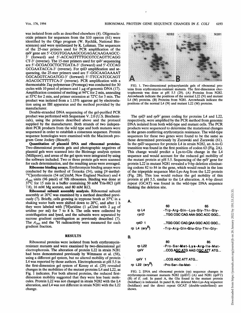
FIG. 1. Two-dimensional polyacrylamide gels of ribosomal pro-teins from erythromycin-resistant mutants. The first-dimension elec-trophoresis was done at pH 5.5 (29). (A) Proteins from N282.Arrowheads indicate the positions of the normal L22 (N) and mutantL4 (M) proteins. (B) Proteins from N281. Arrowheads indicate thepositions of the normal L4 (N) and mutant L22 (M) proteins.
The rplD and rplV genes coding for proteins IA and L22,respectively, were amplified by the PCR method from genomicDNA isolated from both wild-type and mutant cells. The PCRproducts were sequenced to determine the mutational changesin the genes conferring erythromycin resistance. The wild-typesequences for these two genes were found to be the same asthose determined previously by Zurawski and Zurawski (61).In the rplD sequence for protein L4 in strain N282, an A-to-Gtransition was found in the first position of codon 63 (Fig. 2A).This change would predict a Lys-to-Glu change in the L4sequence and would account for the reduced gel mobility ofthe mutant protein at pH 5.5. Sequencing of the rplV gene forprotein L22 in mutant N281 revealed a 9-bp deletion eliminat-ing codons 82 to 84 in the gene, which would result in the lossof the tripeptide sequence Met-Lys-Arg from the L22 protein(Fig. 2B). This loss would reduce the gel mobility of thisprotein at pH 5.5, similar to the L4 alteration. A 4-bp directrepeat (GCAT) was found in the wild-type DNA sequenceflanking the deletion site.
A.
rp L4rpIlD
rpID 1
rp L4 (eryR)
60 65-Trp-Arg-GIn--Lys-Gly-Thr-Gly-...TGG CGC CAG AM GGC ACC GGC...
...TGG CGC CAG AA GGC ACC GGC...-Trp-Arg-G In -GI u-Gly-Thr-G ly-
RESULTS
Ribosomal proteins were isolated from both erythromycin-resistant mutants and were examined by two-dimensional gelelectrophoresis. The alteration of protein L22 in strain N281had been demonstrated previously by Wittmann et al. (58),using a different gel system, but no altered mobility of proteinL4 was reported by those authors. Electrophoresis at pH 5.5 inthe first-dimension gel system of Kenny et al. (29) revealedchanges in the mobilities of the mutant proteins L4 and L22, asFig. 1 indicates. For both altered proteins, the reduced first-dimension mobility suggested that they were less basic mole-cules. Protein L22 was not changed in strain N282 with the L4alteration, and LA was not different in strain N281 with the L22change.
EL
rp L22rpiV
rpIV 1rp L22 (eryR)
80 85-Pro-Ser-M et-Lys- A rg- I e-Met-...CCG AjQ;C TG MG CPGO AlT ATG...
...CCG AGC ATT ATG...-Pro-Ser--lie-Met-
FIG. 2. DNA and ribosomal protein (rp) sequence changes inerythromycin-resistant mutants N282 (rplDl) (A) and N281 (rplVl)(B) of E. coli. In panel A, the Glu found in the mutant protein(boldface) is indicated. In panel B, the deleted Met-Lys-Arg sequence(boldface) and the direct repeat GCAT (double-underlined) areshown.
VOL. 176, 1994
Dow
nloa
ded
from
http
s://j
ourn
als.
asm
.org
/jour
nal/j
b on
26
Janu
ary
2022
by
191.
53.2
53.3
3.
6194 CHITUM AND CHAMPNEY
TABLE 1. Growth rates of erythromycin-resistant mutants andplasmid-complemented strains in the presence
and absence of erythromycin
Doubling time (min) at the indicatedStrain drug concn (jg/ml)
0 300 450 600
N281 45 60 90 105N281(pLF1.0) 40 85 160 300N281(pLF4.6) 45 65 90 110N282 45 55 75 115N282(pLF1.0) 50 65 75 115N282(pLF4.6) 35 65 70 130
Complementation of the mutants with plasmids bearingwild-type alleles of the appropriate genes was performed.EcoRI DNA fragments from the SlO ribosomal protein operonwere cloned into pBR325 to give plasmids pLF1.0, containingonly the rplVl gene, and pLF4.6, containing the rplB+, rplD+,and rplW' genes for proteins L2, LA, and L23, respectively(57). The growth rates of the mutant cells with and withoutcomplementing plasmids were measured in the presence andabsence of erythromycin. As Table 1 indicates, both strainsshowed reduced growth rates with increasing concentrations oferythromycin. The mutants grew about 2.5 times more slowlyat 600 ,ug/ml. Each strain became more sensitive to erythro-mycin in the presence of the wild-type gene. N281(pLF1.0)grew 7.5 times more slowly at this final concentration, whileN282(pLF4.6) grew 3.7 times more slowly. In comparison withN281, N281(pLF1.0) was more affected by higher concentra-tions of the drug than N282(pLF4.6) was in comparison withN282. In each mutant, the noncomplementing plasmid had noeffect on the growth rate in the presence of erythromycin(Table 1). These results reflect the known dominance of
TABLE 2. Ribosomal protein distribution and erythromycinbinding for wild-type, mutant, and complemented strains
Erythro- Protein distribution' ErythromycinStramycina Wild type Mutant binding
SK901 - 100 0 75 (13)N282 - 0 100 3.5 (4)N282(pLF4.6) + 16 (4.9) 84 (4.9) 7.4 (5)N282(pLF4.6) - 72(2.4) 28 (2.4) 55 (8)N281 - 0 100 61(7)N281(pLF1.0) + 37(1.1) 63 (1.1) 33 (13)N281(pLF1.0) - 100 0 69 (8)
a Cells were grown without (-) or with (+) erythromycin (300 jig/ml).b Fractions of wild-type and mutant proteins in two-dimensional gels, with
standard deviations in parentheses.c Percent 1[4Clerythromycin binding (pmol/100 pmol of 70S subunit). Values
are averages for the numbers of assays indicated in parentheses.
erythromycin sensitivity over resistance in merodiploid cells(40).Ribosomes were isolated from the complemented mutant
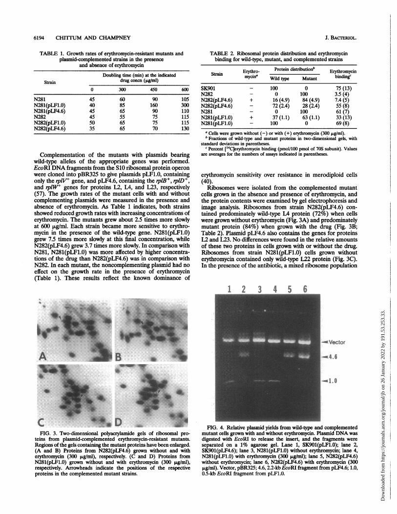
cells grown in the absence and presence of erythromycin, andthe protein contents were examined by gel electrophoresis andimage analysis. Ribosomes from strain N282(pLF4.6) con-tained predominately wild-type L4 protein (72%) when cellswere grown without erythromycin (Fig. 3A) and predominatelymutant protein (84%) when grown with the drug (Fig. 3B;Table 2). Plasmid pLF4.6 also contains the genes for proteinsL2 and L23. No differences were found in the relative amountsof these two proteins in cells grown with or without the drug.Ribosomes from strain N281(pLF1.0) cells grown withouterythromycin contained only wild-type L22 protein (Fig. 3C).In the presence of the antibiotic, a mixed ribosome population
1 ' I Aa r
cvctor
w4.6
FIG. 3. Two-duimensional polyacrylamide, gels of ribosomal pro-teins from plasmid-complemented erythromycin-resistant mutants.Regions of the gels containing the mutant proteins have been enlarged.(A and B) Proteins from N282(pLF4.6) grown without and witherythromycin (300 t±g/ml), respectively (C and D) Proteins fromN281(pLF1.0) grown without and with erythromycin (300 p~gfml),respectively. Arrowheads indicate the positions of the respectiveproteins in the complemented mutant strains.
-ml.,
FIG. 4. Relative plasmid yields from wild-type and complementedmutant cells grown with and without erythromycin. Plasmid DNA wasdigested with EcoRI to release the insert, and the fragments wereseparated on a 1% agarose gel. Lane 1, SK901(pLF1.0); lane 2,SK901(pLF4.6); lane 3, N281(pLF1.0) without erythromycin; lane 4,N281(pLF1.0) with erythromycin (300 ,ug/ml); lane 5, N282(pLF4.6)without erythromycin; lane 6, N282(pLF4.6) with erythromycin (300,ug/ml). Vector, pBR325; 4.6, 2.2-kb EcoRI fragment from pLF4.6; 1.0,0.5-kb EcoRI fragment from pLF1.0.
J. BAcTERiOL.
Dow
nloa
ded
from
http
s://j
ourn
als.
asm
.org
/jour
nal/j
b on
26
Janu
ary
2022
by
191.
53.2
53.3
3.
RIBOSOMAL PROTEIN GENE SEQUENCE CHANGES IN E. COLI 6195
Ec0w0~ac'U
e
Fraction Number
Ec0w
0so
2y
50000
40000
EQE30000 U
* D
1.c 0mo20000
10000
0 10 20 30Fraction Number
60000
50000
40000 EE c0.
w
30000 a ,28
x e20000 X(
10000
0 10 20 30
Fraction Number0 10 20 30
Fraction Number
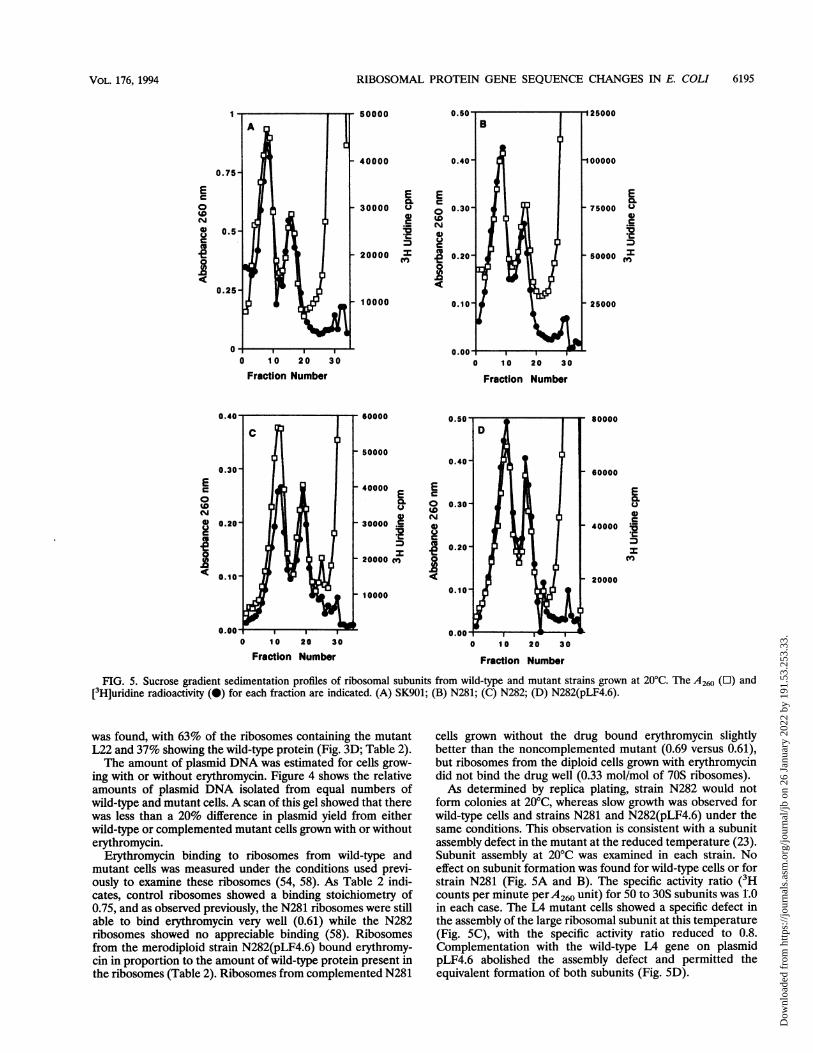
FIG. 5. Sucrose gradient sedimentation profiles of ribosomal subunits from wild-type and mutant strains grown at 20'C. The A260 ([1) and[3H]uridine radioactivity (0) for each fraction are indicated. (A) SK901; (B) N281; (C) N282; (D) N282(pLF4.6).
was found, with 63% of the ribosomes containing the mutantL22 and 37% showing the wild-type protein (Fig. 3D; Table 2).The amount of plasmid DNA was estimated for cells grow-
ing with or without erythromycin. Figure 4 shows the relativeamounts of plasmid DNA isolated from equal numbers ofwild-type and mutant cells. A scan of this gel showed that therewas less than a 20% difference in plasmid yield from eitherwild-type or complemented mutant cells grown with or withouterythromycin.
Erythromycin binding to ribosomes from wild-type andmutant cells was measured under the conditions used previ-ously to examine these ribosomes (54, 58). As Table 2 indi-cates, control ribosomes showed a binding stoichiometry of0.75, and as observed previously, the N281 ribosomes were stillable to bind erythromycin very well (0.61) while the N282ribosomes showed no appreciable binding (58). Ribosomesfrom the merodiploid strain N282(pLF4.6) bound erythromy-cin in proportion to the amount of wild-type protein present inthe ribosomes (Table 2). Ribosomes from complemented N281
cells grown without the drug bound erythromycin slightlybetter than the noncomplemented mutant (0.69 versus 0.61),but ribosomes from the diploid cells grown with erythromycindid not bind the drug well (0.33 mol/mol of 70S ribosomes).As determined by replica plating, strain N282 would not
form colonies at 20'C, whereas slow growth was observed forwild-type cells and strains N281 and N282(pLF4.6) under thesame conditions. This observation is consistent with a subunitassembly defect in the mutant at the reduced temperature (23).Subunit assembly at 20'C was examined in each strain. Noeffect on subunit formation was found for wild-type cells or forstrain N281 (Fig. 5A and B). The specific activity ratio (3Hcounts per minute perA260 unit) for 50 to 30S subunits was 1.0in each case. The L4 mutant cells showed a specific defect inthe assembly of the large ribosomal subunit at this temperature(Fig. SC), with the specific activity ratio reduced to 0.8.Complementation with the wild-type L4 gene on plasmidpLF4.6 abolished the assembly defect and permitted theequivalent formation of both subunits (Fig. SD).
E._U0
t.Is
80000
60000
E
0U
40000 :A
20000
VOL. 176, 1994
Dow
nloa
ded
from
http
s://j
ourn
als.
asm
.org
/jour
nal/j
b on
26
Janu
ary
2022
by
191.
53.2
53.3
3.
6196 CHITITM AND CHAMPNEY
TABLE 3. Ribosomal protein sequence similarities
Protein and % Refer-organism Sequence" Similarity ence
IAE. coil PWRQKGTGR 100 (201) 61Y pseudotuberculosis PWRQKGTGR 94.5 (210) 22M. capricolum PWKQKGTGL 35.8 (193) 41
L22E. coli GPSMBIMP 100(110) 61M. capricolum GPTLKRFRP 54.1(109) 41B. stearothermophilus GPTLKIRFRP 52.8 (108) 30Pea (chloro.)C GKTLKRVRA 43.6 (110) 21Liverwort (chloro.) GIFFKRFQP 41.6 (101) 19C paradoaw GPTLKRFRP 41.4 (99) 37Red alga (chloro.) GPKLKRFQP 40.6 (101) 28Spinach (chloro.) GITLKKVKP 38.1(97) 60Tobacco (chloro.) GTTVKKLP 37.6 (101) 53Rice (chloro.) STIMNKFRP 33.3 (102) 27Rat (liver) APKMRRRSG 32(109) 32Halobacterium m. VGESQGRKP 17.8 (90) 3
a Residues altered in erythromycin-resistant E. col ribosomal proteins areunderlined. Identical sequences in other organisms are in boldface.
b Percent overall sequence homology, with the total number of amino acids ineach protein given in parentheses.
I chloro., chloroplast.
The predicted amino acid sequences in L4 and L22 associ-ated with erythromycin resistance were compared with se-quences reported for homologous ribosomal proteins fromother organisms. These results are presented in Table 3. Thesequence around the altered site in the L4 sequence is highlyconserved, showing complete identity in Yersinia pseudotuber-culosis and nearly complete identity in Mycoplasma capricolum(Table 3). For protein L22, a strong conservation of thesequence around the Met-Lys-Arg deletion site was found forL22 homologs in two other prokaryotes (Bacillus stearother-mophilus and Mycoplasma capricolum). In Cyanophora para-doxa and in six chloroplast genes, additional significant simi-larity was observed (Table 3). Ribosomes from these cells andplastids are known to be sensitive to erythromycin (36, 42).Little similarity to L22-like sequences from archaebacteria andrats was found (3, 32).
DISCUSSION
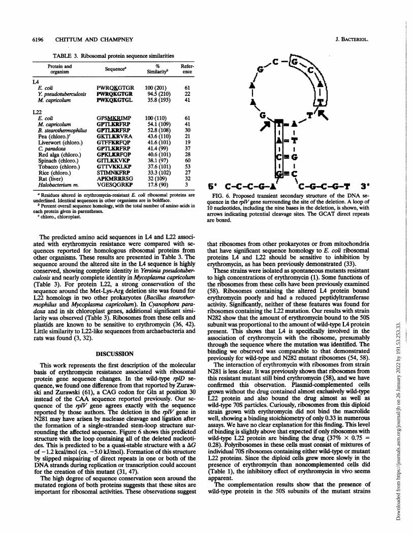
This work represents the first description of the molecularbasis of erythromycin resistance associated with ribosomalprotein gene sequence changes. In the wild-type rplD se-quence, we found one difference from that reported by Zuraw-ski and Zurawski (61), a CAG codon for Gin at position 30instead of the CAA sequence reported previously. Our se-quence of the rplV gene agrees exactly with the sequencereported by those authors. The deletion in the rplV gene inN281 may have arisen by nuclease cleavage and ligation afterthe formation of a single-stranded stem-loop structure sur-rounding the affected sequence. Figure 6 shows this predictedstructure with the loop contain-ing aIl of the deleted nucleoti-des. This is predicted to be a quasi-stable structure with a AGof -1.2 kcal/mol (ca. -5.0 kJ/mol). Formation of this structureby slipped mispairing of direct repeats in one or both of theDNA strands during replication or transcription could accountfor the creation of this mutant (31, 47).The high degree of sequence conservation seen around the
mutated regions of both proteins suggests that these sites areimportant for ribosomal activities. These observations suggest
G
OF I
CAI In
6' C--C-G-A C-4G-C-GT 3FIG. 6. Proposed transient secondary structure of the DNA se-
quence in the pilVgene surrounding the site of the deletion. A loop of10 nucleotides, including the nine bases in the deletion, is shown, witharrows indicating potential cleavage sites. The GCAT direct repeatsare boxed.
that ribosomes from other prokaryotes or from mitochondriathat have significant sequence homology to E. coli ribosomalproteins L4 and L22 should be sensitive to inhibition byerythromycin, as has been previously demonstrated (33).These strains were isolated as spontaneous mutants resistant
to high concentrations of erythromycin (1). Some functions ofthe ribosomes from these cells have been previously examined(58). Ribosomes containing the altered LA protein bounderythromycin poorly and had a reduced peptidyltransferaseactivity. Significantly, neither of these features was found forribosomes containing the L22 mutation. Our results with strainN282 show that the amount of erythromycin bound to the 50Ssubunit was proportional to the amount ofwild-type L4 proteinpresent. This shows that L4 is specifically involved in theassociation of erythromycin with the ribosome, presumablythrough the sequence where the mutation was identified. Thebinding we observed was comparable to that demonstratedpreviously for wild-type and N282 mutant ribosomes (54, 58).The interaction of erythromycin with ribosomes from strain
N281 is less clear. It was previously shown that ribosomes fromthis resistant mutant still bind erythromycin (58), and we haveconfirmed this observation. Plasmid-complemented cellsgrown without the drug contained almost exclusively wild-typeL22 protein and also bound the drug almost as well aswild-type 70S particles. Curiously, ribosomes from this diploidstrain grown with erythromycin did not bind the macrolidewell, showing a binding stoichiometry of only 0.33 in numerousassays. We have no clear explanation for this finding. This levelof binding is slightly above that expected if only ribosomes withwild-type L22 protein are binding the drug (37% X 0.75 =0.28). Polyribosomes in these cells must consist of mixtures ofindividual 70S ribosomes containing either wild-type or mutantL22 proteins. Since the diploid cells grew more slowly in thepresence of erythromycin than noncomplemented cells did(Table 1), the inhibitory effect of erythromycin in vivo seemsapparent.The complementation results show that the presence of
wild-type protein in the 50S subunits of the mutant strains
J. BAcTERIOL.
Dow
nloa
ded
from
http
s://j
ourn
als.
asm
.org
/jour
nal/j
b on
26
Janu
ary
2022
by
191.
53.2
53.3
3.
RIBOSOMAL PROTEIN GENE SEQUENCE CHANGES IN E. COLI 6197
leads to erythromycin sensitivity, with the growth rate dimin-ished in proportion to the wild-type-protein content of theparticle. In particular, the reduction in growth rate for thecomplemented mutants at 300 ptg of erythromycin per ml [40%for N281(pLF1.0) and 18% for N282(pLF4.6); Table 1] isreflected closely in the amount of wild-type L22 or L4 proteinpresent in the ribosomes (37 and 16%, respectively; Table 2)from cells grown at this same drug concentration.
Presumably, this reflects a change in the amounts of theseproteins assembled into the 50S particle, since the relativeamounts of either plasmid were in wild-type cells and in themutants growing with or without erythromycin were not dif-ferent. This is consistent with transcription of mRNA for theseproteins from the chloramphenicol transacetylase promoter inthis plasmid, which is not under ribosomal protein control (6)but is controlled by catabolite repression (34). A major effectof L4 overproduction on autogenous regulation of expressionof the chromosomal S10 operon. is unlikely, since the presenceof the pLF4.6 plasmid in either wild-type cells or strain N281did not influence the growth rate or erythromycin sensitivity.This suggests that the change in ribosomal protein content isrelated to the number of SOS subunits able to assimilate eitherform of the protein in the presence of erythromycin.We have shown elsewhere that erythromycin specifically
inhibits the assembly of the 50S subunit in growing wild-type E.coli cells (10). Mutant strain N281 with the L22 alteration ispartially resistant to this effect and requires about a fivefold-greater concentration of erythromycin for assembly to beaffected. Strain N282 is virtually immune to inhibition ofassembly by erythromycin. In diploid cells growing in theabsence of erythromycin, wild-type L4 and L22 proteins arepresumably preferentially assembled into new 50S subunits, asTable 2 indicates. Mutant proteins may be at some type ofassembly disadvantage, as shown by the cold-sensitive assemblydefect in the IA mutant strain. In the presence of erythromy-cin, incorporation of wild-type proteins into the particle wouldlead to erythromycin sensitivity in translation and to a reduc-tion in cellular growth rate, as shown in Table 1. This suggeststhat mutant 50S subunits would be preferentially formed in thepresence of the antibiotic (Table 2). We intend to examineerythromycin effects on ribosomal protein-RNA interactionsand to measure the relative rates of ribosomal protein synthe-sis in these complemented mutants to more closely examinethis possibility.
In conclusion, we have identified critical parts of the se-quences of two different ribosomal proteins necessary for theinteraction of the macrolide antibiotic erythromycin with thebacterial ribosome. It seems clear that the binding of erythro-mycin to the 50S subunit involves sequences in both proteinsLA and L22 and nucleotides in domain V of the 23S RNAsecondary structure (15, 18, 50). These observations shouldhelp in identifying the critical amino acid and nucleotideresidues involved in the binding of this compound and itsinhibitory effects on translation and ribosome formation.
ACKNOWLEDGMENTSWe thank the late David Apirion for strains and Kent Lohman for
PCR primers. We also appreciate the assistance of Eugenia Posey andthe advice and comments of Dumi Ratnasinghe and Phillip R. Musichduring the course of this work.
REFERENCES1. Apirion, D. 1967. Three genes that affect Escherichia coli ribo-
somes. J. Mol. Biol. 30:255-275.2. Arevalo, M. A., F. Tejedor, F. Polo, and J. P. G. Ballesta. 1988.
Protein components of the erythromycin binding site in bacterial
protein synthesis. J. Biol. Chem. 263:58-63.3. Arndt, E., W. Kroemer, and T. Hatakeyama. 1990. Organization
and nucleotide sequence of a gene cluster coding for eightribosomal proteins in the archaebacterium Halobactenum maris-mortui. J. Biol. Chem. 265:3034-3039.
4. Ausubel, F., R Brent, R Kingston, D. Moore, J. Seidman, J.Smith, and K. Struhl (ed.). 1989. Short protocols in molecularbiology. Greene Publishing Associates and Wiley-Interscience,New York.
5. Barritault, D. S., A. Expert-Bezancon, M.-F. Guerin, and D.Hayes. 1976. The use of acetone precipitation in the isolation ofribosomal proteins. Eur. J. Biochem. 63:131-135.
6. Bolivar, F. 1978. Construction and characterization of new cloningvehicles. III. Derivatives of pBR322 carrying unique EcoRi sitesfor selection of EcoRi generated recombinant DNA molecules.Gene 4:121-136.
7. Champney, W. S. 1979. Localized mutagenesis for the isolation oftemperature-sensitive mutants of Eschenichia coli affected in pro-tein synthesis. Genetics 91:215-227.
8. Champney, W. S. 1980. Protein synthesis defects in temperature-sensitive mutants of Escherichia coli with altered ribosomal pro-teins. Biochim. Biophys. Acta 609:464-474.
9. Chinali, G., E. Nyssen, M. DiGiambattista, and C. Cocito. 1988.Action of erythromycin and virginiamycin S on polypeptide syn-thesis in cell-free systems. Biochim. Biophys. Acta 951:42-52.
10. Chittum, H. S., and W. S. Champney. Erythromycin inhibits theassembly of the large ribosomal subunit in Escherichia coli. Curr.Microbiol., in press.
11. Cooperman, B. S., C. J. Weitzmann, and C. L. Fernandez. 1990.Antibiotic probes of Escherichia coli peptidyltransferase, p. 491-501. In W. E. Hill, A. Dahlberg, R. A. Garrett, P. B. Moore, D.Schlessinger, and J. R. Warner (ed.), The ribosome: structure,function, and evolution. American Society for Microbiology,Washington, D.C.
12. Corcoran, J. W. 1984. Mode of action and resistance mechanismsof macrolides, p. 231-259. In S. Omura (ed.), Macrolide antibiot-ics. Academic Press, Orlando, Fla.
13. Cundliffe, E. 1987. On the nature of antibiotic binding sites inribosomes. Biochimie 69:863-869.
14. Cundliffe, E. 1990. Recognition sites for antibiotics within rRNA,p. 479-490. In W. E. Hill, A. Dahlberg, R. A. Garrett, P. B. Moore,D. Schlessinger, and J. R. Warner (ed.), The ribosome: structure,function, and evolution. American Society for Microbiology,Washington, D.C.
15. Douthwaite, S. 1992. Functional interactions within 23S rRNAinvolving the peptidyltransferase center. J. Bacteriol. 174:1333-1338.
16. Douthwaite, S., J. B. Prince, and H. F. Noller. 1985. Evidence forfunctional interaction between domains II and V of 23S ribosomalRNA from an erythromycin-resistant mutant. Proc. Natl. Acad.Sci. USA 82:8330-8334.
17. Ehrlich, H. A., D. H. Gelfand, and R S. Saiki. 1988. Specific DNAamplification. Nature (London) 331:461-462.
18. Ettayebi, M., S. M. Prasad, and E. A. Morgan. 1985. Chloram-phenicol-erythromycin resistance mutations in a 23S rRNA geneof Escherichia coli. J. Bacteriol. 162:551-557.
19. Fukuzawa, H., T. Kohchi, T. Sano, H. Shirai, K. Umesono, H.Inokuchi, H. Ozeki, and K. Ohyama. 1988. Structure and organi-zation of Marchantia polymorpha chloroplast genome. III. Geneorganization of the large single copy region from rbcL to tmI(CAU). J. Mol. Biol. 203:333-351.
20. Gale, E. F., E. Cundliffe, P. E. Reynolds, M. H. Richmond, andM. J. Waring. 1981. The molecular basis of antibiotic action. JohnWiley & Sons, New York.
21. Gantt, J. S., S. L. Baldauf, P. J. Calie, N. F. Weeden, and J. D.Palmer. 1989. Transfer of rpl22 to the nucleus greatly preceded itsloss from the chloroplast and involved the gain of an intron.EMBO J. 10:3073-3078.
22. Gross, U., J. H. Chen, D. H. Kono, J. G. Lobo, and D. T. Y. Yu.1989. High degree of conservation between Yersinia pseudotuber-culosis and Escherichia coli. Nucleic Acids Res. 17:3601-3602.
23. Guthrie, C., H. Nashimoto, and M. Nomura. 1969. Structure andfunction of E. coli ribosomes. VIII. Cold-sensitive mutants defec-
VOL. 176, 1994
Dow
nloa
ded
from
http
s://j
ourn
als.
asm
.org
/jour
nal/j
b on
26
Janu
ary
2022
by
191.
53.2
53.3
3.
6198 CHITIUM AND CHAMPNEY
tive in ribosomal assembly. Proc. Natl. Acad. Sci. USA 63:384-391.24. Hampl, H., H. Schultze, and K. HL Nierhaus. 1981. Ribosomal
components from Escherichia coli SOS subunits involved in thereconstitution of peptidyltransferase activity. J. Biol. Chem. 256:2284-2288.
25. Hardy, S. J. S., C. G. Kurland, P. Voynow, and G. Mora. 1969. Theribosomal proteins of Escherchia coli. I. Purification of the 30Sribosomal proteins. Biochemistry 8:2897-2905;
26. Herold, M., and K. H. Nierhaus. 1987. Incorporation of sixadditional proteins to complete the assembly map of the SOSsubunit from Escherichia coli ribosomes. J. Biol. Chem. 262:8826-8833.
27. Hiratsuka, J., H. Shimada, R Whittier, T. Ishibashi, M. Saka-moto, M. Mori, C. Kondo, Y. Hoji, C. R. Sun, B. Y. Meng, Y. Q.Li, A. Kanno, Y. Nishizawa, A. Hirai, K. Shinozaki, and M.Sugiura. 1989. The complete sequence of the rice (Oryza sativa)chloroplast genome: intermolecular recombination between dis-tinct tRNA genes accounts for a major plastid DNA inversionduring the evolution of cereals. Mol. Gen. Genet. 217:185-194.
28. Kao, J. S., and M. Wu. 1990. The sequence of the plastid encodedrpl22 protein in marine macroalgae Gracilaria tenuistipitata. Nu-cleic Acids Res. 18:3067.
29. Kenny, J. W., J. M. Lambert, and R. R. Traut. 1979. Cross-linkingof ribosomes using 2-iminothiolane (methyl-4 mercaptobutyrimi-date) and identification of cross-linked proteins by diagonalpolyacrylamide sodium dodecyl gel electrophoresis. Methods En-zymol. 59534-550.
30. Kromer, W. J., T. Hatakeyama, and M. Kimura. 1990. Nucleotidesequences of Bacillus stearothermophilus ribosomal protein genes:part of the S10 operon. Biol. Chem. Hoppe-Seyler 371:631-636.
31. Kunkel, T. A. 1990. Misalignment-mediated DNA synthesis errors.Biochemistry 29:8003-8011.
32. Laine, R. 0., P. J. Laipis, N. F. Shay, and M. S. Kilberg. 1991.Identification of an amino acid-regulated mRNA from rat liver asthe mammalian equivalent of bacterial ribosomal protein L22. J.Biol. Chem. 266:16969-16972.
33. Lamb, A. J., G. D. Clark-Walker, and A. W. Linnane. 1968. Thedifferentiation of mitochondrial and cytoplasmic protein synthe-sizing systems in vitro by antibiotics. Biochim. Biophys. Acta161:415-427.
34. LeGrice, S. F. J., and H. Matzura. 1981. Binding of RNApolymerase and the catabolite gene activator protein within the catpromoter in Escherichia coli. J. Mol. Biol. 150:185-196.
35. Mao, J. C.-H., and M. Putterman. 1969. The intermolecularcomplex of erythromycin and the ribosome. J. Mol. Biol. 44:347-361.
36. Mets, L. J., and L. Bogorad. 1971. Mendelian and uniparentalalterations in erythromycin binding by plastid ribosomes. Science174:707-709.
37.. Michalowski, C. B., B. Pfanzagal, W. Loefelhardt, and H. J.Bohnert. 1990. The cyanelle S10 spc nosmal protein gene operonfrom Cyanophora panzdara. Mol. Gen. Genet. 224:222-231.
38. Miller, J. H. 1972. Experiments in molecular genetics. Cold SpringHarbor Laboratory, Cold Spring Harbor, N.Y.
39. Nakayama, L. 1984. Macrolides in clinical practice, p. 261-300. InS. Omura (ed.), Macrolide antibiotics. Academic Press, Orlando,Fla.
40. Nomura, M., and F. Engbaek. 1972. Expression of ribosomalprotein genes as analyzed by bacteriophage Mu-induced muta-tions. Proc. Natl. Acad. Sci. USA 69t1526-1530.
41. Ohkubo, S., A. Muto, Y. Kawauchi, F. Yamao, and S. Osawa. 1987.The ribosomal protein gene cluster of Mycoplasma capricolum.Mol. Gen. Genet. 210:314-322.
42. Omura, S. (ed.). 1984. Macrolide antibiotics. Academic Press,Orlando, Fla.
43. Pardo, D., and R. Rosset. 1974. Genetic studies of erythromycin-resistant mutants of Escherichia coil. Mol. Gen. Genet. 135:257-268.
44. Pardo, D., and R. Rosset. 1977. New ribosomal mutation whichaffects the two ribosomal subunits in Escherichia coli. Mol. Gen.Genet. 153:199-204.
45. Pardo, D., and R Rosset. 1977. Properties of ribosomes fromerythromycin resistant mutants of Escherichia coil. Mol. Gen.Genet. 156:267-271.
46. Pestka, S. 1974. Binding of [14Clerythromycin to Escherichia coliribosomes. Antimicrob. Agents Chemother. 6:474-478.
47. Ripley, 1L S. 1982. Model for the participation of quasi-palin-dromic DNA sequences in frameshift mutation. Proc. Natl. Acad.Sci. USA 79:4128-4132.
48. Schuier, J., H.-S. Gewitz, S.-E. Behrens, A. Lee, C. Ginther, and T.Leighton. 1990. Isolation and characterization of Bacillus stearo-thermophilus 30S and 50S ribosomal protein mutations. J. Bacte-riol. 172:7306-7309.
49. Sharrock, R A., T. Leighton, and 'H. G. Wittmann. 1981. Macro-lide and aminoglycoside antibiotic resistance mutations in theBacillus subtilis ribosome resulting in temperature-sensitive sporu-lation. Mol. Gen. Genet. 183:538-543.
50. Sigmund, C. D., and B. A. Morgan. 1982. Erythromycin resistancedue to a mutation in a ribosomal RNA operon of Escherichia coli.Proc. NatI. Acad. Sci. USA 79:5602-5606.
51. Silhavy, T. J., M. L. Berman, and L. W. Enquist. 1984. Experi-ments with gene fusions. Cold Spring Harbor Laboratory, ColdSpring Harbor, N.Y.
52. Tal, P.-C., and B. D. Davis. 1979. Action of antibiotics onchain-initiating and on chain-elongating ribosomes. Methods En-zymol. 59:.851-862.
53. Tanaka, M., T. Wakasugi, M. Sugita, K. Shinozaki, and M.Suginra. 1986. Genes for the eight ribosomal proteins are clus-tered on the chloroplast genome of tobacco (Nicotiana tabacum):similarity to the SlO and spc operons of Escherichia coli. Proc.Nat. Acad. Sci. USA 83:60304034.
54. Teraoka, H. 1970. A reversible change in the ability of Escherichiacoil ribosomes to bind to erythromycin. J. Mol. Biol. 4&511-515.
55. Teraoka, H., and K. H. Nierhaus. 1978. Proteins from Escherichiacoli ribosomes involved in the binding of erythromycin. J. Mol.Biol. 126:185-193.
56. Tipper, D. J., C. W. Johnson, C. L. Ginther, T. Leighton, and H. G.Wittmann. 1977. Erythromycin resistant mutations in Bacillussubtilis cause temperature sensitive sporulation. Mol. Gen. Genet.150:147-159.
57. Watson, J. C., and S. J. Surzycki. 1983. Both the chloroplast andnuclear genomes of Chlamydomonas reinhardi share homologywith Escherichia coli genes for transcriptional and translationalcomponents. Curr. Genet. 7:201-210.
58. Wittmann, H. G., G. Stoaer, D. Apirion, L Rosen, K. Tanaka, M.Tamaki, R Takata, S. Dekio, E. Otaka, and S. Osawa. 1973.Biochemical and genetic studies on two different types of eryth-romycin resistant mutants of Escherichia coli with altered ribo-somal proteins. Mol. Gen. Genet. 127:175-189.
59. Zengel, J. M., and L Lindahl. 1993. Domain I of 23S rRNAcompetes with a paused transcription complex for ribosomalprotein I4 of Escherichia coli. Nucleic Acids Res. 21:2429-2435.
60. Zhou, D. X*, F. M. Quigley, 0. Massenet, and R Mache. 1989.Cotranscription of the S10- and spc-like operons in spinachchloroplasts and identification of three of their gene products.Mol. Gen. Genet. 216:439 445.
61. Zurawski, G., and S. M. Zurawski. 1985. Structure of the Esche-richia coli S10 ribosomal protein operon. Nucleic Acids Res.13:45214526.
J. BAcrERIOL.
Dow
nloa
ded
from
http
s://j
ourn
als.
asm
.org
/jour
nal/j
b on
26
Janu
ary
2022
by
191.
53.2
53.3
3.