Embed Size (px)
Citation preview

Rho-dependent Termination of ssrS (6S RNA) Transcription inEscherichia coliIMPLICATION FOR 3� PROCESSING OF 6S RNA AND EXPRESSION OF DOWNSTREAM ygfA(PUTATIVE 5-FORMYL-TETRAHYDROFOLATE CYCLO-LIGASE)*
Received for publication, June 3, 2010, and in revised form, October 26, 2010 Published, JBC Papers in Press, October 29, 2010, DOI 10.1074/jbc.M110.150201
Huiseok Chae‡1, Kook Han‡1, Kwang-sun Kim§, Hongmarn Park‡, Jungmin Lee‡, and Younghoon Lee‡2
From the ‡Department of Chemistry, KAIST, Daejeon 305-701, Korea and §Korea Research Institute of Bioscience andBiotechnology (KRIBB), Daejeon 305-806, Korea
It is well known that 6S RNA, a global regulatory noncodingRNA that modulates gene expression in response to the cellu-lar stresses in Escherichia coli, is generated by processing fromprimary ssrS (6S RNA) transcripts derived from two differentpromoters. The 5� processing of 6S RNA from primary tran-scripts has been well studied; however, it remains unclear howthe 3�-end of this RNA is generated although previous studieshave suggested that exoribonucleolytic trimming is necessaryfor 3� processing. Here, we describe several Rho-dependenttermination sites located �90 bases downstream of the mature3�-end of 6S RNA. Our data suggest that the 3�-end of 6S RNAis generated via exoribonucleolytic trimming, rather than en-doribonucleolytic cleavage, following the transcription termi-nation events. The termination sites identified in this study arewithin the open reading frame of the downstream ygfA (puta-tive 5-formyl-tetrahydrofolate cyclo-ligase) gene, a part of thehighly conserved bacterial operon ssrS-ygfA, which is up-regu-lated during the biofilm formation. Our findings reveal thatygfA expression, which also aids the formation of multidrug-tolerant persister cells, could be regulated by Rho-dependenttermination activity in the cell.
Small noncoding RNAs (sRNAs)3 mediate a number of cel-lular processes in bacteria, such as protein tagging for degra-dation, modulation of RNA polymerase activity, regulation ofmRNA stability and translation, and secretion (1–4). ManysRNAs are transcribed as primary transcripts by RNA poly-merase (RNAP), and the transcripts are then subjected to pro-cessing by removing extra residues at the 5�- and/or 3�-endsto form functional mature forms (5).The 6S RNA was first identified as an abundant sRNA in
Escherichia coli, migrating as an 11S particle without ribo-some association (6). A recent study revealed that 6S RNA
binds to RNAP �70-holoenzyme (E�70) and represses tran-scription from �70-dependent promoters and activates �S-de-pendent promoters (7–9). The 6S RNA is highly inducedduring stationary growth, during which time it plays an im-portant role in modifying the utilization of E�70 to E�S (7, 8,10). The characteristic secondary structure of 6S RNA con-sists of a largely double-stranded helix and a central bulge (11,12). This highly conserved structure resembles DNA tem-plates in terms of its open promoter complex and mighttherefore mimic DNA promoters to encourage the binding ofE�70 (7, 8). Interestingly, 6S RNA can act as a template for thetranscription of pRNA, a very short RNA molecule (10).6S RNA is cotranscribed with ygfA, a gene expressed as a
second gene from the ssrS promoters, that encodes a putative5-formyl-tetrahydrofolate cyclo-ligase (7, 13). This dual-func-tional operon structure is highly conserved in �- and �-pro-teobacteria, as well as in certain members of the class �-pro-teobacteria (14, 15). Recent reports have suggested that ygfAexpression aids the formation of persister cells (13) and is up-regulated during the biofilm formation (16). The expressionof ygfA is of particular interest, considering that the antibioticrecalcitrance of biofilm infections is largely caused by per-sister cells. However, the mechanisms of ygfA regulation re-main unclear.The transcription of primary 6S RNA transcripts and
processing of their 5�-ends has been well characterized inE. coli (17). Two alternative promoters (i.e. a proximal ca-nonical �70-dependent ssrS P1 and a distal �70- and �S-de-pendent P2) are involved in the transcription of 6S RNA.These promoters produce two transcripts: a short P1 tran-script that begins at nucleotide position �9 and a long P2transcript that begins at position �224 relative to the ma-ture 5�-end of 6S RNA (�1). Interestingly, transcriptionfrom these promoters changes in response to cellularstress: P1 transcription is the predominant type of tran-scription during exponential phase, while P2 transcriptionincreases upon entry into stationary phase (17). Further-more, the 5�-ends of the transcripts are removed by twofunctionally similar enzymes with different specificities:RNase E acts on the long transcript, while RNase E andRNase G act on the short transcript (17). On the otherhand, the mechanism responsible for the formation of the3�-end of 6S RNA is not clear, although it has been shownthat exoribonucleases are involved in a plausible 3� trimming
* This work was supported by the 21C Frontier Microbial Genomics andApplication Center Program (MG08-0201-2-0) and the National ResearchFoundation of Korea (NRF) Grant by the Korea government (MEST) (2010-0000241; 2010-0029167).
1 Both authors contributed equally to this work.2 To whom correspondence should be addressed: Dept. of Chemistry,
KAIST, Daejeon 305-701, Korea. Fax: 82-42-350-2810; E-mail: [email protected].
3 The abbreviations used are: sRNA, small noncoding RNA; CAT, chloram-phenicol acetyl transferase; IC50, the concentration of chloramphenicolrequired for 50% inhibition of growth; RACE, rapid amplification of cDNAends.
THE JOURNAL OF BIOLOGICAL CHEMISTRY VOL. 286, NO. 1, pp. 114 –122, January 7, 2011© 2011 by The American Society for Biochemistry and Molecular Biology, Inc. Printed in the U.S.A.
114 JOURNAL OF BIOLOGICAL CHEMISTRY VOLUME 286 • NUMBER 1 • JANUARY 7, 2011
by guest on May 29, 2018
http://ww
w.jbc.org/
Dow
nloaded from

mechanism that forms heterogeneous 3�-ends from �184 to�191 (17, 18).Considering that 6S RNA is transcribed as part of a dicis-
tronic ssrS-ygfAmessage, the 3� processing of 6S RNA likelyoccurs via endoribonuclease cleavage followed by exoribonu-cleolytic trimming. However, this model is not consistentwith past observations that formation of the 3�-end of 6SRNA is not altered in the endoribonuclease RNase E-, RNaseG-, or RNase III-deficient cells (17).In the present study, we found that in vitro transcription
from the ssrS promoter continues beyond ygfA and can ex-tend into sibC, a gene that encodes an anti-toxin small RNA(19, 20). However, in vivo studies suggested that transcriptionfrom the ssrS promoters results in very low levels of ygfA ex-pression. This low expression could reflect either internalcleavage of the polycistronic RNA or termination of transcrip-tion somewhere beyond the 3�-end of 6S RNA in the cell. Weidentified several Rho-dependent termination sites located�90 bases downstream of the 3�-end of 6S RNA. No accumu-lation of Rho-terminated ssrS transcripts in cells lacking thefour endoribocucleases, RNase E, RNase G, RNase III, andRNase P suggest that the 3�-end of 6S RNA is generated viaexoribonucleolytic trimming after termination. The locationof Rho-dependent termination sites within the N-terminal
coding region of ygfA implies that ygfA expression depends onssrS transcription continuing through the termination sites.
EXPERIMENTAL PROCEDURES
Bacterial Strains-E. coli—K-12 strain JM109 was used forthe construction of plasmids and for in vivo analysis of ssrStranscription. Plasmid-borne transcripts were analyzed using6S RNA knock-out strains. MG1655 ssrS::kan was constructedbased on the previously described report (21) with a corre-sponding primer pair (Table 1). The 6S RNA knock-out deriv-atives of the following endoribonuclease-deficient strainswere constructed by employing bacteriophage P1-mediatedtransduction with MG1655 ssrS::kan as the donor strain (21):GW10 (rne� rng�), GW11 (rne� rng::cat), GW20 (GW10 rne-1), GW21 (rne-1 rng::cat) for RNase E or/and RNase G (22);SDF204 (rnc�) and SDF205 (rnc105) for RNase III (23);NHY312 (rnpA�) and NHY322 (rnpA49) for RNase P (24).Preparation of Total Cellular RNA-E. coli—Overnight cul-
tures grown at 37 °C were diluted 1:100 in LB broth contain-ing ampicillin (50 �g/ml) or tetracycline (10 �g/ml) andgrown to an A600 of 0.5 at the same temperature. If necessary,IPTG was added to the cell culture at 1 mM of final concentra-tion and the culture was incubated further for 30 min. In thecase of rne-1 or rnpA49 cells, the cells were grown at 30 °C
TABLE 1Oligonucleotides used in this study
Primer Sequence (5� to 3�) Use
6SKO_up CCA CAA GAA TGT GGC GCT CCG CGG TTG GTG AGT CAGAAG AAC TCG TCA AG
Construction of MG1655 ssrS::kan
6SKO_dn GCA GTT TTA AGG CTT CTC GGA CGG ACC GAG CAT GTATGG ACA GCA AGC GAA CCG
ssrS-92_F CGG GGA TCC TTA CTT GAA CAA GGT CGC A DNA templates for in vitro transcription (Figs. 1, 2, and 4) orconstruction of pCAT (ssrS-CAT fusion) plasmids (Fig. 2)
ssrS�185_R CCC AAG CTT GGG AAT CTC CGA GAT GCC DNA templates for in vitro transcription (Fig. 1) orconstruction of pCAT (ssrS-CAT fusion) plasmids (Fig. 2)ssrS�238_R CCC AAG CTT ATA ATG TCA GTG GGA GTT CTG
ssrS�260_R CCC AAG CTT ATT TTG CGG ATT TCT TGT CGssrS�330_R CCC AAG CTT CCG GGT AGC GGC TTGssrS�348_R CCC AAG CTT CGG GGG ATA AGT CAT CATssrS�448_R CCC AAG CTT ATA CGC GCT TAC CGG CGCssrS�548_R CCC AAG CTT GGC TCA TGG ATC TTC AAC CTssrS�648_R CCC AAG CTT GCC CAT TCC CAG GCG CTG AssrS�748_R CCC AAG CTT CAA CGG GGA GTT TTT CCA CssrS�780_R CCC AAG CTT AAC CAC CGC AGG AAG AGG GGssrS�848_R CCC AAG CTT GAA TCA ACG ATG TCA ATC AGssrS�931_R CCC AAG CTT CTC ATA AGT TTC AGC GCT TAssrS�1040_R CCC AAG CTT TTA CGA TGG CAG GGC AGC ATssrS�781_F CGC GGA TCC ACA CCG TCG AAA GTC TGG GAcat_R ACG GTG GTA TAT CCA GTG AT DNA templates for in vitro transcription (Fig. 4)a6S�185 GGG AAT CTC CGA GAT GCC GCC Anti-6S RNA probe or DNA templates for in vitro
transcription (Figs. 3, 4, and 6)atRNA_arg ACC TGT ACT CTA TCC AAC Anti-tRNAArg probe (Fig. 3)tRNA_F TAC AAG CTT GCG CTC GTA GCT CAG Construction of ssrS-tRNAArg-CAT fusion plasmids (Fig. 3)tRNA_R TAT AAG CTT TGG TGC GCC CGG CGGTyrT_F GCG TCT TTG TTT ACG GTA ATC G tyrT template for in vitro transcription (Fig. 4)TyrT_R GAT TCG TTT GAG AAT TCC GGG GERIT76S CGG AAT TCT TAA TAC GAC TCA CTA TAG GAT TTC TCT
GAG ATG TTCDNA templates for in vitro transcription (Fig. 4)
ARrho1_F GGG GAC GTC ATT CAT AGT GGT GTG AGT TCT TAA ACTTGG AGT ACG TAA AAA CCC
Construction of anti-rho expression plasmid (Fig. 5)
pnMTXbaI_R GCT CTA GAA AAG CAA AAA CCC GCC GAA GCG GGT TTTTAC GTA CTC CG
Rho216_R GTA GGA GCT GTC TGC GGA AC Reverse transcription of rhomRNA (Fig. 5)6S�733_R CCA CCA ACT GAC AAT CAT GC Reverse transcription of ssrS-ygfA dicistronic RNA (Fig. 5)Rho20_F AGA ATA CGC CGG TTT CTG AG RT- or qRT-PCR of rhomRNA (Fig. 5)Rho196_R GGA GGA AAC CAA ATC CAT CC6S�104_F AAG CCT TAA AAC TGC GAC GA RT- or qRT-PCR of ssrS-ygfA dicistronic RNA (Fig. 5)6S�316_R GTT GAC CCA TTT CCT GCT GT5S_F TGC CTG GCG GCC GTA GCG CGG RT- or qRT-PCR of 5S RNA (Fig. 5)5S_R ATG CCT GGC AGT TCC CTA CTC TC
Rho-dependent Termination of ssrS Transcription in E. coli
JANUARY 7, 2011 • VOLUME 286 • NUMBER 1 JOURNAL OF BIOLOGICAL CHEMISTRY 115
by guest on May 29, 2018
http://ww
w.jbc.org/
Dow
nloaded from

and then shifted to 44 °C for 1 h. rng::cat cells also treated asabove to compare with rne-1 effect even though the strainitself is not temperature-sensitive. Total cellular RNA wasisolated by hot phenol extraction as described previously (25).Northern Blot Analysis—Total cellular RNA (15 �g) was
fractionated on a 5% polyacrylamide gel/7 M urea and electro-transferred onto a Hybond-XL membrane (GE Healthcare).5�-End-radiolabeled oligonucleotides a6S�185 and atRNA_arg (Table 1) used as probes for 6S RNA and Brevibacteriumalbidum tRNAArg, respectively. Hybridization was performedaccording to the manufacturer’s instructions. The Northernblots were visualized and quantified using Image AnalyzerFLA7000 (Fuji).In Vitro Transcription by E. coli RNA Polymerase—The lin-
ear DNA templates used for in vitro transcription reactionswere obtained with chromosomal or plasmid DNA via PCRwith corresponding primer pairs (Table 1). Some primers hadextra linker sequences at the 5�-end. In vitro transcriptionreaction was conducted using E. coli E�70 (Epicenter) accord-ing to the manufacturers’ instructions with minor modifica-tions. Briefly, the DNA templates (6 nM) were incubated at37 °C for 5 min in reaction buffer (40 mM Tris-HCl, pH 7.5,150 mM KCl, 10 mM MgCl2, 0.05% Triton X-100, 10 mM DTT)containing E�70 (1 unit) and 2 mM ATP. The reaction wasthen initiated by adding mixtures containing rNTPs (0.5 mM
rGTP, 0.5 mM rUTP, and 0.025 mM rCTP including 10 �Ci of[�-32P]CTP) and rRNasin (4 units, Promega). After 25 min,the reactions were terminated by phenol-chloroform extrac-tion and ethanol precipitation. The pellet was resuspendedwith gel-loading buffer II (Ambion) and loaded on a 5% poly-acrylamide/8 M urea sequencing gel. The gel was visualizedand analyzed by FLA7000 (Fuji).Analysis of Transcription from ssrS-P1 Promoter in Vivo—
ssrS-P1 promoter-containing DNA fragments (i.e. rangingfrom nucleotide position �92 to various downstream bases)were obtained via PCR amplification of genomic DNA withcorresponding primer pairs (Table 1). The resulting PCRproducts were digested with BamHI/HindIII and ligated intopKK232–8 (GE Healthcare) to generate pCAT plasmids. Cellscontaining fusion plasmids were exponentially grown in LBbroth (1:300 dilution) of different chloramphenicol concen-trations. The IC50 was measured as previously described (26).Alternatively, a B. albidum tRNAArg sequence (27) was am-
plified by PCR with a corresponding primer pair (Table 1) andinserted immediately upstream of the CAT gene in the pCATplasmids to measure the exogenous amount of tRNAArg (i.e.as an indicator of transcription activity). Total RNAs isolatedfrom cells containing the exogenous tRNA gene were sub-jected to Northern blot analysis as described above.Analysis of Rho-dependent Termination—His-tagged Rho
and IcdA (isocitrate dehydrogenase) proteins were purifiedfrom cells containing ASKA-rho and ASKA-icdA plasmids,respectively, as previously reported (28). The linear DNAtemplates were prepared and subjected to in vitro transcrip-tion analysis as described above except that Rho, or IcdA pro-tein as a control, was added at 40 nM.Gel Mobility Shift Assay—DNA templates for in vitro tran-
scription of 6S RNA and precursor 6S RNA carrying the
downstream sequence to �330 were obtained via PCR usingpCAT330 DNA with primer pairs of ERIT76S/a6S�185 andERIT76S/cat_R (Table 1), respectively. In vitro transcriptionwas carried out using T7 RNA polymerase (Promega). Gel-purified 6S RNA and precursor 6S RNA were 5�-end labeledwith [�-32P]ATP and re-purified by gel elution. Labeled RNAs(2 nM) were incubated with the purified Rho protein in 10 �lof binding buffer (10 mM Tris-HCl, pH 8.0, 1 mM MgCl, 1 mM
DTT, 100 mM NaCl, 0.06% Triton X-100, 0.25% glycerol, 10�g of ytRNA) for 15 min at 25 °C. The reaction mixtures werethen analyzed on 5% polyacrylamide gels, as described previ-ously (20).3�-RACE Assay—3� RACE analysis was performed on the
in vitro transcribed RNAs in the presence of Rho proteinwith the ssrS-containing DNA ranging from �92 to �448as previously described (29), with following modifications.The Adaptor-ligated RNA was reverse transcribed and PCRamplified using a One-Step RT-PCR PreMix kit (Intron)according to the manufacturer’s instructions. The PCRproducts were separated on a 2% agarose gel, purified, andanalyzed by DNA sequencing after cloning into a pGEM-T-easy vector (Promega).Analysis of Rho-knockdown Effects on ygfA Expression—
Plasmid pAKA (20), a derivative of pACYC184, was used toconstruct an anti-rho RNA expression plasmid. The anti-rhoRNA sequence linked to the rnpB terminator was amplifiedwith a primer pair of ARrho1_F/pnMTXbaI_R by PCR andcloned into the AatII/EcoRI sites of pAKA so that anti-rhoRNA could be induced by IPTG. Total RNAs were isolatedfrom cells containing the anti-rho RNA expression plasmidpARRho after the IPTG induction for 30 min as describedabove, and treated with Turbo DNase (Ambion) to removecontaminating DNA. DNase was heat-inactivated and RNAsamples were subjected to reverse transcription with MMLVRT (Enzynomics) with primers Rho216_R for rhomRNA,6S�733_R for ssrS-ygfA dicistronic RNA, and 5S_R for 5SrRNA. The resulting cDNAs were amplified using Taq poly-merase premix (Solgent) with primer pairs Rho20_F/Rho196_R for rhomRNA, 6S�104_F/6S�316_R for ssrS-ygfAdicistronic transcripts, and 5S_F/5S_R for 5S rRNA. Theprimers used were listed in Table 1. The PCR products wereelectrophoresed on 2% agarose gels, stained by SYBR Safe (In-vitrogen), and photographed by GelDoc 1000 (Bio-Rad).Quantitative real-time RT-PCR was done by ABI 7500 Real-time PCR (Applied Biosystems) with QuantiTect SYBR GreenPCR premix kit (Qiagen) in triplicate experiments. The abun-dance of each RNA was normalized to the amount of 5S RNAand represented as a fold change. Data were analyzed usingABI 7500 SDS software (Applied Biosystems, ver. 1.3). Thecycle threshold (CT) values obtained were an average of thetriplicates.
RESULTS
In Vitro Transcription of ssrS Extends to sibC—Although ithas been reported that ssrS and ygfA are co-transcribed, itremains unknown where this transcription is terminated. Toexamine the presence of intrinsic termination sites in vitrofirst, we prepared DNA templates that extended downstream
Rho-dependent Termination of ssrS Transcription in E. coli
116 JOURNAL OF BIOLOGICAL CHEMISTRY VOLUME 286 • NUMBER 1 • JANUARY 7, 2011
by guest on May 29, 2018
http://ww
w.jbc.org/
Dow
nloaded from

of the SibC sRNA gene and used these templates for in vitrotranscription (Fig. 1). The DNA constructs were designed toinitiate transcription at the ssrS P1 promoter. Because all theDNA constructs had a 9-bp linker sequence at the ends, run-off transcripts were expected to have extra 9 nucleotides. TheConstructs 1 and 2 produced transcripts (marked with a andb), with the same sizes as the P1 run-off transcripts of ex-pected 256 and 866 nucleotides, respectively. Construct 4contained the sibC transcription unit where transcription isinitiated at position �869 and terminates at �1009 by an in-
trinsic terminator (TsibC) (19, 20). This construct generated atranscript (marked with c) with the estimated size of 141 nu-cleotides. We did not observe apparent run-off transcripts ofexpected 181 nucleotides in this construct, suggesting that thesibC terminator is very effective. Construct 5 generated twotranscripts (marked with d and c) of estimated 1018 and 141nucleotides, which would initiate from the ssrS P1 and sibCpromoter, respectively, but both of which would terminatedat the TsibC. In Construct 6, which lacks the TsibC, we ob-served a run-off transcript (marked with e) of estimated 949
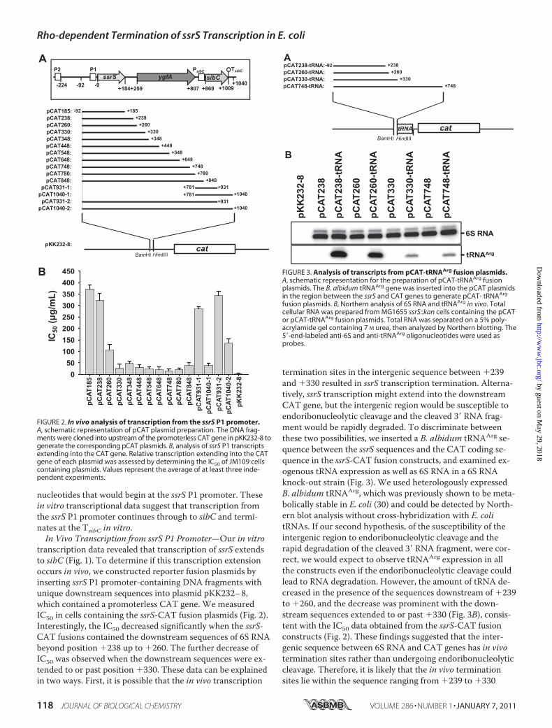
FIGURE 1. In vitro transcription from the ssrS P1 promoter. A, schematic arrangement of the ssrS transcription unit and DNA templates used for in vitrotranscription. Two ssrS promoters, P1 and P2, and the downstream ygfA and sibC genes are indicated. Six DNA templates (i.e. 1– 6) were prepared by PCRand used as DNA templates for in vitro transcription with �70 RNA polymerase. Numbers are given relative to the 5� nucleotide within the mature 6S RNA,which is considered �1. The ssrS P1 promoter starts transcription at �9, while the sibC gene has a promoter (PsibC) and an intrinsic terminator (TsibC), whichallow for transcription initiation and termination at �869 and �1009, respectively. B, in vitro transcription products were analyzed on a 5% polyacrylamidegel containing 7 M urea. The template numbers used for in vitro transcription are indicated above each lane. Major transcripts were marked with a to e.C, E. coli ssrS transcription unit. The 6S RNA and SibC RNA sequences encoded by ssrS and sibC, respectively, are shown in bold. Rho-dependent terminationsites are indicated by vertical arrows and the sizes of arrows indicate the approximate relative magnitude of termination at each site, as determined by 3�-RACE. Boxed C residues indicate repeated C residues with 12 � 1 spacing required for Rho-dependent termination. S/D indicates a putative ribosome bind-ing site of ygfA mRNA.
Rho-dependent Termination of ssrS Transcription in E. coli
JANUARY 7, 2011 • VOLUME 286 • NUMBER 1 JOURNAL OF BIOLOGICAL CHEMISTRY 117
by guest on May 29, 2018
http://ww
w.jbc.org/
Dow
nloaded from
nucleotides that would begin at the ssrS P1 promoter. Thesein vitro transcriptional data suggest that transcription fromthe ssrS P1 promoter continues through to sibC and termi-nates at the TsibC in vitro.In Vivo Transcription from ssrS P1 Promoter—Our in vitro
transcription data revealed that transcription of ssrS extendsto sibC (Fig. 1). To determine if this transcription extensionoccurs in vivo, we constructed reporter fusion plasmids byinserting ssrS P1 promoter-containing DNA fragments withunique downstream sequences into plasmid pKK232–8,which contained a promoterless CAT gene. We measuredIC50 in cells containing the ssrS-CAT fusion plasmids (Fig. 2).Interestingly, the IC50 decreased significantly when the ssrS-CAT fusions contained the downstream sequences of 6S RNAbeyond position �238 up to �260. The further decrease ofIC50 was observed when the downstream sequences were ex-tended to or past position �330. These data can be explainedin two ways. First, it is possible that the in vivo transcription
termination sites in the intergenic sequence between �239and �330 resulted in ssrS transcription termination. Alterna-tively, ssrS transcription might extend into the downstreamCAT gene, but the intergenic region would be susceptible toendoribonucleolytic cleavage and the cleaved 3� RNA frag-ment would be rapidly degraded. To discriminate betweenthese two possibilities, we inserted a B. albidum tRNAArg se-quence between the ssrS sequences and the CAT coding se-quence in the ssrS-CAT fusion constructs, and examined ex-ogenous tRNA expression as well as 6S RNA in a 6S RNAknock-out strain (Fig. 3). We used heterologously expressedB. albidum tRNAArg, which was previously shown to be meta-bolically stable in E. coli (30) and could be detected by North-ern blot analysis without cross-hybridization with E. colitRNAs. If our second hypothesis, of the susceptibility of theintergenic region to endoribonucleolytic cleavage and therapid degradation of the cleaved 3� RNA fragment, were cor-rect, we would expect to observe tRNAArg expression in allthe constructs even if the endoribonucleolytic cleavage couldlead to RNA degradation. However, the amount of tRNA de-creased in the presence of the sequences downstream of �239to �260, and the decrease was prominent with the down-stream sequences extended to or past �330 (Fig. 3B), consis-tent with the IC50 data obtained from the ssrS-CAT fusionconstructs (Fig. 2). These findings suggested that the inter-genic sequence between 6S RNA and CAT genes has in vivotermination sites rather than undergoing endoribonucleolyticcleavage. Therefore, it is likely that the in vivo terminationsites lie within the sequence ranging from �239 to �330
FIGURE 2. In vivo analysis of transcription from the ssrS P1 promoter.A, schematic representation of pCAT plasmid preparation. The DNA frag-ments were cloned into upstream of the promoterless CAT gene in pKK232-8 togenerate the corresponding pCAT plasmids. B, analysis of ssrS P1 transcriptsextending into the CAT gene. Relative transcription extending into the CATgene of each plasmid was assessed by determining the IC50 of JM109 cellscontaining plasmids. Values represent the average of at least three inde-pendent experiments.
FIGURE 3. Analysis of transcripts from pCAT-tRNAArg fusion plasmids.A, schematic representation for the preparation of pCAT-tRNAArg fusionplasmids. The B. albidum tRNAArg gene was inserted into the pCAT plasmidsin the region between the ssrS and CAT genes to generate pCAT- tRNAArg
fusion plasmids. B, Northern analysis of 6S RNA and tRNAArg in vivo. Totalcellular RNA was prepared from MG1655 ssrS::kan cells containing the pCATor pCAT-tRNAArg fusion plasmids. Total RNA was separated on a 5% poly-acrylamide gel containing 7 M urea, then analyzed by Northern blotting. The5�-end-labeled anti-6S and anti-tRNAArg oligonucleotides were used asprobes.
Rho-dependent Termination of ssrS Transcription in E. coli
118 JOURNAL OF BIOLOGICAL CHEMISTRY VOLUME 286 • NUMBER 1 • JANUARY 7, 2011
by guest on May 29, 2018
http://ww
w.jbc.org/
Dow
nloaded from
(minor sites in the region of �239 to �260 and major sitesin �261 to �330).Rho Factor Is Involved in Termination of 6S RNA—It is
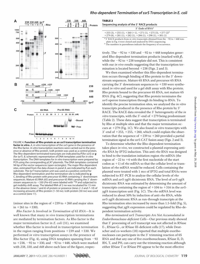
well known that many in vivo transcription terminationsare mediated by termination factors. As Rho factor is themajor termination factor in E. coli (31), we examinedwhether Rho factor is involved in transcription terminationin the region ranging from positions �239 and �330. Weperformed in vitro transcription assays in the presence ofRho factor (Fig. 4), using DNA templates spanning from �92to �238, �92 to �330, and �92 to �448, which were markedwith 238, 330, and 448 above each lane of the figure, respec-
tively. The �92 to �330 and �92 to �448 templates gener-ated Rho-dependent termination products (marked with f),while the �92 to �238 template did not. This is consistentwith our in vivo results suggesting that the transcription ter-mination is located beyond �238 (Figs. 2 and 3).We then examined whether this Rho-dependent termina-
tion occurs through binding of Rho protein to the 3� down-stream sequences. Mature 6S RNA and precursor 6S RNAcarrying the 3� downstream sequences to �330 were synthe-sized in vitro and used for a gel shift assay with Rho protein.Rho protein bound to the precursor 6S RNA, not mature 6SRNA (Fig. 4C), suggesting that Rho protein terminates thessrS operon transcription through its binding to RNA. Toidentify the precise termination sites, we analyzed the in vitrotranscripts produced in the presence of Rho protein by 3�RACE. The RACE data revealed the 3� heterogeneity of the invitro transcripts, with the 3�-end of �279 being predominant(Table 2). These data suggest that transcription is terminatedby Rho at multiple sites and that the major termination oc-curs at �279 (Fig. 1C). We also found in vitro transcripts with3�-end of �253, �255, �260, which could explain the obser-vation that the sequence of �239 to �260 provided a partialtermination signal in the ssrS-CAT fusion assay (Figs. 2 and 3).To determine whether the Rho-dependent termination
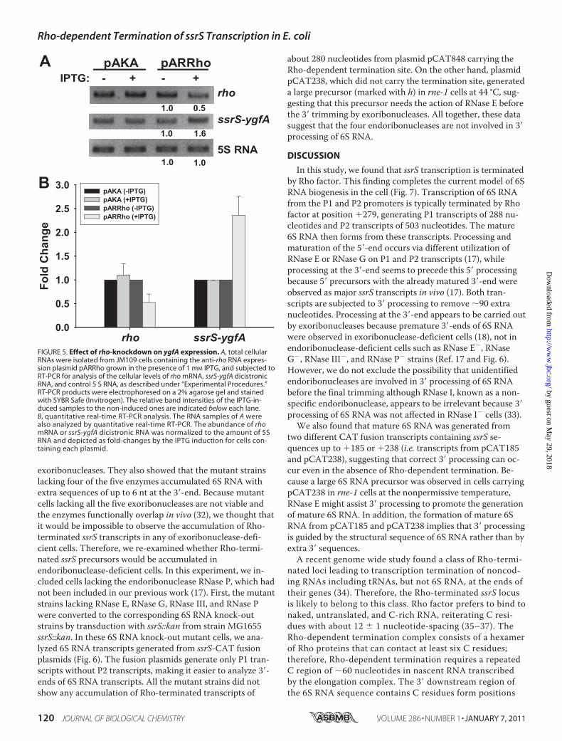
takes place in vivo, we constructed a plasmid expressing anti-rho RNA by IPTG induction. This anti-rho RNA was designedto bind to the translation initiation region rhomRNA (theregion of �22 to �6 with the first nucleotide of the startcodon as �1) of rhomRNA so that the cellular level or trans-lation of rhomRNA would be reduced. Cells containing theplasmid were treated with 1 mM of IPTG and total RNAs weresubjected to RT-PCR to analyze the cellular levels of rhomRNA and ssrS-ygfA dicistronic RNA. The level of ssrS-ygfAdicistronic RNA was estimated by determining the amount oftranscripts containing the region of �104 to �316 in the ssrS-ygfA transcription unit (Fig. 1C). The rhomRNA level wasreduced to about 50% by induction of anti-rho RNA, whilessrS-ygfA dicistronic RNA as run-through transcripts at theRho termination sites increased by more than 1.5-fold (Fig. 5),suggesting that ygfA expression could be regulated by Rho-de-pendent termination activity.Rho-terminaterd ssrS Transcripts Are Not Accumulated in
Endoribonuclease-deficient Cells—Our previous study showedthat 3� processing of ssrS transcript was not affected in RNaseE-, RNase G-, or RNase III-deficient cells (17), while Deut-scher and co-workers (18) reported that multiple exoribo-nucleases can participate in the 3� trimming reaction of 6SRNA and that any one of five exoribonucleases, RNase II, D,BN, T, and PH, can carry out the trimming reaction althougheither RNase T or RNase PH appear to be the most effective
FIGURE 4. Function of Rho protein as an ssrS transcription terminationfactor in vitro. A, in vitro transcription of the ssrS gene in the presence ofthe Rho factor. In vitro transcription reactions were carried out in the pres-ence or absence of Rho protein. IcdA protein was used as a control protein.The Rho-dependent terminated transcripts are indicated by f (for ssrS) or g(for tyrT). B, schematic representation of DNA templates used for in vitrotranscription. The DNA templates for in vitro transcription were prepared byPCR using the corresponding pCAT plasmids. The DNA templates contained94 bp of the vector sequences (open rectangles). The major Rho-dependentsites, estimated from the data shown in panel A, are indicated by f on eachsubstrate. The tyrT transcription unit was used as a positive control forRho-dependent termination and the termination site is indicated by g.C, binding of Rho protein with precursor 6S RNA containing 3� downstreamsequences. Mature 6S RNA (6S) and precursor 6S RNA carrying the 3� down-stream sequences to �330 (Pre-6S) were labeled with 32P and subjected togel mobility shift assay. The labeled RNA of 2 nM was incubated for 15 minin the absence (lanes 1 and 6) of protein or presence (lanes 2–5 and 7–10) ofincreasing amounts of Rho protein (1–50 nM). IcdA protein (50 nM) was usedas a control (lane 11).
TABLE 2Sequencing analysis of the 3� RACE products
3�-Ends of RNAa
�253 (3), �255 (1), �260 (�1), �271 (1), �275 (3), �277 (2)b�279 (8), �281 (1), �282 (1), �284 (1), �298 (1), �301 (1)a3� RACE products from in vitro transcripts obtained from the �92 to �448 tem-plate in Fig. 4 were cloned and analyzed by DNA sequencing.
b The numbers in parentheses indicate the frequency of occurrence.
Rho-dependent Termination of ssrS Transcription in E. coli
JANUARY 7, 2011 • VOLUME 286 • NUMBER 1 JOURNAL OF BIOLOGICAL CHEMISTRY 119
by guest on May 29, 2018
http://ww
w.jbc.org/
Dow
nloaded from
exoribonucleases. They also showed that the mutant strainslacking four of the five enzymes accumulated 6S RNA withextra sequences of up to 6 nt at the 3�-end. Because mutantcells lacking all the five exoribonucleases are not viable andthe enzymes functionally overlap in vivo (32), we thought thatit would be impossible to observe the accumulation of Rho-terminated ssrS transcripts in any of exoribonuclease-defi-cient cells. Therefore, we re-examined whether Rho-termi-nated ssrS precursors would be accumulated inendoribonuclease-deficient cells. In this experiment, we in-cluded cells lacking the endoribonuclease RNase P, which hadnot been included in our previous work (17). First, the mutantstrains lacking RNase E, RNase G, RNase III, and RNase Pwere converted to the corresponding 6S RNA knock-outstrains by transduction with ssrS::kan from strain MG1655ssrS::kan. In these 6S RNA knock-out mutant cells, we ana-lyzed 6S RNA transcripts generated from ssrS-CAT fusionplasmids (Fig. 6). The fusion plasmids generate only P1 tran-scripts without P2 transcripts, making it easier to analyze 3�-ends of 6S RNA transcripts. All the mutant strains did notshow any accumulation of Rho-terminated transcripts of
about 280 nucleotides from plasmid pCAT848 carrying theRho-dependent termination site. On the other hand, plasmidpCAT238, which did not carry the termination site, generateda large precursor (marked with h) in rne-1 cells at 44 °C, sug-gesting that this precursor needs the action of RNase E beforethe 3� trimming by exoribonucleases. All together, these datasuggest that the four endoribonucleases are not involved in 3�processing of 6S RNA.
DISCUSSION
In this study, we found that ssrS transcription is terminatedby Rho factor. This finding completes the current model of 6SRNA biogenesis in the cell (Fig. 7). Transcription of 6S RNAfrom the P1 and P2 promoters is typically terminated by Rhofactor at position �279, generating P1 transcripts of 288 nu-cleotides and P2 transcripts of 503 nucleotides. The mature6S RNA then forms from these transcripts. Processing andmaturation of the 5�-end occurs via different utilization ofRNase E or RNase G on P1 and P2 transcripts (17), whileprocessing at the 3�-end seems to precede this 5� processingbecause 5� precursors with the already matured 3�-end wereobserved as major ssrS transcripts in vivo (17). Both tran-scripts are subjected to 3� processing to remove �90 extranucleotides. Processing at the 3�-end appears to be carried outby exoribonucleases because premature 3�-ends of 6S RNAwere observed in exoribonuclease-deficient cells (18), not inendoribonuclease-deficient cells such as RNase E�, RNaseG�, RNase III�, and RNase P� strains (Ref. 17 and Fig. 6).However, we do not exclude the possibility that unidentifiedendoribonucleases are involved in 3� processing of 6S RNAbefore the final trimming although RNase I, known as a non-specific endoribonuclease, appears to be irrelevant because 3�processing of 6S RNA was not affected in RNase I� cells (33).
We also found that mature 6S RNA was generated fromtwo different CAT fusion transcripts containing ssrS se-quences up to �185 or �238 (i.e. transcripts from pCAT185and pCAT238), suggesting that correct 3� processing can oc-cur even in the absence of Rho-dependent termination. Be-cause a large 6S RNA precursor was observed in cells carryingpCAT238 in rne-1 cells at the nonpermissive temperature,RNase E might assist 3� processing to promote the generationof mature 6S RNA. In addition, the formation of mature 6SRNA from pCAT185 and pCAT238 implies that 3� processingis guided by the structural sequence of 6S RNA rather than byextra 3� sequences.A recent genome wide study found a class of Rho-termi-
nated loci leading to transcription termination of noncod-ing RNAs including tRNAs, but not 6S RNA, at the ends oftheir genes (34). Therefore, the Rho-terminated ssrS locusis likely to belong to this class. Rho factor prefers to bind tonaked, untranslated, and C-rich RNA, reiterating C resi-dues with about 12 � 1 nucleotide-spacing (35–37). TheRho-dependent termination complex consists of a hexamerof Rho proteins that can contact at least six C residues;therefore, Rho-dependent termination requires a repeatedC region of �60 nucleotides in nascent RNA transcribedby the elongation complex. The 3� downstream region ofthe 6S RNA sequence contains C residues form positions
FIGURE 5. Effect of rho-knockdown on ygfA expression. A, total cellularRNAs were isolated from JM109 cells containing the anti-rho RNA expres-sion plasmid pARRho grown in the presence of 1 mM IPTG, and subjected toRT-PCR for analysis of the cellular levels of rho mRNA, ssrS-ygfA dicistronicRNA, and control 5 S RNA, as described under “Experimental Procedures.”RT-PCR products were electrophoresed on a 2% agarose gel and stainedwith SYBR Safe (Invitrogen). The relative band intensities of the IPTG-in-duced samples to the non-induced ones are indicated below each lane.B, quantitative real-time RT-PCR analysis. The RNA samples of A werealso analyzed by quantitative real-time RT-PCR. The abundance of rhomRNA or ssrS-ygfA dicistronic RNA was normalized to the amount of 5SRNA and depicted as fold-changes by the IPTG induction for cells con-taining each plasmid.
Rho-dependent Termination of ssrS Transcription in E. coli
120 JOURNAL OF BIOLOGICAL CHEMISTRY VOLUME 286 • NUMBER 1 • JANUARY 7, 2011
by guest on May 29, 2018
http://ww
w.jbc.org/
Dow
nloaded from

�193 to �313 with 12 � 1 nucleotide spacing (Fig. 1C).Therefore, it is likely that the C-residues ranging from�193 to �265 interact with Rho factor to terminate thetranscription. Rho-dependent termination sites are locatedwithin the ygfA open reading frame. The definitive genefunction of ygfA is not yet known, but encodes a proteinwith a high degree of sequence identity to mammalian5-formyl-tetrahydrofolate cyclo-ligase (7, 13). As ygfA does
not have its own promoter, its expression should dependon transcription initiation from the ssrS promoters (14,17), as well as antitermination at Rho-dependent termina-tion sites. Consequently, ygfA expression can be regulatedby the initiation rate from the ssrS promoters and the ac-tivity of Rho factor in the cell. When a rho-knockdown ex-periment was performed by expressing anti-rho RNA, weobserved the increase of ssrS-ygfA dicistronic transcriptsformed by read-through transcription at the Rho-depen-dent termination sites. Recently, Nudler’s group also re-ported a significant increase in ygfA mRNA when RNAsfrom cells treated with bicyclomycin, which inhibits Rhofactor function, were subjected to microarray analysis (38).It is likely, therefore, Rho factor participates not only intranscription termination for generating 3�-ends of 6SRNA from the termination sites but also in regulating ygfAexpression. It is noteworthy that ygfA expression is impli-cated in the formation of persister cells, which contributeto the antibiotic resistance of biofilm infections (13).Moreover, several microarray analyses showed that ygfAgene is up-regulated during the biofilm formation (16, 39,40). Thus, antitermination of ssrS transcripts at Rho-de-pendent termination sites under some Rho-factor functionsuppressing conditions could play an essential role in per-sister cell and biofilm formations.
Acknowledgments—We thank both the National Institute of Genet-ics (Shizuoka, Japan) and Dr. Wachi for providing us the ASKAplasmids and RNase E/RNase G mutant strains, respectively.
FIGURE 6. Effect of endoribonucleases on 3� processing of 6S RNA. Total cellular RNAs were prepared from RNase G mutant (rng::cat) (A), RNase E mutant(rne-1) and RNase E/G double mutant (rne-1, rng::cat) (B), RNase III mutant (rnc105) (C), and RNase P mutant (rnpA49) strains (D) along with the correspond-ing wild-type isogenic strains containing pCAT238 or pCAT848. Each RNA sample was fractionated on a 5% polyacrylamide gel and 6S RNA transcripts wereanalyzed by Northern blot. The strains, plasmids, and growth temperatures are indicated above lanes. A large 6S RNA transcript accumulated in rne-1 cellsat 44 °C is indicated by h. Low Range ssRNA Ladder (NEB) was used as RNA size markers. P, P1 6S RNA transcripts with 3�-processed ends. M, mature 6S RNA.
FIGURE 7. A model of 6S RNA biogenesis via regulation of Rho-depen-dent termination of ssrS transcription. The 6S RNA is transcribed fromtwo tandem promoters, the �70-dependent P1 promoter and the �70/�S-dual dependent P2 promoter. Transcription from the ssrS promoters is ter-minated at Rho termination sites. Exoribonucleases first mediate 3� process-ing of primary ssrS transcripts. The P1 transcripts are further processed byRNases E or G, and the P2 transcripts are processed by RNase E.
Rho-dependent Termination of ssrS Transcription in E. coli
JANUARY 7, 2011 • VOLUME 286 • NUMBER 1 JOURNAL OF BIOLOGICAL CHEMISTRY 121
by guest on May 29, 2018
http://ww
w.jbc.org/
Dow
nloaded from

REFERENCES1. Eddy, S. R. (2001) Nat. Rev. Genet. 2, 919–9292. Gottesman, S. (2004) Annu. Rev. Microbiol. 58, 303–3283. Storz, G., Opdyke, J. A., and Zhang, A. (2004) Curr. Opin. Microbiol. 7,
140–1444. Wassarman, K. M. (2002) Cell 109, 141–1445. Deutscher, M. P. (1993) in Nucleases (Linn, S. M., Lloyd, R. S., and Rob-
erts, R. J., eds) Cold Spring Harbor Laboratory Press, Plainview, NY6. Lee, S. Y., Bailey, S. C., and Apirion, D. (1978) J. Bacteriol. 133,
1015–10237. Wassarman, K. M., and Storz, G. (2000) Cell 101, 613–6238. Wassarman, K. M. (2007)Mol. Microbiol. 65, 1425–14319. Trotochaud, A. E., and Wassarman, K. M. (2004) J. Bacteriol. 186,
4978–498510. Wassarman, K. M., and Saecker, R. M. (2006) Science 314, 1601–160311. Brownlee, G. G. (1971) Nat. New Biol. 229, 147–14912. Trotochaud, A. E., and Wassarman, K. M. (2005) Nat. Struct. Mol. Biol.
12, 313–31913. Hansen, S., Lewis, K., and Vulic, M. (2008) Antimicrob. Agents Che-
mother. 52, 2718–272614. Hsu, L. M., Zagorski, J., Wang, Z., and Fournier, M. J. (1985) J. Bacteriol.
161, 1162–117015. Willkomm, D. K., Minnerup, J., Huttenhofer, A., and Hartmann, R. K.
(2005) Nucleic Acids Res. 33, 1949–196016. Ren, D., Bedzyk, L. A., Thomas, S. M., Ye, R. W., and Wood, T. K. (2004)
Appl. Microbiol. Biotechnol. 64, 515–52417. Kim, K. S., and Lee, Y. (2004) Nucleic Acids Res. 32, 6057–606818. Li, Z., Pandit, S., and Deutscher, M. P. (1998) Proc. Natl. Acad. Sci.
U.S.A. 95, 2856–286119. Fozo, E. M., Kawano, M., Fontaine, F., Kaya, Y., Mendieta, K. S., Jones,
K. L., Ocampo, A., Rudd, K. E., and Storz, G. (2008)Mol. Microbiol. 70,1076–1093
20. Han, K., Kim, K. S., Bak, G., Park, H., and Lee, Y. (2010) Nucleic AcidsRes. 38, 5851–5866
21. Yu, D., Ellis, H. M., Lee, E. C., Jenkins, N. A., Copeland, N. G., andCourt, D. L. (2000) Proc. Natl. Acad. Sci. U.S.A. 97, 5978–5983
22. Wachi, M., Umitsuki, G., and Nakai, K. (1997)Mol. Gen. Genet. 253,515–519
23. Dasgupta, S., Fernandez, L., Kameyama, L., Inada, T., Nakamura, Y.,Pappas, A., and Court, D. L. (1998)Mol. Microbiol. 28, 629–640
24. Kirsebom, L. A., Baer, M. F., and Altman, S. (1988) J. Mol. Biol. 204,879–888
25. Kim, S., Kim, H., Park, I., and Lee, Y. (1996) J. Biol. Chem. 271,19330–19337
26. Park, J. W., Jung, Y., Lee, S. J., Jin, D. J., Lee, Y., and Lee, S. J. (2002) Bio-chem. Biophys. Res. Commun. 290, 1183–1187
27. Kim, M. S., Kim, S., Kim, S. C., Lee, Y. M., Jeon, E. S., Park, C. U., andLee, Y. (1997)Mol. Gen. Genet. 254, 464–468
28. Kitagawa, M., Ara, T., Arifuzzaman, M., Ioka-Nakamichi, T., Inamoto,E., Toyonaga, H., and Mori, H. (2005) DNA Res. 12, 291–299
29. Argaman, L., Hershberg, R., Vogel, J., Bejerano, G., Wagner, E. G., Mar-galit, H., and Altuvia, S. (2001) Curr. Biol. 11, 941–950
30. Kim, M. S., Park, B. H., Kim, S., Lee, Y. J., Chung, J. H., and Lee, Y.(1998) Biochem. Mol. Biol. Int. 46, 1153–1160
31. Ciampi, M. S. (2006)Microbiology 152, 2515–252832. Kelly, K. O., and Deutscher, M. P. (1992) J. Bacteriol. 174, 6682–668433. Kim, Y. (2009) Regulation of RNase P RNA Biogenesis Ph.D. Dissertation,
KAIST34. Peters, J. M., Mooney, R. A., Kuan, P. F., Rowland, J. L., Keles, S., and
Landick, R. (2009) Proc. Natl. Acad. Sci. U.S.A. 106, 15406–1541135. Alifano, P., Rivellini, F., Limauro, D., Bruni, C. B., and Carlomagno, M. S.
(1991) Cell 64, 553–56336. Graham, J. E., and Richardson, J. P. (1998) J. Biol. Chem. 273,
20764–2076937. Graham, J. E. (2004) Nucleic Acids Res. 32, 3093–310038. Cardinale, C. J., Washburn, R. S., Tadigotla, V. R., Brown, L. M., Gottes-
man, M. E., and Nudler, E. (2008) Science 320, 935–93839. Ito, A., Taniuchi, A., May, T., Kawata, K., and Okabe, S. (2009) Appl.
Environ. Microbiol. 75, 4093–410040. Ito, A., May, T., Taniuchi, A., Kawata, K., and Okabe, S. (2009) Biotech-
nol. Bioeng. 103, 975–983
Rho-dependent Termination of ssrS Transcription in E. coli
122 JOURNAL OF BIOLOGICAL CHEMISTRY VOLUME 286 • NUMBER 1 • JANUARY 7, 2011
by guest on May 29, 2018
http://ww
w.jbc.org/
Dow
nloaded from

Younghoon LeeHuiseok Chae, Kook Han, Kwang-sun Kim, Hongmarn Park, Jungmin Lee and
CYCLO-LIGASE)DOWNSTREAM ygfA (PUTATIVE 5-FORMYL-TETRAHYDROFOLATE
PROCESSING OF 6S RNA AND EXPRESSION OF′IMPLICATION FOR 3:Escherichia coli (6S RNA) Transcription in ssrSRho-dependent Termination of
doi: 10.1074/jbc.M110.150201 originally published online October 29, 20102011, 286:114-122.J. Biol. Chem.
10.1074/jbc.M110.150201Access the most updated version of this article at doi:
Alerts:
When a correction for this article is posted•
When this article is cited•
to choose from all of JBC's e-mail alertsClick here
http://www.jbc.org/content/286/1/114.full.html#ref-list-1
This article cites 38 references, 13 of which can be accessed free at
by guest on May 29, 2018
http://ww
w.jbc.org/
Dow
nloaded from