Embed Size (px)
Citation preview
REVIEW
The poly(C)-binding proteins: A multiplicity offunctions and a search for mechanisms
ALEKSANDR V. MAKEYEV and STEPHEN A. LIEBHABERDepartments of Genetics and Medicine, University of Pennsylvania School of Medicine,Philadelphia, Pennsylvania 19104, USA
ABSTRACT
The poly(C) binding proteins (PCBPs) are encoded at five dispersed loci in the mouse and human genomes. Theseproteins, which can be divided into two groups, hnRNPs K/J and the aCPs (aCP1-4), are linked by a commonevolutionary history, a shared triple KH domain configuration, and by their poly(C) binding specificity. Given theseconserved characteristics it is remarkable to find a substantial diversity in PCBP functions. The roles of theseproteins in mRNA stabilization, translational activation, and translational silencing suggest a complex and diverse setof post-transcriptional control pathways. Their additional putative functions in transcriptional control and as struc-tural components of important DNA-protein complexes further support their remarkable structural and functionalversatility. Clearly the identification of additional binding targets and delineation of corresponding control mecha-nisms and effector pathways will establish highly informative models for further exploration.
Keywords: aCP; hnRNP K; mRNA stability; mRNA translation; poly(C) binding proteins; post-transcriptionalcontrols
PERSPECTIVE
It has long been noted that certain RNA-binding pro-teins can be characterized, grouped, and purified onthe basis of their binding to nucleic acid homopolymers(Swanson & Dreyfuss, 1988)+ A defined set of RNA-binding proteins is characterized by high affinity andsequence-specific interaction with poly(C)+ Thesepoly(C)-binding proteins (PCBPs) comprise two sub-sets in mammalian cells; hnRNPs K/J (Matunis et al+,1992) and the aCP proteins (a-complex proteins)+ Theexact structural and genetic relationship of hnRNP Kand J remains to be fully defined+ The aCPs are en-coded at four dispersed loci, with additional isoformsgenerated via alternative splicing (Makeyev & Lieb-haber, 2000)+ The PCBPs studied in greatest detail arehnRNP K, aCP-1, and aCP-2+ The latter two proteinsare alternatively referred to as PCBP1 and PCBP2 orhnRNP-E1 and hnRNP-E2 (Kiledjian et al+, 1995; Lefferset al+, 1995)+ Recent studies, summarized in this re-view, reveal that the PCBPs are involved in a remark-
able array of biological processes+ Members of thisprotein family are linked to mRNA stabilization (Weiss& Liebhaber, 1994, 1995; Holcik & Liebhaber, 1997),translational silencing (Ostareck et al+, 1997; Collieret al+, 1998), and translational enhancement (Blyn et al+,1997; Gamarnik & Andino, 1997)+ These proteins alsoappear to be involved as determinants of transcrip-tional controls (Michelotti et al+, 1996; Tomonaga & Lev-ens, 1996) and apoptotic pathways (Charroux et al+,1999; Zhu & Chen, 2000) and constitute candidate struc-tural components of recombination complexes withinretrotransposon long terminal repeats (Goller et al+,1994) and in telomere complexes (Lacroix et al+, 2000)+The common denominator of PCBP activities appearsto reflect binding to C-rich single-strand motifs+ Deter-mining how such interactions might factor into this broadarray of diverse biologic functions constitutes a majorchallenge to current research efforts+
Initial studies of PCBPs have been previously re-viewed (Bomsztyk et al+, 1997; Ostareck-Lederer et al+,1998)+ This article will focus with particular attention onthree areas of recent interest: (1) identification and char-acterization of novel PCBP isoforms and posttransla-tional modifications that may underlie their functionaldiversity; (2) new insights into the evolutionary history
Reprint requests to: Dr+ Stephen A+ Liebhaber, University of Penn-sylvania School of Medicine, Department of Genetics, CRB—Room428, 415 Curie Blvd+, Philadelphia, Pennsylvania 19104-6148, USA;e-mail: liebhabe@mail+med+upenn+edu+
RNA (2002), 8:265–278+ Cambridge University Press+ Printed in the USA+Copyright © 2002 RNA Society+DOI: 10+1017+S1355838202024627
265
of the PCBPs that may shed light on their conservedstructure-function relationships, and (3) the expandingspectrum of PCBP functions in transcriptional and post-transcriptional controls+
hnRNP K AND THE aCPS REPRESENTDISTINCT PCBP SUBSETS WITHCOMMON EVOLUTIONARY ORIGINS
Shared structures and origins of the PCBPs
PCBPs belong to the KH domain superfamily of nu-cleic acid-binding proteins+ The KH (hnRNP K homol-ogy) domain was initially identified in hnRNP K andhas subsequently been noted in a wide range of RNA-binding proteins in organisms extending throughoutthe procaryotic and eukaryotic evolutionary spectrum(for a recent review, see Adinolfi et al+, 1999)+ The KHdomain can occur in a protein as a single unit or inmultiple copies+ Isolated KH domains can act as inde-pendent nucleic acid-binding units and can dictate, ei-ther independently or in concert, a wide spectrum ofnucleic acid-binding specificities+
The PCBPs share an overall anatomy consisting oftwo KH domains grouped near the N-terminus and a
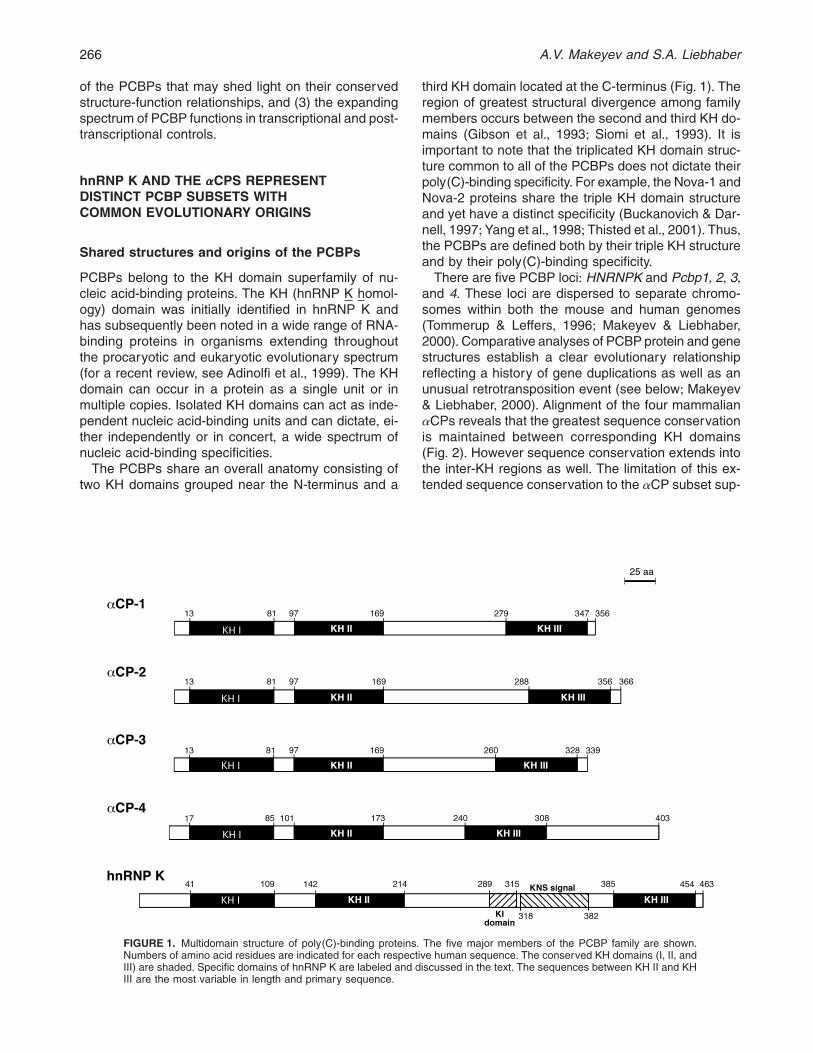
third KH domain located at the C-terminus (Fig+ 1)+ Theregion of greatest structural divergence among familymembers occurs between the second and third KH do-mains (Gibson et al+, 1993; Siomi et al+, 1993)+ It isimportant to note that the triplicated KH domain struc-ture common to all of the PCBPs does not dictate theirpoly(C)-binding specificity+ For example, the Nova-1 andNova-2 proteins share the triple KH domain structureand yet have a distinct specificity (Buckanovich & Dar-nell, 1997; Yang et al+, 1998; Thisted et al+, 2001)+ Thus,the PCBPs are defined both by their triple KH structureand by their poly(C)-binding specificity+
There are five PCBP loci: HNRNPK and Pcbp1, 2, 3,and 4+ These loci are dispersed to separate chromo-somes within both the mouse and human genomes(Tommerup & Leffers, 1996; Makeyev & Liebhaber,2000)+Comparative analyses of PCBP protein and genestructures establish a clear evolutionary relationshipreflecting a history of gene duplications as well as anunusual retrotransposition event (see below; Makeyev& Liebhaber, 2000)+ Alignment of the four mammalianaCPs reveals that the greatest sequence conservationis maintained between corresponding KH domains(Fig+ 2)+ However sequence conservation extends intothe inter-KH regions as well+ The limitation of this ex-tended sequence conservation to the aCP subset sup-
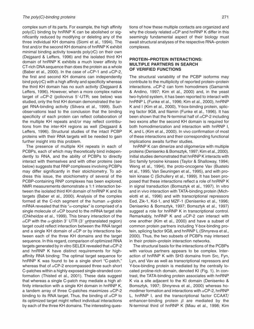
FIGURE 1. Multidomain structure of poly(C)-binding proteins+ The five major members of the PCBP family are shown+Numbers of amino acid residues are indicated for each respective human sequence+ The conserved KH domains (I, II, andIII) are shaded+ Specific domains of hnRNP K are labeled and discussed in the text+ The sequences between KH II and KHIII are the most variable in length and primary sequence+
266 A.V. Makeyev and S.A. Liebhaber
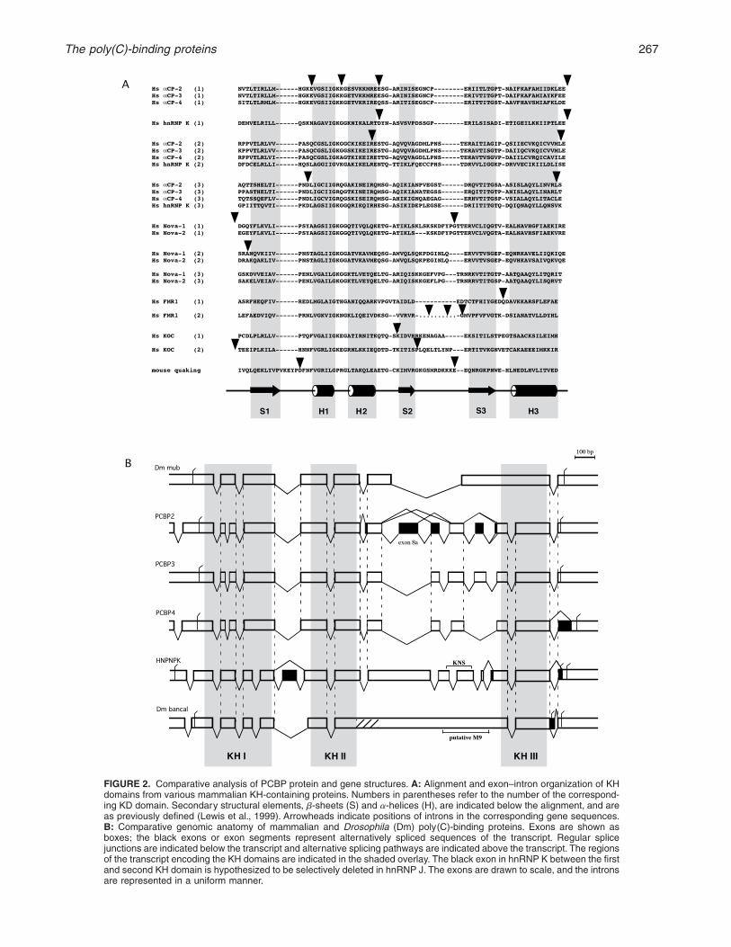
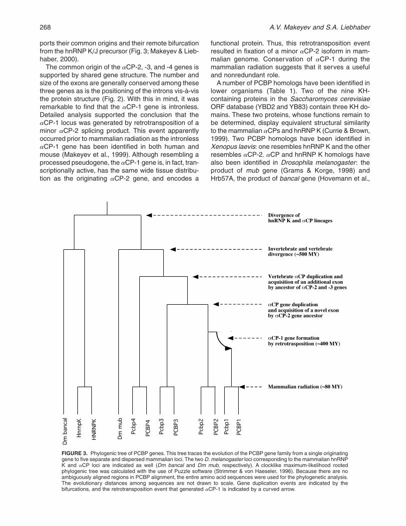
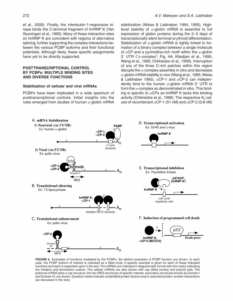
FIGURE 2. Comparative analysis of PCBP protein and gene structures+ A: Alignment and exon–intron organization of KHdomains from various mammalian KH-containing proteins+ Numbers in parentheses refer to the number of the correspond-ing KD domain+ Secondary structural elements, b-sheets (S) and a-helices (H), are indicated below the alignment, and areas previously defined (Lewis et al+, 1999)+ Arrowheads indicate positions of introns in the corresponding gene sequences+B: Comparative genomic anatomy of mammalian and Drosophila (Dm) poly(C)-binding proteins+ Exons are shown asboxes; the black exons or exon segments represent alternatively spliced sequences of the transcript+ Regular splicejunctions are indicated below the transcript and alternative splicing pathways are indicated above the transcript+ The regionsof the transcript encoding the KH domains are indicated in the shaded overlay+ The black exon in hnRNP K between the firstand second KH domain is hypothesized to be selectively deleted in hnRNP J+ The exons are drawn to scale, and the intronsare represented in a uniform manner+
The poly(C)-binding proteins 267
ports their common origins and their remote bifurcationfrom the hnRNP K/J precursor (Fig+ 3;Makeyev & Lieb-haber, 2000)+
The common origin of the aCP-2, -3, and -4 genes issupported by shared gene structure+ The number andsize of the exons are generally conserved among thesethree genes as is the positioning of the introns vis-à-visthe protein structure (Fig+ 2)+ With this in mind, it wasremarkable to find that the aCP-1 gene is intronless+Detailed analysis supported the conclusion that theaCP-1 locus was generated by retrotransposition of aminor aCP-2 splicing product+ This event apparentlyoccurred prior to mammalian radiation as the intronlessaCP-1 gene has been identified in both human andmouse (Makeyev et al+, 1999)+ Although resembling aprocessed pseudogene, the aCP-1 gene is, in fact, tran-scriptionally active, has the same wide tissue distribu-tion as the originating aCP-2 gene, and encodes a
functional protein+ Thus, this retrotransposition eventresulted in fixation of a minor aCP-2 isoform in mam-malian genome+ Conservation of aCP-1 during themammalian radiation suggests that it serves a usefuland nonredundant role+
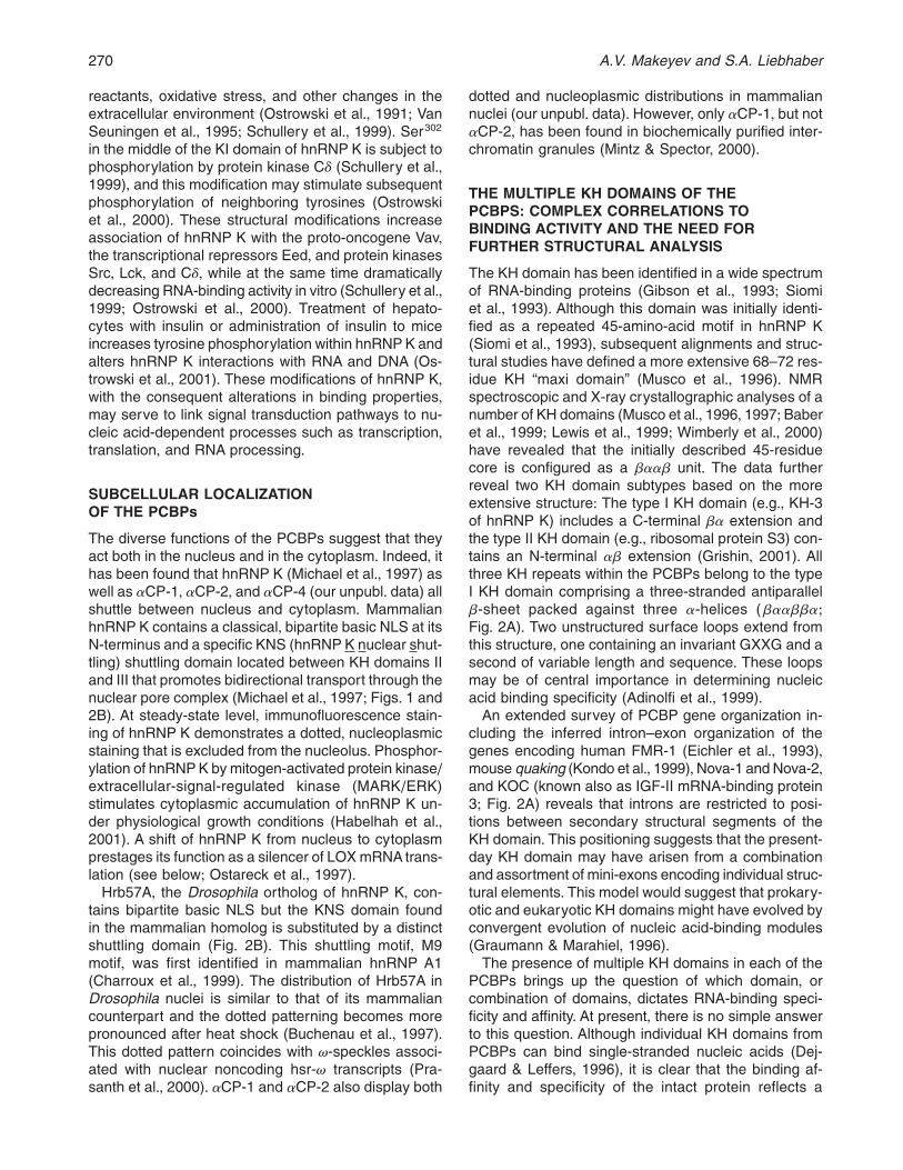
A number of PCBP homologs have been identified inlower organisms (Table 1)+ Two of the nine KH-containing proteins in the Saccharomyces cerevisiaeORF database (YBD2 and YB83) contain three KH do-mains+ These two proteins, whose functions remain tobe determined, display equivalent structural similarityto the mammalian aCPs and hnRNP K (Currie & Brown,1999)+ Two PCBP homologs have been identified inXenopus laevis: one resembles hnRNP K and the otherresembles aCP-2+ aCP and hnRNP K homologs havealso been identified in Drosophila melanogaster: theproduct of mub gene (Grams & Korge, 1998) andHrb57A, the product of bancal gene (Hovemann et al+,
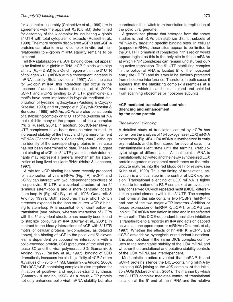
FIGURE 3. Phylogenic tree of PCBP genes+ This tree traces the evolution of the PCBP gene family from a single originatinggene to five separate and dispersed mammalian loci+ The two D. melanogaster loci corresponding to the mammalian hnRNPK and aCP loci are indicated as well (Dm bancal and Dm mub, respectively)+ A clocklike maximum-likelihood rootedphylogenic tree was calculated with the use of Puzzle software (Strimmer & von Haeseler, 1996)+ Because there are noambiguously aligned regions in PCBP alignment, the entire amino acid sequences were used for the phylogenetic analysis+The evolutionary distances among sequences are not drawn to scale+ Gene duplication events are indicated by thebifurcations, and the retrotransposition event that generated aCP-1 is indicated by a curved arrow+
268 A.V. Makeyev and S.A. Liebhaber
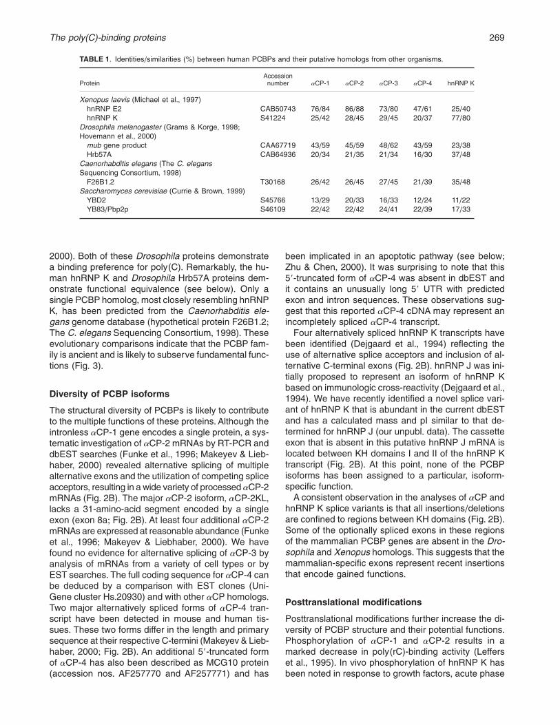
2000)+ Both of these Drosophila proteins demonstratea binding preference for poly(C)+ Remarkably, the hu-man hnRNP K and Drosophila Hrb57A proteins dem-onstrate functional equivalence (see below)+ Only asingle PCBP homolog,most closely resembling hnRNPK, has been predicted from the Caenorhabditis ele-gans genome database (hypothetical protein F26B1+2;The C. elegans Sequencing Consortium, 1998)+ Theseevolutionary comparisons indicate that the PCBP fam-ily is ancient and is likely to subserve fundamental func-tions (Fig+ 3)+
Diversity of PCBP isoforms
The structural diversity of PCBPs is likely to contributeto the multiple functions of these proteins+Although theintronless aCP-1 gene encodes a single protein, a sys-tematic investigation of aCP-2 mRNAs by RT-PCR anddbEST searches (Funke et al+, 1996; Makeyev & Lieb-haber, 2000) revealed alternative splicing of multiplealternative exons and the utilization of competing spliceacceptors, resulting in a wide variety of processed aCP-2mRNAs (Fig+ 2B)+ The major aCP-2 isoform, aCP-2KL,lacks a 31-amino-acid segment encoded by a singleexon (exon 8a; Fig+ 2B)+ At least four additional aCP-2mRNAs are expressed at reasonable abundance (Funkeet al+, 1996; Makeyev & Liebhaber, 2000)+ We havefound no evidence for alternative splicing of aCP-3 byanalysis of mRNAs from a variety of cell types or byEST searches+ The full coding sequence for aCP-4 canbe deduced by a comparison with EST clones (Uni-Gene cluster Hs+20930) and with other aCP homologs+Two major alternatively spliced forms of aCP-4 tran-script have been detected in mouse and human tis-sues+ These two forms differ in the length and primarysequence at their respective C-termini (Makeyev & Lieb-haber, 2000; Fig+ 2B)+ An additional 59-truncated formof aCP-4 has also been described as MCG10 protein(accession nos+ AF257770 and AF257771) and has
been implicated in an apoptotic pathway (see below;Zhu & Chen, 2000)+ It was surprising to note that this59-truncated form of aCP-4 was absent in dbEST andit contains an unusually long 59 UTR with predictedexon and intron sequences+ These observations sug-gest that this reported aCP-4 cDNA may represent anincompletely spliced aCP-4 transcript+
Four alternatively spliced hnRNP K transcripts havebeen identified (Dejgaard et al+, 1994) reflecting theuse of alternative splice acceptors and inclusion of al-ternative C-terminal exons (Fig+ 2B)+ hnRNP J was ini-tially proposed to represent an isoform of hnRNP Kbased on immunologic cross-reactivity (Dejgaard et al+,1994)+ We have recently identified a novel splice vari-ant of hnRNP K that is abundant in the current dbESTand has a calculated mass and pI similar to that de-termined for hnRNP J (our unpubl+ data)+ The cassetteexon that is absent in this putative hnRNP J mRNA islocated between KH domains I and II of the hnRNP Ktranscript (Fig+ 2B)+ At this point, none of the PCBPisoforms has been assigned to a particular, isoform-specific function+
A consistent observation in the analyses of aCP andhnRNP K splice variants is that all insertions/deletionsare confined to regions between KH domains (Fig+ 2B)+Some of the optionally spliced exons in these regionsof the mammalian PCBP genes are absent in the Dro-sophila and Xenopus homologs+ This suggests that themammalian-specific exons represent recent insertionsthat encode gained functions+
Posttranslational modifications
Posttranslational modifications further increase the di-versity of PCBP structure and their potential functions+Phosphorylation of aCP-1 and aCP-2 results in amarked decrease in poly(rC)-binding activity (Lefferset al+, 1995)+ In vivo phosphorylation of hnRNP K hasbeen noted in response to growth factors, acute phase
TABLE 1+ Identities/similarities (%) between human PCBPs and their putative homologs from other organisms+
ProteinAccession
number aCP-1 aCP-2 aCP-3 aCP-4 hnRNP K
Xenopus laevis (Michael et al+, 1997)hnRNP E2 CAB50743 76/84 86/88 73/80 47/61 25/40hnRNP K S41224 25/42 28/45 29/45 20/37 77/80
Drosophila melanogaster (Grams & Korge, 1998;Hovemann et al+, 2000)
mub gene product CAA67719 43/59 45/59 48/62 43/59 23/38Hrb57A CAB64936 20/34 21/35 21/34 16/30 37/48
Caenorhabditis elegans (The C. elegansSequencing Consortium, 1998)
F26B1+2 T30168 26/42 26/45 27/45 21/39 35/48Saccharomyces cerevisiae (Currie & Brown, 1999)
YBD2 S45766 13/29 20/33 16/33 12/24 11/22YB83/Pbp2p S46109 22/42 22/42 24/41 22/39 17/33
The poly(C)-binding proteins 269
reactants, oxidative stress, and other changes in theextracellular environment (Ostrowski et al+, 1991; VanSeuningen et al+, 1995; Schullery et al+, 1999)+ Ser302
in the middle of the KI domain of hnRNP K is subject tophosphorylation by protein kinase Cd (Schullery et al+,1999), and this modification may stimulate subsequentphosphorylation of neighboring tyrosines (Ostrowskiet al+, 2000)+ These structural modifications increaseassociation of hnRNP K with the proto-oncogene Vav,the transcriptional repressors Eed, and protein kinasesSrc, Lck, and Cd, while at the same time dramaticallydecreasing RNA-binding activity in vitro (Schullery et al+,1999; Ostrowski et al+, 2000)+ Treatment of hepato-cytes with insulin or administration of insulin to miceincreases tyrosine phosphorylation within hnRNP K andalters hnRNP K interactions with RNA and DNA (Os-trowski et al+, 2001)+ These modifications of hnRNP K,with the consequent alterations in binding properties,may serve to link signal transduction pathways to nu-cleic acid-dependent processes such as transcription,translation, and RNA processing+
SUBCELLULAR LOCALIZATIONOF THE PCBPs
The diverse functions of the PCBPs suggest that theyact both in the nucleus and in the cytoplasm+ Indeed, ithas been found that hnRNP K (Michael et al+, 1997) aswell as aCP-1, aCP-2, and aCP-4 (our unpubl+ data) allshuttle between nucleus and cytoplasm+ MammalianhnRNP K contains a classical, bipartite basic NLS at itsN-terminus and a specific KNS (hnRNP K nuclear shut-tling) shuttling domain located between KH domains IIand III that promotes bidirectional transport through thenuclear pore complex (Michael et al+, 1997; Figs+ 1 and2B)+ At steady-state level, immunofluorescence stain-ing of hnRNP K demonstrates a dotted, nucleoplasmicstaining that is excluded from the nucleolus+ Phosphor-ylation of hnRNP K by mitogen-activated protein kinase/extracellular-signal-regulated kinase (MARK/ERK)stimulates cytoplasmic accumulation of hnRNP K un-der physiological growth conditions (Habelhah et al+,2001)+ A shift of hnRNP K from nucleus to cytoplasmprestages its function as a silencer of LOX mRNA trans-lation (see below; Ostareck et al+, 1997)+
Hrb57A, the Drosophila ortholog of hnRNP K, con-tains bipartite basic NLS but the KNS domain foundin the mammalian homolog is substituted by a distinctshuttling domain (Fig+ 2B)+ This shuttling motif, M9motif, was first identified in mammalian hnRNP A1(Charroux et al+, 1999)+ The distribution of Hrb57A inDrosophila nuclei is similar to that of its mammaliancounterpart and the dotted patterning becomes morepronounced after heat shock (Buchenau et al+, 1997)+This dotted pattern coincides with v-speckles associ-ated with nuclear noncoding hsr-v transcripts (Pra-santh et al+, 2000)+ aCP-1 and aCP-2 also display both
dotted and nucleoplasmic distributions in mammaliannuclei (our unpubl+ data)+ However, only aCP-1, but notaCP-2, has been found in biochemically purified inter-chromatin granules (Mintz & Spector, 2000)+
THE MULTIPLE KH DOMAINS OF THEPCBPS: COMPLEX CORRELATIONS TOBINDING ACTIVITY AND THE NEED FORFURTHER STRUCTURAL ANALYSIS
The KH domain has been identified in a wide spectrumof RNA-binding proteins (Gibson et al+, 1993; Siomiet al+, 1993)+ Although this domain was initially identi-fied as a repeated 45-amino-acid motif in hnRNP K(Siomi et al+, 1993), subsequent alignments and struc-tural studies have defined a more extensive 68–72 res-idue KH “maxi domain” (Musco et al+, 1996)+ NMRspectroscopic and X-ray crystallographic analyses of anumber of KH domains (Musco et al+, 1996, 1997; Baberet al+, 1999; Lewis et al+, 1999; Wimberly et al+, 2000)have revealed that the initially described 45-residuecore is configured as a baab unit+ The data furtherreveal two KH domain subtypes based on the moreextensive structure: The type I KH domain (e+g+, KH-3of hnRNP K) includes a C-terminal ba extension andthe type II KH domain (e+g+, ribosomal protein S3) con-tains an N-terminal ab extension (Grishin, 2001)+ Allthree KH repeats within the PCBPs belong to the typeI KH domain comprising a three-stranded antiparallelb-sheet packed against three a-helices (baabba;Fig+ 2A)+ Two unstructured surface loops extend fromthis structure, one containing an invariant GXXG and asecond of variable length and sequence+ These loopsmay be of central importance in determining nucleicacid binding specificity (Adinolfi et al+, 1999)+
An extended survey of PCBP gene organization in-cluding the inferred intron–exon organization of thegenes encoding human FMR-1 (Eichler et al+, 1993),mouse quaking (Kondo et al+, 1999),Nova-1 and Nova-2,and KOC (known also as IGF-II mRNA-binding protein3; Fig+ 2A) reveals that introns are restricted to posi-tions between secondary structural segments of theKH domain+ This positioning suggests that the present-day KH domain may have arisen from a combinationand assortment of mini-exons encoding individual struc-tural elements+ This model would suggest that prokary-otic and eukaryotic KH domains might have evolved byconvergent evolution of nucleic acid-binding modules(Graumann & Marahiel, 1996)+
The presence of multiple KH domains in each of thePCBPs brings up the question of which domain, orcombination of domains, dictates RNA-binding speci-ficity and affinity+ At present, there is no simple answerto this question+ Although individual KH domains fromPCBPs can bind single-stranded nucleic acids (Dej-gaard & Leffers, 1996), it is clear that the binding af-finity and specificity of the intact protein reflects a
270 A.V. Makeyev and S.A. Liebhaber
complex sum of its parts+ For example, the high affinitypoly(C) binding by hnRNP K can be abolished or sig-nificantly reduced by modifying or deleting any of thethree individual KH domains (Siomi et al+, 1994)+ Thefirst and/or the second KH domains of hnRNP K exhibitminimal binding activity towards poly(rC) on their own(Dejgaard & Leffers, 1996) and the isolated third KHdomain of hnRNP K exhibits a much lower affinity toCT-rich DNA sequence than does the protein as a whole(Baber et al+, 2000)+ In the case of aCP-1 and aCP-2,the first and second KH domains can independentlybind poly(rC) with a high affinity and specificity whereasthe third KH domain has no such activity (Dejgaard &Leffers, 1996)+ However, when a more complex nativetarget of aCP-2 (poliovirus 59-UTR; see below) wasstudied, only the first KH domain demonstrated the tar-get RNA-binding activity (Silvera et al+, 1999)+ Suchobservations lead to the conclusion that the bindingspecificity of each protein can reflect collaboration ofthe multiple KH repeats and/or may reflect contribu-tions from the inter-KH regions as well (Dejgaard &Leffers, 1996)+ Structural studies of the intact PCBPproteins with their RNA targets will be needed to gainfurther insight into this problem+
The presence of multiple KH repeats in each ofPCBPs, each of which may theoretically bind indepen-dently to RNA, and the ability of PCBPs to directlyinteract with themselves and with other proteins (seebelow) suggests that RNP complexes involving PCBPsmay differ significantly in their stoichiometry+ To ad-dress this issue, the stoichiometry of several of thePCBP-containing RNP complexes has been explored+NMR measurements demonstrate a 1:1 interaction be-tween the isolated third KH domain of hnRNP K and itstargets (Baber et al+, 2000)+ Analysis of the complexformed at the C-rich segment of the human a-globinmRNA revealed that this “a-complex” is comprised of asingle molecule of aCP bound to the mRNA target site(Chkheidze et al+, 1999)+ This binary interaction of theaCP with the a-globin 39 UTR (39 untranslated region)target could reflect interaction between the RNA targetand a single KH domain of aCP or by interactions be-tween each of the three KH domains and the targetsequence+ In this regard, comparison of optimized RNAtargets generated by in vitro SELEX revealed that aCP-2and hnRNP K have distinct requirements for high-affinity RNA binding: The optimal target sequence forhnRNP K was found to be a single short “C-patch,”whereas that of aCP-2 encompassed three such shortC-patches within a highly exposed single-stranded con-formation (Thisted et al+, 2001)+ These data suggestthat whereas a single C-patch may mediate a high af-finity interaction with a single KH domain in hnRNP K,a tandem array of three C-patches maximizes aCP-2binding to its RNA target+ Thus, the binding of aCP toits optimized target might reflect individual interactionsby each of the three KH domains+ The interesting ques-
tions of how these multiple contacts are organized andwhy the closely related aCP and hnRNP K differ in thisseemingly fundamental aspect of their biology mustawait structural analyses of the respective RNA–proteincomplexes+
PROTEIN–PROTEIN INTERACTIONS:MULTIPLE PARTNERS IN SEARCHOF VERIFIED FUNCTIONS
The structural variability of the PCBP isoforms maycontribute to the multiplicity of reported protein–proteininteractions+ aCP-2 can form homodimers (Gamarnik& Andino, 1997; Kim et al+, 2000) and, in the yeasttwo-hybrid system, it has been reported to interact withhnRNP L (Funke et al+, 1996; Kim et al+, 2000), hnRNPK and I (Kim et al+, 2000), Y-box-binding protein, splic-ing factor 9G8, and filamin (Funke et al+, 1996)+ It hasbeen shown that the N-terminal half of aCP-2 includingtwo exons after the second KH domain is required forboth homodimerization and interaction with hnRNP I,K, and L (Kim et al+, 2000)+ In vivo confirmation of mostof these interactions and their corresponding functionalimplications awaits further studies+
hnRNP K can dimerize and oligomerize with multipleproteins (Denisenko & Bomsztyk, 1997;Kim et al+, 2000)+Initial studies demonstrated that hnRNP K interacts withSrc family tyrosine kinases (Taylor & Shalloway, 1994;Weng et al+, 1994), the proto-oncogene Vav (Busteloet al+, 1995; Van Seuningen et al+, 1995), and with pro-tein kinase C (Schullery et al+, 1999)+ It has been pro-posed that these interactions reflect a role of hnRNP Kin signal transduction (Bomsztyk et al+, 1997)+ In vitroand in vivo interaction with TATA-binding protein (Mich-elotti et al+, 1996) and with transcriptional repressorsEed, Zik-1, Kid-1, and MZF-1 (Denisenko et al+, 1996;Denisenko & Bomsztyk, 1997; Bomsztyk et al+, 1997)suggest a role for hnRNP K in transcriptional control+Remarkably, hnRNP K and aCP-2 can interact withone another (Kim et al+, 2000) and have a subset ofcommon protein partners including Y-box-binding pro-tein, splicing factor 9G8, and hnRNP L (Shnyreva et al+,2000)+ Thus, the two subsets of PCBPs may intersectin their protein–protein interaction networks+
The structural basis for the interactions of the PCBPswith various partners appears to be complex+ Inter-action of hnRNP K with SH3 domains from Src, Fyn,Lyn, and Vav as well as transcriptional repressors andY-box-binding protein is mediated by the centrally lo-cated proline-rich domain, denoted KI (Fig+ 1)+ In con-trast, the TATA-binding protein associates with hnRNPK via a site adjacent to the KI domain (Denisenko &Bomsztyk, 1997; Shnyreva et al+, 2000) whereas ho-modimer formation and interactions with aCP-2, hnRNPL, hnRNP I, and the transcriptional factor CCAAT/enhancer-binding protein b are mediated by theN-terminal third of hnRNP K (Miau et al+, 1998; Kim
The poly(C)-binding proteins 271
et al+, 2000)+ Finally, the interleukin-1-responsive ki-nase binds the C-terminal fragment of hnRNP K (VanSeuningen et al+, 1995)+Many of these interaction siteson hnRNP K are coincident with regions of alternativesplicing, further supporting the complex interactions be-tween the various PCBP isoforms and their functionalpotentials+ Although likely, these specific assignmentshave yet to be directly supported+
POSTTRANSCRIPTIONAL CONTROLBY PCBPs: MULTIPLE BINDING SITESAND DIVERSE FUNCTIONS
Stabilization of cellular and viral mRNAs
PCBPs have been implicated in a wide spectrum ofposttranscriptional controls+ Initial insights into theroles emerged from studies of human a-globin mRNA
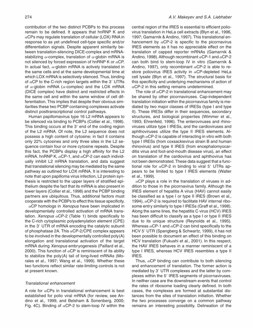
stabilization (Weiss & Liebhaber, 1994, 1995)+ High-level stability of a-globin mRNA is essential to fullexpression of globin proteins during the 2–3 days oftranscriptionally silent terminal erythroid differentiation+Stabilization of a-globin mRNA is tightly linked to for-mation of a binary complex between a single moleculeof aCP and a pyrimidine-rich motif within the a-globin39 UTR (“a-complex”; Fig+ 4A; Kiledjian et al+, 1995;Wang et al+, 1995; Chkheidze et al+, 1999)+ Interruptionof any of the three C-rich patches within this regiondisrupts the a-complex assembly in vitro and decreasesa-globin mRNA stability in vivo (Wang et al+, 1995;Weiss& Liebhaber 1995)+ aCP-1 and aCP-2 can indepen-dently bind to the human a-globin mRNA 39 UTR toform the a-complex as demonstrated in vitro+ This bind-ing is specific to aCPs as hnRNP K lacks this bindingactivity (Chkheidze et al+, 1999)+ The respective Kd val-ues of recombinant aCP-1 (51 nM) and aCP-2 (0+8 nM)
FIGURE 4. Examples of functions mediated by the PCBPs+ Six distinct examples of PCBP function are shown+ In eachcase, the PCBP isoform of interest is indicated as a filled circle+ A specific example is given for each of these indicatedfunctions and each is expanded upon in the text+ The mRNAs are indicated in diagrammatic format with tick marks indicatingthe initiation and termination codons+ The cellular mRNAs are also shown with cap (filled circles) and poly(A) tails+ Thepolioviral mRNA lacks a cap structure; the two IRES structures of specific interest, secondary structures known as Domain Iand Domain IV, are shown+Question marks indicate unidentified protein factors and/or assumed protein–protein interactions(as discussed in the text)+
272 A.V. Makeyev and S.A. Liebhaber
for a-complex assembly (Chkheidze et al+, 1999) are inagreement with the apparent Kd (0+5 nM) determinedfor assembly of the a-complex by incubating a-globin39 UTR with total cytoplasmic extracts (Russell et al+,1998)+ The more recently discovered aCP-3 and aCP-4proteins can also form an a-complex in vitro but theirrelationship to a-globin mRNA stability remains to beexplored+
mRNA stabilization via aCP binding does not appearto be limited to a-globin mRNA+ aCP-2 binds with highaffinity (Kd; 2 nM) to a C-rich region within the 39 UTRof collagen a1 (I) mRNA with a consequent increase inmRNA stability (Stefanovic et al+, 1997)+ As is the casefor a-globin mRNA, this interaction can occur in theabsence of additional factors (Lindquist et al+, 2000)+aCP-1 and aCP-2 binding to 39 UTR pyrimidine-richmotifs have been implicated in hypoxia-mediated sta-bilization of tyrosine hydroxylase (Paulding & Czyzyk-Krzeska, 1999) and erythropoietin (Czyzyk-Krzeska &Bendixen, 1999) mRNAs+ aCPs are also constituentsof a stabilizing complex on 39UTR of the b-globin mRNAthat exhibits many of the properties of the a-complex(Yu & Russell, 2001)+ In addition, poly(C)-sensitive 39UTR complexes have been demonstrated to mediateincreased stability of the heavy and light neurofilamentmRNAs (Canete-Soler & Schlaepfer, 2000) althoughthe identity of the corresponding proteins in this casehas not been determined to date+ These data suggestthat binding of aCPs to 39 UTR pyrimidine-rich determi-nants may represent a general mechanism for stabil-ization of long-lived cellular mRNAs (Holcik & Liebhaber,1997)+
A role for a-CP binding has been recently proposedfor stabilization of viral mRNAs (Fig+ 4A)+ aCP-1 andaCP-2 can interact with two independent structures ofthe polioviral 59 UTR: a cloverleaf structure at the 59terminus (stem-loop I) and a more centrally locatedstem-loop IV (Fig+ 4C; Blyn et al+, 1996; Gamarnik &Andino, 1997)+ Both structures have short C-richstretches exposed in the loop structures+ aCP-2 bind-ing to stem-loop IV is essential for efficient poliovirustranslation (see below), whereas interaction of aCPswith the 59 cloverleaf structure has recently been foundto stabilize poliovirus mRNA (Murray et al+, 2001)+ Incontrast to the binary interactions of aCP with 39 UTRmotifs of cellular proteins (a-complexes; as detailedabove), the binding of aCP to the polio viral 59 clover-leaf is dependent on cooperative interactions with apolio-encoded protein, 3CD (precursor of the viral pro-tease 3C and the viral polymerase 3D; Gamarnik &Andino, 1997; Parsley et al+, 1997); binding of 3CDdramatically increases the binding affinity of aCP-2 (fromKd values of;95 to;1 nM;Gamarnik & Andino, 2000)+This 3CD/aCP-containing complex is also required forinitiation of positive- and negative-strand synthesis(Gamarnik & Andino, 1998)+ As a result, aCP proteinnot only enhances polio viral mRNA stability but also
coordinates the switch from translation to replication ofthe polio viral genome+
A generalized picture that emerges from the abovestudies is that aCPs can stabilize distinct subsets ofmRNAs by targeting specific binding sites+ In cellular(capped) mRNAs, these sites appear to be limited tothe 39UTR+ Formation of complexes in this region wouldappear logical as this is the only site in these mRNAsat which RNP complexes can remain undisturbed dur-ing active translation+ The 59 UTR stabilizing complexin the polioviral RNA is located 59 of the ribosomalentry site (IRES) and thus would be similarly protectedfrom ribosome interference+ Therefore, in both cases itappears that the stabilizing complex assembles at aposition in which it can be maintained and shieldedfrom scanning ribosomes or ribosome subunits+
aCP-mediated translational controls:Silencing and enhancementby the same protein
Translational silencing
A detailed study of translation control by aCPs hascome from the analysis of 15-lipoxygenase (LOX) mRNAexpression (Fig+ 4B)+ LOX mRNA is synthesized in earlyerythroblasts and is then stored for several days in atranslationally silent state until the terminal (reticulo-cyte) stage of differentiation+ At that point, mRNA istranslationally activated and the newly synthesized LOXprotein degrades microsomal membranes as the retic-ulocyte matures into the red blood cell (for review, seeKuhn et al+, 1999)+ Thus the timing of translational ac-tivation is a critical step in the control of LOX expres-sion+ Translational silencing of LOX mRNA is tightlylinked to formation of a RNP complex at an evolution-arily conserved CU-rich repeated motif (DICE, differen-tiation control element) within the 39 UTR+ The complexthat forms at this site contains two PCBPs; hnRNP Kand one of the two major aCP isoforms+ Addition orforced expression of hnRNP K, aCP-1, or aCP-2 caninhibit LOX mRNA translation in vitro and in transfectedHeLa cells+ This DICE-dependent translation inhibitionis transferable to a reporter mRNA and acts on cappedas well as uncapped reporter mRNAs (Ostareck et al+,1997)+ Whether the effects of hnRNP K, aCP-1, andaCP-2 are additive, synergistic, or redundant is not clear+It is also not clear if the same DICE complex contrib-utes to the remarkable stability of the LOX mRNA andwhether the translational and putative stability controlsof the LOX mRNA are interdependent+
Mechanistic studies revealed that hnRNP K andaCP-1 proteins silence the DICE-containing mRNA byinhibiting 60S joining to the 40S complex at the initia-tion AUG (Ostareck et al+, 2001)+ The manner by whichthe 39 UTR complex mediates control of translationalinitiation at the 59 end of the mRNA and the relative
The poly(C)-binding proteins 273
contribution of the two distinct PCBPs to this processremain to be defined+ It appears that hnRNP K andaCPs may regulate translation of cellular (LOX) RNA inresponse to as yet undefined cell-type specific and/ordifferentiation signals+ Despite apparent similarity be-tween translation-silencing DICE-complex and mRNA-stabilizing a-complex, translation of a-globin mRNA isnot silenced by forced expression of hnRNP K or aCP+In actual fact, a-globin mRNA is actively translated inthe same cells and at the same developmental time atwhich LOX mRNA is selectively silenced+ Thus, bindingof aCP to the C-rich region targets within the 39 UTRsof a-globin mRNA (a-complex) and the LOX mRNA(DICE complex) have distinct and restricted effects inthe same cell and within the same window of cell dif-ferentiation+ This implies that despite their obvious sim-ilarities these two PCBP containing complexes activatedistinct posttranscriptional control pathways+
Human papillomavirus type 16 L2 mRNA appears tobe silenced via binding to PCBPs (Collier et al+, 1998)+This binding occurs at the 39 end of the coding regionof the L2 mRNA+ Of note, the L2 sequence does notpossess a high content of cytosine; in fact it containsonly 22% cytosines and only three sites in the L2 se-quence contain four or more cytosine repeats+ Despitethis fact, the PCBPs display a high affinity for the L2mRNA+ hnRNP K, aCP-1, and aCP-2 can each individ-ually inhibit L2 mRNA translation, and data suggestthat translational silencing may be mediated by the samepathway as outlined for LOX mRNA+ It is interesting tonote that upon papilloma virus infection, L2 protein syn-thesis is restricted to the upper layers of stratified epi-thelium despite the fact that its mRNA is also present inlower layers (Collier et al+, 1998) and the PCBP bindingpartners are ubiquitous+ Thus, additional factors mustcooperate with the PCBPs to effect this tissue specificity+aCP homologs in Xenopus have been implicated in
developmentally controlled activation of mRNA trans-lation+ Xenopus aCP-2 (Table 1) binds specifically tothe C-rich cytoplasmic polyadenylation element (CPE)in the 39 UTR of mRNA encoding the catalytic subunitof phosphatase 2A+ This aCP-2/CPE complex appearsto be involved in the developmentally controlled poly(A)elongation and translational activation of the targetmRNA during Xenopus embryogenesis (Paillard et al+,2000)+ This function of aCP is reminiscent of its abilityto stabilize the poly(A) tail of long-lived mRNAs (Mo-rales et al+, 1997; Wang et al+, 1999)+ Whether thesetwo functions reflect similar rate-limiting controls is notat present known+
Translational enhancement
A role for aCPs in translational enhancement is bestestablished for polio viral mRNA (for review, see An-dino et al+, 1999, and Belsham & Sonenberg, 2000;Fig+ 4C)+ Binding of aCP-2 to stem-loop IV within the
central region of the IRES is essential to efficient polio-virus translation in HeLa cell extracts (Blyn et al+, 1996,1997; Gamarnik & Andino, 1997)+ This translational en-hancement by aCP-2 is specific to the picornavirusIRES elements as it has no appreciable effect on thetranslation of capped reporter mRNAs (Gamarnik &Andino, 1998)+Although recombinant aCP-1 and aCP-2can both bind to stem-loop IV in vitro (Gamarnik &Andino, 1997), only recombinant aCP-2 is able to re-store poliovirus IRES activity in aCP-depleted HeLacell lysate (Blyn et al+, 1997)+ The structural basis forthis specificity and underlying mechanisms of action ofaCP-2 in this setting remains undetermined+
The role of aCP-2 in translational enhancement maybe shared by other picornaviruses+ Cap-independenttranslation initiation within the picornavirus family is me-diated by two major classes of IRESs (type I and typeII)+ These IRESs differ in their sequences, secondarystructures, and biological properties (Wimmer et al+,1993; Ehrenfeld, 1996)+ The enteroviruses and rhino-viruses utilize type I IRESs, and the cardioviruses andaphthoviruses utilize the type II IRES elements+ Al-though aCP-2 is capable of interacting in vitro with bothtype I IRESs (from coxsackievirus strain B and humanrhinovirus) and type II IRES (from encephalomyocar-ditis virus and foot-and-mouth disease virus), an effecton translation of the cardiovirus and aphthovirus hasnot been demonstrated+ These data suggest that a func-tional role for aCP-2 in binding to viral 59 UTRs ap-pears to be limited to type I IRES elements (Walteret al+, 1999)+aCP plays a role in the translation of viruses in ad-
dition to those in the picornavirus family+ Although theIRES element of hepatitis A virus (HAV) cannot easilybe classified as a type I or type II IRES (Brown et al+,1994), aCP-2 is required to facilitate HAV internal ribo-some entry similarly to type I IRESs (Graff et al+, 1998)+Along the same lines, the hepatitis C virus (HCV) IREShas been difficult to classify as a type I or type II IRESdue to its unique structure (Reynolds et al+, 1995)+Whereas aCP-1 and aCP-2 can bind specifically to theHCV 59 UTR (Spangberg & Schwartz, 1999), it has notbeen possible to document an effect of this binding onHCV translation (Fukushi et al+, 2001)+ In this respect,the HAV IRES behaves in a manner reminiscent of atype I IRES, whereas HCV IRES resembles a type IIIRES+
Thus, aCP binding can contribute to both silencingand enhancement of translation+ The former action ismediated by 39 UTR complexes and the latter by com-plexes within the 59 IRES segments of picornaviruses+In neither case are the downstream events that controlthe rates of ribosome loading clearly defined+ In bothcases, the complexes are formed at substantial dis-tances from the sites of translation initiation+ Whetherthe two processes converge on a common pathwayremains an interesting possibility+ Delineation of the
274 A.V. Makeyev and S.A. Liebhaber
relevant pathways should be highly informative asthey may define novel aspects of translational bio-chemistry and long-range interactions mediated bymRNP complexes+
TRANSCRIPTIONAL CONTROLS:DIVERSE ROLES AND MECHANISMS
In addition to their roles in mRNA stability and transla-tional controls, the PCBPs appear to have diverse func-tions in the more proximal events in gene expression+hnRNP K has been implicated in multiple aspects oftranscriptional regulation+ hnRNP K has a specific bind-ing site on the SV40 early promoter (Gaillard et al+,1994) and in the pyrimidine-rich strand of the CT ele-ment in the promoter of human c-myc gene (Fig+ 4D;Tomonaga & Levens, 1996)+ In both of these cases,this interaction activates transcription in in vitro sys-tems apparently by an hnRNP K-dependent assemblyof the TFIID complex at target promoters (Michelottiet al+, 1996)+ A competition/displacement model hasbeen suggested to explain the observed differences inthe effects of hnRNP K on Sp1- and Sp3-mediatedtranscriptional activation of the neuronal nicotinic ace-tylcholine receptor promoter (Du et al+, 1998)+ Accord-ing to this model, hnRNP K selectively blocks access ofSp1, but not Sp3, to an E2 element+ In the case of thethymidine kinase promoter, hnRNP K itself cannot phys-ically interact with promoter but may repress transcrip-tion by inhibiting the binding of other trans-factors tothe cell cycle regulatory determinant of this promoter(Fig+ 4E; Lau et al+, 2000)+ In addition, hnRNP K hasbeen reported to directly interact in vitro and in vivowith zinc-finger transcriptional repressor Zik-1 (De-nisenko et al+, 1996) and with other structurally relatedtranscriptional repressors such as Kid-1 and MZF-1(Bomsztyk et al+, 1997)+ Thus, the abilities of hnRNP Kto interact with both nucleic acid and protein targets isessential for its transcriptional functions+
hnRNP K has recently been identified among a limitednumber of proteins binding specifically to the homo-pyrimidine strand of d(GA{TC)n sequences (Garcia-Bassets et al+, 1999)+ Alternating (GA{TC)n DNAsequences are abundant in eukaryotic genomes, canform non-B-DNA conformers in vitro, and are often foundin gene regulatory regions+ Regions of single-strandedDNA in non-B-DNA conformers have a tendency to re-store their regular double-stranded sequence+ By sta-bilizing a single-stranded bubble in such a region,hnRNP K may increase DNA flexibility and allow otherDNA-binding proteins to align more easily with the ba-sal transcriptional machinery (Garcia-Bassets et al+,1999)+ In these capacities, hnRNP K may serve as an“architectural” transcription factor+ Such a role has beenspecifically suggested for hnRNP K in regulation of thec-myc promoter (Michelotti et al+, 1996)+A similar struc-tural function may be attributed by hnRNP K binding at
the telomere; in vitro studies reveal high-affinity bindingof this protein to a cytosine-rich oligonucleotide thatmimics a special intramolecular folded structure called“i-DNA” in the telomere (Lacroix et al+, 2000)+ Whetherthis telomere interaction occurs in vivo is not clear+
ROLES IN APOPTOTIC ANDDEVELOPMENTAL PATHWAYS
Progress toward an understanding of hnRNP K func-tions in development have been significantly aided byanalysis of the Drosophila ortholog, Hrb57A, the prod-uct of bancal gene (Table 1)+ Hrb57A possesses simi-larity in its structure and in nucleotide binding specificityto mammalian hnRNP K (Hovemann et al+, 2000)+ Nulland weak alleles of bancal result in Drosophila adultswith shortened appendages as a consequence of areduction in the number of cell divisions in the imaginaldiscs; remarkably this developmental phenotype canbe reversed by expression of the human hnRNP Ktransgene (Charroux et al+, 1999)+ Moreover, over-expression of either Hrb57A or human hnRNP K in-duced cell death in imaginal discs and this effect canbe overcome by coexpression of the anti-apoptotic P35protein (Charroux et al+, 1999)+ Of note in this regard,forced expression of human hnRNP K decreases thelevel of the endogenous Hrb57A+ These data indicatethat hnRNP K is functionally involved in the control ofcell division and apoptotic pathways and that its criticallevel may be controlled by negative feedback mecha-nisms (Charroux et al+, 1999)+
In marked contrast to the studies of the hnRNP Khomologs, there are no clear phenotypes associatedwith deletion mutants of the aCP homolog (mub) inDrosophila (Table 1; Fig+ 2B; Grams & Korge, 1998)+
Analysis of human PCBPs supports their involve-ment in cell growth control+ hnRNP K is a defined targetof epidermal cell growth factors produced by humanbreast cancer cells and in this context hnRNP K canexert positive controls over the rate of cell proliferation(Mandal et al+, 2001)+ A protein encoded by a splicevariant of the aCP-4 transcript, MCG10, has recentlybeen reported to participate in an apoptotic pathway inhuman cells (Fig+ 4F)+ Elevated levels of MCG10 wereinduced in a p53-dependent manner in cells followingDNA damage and forced expression of MCG10 in-duced apoptosis and cell cycle arrest at G2-M (Zhu &Chen, 2000)+ These data suggest that an aCP-4 iso-form, MCG10, is a potential mediator of p53 tumorsuppression+
PCBP BIOLOGY: WHERE WE AREAND WHERE WE MIGHT GO
The five PCBP genes encode a well-defined subset ofsingle-strand nucleic acid-binding proteins+ These pro-
The poly(C)-binding proteins 275
teins are structurally related by their triplicated KH do-mains, by their common evolutionary origins, and by ashared preference for C-rich binding sites+ Current dataclearly demonstrates that these proteins play importantand diverse roles in gene expression at both transcrip-tional and posttranscriptional levels+ A number of gen-eral models have been put forward to unify the actionsof one or more of these proteins+ For example, it hasbeen suggested that the seemingly disparate functionsof hnRNP K at multiple levels of gene can be unified byconsidering it as a RNA- and DNA-regulated dockingplatform linking sequence-specific nucleic acid bindingwith specific protein–protein interactions and servingas a fulcrum for multilateral molecular cross-talk (Bom-sztyk et al+, 1997)+ Although of significant utility, thismodel is difficult to fully reconcile with the observationthat mammalian and Drosophila hnRNP K proteins dem-onstrate functional equivalence despite their apparentabsence of structural homology in the auxiliary do-mains responsible for protein–protein interactions+
Another model suggests that the family of PCBPsmay serve the general role of chaperones that stabilizecomplex secondary and tertiary RNA structures orsingle-stranded DNA structures within promoters or attelomeres to facilitate single-strand-dependent inter-actions with a variety of trans-factors+ This model hasbeen proposed, for example, in the regulation of thec-myc promoter by hnRNP K (Michelotti et al+, 1996)and for facilitation of internal ribosome entry on thepicornavirus IRES (Belsham & Sonenberg, 2000)+How-ever, recent studies from our laboratory indicate thatsome members of this family, in particular aCP-1 andaCP-2, can function in their mRNA stabilization role inthe total absence of their RNA binding function (J+ Kong& S+A+ Liebhaber, in prep+)+ Thus, the chaperone func-tions of the PCBPs, if important in mRNA stabilization,would only represent one facet of their function+
From all these considerations, it becomes apparentthat the functional diversity and spectrum of actions ofthe PCBPs are quite wide and may not lend them-selves to unifying models at present+ The flip side of theapparent shortfall is that there remains a rich mine ofstructural and mechanistic questions yet to be ex-plored+ These studies should reveal, in the not too dis-tant future, the complexity of interactions and pathwaysutilized by this family in its extensive and multiple con-tributions to the control of cell function+
Received August 30, 2001; returned for revisionOctober 16, 2001; revised manuscript receivedDecember 21, 2001
ACKNOWLEDGMENTS
The authors would like to thank Dr+ Nancy E+ Cooke andDr+ Shelly Waggoner for critical reviews of this manuscript+
REFERENCES
Adinolfi S, Bagni C, Castiglione Morelli MA, Fraternali F, Musco G,Pastore A+ 1999+ Novel RNA-binding motif: The KH module+ Bio-polymers 51:153–164+
Andino R, Boddeker N, Silvera D, Gamarnik AV+ 1999+ Intracellulardeterminants of picornavirus replication+ Trends Microbiol 7:76–82+
Baber JL, Levens D, Libutti D,Tjandra N+ 2000+Chemical shift mappedDNA-binding sites and 15N relaxation analysis of the C-terminalKH domain of heterogeneous nuclear ribonucleoprotein K+ Bio-chemistry 39:6022–6032+
Baber JL, Libutti D, Levens D, Tjandra N+ 1999+ High precision so-lution structure of the C-terminal KH domain of heterogeneousnuclear ribonucleoprotein K, a c-myc transcription factor+ J MolBiol 289:949–962+
Belsham GJ, Sonenberg N+ 2000+ Picornavirus RNA translation:Rolesfor cellular proteins+ Trends Microbiol 8:330–335+
Blyn LB, Swiderek KM, Richards O, Stahl DC, Semler BL, EhrenfeldE+ 1996+ Poly(rC) binding protein 2 binds to stem-loop IV of thepoliovirus RNA 59 noncoding region: Identification by automatedliquid chromatography-tandem mass spectrometry+ Proc Natl AcadSci USA 93:11115–11120+
Blyn LB, Towner JS, Semler BL, Ehrenfeld E+ 1997+ Requirement ofpoly(rC) binding protein 2 for translation of poliovirus RNA+ J Virol71:6243–6246+
Bomsztyk K, Van Seuningen I, Suzuki H, Denisenko O, Ostrowski J+1997+ Diverse molecular interactions of the hnRNP K protein+FEBS Lett 403:113–115+
Brown EA, Zajac AJ, Lemon SM+ 1994+ In vitro characterization of aninternal ribosomal entry site (IRES) present within the 59 non-translated region of hepatitis A virus RNA: Comparison with theIRES of encephalomyocarditis virus+ J Virol 68:1066–1074+
Buchenau P, Saumweber H, Arndt-Jovin DJ+ 1997+ The dynamicnuclear redistribution of an hnRNP K-homologous protein duringDrosophila embryo development and heat shock+ Flexibility oftranscription sites in vivo+ J Cell Biol 137:291–303+
Buckanovich RJ, Darnell RB+ 1997+ The neuronal RNA binding pro-tein Nova-1 recognizes specific RNA targets in vitro and in vivo+Mol Cell Biol 17:3194–3201+
Bustelo XR, Suen KL, Michael WM, Dreyfuss G, Barbacid M+ 1995+Association of the vav proto-oncogene product with poly(rC)-specific RNA-binding proteins+ Mol Cell Biol 15:1324–1332+
Canete-Soler R,Schlaepfer WW+ 2000+Similar poly(C)-sensitive RNA-binding complexes regulate the stability of the heavy and lightneurofilament mRNAs+ Brain Res 867:265–279+
Charroux B, Angelats C, Fasano L, Kerridge S, Vola C+ 1999+ Thelevels of the bancal product, a Drosophila homologue of verte-brate hnRNP K protein, affect cell proliferation and apoptosis inimaginal disc cells+ Mol Cell Biol 19:7846–7856+
Chkheidze AN, Lyakhov DL, Makeyev AV, Morales J, Kong J, Lieb-haber SA+ 1999+ Assembly of the alpha-globin mRNA stabilitycomplex reflects binary interaction between the pyrimidine-rich 39untranslated region determinant and poly(C) binding protein al-phaCP+ Mol Cell Biol 19:4572–4581+
Collier B,Goobar-Larsson L, Sokolowski M, Schwartz S+ 1998+ Trans-lational inhibition in vitro of human papillomavirus type 16 L2mRNA mediated through interaction with heterogenous ribonu-cleoprotein K and poly(rC)-binding proteins 1 and 2+ J Biol Chem273:22648–22656+
Currie JR, Brown WT+ 1999+ KH domain-containing proteins of yeast:Absence of a fragile X gene homologue+ Am J Med Genet 84:272–276+
Czyzyk-Krzeska MF, Bendixen AC+ 1999+ Identification of the poly(C)binding protein in the complex associated with the 39 untranslatedregion of erythropoietin messenger RNA+ Blood 93:2111–2120+
Dejgaard K, Leffers H+ 1996+ Characterisation of the nucleic-acid-binding activity of KH domains+ Different properties of differentdomains+ Eur J Biochem 241:425–431+
Dejgaard K, Leffers H, Rasmussen HH,Madsen P, Kruse TA, GesserB, Nielsen H, Celis JE+ 1994+ Identification, molecular cloning,expression and chromosome mapping of a family of transforma-tion upregulated hnRNP-K proteins derived by alternative splic-ing+ J Mol Biol 236:33–48+
Denisenko ON, Bomsztyk K+ 1997+ The product of the murine homo-log of the Drosophila extra sex combs gene displays transcrip-tional repressor activity+ Mol Cell Biol 17:4707–4717+
276 A.V. Makeyev and S.A. Liebhaber
Denisenko ON,O’Neill B,Ostrowski J, Van Seuningen I, Bomsztyk K+1996+ Zik1, a transcriptional repressor that interacts with the het-erogeneous nuclear ribonucleoprotein particle K protein+ J BiolChem 271:27701–27706+
Du Q, Melnikova IN, Gardner PD+ 1998+ Differential effects of het-erogeneous nuclear ribonucleoprotein K on Sp1- and Sp3-mediated transcriptional activation of a neuronal nicotinicacetylcholine receptor promoter+ J Biol Chem 273:19877–19883+
Ehrenfeld E+ 1996+ Initiation of translation by picornavirus RNAs+ In:Hershey JWB, Mathews MB, Sonenberg N, eds+ Translationalcontrol+ Cold Spring Harbor, New York: Cold Spring Harbor Lab-oratory Press+ pp 549–573+
Eichler EE, Richards S, Gibbs RA, Nelson DL+ 1993+ Fine structureof the human FMR1 gene+ Hum Mol Genet 2:1147–1153+
Fukushi S, Okada M, Kageyama T, Hoshino FB, Nagai K, KatayamaK+ 2001+ Interaction of poly(rC)-binding protein 2 with the 59-terminal stem loop of the hepatitis C-virus genome+ Virus Res73:67–79+
Funke B, Zuleger B, Benavente R, Schuster T, Goller M, Stevenin J,Horak I+ 1996+ The mouse poly(C)-binding protein exists in mul-tiple isoforms and interacts with several RNA-binding proteins+Nucleic Acids Res 24:3821–3828+
Gaillard C, Cabannes E, Strauss F+ 1994+ Identity of the RNA-bindingprotein K of hnRNP particles with protein H16, a sequence-specific single strand DNA-binding protein+ Nucleic Acids Res22:4183–4186+
Gamarnik AV, Andino R+ 1997+ Two functional complexes formed byKH domain containing proteins with the 59 noncoding region ofpoliovirus RNA+ RNA 3:882–892+
Gamarnik AV, Andino R+ 1998+ Switch from translation to RNAreplication in a positive-stranded RNA virus+ Genes & Dev 12:2293–2304+
Gamarnik AV, Andino R+ 2000+ Interactions of viral protein 3CD andpoly(rC) binding protein with the 59 untranslated region of thepoliovirus genome+ J Virol 74:2219–2226+
Garcia-Bassets I, Ortiz-Lombardia M, Pagans S, Romero A, CanalsF, Avilés FX, Azorin F+ 1999+ The identification of nuclear proteinsthat bind the homopyrimidine strand of d(GA{TC)n DNA se-quences, but not the homopurine strand+ Nucleic Acids Res27:3267–3275+
Gibson TJ, Thompson JD, Heringa J+ 1993+ The KH domain occursin a diverse set of RNA-binding proteins that include the antiter-minator NusA and is probably involved in binding to nucleic acid+FEBS Lett 324:361–366+
Goller M, Funke B, Gehe-Becker C, Kroger B, Lottspeich F, Horak I+1994+ Murine protein which binds preferentially to oligo-C-richsingle-stranded nucleic acids+ Nucleic Acids Res 22:1885–1889+
Graff J, Cha J, Blyn LB, Ehrenfeld E+ 1998+ Interaction of poly(rC)binding protein 2 with the 59 noncoding region of hepatitis A virusRNA and its effects on translation+ J Virol 72:9668–9675+
Grams R, Korge G+ 1998+ The mub gene encodes a protein contain-ing three KH domains and is expressed in the mushroom bodiesof Drosophila melanogaster+ Gene 215:191–201+
Graumann P, Marahiel MA+ 1996+ A case of convergent evolution ofnucleic acid binding modules+ Bioessays 18:309–315+
Grishin NV+ 2001+ KH domain: One motif, two folds+ Nucleic AcidsRes 29:638–643+
Habelhah H, Shah K, Huang L, Ostareck-Lederer A, Burlingame AL,Shokat KM, Hentze MW, Ronai Z+ 2001+ ERK phosphorylationdrives cytoplasmic accumulation of hnRNP-K and inhibition ofmRNA translation+ Nat Cell Biol 3:325–330+
Holcik M, Liebhaber SA+ 1997+ Four highly stable eukaryotic mRNAsassemble 39 untranslated region RNA–protein complexes shar-ing cis and trans components+ Proc Natl Acad Sci USA 94:2410–2414+
Hovemann BT, Reim I, Werner S, Katz S, Saumweber H+ 2000+ Theprotein Hrb57A of Drosophila melanogaster closely related tohnRNP K from vertebrates is present at sites active in transcrip-tion and coprecipitates with four RNA-binding proteins+ Gene245:127–137+
Kiledjian M, Wang X, Liebhaber SA+ 1995+ Identification of two KHdomain proteins in the alpha-globin mRNP stability complex+EMBOJ 14:4357–4364+
Kim JH, Hahm B, Kim YK, Choi M, Jang SK+ 2000+ Protein–proteininteraction among hnRNPs shuttling between nucleus and cyto-plasm+ J Mol Biol 298:395–405+
Kondo T, Furuta T, Mitsunaga K, Ebersole TA, Shichiri M,Wu J, ArtztK, Yamamura K, Abe K+ 1999+ Genomic organization and expres-sion analysis of the mouse qkI locus+Mamm Genome 10:662–669+
Kuhn H, Heydeck D, Brinckman R, Trebus F+ (1999) Regulation ofcellular 15-lipoxygenase activity on pretranslational, translational,and posttranslational levels+ Lipids 34 (Suppl+):S273–S279+
Lacroix L, Lienard H, Labourier E, Djavaheri-Mergny M, Lacoste J,Leffers H, Tazi J, Helene C,Mergny JL+ 2000+ Identification of twohuman nuclear proteins that recognize the cytosine-rich strand ofhuman telomeres in vitro+ Nucleic Acids Res 28:1564–1575+
Lau JS, Baumeister P, Kim E, Roy B, Hsieh TY, Lai M, Lee AS+ 2000+Heterogeneous nuclear ribonucleoproteins as regulators of geneexpression through interactions with the human thymidine kinasepromoter+ J Cell Biochem 79:395–406+
Leffers H, Dejgaard K, Celis JE+ 1995+ Characterisation of two majorcellular poly(rC)-binding human proteins, each containing threeK-homologous (KH) domains+ Eur J Biochem 230:447–453+
Lewis HA, Chen H, Edo C, Buckanovich RJ, Yang YY, Musunuru K,Zhong R,Darnell RB,Burley SK+ 1999+Crystal structures of Nova-1and Nova-2 K-homology RNA-binding domains+ Structure FoldDes 7:191–203+
Lindquist JN, Kauschke SG, Stefanovic B, Burchardt ER, BrennerDA+ 2000+Characterization of the interaction between alphaCP(2)and the 39-untranslated region of collagen alpha1(I) mRNA+ Nu-cleic Acids Res 28:4306–4316+
Makeyev AV, Chkheidze AN, Liebhaber SA+ 1999+ A set of highlyconserved RNA-binding proteins, aCP-1 and aCP-2, implicatedin mRNA stabilization, are coexpressed from an intronless geneand its intron-containing paralog+ J Biol Chem 274:24849–24857+
Makeyev AV, Liebhaber SA+ 2000+ Identification of two novel mam-malian genes establishes a subfamily of KH-domain RNA-bindingproteins+ Genomics 67:301–316+
Mandal M, Vadlamudi R, Nguyen D, Wang RA, Costa L, Bagheri-Yarmand R, Mendelsohn J, Kumar R+ 2001+ Growth factors reg-ulate heterogeneous nuclear ribonucleoprotein k expression andfunction+ J Biol Chem 276:9699–9704+
Matunis MJ, Michael WM, Dreyfuss G+ 1992+ Characterization andprimary structure of the poly(C)-binding heterogeneous nuclearribonucleoprotein complex K protein+ Mol Cell Biol 12:164–171+
Miau LH, Chang CJ, Shen BJ, Tsai WH, Lee SC+ 1998+ Identificationof heterogeneous nuclear ribonucleoprotein K (hnRNP K) as arepressor of C/EBPbeta-mediated gene activation+ J Biol Chem273:10784–10791+
Michael WM, Eder PS, Dreyfuss G+ 1997+ The K nuclear shuttlingdomain: A novel signal for nuclear import and nuclear export inthe hnRNP K protein+ EMBO J 16:3587–3598+
Michelotti EF, Michelotti GA, Aronsohn AI, Levens D+ 1996+ Hetero-geneous nuclear ribonucleoprotein K is a transcription factor+ MolCell Biol 16:2350–2360+
Mintz PJ, Spector DL+ 2000+ Compartmentalization of RNA process-ing factors within nuclear speckles+ J Struct Biol 129:241–251+
Morales J, Russell JE, Liebhaber SA+ 1997+ Destabilization of humanalpha-globin mRNA by translation anti-termination is controlledduring erythroid differentiation and is paralleled by phased short-ening of the poly(A) tail+ J Biol Chem 272:6607–6613+
Murray KE, Roberts AW, Barton DJ+ 2001+ Poly(rC) binding proteinsmediate poliovirus mRNA stability+ RNA 7:1126–1141+
Musco G, Kharrat A, Stier G, Fraternali F, Gibson TJ, Nilges M,Pastore A+ 1997+ The solution structure of the first KH domain ofFMR1, the protein responsible for the fragile X syndrome+ NatStruct Biol 4:712–716+
Musco G, Stier G, Joseph C, Castiglione Morelli MA, Nilges M, Gib-son TJ, Pastore A+ 1996+ Three-dimensional structure and stabil-ity of the KH domain:Molecular insights into the fragile X syndrome+Cell 85:237–245+
Ostareck DH, Ostareck-Lederer A, Shatsky IN, Hentze MW+ 2001+Lipoxygenase mRNA silencing in erythroid differentiation+ The39UTR regulatory complex controls 60S ribosomal subunit join-ing+ Cell 104:281–290+
Ostareck DH, Ostareck-Lederer A, Wilm M, Thiele BJ, Mann M,Hentze MW+ 1997+ mRNA silencing in erythroid differentiation:hnRNP K and hnRNP E1 regulate 15-lipoxygenase translationfrom the 39 end+ Cell 89:597–606+
Ostareck-Lederer A, Ostareck DH, Hentze MW+ 1998+ Cytoplasmicregulatory functions of the KH-domain proteins hnRNPs K andE1/E2+ Trends Biochem Sci 23:409–411+
The poly(C)-binding proteins 277
Ostrowski J, Kawata Y, Schullery DS,Denisenko ON,Higaki Y,AbrassCK, Bomsztyk K+ 2001+ Insulin alters heterogeneous nuclear ri-bonucleoprotein K protein binding to DNA and RNA+ Proc NatlAcad Sci USA 98:9044–9049+
Ostrowski J, Schullery DS, Denisenko ON, Higaki Y,Watts J, Aeber-sold R, Stempka L, Gschwendt M, Bomsztyk K+ 2000+ Role oftyrosine phosphorylation in the regulation of the interaction ofheterogenous nuclear ribonucleoprotein K protein with its proteinand RNA partners+ J Biol Chem 275:3619–3628+
Ostrowski J, Sims JE, Sibley CH, Valentine MA, Dower SK, MeierKE, Bomsztyk K+ 1991+A serine/threonine kinase activity is closelyassociated with a 65-kDa phosphoprotein specifically recognizedby the kB enhancer element+ J Biol Chem 266:12722–12733+
Paillard L,Maniey D, Lachaume P, Legagneux V, Osborne HB+ 2000+Identification of a C-rich element as a novel cytoplasmic polyad-enylation element in Xenopus embryos+ Mech Dev 93:117–125+
Parsley TB, Towner JS, Blyn LB, Ehrenfeld E, Semler BL+ 1997+ Poly(rC) binding protein 2 forms a ternary complex with the 59-terminal sequences of poliovirus RNA and the viral 3CD protein-ase+ RNA 3:1124–1134+
Paulding WR, Czyzyk-Krzeska MF+ 1999+ Regulation of tyrosine hy-droxylase mRNA stability by protein-binding, pyrimidine-rich se-quence in the 39-untranslated region+ J Biol Chem 274:2532–2538+
Prasanth KV, Rajendra TK, Lal AK, Lakhotia SC+ 2000+ Omegaspeckles—a novel class of nuclear speckles containing hnRNPsassociated with noncoding hsr-omega RNA in Drosophila+ J CellSci 113:3485–3497+
Reynolds JE, Kaminski A, Kettinen HJ, Grace K, Clarke BE, CarrollAR, Rowlands DJ, Jackson RJ+ 1995+ Unique features of internalinitiation of hepatitis C virus RNA translation+ EMBO J 14:6010–6020+
Russell JE, Morales J, Makeyev AV, Liebhaber SA+ 1998+ Sequencedivergence in the 39 untranslated regions of human zeta- andalpha-globin mRNAs mediates a difference in their stabilities andcontributes to efficient alpha-to-zeta gene development switch-ing+ Mol Cell Biol 18:2173–2183+
Schullery DS, Ostrowski J, Denisenko ON, Stempka L, Shnyreva M,Suzuki H, Gschwendt M, Bomsztyk K+ 1999+ Regulated inter-action of protein kinase Cd with the heterogeneous nuclear ribo-nucleoprotein K protein+ J Biol Chem 274:15101–15109+
Shnyreva M, Schullery DS, Suzuki H, Higaki Y, Bomsztyk K+ 2000+Interaction of two multifunctional proteins+ Heterogeneous nu-clear ribonucleoprotein K and Y-box-binding protein+ J Biol Chem275:15498–15503+
Silvera D, Gamarnik AV,Andino R+ 1999+ The N-terminal K homologydomain of the poly(rC)-binding protein is a major determinant forbinding to the poliovirus 59-untranslated region and acts as aninhibitor of viral translation+ J Biol Chem 274:38163–38170+
Siomi H, Choi M, Siomi MC, Nussbaum RL, Dreyfuss G+ 1994+ Es-sential role for KH domains in RNA binding: Impaired RNA bind-ing by a mutation in the KH domain of FMR1 that causes fragileX syndrome+ Cell 77:33–39+
Siomi H, Matunis MJ, Michael WM, Dreyfuss G+ 1993+ The pre-mRNA binding K protein contains a novel evolutionarily con-served motif+ Nucleic Acids Res 21:1193–1198+
Spangberg K, Schwartz S+ 1999+ Poly(C)-binding protein interactswith the hepatitis C virus 59 untranslated region+ J Gen Virol80:1371–1376+
Stefanovic B, Hellerbrand C, Holcik M, Briendl M, Liebhaber SA,Brenner DA+ 1997+ Posttranscriptional regulation of collagenalpha1(I) mRNA in hepatic stellate cells+ Mol Cell Biol 17:5201–5209+
Strimmer K, von Haeseler A+ 1996+ Quartet puzzling: A quartetmaximum-likelihood method for reconstructing tree topologies+Mol Biol Evol 13:964–969+
Swanson MS, Dreyfuss G+ 1988+ Classification and purification ofproteins of heterogeneous nuclear ribonucleoprotein particles byRNA-binding specificities+ Mol Cell Biol 8:2237–2241+
Taylor SJ, Shalloway D+ 1994+ An RNA-binding protein associatedwith Src through its SH2 and SH3 domains in mitosis+ Nature368:867–871+
The C. elegans Sequencing Consortium+ 1998+ Genome sequenceof the nematode C. elegans: A platform for investigating biology+Science 282:2012–2018+
Thisted T, Lyakhov DL, Liebhaber SA+ 2001+ Optimized RNA targetsof two closely-related triple KH-domain proteins, hnRNP K andalpha-CP-2KL suggest distinct modes of RNA recognition+ J BiolChem 276:17484–17496+
Tommerup N, Leffers H+ 1996+ Assignment of human KH-box-containing genes by in situ hybridization: HNRNPK maps to9q21+32-q21+33, PCBP1 to 2p12-p13, and PCBP2 to 12q13+12-q13+13, distal to FRA12A+ Genomics 32:297–298+
Tomonaga T, Levens D+ 1996+ Activating transcription from singlestranded DNA+ Proc Natl Acad Sci USA 93:5830–5835+
Van Seuningen I, Ostrowski J, Bustelo XR, Sleath PR, Bomsztyk K+1995+ The K protein domain that recruits the interleukin1-responsive K protein kinase lies adjacent to a cluster of c-Srcand Vav SH3-binding sites+ Implications that K protein acts as adocking platform+ J Biol Chem 270:26976–26985+
Walter BL, Nguyen JH, Ehrenfeld E, Semler BL+ 1999+ Differentialutilization of poly(rC) binding protein 2 in translation directed bypicornavirus IRES elements+ RNA 5:1570–1585+
Wang X, Kiledjian M, Weiss IM, Liebhaber SA+ 1995+ Detection andcharacterization of a 39 untranslated region ribonucleoprotein com-plex associated with human alpha-globin mRNA stability+Mol CellBiol 15:1769–1777+
Wang Z, Day N, Trifillis P, Kiledjian M+ 1999+ An mRNA stability com-plex functions with poly(A)-binding protein to stabilize mRNA invitro+ Mol Cell Biol 19:4552–4560+
Weiss IM, Liebhaber SA+ 1994+ Erythroid cell-specific determinantsof alpha-globin mRNA stability+ Mol Cell Biol 14:8123–8132+
Weiss IM, Liebhaber SA+ 1995+ Erythroid cell-specific mRNA stabilityelements in the alpha 2-globin 39 nontranslated region+ Mol CellBiol 15:2457–2465+
Weng Z, Thomas SM, Rickles RJ, Taylor JA, Brauer AW, Seidel-Dugan C, Michael WM, Dreyfuss G, Brugge JS+ 1994+ Identifica-tion of Src, Fyn, and Lyn SH3-binding proteins: Implications for afunction of SH3 domains+ Mol Cell Biol 14:4509–4521+
Wimberly BT, Brodersen DE, Clemons WM Jr, Morgan-Warren RJ,Carter AP, Vonrhein C, Hartsch T, Ramakrishnan V+ 2000+ Struc-ture of the 30S ribosomal subunit+ Nature 407:327–339+
Wimmer E, Hellen CU, Cao X+ 1993+ Genetics of poliovirus+ AnnuRev Genet 27:353–436+
Yang YY, Yin GL, Darnell RB+ 1998+ The neuronal RNA-binding pro-tein Nova-2 is implicated as the autoantigen targeted in POMApatients with dementia+ Proc Natl Acad Sci USA 95:13254–13259+
Yu J, Russell JE+ 2001+ Structural and functional analysis of an mrnpcomplex that mediates the high stability of human beta-globinmRNA+ Mol Cell Biol 21:5879–5888+
Zhu J, Chen X+ 2000+ MCG10, a novel p53 target gene that encodesa KH domain RNA-binding protein, is capable of inducing apop-tosis and cell cycle arrest in G(2)-M+ Mol Cell Biol 20:5602–5618+
278 A.V. Makeyev and S.A. Liebhaber