Embed Size (px)
Citation preview

1
The role of short-chain fatty acids in the interplay between diet, gut microbiota and host
energy metabolism
Gijs den Besten1,3, Karen van Eunen1,3, Albert K. Groen1,2,3, Koen Venema3,4, Dirk-Jan
Reijngoud1,2,3 and Barbara M. Bakker1,3,*
1Center for Liver, Digestive and Metabolic Diseases, Department of Pediatrics, 2Department of
Laboratory Medicine, University of Groningen, University Medical Center Groningen, Groningen,
The Netherlands
3Netherlands Consortium for Systems Biology, Amsterdam, The Netherlands
4TNO P.O. Box 360, 3700 AJ Zeist, The Netherlands
*Corresponding author: Barbara M. Bakker, Hanzeplein 1, 9713 GZ Groningen, The Netherlands,
Phone: +31(0)50 3611542; Fax: +31(0)50 3611746; E-mail: [email protected]
Running footline: Short-chain fatty acids and host energy metabolism
Abbreviations: SCFA, short-chain fatty acid; PEP, phosphoenolpyruvate; GAPDH,
glyceraldehyde-3-phosphate dehydrogenase; MCT, monocarboxylate transporters; SMCT, sodium-
dependent monocarboxylate transporter; GPR, G protein-coupled receptor; Ffar, free fatty acid
receptor; PYY, peptide YY; GLP-1, Glucagon-like peptide-1; AMPK, AMP-activated protein
kinase; PGC-1α, peroxisome proliferator-activated receptor gamma coactivator 1-alpha; PKA,
protein kinase A; HSL, hormone-sensitive lipase
by guest, on May 21, 2018
ww
w.jlr.org
Dow
nloaded from

2
Abstract
Short-chain fatty acids (SCFAs), the end products of fermentation of dietary fibers by the
anaerobic intestinal microbiota, have been shown to exert multiple beneficial effects on mammalian
energy metabolism. The mechanisms underlying these effects are subject of intensive research and
encompass the complex interplay between diet, gut microbiota and host energy metabolism. This
review summarizes the role of SCFAs in host energy metabolism, starting from the production by
the gut microbiota, the uptake by the host and ending with the effects on host metabolism. There are
interesting leads on the underlying molecular mechanisms, but also many apparently contradictory
results. A coherent understanding of the multilevel network in which SCFAs exert their effects, is
hampered by the lack of quantitative data on actual fluxes of SCFAs and metabolic processes
regulated by SCFAs. In this review we address questions that, when answered, will bring us a great
step forward to elucidate the role of SCFAs in mammalian energy metabolism.
Supplementary key words: fatty acids, diet, nutritional fiber, bacterial SCFA metabolism, SCFA
fluxes and concentrations
by guest, on May 21, 2018
ww
w.jlr.org
Dow
nloaded from

3
Introduction
The decrease in physical exercise and increase in energy intake, especially seen in the
Western world, disrupts the energy balance in humans and can lead to a complex of symptoms
collectively denoted as the metabolic syndrome. The key characteristics of the metabolic syndrome
are obesity, loss of glycemic control, dyslipidemia and hypertension (1). Due to the complex,
multifactorial etiology of the metabolic syndrome the exact mechanisms underlying the different
comorbidities are not yet completely known. Recently, dietary fibers have raised much interest, as
they exert beneficial effects on body weight, food intake, glucose homeostasis, and insulin
sensitivity (2-4). Epidemiological studies show an association between a higher fiber intake and a
reduced risk of irritable bowel syndrome, inflammatory bowel disease, cardiovascular disease,
diabetes, and colon cancer (1).
Humans lack the enzymes to degrade the bulk of dietary fibers. Therefore these non
digestable carbohydrates pass the upper gastro-intestinal tract unaffected and are fermented in the
cecum and the large intestine by the anaerobic cecal and colonic microbiota. Fermentation results in
multiple groups of metabolites (elegantly reviewed by Nicholson et al. (5)) of which short-chain
fatty acids (SCFAs) are the major group (6). To the microbial community SCFAs are a necessary
waste product, required to balance redox equivalent production in the anaerobic environment of the
gut (7). SCFAs are saturated aliphatic organic acids that consist of 1-6 carbons of which acetate
(C2), propionate (C3) and butyrate (C4) are the most abundant (≥95%) (8). Acetate, propionate and
butyrate are present in an approximate molar ratio of 60 : 20 : 20 in the colon and stool (9-11).
Depending on the diet, the total concentration of SCFAs decreases from 70-140 mM in the proximal
colon to 20-70 mM in the distal colon (12). A unique series of measurements in sudden-death
victims (n = 6) showed that the acetate : propionate : butyrate ratio in humans was similar in the
proximal and distal regions of the large intestine (11). In the cecum and large intestine, 95% of the
by guest, on May 21, 2018
ww
w.jlr.org
Dow
nloaded from

4
produced SCFAs are rapidly absorbed by the colonocytes while the remaining 5% is secreted in the
feces (12-15).
In the last decades it became apparent that SCFAs might play a key role in the prevention
and treatment of the metabolic syndrome, bowel disorders and certain types of cancer (16-22). In
clinical studies SCFA administration positively influenced the treatment of ulcerative colitis,
Crohn’s disease and antibiotic-associated diarrhea (10,23-27). The molecular mechanisms by which
SCFAs induce these effects are an active field of research. In this review we will discuss the role of
SCFAs in the interplay between diet, gut microbiota and regulation of host energy metabolism. We
will argue that an integrated understanding will require more quantitative data on the SCFA flux
across the intestinal wall and the impact this has on host metabolism.
The diet
Gut bacteria in the cecum and large intestine produce SCFAs mainly from non-digestible
carbohydrates that pass the small intestine unaffected. The different types and amounts of non-
digestible carbohydrates that reach the cecum and large intestine depend on the daily intake and
type of food. The major components of fiber that pass the upper gut are plant cell-wall
polysaccharides, oligosaccharides and resistant starches (28). The average human diet in Western
societies contains approximately 20-25 g fiber/day (29). In diets that are high in fruit and
vegetables, the fiber content may reach 60 g/day (30). Fermentation of the carbohydrates reaching
the cecum yield 400-600 mmol SCFAs/day, which amounts to a production of SCFAs of 0.24-0.38
kg body weight-1 h-1, equivalent to ~10% of the human caloric requirements (31).
The amount and type of fiber consumed has dramatic effects on the composition of the
intestinal microbiota and consequently on the type and amount of SCFAs produced. For the host the
in vivo SCFA production rates as well as the intestinal SCFA concentrations on different fibers are
most relevant. As we discuss in the next sections information on the cecal SCFA content is
by guest, on May 21, 2018
ww
w.jlr.org
Dow
nloaded from

5
available in model organisms but there is limited information about in vivo production rates. In
contrast, in humans measurement of the cecal SCFA concentration is almost impossible and in most
cases conclusions about cecal and colonic metabolism are deduced from fecal content and in vitro
studies.
Concentrations of short-chain fatty acids as a function of diet
In Table 1 we list the effect of fibers on intestinal SCFA concentrations in various studies.
Although units and information on dietary composition differ, this overview allows a number of
conclusions. In rat, the addition of fiber resulted in increased cecal SCFA concentrations compared
to control diets. The cecal concentrations depend on the type of fiber used but also on the daily fiber
intake. There is no linear correlation between fiber intake and SCFA concentration in the cecum
(32). Cecal SCFA concentrations increased when 10% of dietary wheat starch was replaced by
inulin, but decreased again when fiber content was increased to 20% inulin (Table 1). In contrast to
other fibers, inulin shifted the relative production of SCFAs from acetate to propionate and butyrate
(32,33). In pigs, compared to rats a better model for the human gastrointestinal tract, the increase in
daily fiber intake is less reflected in the cecal SCFA concentrations (Table 1) (34,35). However, in
studies with pigs, the increase in daily fiber intake per kg body weight is much less compared to
therat studies described above. Indeed, by increasing the daily fiber intake even more the cecal
SCFA concentrations were also increased in pigs (Table 1) (36,37).
In humans the effect of dietary fiber intake has been studied mainly by measuring the
SCFA concentrations in feces followed by calculating the total rate of SCFA excretion. Fecal
secretion rates for SCFA are in the range of 10-30 mmol/day for diets with high fiber content
compared to 5-15 mmol/day for control diets (38-42). In most studies, acetate is the predominant
SCFA in the feces, followed by propionate and butyrate. It is important to note that fecal SCFA
concentrations do not reflect their concentration and production rate in the intestine as most SCFAs
by guest, on May 21, 2018
ww
w.jlr.org
Dow
nloaded from

6
are taken up by the host therefore fecal SCFA excretion provides little information about actual
intestinal SCFA metabolism.
Production fluxes of short-chain fatty acids
Since SCFA production is difficult to measure in vivo, most experiments have been done
in vitro with intestinal or fecal microbiota as inoculum (Table 2). In vitro fermentation, however,
differs from the in vivo situation, since (i) during isolation of microbiota the diversity alters
dramatically and (ii) products accumulate during fermentation.
Using microbiota obtained from pig intestine, studies showed pronounced differences in
SCFA production rates from different fibers (Table 2) (43,44). In addition, in vitro SCFA
production by inocula derived from swine ileum were higher when the swine were put on
galactooligosaccharide diets for six weeks compared to production rates before adaptation (Table 2)
(44). Studies using human feces as inoculum show less pronounced effects of fiber type (45,46). It
is unclear if this is due to the type of fiber or the origin of the microbiota. Titration with lactulose
yielded an optimum SCFA production rate at 7.5 mg/ml (Table 2), reminiscent of the in vivo effect
of inulin supply on the cecal SCFA concentrations (Table 1).
As far as we know, Bergman et al. (47) performed the most accurate in vivo determination
of SCFA production rates. In three separate experiments they infused radiolabeled acetate,
propionate or butyrate into the rumen of continuously dried grass fed sheep. Combining the data of
the radioactivity of the SCFAs in the rumen they found production rates of 2.9, 0.8 and 0.5 mmol
kg body weight-1 h-1 for acetate, propionate and butyrate, respectively (Table 2). These values can
not be translated to humans, since fiber fermentation in ruminants has a more prominent role than it
has in non-ruminants (31), yet this methodology is well suited to be applied more widely in
laboratory animals. Alternative techniques to measure intestinal SCFA fluxes are indirect and
subject to controversy. In these studies isotope dilution of intravenenously infused 13C-labeled
by guest, on May 21, 2018
ww
w.jlr.org
Dow
nloaded from

7
SCFAs was monitored (48,49). The obtained values reflect the rate of appearance of SCFAs in the
peripheral circulation, after first-pass extraction by the splanchic bed, and thereby underestimate
SCFA production by gut microbiota. Pouteau et al. (48) performed whole body stable-isotope-
dilution studies in fasted humans before and after giving them 20 gram of pure lactulose. From the
difference in whole-body production between both situations they estimated the colonic acetate
production rate to be 0.2 mmol kg body weight-1 h-1. Isotope studies in children who are unable to
metabolize propionate due to a nonfunctional propionyl-CoA carboxylase, showed that gut
microbiota produce approximately 0.05 mmol kg body weight-1 h-1 propionate (50). This estimated
propionate production rate is 4-fold lower than the reported acetate production rate. Although we
are aware that these studies cannot be compared directly, we note that this ratio is similar to the
average acetate : propionate ratio of 3 in cecal concentration (Table 1).
The production of SCFAs by gut microbiota
Changes in dietary fibers drive changes in the composition of gut microbiota. Although
diet is a major determinant of the colonic microbiome, also the host genetic background and the
colonic milieu exert a strong influence on the microbial composition in the large intestine (51-54).
The microbial activity in turn also affects the colonic milieu. Together, this causes a strong
variation of the microbial population between individuals. In this section we will discuss this
variation, the mechanisms of microbial SCFA production and the interaction between microbial
composition, microbial SCFA production and the colonic milieu.
The composition of gut microbiota
Infants are born without gut microbiota, but rapidly after birth the infant’s gut is
conventionalized by bacteria coming from the mother and the surrounding environment. The
composition of the microbiota stays unstable until the age of approximately 3-4 years, when it
by guest, on May 21, 2018
ww
w.jlr.org
Dow
nloaded from

8
becomes mature. Colonization of the gut has two major benefits. First, the microbiota educate the
immune system and increase the tolerance to microbial immunodeterminants (55). Second, the
microbiota act as a metabolic organ that can break down otherwise indigestible food components,
degrade potentially toxic food compounds like oxalate, and synthesize certain vitamins and amino
acids (55). Each individual has a unique microbiome of which the composition is influenced by the
host genotype and physiogy, the colonization history, environmental factors, food and drugs (e.g.
antibiotics) (56). A recent metabolic reconstruction based on the data of the Human Microbiome
Consortium clearly showed, however, that metabolic functionality was rather constant over a group
of studied individuals since many biochemical pathways are redundant between alternative
members of the microbiome (57).
Bacteria constitute with 1014 citizens the most dominant and most diverse group of
microorganisms present in the human colon. Based on variation in 16S ribosomal RNA genes, it
was assessed that there may be between 500-1000 different species present, which belong to more
than 70 genera (55). The three phyla Bacteroidetes (Gram-negative), Firmicutes (Gram-postive),
and Actinobacteria (Gram-positive), are the most abundant in the intestine. The Bacteroidetes
phylum mainly produces acetate and propionate, whereas the Firmicutes phylum has butyrate as its
primary metabolic end product (58). Most bacterial activity occurs in the proximal colon where
substrate availability is highest. Towards the distal colon, the availability of substrate declines and
the extraction of free water reduces diffusion of substrates and microbial products. This makes the
proximal part of the colon the principal site of fermentation. Particularly non-digestable
carbohydrates are fermented in the proximal colon by saccharolytic bacteria, mainly primary
fermenters like Bacteroidetes. This fermentation results in SCFAs together with the gases CO2 and
H2 (12). The Bacteroidetes are part of a community, stabilized by mutual cross feeding, where other
members of the community consume these gasses. For instance Archaea produce CH4 from CO2
and H2, while acetogens convert CO2 into acetate.
by guest, on May 21, 2018
ww
w.jlr.org
Dow
nloaded from

9
Nitrogen is essential for bacterial growth and the hydrolysis of host-derived urea to NH3 is
a major nitrogen source. Almost 50% of the urea produced by the host is hydrolysed in the lumen of
the large intestine (59). Fermentation of bacterial proteins and amino acids derived from primary
fermenters like Bacteroidetes occurs in the more distal part of the colon by secondary fermenters:
the proteolytic bacteria. Degradation of proteins and amino acids results in branched-chain fatty
acids, accompanied by potentially toxic metabolites such as amines, phenolic compounds, and
volatile sulfur compounds (60).
The bacterial pathways of anaerobic SCFA production
The microbiota hydrolyse non-digestible carbohydrates into oligosaccharides and then
monosaccharides, which they ferment in the anaerobic environment of the gut. Major bacterial
metabolic routes are the Embden-Meyerhof-Parnas pathway (glycolysis, for six-carbon sugars) and
the pentose-phosphate pathway (for five-carbon sugars), which convert monosaccharides into
phosphoenolpyruvate (PEP) (61). Subsequently, PEP is converted into fermentation products such
as organics acids or alcohols.
At the level of glyceraldehyde-3-phosphate dehydrogenase (GAPDH) the electron carrier
NADH is formed. Anaerobially, there are three types of pathways to get rid of excess reducing
equivalents (Figure 1A). The first is the classical fermentation pathway where pyruvate is reduced
to e.g. lactate or ethanol thereby oxidizing NADH. Secondly, many primary fermenters sink their
excess of reducing equivalents into molecular H2 (62). Two major routes are used to generate H2:
(1) an exergonic (∆Go’<0) route via pyruvate:ferredoxin oxidoreductase and ferredoxin
hydrogenase and (2) an endergonic (∆Go’>0) route via NADH:ferredoxin oxidoreductase and
ferredoxin hydrogenase. The latter proceeds only at a low H2 pressure in the lumen of the large
intestine. Consequently, H2-consuming bacteria drive the metabolism of primary fermenters by
depleting H2 (58). The third type of pathway is a primitive anaerobic electron transport chain
by guest, on May 21, 2018
ww
w.jlr.org
Dow
nloaded from

10
(63,64). It starts with the carboxylation of PEP and the resulting oxaloacetate is reduced to
fumarate. Subsequently fumarate accepts elecrons from NADH via a simple electron-transfer chain
between NADH and fumarate. Two enzymes constitute this chain: NADH dehydrogenase and
fumarate reductase. Protons are transported across the cell membrane by the NADH dehydrogenase,
which are then used for chemiosmotic ATP synthesis. When the partial pressure of CO2 is low,
succinate, the product of fumarate reductase is converted into methylmalonate, which is cleaved
into propionate and CO2. The latter can be recycled into PEP via carboxylation to form
oxaloacetate.
Major end products of the described fermentation pathways are the SCFAs. A major part
of the pyruvate is converted to acetyl-CoA with the concominant formation of H2 and CO2. Acetate
is either formed by hydrolysis of acetyl-CoA or from CO2 via the Wood-Ljungdahl pathway (Figure
1B), in which CO2 is reduced to CO and converted with a methyl group and CoASH to acetyl-CoA
(65,66). Propionate can be formed via the primitive electron transfer chain using
phosphoenolpyruvate as decribed above or by the reduction of lactate to propionate, the latter being
called the acrylate pathway (61). Both pathways reduce additional NADH compared to the
fermentation to lactate (Figure 1B). Formation of butyrate starts from condensation of two
molecules of acetyl-CoA and subsequent reduction to butyryl-CoA (Figure 1C). Lactate-utilizing
bacteria can produce butyrate by first producing acetyl-CoA from lactate (67). In the so-called
classical pathway the enzymes phosphotransbutyrylase and butyrate kinase convert butyryl-CoA to
butyrate and CoASH with the concomitant formation of ATP (68). However, recently an alternative
pathway was discovered in which butyryl-CoA is converted by butyryl-CoA:acetate CoA-
transferase to butyrate (69). The conversion utilizes exogenously derived acetate and generates
butyrate and acetyl-CoA. This finding was supported by labeling studies, which showed that there
was cross-feeding between acetate-producing and butyrate-producing bacteria (70,71). The
alternative pathway appears to dominate over the classical pathway in human gut microbiota (69).
by guest, on May 21, 2018
ww
w.jlr.org
Dow
nloaded from

11
For the production of SCFAs it is important that the gut microbiota works as a community,
but also that the gut microbiota has symbiotic associations with the host. The molecular H2 that is
produced during acetate formation must be used by other bacteria in the community to avoid
accumulation of H2 which would inhibit the ability of primary fermenters to oxidize NADH. The
CO2 that is needed in the primitive electron transfer chain is partly provided by the host. Humans
produce on the average about 0.7 kg of CO2 per day (72). Part of that production is excreted into the
lumen of the gut as HCO3- in exchange for SCFA anions (see below and Figure 2). Most likely this
is an important pH regulatory mechanism since protons in the lumen of the gut, formed during the
production of SCFAs are neutralized by bicarbonate under the formation of CO2.
Although much is known about the biochemistry of the conversion of carbohydrates into
SCFAs by the bacteria composing the microbial community, there is paucity of data on the
production rates of SCFAs by the gut microbial community as a whole. This is largely due to the
inability to sample the large intestine of man. Therefore, and as discussed in the previous section,
the supply rate of SCFAs to the host remains enigmatic. There is a pressing need of measurement of
true production rates of SCFAs, and the degree by which specific carbohydrates and microbiota
influence mass and composition of SCFAs.
The mutual relationship between microbial composition, microbial SCFA production and the
colonic milieu
The diet and the intestinal mileu interact in a complex way with the bacterial population in
the gut. Fibers that lead to high amounts of SCFAs, lower the pH in the colon, which in turn affects
the composition of the colonic microbiota and thereby the SCFA production.
Since most SCFAs are absorbed by the host in exchange for bicarbonate, the luminal pH is
the result of the microbial SCFA production and the neutralizing capacity of bicarbonate. As the
concentration of SCFAs decline from the proximal to the distal colon, the pH increases from cecum
by guest, on May 21, 2018
ww
w.jlr.org
Dow
nloaded from

12
to rectum (11,73-75). The drop in pH from the ileum to the cecum due to the higher SCFA
concentrations has two effects. First, both in vitro and animal studies showed that lower pH values
change gut microbiota composition and secondly, it prevents overgrowth by pH-sensitive
pathogenic bacteria like enterobacteriaceae and clostridia (51,52,54). Studies of human fecal
microbial communities showed that at pH 5.5 the butyrate-producing bacteria such as Roseburia
spp. and Faecalibacterium prausnitzii, both belonging to the Firmicutes phylum, comprised 20% of
total population (53). When fermentable dietary fibers become limiting in the more distal parts of
the large intestine, the luminal pH inceases to 6.5 and the butyrate-producing bacteria almost
completely disappear and the acetate- and propionate-producing Bacteroides-related bacteria
become dominant (53).
The interplay between diet, gut microbiota and SCFA production was also found in rats fed
different fibers. The cecum content differed in SCFA concentration, pH and microbiota composition
in the different fiber groups (76). After 14 days of diet the rats fed oligosaccharide-containing diets
showed higher cecal SCFA pools while pH was lower compared to control or cellulose diets. The
oligosaccharide-containing diets resulted also in altered microbiota compositions as cecal
bifidobacteria and total amounts of anaerobes were higher whereas total aerobes were lower
compared to rats fed the control diet. In addition, in vitro SCFA production rates from swine ileum
were higher when the swine were put on galactooligosaccharide diets for six weeks compared to
production rates before adaptation (Table 2) (44). Microbiota analysis showed that fecal
bifidobacteria and lactobacilli were increased after adaptation. The changes in the intestinal lumen
pH also affects the transport of SCFAs from the lumen to the colonocytes (8), which we will
discuss more extensively below in the section about SCFA metabolism by the host.
The effects of SCFAs on host metabolism
by guest, on May 21, 2018
ww
w.jlr.org
Dow
nloaded from

13
SCFAs produced by the microbiota in the cecum and the colon can be found in hepatic,
portal and peripheral blood (11,77). These SCFAs affect lipid, glucose and cholesterol metabolism
in various tissues (17,78-80). These results indicate that SCFAs are transported from the intestinal
lumen into the blood compartement of the host and are taken up by organs where they act as
substrates or signal molecules. SCFA transport has been studied mostly in colonocytes, the first
host cells that take up SCFAs and which depend largely on butyrate for their energy supply (10).
SCFA receptors constitute a new and rapidly growing field of research as more functions of these
receptors are discovered (81-83). In this section we will discuss the transport and metabolism of
SCFA by the host as well as their regulatory role in energy metabolism of the host.
Transport of short-chain fatty acids across host cell membranes
Most SCFAs transport studies have been performed in colonocytes, which form the cecal
and colonic epithelium and are exposed to the highest SCFA concentrations. SCFAs are transported
across the apical and the basolateral membranes of colonocytes (Figure 2). For the apical uptake of
SCFAs two mechanisms have been proposed, namely passive diffusion of undissociated SCFAs and
active transport of dissociated SCFA anions mediated by a number of different transporters. With a
pKa of ~4.8 and at a luminal pH around 6.0 (pH 5.5-6.5) only a very small part of SCFAs is present
in the undissociated form (84). Thus it is unlikely that passive diffusion of the undissociated form
plays a major role.
Three mechanisms have been described for the transport of the SCFA anions across the
apical membrane of the epithelial cells lining the gut. The first type of transporter couples the
import of SCFA anions to HCO3- secretion into the intestinal lumen (Figure 2). Conclusive
evidence for the existence of SCFA-HCO3- exchange was obtained from vesicle studies in which it
was shown that SCFA-HCO3- exchange was independent of Cl--HCO3
- exhange and Na+ transport
(85-88). The identity of the transporter, however, remains unknown. Secondly, members of the
by guest, on May 21, 2018
ww
w.jlr.org
Dow
nloaded from

14
family of monocarboxylate transporters (MCT) catalyse SCFA anion cotransport with cations
(85,89). MCT1 (SLC16A1) was found in the apical membranes of enterocytes and it transports
SCFAs in an H+-dependent, electroneutral manner but it can also transport lactate and pyruvate
(Figure 2) (90). Finally, the electrogenic, sodium-dependent monocarboxylate transporter (SMCT)
1 (SLC5A8) is expressed along the entire length of the large intestine and is located in the apical
membrane (91). SCFA anion transport by SMCT1 is coupled to Na+ transport in a 1:2 stoichiometry
and stimulates Cl- and water absorption (Figure 2) (92). SMCT1 transports butyrate faster than
propionate and acetate. Recently, Gonçalves et al. (93) showed that SCFA anion transport by
MCT1 dominates over transport by SMCT1. An increased transport of SCFA anions across the
apical membrane enhances the activity and expression of NHE3, an apical Na+/H+ exchanger, and
thereby stimulates sodium and water absorption as well as deacidification of the cell (94).
The part of SCFAs that is not consumed by the coloncytes is transported across the
basolateral membrane. SCFA anion transport across the basolateral membrane is probably mediated
via SCFA--HCO3- antiport and cation-SCFA anion symport (Figure 2). The basolateral SCFA--
HCO3- antiporter is distinct from the apical SCFA--HCO3
- antiporter as indicated by their different
Km values for butyrate, which are 1.5 mM and 17.5 mM for the apical and the basolateral
exchanger, respectively (87,95). Immunoblotting revealed that MCT4 (SLC16A3) and MCT5
(SLC16A4) were localized to the basolateral membrane (96). MCT4 transports SCFA anions in an
H+-dependent electroneutral manner but has a lower substrate affinity compared to MCT1 (97).
Whether MCT5 is capable of transporting SCFAs is not known, as MCT5 has not been functionally
characterized yet. Because the intracellular pH is higher than the pH in the intestinal lumen, all
intracellular SCFAs are present in the dissociated form, implying that transport at the basolateral
side should be via transporters only since no diffusion can occur.
The transporters for the uptake of SCFAs from the blood into the tissues remain largely
unknown. Recently, the organic anion transporters OAT2 and OAT7 were found to transport
by guest, on May 21, 2018
ww
w.jlr.org
Dow
nloaded from

15
propionate and butyrate, respectively, across the sinusoidal membrane of hepatocytes (98,99). For a
better understanding of the role of SCFAs in various tissues, the uptake by the different organs
should be investigated further.
Short-chain fatty acids as a source of energy
When taken up, a large part of the SCFAs is used as a source of energy. In humans,
SCFAs provide ~10% of the daily caloric requirements (31). CO2 production measurements in
isolated colonocytes showed that colonocytes derive 60-70% of their energy supply from SCFA
oxidation (100). The general idea is that colonocytes prefer butyrate to acetate and propionate and
oxidize it to ketone bodies and CO2 (100). This is based on the relatively high affinity of the
colonocytes for butyrate. However, isolated colonocytes from humans and rats showed a maximum
flux of 0.6, 0.2 and 0.4 µmol/min/g cell weight and a K0.5 of approximately 0.6, 0.4 and 0.1 mM for
acetate, propionate and butyrate, respectively (101,102). This indicates that under physiological
conditions, with a relative high colonic concentration of acetate compared to butyrate, acetate is at
least as important as butyrate for the energy supply in colonocytes. In sudden death victims (n = 6),
molar fractions of SCFAs in the hepatic portal vein were found to be 69: 23: 8 for acetate:
propionate: butyrate, as compared to 57: 22: 21 in the large intestine (11). This is generally
attributed to consumption of a large part of butyrate by colonocytes. Donohoe et al. (18) showed
that colonocytes of germ-free mice exhibit a deficit in mitochondrial respiration and undergo
autophagy. By introducing the butyrate producing strain Butyrivibrio fibrisolvens into germ-free
mice or by adding butyrate to isolated colonocytes of germ-free mice they rescued the colonocytes
from both the deficit in mitochondrial respiration and from autophagy. In the presence of an
inhibitor for fatty acid oxidation butyrate was unable to suppress autophagy. From this it was
concluded that the rescue was due to butyrate acting as an energy source rather than as a regulator.
by guest, on May 21, 2018
ww
w.jlr.org
Dow
nloaded from

16
Exogenous acetate formed by colonic bacterial fermentation enters the blood compartment
and is mixed with endogenous acetate released by tissues and organs (103,104). Up to 70% of the
acetate is taken up by the liver (105) where it is not only used as an energy source, but also as a
substrate for the synthesis of cholesterol, long-chain fatty acids and as a co-substrate for glutamine
and glutamate synthesis. Other tissues including heart, adipose tissue, kidney and muscle
metabolize the remainder of acetate (104).
To prevent high SCFA concentrations in blood, the liver clears the major part of
propionate and butyrate from the portal circulation (105). Propionate acts as a precursor for
gluconeogenesis in the liver (6). In ruminants, with isotope dilution techniques the contribution of
propionate to glucose synthesis was calculated to vary between 45 and 60% (106). It is unclear if
this is similar in non-ruminants, because ruminants depend for 80% of their maintenance energy on
SCFAs (31). After conversion of propionate into propionyl-CoA by propanoate:CoA ligase (AMP-
forming), propionyl-CoA is converted to succinyl-CoA in three consecutive steps catalyzed by
propionyl-CoA carboxylase, methylmalonyl-CoA epimerase and methylmalonyl-CoA mutase.
Succinyl-CoA enters the tricarboxylic acid (TCA) cycle and is converted to oxaloacetate, the
precursor of gluconeogenesis (107). In humans the extent to which propionate contributes to energy
metabolism is unknown due to the lack of data on true production rates of propionate.
Concentrations of propionate in portal blood and hepatic venous blood suggest that around 30% of
propionate is taken up by the liver (11,105). Peripheral tissues take up the remainder of propionate
because peripheral venous blood levels were 23% lower compared to hepatic venous blood levels.
In another study it was estimated that humans use 50% of the propionate as a substrate for hepatic
gluconeogenesis (108). The general view is that the liver clears a large fraction of propionate from
the portal circulation but absolute values are still unknown.
As discussed above, the major part of butyrate is used as fuel for colonocytes. The
remainder is mostly oxidized by hepatocytes, which prevents toxic systemic concentrations (107).
by guest, on May 21, 2018
ww
w.jlr.org
Dow
nloaded from

17
Receptors of short-chain fatty acids
Next to the role as substrates, SCFA concentrations are also sensed by specific G protein-
coupled receptors (GPRs), which are involved in the regulation of lipid and glucose metabolism.
GPR41 and GPR43 were identified as SCFA receptors (109,110) and two other GPRs of the same
subfamily, GPR40 and GPR42, were found to be receptors for medium- and long-chain fatty acids
and an open reading frame pseudogene of GPR41, respectively (81). After the discovery of the
SCFA receptors, GPR41 was renamed free fatty acid receptor 3 (Ffar3) and GPR43 became Ffar2.
Ffar2 and Ffar3 only share 33% amino acid identity and they differ in affinity for SCFAs, tissue
distribution and physiological roles. The distinct chain length:activity relationship for Ffar2 is
acetate = propionate > butyrate and for Ffar3 butyrate = propionate > acetate (109,110). The two
receptors have distinct G protein-coupling specificities: Ffar2 couples to both pertussis toxin-
sensitive (Gi/o) and -insensitive (Gq) G proteins and Ffar3 only to Gi/o proteins (109,110). Nilsson et
al. (111) showed by a [Ca2+] mobilization assay in the presence of pertussis toxin that Ffar2 signals
approximately for 70% through the Gi/o pathway.
The highest mRNA expression of Ffar2 was found in immune cells such as monocytes, B-
lymphocytes, and polymorphonuclear cells (109-111). In addition, considerable mRNA expression
was found in white and brown adipose tissue, bone marrow, spleen, pancreas and large intestine
(112). Ffar3 has a more widespread expression pattern than Ffar2, with the highest expression in
adipose tissue. High mRNA expression was also found in the spleen, pancreas, lymph nodes, bone
marrow and polymorphonuclear cells (109-111). It is not known whether Ffar2 and Ffar3 reside on
apical or basolateral membranes.
As discussed below, SCFA-Ffar pathways turn out to be involved in regulation of lipid
and glucose metabolism (113-120).
Regulation of fatty acid metabolism by short-chain fatty acids
by guest, on May 21, 2018
ww
w.jlr.org
Dow
nloaded from

18
SCFAs regulate the balance between fatty acid synthesis, fatty-acid oxidation and lipolysis
in the body. Fatty acid oxidation is activated by SCFAs, while de novo synthesis and lipolysis are
inhibited. The net result is a reduction of the concentrations of free fatty acids in plasma (114) and a
decrease in body weight (17,121-124). In this section we discuss the signaling pathways that
mediate this regulation. Besides the receptors Ffar2 and Ffar3 that we discussed above, AMP-
activated protein kinase plays an important role in this regulation, as summarized in Figure 3.
SCFAs have been shown to increase the AMPK activity in liver and muscle tissue
(17,121,125). Activation of AMPK triggers PPARγ coactivator (PGC)-1α expression which is
known to control the transcriptional activity of several transcription factors such as PPARα,
PPARδ, PPARγ, LXR and FXR, all important in regulation of cholesterol, lipid and glucose
metabolism (126,127). As a consequence, fatty acid oxidation is enhanced in both tissues and de
novo fatty acid synthesis in the liver is decreased. In addition, SCFAs have been shown to increase
PGC-1α and uncoupling protein (UCP) -1 protein expression in brown adipose tissue, thereby
increasing thermogenesis and fatty acid oxidation (17). If AMPK is involved in this effect of
SCFAs, however, is still unknown. SCFAs activate AMPK either directly by increasing the
AMP/ATP ratio or indirectly via the Ffar2-leptin pathway. In vitro studies showed that SCFAs
increased the AMP/ATP ratio and AMPK activity in both muscle and liver cells in a leptin-
independent manner (17,123), but the mechanism is still unknown. In vitro and in vivo experiments
showed that SCFAs increase leptin expression via a Ffar2-dependent pathway (113,118,119).
Leptin, an adipokine that regulates energy expenditure and food intake, stimulates fatty acid
oxidation by increasing the AMP/ATP ratio and AMPK activity in liver and muscle tissue
(128,129). To what extent the AMPK-activated effect of SCFAs in vivo is regulated via leptin or via
the leptin-independent mechanism is still unknown.
Hepatic fatty acid lipolysis seems to be unaffected by SCFAs but lipolysis in adipose
tissue is strongly reduced by SCFAs (113,114,121,130). In isolated adipocytes acetate and
by guest, on May 21, 2018
ww
w.jlr.org
Dow
nloaded from

19
propionate were found to inhibit lipolysis via Ffar2 activation (113,114). A reduction of lipolysis is
consistent with data from human studies where intravenous administration of acetate and propionate
reduced plasma free fatty acids and glycerol (131,132). Ffar2 mediated inhibition of lipolysis is
most likely through inactivation of the hormone sensitive lipase, which hydrolyzes triglycerides and
is one of the key molecules controlling lipolysis in adipose tissue (133). Binding of SCFAs to Ffar2
leads to the dissociation and thereby activation of the Gi/o protein. Gi/o protein inhibits adenylate
cyclase and thereby reduces the production of cAMP from ATP, which subsequently decreases the
activity of protein kinase A (PKA) (134). A decrease of PKA activity leads to dephosphorylation
and deactivation of hormone-sensitive lipase (HSL) in adipose tissue (133). Consistently,
administration of high-resistant starch to humans resulted in higher plasma SCFA concentrations
and lower hormone-sensitive lipase activity in adipose tissue (130).
Ffar2 also plays an important role in storage of fat in white adipose tissue as shown
recently by Kimura et al. (135). Ffar2-deficient mice are obese on a normal diet, whereas mice
overexpressing Ffar2 specifically in adipose tissue are protected against dietary-induced obesity.
The authors concluded that SCFA activation of adipose specific Ffar2 suppresses insulin signaling
by inhibition of Akt phosphorylation, which inhibits fat storage in adipose tissue and promotes the
metabolism of lipids and glucose in other tissues. Indeed, liver triglycerides were decreased and
mRNA levels of genes involved in fatty acid oxidation in muscle tissue were increased in mice
overexpressing adipose-specific Ffar2 compared to wild-type controls. In addition, total body
energy expenditure was also increased together with a decreased RER value, indicating increased
fatty acid oxidation. Unfortunately, no data was provided on AMP/ATP ratio, AMPK activity and
actual organ-specific fatty acid oxidation fluxes. Surprisingly, plasma leptin levels in adult mice
overexpressing adipose-specific Ffar2 were lower than in wild-type mice. Although SCFAs have
been shown to increase leptin expression via a Ffar2-dependent pathway, it is possible that the
decreased leptin levels are simply a result of decreased adipose tissue (136).
by guest, on May 21, 2018
ww
w.jlr.org
Dow
nloaded from

20
In conclusion, it has been shown convincingly that the prevention of dietary-induced
obesity by SCFA can be attributed to an increase of fatty acid oxidation in multiple tissues and a
decrease of fat storage in white adipose tissue. However, our understanding of the molecular
mechanisms is still incomplete.
Regulation of glucose metabolism by short-chain fatty acids
The scarce data available on the effect of SCFAs on glucose metabolism reveal a decrease
of plasma glucose levels possibly via multiple mechanisms. The plasma glucose level is determined
by uptake via the food, gluconeogenesis, and uptake by multiple organs. Again, the Ffar receptors
and AMPK are involved in transduction of the effects of SCFAs (Figure 4). In addition, the gut
hormones peptide YY (PYY) and glucagon-like peptide-1 (GLP-1) play an important role in the
communication between tissues.
Oral administration of acetate and propionate reduced glycemia in diabetic hyperglycemic
KK-A(y) mice and normal rats (137,138). There is indirect evidence for a reduced gluconeogenesis
by the liver. Activation of the hepatic AMPK pathway decreased gene expression of the
gluconeogenic enzymes glucose 6-phosphatase (G6Pase) and phosphoenolpyruvate carboxykinase
(PEPCK). Unfortunately, the gluconeogenic flux was not measured but fasting plasma glucose and
insulin levels were decreased together with an increased glucose tolerance (137).
SCFAs may also affect plasma glucose levels by increasing the gut hormones PYY and
GLP-1 via activation of the receptors Ffar2 and Ffar3. PYY is known as a satiety hormone but it
also reinforces the insulin action on glucose disposal in muscle and adipose tissue (139-141). In
human and rat colon samples, the SCFA receptors Ffar2 and Ffar3 co-localize with enteroendocrine
L-cells containing PYY (142-144). In line, Ffar2 and Ffar3 knockout mice showed reduced colonic
PYY expression and whole-body glucose tolerance (145). Intracolonic infusions of SCFAs in rats
and pigs increased blood concentrations of PYY but unfortunately no data on glucose metabolism
by guest, on May 21, 2018
ww
w.jlr.org
Dow
nloaded from
21
was reported (146,147). GLP-1 indirectly regulates blood glucose levels by increasing the secretion
of insulin and decreasing the secretion of glucagon by the pancreas (148). Intracolonic infusions of
SCFAs and intake of fibers both increased plasma GLP-1 concentrations and glucose uptake by
adipose tissue (147,149-151). In addition, mice lacking Ffar2 or Ffar3 exhibited reduced SCFA-
triggered GLP-1 secretion in vitro and in vivo and a parallel impairment of glucose tolerance (145).
In summary, SCFAs seem to beneficially affect glucose metabolism by normalizing
plasma glucose levels and increasing glucose handling. To what extent these effects occur directly
via a hepatic AMPK regulation pathway or indirectly via the gut derived hormones PYY and GLP-1
is not clear.
Regulation of cholesterol metabolism by short-chain fatty acids
SCFAs have been shown to reduce plasma concentrations of cholesterol in rodents and
humans (78,79,124). Cholesterol is synthesized from its precursor unit acetyl-CoA via a complex
metabolic pathway in which 3-hydroxy-3-methylglutaryl-CoA reductase is the rate-limiting enzyme
(152).
In vitro studies showed that propionate lowered cholesterol synthesis rate by decreasing
the enzyme activity of hepatic 3-hydroxy-3-methylglutaryl-CoA synthase and 3-hydroxy-3-
methylglutaryl-CoA reductase (152,153). In addition, in vivo experiments using 3H2O as a tracer
showed that total cholesterol synthesis rate was decreased in rat livers upon addition of dietary
propionate (154,155). The role of acetate in cholesterol homeostasis has received less attention, but
Fushimi et al. (78) showed that serum cholesterol levels are affected by acetate. Rats receiving a
diet containing 1% (w/w) cholesterol showed significantly less increased serum cholesterol levels
when the diet was supplemented with 0.3% (w/w) acetate. In the liver, the protein concentration of
HMGCS was reduced and the mRNA level of cholesterol 7α-hydroxylase (CYP7A1) was increased
upon addition of acetate. CYP7A1 is involved in the conversion of cholesterol to bile acid, and acts
by guest, on May 21, 2018
ww
w.jlr.org
Dow
nloaded from

22
as a sink for cholesterol. In line with this observation, acetate supplementation decreased
hypercholesterolemia in humans (124).
Since AMPK activation has also been reported to inhibit HMGCR activity and reduce
cholesterol levels in isolated rat hepatocytes (156-158), it is not unlikely that the cholesterol-
lowering effects described are mediated through AMPK activation by SCFAs, just like the effects
of SCFAs on fatty acid and glucose metabolism.
The interplay between gut microbiota, SCFA concentrations and host energy metabolism
The complexity of the interactions between gut microbiota, SCFA concentrations and host
energy metabolism is illustrated by contradictory reports in obese and germ-free subjects. Dietary
administration of SCFAs protected mice from diet-induced obesity and insulin resistance
(17,121,122). Yet, genetically obese ob/ob mice and obese human subjects have increased amounts
of cecal and fecal SCFAs (159-161). It is unclear whether the beneficial effect of SCFAs is
somehow compromised in obese subjects, or whether the effect is simply not strong enough to
compensate for an adverse diet or genetic predisposition.
Another puzzling result is that despite very low SCFA levels germ-free mice and rats are
protected from diet-induced obesity (162,163). In this respect it is important to note that the type of
microbiota is of key importance. A conventionalization study of adult germ-free mice with
microbiota from obese mice (ob/ob) exhibited a significantly greater percentage increase in total
body fat than colonization with microbiota from lean (+/+) donors (159). The obese animals in this
study showed a 50% reduction in relative abundance of the Bacteroidetes (i.e. acetate and
propionate producers), whereas the Firmicutes (i.e. butyrate producers) were proportionally
increased compared to the lean counterparts. Comparative metagenomic analysis predicted that the
microbiota from ob/ob mice had an increased fermentation capacity, which was also shown in
increased cecal concentrations of SCFA. From this, the authors hypothesized that the efficiency of
energy harvesting from the diet is increased by a change in gut microbiota, which then might lead to
by guest, on May 21, 2018
ww
w.jlr.org
Dow
nloaded from

23
obesity. To test this hypothesis, Murphy et al. (160) fed wild-type mice and ob/ob mice either a
high-fat or a low-fat diet and investigated the relationship between the microbial composition and
energy harvesting capacity (deduced from the cecal SCFA concentrations). The decrease of
Bacteroidetes and the increase of Firmicutes were confirmed, but they did not see a direct
association between changes in the microbiota and markers of energy harvesting. This and other
studies are, however, limited by a lack of flux data. A priori there is no reason why cecal SCFA
concentrations reflect the SCFA flux to the host and thereby the additional energy harvest.
In humans the distinct relation between the Firmicutes:Bacteroidetes ratio and obesity is
less clear. Multiple studies found a higher Firmicutes:Bacteroidetes ratio and an increased amount
of SCFAs in the stool samples of obese people compared to lean people (159,164,165). In contrast,
Schwiertz et al. (161) found no changes in Firmicutes but an increase in Bacteroidetes in obese
humans compared to lean humans. In agreement with earlier studies, the latter also reported an
increase in total fecal SCFA concentrations in obese humans.
Outlook
SCFAs have unambiguously been shown to exert multiple beneficial effects on various
aspects of mammalian energy metabolism. An ever increasing detail in our understanding of the
underlying molecular mechanisms, however, does not yet allow to understand paradoxical results in
physiological studies. Partly this is caused by the lack of human data, since not all results obtained
in rodents can be directly translated to humans. More fundamentally, the field is severely hampered
by the lack of data on actual fluxes of SCFAs and metabolic processes regulated by SCFAs. Most
studies report concentrations of metabolites (fatty acids, glucose, cholesterol, etc.) or transcript
levels but these do not necessarily reflect flux changes. A number of questions need to be
addressed:
by guest, on May 21, 2018
ww
w.jlr.org
Dow
nloaded from

24
• What are the in vivo SCFA production and uptake fluxes under different conditions
(i.e. with different fibers, with different microbiota or in different disease models)?
• How do these SCFAs then affect glucose and lipid fluxes via their dual role as
substrates and regulators?
• Can we quantify the role of different tissues and hormones?
• Does the demand of the host for specific SCFAs drive a change microbial
metabolism?
• At which timescales are different, apparently contradictory effects working?
A quantitative and time-resolved approach to the these questions should bring us a great
step forward to elucidate the role of SCFAs in mammalian energy metabolism.
Acknowledgements
This work was funded by the Netherlands Genomics Initiative via the Netherlands Consortium for
Systems Biology.
by guest, on May 21, 2018
ww
w.jlr.org
Dow
nloaded from

25
References
(1) Galisteo, M., J. Duarte, and A. Zarzuelo. 2008. Effects of dietary fibers on disturbances
clustered in the metabolic syndrome. J Nutr Biochem 19: 71-84.
(2) Venn, B. J., J. I. Mann. 2004. Cereal grains, legumes and diabetes. Eur J Clin Nutr 58: 1443-
1461.
(3) Delzenne, N. M., P. D. Cani. 2005. A place for dietary fibre in the management of the metabolic
syndrome. Curr Opin Clin Nutr Metab Care 8: 636-640.
(4) Marlett, J. A., M. I. McBurney, and J. L. Slavin. 2002. Position of the American Dietetic
Association: Health Implications of Dietary Fiber. J Am Diet Assoc 102: 993-1000.
(5) Nicholson, J. K., E. Holmes, J. Kinross, R. Burcelin, G. Gibson, W. Jia, and S. Pettersson. 2012.
Host-Gut Microbiota Metabolic Interactions. Science 336: 1262-1267.
(6) Roy, C. C., C. L. Kien, L. Bouthillier, and E. Levy. 2006. Short-Chain Fatty Acids: Ready for
Prime Time? Nutr Clin Pract 21: 351-366.
(7) van Hoek, M., R. Merks. 2012. Redox balance is key to explaining full vs. partial switching to
low-yield metabolism. BMC Systems Biology 6: 22.
(8) Cook, S. I., J. H. Sellin. 1998. Review article: short chain fatty acids in health and disease.
Aliment Pharmacol Ther 12: 499-507.
(9) Hijova, E., A. Chmelarova. 2007. Short chain fatty acids and colonic health. Bratisl Lek Listy
108: 354-358.
(10) Binder, H. J. 2010. Role of Colonic Short-Chain Fatty Acid Transport in Diarrhea. Annu Rev
Physiol 72: 297-313.
by guest, on May 21, 2018
ww
w.jlr.org
Dow
nloaded from

26
(11) Cummings, J. H., E. W. Pomare, W. J. Branch, C. P. Naylor, and G. T. Macfarlane. 1987.
Short chain fatty acids in human large intestine, portal, hepatic and venous blood. Gut 28: 1221-
1227.
(12) Topping, D. L., P. M. Clifton. 2001. Short-Chain Fatty Acids and Human Colonic Function:
Roles of Resistant Starch and Nonstarch Polysaccharides. Physiol Rev 81: 1031-1064.
(13) Ruppin, H., S. Bar-Meir, K. H. Soergel, C. M. Wood, and M. G. J. Schmitt. 1980. Absorption
of short-chain fatty acids by the colon. Gastroenterology 78: 1500-1507.
(14) Dawson, A. M., C. D. Holdsworth, and J. Webb. 1964. Absorption of Short Chain Fatty Acids
in Man. Exp Biol Med 117: 97-100.
(15) Rechkemmer, G., K. Rönnau, and W. V. Engelhardt. 1988. Fermentation of polysaccharides
and absorption of short chain fatty acids in the mammalian hindgut. Comp Biochem Physiol A
Comp Physiol 90: 563-568.
(16) Hu, G., G. Chen, H. Xu, R. Ge, and J. Lin. 2010. Activation of the AMP activated protein
kinase by short-chain fatty acids is the main mechanism underlying the beneficial effect of a high
fiber diet on the metabolic syndrome. Med Hypotheses 74: 123-126.
(17) Gao, Z., J. Yin, J. Zhang, R. E. Ward, R. J. Martin, M. Lefevre, W. T. Cefalu, and J. Ye. 2009.
Butyrate Improves Insulin Sensitivity and Increases Energy Expenditure in Mice. Diabetes 58:
1509-1517.
(18) Donohoe, D. R., N. Garge, X. Zhang, W. Sun, T. M. O'Connell, M. K. Bunger, and S. J.
Bultman. 2011. The Microbiome and Butyrate Regulate Energy Metabolism and Autophagy in the
Mammalian Colon. Cell Metabolism 13: 517-526.
by guest, on May 21, 2018
ww
w.jlr.org
Dow
nloaded from

27
(19) Blouin, J. M., G. Penot, M. Collinet, M. Nacfer, C. Forest, P. Laurent-Puig, X. Coumoul, R.
Barouki, C. Benelli, and S. Bortoli. 2010. Butyrate elicits a metabolic switch in human colon cancer
cells by targeting the pyruvate dehydrogenase complex. Int J Cancer 128: 2591-2601.
(20) Scharlau, D., A. Borowicki, N. Habermann, T. Hofmann, S. Klenow, C. Miene, U. Munjal, K.
Stein, and M. Glei. 2009. Mechanisms of primary cancer prevention by butyrate and other products
formed during gut flora-mediated fermentation of dietary fibre. Mutat Res 682: 39-53.
(21) Tang, Y., Y. Chen, H. Jiang, G. T. Robbins, and D. Nie. 2010. G-protein-coupled receptor for
short-chain fatty acids suppresses colon cancer. Int J Cancer 128: 847-856.
(22) Hamer, H. M., D. Jonkers, K. Venema, S. Vanhoutvin, F. J. Troost, and R. -. Brummer. 2008.
Review article: the role of butyrate on colonic function. Aliment Pharmacol Ther 27: 104-119.
(23) Harig, J. M., K. H. Soergel, R. A. Komorowski, and C. M. Wood. 1989. Treatment of
diversion colitis with short-chain-fatty acid irrigation. N Engl J Med 320: 23-28.
(24) Breuer, R. I., S. K. Buto, M. L. Christ, J. Bean, P. Vernia, P. Paoluzi, M. C. Di Paolo, and R.
Caprilli. 1991. Rectal irrigation with short-chain fatty acids for distal ulcerative colitis. Preliminary
report. Dig Dis Sci 36: 185-187.
(25) Vernia, P., A. Marcheggiano, R. Caprilli, G. Frieri, G. Corrao, D. Valpiani, M. C. Di Paolo, P.
Paoluzi, and A. Torsoli. 1995. Short-chain fatty acid topical treatment in distal ulcerative colitis.
Aliment Pharmacol Ther 9: 309-313.
(26) Scheppach, W. 1996. Treatment of distal ulcerative colitis with short-chain fatty acid enemas.
A placebo-controlled trial. German-Austrian SCFA Study Group. Dig Dis Sci 41: 2254-2259.
by guest, on May 21, 2018
ww
w.jlr.org
Dow
nloaded from

28
(27) Di Sabatino, A., R. Morera, R. Ciccocioppo, P. Cazzola, S. Gotti, F. P. Tinozzi, S. Tinozzi, and
G. R. Corazza. 2005. Oral butyrate for mildly to moderately active Crohn's disease. Aliment
Pharmacol Ther 22: 789-794.
(28) Flint, H. J., E. A. Bayer, M. T. Rincon, R. Lamed, and B. A. White. 2008. Polysaccharide
utilization by gut bacteria potential for new insights from genomic analysis. Nat Rev Microbiol 6:
121-131.
(29) Bingham, S. A., N. E. Day, R. Luben, P. Ferrari, N. Slimani, T. Norat, F. Clavel-Chapelon, E.
Kesse, A. Nieters, H. Boeing, A. Tjϕnneland, K. Overvad, C. Martinez, M. Dorronsoro, C. A.
Gonzalez, T. J. Key, A. Trichopoulou, A. Naska, P. Vineis, R. Tumino, V. Krogh, H. B. Bueno-de-
Mesquita, P. H. M. Peeters, G. Berglund, G. Hallmans, E. Lund, G. Skeie, R. Kaaks, and E. Riboli.
2003. Dietary fibre in food and protection against colorectal cancer in the European Prospective
Investigation into Cancer and Nutrition (EPIC): an observational study. The Lancet 361: 1496-
1501.
(30) Musso, G., R. Gambino, and M. Cassader. 2011. Interactions between gut microbiota and host
metabolism predisposing to obesity and diabetes. Annu Rev Med 62: 361-380.
(31) Bergman, E. N. 1990. Energy contributions of volatile fatty acids from the gastrointestinal tract
in various species. Physiol Rev 70: 567-590.
(32) Levrat, M. A., C. Rémésy, and C. Demigné. 1991. High Propionic Acid Fermentations and
Mineral Accumulation in the Cecum of Rats Adapted to Different Levels of Inulin. J Nutr 121:
1730-1737.
by guest, on May 21, 2018
ww
w.jlr.org
Dow
nloaded from

29
(33) Hedemann, M. S., P. K. Theil, and K. E. Bach Knudsen. 2009. The thickness of the intestinal
mucous layer in the colon of rats fed various sources of non-digestible carbohydrates. Br J Nutr
102: 117-125.
(34) Marsono, Y., R. J. Illman, J. M. Clarke, R. P. Trimble, and D. L. Topping. 1993. Plasma lipids
and large bowel volatile fatty acids in pigs fed on white rice, brown rice and rice bran. Br J Nutr 70:
503-513.
(35) Bird, A. R., T. Hayakawa, Y. Marsono, J. M. Gooden, I. R. Record, R. L. Correll, and D. L.
Topping. 2000. Coarse Brown Rice Increases Fecal and Large Bowel Short-Chain Fatty Acids and
Starch but Lowers Calcium in the Large Bowel of Pigs. J Nutr 130: 1780-1787.
(36) Haenen, D., J. Zhang, C. Souza da Silva, G. Bosch, I. M. van der Meer, J. van Arkel, J. J. van
den Borne, O. Pérez Gutiérrez, H. Smidt, B. Kemp, M. Müller, and G. J. Hooiveld. 2013. A Diet
High in Resistant Starch Modulates Microbiota Composition, SCFA Concentrations, and Gene
Expression in Pig Intestine. J Nutr 143: 274-283.
(37) Topping, D. L., R. J. Illman, J. M. Clarke, R. P. Trimble, K. A. Jackson, and Y. Marsono.
1993. Dietary Fat and Fiber Alter Large Bowel and Portal Venous Volatile Fatty Acids and Plasma
Cholesterol but Not Biliary Steroids in Pigs. J Nutr 123: 133-143.
(38) Cummings, J. H., E. R. Beatty, S. M. Kingman, S. A. Bingham, and H. N. Englyst. 1996.
Digestion and physiological properties of resistant starch in the human large bowel. Br J Nutr 75:
733-747.
(39) van Munster, I. P., A. Tangerman, and F. M. Nagengast. 1994. Effect of resistant starch on
colonic fermentation, bile acid metabolism, and mucosal proliferation. Dig Dis Sci 39: 834-842.
by guest, on May 21, 2018
ww
w.jlr.org
Dow
nloaded from

30
(40) Holt, P. R., E. Atillasoy, J. Lindenbaum, S. B. Ho, J. R. Lupton, D. McMahon, and S. F. Moss.
1996. Effects of acarbose on fecal nutrients, colonic pH, and short-chain fatty acids and rectal
proliferative indices. Metabolism 45: 1179-1187.
(41) Fleming, S. E., A. U. O'Donnell, and J. A. Perman. 1985. Influence of frequent and long-term
bean consumption on colonic function and fermentation. Am J Clin Nutr 41: 909-918.
(42) Jenkins, D. J. A., V. Vuksan, C. W. C. Kendall, P. Wursch, R. Jeffcoat, S. Waring, C. C.
Mehling, E. Vidgen, L. S. A. Augustin, and E. Wong. 1998. Physiological Effects of Resistant
Starches on Fecal Bulk, Short Chain Fatty Acids, Blood Lipids and Glycemic Index. J Am Coll Nutr
17: 609-616.
(43) Holtug, K., H. S. Rasmussen, and P. B. Mortensen. 1992. An in vitro study of short-chain fatty
acid concentrations, production and absorption in pig (Sus scrofa) colon. Comp Biochem Physiol
103: 189-197.
(44) Smiricky-Tjardes, M. R., C. M. Grieshop, E. A. Flickinger, L. L. Bauer, and G. C. Fahey.
2003. Dietary galactooligosaccharides affect ileal and total-tract nutrient digestibility, ileal and fecal
bacterial concentrations, and ileal fermentative characteristics of growing pigs. J Anim Sci 81:
2535-2545.
(45) Velázquez, M., C. Davies, R. Marett, J. L. Slavin, and J. M. Feirtag. 2000. Effect of
Oligosaccharides and Fibre Substitutes on Short-chain Fatty Acid Production by Human Faecal
Microflora. Anaerobe 6: 87-92.
(46) Bourquin, L. D., E. C. Titgemeyer, K. A. Garleb, and G. C. Fahey. 1992. Short-Chain Fatty
Acid Production and Fiber Degradation by Human Colonic Bacteria: Effects of Substrate and Cell
Wall Fractionation Procedures. J Nutr 122: 1508-1520.
by guest, on May 21, 2018
ww
w.jlr.org
Dow
nloaded from
31
(47) Bergman, E. N., R. S. Reid, M. G. Murray, J. M. Brockway, and F. G. Whitelaw. 1965.
Interconversions and production of volatile fatty acids in the sheep rumen. Biochem J 1: 53-58.
(48) Pouteau, E., K. Vahedi, B. Messing, B. Flourie, P. Nguyen, D. Darmaun, and M. Krempf.
1998. Production rate of acetate during colonic fermentation of lactulose: a stable-isotope study in
humans. Am J Clin Nutr 68: 1276-1283.
(49) Pouteau, E., P. Nguyen, O. Ballevre, and M. Krempf. 2003. Production rates and metabolism
of short-chain fatty acids in the colon and whole body using stable isotopes. Proc Nutr Soc 62: 87-
93.
(50) Thompson, G. N., J. H. Walter, J. -. Bresson, G. C. Ford, S. L. Lyonnet, R. A. Chalmers, J. -.
Saudubray, J. V. Leonard, and D. Halliday. 1990. Sources of propionate in inborn errors of
propionate metabolism. Metab Clin Exp 39: 1133-1137.
(51) Cherrington, C. A., M. Hinton, G. R. Pearson, and I. Chopra. 1991. Short-chain organic acids
at ph 5.0 kill Escherichia coli and Salmonella spp. without causing membrane perturbation. J Appl
Bacteriol 70: 161-165.
(52) Prohászka, L., B. M. Jayarao, A. Fábián, and S. Kovács. 1991. The Role of Intestinal Volatile
Fatty Acids in the Salmonella Shedding of Pigs. J Vet Med B 37: 570-574.
(53) Walker, A. W., S. H. Duncan, E. C. McWilliam Leitch, M. W. Child, and H. J. Flint. 2005. pH
and Peptide Supply Can Radically Alter Bacterial Populations and Short-Chain Fatty Acid Ratios
within Microbial Communities from the Human Colon. Appl Environ Microbiol 71: 3692-3700.
(54) Duncan, S. H., P. Louis, J. M. Thomson, and H. J. Flint. 2009. The role of pH in determining
the species composition of the human colonic microbiota. Environ Microbiol 11: 2112-2122.
by guest, on May 21, 2018
ww
w.jlr.org
Dow
nloaded from

32
(55) Xu, J., J. I. Gordon. 2003. Honor thy symbionts. PNAS 100: 10452-10459.
(56) Zoetendal, E. G., A. D. L. Akkermans, W. M. Akkermans-van Vliet, J. A. G. M. de Visser, and
W. M. de Vos. 2001. The host genotype affects the bacterial community in the human
gastrointestinal tract. Microb Ecol Health Dis 13: 129-134.
(57) Abubucker, S., N. Segata, J. Goll, A. M. Schubert, and J. Izard. 2012. Metabolic
Reconstruction for Metagenomic Data and Its Application to the Human Microbiome. PLoS
Comput Biol 8: e1002358.
(58) Macfarlane, S., G. T. Macfarlane. 2003. Regulation of short-chain fatty acid production. Proc
Nutr Soc 62: 67-72.
(59) Veeneman, J. M., H. A. Kingma, F. Stellaard, P. E. de Jong, D. Reijngoud, and R. M.
Huisman. 2004. Comparison of amino acid oxidation and urea metabolism in haemodialysis
patients during fasting and meal intake. Nephrol Dial Transplant 19: 1533-1541.
(60) Millet, S., M. J. Van Oeckel, M. Aluwé, E. Delezie, and D. L. De Brabander. 2010. Prediction
of in vivo short-chain fatty acid production in hindgut fermenting mammals: problems and pitfalls.
Crit Rev Food Sci Nutr 50: 605-619.
(61) Miller, T. L., M. J. Wolin. 1996. Pathways of acetate, propionate, and butyrate formation by
the human fecal microbial flora. Appl Environ Microbiol 62: 1589-1592.
(62) Fischbach, M. A., J. L. Sonnenburg. 2011. Eating For Two: How Metabolism Establishes
Interspecies Interactions in the Gut. Cell Host Microbe 10: 336-347.
(63) Macy, J. M., L. G. Ljungdahl, and G. Gottschalk. 1978. Pathway of Succinate and Propionate
Formation in Bacteroides fragilis. J Bacteriol 134: 84-91.
by guest, on May 21, 2018
ww
w.jlr.org
Dow
nloaded from

33
(64) Macy, J. M., I. Probst. 1979. The biology of gastrointestinal bacteroides. Annu Rev Microbiol
33: 561-594.
(65) Pryde, S. E., S. H. Duncan, G. L. Hold, C. S. Stewart, and H. J. Flint. 2002. The microbiology
of butyrate formation in the human colon. FEMS Microbiol Lett 217: 133-139.
(66) Ragsdale, S. W., E. Pierce. 2008. Acetogenesis and the Wood–Ljungdahl pathway of CO2
fixation. Biochim Biophys Acta 1784: 1873-1898.
(67) Duncan, S. H., P. Louis, and H. J. Flint. 2004. Lactate-Utilizing Bacteria, Isolated from Human
Feces, That Produce Butyrate as a Major Fermentation Product. Appl Environ Microbiol 70: 5810-
5817.
(68) Louis, P., H. J. Flint. 2009. Diversity, metabolism and microbial ecology of butyrate-producing
bacteria from the human large intestine. FEMS Microbiol Lett 294: 1-8.
(69) Duncan, S. H., A. Barcenilla, C. S. Stewart, S. E. Pryde, and H. J. Flint. 2002. Acetate
Utilization and Butyryl Coenzyme A (CoA):Acetate-CoA Transferase in Butyrate-Producing
Bacteria from the Human Large Intestine. Appl Environ Microbiol 68: 5186-5190.
(70) Venema, K. 2010. Role of gut microbiota in the control of energy and carbohydrate
metabolism. Curr Opin Clin Nutr Metab Care 13: 432-438.
(71) Duncan, S. H., G. Holtrop, G. E. Lobley, A. G. Calder, C. S. Stewart, and H. J. Flint. 2004.
Contribution of acetate to butyrate formation by human faecal bacteria. Br J Nutr 91: 915-923.
(72) El-Khoury, A. E., M. Sánchez, N. K. Fukagawa, R. E. Gleason, and V. R. Young. 1994.
Similar 24-h Pattern and Rate of Carbon Dioxide Production, by Indirect Calorimetry vs. Stable
by guest, on May 21, 2018
ww
w.jlr.org
Dow
nloaded from

34
Isotope Dilution, in Healthy Adults under Standardized Metabolic Conditions. J Nutr 124: 1615-
1627.
(73) Annison, G., R. J. Illman, and D. L. Topping. 2003. Acetylated, propionylated or butyrylated
starches raise large bowel short-chain fatty acids preferentially when fed to rats. J Nutr 133: 3523-
3528.
(74) Jacobs, L. R., J. R. Lupton. 1986. Relationship between Colonic Luminal pH, Cell
Proliferation, and Colon Carcinogenesis in 1,2-Dimethylhydrazine Treated Rats Fed High Fiber
Diets. Cancer Res 46: 1727-1734.
(75) Ward, F. W., M. E. Coates. 1987. Gastrointestinal pH measurement in rats: influence of the
microbial flora, diet and fasting. Lab Anim 21: 216-222.
(76) Campbell, J. M., G. C. Fahey, and B. W. Wolf. 1997. Selected Indigestible Oligosaccharides
Affect Large Bowel Mass, Cecal and Fecal Short-Chain Fatty Acids, pH and Microflora in Rats. J
Nutr 127: 130-136.
(77) Murase, M., Y. Kimura, and Y. Nagata. 1995. Determination of portal short-chain fatty acids
in rats fed various dietary fibers by capillary gas chromatography. J Chromatogr B Biomed Appl
664: 415-420.
(78) Fushimi, T., K. Suruga, Y. Oshima, M. Fukiharu, Y. Tsukamoto, and T. Goda. 2006. Dietary
acetic acid reduces serum cholesterol and triacylglycerols in rats fed a cholesterol-rich diet. Br J
Nutr 95: 916-924.
(79) Demigné, C., C. Morand, M. A. Levrat, C. Besson, C. Moundras, and C. Rémésy. 1995. Effect
of propionate on fatty acid and cholesterol synthesis and on acetate metabolism in isolated rat
hepatocytes. Br J Nutr 74: 209-219.
by guest, on May 21, 2018
ww
w.jlr.org
Dow
nloaded from

35
(80) Todesco, T., A. V. Rao, O. Bosello, and D. J. Jenkins. 1991. Propionate lowers blood glucose
and alters lipid metabolism in healthy subjects. Am J Clin Nutr 54: 860-865.
(81) Stoddart, L. A., N. J. Smith, and G. Milligan. 2008. International Union of Pharmacology.
LXXI. Free Fatty Acid Receptors FFA1, -2, and -3: Pharmacology and Pathophysiological
Functions. Pharmacol Rev 60: 405-417.
(82) Ulven, T. 2012. Short-chain free fatty acid receptors FFA2/GPR43 and FFA3/GPR41 as new
potential therapeutic targets. Front Endocrinol 3: 1-11.
(83) Tazoe, H., Y. Otomo, I. Kaji, R. Tanaka, S. I. Karaki, and A. Kuwahara. 2008. Roles of short-
chain fatty acids receptors, GPR41 and GPR43 on colonic functions. J Physiol Pharmacol 59: 251-
262.
(84) Sellin, J. H. 1999. SCFAs: The Enigma of Weak Electrolyte Transport in the Colon. News
Physiol Sci 14: 58-64.
(85) Vidyasagar, S., C. Barmeyer, J. Geibel, H. J. Binder, and V. M. Rajendran. 2005. Role of
short-chain fatty acids in colonic HCO3 secretion. Am J Physiol Gastrointest Liver Physiol 288:
G1217-G1226.
(86) Mascolo, N., V. M. Rajendran, and H. J. Binder. 1991. Mechanism of short-chain fatty acid
uptake by apical membrane vesicles of rat distal colon. Gastroenterology 101: 331-338.
(87) Harig, J. M., E. K. Ng, P. K. Dudeja, T. A. Brasitus, and K. Ramaswamy. 1996. Transport of
n-butyrate into human colonic luminal membrane vesicles. Am J Physiol Gastrointest Liver Physiol
271: G415-G422.
by guest, on May 21, 2018
ww
w.jlr.org
Dow
nloaded from

36
(88) Harig, J. M., K. H. Soergel, J. A. Barry, and K. Ramaswamy. 1991. Transport of propionate by
human ileal brush-border membrane vesicles. Am J Physiol Gastrointest Liver Physiol 260: G776-
G782.
(89) Hadjiagapiou, C., L. Schmidt, P. K. Dudeja, T. J. Layden, and K. Ramaswamy. 2001.
Mechanism(s) of butyrate transport in Caco-2 cells: role of monocarboxylate transporter 1. Am J
Physiol Gastrointest Liver Physiol 279: G775-780.
(90) Teramae, H., T. Yoshikawa, R. Inoue, K. Ushida, K. Takebe, J. Nio-Kobayashi, and T.
Iwanaga. 2010. The cellular expression of SMCT2 and its comparison with other transporters for
monocarboxylates in the mouse digestive tract. Biomed Res 31: 239-249.
(91) Takebe, K., J. Nio, M. Morimatsu, S. Karaki, A. Kuwahara, I. Kato, and T. Iwanaga. 2005.
Histochemical demonstration of a Na+-coupled transporter for short-chain fatty acids (Slc5a8) in
the intestine and kidney of the mouse. Biomed Res 26: 213-221.
(92) Gupta, N., P. M. Martin, P. D. Prasad, and V. Ganapathy. 2006. SLC5A8 (SMCT1)-mediated
transport of butyrate forms the basis for the tumor suppressive function of the transporter. Life Sci.
78: 2419-2425.
(93) Gonçalves, P., J. R. Araújo, and F. Martel. 2011. Characterization of Butyrate Uptake by
Nontransformed Intestinal Epithelial Cell Lines. J Membr Biol 240: 35-46.
(94) Musch, M. W., C. Bookstein, Y. Xie, J. H. Sellin, and E. B. Chang. 2001. SCFA increase
intestinal Na absorption by induction of NHE3 in rat colon and human intestinal C2/bbe cells. Am J
Physiol Gastrointest Liver Physiol 280: G687-G693.
by guest, on May 21, 2018
ww
w.jlr.org
Dow
nloaded from

37
(95) Tyagi, S., J. Venugopalakrishnan, K. Ramaswamy, and P. K. Dudeja. 2002. Mechanism of n-
butyrate uptake in the human proximal colonic basolateral membranes. Am J Physiol Gastrointest
Liver Physiol 282: G676-G682.
(96) Gill, R. K., S. Saksena, W. A. Alrefai, Z. Sarwar, J. L. Goldstein, R. E. Carroll, K.
Ramaswamy, and P. K. Dudeja. 2005. Expression and membrane localization of MCT isoforms
along the length of the human intestine. Am J Physiol Cell Physiol 289: C846-852.
(97) Halestrap, A. P., D. Meredith. 2004. The SLC16 gene family-from monocarboxylate
transporters (MCTs) to aromatic amino acid transporters and beyond. Pflugers Arch 447: 619-628.
(98) Islam, R., N. Anzai, N. Ahmed, B. Ellapan, C. J. Jin, S. Srivastava, D. Miura, T. Fukutomi, Y.
Kanai, and H. Endou. 2008. Mouse Organic Anion Transporter 2 (mOat2) Mediates the Transport
of Short Chain Fatty Acid Propionate. J Pharmacol Sci 106: 525-528.
(99) Shin, H. J., N. Anzai, A. Enomoto, X. He, D. K. Kim, H. Endou, and Y. Kanai. 2007. Novel
liver-specific organic anion transporter OAT7 that operates the exchange of sulfate conjugates for
short chain fatty acid butyrate. Hepatology 45: 1046-1055.
(100) Roediger, W. E. 1982. Utilization of nutrients by isolated epithelial cells of the rat colon.
Gastroenterology 83: 424-429.
(101) Clausen, M. R., P. B. Mortensen. 1995. Kinetic studies on colonocyte metabolism of short
chain fatty acids and glucose in ulcerative colitis. Gut 37: 684-689.
(102) Jørgensen, J. R., M. R. Clausen, and P. B. Mortensen. 1997. Oxidation of short and medium
chain C2-C8 fatty acids in Sprague-Dawley rat colonocytes. Gut 40: 400-405.
by guest, on May 21, 2018
ww
w.jlr.org
Dow
nloaded from

38
(103) Ballard, F. J. 1972. Supply and utilization of acetate in mammals. Am J Clin Nutr 25: 773-
779.
(104) Knowles, S. E., I. G. Jarrett, O. H. Filsell, and F. J. Ballard. 1974. Production and utilization
of acetate in mammals. Biochem J 142: 401-411.
(105) Bloemen, J. G., K. Venema, M. C. van de Poll, S. W. Olde Damink, W. A. Buurman, and C.
H. Dejong. 2009. Short chain fatty acids exchange across the gut and liver in humans measured at
surgery. Clin Nutr 28: 657-661.
(106) Wiltrout, D. W., L. D. Satter. 1972. Contribution of Propionate to Glucose Synthesis in the
Lactating and Nonlactating Cow. J. Dairy Sci. 55: 307-317.
(107) Bloemen, J. G., S. W. M. Olde Damink, K. Venema, W. A. Buurman, R. Jalan, and C. H. C.
Dejong. 2010. Short chain fatty acids exchange: Is the cirrhotic, dysfunctional liver still able to
clear them? Clin Nutr 29: 365-369.
(108) Reilly, K. J., J. L. Rombeau. 1993. Metabolism and potential clinical applications of short-
chain fatty acids. Clin Nutr 12: S97-S105.
(109) Brown, A. J., S. M. Goldsworthy, A. A. Barnes, M. M. Eilert, L. Tcheang, D. Daniels, A. I.
Muir, M. J. Wigglesworth, I. Kinghorn, N. J. Fraser, N. B. Pike, J. C. Strum, K. M. Steplewski, P.
R. Murdock, J. C. Holder, F. H. Marshall, P. G. Szekeres, S. Wilson, D. M. Ignar, S. M. Foord, A.
Wise, and S. J. Dowell. 2003. The Orphan G Protein-coupled Receptors GPR41 and GPR43 Are
Activated by Propionate and Other Short Chain Carboxylic Acids. JBC 278: 11312-11319.
(110) Le Poul, E., C. Loison, S. Struyf, J. Y. Springael, V. Lannoy, M. E. Decobecq, S. Brezillon,
V. Dupriez, G. Vassart, J. Van Damme, M. Parmentier, and M. Detheux. 2003. Functional
by guest, on May 21, 2018
ww
w.jlr.org
Dow
nloaded from

39
Characterization of Human Receptors for Short Chain Fatty Acids and Their Role in
Polymorphonuclear Cell Activation. JBC 278: 25481-25489.
(111) Nilsson, N. E., K. Kotarsky, C. Owman, and B. Olde. 2003. Identification of a free fatty acid
receptor, FFA2R, expressed on leukocytes and activated by short-chain fatty acids. Biochem
Biophys Res Commun 303: 1047-1052.
(112) Regard, J. B., I. T. Sato, and S. R. Coughlin. 2008. Anatomical Profiling of G Protein-
Coupled Receptor Expression. Cell 135: 561-571.
(113) Hong, Y., Y. Nishimura, D. Hishikawa, H. Tsuzuki, H. Miyahara, C. Gotoh, K. Choi, D. D.
Feng, C. Chen, H. Lee, K. Katoh, S. Roh, and S. Sasaki. 2005. Acetate and Propionate Short Chain
Fatty Acids Stimulate Adipogenesis via GPCR43. Endocrinology 146: 5092-5099.
(114) Ge, H., X. Li, J. Weiszmann, P. Wang, H. Baribault, J. Chen, H. Tian, and Y. Li. 2008.
Activation of G Protein-Coupled Receptor 43 in Adipocytes Leads to Inhibition of Lipolysis and
Suppression of Plasma Free Fatty Acids. Endocrinology 149: 4519-4526.
(115) Samuel, B. S., A. Shaito, T. Motoike, F. E. Rey, F. Backhed, J. K. Manchester, R. E.
Hammer, S. C. Williams, J. Crowley, M. Yanagisawa, and J. I. Gordon. 2008. Effects of the gut
microbiota on host adiposity are modulated by the short-chain fatty-acid binding G protein-coupled
receptor, Gpr41. PNAS 105: 16767-16772.
(116) Sleeth, M. L., E. L. Thompson, H. E. Ford, S. E. K. Zac-Varghese, and G. Frost. 2010. Free
fatty acid receptor 2 and nutrient sensing a proposed role for fibre, fermentable carbohydrates and
short-chain fatty acids in appetite regulation. Nutr Res Rev 23: 135-145.
by guest, on May 21, 2018
ww
w.jlr.org
Dow
nloaded from

40
(117) Bjursell, M., T. Admyre, M. Goransson, A. E. Marley, D. M. Smith, J. Oscarsson, and M.
Bohlooly-Y. 2011. Improved glucose control and reduced body fat mass in free fatty acid receptor 2
(Ffar2) deficient mice fed a high fat diet. Am J Physiol Endocrinol Metab 300: E211-E220.
(118) Zaibi, M. S., C. J. Stocker, J. O’Dowd, A. Davies, M. Bellahcene, M. A. Cawthorne, A. J. H.
Brown, D. M. Smith, and J. R. S. Arch. 2010. Roles of GPR41 and GPR43 in leptin secretory
responses of murine adipocytes to short chain fatty acids. FEBS Lett 584: 2381-2386.
(119) Xiong, Y., N. Miyamoto, K. Shibata, M. A. Valasek, T. Motoike, R. M. Kedzierski, and M.
Yanagisawa. 2004. Short-chain fatty acids stimulate leptin production in adipocytes through the G
protein-coupled receptor GPR41. PNAS 101: 1045-1050.
(120) Covington, D. K., C. A. Briscoe, A. J. Brown, and C. K. Jayawickreme. 2006. The G-protein-
coupled receptor 40 family (GPR40-GPR43) and its role in nutrient sensing. Biochem Soc Trans 34:
770-773.
(121) Yamashita, H., K. Fujisawa, E. Ito, S. Idei, N. Kawaguchi, M. Kimoto, M. Hiemori, and H.
Tsuji. 2007. Improvement of obesity and glucose tolerance by acetate in Type 2 diabetic Otsuka
Long-Evans Tokushima Fatty (OLETF) rats. Biosci Biotechnol Biochem 71: 1236-1243.
(122) Lin, H. V., A. Frassetto, E. J. J. Kowalik, A. R. Nawrocki, M. M. Lu, J. R. Kosinski, J. A.
Hubert, D. Szeto, X. Yao, G. Forrest, and D. J. Marsh. 2012. Butyrate and Propionate Protect
against Diet-Induced Obesity and Regulate Gut Hormones via Free Fatty Acid Receptor 3-
Independent Mechanisms. PLoS ONE 7: Epub 2012 Apr 10.
(123) Kondo, T., M. Kishi, T. Fushimi, and T. Kaga. 2009. Acetic Acid Upregulates the Expression
of Genes for Fatty Acid Oxidation Enzymes in Liver To Suppress Body Fat Accumulation. J Agric
Food Chem 57: 5982-5986.
by guest, on May 21, 2018
ww
w.jlr.org
Dow
nloaded from
41
(124) Kondo, T., M. Kishi, T. Fushimi, S. Ugajin, and T. Kaga. 2009. Vinegar Intake Reduces
Body Weight, Body Fat Mass, and Serum Triglyceride Levels in Obese Japanese Subjects. Biosci
Biotechnol Biochem 73: 1837-1843.
(125) Zydowo, M. M., R. T. Smoleński, and J. Swierczyński. 1993. Acetate-induced changes of
adenine nucleotide levels in rat liver. Metabolism 42: 644-648.
(126) Lin, J., C. Handschin, and B. M. Spiegelman. 2005. Metabolic control through the PGC-1
family of transcription coactivators. Cell Metab 1: 361-370.
(127) Jäger, S., C. Handschin, J. St.-Pierre, and B. M. Spiegelman. 2007. AMP-activated protein
kinase (AMPK) action in skeletal muscle via direct phosphorylation of PGC-1α. PNAS 104: 12017-
12022.
(128) Minokoshi, Y., Y. B. Kim, O. D. Peroni, L. G. Fryer, C. Müller, D. Carling, and B. B. Kahn.
2002. Leptin stimulates fatty-acid oxidation by activating AMP-activated protein kinase. Nature
415: 339-343.
(129) Yu, X., S. McCorkle, M. Wang, Y. Lee, J. Li, A. K. Saha, R. H. Unger, and N. B. Ruderman.
2004. Leptinomimetic effects of the AMP kinase activator AICAR in leptin-resistant rats:
prevention of diabetes and ectopic lipid deposition. Diabetologia 47: 2012-2021.
(130) Robertson, M. D., A. S. Bickerton, A. L. Dennis, H. Vidal, and K. N. Frayn. 2005. Insulin-
sensitizing effects of dietary resistant starch and effects on skeletal muscle and adipose tissue
metabolism. Am J Clin Nutr 82: 559-567.
(131) Suokas, A., M. Kupari, J. Heikkilä, K. Lindros, and R. Ylikahri. 1988. Acute cardiovascular
and metabolic effects of acetate in men. Alcohol Clin Exp Res 12: 52-58.
by guest, on May 21, 2018
ww
w.jlr.org
Dow
nloaded from

42
(132) Al-Lahham, S. H., M. P. Peppelenbosch, H. Roelofsen, R. J. Vonk, and K. Venema. 2010.
Biological effects of propionic acid in humans; metabolism, potential applications and underlying
mechanisms. Biochim Biophys Acta 1801: 1175-1183.
(133) Carmen, G. Y., S. M. Víctor. 2006. Signalling mechanisms regulating lipolysis. Cell Signal
18: 401-408.
(134) Houslay, M. D., G. Milligan. 1997. Tailoring cAMP-signalling responses through isoform
multiplicity. Trends Biochem Sci 22: 217-224.
(135) Kimura, I., K. Ozawa, D. Inoue, T. Imamura, K. Kimura, T. Maeda, K. Terasawa, D.
Kashihara, K. Hirano, T. Tani, T. Takahashi, S. Miyauchi, G. Shioi, H. Inoue, and G. Tsujimoto.
2013. The gut microbiota suppresses insulin-mediated fat accumulation via the short-chain fatty
acid receptor GPR43. Nat Commun 4: 1829.
(136) Friedman, J. M., J. L. Halaas. 1998. Leptin and the regulation of body weight in mammals.
Nature 395: 763-770.
(137) Sakakibara, S., T. Yamauchi, Y. Oshima, Y. Tsukamoto, and T. Kadowaki. 2006. Acetic acid
activates hepatic AMPK and reduces hyperglycemia in diabetic KK-A(y) mice. Biochem Biophys
Res Commun 344: 597-604.
(138) Boillot, J., C. Alamowitch, A. M. Berger, J. Luo, F. Bruzzo, F. R. Bornet, and G. Slama.
1995. Effects of dietary propionate on hepatic glucose production, whole-body glucose utilization,
carbohydrate and lipid metabolism in normal rats. Br J Nutr 73: 241-251.
(139) Batterham, R. L., M. A. Cowley, C. J. Small, H. Herzog, M. A. Cohen, C. L. Dakin, A. M.
Wren, A. E. Brynes, M. J. Low, M. A. Ghatei, R. D. Cone, and S. R. Bloom. 2002. Gut hormone
PYY(3-36) physiologically inhibits food intake. Nature 418: 650-654.
by guest, on May 21, 2018
ww
w.jlr.org
Dow
nloaded from
43
(140) van den Hoek, A. M., A. C. Heijboer, E. P. M. Corssmit, P. J. Voshol, J. A. Romijn, L. M.
Havekes, and H. Pijl. 2004. PYY3–36 Reinforces Insulin Action on Glucose Disposal in Mice Fed a
High-Fat Diet. Diabetes 53: 1949-1952.
(141) Boey, D., S. Lin, T. Karl, P. Baldock, N. Lee, R. Enriquez, M. Couzens, K. Slack, R.
Dallmann, A. Sainsbury, and H. Herzog. 2006. Peptide YY ablation in mice leads to the
development of hyperinsulinaemia and obesity. Diabetologia 49: 1360-1370.
(142) Karaki, S., H. Tazoe, H. Hayashi, H. Kashiwabara, K. Tooyama, Y. Suzuki, and A.
Kuwahara. 2008. Expression of the short-chain fatty acid receptor, GPR43, in the human colon. J
Mol Histol 39: 135-142.
(143) Karaki, S., R. Mitsui, H. Hayashi, I. Kato, H. Sugiya, T. Iwanaga, J. B. Furness, and A.
Kuwahara. 2006. Short-chain fatty acid receptor, GPR43, is expressed by enteroendocrine cells and
mucosal mast cells in rat intestine. Cell Tissue Res 324: 353-360.
(144) Karaki, S., H. Tazoe, I. Kaji, Y. Otomo, T. Yajima, and A. Kuwahara. 2008. Contractile and
Secretory Responses of Luminal Short-Chain Fatty Acids and the Expression of These Receptors,
GPR41 and GPR43, in the Human Small and Large Intestines. Gastroenterology 134: A-368.
(145) Tolhurst, G., H. Heffron, Y. S. Lam, H. E. Parker, A. M. Habib, E. Diakogiannaki, J.
Cameron, J. Grosse, F. Reimann, and F. M. Gribble. 2011. Short-Chain Fatty Acids Stimulate
Glucagon-Like Peptide-1 Secretion via the G-Protein–Coupled Receptor FFAR2. Diabetes 61: 364-
371.
(146) Cherbut, C., L. Ferrier, C. Rozé, Y. Anini, H. Blottière, G. Lecannu, and J. Galmiche. 1998.
Short-chain fatty acids modify colonic motility through nerves and polypeptide YY release in the
rat. Am J Physiol 275: G1415-G1422.
by guest, on May 21, 2018
ww
w.jlr.org
Dow
nloaded from
44
(147) Cuche, G., J. C. Cuber, and C. H. Malbert. 2000. Ileal short-chain fatty acids inhibit gastric
motility by a humoral pathway. Am J Physiol Gastrointest Liver Physiol 279: G925-G930.
(148) Barrera, J. G., D. A. Sandoval, D. A. D'Alessio, and R. J. Seele. 2011. GLP-1 and energy
balance: an integrated model of short-term and long-term control. Nat Rev Endocrinol 7: 507-516.
(149) Zhou, J., R. J. Martin, R. T. Tulley, A. M. Raggio, K. L. McCutcheon, L. Shen, S. C. Danna,
S. Tripathy, M. Hegsted, and M. J. Keenan. 2008. Dietary resistant starch upregulates total GLP-1
and PYY in a sustained day-long manner through fermentation in rodents. Am J Physiol Endocrinol
Metab 295: E1160-E1166.
(150) Hooda, S., J. J. Matte, T. Vasanthan, and R. T. Zijlstra. 2010. Dietary Oat β-Glucan Reduces
Peak Net Glucose Flux and Insulin Production and Modulates Plasma Incretin in Portal-Vein
Catheterized Grower Pigs. J Nutr 140: 1564-1569.
(151) Freeland, K. R., T. M. Wolever. 2010. Acute effects of intravenous and rectal acetate on
glucagon-like peptide-1, peptide YY, ghrelin, adiponectin and tumour necrosis factor-alpha. Br J
Nutr 103: 460-466.
(152) Rodwell, V. W., J. L. Nordstrom, and J. J. Mitschelen. 1976. Regulation of HMG-CoA
reductase. Adv Lipid Res 1: 1-74.
(153) Bush, R. S., L. P. Milligan. 1971. Study of the mechanism of inhibition of ketogenesis by
propionate in bovine liver. Can J Anim Sci 51: 121-127.
(154) Wright, R. S., J. W. Anderson, and S. R. Bridges. 1990. Propionate inhibits hepatocyte lipid
synthesis. Proc Soc Exp Biol Med 195: 26-29.
by guest, on May 21, 2018
ww
w.jlr.org
Dow
nloaded from

45
(155) Hara, H., S. Haga, Y. Aoyama, and S. Kiriyama. 1999. Short-Chain Fatty Acids Suppress
Cholesterol Synthesis in Rat Liver and Intestine. J Nutr 129: 942-948.
(156) Carling, D., P. R. Clarke, V. A. Zammit, and D. G. Hardie. 1989. Purification and
characterization of the AMP-activated protein kinase. Eur J Biochem 186: 129-136.
(157) Corton, J. M., J. G. Gillespie, and D. G. Hardie. 1994. Role of the AMP-activated protein
kinase in the cellular stress response. Curr Biol 4: 315-324.
(158) Henin, N., M. F. Vincent, H. E. Gruber, and G. Van den Berghe. 1995. Inhibition of fatty acid
and cholesterol synthesis by stimulation of AMP-activated protein kinase. FASEB J 9: 541-546.
(159) Turnbaugh, P. J., R. E. Ley, M. A. Mahowald, V. Magrini, E. R. Mardis, and J. I. Gordon.
2006. An obesity-associated gut microbiome with increased capacity for energy harvest. Nature
444: 1027-1031.
(160) Murphy, E. F., P. D. Cotter, S. Healy, T. M. Marques, O. O'Sullivan, F. Fouhy, S. F. Clarke,
P. W. O'Toole, E. M. Quigley, C. Stanton, P. R. Ross, R. M. O'Doherty, and F. Shanahan. 2010.
Composition and energy harvesting capacity of the gut microbiota: relationship to diet, obesity and
time in mouse models. Gut 59: 1635-1642.
(161) Schwiertz, A., D. Taras, K. Schäfer, S. Beijer, N. A. Bos, C. Donus, and P. D. Hardt. 2010.
Microbiota and SCFA in Lean and Overweight Healthy Subjects. Obesity 18: 190-195.
(162) Bäckhed, F., J. K. Manchester, C. F. Semenkovich, and J. I. Gordon. 2007. Mechanisms
underlying the resistance to diet-induced obesity in germ-free mice. PNAS 104: 979-984.
(163) Høverstad, T., T. Midtvedt. 1986. Short-Chain Fatty Acids in Germfree Mice and Rats. J Nutr
116: 1772-1776.
by guest, on May 21, 2018
ww
w.jlr.org
Dow
nloaded from

46
(164) Ley, R. E., F. BÃckhed, P. Turnbaugh, C. A. Lozupone, R. D. Knight, and J. I. Gordon. 2005.
Obesity alters gut microbial ecology. PNAS 102: 11070-11075.
(165) Ley, R. E., P. J. Turnbaugh, S. Klein, and J. I. Gordon. 2006. Microbial ecology: Human gut
microbes associated with obesity. Nature 444: 1022-1023.
(166) Khan, K. M., C. A. Edwards. 2002. Effect of Substrate Concentration on Short Chain Fatty
Acid Production in In vitro Cultures of Human Faeces. MEHD 14: 160-164.
by guest, on May 21, 2018
ww
w.jlr.org
Dow
nloaded from

47
Figure legends
Figure 1 Schematic overview of the three pathways that gut microbes use to get rid of excess
reducing equivalents (A); 1) pyruvate reduced to lactate thereby reducing NADH, 2a)
pyruvate:ferredoxin oxidoreductase and hydrogenase or 2b) NADH:ferredoxin oxidoreductase and
hydrogenase to sink reducing equivalents into molecular H2, 3) primitive anaerobic electron
transport chain for reducing NADH. (B and C) Schematic overview of the production of acetate,
propionate and butyrate from carbohydrates. (B) Acetate is either formed directly from acetyl CoA
or via the Wood-Ljungdahl pathway using formate. Propionate can be formed from
phosphoenolpyruvate (PEP) through the succinate decarboxylation pathway or through the acrylate
pathway in which lactate is reduced to propionate. (C) Condensation of two molecules of acetyl
CoA results in butyrate by the enzym butyrate-kinase or by utilising exogenously derived acetate
using the enzyme butyryl-CoA:acetate-CoA-transferase.
Figure 2 Schematic overview of the proposed transport mechanisms of SCFAs in colonocytes.
Across the apical membrane the major part is transported in the dissociated form by an HCO3-
exchanger of unknown identity or by one of the known symporters MCT1 or SMCT1. A small part
may be transported via passive diffusion (spiral). The part of SCFAs that is not oxidized by the
colonocytes is transported across the basolateral membrane. The basolateral transport can be
mediated by an unknown HCO3- exchanger, MCT4 or MCT5.
Figure 3 Schematic overview of the proposed mechanisms by which SCFAs increase fatty acid
oxidation in liver, muscle and brown adipose tissue. In muscle and liver, SCFAs activates AMPK
directly by increasing the AMP/ATP ratio and indirectly via the Ffar2-leptin pathway in white
adipose tissue. In white adipose tissue, SCFAs decrease insulin sensitivity via Ffar2 and thereby
decrease fat storage. In addition, binding of SCFAs to Ffar2 also leads to the release of the Gi/o
by guest, on May 21, 2018
ww
w.jlr.org
Dow
nloaded from

48
protein, subsequent inhibition of adenylate cyclase (AC) and an increase of the ATP/cAMP ratio.
This, in turn, leads to the inhibition of protein kinase A (PKA) and subsequent inhibition of
hormone-sensitive lipase (HSL), leading to a decreased lipolysis and reduced plasma free fatty
acids.
Figure 4 Schematic overview of the proposed mechanisms by which SCFAs effect glucose
metabolism. In the colon SCFAs can increase PYY and GLP-1 expression via Ffar2 and Ffar3.
PYY has been shown to increase glucose uptake in muscle and adipose tissue whereas GLP-1
increases insulin and decreases glucagon production in the pancreas. In addition, SCFAs have been
shown to decrease hepatic gluconeogenesis by increasing the AMPK activity.
by guest, on May 21, 2018
ww
w.jlr.org
Dow
nloaded from

49
Table 1 Total and molar percentage SCFAs after dietary intervention with various types of fibers in
different organisms.
Diet
Daily fiber
intake (g/day)
Concentration SCFAs
Molar percentage
Acetate:Propionate:Butyrate
Reference
Rat cecum
Control
Cellulose
Oligofructose
Fructooligosaccharides
Xylooligosaccharides
0
1
1
1
1
mmol/L
41
36
50
61
46
72:14:14
72:14:14
71:10:19
74:9:17
76:11:13
(76)
Control
5% inulin
10% inulin
20% inulin
0
1
2
4
mmol/L
85
143
156
107
65:23:12
45:34:21
43:37:20
40:34:26
(32)
Control
Cellulose
Pectin
Inulin
Resistant starch
Barley hulls
0
2
3
2
5
2
µmol/g
47
48
71
57
92
62
77:8:15
83:6:11
84:11:5
63:18:19
76:7:17
78:5:17
(33)
Pig cecum
mmol/L
by guest, on May 21, 2018
ww
w.jlr.org
Dow
nloaded from

50
White rice (control)
Brown rice
Rice bran
Rice bran plus rice oil
20
43
36
43
80
75
68
76
64:25:11
66:27:7
67:25:8
68:22:10
(34)
White rice
Panboiled brown rice
33
33
mmol/L
69
63
68:26:6
62:32:6
(35)
Control
Wheat bran
Oat bran
Baked beans
14
42
44
45
mmol/L
70
131
92
124
67:22:11
60:31:9
59:32:9
65:30:5
(37)
Control
Resistant starch
Not reported
Not reported
mmol/L
100
140
73:20:6
73:22:5
(36)
by guest, on May 21, 2018
ww
w.jlr.org
Dow
nloaded from

51
Table 2 In vitro SCFA production rates from different organisms.
Substrate
Inoculum
Origin
SCFA production rate
Reference
Acetate Propionate Butyrate Total SCFAs
mmol/L reaction volume/h
Cellulose
Wheat bran
Isphagula husk
Gum arabic
Pig
colon
1.7
5.8
8.3
5.8
2.5
4.2
8.3
5.8
0.8
3.1
1.0
0.4
5.0
13.1
17.6
12.0
(43)
mmol/g substrate/h
Raffinos+Stachyose
Non-Oligosaccharides soy solubles
Soy solubles
Non-Oligosaccharide tGOS
Transgalactooligosaccharides
After 6-wk adaptation
Raffinos+Stachyose
Non-Oligosaccharides soy solubles
Soy solubles
Non-Oligosaccharide tGOS
Transgalactooligosaccharides
Swine
ileum
0.17
0.17
0.12
0.15
0.07
0.30
0.17
0.12
0.18
0.07
0.08
0.05
0.02
0.05
0.02
0.20
0.07
0.05
0.13
0.05
0.08
0.05
0.03
0.05
0.02
0.07
0.03
0.02
0.03
0.02
0.33
0.27
0.17
0.25
0.10
0.57
0.25
0.18
0.35
0.13
(44)
mmol/L reaction volume/h
Glucose
Soy oligosaccharide
Fructooligosaccharide
Human
feces
21.2
12.3
15.8
5.0
6.6
5.5
2.2
5.8
2.8
28.3
24.6
24.1
(45)
by guest, on May 21, 2018
ww
w.jlr.org
Dow
nloaded from
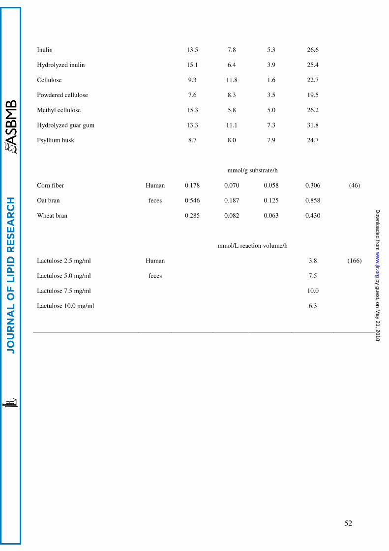
52
Inulin
Hydrolyzed inulin
Cellulose
Powdered cellulose
Methyl cellulose
Hydrolyzed guar gum
Psyllium husk
13.5
15.1
9.3
7.6
15.3
13.3
8.7
7.8
6.4
11.8
8.3
5.8
11.1
8.0
5.3
3.9
1.6
3.5
5.0
7.3
7.9
26.6
25.4
22.7
19.5
26.2
31.8
24.7
mmol/g substrate/h
Corn fiber
Oat bran
Wheat bran
Human
feces
0.178
0.546
0.285
0.070
0.187
0.082
0.058
0.125
0.063
0.306
0.858
0.430
(46)
mmol/L reaction volume/h
Lactulose 2.5 mg/ml
Lactulose 5.0 mg/ml
Lactulose 7.5 mg/ml
Lactulose 10.0 mg/ml
Human
feces
3.8
7.5
10.0
6.3
(166)
by guest, on May 21, 2018
ww
w.jlr.org
Dow
nloaded from

![Magnetoliposomes Loaded with Poly-Unsaturated Fatty Acids ... · reported using preparations of omega-3 fatty acids [29-35], a kind of long chain polyunsaturated fatty acids (PUFAs)](https://img.pdfslide.us/doc/110x75/5f8b025f23ab9a27c5624e1e/magnetoliposomes-loaded-with-poly-unsaturated-fatty-acids-reported-using-preparations.jpg)