Embed Size (px)
Citation preview
REVIEW Open Access
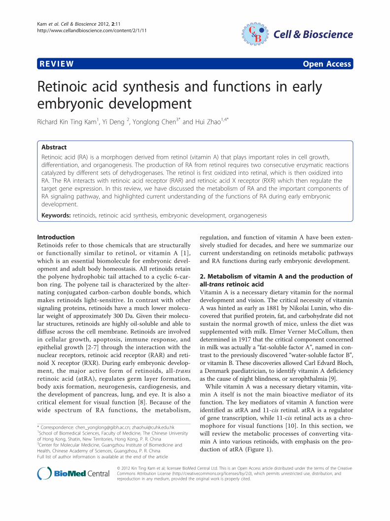
Retinoic acid synthesis and functions in earlyembryonic developmentRichard Kin Ting Kam1, Yi Deng 2, Yonglong Chen3* and Hui Zhao1,4*
Abstract
Retinoic acid (RA) is a morphogen derived from retinol (vitamin A) that plays important roles in cell growth,differentiation, and organogenesis. The production of RA from retinol requires two consecutive enzymatic reactionscatalyzed by different sets of dehydrogenases. The retinol is first oxidized into retinal, which is then oxidized intoRA. The RA interacts with retinoic acid receptor (RAR) and retinoic acid X receptor (RXR) which then regulate thetarget gene expression. In this review, we have discussed the metabolism of RA and the important components ofRA signaling pathway, and highlighted current understanding of the functions of RA during early embryonicdevelopment.
Keywords: retinoids, retinoic acid synthesis, embryonic development, organogenesis
IntroductionRetinoids refer to those chemicals that are structurallyor functionally similar to retinol, or vitamin A [1],which is an essential biomolecule for embryonic devel-opment and adult body homeostasis. All retinoids retainthe polyene hydrophobic tail attached to a cyclic 6-car-bon ring. The polyene tail is characterized by the alter-nating conjugated carbon-carbon double bonds, whichmakes retinoids light-sensitive. In contrast with othersignaling proteins, retinoids have a much lower molecu-lar weight of approximately 300 Da. Given their molecu-lar structures, retinoids are highly oil-soluble and able todiffuse across the cell membrane. Retinoids are involvedin cellular growth, apoptosis, immune response, andepithelial growth [2-7] through the interaction with thenuclear receptors, retinoic acid receptor (RAR) and reti-noid X receptor (RXR). During early embryonic develop-ment, the major active form of retinoids, all-transretinoic acid (atRA), regulates germ layer formation,body axis formation, neurogenesis, cardiogenesis, andthe development of pancreas, lung, and eye. It is also acritical element for visual function [8]. Because of thewide spectrum of RA functions, the metabolism,
regulation, and function of vitamin A have been exten-sively studied for decades, and here we summarize ourcurrent understanding on retinoids metabolic pathwaysand RA functions during early embryonic development.
2. Metabolism of vitamin A and the production ofall-trans retinoic acidVitamin A is a necessary dietary vitamin for the normaldevelopment and vision. The critical necessity of vitaminA was hinted as early as 1881 by Nikolai Lunin, who dis-covered that purified protein, fat, and carbohydrate did notsustain the normal growth of mice, unless the diet wassupplemented with milk. Elmer Verner McCollum, thendetermined in 1917 that the critical component concernedin milk was actually a “fat-soluble factor A”, named in con-trast to the previously discovered “water-soluble factor B”,or vitamin B. These discoveries allowed Carl Edvard Bloch,a Denmark paediatrician, to identify vitamin A deficiencyas the cause of night blindness, or xerophthalmia [9].While vitamin A was a necessary dietary vitamin, vita-
min A itself is not the main bioactive mediator of itsfunction. The key mediators of vitamin A function wereidentified as atRA and 11-cis retinal. atRA is a regulatorof gene transcription, while 11-cis retinal acts as a chro-mophore for visual functions [10]. In this section, wewill review the metabolic processes of converting vita-min A into various retinoids, with emphasis on the pro-duction of atRA (Figure 1).
* Correspondence: [email protected]; [email protected] of Biomedical Sciences, Faculty of Medicine, The Chinese Universityof Hong Kong, Shatin, New Territories, Hong Kong, P. R. China3Center for Molecular Medicine, Guangzhou Institute of Biomedicine andHealth, Chinese Academy of Sciences, Guangzhou, P. R. ChinaFull list of author information is available at the end of the article
Kam et al. Cell & Bioscience 2012, 2:11http://www.cellandbioscience.com/content/2/1/11 Cell & Bioscience
© 2012 Kin Ting Kam et al; licensee BioMed Central Ltd. This is an Open Access article distributed under the terms of the CreativeCommons Attribution License (http://creativecommons.org/licenses/by/2.0), which permits unrestricted use, distribution, andreproduction in any medium, provided the original work is properly cited.
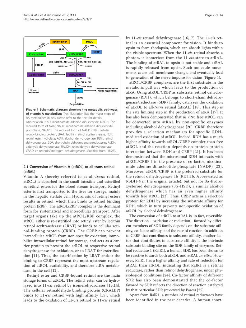
2.1 Conversion of Vitamin A (atROL) to all-trans retinal(atRAL)Vitamin A (hereby referred to as all-trans retinol,atROL) is absorbed in the small intestine and esterifiedas retinyl esters for the blood stream transport. Retinylester is first transported to the liver for storage, mainlyin the hepatic stellate cell. Hydrolysis of retinyl estersresults in retinol, which then binds to retinol bindingprotein (RBP). The atROL/RBP complex is the dominantform for systematical and intercellular transport. Aftertarget organs take up the atROL/RBP complex, theatROL either is re-esterified into retinyl ester by lecithinretinol acyltransferase (LRAT) or binds to cellular reti-nol-binding protein (CRBP). The CRBP can preventintracellular atROL from non-specific oxidation, immo-bilize intracellular retinol for storage, and acts as a car-rier protein to present the atROL to respective retinoldehydrogenase for oxidation, or to LRAT for esterifica-tion [11]. Thus, the esterification by LRAT and/or thebinding to CRBP represent the most upstream regula-tion of atROL availability, and therefore atRA metabo-lism, in the cell [12].Retinyl ester and CRBP-bound retinol are the main
storage forms of atROL. The retinyl ester can be hydro-lyzed into 11-cis retinol by isomerohydrolases [13,14].The cellular retinaldehyde binding protein (CRALBP)binds to 11-cis retinol with high affinity [15], whichleads to the oxidation of 11-cis retinol to 11-cis retinal
by 11-cis retinol dehydrogenase [16,17]. The 11-cis ret-inal is an essential component for vision. It binds toopsin to form rhodopsin, which can absorb lights withinthe visible spectrum. When the 11-cis-retinal absorbs aphoton, it isomerizes from the 11-cis state to atRAL.The binding of atRAL to opsin is not stable and atRALis rapidly released from opsin. Such molecule move-ments cause cell membrane change, and eventually leadto generation of the nerve impulse for vision (Figure 1).atROL/CRBP complexes are the first substrate in the
metabolic pathway which leads to the production ofatRA. Using atROL/CRBP as substrate, retinol dehydro-genase (RDH), which belongs to short-chain dehydro-genase/reductase (SDR) family, catalyzes the oxidationof atROL to all-trans retinal (atRAL) [18]. This step isthe rate limiting step in the production of atRA [19]. Ithas also been demonstrated that in vitro free atROL canbe converted into atRAL by non-specific enzymesincluding alcohol dehydrogenase [20]. CRBP thereforeprovides a selection mechanism for specific RDH-mediated oxidation of atROL. Indeed, RDH has a muchhigher affinity towards atROL/CRBP complex than freeatROL and the reaction depends on protein-proteininteraction between RDH and CRBP [21]. It has beendemonstrated that the microsomal RDH interacts withatROL/CRBP-I in the presence of co-factor, nicotina-mide adenine dinucleotide phosphate (NADP) [22].Moreover, atROL/CRBP is the preferred substrate forthe retinol dehydrogenase 16 (RDH16. Abbreviated asRoDH-4 in the original article), but not for 3a-hydro-xysteroid dehydrogenase (3a-HSD), a similar alcoholdehydrogenase which has an even higher affinitytowards free atROL [23]. Thus, CRBP acts as a selectionprotein for RDH by increasing the substrate affinity forRDH, which in turn prevents non-specific oxidation ofatROL by alcohol dehydrogenase.The conversion of atROL to atRAL is, in fact, reversible.
The direction - oxidation or reduction - favored by differ-ent members of SDR family depends on the substrate affi-nity, co-factor affinity, and the rate of reaction. In additionto CRBP that contributes to substrate affinity, another fac-tor that contributes to substrate affinity is the intrinsicsubstrate binding site on the SDR family of enzymes. Ret-inal reductase 1 (RalR1), a human SDR, has been shown tobe reactive towards both atROL and atRAL in vitro. How-ever, RalR1 has a higher affinity and rate of reduction foratRAL than atROL, indicating that RalR1 is a retinalreductase, rather than retinol dehydrogenase, under phy-siological conditions [24]. Co-factor affinity of differentSDR has also been demonstrated that the co-factorfavored by SDR reflects the direction of reaction catalyzedby that particular SDR (reviewed by Pares) [25].Apart from RalR1, a number of retinal reductases have
been identified in the past decades. A human short-
Figure 1 Schematic diagram showing the metabolic pathwaysof vitamin A metabolism. This illustration lists the major steps ofRA metabolism in cell, please refer to the text for details.Abbreviation: NAD, nicotinamide adenine dinucleotide; NADH, Thereduced form of NAD; NADP, nicotinamide adenine dinucleotidephosphate; NADPH, The reduced form of NADP; CRBP: cellularretinol-binding protein; LRAT: lecithin retinol acyltransferase; REH:retinyl ester hydrolase; ADH: alcohol dehydrogenase; RDH: retinoldehydrogenase; SDR: short-chain dehydrogenase/reductases; ALDH:aldehyde dehydrogenase; RALDH: retinaldehyde dehydrogenase.CRAD: cis-retinoid/androgen dehydrogenase. Modified from [18,25].
Kam et al. Cell & Bioscience 2012, 2:11http://www.cellandbioscience.com/content/2/1/11
Page 2 of 14
chained retinol reductase (retSDR1) identified in a neu-roblastoma cell line has been shown to promote the for-mation of retinyl ester in the presence of exogenousatRAL [26]. A mouse liver peroxisomal SDR termedmouse retinal reductase (RRD) also showed a highatRAL-specific reductase activity in the presence ofCRBP in vitro [27]. This enzyme was induced by peroxi-some proliferator-activated receptor (PPAR), suggestinga relationship between retinoid metabolism and peroxi-some activity [27]. Using in vitro assay, some studieshave identified reductases which showed in vitro retinal-reducing activity. For example, human aldose reductaseand human small intestine aldose reductase can functionas retinal reductase in vitro [28]. Similarly, the mouseshort-chained aldehyde reductase (SCALD) could reduceatRAL and 9-cis retinal in vitro [29]. However, these stu-dies are based on in vitro biochemical analysis only anddo not take into account the substrate selection byCRBP. Therefore, the enzymatic activities on differentretinal reported should not be taken as a direct evidencefor physiological retinal reductases.
2.2 Conversion of atRAL to atRASimilar to atROL, all-trans retinal is also transported byCRBP in the cell, and is then oxidized to atRA. The oxi-dation from atRAL to atRA has been observed as earlyas 1960 [30]. The oxidation of atRAL to atRA wasmediated by various retinaldehyde dehydrogenases(RALDH). At least 3 RALDHs have been identified inhuman, mouse, and Xenopus, with different physiologi-cal functions [31]. Retinaldehyde dehydrogenase 1(Raldh1) is highly expressed in the dorsal retina ofmouse embryos [32], in epithelial tissues of adult miceand Xenopus [33], and in the stomach and small intes-tine of adult rats [34]. Xenopus raldh1 has been shownto be an atRA synthesizing enzyme in a retinoic acidresponsive cell line [35]. RALDH1-/-mice also suggestthat Raldh1 is capable of atRA synthesis [36]. However,knockout of RALDH1 did not severely affect the mor-phology of the retina although RALDH1 is localized inthe dorsal retina [36], indicating that other enzymesmight redundantly share the function of RALDH1.The retinaldehyde dehydrogenase 2 (RALDH2) was
identified in human, mouse, chick, zebrafish and Xenopus[37-39]. Interestingly, RALDH2 was identified as a crucialenzyme for atRA synthesis in different organisms. Knock-out of RALDH2 was embryonic lethal during the post-implantation period in mice [40], suggesting that atRA isessential for normal embryonic development. The pheno-types of RALDH2 knock-out mice include severelyimpaired segmentation of rhombomeres, altered homeo-box gene expression pattern, and defective neural crestcell migration [41]. In zebrafish, knockdown of raldh2caused a down-regulation of retinoic acid signaling,
malformation in the central nervous system, and disrup-tion of left-right asymmetry [42,43]. The raldh2 mutant,neckless (nls), displayed a suppressed formation of themidbrain to hindbrain region, as well as segmentationdefects in rhombomeres [44]. Such defects were attributedto the reduction in atRA signaling [45], since the spatialand temporal pattern of atRA signaling is maintainedmainly by raldh2 and a degradative enzyme cytochromeP450 hydroxylase A1 (cyp26a1) in zebrafish. In Xenopusembryos, ectopic expression of raldh2 caused teratogeniceffects such as the expression of posterior neural markers(en2 and krox20) in the anterior region, which is similar tothat due to atRA toxicity [38], suggesting that raldh2 is animportant enzyme in maintaining atRA homeostasis inembryos. Knockdown of raldh2 in Xenopus embryoscaused a shortening of anteroposterior axis and a posteriorshift of neural marker en2 and krox20 [46]. Collectively,such evidence indicated that raldh2 plays a crucial role inthe anteroposterior patterning of the central nervous sys-tem and trunk axis through regulation of the RA signaling.Retinaldehyde dehydrogenase 3 (RALDH3) has been
identified in human, chick, mouse, zebrafish, and Xeno-pus, and is expressed in the ventral retina across variousspecies [47-51]. Studies in mouse have shown thatRALDH3 was mainly involved in the frontonasal devel-opment and patterning of ocular structures [52]. Micelacking RALDH3 were neonatal lethal, due to therespiratory tract obstruction in nasal region, and theneonatal lethality could be rescued by atRA supplements[53], suggesting that RALDH3 is an atRA synthesisenzyme. In 2007, Halilagic et al. showed that atRA pro-duction by RALDH3 contributed to the correct pattern-ing of the anterior and dorsal boundaries of thedeveloping forebrain [54]. It was further delineated thatRALDH3 knockout mice exhibited loss of dopaminereceptor D2 in the ventral forebrain. These studies sug-gest that RALDH3 is essential for the development ofthe central nervous system and the morphogenesis ofanterior head structures [52].Similar to atROL and atRAL, the metabolism of atRA
is also closely related to retinoid binding protein termedcellular retinoic acid binding proteins (CRABPs).CRABPs bind to intracellular RA and prevent it fromnon-specific degradation [55,56]. There are two speciesof CRABP, CRABP-I and CRABP-II. These carrier pro-teins also ensure the solubility of hydrophobic retinoidin the aqueous intracellular environment. However, arecent study of CRBP-I/CRABP-I/CRABP-II tripleknock-out mice has shown that the main regulator ofretinoid homeostasis was likely to be CRBPs, withCRABPs playing a minor role in this process. Hoegberget al. found that the chemical-induced depletion of totalretinoids in triple knockout mice was more severe thanthe wild type and CRABP-I/CRABP-II double knockout
Kam et al. Cell & Bioscience 2012, 2:11http://www.cellandbioscience.com/content/2/1/11
Page 3 of 14

mice [57], suggesting that CRBP-I is a more potent reg-ulator of retinoid homeostasis. While CRABPs mightnot be critical in regulating total retinoids homeostasis,they participate in mediating RA signaling by transport-ing RA to the nucleus to interact with RARs. CRABP-IIwas shown to be translocated into nucleus upon theligand binding [58], which allows atRA to bind to andactivate RAR, a transcription factor responsible for theRA signaling (Figure 2). Interestingly, the RA signalingis tightly regulated by negative feedback mechanisms asCRABP-II is negatively regulated by atRA [59]. ElevatedRA signaling suppresses the production of CRABPs,which down-regulate the activation of RARs and the RAsignaling. CRABP-I, on the other hand, regulates therate of RA metabolism by presenting RA to the degrad-ing enzyme CYP26A1 [60].
2.3 Degradation of atRAAll-trans retinoic acid is degraded by CYP26 enzymes,which belong to cytochrome P450 hydroxylase family. A
number of CYP26 family including CYP26A1, B1, C1,and D1 have been characterized and all of them possessthe ability to degrade atRA into less bio-active retinoid[61-63]. Rhombomeric alteration defects were onlyobserved by the knockdown of all three cyp26 enzymesin zebrafish [64], suggesting that cyp26a1, b1 and c1 actredundantly in hindbrain patterning. CYP26A1 isinduced by atRA while it promotes the hydroxylation ofatRA into 4-hydroxy retinoic acid, 4-oxo retinoic acid,and 18-hyroxy retinoic acid [45,65-67]. Since RALDH2and CYP26A1 are both regulated by atRA itself, themetabolism of atRA therefore forms an auto-regulatoryloop that regulates and balances atRA levels in embryos.Such regulation not only maintains the endogenousatRA level within a normal range, but also allows theorganisms to respond to exogenous atRA fluctuation.
3. Retinoic acid receptorsatRA is carried into the nucleus by CRABP-II, and inter-acts with RARs, which themselves are transcription
Figure 2 Ilustration of RA and paracrine RA signaling. In serum, retinol is bound to retinol-binding protein 4 (RBP-4) synthesized in the liver.Although retinol is lipid soluble, it enters cells mainly through the interaction with its receptor STRA. In the cell, retinol can either be convertedinto retinyl esters for storage via lecithin retinol acyltransferase (LRAT) or bind to the cellular retinol binding protein (CRBP). The CRBP-boundretinol is oxidized to retinal by either alcohol dehydrogenase (ADH) or retinol dehydrogenase (RDH), and retinal is oxidized to retinoic acid (RA)by retinaldehyde dehydrogenases (RALDH1/2/3). All-trans retinoic acid (atRA) is the major bioactive component among the retinoids. CYP26 canfurther oxidize atRA to 4-oxo-RA for degradation. Cellular retinoic acid-binding protein (CRABP) facilitates transportation of atRA into the nucleuswhere atRA binds its receptors. The ternary complex of ligand-bound RAR and RXR binds to the retinoic acid response element (RARE) andactivates the RA target genes. atRA can diffuse to adjacent cells to activate target gene expression in these cells. RAR can also bind to the liver Xreceptor (LXR), farnesoid X receptor (FXR), and peroxisome proliferator-activated receptor (PPAR) for multiple functions.
Kam et al. Cell & Bioscience 2012, 2:11http://www.cellandbioscience.com/content/2/1/11
Page 4 of 14

factors. RARs belong to retinoid receptor family, whichalso includes another group called retinoid X receptors(RXRs). RARs recognize both atRA and 9-cis retinoicacid, while RXRs only recognize 9-cis retinoic acid.Upon the ligand binding, RAR dimerizes with RXR toform a heterodimer, which then initiate gene transcrip-tion by binding to the retinoic acid response element(RARE) in the promoter region of the targets genes (Fig-ure 2). The RAR family consists of RARa/b/g threemembers that bind to atRA [68-71]. Single knockoutmice that lack each of RARs were not embryonic lethaland did not display the complete spectrum of vitamin Adeficiency phenotype. A disruption in RARa did notcause any observable phenotypic change in a mousemodel [72]. Knockout of RARb caused a reduction inthe body weight and ocular defect, while limb formationremained normal [73]. Double knockout of two RARgsubtypes caused growth deficiency, cartilage dysmorpho-genesis, and vertebrate malformation [74]. These resultsimply that RARs work redundantly and compensate thefunction of each other. Indeed, knockdown of RARacaused an increase in the expression level of RARb andRARg [75]. Only double knockout mutants showed phe-notype close to the symptoms of vitamin A deficiency[76]. The auto-regulatory loop of RAR expression issimilar to that regulating the expression of of RALDH2and CYP26a1. Moreover, RARE has been identified inthe promoter regions of RARa and b [77-79], indicatingthat the expression of these RARs is also under controlof atRA.Similarly, there are also three subtypes of RXRs [80].
RXRa knockout mice were embryonic lethal, potentiallydue to malformation of the heart in utero [81]. RXRbknockout mice were 50% embryonic lethal, and the sur-viving littermates were morphologically normal exceptspermatogenesis defects which rendered the male sterile[82], while the RXRg-null mutant mice were morpholo-gically normal when compared with the wild type [83].Moreover, the mice carrying only one copy of RXRa(RXRa+/-/RXRb-/-/RXRg-/-) were viable, suggestingone copy of RXRa is sufficient to carry out most offunction of the RXRs [83]. Since atRA-bound RAR canform heterodimer with RXR in the absence of 9-cis reti-noic acid and is still active in transcription activities, theimportance of RXRs may not be as critical as RARs.This may explain why only one copy of RXRa is suffi-cient for the mouse embryonic development. Takentogether, these results suggest that each of the RAR sub-types function redundantly and most of the RXR sub-types are not critical for the embryonic development.While RARs mainly mediate the RA signaling, it has
been revealed by many studies that ligand-bound RXRsactivate other signaling pathways by forming heterodi-mer with other nuclear receptors such as liver X
receptor (LXR), farnesoid X receptor (FXR), and PPAR[84-86] (Figure 2). LXR mainly functions as a sensor ofcholesterol levels by recognizing its ligand oxysterols.Overloading cells with cholesterol activates LXR/RXRheterodimers which in turn initiate transcription of tar-get genes, thereby regulate cholesterol transport, uptake,metabolism, and bile acid synthesis in the liver [87,88].FXR can recognize free or conjugated bile acid and thusacts as an intracellular sensor of bile acid to regulate themetabolism of bile acid in the liver. Activation ofliganded-FXR/RXR promotes bile acid efflux and inhi-bits bile acid synthesis [89]. PPAR is a lipid sensingnuclear receptor, recognizing a wide range of fatty acids[90]. These interactions between LXR, FXR, PPAR, andRXR reflect the complexity of RXR functions and theirpotentials on the RA signaling. In addition, not only doRXRs take part promiscuously in multiple signalingpathways, the expression of these nuclear receptors isalso under control of complex feedback loops and cross-talks with other signaling pathways [91]. The heterodi-merization of RXR with RAR, LXR, FXR, or PPAR istherefore mutually competitive, and atRA signaling notonly triggers the transcription of its target genes, butalso competitively suppresses the transcription of others.This may explain the board spectrum of atRA-inducedteratogenicity observed in embryos.
4. Differential expression and gene regulation ofRA metabolic enzymesThe RA metabolic enzymes show distinct differentialexpression pattern during early embryonic development,and interestingly their expression is regulated by the RAsignaling. Detailed descriptions of the expression pat-terns of these genes are beyond the scope of this review.Schematic drawing of expression of rdh10, dhrs3,raldh2, cyp26a1, rara2, and crabp-II at Xenopus gastrula(stage 11) and neurula (stage 14) stages are illustrated inFigure 3, which shows that the RA signaling itself regu-lates expression of the enzymes for RA biosynthesis andelicits the complexity of RA acting as a morphogen inearly embryonic development. Ectopic cyp26 expressioncan be induced by atRA treatment [92], while theembryos treated with atRA showed down-regulation ofraldh2 [38] and rdh10 [46]. Dhrs3 can also be inducedby atRA treatment (RKT Kam, Y Chen, WY Chan andH Zhao. Dhrs3 attenuates the retinoic acid signalingand is required for early embryonic patterning. Sub-mitted). Thus the RA signaling down-regulates theexpression of the enzymes for atRA production, but up-regulates enzymes that can reduce atRA level inembryos. Other components in the RA signaling arealso responsive to atRA treatment. For example, crabp-II was found to be an atRA-inducible gene [93], and wasfound to contain a RARE domain in its promoter region
Kam et al. Cell & Bioscience 2012, 2:11http://www.cellandbioscience.com/content/2/1/11
Page 5 of 14

[94]. Similarly, rara2 was found to be inducible by atRAtreatment in leukemic cell lines [95], and in rat embryosas well [96]. We summarize the regulation of thesegenes by RA signaling in Figure 4.
5. Retinoic acid signaling during early embryonicdevelopmentThe RA signaling pathway has been implicated in var-ious developmental processes. During early embryonicdevelopment, retinoids act as an important morphogenacross different species from invertebrate to metazoanincluding human [97,98]. It participates in regulatingvarious biological processes, such as apoptosis and dif-ferentiation, and cell fate specification.
5.1 Axis formationThe RA signaling has been implicated in embryonic axisformation as well. Evidence shows that it interacts with
Figure 3 Schematic diagram illustrating expression of the genes that are involved in RA biosynthesis and transportation at gastrula(stage 11) and neurula (stage 14) stages of Xenopus embryos. rdh10 is expressed in the circumblastoporal region of Xenopus gastrula, andthe signals at the dorsal side form a zone extending anteriorly. At stage14, the signals are found in trunk paraxial mesoderm region [46]. rdh10 isnot expressed in the notochord. raldh2 is expressed in a form of a ring around the vegetal pole with signals more intense in the dorsalblastpore lip. During neurula stages, raldh2 signals are mainly distributed in the trunk paraxial mesoderm, expanding ventrally [38]. rdh10 andraldh2 display overlapping expression pattern in the trunk paraxial mesoderm, with rdh10 expressed more anteriorly than raldh2. cyp26a isexpressed in two primary domains at stage11, the posterior domain surrounding the blastopore and the anterior domain covering the anteriorpart of prospective neural plate. At stage 14, the anterior cyp26a transcripts is developed into three elements corresponding to the cementgland anlage, the mid-/hindbrain boundary, and the auditory placodes, while the posterior expression domain remains in the circumblastoporalarea, and the developing neural plate is also covered by a gradient of cyp26a signals with the highest present in posterior region [92]. Theexpression domains of radlh2 and cyp26a do not overlap at gastrula and neurula stages of Xenopus embryos. rara2 is expressed in the involutingsurface layer surrounding the blastopore, and becomes stronger as the gastrulation proceeds. At stage 14, rara2 expression is expressedpredominantly in the posterior neural plate of the embryos [167]. The expression of the carbp-II is defined into an anterior and a posteriordomain at gastrula stages. In the anterior domain, Xenopus crabp-II is limited to the dorsal area which generates prospective head structures. Atthe neurula stages, crabp-II is expressed in the prospective telencephalon and rhombencephalon, and the most posterior region of the embryos[168]. The dhrs3 signals form a circumblastoporal ring which is similar to rdh10 at stage 11. The signals in the neural plate form two signalingzones and gradually converge towards the midline, forming two signal strips extending posteriorly. In addition, dhrs3 is expressed in thenotochord at neurula stages [169]. All these drawings are shown in dorsal view and the blue color represents expression signals.
Figure 4 The RA signaling regulates expression of thecomponents involved in RA synthesis and transportation.
Kam et al. Cell & Bioscience 2012, 2:11http://www.cellandbioscience.com/content/2/1/11
Page 6 of 14
Nodal signaling to regulate dorsoventral axis formation.The mice embryos lacking all three Cyp26 genes dis-played secondary body axes due to expansion of theNodal expression domain. In fact, the mouse Nodalgene contains an RARE in the intron 1 that is highlyconserved among mammals [99]. Moreover, pre-gastru-lation mouse embryos express Cyp26s but not Raldhssuggesting that maternal RA is decreased by embryonicCyp26s for proper Nodal expression during embryonicpatterning [99].In addition to the dorsoventral axis formation, cyp26
is also important for restricting the expression of poster-ior genes during the anteroposterior patterning [100].Experiments in zebrafish embryos indicate that reduc-tion of atRA by ectopic cyp26 decreased the expressionof posterior genes meis3. Consistent with this, knock-down of cyp26 led to the anterior expansion of meis3.Interestingly, the expression of cyp26 is suppressed bythe FGF and Wnt signalings, which are involved in thespecification of posterior trunk during the gastrulation.Thus, RA together with FGF and Wnt signalings formsa complex regulatory network to regulate the anteropos-terior axis formation in gastrula embryos, the cyp26being one of the cross-talk genes linking these signalingpathways [100].The RALDHs and CYP26s enzymes also participate in
the anteroposterior patterning of the central nervoussystem by maintaining a gradient of atRA along the axis[101]. Global application of atRA to mouse embryosduring day 7 of gestation caused severe head deforma-tions, such as exencephaly, microcephaly, and anence-phaly, whereas exposure of embryos to atRA during day8 of gestation led to severe caudal truncations reminis-cent of the human caudal regression syndrome[102,103]. Similar phenomena were observed with Xeno-pus embryos as well [104-106].Vertebrates display asymmetric placement of various
internal organs including the heart, liver, spleen, andgut, and the asymmetric development of paired organssuch as brain hemispheres and lungs. Treatment withRA antagonist in mouse led to randomization of heartlooping and perturbed sideness of node [107]. Applica-tion of RA antagonist revealed that the RA signalingsequentially controlled visceral and heart laterality [108].On the other hand, the somites obviously avoid theinfluence of signaling pathways that regulate the left-right asymmetry. The somites, derived from the trunkparaxial mesoderm and sequentially segmented alongthe anteroposterior axis, are formed as bilaterally pairedunits. However, the mice embryos that lack Raldh2exhibited asymmetric somite formation [109], which wasdue to left-right desynchronization of the segmentationclock oscillations. Such defects were also observed inRA deficient chick [110] and zebrafish [43]. It is
therefore postulated that the RA form a protectionzone, protecting the somite from the left-right asym-metric signals to maintain the bilateral symmetry ofsomites columns.
5.2 Neural differentiationatRA has been known for its ability to induce neural dif-ferentiation. During neural differentiation in earlyembryonic development, the pro-neural induction factorNeurogenin 2 (Neurog2) is required for primary sensoryneuron specification [111]. Two RARE identified in thepromoter region of mouse Neurog2 provide evidencethat atRA directly regulates the expression of Neurog2and therefore neural differentiation [112]. In addition, ithas been shown that atRA treatment can induce pluri-potent embryonic carcinoma stem cell line NT2 cells todifferentiate into forebrain, hindbrain, and spinal cordneural progenitors [113], and the NT2 cells displayedGABAergic and glutamatergic phenotype. In line withthis, mouse embryonic stem cells could be induced intoGABAergic neurons by atRA treatment in vitro [114].These in vitro findings were also supported by an invivo study that knockdown of RARa abolished the effectof atRA on dendritic growth [115]. Collectively, thesestudies support the view that atRA is required for neuraldifferentiation in the central nervous system.
5.3 Hindbrain patterningVertebrate hindbrain contains seven rhombomeres. Sev-eral Hox genes have been shown to be involved inrhombomeres formation and/or provide positional iden-tity to specific rhombomeres [116]. Two RAREs havebeen identified in Hoxb1, one of which is located 5’ toHoxb1 promoter and is required for the restrictedexpression in rhombomere 4 [117]. Studies in mouseand fish embryos showed that retinoids could induceectopic expression of Hoxa1 and Hoxb1, which caused arhombomere 2 to 4 transformation [118-120]. In addi-tion, the Hnf1b that suppressed Hoxb1 expression inrhombomere 5 was also regulated by the RA signaling[121,122]. Altogether, these data implicate that the RAsignaling is involved in hindbrain patterning. Indeed,knockout mice lacking Raldh2 led to the conversion ofcaudal hindbrain segments into rhombomere 4 identity[123,124] and the same is true for RA antagonist treatedmice [109]. In chick embryos, treatment by usingincreasing concentrations of RA antagonist, BMS453,showed that successively more posterior rhombomereboundaries required progressively higher concentrationof endogenous retinoic acid for their correct positioning[125]. In zebrafish embryos, rhombomere 5 of the cau-dal hindbrain is well specified but both rhombomere 4and 5 were posteriorly expanded in the absence of RA[42,126]. In Xenopus embryos, injection of cyp26a1
Kam et al. Cell & Bioscience 2012, 2:11http://www.cellandbioscience.com/content/2/1/11
Page 7 of 14
mRNAs into one side of the embryo resulted in altera-tions of the RA signaling at the injected side as indi-cated by posteriorization of krox20, a transcriptionfactor marking the rhombomere 3 and 5 [92], whereasincreasing the RA signaling via ectopic expression ofraldh2 led to anteriorization of both midbrain and hind-brain rhombomere identities in the expense of someforebrain territory [38] (Figure 5). Despite this, studiesin mouse and Xenopus showed that decreased RA sig-naling did not change the mid-/hindbrain boundary(MHB)[41,92], which requires the orchestration of acomplex regulatory network involving the FGF, Wnt,and Shh signalings [127]. On the contrary, the hind-brain/spinal cord boundary seems to be specifieddepending on the RA signaling. In zebrafish cyp26a1mutant embryos, the expression domains of hoxb5a andhoxb6a were expanded rostrally, leading to the expan-sion of rostral spinal cord at the expense of the hind-brain territory [128].
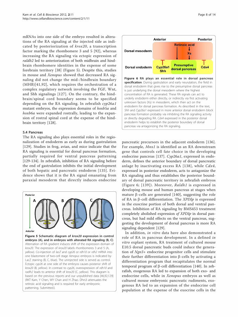
5.4 PancreasThe RA signaling also plays essential roles in the regio-nalization of endoderm as early as during gastrulation[129]. Studies in frog, avian, and mice indicate that theRA signaling is essential for dorsal pancreas formation,partially required for ventral pancreas patterning[129-134]. In zebrafish, inhibition of RA signaling beforethe end of gastrulation inhibits the initial developmentof both hepatic and pancreatic endoderm [135]. Evi-dence shows that it is the RA signal emanating fromparaxial mesoderm that directly induces endocrine
pancreatic precursors in the adjacent endoderm [136].For example, Mnx1 is identified as an RA downstreamgene that controls cell fate choice in the developingendocrine pancreas [137]. Cyp26a1, expressed in endo-derm, defines the anterior boundary of dorsal pancreaticanlage by inactivating excess RA [138], while Cdx4,expressed in posterior endoderm, acts to antagonize theRA signaling and thus establishes the posterior bound-ary of dorsal pancreatic territory in zebrafish embryos(Figure 6; [139]). Moreover, Raldh1 is expressed indeveloping mouse and human pancreas at stages whenmature b-cells are generated [140], suggesting the roleof RA in b-cell differentiation. The XPDIp is expressedin the exocrine portion of both dorsal and ventral pan-creas. Inhibition of RA signaling by BMS453 treatmentcompletely abolished expression of XPDIp in dorsal pan-creas, but had mild effects on the ventral pancreas, sug-gesting the development of dorsal pancreas is more RAsignaling dependent [129].In addition, in vitro data have also demonstrated a
role of RA in pancreas development. In a defined invitro explant system, RA treatment of cultured mouseE10.5 dorsal pancreatic buds could induce the genera-tion of Ngn3+ endocrine progenitor cells and stimulatetheir further differentiation into b-cells by activating adifferentiation program that recapitulates the normaltemporal program of b-cell differentiation [140]. In zeb-rafish, exogenous RA led to expansion of both exo- andendocrine cells, while in Xenopus embryos as well ascultured mouse embryonic pancreatic rudiments, exo-genous RA led to an expansion of the endocrine cellpopulation at the expense of the exocrine cells in the
Figure 5 Schematic diagram of krox20 expression in controlembryos (A), and in embryos with alterated RA signaling (B, C).Alternation of RA gradient induces shift of the expression domain ofkrox20. The expression of krox20 labels rhombomeres 3 and 5 (A,yellow). Co-injection of lacZ and cyp26 or rdh10 or rdh2 mRNA intoone blastomere of two-cell stage Xenopus embryos is indicated byLacZ staining (B, C, blue). The uninjected side is served as control.Ectopic cyp26 at one side of the embryos causes posterior shift ofkrox20 (B, yellow). In contrast to cyp26, overexpression of rdh10 andradlh2 leads to anterior shift of krox20 (C, yellow). This diagram isbased on the previous reports and our unpublished data [46,92,129](RKT Kam, Y Chen, WY Chan and H Zhao. Dhrs3 attenuates theretinoic acid signaling and is required for early embryonicpatterning. Submitted.).
Figure 6 RA plays an essential role in dorsal pancreasspecification. During gastrulation and early neurulation, the field indorsal endoderm that gives rise to the presumptive dorsal pancreasis just underlying the dorsal mesoderm where the highestconcentration of RA is generated. These RA signals can act tounderly endoderm either directly, or indirectly via first activatingunknown factors (Xs) in mesoderm, which then act on theendoderm for dorsal pancreas formation. As described in the text,Shh and Cyp26a1 expressed in more anterior dorsal endoderm blockpancreas formation probably via inhibiting the RA signaling activityor directly degrading RA. Cdx4 expressed in the posterior dorsalendoderm helps to establish the posterior boundary of dorsalpancreas via antagonizing the RA signaling.
Kam et al. Cell & Bioscience 2012, 2:11http://www.cellandbioscience.com/content/2/1/11
Page 8 of 14
dorsal pancreas [129,141,142]. Similarly, the induction ofpancreatic fate by RA in endodermalized naive ectodermdissected from early Xenopus gastrulae only showed thegeneration of early insulin-expressing cells [143,144].Therefore, cautions should be taken to apply RA at theright time and the right dose (close to physiological con-centration in early embryos) for in vitro induction ofmature pancreatic b-cells from embryonic stem cells.
5.5 HeartThe effect of RA on cardiogenesis was first demon-strated in quail that vitamin A deficiency could inducedefective primitive heart development [145]. Further evi-dence shows that RA signaling is required for the for-mation of cardiac progenitors [146], and the correctmodeling of the early heart field and the anteroposteriorpatterning of the heart is controlled by atRA throughthe action of Isl1 and Fgf8 signaling pathway [147,148].Knockout mice embryos lacking Raldh2 showed anabnormal development of the second heart field (SHF)[149], which is a population of undifferentiated cardiacprecursor cells originating from the pharyngeal meso-derm and lying medial to the cardiac crescent (the firstheart field). The expression of SHF markers includingIsl1, Tbx1, and Fgf8, was posteriorly expanded in theseembryos [150], indicating that the RA signaling isrequired for the restriction of SHF. Defects in the for-mation of a proper heart tube might also partiallyexplain why Raldh2-/- is embryonic lethal [148]. Studiesin zebrafish indicated RA functions in the restriction ofcardiac specification [149]. During this process, if RAsignaling was inhibited in lateral mesoderm, the uncom-mitted cells could differentiate into myocardial progeni-tor cells instead of pharyngeal or pancreatic cells [149],leading to the expansion of cardiomyocytes.In addition to the heart development, the RA signaling
has also been implicated in heart regeneration in adultzebrafish. Gene profiling assay indicated that raldh2 wasone of the most highly induced genes in the regenerat-ing heart [151]. After the ventricular injury, raldh2 wasactivated in the endocardium, but its expression wassubsequently restricted in endocardial cells at the injurysite one day post-trauma as the repairing cardiogenesisbegan. By 7 days after amputation, the epicardial cells atthe injury site also expressed raldh2. Inhibition of RARsor expression of an RA-degrading enzyme blockedregenerative cardiomyocyte proliferation [152]. Thesefindings reveal essential roles of RA in repairing thedamaged heart tissue and promoting cardiomyocyteproliferation.
5.6 KidneyThere is increasing evidence indicating that atRA playsimportant roles in kidney development [153-155]. The
kidney field is derived from the intermediate mesoderm.The specification of renal progenitor cells is influencedby RA signals emanating from the paraxial mesoderm[153-155]. Recent studies in Xenopus and zebrafish havedemonstrated that ectopic production of RA increasedthe size of the kidney field, while blocking the pathwayinhibited the kidney specification [86,154]. In Xenopus,pax8 and lhx1,which are the earliest determinants ofpronephric fate are both under control of the RA signal-ing [154,156], while pteg, an early pronephric marker, isa direct target of the RA signaling and an essential fac-tor for pronephric specification [157]. Another criticalfactor for the pronephros development, Wilm’s tumorsuppressor (wt1), is under direct control of the RA sig-naling as well [158,159]. Inhibition of RA by BMS453treatment caused reduction of pronephros field, asrevealed by the decreased expression of the pronephrosmarkers including smp-30 and pax2 at the late tailbudstage [154]. Furthermore, in Xenopus embryos, the pluri-potent animal cap cells treated with RA and activincould differentiate into pronephros tissue [154,160].Likewise, the RA signaling is also essential for directingthe proximodistal patterning of pronephric nephron inzebrafish [161]. The exogenous RA could induce proxi-mal segment fates at the expense of distal fates, whereasinhibition of the RA signaling caused a loss of the proxi-mal segments and an expansion of the distal segments.Interestingly, inactivation of both RARa and RARb in
mice embryos resulted in renal malformations [155].Further evidence showed that RARa and RARb were co-expressed with Ret, a receptor tyrosine kinase involvedin renal development, in renal stromal mesenchyme,where their deletion led to altered stromal cell pattern-ing, impaired ureteric bud growth, and down-regulationof Ret in the ureteric bud. Moreover, studies in miceindicate that the RA signaling in ureteric bud cellsmainly depends on atRA generated through Raldh2 instromal cells [162].
5.7 LungEvidence indicates that disruption of the RA signalingduring early development caused abnormalities of lungformation. Either knockout of Raldh2 or treatment withBMS 493, a RA inhibitor, caused defects of lung forma-tion in mice [163]. During primary lung bud morpho-genesis, although RA is not required for specification oflung cell fate in the endoderm, it is essential for theinduction of the primordial lung buds [163,164]. Duringthe induction of lung bud, the RA signaling inhibits theexpression of Dkk-1, the Wnt signaling inhibitor, in theforegut mesoderm. This inhibition in turn activates Wntsignaling in the lung bud region. At the same time, theRA signaling also inhibits TGF-b signaling. Thebalanced Wnt and TGF-b signalings coordinate with
Kam et al. Cell & Bioscience 2012, 2:11http://www.cellandbioscience.com/content/2/1/11
Page 9 of 14

each other to induce and maintain the optimal expres-sion level of Fgf10 for the induction of the lung primor-dium [165]. Furthermore, airway branching wasaccompanied by down-regulation of the RA signalingpathway [166], thus regional mechanisms that controlRA availability and utilization are important for the lungmorphogenesis.
Concluding remarksTo date, it has been demonstrated that the RA signalingplays sophisticated roles in early embryonic develop-ment. In fact, gastrulation is a very critical period forthe RA signaling to exert its function in the regionaliza-tion of all three germ layers along the anteroposterioraxis. RA is mainly generated in the mesoderm and thuscan be most efficiently used. It is not clear as to howthe RA metabolic enzymes coordinate with each otherto generate the RA gradient at right time, right region,and with right strength during early embryonic develop-ment. Future studies are needed to identify the immedi-ate and direct RA target genes in distinct RA responsivecells and to define the crosstalks between RA and othersignaling pathways, such as hedgehog, FGF, Wnt, andNotch signalings for the formation and regionalizationof three germ layers. Such knowledge will be essentialfor understanding organogenesis and establishing reli-able strategies for stem cell differentiation into specificcell types that can be used for treatment of humandiseases.
AcknowledgementsWe thank members of our laboratories for valuable discussion. This work issupported by GRF grant from the Research Grants Council of Hong Kong(No. CUHK480709) to HZ, the National Basic Research Program of China(2009CB941202), and the fund from the Key Project of KnowledgeInnovation Program of the Chinese Academy of Sciences (KSCX2-YW-R-083)to YLC. RKTK was supported by the Graduate Studentships from The ChineseUniversity of Hong Kong.
Author details1School of Biomedical Sciences, Faculty of Medicine, The Chinese Universityof Hong Kong, Shatin, New Territories, Hong Kong, P. R. China. 2Departmentof Medicine and Therapeutics, Faculty of Medicine, The Chinese University ofHong Kong, Shatin, New Territories, Hong Kong, P. R. China. 3Center forMolecular Medicine, Guangzhou Institute of Biomedicine and Health,Chinese Academy of Sciences, Guangzhou, P. R. China. 4Key Laboratory forRegenerative Medicine, Ministry of Education, Ji Nan University-The ChineseUniversity of Hong Kong, Shatin, New Territories, Hong Kong, P. R. China.
Authors’ contributionsRKTK and HZ planned the manuscript outline. RKTK, HZ and YLC wrote thedraft, YD revised and did proof reading, HZ and YLC finalized themanuscript. All authors read and approve the final manuscript.
Competing interestsThe authors declare that they have no competing interests.
Received: 2 December 2011 Accepted: 22 March 2012Published: 22 March 2012
References1. Tang XH, Gudas LJ: Retinoids, retinoic acid receptors, and cancer. Annu
Rev Pathol 2011, 6:345-364.2. Duriancik DM, Lackey DE, Hoag KA: Vitamin A as a regulator of antigen
presenting cells. J Nutr 140(8):1395-1399.3. Gudas LJ, Wagner JA: Retinoids regulate stem cell differentiation. J Cell
Physiol 226(2):322-330.4. Hogarth CA, Griswold MD: The key role of vitamin A in spermatogenesis.
J Clin Invest 120(4):956-962.5. Mora JR, Iwata M, von Andrian UH: Vitamin effects on the immune
system: vitamins A and D take centre stage. Nat Rev Immunol 2008,8(9):685-698.
6. Mukherjee S, Date A, Patravale V, Korting HC, Roeder A, Weindl G: Retinoidsin the treatment of skin aging: an overview of clinical efficacy andsafety. Clin Interv Aging 2006, 1(4):327-348.
7. Pino-Lagos K, Benson MJ, Noelle RJ: Retinoic acid in the immune system.Ann N Y Acad Sci 2008, 1143:170-187.
8. Davies WL, Hankins MW, Foster RG: Vertebrate ancient opsin andmelanopsin: divergent irradiance detectors. Photochem Photobiol Sci 2010,9(11):1444-1457.
9. Wolf G: The discovery of the visual function of vitamin A. J Nutr 2001,131(6):1647-1650.
10. D’Ambrosio DN, Clugston RD, Blaner WS: Vitamin A Metabolism: AnUpdate. 2011, 3(1):63-103.
11. Penzes P, Napoli JL: Holo-cellular retinol-binding protein: distinction ofligand-binding affinity from efficiency as substrate in retinalbiosynthesis. Biochemistry 1999, 38(7):2088-2093.
12. Liu L, Gudas LJ: Disruption of the lecithin:retinol acyltransferase genemakes mice more susceptible to vitamin A deficiency. J Biol Chem 2005,280(48):40226-40234.
13. Deigner PS, Law WC, Canada FJ, Rando RR: Membranes as the energysource in the endergonic transformation of vitamin A to 11-cis-retinol.Science 1989, 244(4907):968-971.
14. Mata NL, Moghrabi WN, Lee JS, Bui TV, Radu RA, Horwitz J, Travis GH:Rpe65 is a retinyl ester binding protein that presents insoluble substrateto the isomerase in retinal pigment epithelial cells. J Biol Chem 2004,279(1):635-643.
15. Saari JC, Nawrot M, Kennedy BN, Garwin GG, Hurley JB, Huang J, Possin DE,Crabb JW: Visual cycle impairment in cellular retinaldehyde bindingprotein (CRALBP) knockout mice results in delayed dark adaptation.Neuron 2001, 29(3):739-748.
16. Driessen CA, Janssen BP, Winkens HJ, van Vugt AH, de Leeuw TL,Janssen JJ: Cloning and expression of a cDNA encoding bovine retinalpigment epithelial 11-cis retinol dehydrogenase. Invest Ophthalmol Vis Sci1995, 36(10):1988-1996.
17. Simon A, Hellman U, Wernstedt C, Eriksson U: The retinal pigmentepithelial-specific 11-cis retinol dehydrogenase belongs to the family ofshort chain alcohol dehydrogenases. J Biol Chem 1995, 270(3):1107-1112.
18. Duester G: Families of retinoid dehydrogenases regulating vitamin Afunction: production of visual pigment and retinoic acid. Eur J Biochem2000, 267(14):4315-4324.
19. Agadir A, Nau H, Blaner WS: Retinoids: the biochemical and molecularbasis of Vitamin A and retinoid action. Berlin; New York Springer; 1999.
20. Posch KC, Enright WJ, Napoli JL: Retinoic acid synthesis by cytosol fromthe alcohol dehydrogenase negative deermouse. Arch Biochem Biophys1989, 274(1):171-178.
21. Noy N: Retinoid-binding proteins: mediators of retinoid action. Biochem J2000, 348(Pt 3):481-495.
22. Boerman MH, Napoli JL: Characterization of a microsomal retinoldehydrogenase: a short-chain alcohol dehydrogenase with integral andperipheral membrane forms that interacts with holo-CRBP (type I).Biochemistry 1995, 34(21):7027-7037.
23. Lapshina EA, Belyaeva OV, Chumakova OV, Kedishvili NY: Differentialrecognition of the free versus bound retinol by human microsomalretinol/sterol dehydrogenases: characterization of the holo-CRBPdehydrogenase activity of RoDH-4. Biochemistry 2003, 42(3):776-784.
24. Belyaeva OV, Stetsenko AV, Nelson P, Kedishvili NY: Properties of short-chain dehydrogenase/reductase RalR1: characterization of purifiedenzyme, its orientation in the microsomal membrane, and distributionin human tissues and cell lines. Biochemistry 2003, 42(50):14838-14845.
Kam et al. Cell & Bioscience 2012, 2:11http://www.cellandbioscience.com/content/2/1/11
Page 10 of 14
25. Pares X, Farres J, Kedishvili N, Duester G: Medium- and short-chaindehydrogenase/reductase gene and protein families: Medium-chain andshort-chain dehydrogenases/reductases in retinoid metabolism. Cell MolLife Sci 2008, 65(24):3936-3949.
26. Cerignoli F, Guo X, Cardinali B, Rinaldi C, Casaletto J, Frati L, Screpanti I,Gudas LJ, Gulino A, Thiele CJ, et al: retSDR1, a short-chain retinoldehydrogenase/reductase, is retinoic acid-inducible and frequentlydeleted in human neuroblastoma cell lines. Cancer Res 2002,62(4):1196-1204.
27. Lei Z, Chen W, Zhang M, Napoli JL: Reduction of all-trans-retinal in themouse liver peroxisome fraction by the short-chain dehydrogenase/reductase RRD: induction by the PPAR alpha ligand clofibrate.Biochemistry 2003, 42(14):4190-4196.
28. Crosas B, Hyndman DJ, Gallego O, Martras S, Pares X, Flynn TG, Farres J:Human aldose reductase and human small intestine aldose reductaseare efficient retinal reductases: consequences for retinoid metabolism.Biochem J 2003, 373(Pt 3):973-979.
29. Kasus-Jacobi A, Ou J, Bashmakov YK, Shelton JM, Richardson JA,Goldstein JL, Brown MS: Characterization of mouse short-chain aldehydereductase (SCALD), an enzyme regulated by sterol regulatory element-binding proteins. J Biol Chem 2003, 278(34):32380-32389.
30. Dowling JE, Wald G: The role of vitamin A acid. Vitam Horm 1960,18:515-541.
31. Duester G, Mic FA, Molotkov A: Cytosolic retinoid dehydrogenases governubiquitous metabolism of retinol to retinaldehyde followed by tissue-specific metabolism to retinoic acid. Chem Biol Interact 2003, 143-144:201-210.
32. Haselbeck RJ, Hoffmann I, Duester G: Distinct functions for Aldh1 andRaldh2 in the control of ligand production for embryonic retinoidsignaling pathways. Dev Genet 1999, 25(4):353-364.
33. Ang HL, Duester G: Retinoic acid biosynthetic enzyme ALDH1 localizes ina subset of retinoid-dependent tissues during xenopus development.Dev Dyn 1999, 215(3):264-272.
34. Frota-Ruchon A, Marcinkiewicz M, Bhat PV: Localization of retinaldehydrogenase type 1 in the stomach and intestine. Cell Tissue Res 2000,302(3):397-400.
35. Ang HL, Duester G: Stimulation of premature retinoic acid synthesis inXenopus embryos following premature expression of aldehydedehydrogenase ALDH1. Eur J Biochem 1999, 260(1):227-234.
36. Fan X, Molotkov A, Manabe S, Donmoyer CM, Deltour L, Foglio MH,Cuenca AE, Blaner WS, Lipton SA, Duester G: Targeted disruption ofAldh1a1 (Raldh1) provides evidence for a complex mechanism ofretinoic acid synthesis in the developing retina. Mol Cell Biol 2003,23(13):4637-4648.
37. Blentic A, Gale E, Maden M: Retinoic acid signalling centres in the avianembryo identified by sites of expression of synthesising andcatabolising enzymes. Dev Dyn 2003, 227(1):114-127.
38. Chen Y, Pollet N, Niehrs C, Pieler T: Increased XRALDH2 activity has aposteriorizing effect on the central nervous system of Xenopusembryos. Mech Dev 2001, 101(1-2):91-103.
39. Niederreither K, McCaffery P, Drager UC, Chambon P, Dolle P: Restrictedexpression and retinoic acid-induced downregulation of theretinaldehyde dehydrogenase type 2 (RALDH-2) gene during mousedevelopment. Mech Dev 1997, 62(1):67-78.
40. Niederreither K, Subbarayan V, Dolle P, Chambon P: Embryonic retinoicacid synthesis is essential for early mouse post-implantationdevelopment. Nat Genet 1999, 21(4):444-448.
41. Niederreither K, Vermot J, Schuhbaur B, Chambon P, Dolle P: Retinoic acidsynthesis and hindbrain patterning in the mouse embryo. Development2000, 127(1):75-85.
42. Grandel H, Lun K, Rauch GJ, Rhinn M, Piotrowski T, Houart C, Sordino P,Kuchler AM, Schulte-Merker S, Geisler R, et al: Retinoic acid signalling inthe zebrafish embryo is necessary during pre-segmentation stages topattern the anterior-posterior axis of the CNS and to induce a pectoralfin bud. Development 2002, 129(12):2851-2865.
43. Kawakami Y, Raya A, Raya RM, Rodriguez-Esteban C, Belmonte JC: Retinoicacid signalling links left-right asymmetric patterning and bilaterallysymmetric somitogenesis in the zebrafish embryo. Nature 2005,435(7039):165-171.
44. Begemann G, Marx M, Mebus K, Meyer A, Bastmeyer M: Beyond theneckless phenotype: influence of reduced retinoic acid signaling on
motor neuron development in the zebrafish hindbrain. Dev Biol 2004,271(1):119-129.
45. Dobbs-McAuliffe B, Zhao Q, Linney E: Feedback mechanisms regulateretinoic acid production and degradation in the zebrafish embryo. MechDev 2004, 121(4):339-350.
46. Strate I, Min TH, Iliev D, Pera EM: Retinol dehydrogenase 10 is a feedbackregulator of retinoic acid signalling during axis formation and patterningof the central nervous system. Development 2009, 136(3):461-472.
47. Koenig SF, Brentle S, Hamdi K, Fichtner D, Wedlich D, Gradl D: En2, Pax2/5and Tcf-4 transcription factors cooperate in patterning the Xenopusbrain. Dev Biol 340(2):318-328.
48. Lupo G, Liu Y, Qiu R, Chandraratna RA, Barsacchi G, He RQ, Harris WA:Dorsoventral patterning of the Xenopus eye: a collaboration of Retinoid,Hedgehog and FGF receptor signaling. Development 2005,132(7):1737-1748.
49. Mic FA, Molotkov A, Fan X, Cuenca AE, Duester G: RALDH3, aretinaldehyde dehydrogenase that generates retinoic acid, is expressedin the ventral retina, otic vesicle and olfactory pit during mousedevelopment. Mech Dev 2000, 97(1-2):227-230.
50. Pittlik S, Domingues S, Meyer A, Begemann G: Expression of zebrafishaldh1a3 (raldh3) and absence of aldh1a1 in teleosts. Gene Expr Patterns2008, 8(3):141-147.
51. Suzuki R, Shintani T, Sakuta H, Kato A, Ohkawara T, Osumi N, Noda M:Identification of RALDH-3, a novel retinaldehyde dehydrogenase,expressed in the ventral region of the retina. Mech Dev 2000, 98(1-2):37-50.
52. Molotkova N, Molotkov A, Duester G: Role of retinoic acid duringforebrain development begins late when Raldh3 generates retinoic acidin the ventral subventricular zone. Dev Biol 2007, 303(2):601-610.
53. Dupe V, Matt N, Garnier JM, Chambon P, Mark M, Ghyselinck NB: Anewborn lethal defect due to inactivation of retinaldehydedehydrogenase type 3 is prevented by maternal retinoic acid treatment.Proc Natl Acad Sci USA 2003, 100(24):14036-14041.
54. Halilagic A, Ribes V, Ghyselinck NB, Zile MH, Dolle P, Studer M: Retinoidscontrol anterior and dorsal properties in the developing forebrain. DevBiol 2007, 303(1):362-375.
55. Chytil F, Ong DE: Cellular retinol- and retinoic acid-binding proteins invitamin A action. Fed Proc 1979, 38(11):2510-2514.
56. Napoli JL, Boerman MH, Chai X, Zhai Y, Fiorella PD: Enzymes and bindingproteins affecting retinoic acid concentrations. J Steroid Biochem Mol Biol1995, 53(1-6):497-502.
57. Hoegberg P, Schmidt CK, Fletcher N, Nilsson CB, Trossvik C, GerlienkeSchuur A, Brouwer A, Nau H, Ghyselinck NB, Chambon P, et al: Retinoidstatus and responsiveness to 2,3,7,8-tetrachlorodibenzo-p-dioxin (TCDD)in mice lacking retinoid binding protein or retinoid receptor forms.Chem Biol Interact 2005, 156(1):25-39.
58. Sessler RJ, Noy N: A ligand-activated nuclear localization signal in cellularretinoic acid binding protein-II. Mol Cell 2005, 18(3):343-353.
59. Stachurska E, Loboda A, Niderla-Bielinska J, Szperl M, Juszynski M,Jozkowicz A, Dulak J, Ratajska A: Expression of cellular retinoic acid-binding protein I and II (CRABP I and II) in embryonic mouse heartstreated with retinoic acid. Acta Biochim Pol 58(1):19-29.
60. Boylan JF, Gudas LJ: The level of CRABP-I expression influences theamounts and types of all-trans-retinoic acid metabolites in F9teratocarcinoma stem cells. J Biol Chem 1992, 267(30):21486-21491.
61. Gu X, Xu F, Wang X, Gao X, Zhao Q: Molecular cloning and expression ofa novel CYP26 gene (cyp26d1) during zebrafish early development. GeneExpr Patterns 2005, 5(6):733-739.
62. Sakai Y, Luo T, McCaffery P, Hamada H, Drager UC: CYP26A1 and CYP26C1cooperate in degrading retinoic acid within the equatorial retina duringlater eye development. Dev Biol 2004, 276(1):143-157.
63. Takeuchi H, Yokota A, Ohoka Y, Iwata M: Cyp26b1 regulates retinoic acid-dependent signals in T cells and its expression is inhibited bytransforming growth factor-beta. PLoS One 6(1):e16089.
64. Hernandez RE, Putzke AP, Myers JP, Margaretha L, Moens CB: Cyp26enzymes generate the retinoic acid response pattern necessary forhindbrain development. Development 2007, 134(1):177-187.
65. White JA, Beckett-Jones B, Guo YD, Dilworth FJ, Bonasoro J, Jones G,Petkovich M: cDNA cloning of human retinoic acid-metabolizing enzyme(hP450RAI) identifies a novel family of cytochromes P450. J Biol Chem1997, 272(30):18538-18541.
Kam et al. Cell & Bioscience 2012, 2:11http://www.cellandbioscience.com/content/2/1/11
Page 11 of 14
66. de Roos K, Sonneveld E, Compaan B, ten Berge D, Durston AJ, van derSaag PT: Expression of retinoic acid 4-hydroxylase (CYP26) during mouseand Xenopus laevis embryogenesis. Mech Dev 1999, 82(1-2):205-211.
67. Swindell EC, Thaller C, Sockanathan S, Petkovich M, Jessell TM, Eichele G:Complementary domains of retinoic acid production and degradation inthe early chick embryo. Dev Biol 1999, 216(1):282-296.
68. Giguere V, Ong ES, Segui P, Evans RM: Identification of a receptor for themorphogen retinoic acid. Nature 1987, 330(6149):624-629.
69. Petkovich M, Brand NJ, Krust A, Chambon P: A human retinoic acidreceptor which belongs to the family of nuclear receptors. Nature 1987,330(6147):444-450.
70. Brand N, Petkovich M, Krust A, Chambon P, de The H, Marchio A, Tiollais P,Dejean A: Identification of a second human retinoic acid receptor. Nature1988, 332(6167):850-853.
71. Ruberte E, Dolle P, Krust A, Zelent A, Morriss-Kay G, Chambon P: Specificspatial and temporal distribution of retinoic acid receptor gammatranscripts during mouse embryogenesis. Development 1990,108(2):213-222.
72. Li E, Sucov HM, Lee KF, Evans RM, Jaenisch R: Normal development andgrowth of mice carrying a targeted disruption of the alpha 1 retinoicacid receptor gene. Proc Natl Acad Sci USA 1993, 90(4):1590-1594.
73. Ghyselinck NB, Dupe V, Dierich A, Messaddeq N, Garnier JM, Rochette-Egly C, Chambon P, Mark M: Role of the retinoic acid receptor beta(RARbeta) during mouse development. Int J Dev Biol 1997, 41(3):425-447.
74. Subbarayan V, Kastner P, Mark M, Dierich A, Gorry P, Chambon P: Limitedspecificity and large overlap of the functions of the mouse RAR gamma1 and RAR gamma 2 isoforms. Mech Dev 1997, 66(1-2):131-142.
75. Manshouri T, Yang Y, Lin H, Stass SA, Glassman AB, Keating MJ, Albitar M:Downregulation of RAR alpha in mice by antisense transgene leads to acompensatory increase in RAR beta and RAR gamma and developmentof lymphoma. Blood 1997, 89(7):2507-2515.
76. Mark M, Ghyselinck NB, Wendling O, Dupe V, Mascrez B, Kastner P,Chambon P: A genetic dissection of the retinoid signalling pathway inthe mouse. Proc Nutr Soc 1999, 58(3):609-613.
77. Chomienne C, Balitrand N, Ballerini P, Castaigne S, de The H, Degos L: All-trans retinoic acid modulates the retinoic acid receptor-alpha inpromyelocytic cells. J Clin Invest 1991, 88(6):2150-2154.
78. Kamei Y, Kawada T, Kazuki R, Sugimoto E: Retinoic acid receptor gamma 2gene expression is up-regulated by retinoic acid in 3T3-L1preadipocytes. Biochem J 1993, 293(Pt 3):807-812.
79. Sucov HM, Murakami KK, Evans RM: Characterization of an autoregulatedresponse element in the mouse retinoic acid receptor type beta gene.Proc Natl Acad Sci USA 1990, 87(14):5392-5396.
80. Chawla A, Repa JJ, Evans RM, Mangelsdorf DJ: Nuclear receptors and lipidphysiology: opening the X-files. Science 2001, 294(5548):1866-1870.
81. Kastner P, Messaddeq N, Mark M, Wendling O, Grondona JM, Ward S,Ghyselinck N, Chambon P: Vitamin A deficiency and mutations ofRXRalpha, RXRbeta and RARalpha lead to early differentiation ofembryonic ventricular cardiomyocytes. Development 1997,124(23):4749-4758.
82. Kastner P, Mark M, Leid M, Gansmuller A, Chin W, Grondona JM, Decimo D,Krezel W, Dierich A, Chambon P: Abnormal spermatogenesis in RXR betamutant mice. Genes Dev 1996, 10(1):80-92.
83. Krezel W, Dupe V, Mark M, Dierich A, Kastner P, Chambon P: RXR gammanull mice are apparently normal and compound RXR alpha +/-/RXR beta-/-/RXR gamma -/- mutant mice are viable. Proc Natl Acad Sci USA 1996,93(17):9010-9014.
84. Mello T, Polvani S, Galli A: Peroxisome proliferator-activated receptor andretinoic x receptor in alcoholic liver disease. PPAR Res 2009, 2009:748174.
85. Rizzo G, Renga B, Antonelli E, Passeri D, Pellicciari R, Fiorucci S: The methyltransferase PRMT1 functions as co-activator of farnesoid X receptor(FXR)/9-cis retinoid X receptor and regulates transcription of FXRresponsive genes. Mol Pharmacol 2005, 68(2):551-558.
86. Wingert RA, Davidson AJ: The zebrafish pronephros: a model to studynephron segmentation. Kidney Int 2008, 73(10):1120-1127.
87. Lagu B, Pio B, Lebedev R, Yang M, Pelton PD: RXR-LXR heterodimermodulators for the potential treatment of dyslipidemia. Bioorg Med ChemLett 2007, 17(12):3497-3503.
88. Zhao C, Dahlman-Wright K: Liver X receptor in cholesterol metabolism.J Endocrinol 204(3):233-240.
89. Desvergne B: RXR: from partnership to leadership in metabolicregulations. Vitam Horm 2007, 75:1-32.
90. Desvergne B, Wahli W: Peroxisome proliferator-activated receptors:nuclear control of metabolism. Endocr Rev 1999, 20(5):649-688.
91. Desvergne B, Michalik L, Wahli W: Transcriptional regulation ofmetabolism. Physiol Rev 2006, 86(2):465-514.
92. Hollemann T, Chen Y, Grunz H, Pieler T: Regionalized metabolic activityestablishes boundaries of retinoic acid signalling. EMBO J 1998,17(24):7361-7372.
93. Astrom A, Pettersson U, Voorhees JJ: Structure of the human cellularretinoic acid-binding protein II gene. Early transcriptional regulation byretinoic acid. J Biol Chem 1992, 267(35):25251-25255.
94. Durand B, Saunders M, Leroy P, Leid M, Chambon P: All-trans and 9-cisretinoic acid induction of CRABPII transcription is mediated by RAR-RXRheterodimers bound to DR1 and DR2 repeated motifs. Cell 1992,71(1):73-85.
95. Zhu J, Heyworth CM, Glasow A, Huang QH, Petrie K, Lanotte M, Benoit G,Gallagher R, Waxman S, Enver T, et al: Lineage restriction of the RARalphagene expression in myeloid differentiation. Blood 2001, 98(8):2563-2567.
96. Takeyama K, Kojima R, Ohashi R, Sato T, Mano H, Masushige S, Kato S:Retinoic acid differentially up-regulates the gene expression of retinoicacid receptor alpha and gamma isoforms in embryo and adult rats.Biochem Biophys Res Commun 1996, 222(2):395-400.
97. Halme A, Cheng M, Hariharan IK: Retinoids regulate a developmentalcheckpoint for tissue regeneration in Drosophila. Curr Biol 20(5):458-463.
98. Maden M: Retinoids in nonmammalian embryos. Methods Mol Biol 2008,461:541-559.
99. Uehara M, Yashiro K, Takaoka K, Yamamoto M, Hamada H: Removal ofmaternal retinoic acid by embryonic CYP26 is required for correct Nodalexpression during early embryonic patterning. Genes Dev 2009,23(14):1689-1698.
100. Kudoh T, Wilson SW, Dawid IB: Distinct roles for Fgf, Wnt and retinoicacid in posteriorizing the neural ectoderm. Development 2002,129(18):4335-4346.
101. White RJ, Schilling TF: How degrading: Cyp26s in hindbrain development.Dev Dyn 2008, 237(10):2775-2790.
102. Kessel M, Gruss P: Homeotic transformations of murine vertebrae andconcomitant alteration of Hox codes induced by retinoic acid. Cell 1991,67(1):89-104.
103. Padmanabhan R: Retinoic acid-induced caudal regression syndrome inthe mouse fetus. Reprod Toxicol 1998, 12(2):139-151.
104. Durston AJ, Timmermans JP, Hage WJ, Hendriks HF, de Vries NJ,Heideveld M, Nieuwkoop PD: Retinoic acid causes an anteroposteriortransformation in the developing central nervous system. Nature 1989,340(6229):140-144.
105. Sive HL, Draper BW, Harland RM, Weintraub H: Identification of a retinoicacid-sensitive period during primary axis formation in Xenopus laevis.Genes Dev 1990, 4(6):932-942.
106. Ruiz i Altaba A, Jessell TM: Retinoic acid modifies the pattern of celldifferentiation in the central nervous system of neurula stage Xenopusembryos. Development 1991, 112(4):945-958.
107. Chazaud C, Chambon P, Dolle P: Retinoic acid is required in the mouseembryo for left-right asymmetry determination and heartmorphogenesis. Development 1999, 126(12):2589-2596.
108. Huang S, Ma J, Liu X, Zhang Y, Luo L: Retinoic acid signaling sequentiallycontrols visceral and heart laterality in zebrafish. J Biol Chem286(32):28533-28543.
109. Vermot J, Gallego Llamas J, Fraulob V, Niederreither K, Chambon P, Dolle P:Retinoic acid controls the bilateral symmetry of somite formation in themouse embryo. Science 2005, 308(5721):563-566.
110. Vermot J, Pourquie O: Retinoic acid coordinates somitogenesis and left-right patterning in vertebrate embryos. Nature 2005, 435(7039):215-220.
111. Anderson DJ: Lineages and transcription factors in the specification ofvertebrate primary sensory neurons. Curr Opin Neurobiol 1999,9(5):517-524.
112. Ribes V, Stutzmann F, Bianchetti L, Guillemot F, Dolle P, Le Roux I:Combinatorial signalling controls Neurogenin2 expression at the onsetof spinal neurogenesis. Dev Biol 2008, 321(2):470-481.
113. Coyle DE, Li J, Baccei M: Regional differentiation of retinoic acid-inducedhuman pluripotent embryonic carcinoma stem cell neurons. PLoS One2011, 6(1):e16174.
Kam et al. Cell & Bioscience 2012, 2:11http://www.cellandbioscience.com/content/2/1/11
Page 12 of 14
114. Shan ZY, Liu F, Lei L, Li QM, Jin LH, Wu YS, Li X, Shen JL: Generation ofdorsal spinal cord GABAergic neurons from mouse embryonic stemcells. Cell Reprogram 2011, 13(1):85-91.
115. Chen N, Napoli JL: All-trans-retinoic acid stimulates translation andinduces spine formation in hippocampal neurons through a membrane-associated RARalpha. FASEB J 2008, 22(1):236-245.
116. Krumlauf R: Hox genes and pattern formation in the branchial region ofthe vertebrate head. Trends Genet 1993, 9(4):106-112.
117. Studer M, Lumsden A, Ariza-McNaughton L, Bradley A, Krumlauf R: Alteredsegmental identity and abnormal migration of motor neurons in micelacking Hoxb-1. Nature 1996, 384(6610):630-634.
118. Marshall H, Nonchev S, Sham MH, Muchamore I, Lumsden A, Krumlauf R:Retinoic acid alters hindbrain Hox code and induces transformation ofrhombomeres 2/3 into a 4/5 identity. Nature 1992, 360(6406):737-741.
119. Kessel M: Reversal of axonal pathways from rhombomere 3 correlateswith extra Hox expression domains. Neuron 1993, 10(3):379-393.
120. Hill J, Clarke JD, Vargesson N, Jowett T, Holder N: Exogenous retinoic acidcauses specific alterations in the development of the midbrain andhindbrain of the zebrafish embryo including positional respecification ofthe Mauthner neuron. Mech Dev 1995, 50(1):3-16.
121. Hernandez RE, Rikhof HA, Bachmann R, Moens CB: vhnf1 integrates globalRA patterning and local FGF signals to direct posterior hindbraindevelopment in zebrafish. Development 2004, 131(18):4511-4520.
122. Wiellette EL, Sive H: vhnf1 and Fgf signals synergize to specifyrhombomere identity in the zebrafish hindbrain. Development 2003,130(16):3821-3829.
123. Begemann G, Meyer A: Hindbrain patterning revisited: timing and effectsof retinoic acid signalling. Bioessays 2001, 23(11):981-986.
124. Gavalas A: ArRAnging the hindbrain. Trends Neurosci 2002, 25(2):61-64.125. Dupe V, Lumsden A: Hindbrain patterning involves graded responses to
retinoic acid signalling. Development 2001, 128(12):2199-2208.126. Begemann G, Schilling TF, Rauch GJ, Geisler R, Ingham PW: The zebrafish
neckless mutation reveals a requirement for raldh2 in mesodermalsignals that pattern the hindbrain. Development 2001, 128(16):3081-3094.
127. Partanen J: FGF signalling pathways in development of the midbrain andanterior hindbrain. J Neurochem 2007, 101(5):1185-1193.
128. Emoto Y, Wada H, Okamoto H, Kudo A, Imai Y: Retinoic acid-metabolizingenzyme Cyp26a1 is essential for determining territories of hindbrain andspinal cord in zebrafish. Dev Biol 2005, 278(2):415-427.
129. Chen Y, Pan FC, Brandes N, Afelik S, Solter M, Pieler T: Retinoic acidsignaling is essential for pancreas development and promotesendocrine at the expense of exocrine cell differentiation in Xenopus.Dev Biol 2004, 271(1):144-160.
130. Stafford D, Hornbruch A, Mueller PR, Prince VE: A conserved role forretinoid signaling in vertebrate pancreas development. Dev Genes Evol2004, 214(9):432-441.
131. Martin M, Gallego-Llamas J, Ribes V, Kedinger M, Niederreither K,Chambon P, Dolle P, Gradwohl G: Dorsal pancreas agenesis in retinoicacid-deficient Raldh2 mutant mice. Dev Biol 2005, 284(2):399-411.
132. Molotkov A, Molotkova N, Duester G: Retinoic acid generated by Raldh2in mesoderm is required for mouse dorsal endodermal pancreasdevelopment. Dev Dyn 2005, 232(4):950-957.
133. Pan FC, Chen Y, Bayha E, Pieler T: Retinoic acid-mediated patterning ofthe pre-pancreatic endoderm in Xenopus operates via direct andindirect mechanisms. Mech Dev 2007, 124(7-8):518-531.
134. Bayha E, Jorgensen MC, Serup P, Grapin-Botton A: Retinoic acid signalingorganizes endodermal organ specification along the entire antero-posterior axis. PLoS One 2009, 4(6):e5845.
135. Stafford D, Prince VE: Retinoic acid signaling is required for a critical earlystep in zebrafish pancreatic development. Curr Biol 2002,12(14):1215-1220.
136. Stafford D, White RJ, Kinkel MD, Linville A, Schilling TF, Prince VE: Retinoidssignal directly to zebrafish endoderm to specify insulin-expressing beta-cells. Development 2006, 133(5):949-956.
137. Dalgin G, Ward AB, Hao le T, Beattie CE, Nechiporuk A, Prince VE: Zebrafishmnx1 controls cell fate choice in the developing endocrine pancreas.Development 138(21):4597-4608.
138. Kinkel MD, Prince VE: On the diabetic menu: zebrafish as a model forpancreas development and function. Bioessays 2009, 31(2):139-152.
139. Kinkel MD, Eames SC, Alonzo MR, Prince VE: Cdx4 is required in theendoderm to localize the pancreas and limit beta-cell number.Development 2008, 135(5):919-929.
140. Ostrom M, Loffler KA, Edfalk S, Selander L, Dahl U, Ricordi C, Jeon J, Correa-Medina M, Diez J, Edlund H: Retinoic acid promotes the generation ofpancreatic endocrine progenitor cells and their further differentiationinto beta-cells. PLoS One 2008, 3(7):e2841.
141. Tulachan SS, Doi R, Kawaguchi Y, Tsuji S, Nakajima S, Masui T, Koizumi M,Toyoda E, Mori T, Ito D, et al: All-trans retinoic acid induces differentiationof ducts and endocrine cells by mesenchymal/epithelial interactions inembryonic pancreas. Diabetes 2003, 52(1):76-84.
142. Shen CN, Marguerie A, Chien CY, Dickson C, Slack JM, Tosh D: All-transretinoic acid suppresses exocrine differentiation and branchingmorphogenesis in the embryonic pancreas. Differentiation 2007,75(1):62-74.
143. Moriya N, Komazaki S, Takahashi S, Yokota C, Asashima M: In vitro pancreasformation from Xenopus ectoderm treated with activin and retinoicacid. Dev Growth Differ 2000, 42(6):593-602.
144. Asashima M, Michiue T, Kurisaki A: Elucidation of the role of activin inorganogenesis using a multiple organ induction system with amphibianand mouse undifferentiated cells in vitro. Dev Growth Differ 2008,50(Suppl 1):S35-45.
145. Heine UI, Roberts AB, Munoz EF, Roche NS, Sporn MB: Effects of retinoiddeficiency on the development of the heart and vascular system of thequail embryo. Virchows Arch B Cell Pathol Incl Mol Pathol 1985,50(2):135-152.
146. Lin SC, Dolle P, Ryckebusch L, Noseda M, Zaffran S, Schneider MD,Niederreither K: Endogenous retinoic acid regulates cardiac progenitordifferentiation. Proc Natl Acad Sci USA 2010, 107(20):9234-9239.
147. Sirbu IO, Zhao X, Duester G: Retinoic acid controls heart anteroposteriorpatterning by down-regulating Isl1 through the Fgf8 pathway. Dev Dyn2008, 237(6):1627-1635.
148. Stachurska E, Loboda A, Niderla-Bielinska J, Szperl M, Juszynski M,Jozkowicz A, Dulak J, Ratajska A: Expression of cellular retinoic acid-binding protein I and II (CRABP I and II) in embryonic mouse heartstreated with retinoic acid. Acta Biochim Pol 2011, 58(1):19-29.
149. Keegan BR, Feldman JL, Begemann G, Ingham PW, Yelon D: Retinoic acidsignaling restricts the cardiac progenitor pool. Science 2005,307(5707):247-249.
150. Ryckebusch L, Wang Z, Bertrand N, Lin SC, Chi X, Schwartz R, Zaffran S,Niederreither K: Retinoic acid deficiency alters second heart fieldformation. Proc Natl Acad Sci USA 2008, 105(8):2913-2918.
151. Mathew LK, Sengupta S, Franzosa JA, Perry J, La Du J, Andreasen EA,Tanguay RL: Comparative expression profiling reveals an essential rolefor raldh2 in epimorphic regeneration. J Biol Chem 2009,284(48):33642-33653.
152. Kikuchi K, Holdway JE, Major RJ, Blum N, Dahn RD, Begemann G, Poss KD:Retinoic acid production by endocardium and epicardium is an injuryresponse essential for zebrafish heart regeneration. Dev Cell20(3):397-404.
153. Serluca FC, Fishman MC: Pre-pattern in the pronephric kidney field ofzebrafish. Development 2001, 128(12):2233-2241.
154. Cartry J, Nichane M, Ribes V, Colas A, Riou JF, Pieler T, Dolle P, Bellefroid EJ,Umbhauer M: Retinoic acid signalling is required for specification ofpronephric cell fate. Dev Biol 2006, 299(1):35-51.
155. Mendelsohn C, Lohnes D, Decimo D, Lufkin T, LeMeur M, Chambon P,Mark M: Function of the retinoic acid receptors (RARs) duringdevelopment (II). Multiple abnormalities at various stages oforganogenesis in RAR double mutants. Development 1994,120(10):2749-2771.
156. Carroll TJ, Vize PD: Synergism between Pax-8 and lim-1 in embryonickidney development. Dev Biol 1999, 214(1):46-59.
157. Lee SJ, Kim S, Choi SC, Han JK: XPteg (Xenopus proximal tubules-expressed gene) is essential for pronephric mesoderm specification andtubulogenesis. Mech Dev 2010, 127(1-2):49-61.
158. Bollig F, Perner B, Besenbeck B, Kothe S, Ebert C, Taudien S, Englert C: Ahighly conserved retinoic acid responsive element controls wt1aexpression in the zebrafish pronephros. Development 2009,136(17):2883-2892.
Kam et al. Cell & Bioscience 2012, 2:11http://www.cellandbioscience.com/content/2/1/11
Page 13 of 14
159. Perner B, Englert C, Bollig F: The Wilms tumor genes wt1a and wt1bcontrol different steps during formation of the zebrafish pronephros.Dev Biol 2007, 309(1):87-96.
160. Osafune K, Nishinakamura R, Komazaki S, Asashima M: In vitro induction ofthe pronephric duct in Xenopus explants. Dev Growth Differ 2002,44(2):161-167.
161. Wingert RA, Selleck R, Yu J, Song HD, Chen Z, Song A, Zhou Y, Thisse B,Thisse C, McMahon AP, et al: The cdx genes and retinoic acid control thepositioning and segmentation of the zebrafish pronephros. PLoS Genet2007, 3(10):1922-1938.
162. Rosselot C, Spraggon L, Chia I, Batourina E, Riccio P, Lu B, Niederreither K,Dolle P, Duester G, Chambon P, et al: Non-cell-autonomous retinoidsignaling is crucial for renal development. Development 137(2):283-292.
163. Desai TJ, Malpel S, Flentke GR, Smith SM, Cardoso WV: Retinoic acidselectively regulates Fgf10 expression and maintains cell identity in theprospective lung field of the developing foregut. Dev Biol 2004,273(2):402-415.
164. Desai TJ, Chen F, Lu J, Qian J, Niederreither K, Dolle P, Chambon P,Cardoso WV: Distinct roles for retinoic acid receptors alpha and beta inearly lung morphogenesis. Dev Biol 2006, 291(1):12-24.
165. Chen F, Cao Y, Qian J, Shao F, Niederreither K, Cardoso WV: A retinoic acid-dependent network in the foregut controls formation of the mouselung primordium. J Clin Invest 120(6):2040-2048.
166. Malpel S, Mendelsohn C, Cardoso WV: Regulation of retinoic acidsignaling during lung morphogenesis. Development 2000,127(14):3057-3067.
167. Shiotsugu J, Katsuyama Y, Arima K, Baxter A, Koide T, Song J,Chandraratna RA, Blumberg B: Multiple points of interaction betweenretinoic acid and FGF signaling during embryonic axis formation.Development 2004, 131(11):2653-2667.
168. Ho L, Mercola M, Gudas LJ: Xenopus laevis cellular retinoic acid-bindingprotein: temporal and spatial expression pattern during earlyembryogenesis. Mech Dev 1994, 47(1):53-64.
169. Kam RK, Chen Y, Chan SO, Chan WY, Dawid IB, Zhao H: Developmentalexpression of Xenopus short-chain dehydrogenase/reductase 3. Int J DevBiol 2010, 54(8-9):1355-1360.
doi:10.1186/2045-3701-2-11Cite this article as: Kam et al.: Retinoic acid synthesis and functions inearly embryonic development. Cell & Bioscience 2012 2:11.
Submit your next manuscript to BioMed Centraland take full advantage of:
• Convenient online submission
• Thorough peer review
• No space constraints or color figure charges
• Immediate publication on acceptance
• Inclusion in PubMed, CAS, Scopus and Google Scholar
• Research which is freely available for redistribution
Submit your manuscript at www.biomedcentral.com/submit
Kam et al. Cell & Bioscience 2012, 2:11http://www.cellandbioscience.com/content/2/1/11
Page 14 of 14


![Acquisition of Retinoic Acid Signaling Pathway and … SCIENCE 2003 Zoological Society of Japan 20: 809–818 (2003) [REVIEW] Acquisition of Retinoic Acid Signaling Pathway and Innovation](https://img.pdfslide.us/doc/110x75/5aad60017f8b9a693f8e43eb/acquisition-of-retinoic-acid-signaling-pathway-and-science-2003-zoological-society.jpg)