Embed Size (px)
Citation preview
9
REVIEW OF LITERATURE
2.1 HISTORY OF DENGUE
Dengue fever is an ancient disease. The earliest record found to date is in a
Chinese encyclopedia of disease, symptoms and remedies, edited in 610 A.D and
again in 992 A.D (Nobuchi 1979). The vast expansion of shipping and the
development of port cities in the 18th
and 19th
centuries, the mosquito vector, Aedes
aegypti and the dengue viruses spread to new geographic areas causing major
epidemics. The ecological disruption occurred in the Southeast Asia and pacific
theaters during and following World War II, created ideal conditions for viral
transmission and an increase of mosquito borne disease and it was in the setting
that a global pandemic of dengue began. Dengue virus was first isolated in Japan in
1943, by inoculation of serum of patients in suckling mice (Kimura and Hotia
1944). In 1944, the virus was isolated from the sera of US soldiers at many parts of
the world including Calcutta (Sabin and Schlesinger, 1945). The severe form of
dengue, called DHF epidemic occurred first time in Manila, Philippines in 1953 to
1954 ( Rigau-Perez et al., 1998).
In Asia, epidemic dengue has expanded geographically from Southeast
Asian countries west to India, Srilanka, the Maldives and Pakistan and east to
China (Gubler, 1998a, 1998b). By the 1980s, the American region was
experiencing major epidemics of dengue in countries that had been free of the
disease for 35 to 130 years (Gubler, 1987). Before 1980, little was known about the
distribution of dengue virus in Africa. Since then, however, major epidemics
caused by all four serotypes have occurred in both East and West Africa (Gubler
10
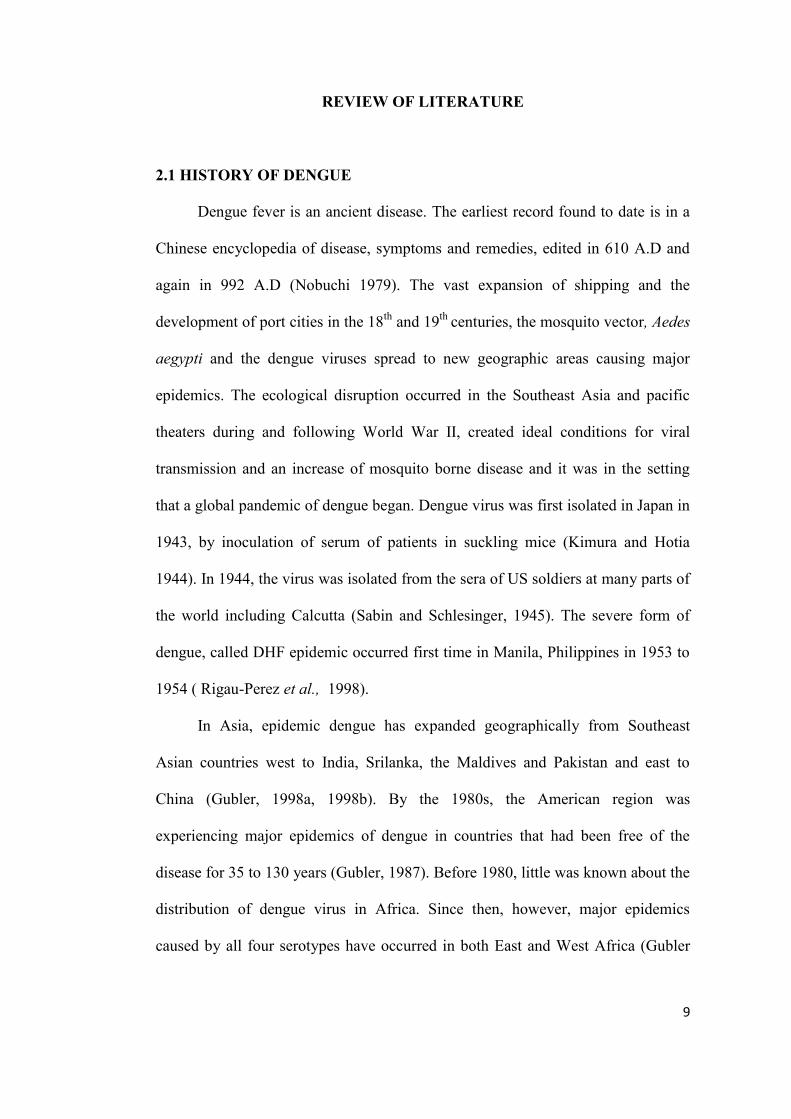
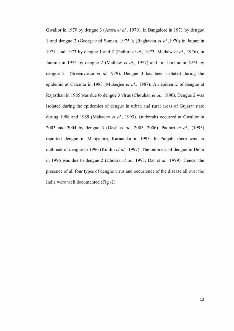
and Trent 1994). In 1997, dengue viruses and Aedes aegypti mosquitoes had a
worldwide distribution in tropical and subtropical countries of the world .Fig 1.
Figure -1
Dengue affected areas worldwide (WHO)
11
2.2 DENGUE IN INDIA
Dengue viruses have been persisting in India year after year since 1956. In
Tamilnadu, the first major outbreak of dengue was noticed in Vellore, South Arcot
district in 1961 and the viral etiology was established later by the isolation of
dengue virus (Carey et al., 1966). The first virologically proved epidemic of DF in
India occurred in Calcutta and eastern coast of India in 1963 - 1964 (Sarkar et al.,
1964; Chatterjee et al., 1965; Carey et al., 1966). Then, the dengue infection spread
northwards and reached Delhi in 1967 (Balaya et al., 1969). Subsequently, the
whole country was involved with wide spread epidemics followed by endemic or
hyper endemic prevalence of all four serotypes of dengue virus. After the
occurrence of first epidemic in 1961, Vellore experienced outbreaks in the year
1964, 1966 and 1968. The virological investigations carried out during that period
proved the presence of dengue 2 in 1964 outbreak (De Ranitz et al., 1965; Carey et
al., 1969). Dengue 3 virus was isolated in 1966 outbreak (Myer et al., 1969) and all
four types of dengue virus in 1968 outbreak (Myer et al., 1970). The epidemic at
Vishakapatnam in 1964 was due to dengue 2 virus (Krishnamurthy et al., 1965;
Paul et al., 1965).
The epidemic of dengue in Nagpur in 1965 documented the presence of
dengue 4 virus in that region (Rodrigues et al., 1972). In the same year, another
outbreak was observed in Madras which was caused by dengue 3 viruses (Myers et
al., 1968). Later, outbreaks of dengue occurred in Jabalpur (MP) by dengue virus
3 in 1966 (Sehgal et al., 1967, Rodrigues et al., 1973) in Asansol in 1967 by
dengue 2 and 4, in Delhi in 1967 by dengue 2 (Balya et al., 1969), in Kanpur in
1968 and 1969 by dengue 4 and dengue 2 (Chaturvedi et al.,1970 ; Chaturvedi et
al.,1972), in Ajmeer in 1969 by dengue 1 and dengue 3 (Ghosh et al., 1974), in
12
Gwalior in 1970 by dengue 3 (Arora et al., 1970), in Bangalore in 1971 by dengue
1 and dengue 2 (George and Soman, 1975 ); (Raghavan et al.,1970) in Jaipur in
1971 and 1973 by dengue 1 and 2 (Padbiri et al., 1973; Mathew et al., 1976), in
Jammu in 1974 by dengue 2 (Mathew et al., 1977) and in Trichur in 1974 by
dengue 2 (Sreenivasan et al.,1979). Dengue 3 has been isolated during the
epidemic at Calcutta in 1983 (Mukerjee et al., 1987). An epidemic of dengue at
Rajasthan in 1985 was due to dengue 3 virus (Chouhan et al., 1990). Dengue 2 was
isolated during the epidemics of dengue in urban and rural areas of Gujarat state
during 1988 and 1989 (Mahadev et al., 1993). Outbreaks occurred at Gwalior in
2003 and 2004 by dengue 3 (Dash et al., 2005, 2006). Padbiri et al., (1995)
reported dengue in Mangalore, Karnataka in 1993. In Punjab, there was an
outbreak of dengue in 1996 (Kuldip et al., 1997). The outbreak of dengue in Delhi
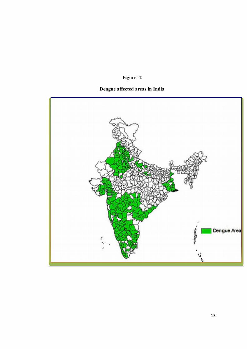
in 1996 was due to dengue 2 (Chusak et al., 1993; Dar et al., 1999). Hence, the
presence of all four types of dengue virus and occurrence of the disease all over the
India were well documented (Fig -2).
13
Figure -2
Dengue affected areas in India
14
2.3 DENGUE INFECTION IN TAMILNADU
As stated earlier, in Tamilnadu, the outbreaks of dengue were noticed in
Vellore, North Arcot district in 1961 (Carey et al., 1964, 1966 and 1968; Carey et
al., 1969; Myers et al., 1970). During this period, an outbreak was also noticed in
Madras (Chennai) in 1965 (Myers et al., 1968). The occurrence of dengue fever
was reported in villages in Dharmapuri district, Tamilnadu in 1997 (Kader et al.,
1997) and in 2001 (Victor et al., 2002). The presence of dengue fever for the first
time in Coimbatore and Erode district of Tamilnadu was reported during 1998 by
Singh et al .,(2000). There was an outbreak of dengue in Chennai in 2001 which
was caused by dengue 2 and dengue 3 viruses. (Kabilan et al., 2005). The outbreak
of dengue in Kanyakumari district in July 2003 proved the presence of dengue
serotype 3 virus in that area (Paramasivam et al., 2006).
2.4 CLINICAL DIAGNOSIS OF DENGUE INFECTION
Dengue virus infection in human causes a spectrum of illness ranging from
inapparent or mild febrile illness to severe and fatal haemorrhagic disease. Typical
clinical symptoms noted during outbreaks in India were pyrexia of 3 to 5 days
duration which at times prolonged to 10 days, gastro intestinal disorders, myalgia,
arthralgia and rash. In recent years, several studies on clinical manifestation of
dengue infection have been published (Hayes et al., 1988) which provide the basis
for the prompt diagnosis of dengue infection.
Virological investigation was made by Bhattacharjee et al., (1995) to
establish the etiologic agent of a febrile outbreak amongst a floating population of
C.R.P.F. Jawans, stationed at Calcutta during May-July, 1993. The illness was
15
associated with fever, severe headache, body ache and arthralgia which lasted for
2-4 days in most of the cases.
An epidemic of febrile illness with haemorrhagic manifestation occurred in
Mangalore city, Karnataka, India in 1993. The most observed clinical symptoms
were pyrexia, myalgia, arthralgia and headache, palatile petechia, maculopapular,
rash and facial flush. (Padbiri et al., 1995).
A prospective observational study conducted by Kalayanarooj et al, (1997)
in Bangladesh in 1994 revealed that 18 % of the dengue fever patients had
anorexia, 61 % had nausea, 66 % had vomiting, 28 % had abdominal pain and 80
% had headache. A comparative analysis of clinical symptoms between DF
patients and patients with OFI showed that DF patients were more likely to report
anorexia, nausea and vomiting than patients with OFI. However, the history of
headache, abdominal pain and bleeding were not significantly different between
these groups. Among the study groups the male: female ratio for dengue and OFI
patients were 1:1.12 and 1:1.45 respectively. The mean ages for DF and OFI
patients were 8.01(±3.15) and 6.2 (±3.0) years respectively.
Clinical symptoms of 57 hospitalised laboratory positive patients with DHF
in Puerto Rico in 1991 were fever (100 %), rash (47.4 %), hepatomegaly (10.5 %)
and petechiae (45.6 %). The male to female ratio was 1.5:1 and the mean age of the
patients was 38 years (Rigau-Perz 1997).
Deparis et al., (1998) conducted a prospective study from 1996 to 1997 in
Polynesia and found that fever (90 %), headache (92.5 %), arthralgia (84 %),
abdominal pain (61 %) and vomiting (57 %) were the commonest symptoms.
Appearance of rash (18 %), prutus (17 %), diarrhea (17 %), hepatomegaly (8 %)
and respiratory illness (19.5 %) were also observed. The association of macular
16
rash, purities, low platelet count and leukocytopenia was statistically predictive of
dengue but not clinically since these four signs occur in many viral infections.
Gascon et al, (1998) analysed 57 Spanish travelers with dengue infection
and reported the following clinical characteristics; fever and asthenia (100 %),
headache (98 %), myalgia (84 %), arthralgia (72 %), rash (61 %) and retroorbital
pain (65 %).
A work on clinical profile of dengue hemorrhagic fever in adults during
1996 outbreak in Delhi, India showed that dengue-2 was responsible for this
outbreak. All 98 patients (100 %) had fever .Other symptoms reported were body
aches (45.9 %), abdominal pain (38.7 %), purpura (33.6 %), epitasis (32.6 %),
melaena (26.5 %), haematemesis (22.4 %) and ecchymoses (20 %) (Sharma et al.,
1998).
Agarwal et al., (1999) described the clinical diagnosis of dengue fever in
206 patients with dengue fever during the 1996 epidemic in Lucknow, India. It was
found that 97 %, of the patients had severe frontal headache. 90 % had myalgia.
Skin rash was seen in 40 % of the patients, vomiting in 29 % of the patients and
arthralgia in knee and hip joints in 9 % of the patients. Anuria was present in 2
patients. Lymphadenopathy was noted in 14 %, hepatomegaly in 4 % (associated
with mild jaundice in one patient) and splenomegaly in 2 % of the patients.
Involvement of the heart and lungs was seen in one patient.
Horvath et al., (1999) studied the clinical features of hospitalized patients
during dengue 3 epidemics in north Queensland, Australia in 1997-1999 and found
that 85 % of the study population had a maximum temperature of ≥ 38 ºc.
Headache (95 %) and masculoskeletal ache (99 %) were very common. Rash was
observed in 66 % of the patients. Gastro intestinal symptoms such as nausea (92
17
%), diarrhea (65 %) and altered taste (93 %) were also common presentations.
Photophobia (45 %), cough (49 %), bleeding (63 %) and prutis (87 %) were also
observed.
The study conducted by Harris et al., (2000) on clinical epidemics of dengue
in the 1998 epidemic in Nicaragua reported that patients had myalgia (64 %),
arthralgia (62 %), vomiting (50 %), diarrhea (16 %) and abdominal pain (46 %).
Four percent of the patients had hepatomegaly.
Hepatic function of 61 children diagnosed to have dengue infection aged 2
months to 12 years comprising 37 cases of dengue fever, 16 with DHF and 8 with
DSS were prospectively studied during the acute attack in Delhi during 1999
(Mohan et al .,2000). Hepatomegaly (74 %), epitaxis (26 %), Jaundice (25 %) and
petechial rashes (18 %) were the common clinical manifestation of DF.
Eong (2001), in their findings in changing pattern of dengue transmission in
Singapore reported that males were prone to the dengue infection more than
females.
Rongrungruang and Leelarasamee (2001) made a study on characteristics
and outcome of adult patients with symptomatic dengue viral infection in Thailand
from 1990 -1997 and found that fever (100 %), myalgia (77.7 %), nausea/vomiting
(74.2 %), headache (66.5 %), abdominal pain (48 %), respiratory tract symptoms
(6.5 %) were the more common symptoms. Diarrhea (21.5 %), rigor (18.9 %),
petecheae (52.8 %), maculopapular rash (9.9 %), hepatomegaly (47.5 %) and
jaundice (1.7 %) were also observed.
A prospective study conducted by Endy et al., (2002) with primary school
children in Thailand from 1998 to 2000 and found that head ache (64 %) was the
most common presenting symptoms in children with acute dengue virus infection
18
followed by cough (43 %), rhinorrhea (35 %) lethargy (32 %), anorexia (25 %),
muscular pain (23 %), vomiting (20 %) and nausea (19 %). Other symptoms
include rash (5 %), abdominal pain (17 %), diarrhea (4 %), joint pain (15 %) and
bleeding (2 %). Headache, although the most common symptom reported for acute
dengue infection was less frequently in dengue affected children than children with
OFI. Similarly, rhinitis, cough and diarrhea were more in children with OFI than
children with dengue infection.
Kalayanarooj et al., (2002) recorded the following clinical symptoms in
patients with DF admitted in the hospital during the period 1995-1999 in Thailand.
Abdominal pain was the most common complaint (30.1 %) in DF patients. Myalgia
was seen in 12.9 % of the patients. Maculopapular rash during the febrile phase
was found in 11.6 % of the DF patients. Typical convalescence rash was found in
19.4 % of the patients. Hepatomegaly was seen in 80.4 % of the patients. The mean
age for DF cases was observed as 7.9 years. The age group 5-9 years was the most
affected group by dengue fever. No gender difference was observed in DF patients.
Narayanan et al., (2002) made a study to identify symptoms, signs and
laboratory values of dengue fever during an outbreak of dengue at Chennai in 2001
and reported that children who developed complications had more fever, body pain
and bleeding than children with dengue and did not have complications. The mean
age of their study population was 6.07 (with shock) and 6.96 (without shock).
Liu et al., (2003) made a study on a large outbreak of dengue fever occurred
in Taiwan during 2001. The patients had fever (90.8 %), body pain (57.5 %),
abdominal pain (50 %), head ache (45.8 %), cough (39.2 %), skin rash (26.7 %),
arthralgia (19.2 %), dizziness (10.8 %), myalgia (11.7 %) and retro orbital pain
(10.8 %).
19
Wu et al., (2003) in their retrospective study on 10 dengue patients with
acalculous cholescystitis from 2001 to 2003 in Taiwan recorded fever in all the
patients, diarrhea in 50 % of the patients, vomiting, myalgia and arthralgia in 40 %
of the patients, rash in 30 % of the patients and chill in 20 % of the patients.
Hepatomegaly was absent in all the patients. The mean age of the patients was 48 ±
15 years.
Kabilan et al., (2003), in their study on dengue disease spectrum among
infants in the year 2001 dengue epidemic in Chennai found fever, hepatomegaly
and rash in 100 %, 93.1 % and 55.2 % of the infants respectively. Oedema of the
lower extremity, retro orbital puffiness and vomiting were seen in 17.2 %, 27.6 %
and 24.1 % of the infants respectively. The age group of the study population was 1
to 11 months (mean age 7 months).
A cross – sectional study undertaken in children under 13 years in Colima by
Mendez and Gonzalez (2003) during the period 1992-2002 (10 years) showed that
the most important clinical features were fever and hemorrhagic manifestation (100
%), vomiting (60 %), abdominal pain (37 %), head ache (50 %), osteomyatea (
40.8 %) and macular rash (29 %).
In Thailand, Watt et al., (2003) observed lymphadenopathy (66 %), bleeding
gums (26 %) epistatis (9 %), haematesis / malena (6 %), erythematous rash (4 %)
and petechia (9 %) in the patients with dengue infection.
Pervin et al., (2004) conducted a study on clinical and laboratory aspects of
clinically suspected cases of dengue fever during 2000 dengue outbreak in Dhaka,
Bangladesh. The clinical profile of the patients showed that the mean body
temperature of the dengue patients was 101.5±1.4. Other common symptoms
included were myalgia (84.5 %), vomiting (36 %), abdominal pain (6 %), headache
20
(95 %), arthralgia (68 %), lethargy (80.4 %) and reteroorbital pain (49.5 %).
Enlarged liver was seen in 13 % of the dengue patients. The involvement of all age
group especially an adult predominance was observed. The mean age of the dengue
patients was 29 ± 12.9 years and most belonged to 20 -29 year age group.
A study conducted by Khanna et al., (2004) during an outbreak of dengue in
Delhi in 2003 revealed that fever was found in all the patients. Other symptoms
were headache (73.3 %), retro orbital pain (46.6 %), conjunctival effusion (66.6
%), arthralgia (10 %) and abdominal pain (100 %). Their study population included
35 males and 20 females. The mean age of the patients was 35.5 years (range 20 -
67 years).
It is seen from the study by Lai et al., (2004) on the characteristics of a DHF
outbreak in 2001 in Taiwan that the dengue patients presented myalgia (77 %),
arthralgia (61 %), vomiting and diarrhea (15 %) and the abdominal pain (54 %).
Enlarged liver was seen in 21 % of the patients.
In southern Vietnam, it was found that during 1996 to 1998, patients with
dengue were presented with sore throat (6 %), cough (16 %), anorexia (64 %),
vomiting (64 %), abdominal pain (62 %), diarrhea (10 %), headache (32 %),
muscle pain (10 %) and joint pain (3 %) (Phuong et al., 2004).
A retrospective study among German travelers during the period 1993- 2001
by Teichmann et al., (2004) showed that all the patients had fever and prostration
(100 %), Other symptoms were headache (86 %), arthralgia (79 %), rash (66 %)
and myalgia (48 %).
Wichmann et al .,(2004) found that out of 347 patients with serological
confirmed dengue infection during an epidemic in 2001 in Thailand, 26 % had
cough,15 % had headache, 8 % had myalgia, 40 % had hepatomegaly, 21 % had
21
petechiae, 57 % had nausea and 59 % had vomiting. All patients were presented
with fever (100 %). Forty percent of patients had hepatomegaly. The total number
of patients included in their study was 347 of which 287 were children and 60 were
adults. The male: female ratio was 1.08:1 and the median age was 10 (4 months to
66 years).
A comparative study of clinical features between children with dengue fever
and DHF in Manila in 1999 by Carlos et al., (2005) revealed that children with
dengue fever had abdominal pain (39.1 %), epistaxis (19.7 %), petchiae (81.6 %)
and gum bleeding (4.7 %).The mean age of the patients was 9.9 and male: female
ratio was 1:0.49.
A study was conducted during a large urban epidemic of dengue fever in
Kollam city of Kerala in 2003 by Daniel et al., (2005).The clinical manifestations
found were fever (96.8 %), headache (77.2 %) sore throat (5.2 %), diarrhea (15.2
%), abdominal pain (62.4 %), skin rashes (13.2 %) and pruritis (10.4 %). The study
population consisted of 130 males and 120 females. The patients mean age was
12.6 ± 20 and the mean duration of symptoms was 6 days.
Espinoza –Gomez et al., (2005), in their study on clinical pattern of
hospitalised patients during dengue epidemic in Colima, Mexico observed bleeding
and hepatomegaly in patients with dengue infections. The mean age of the dengue
confirmed patients was 23.3 years in 137 males and 21.6 years in 152 males.
Gonzalez et al., (2005) reported that during a dengue -3 epidemics in 2001
to 2002 at Havana city Cuba, fever (100 %), headache (92.1 %), myalgia (76.3 %),
arthralgia (73.7 %) and retro orbital pain (57 %) were the most frequent general
symptoms. Vomiting and abdominal pain were observed in 59.2 % and 48.6 % of
cases respectively. Hepatomegaly was seen in 1.8 % of the patients. All the
22
patients studied were adults (16 -64 years).The maximum incidence was seen in
ages 25 to 44 years. Thirty one patients were males and sixteen were females.
Seet et al., (2005) observed that during an outbreak of dengue infection in
China, the patients had vomiting (38 %), diarrhea (41 %) and abdominal pain (21
%). No one was found to have hepatomegaly.
Singh et al., (2005) found that during an outbreak of dengue fever in Delhi,
India in 2003, fever was presented in all the febrile cases with an average duration
of fever being 4.5 ± 1.2 days. Other symptoms include headache (61.6 %),
backache (57.8 %), vomiting (50.8 %), abdominal pain (21 %), haemorrhagic
manifestation in the form of a positive tourniquet test (21 %), gum bleeding and
epitaxis (40 %), skin rashes (20 %) and malaena (14 %).
A comparison of the prevalence of various signs and symptoms of dengue
between age groups of 0 - 54 in California from 1999 to 2001 revealed that
headache, arthralgia, myalgia and retro orbital pain were present in more than 60 %
of children and adults with confirmed dengue. External bleeding and chills were
also present in more than 50 % of children. Fever, external haemorrhagic
manifestations and rash were present in more than 50 % of infants (Hammond et
al., 2005).
IraShah and Kathira (2005) undertook a prospective study in Mumbai, India
in 2003 to determine the clinical features of dengue. The mean age of presentation
was 4.9 years. Fever (100 %), hepatomegaly (47.1 %), vomiting (50 %), bleeding
(38 %), tenderness (38.2 %), and erythematous rash (14.7 %) were seen in the
patients. Thrombocytopenia was the predominant clinical features observed.
A study on profile of liver involvement in dengue virus infection during an
outbreak of dengue infection in Lucknow, India from September 2003 to
23
December 2003 reported fever in all the patients (100 %) with a median duration of
6 days. In addition, 53 % of the patients had petechial rash, 36 % had pain in the
right hypochondrium, 22 % had gastrointestinal bleeding and 18 % had
neurological symptoms. Hepatomegaly was observed in 24 % of the patients and
none had splenomegaly. The median age of the patients was 33 (range 7-65) years.
Of the 45 patients, 87 % were adults (> 18 yrs) (Itha et al., 2005).
Kularatne et al., (2005) reported that patients with dengue fever in Srilanka
in 2001 had myalgia (74 %) and flushed appearance (62 %). The mean age of the
404 patients studied over a period of 2 years was 30 years and the mean duration of
fever was 7 days.
A comparative study of clinical symptoms between the dengue and non–
dengue patients admitted in a hospital in Bangkok from 1999 -2000 revealed that
days of fever and clinical manifestation were not significantly different between
them. Out of 49 patients with dengue 28 were males and 21 were females with a
mean age of 8.8± 3.5 years (Mekmullica et al., 2005).
Ahmed et al., (2006) in their study on dengue fever outbreak in Karachi
from 2005 to 2006 reported fever in all the patients. Chills and rigor were noticed
in 80 %, myalgia in 67 %, headache in 54 %, pharyngitis in 35 %, rash in 28 % and
bleeding manifestation in 2 % of the patients. Hepatomegaly and
lymphoadenopathy were observed in 0.5 % of the patients. Age of their patients
ranged between 14 and 67 years (mean 31 years) and all were males.
A prospective study carried out by Ayyub et al., (2006) on characteristics of
dengue fever in Jeddah, Saudi Arabia from May 2004 till April 2005. They found
that the commonest clinical presentation was fever (100 %), headache (48.72 %),
myalgia (66.7 %) and vomiting (25.64 %). Rash, hemorrhagic manifestations and
24
positive tourniquet test were relatively uncommon. Hepato splenomegaly (12.56
%) was also uncommon.
Itoda et al., (2006) made a study on clinical features of the imported cases of
dengue fever in Japan between 1985 and 2000. They found fever in all the cases
and the mean duration of fever was 5.6 ±2 days. Small macular rashes were seen in
82 % of the patients. Skin itching was present in 74 % of the cases. Headache (90
%), profuse sweating (61 %), myalgia (60 %), retro orbital pain (55 %), chills (56
%), diarrhea (53 %), hemorrhagic manifestation (45 %) and positive tourniquet test
(30 %) were the other clinical symptoms reported. The mean age of their study
groups was 31 ±10.5 years with a range from 18 to 62 years. The numbers of males
were 44 and females were 18.
In Taiwan, patients with DHF/DSS admitted in the hospital in 2002 showed
clinical symptoms such as fever (100 % ), myalgia (35.7 %), nausea and vomiting
(35.7 %), chills (28.6 %), skin rashes (21.4 %), diarrhea (21.4 %), headache (14.3
%) and dizziness (7 %). The median age of the patients was 44 years with range of
15 -68 years (Khor et al., 2006).
Clinical characteristics of dengue and DHF in a medical centre of southern
Taiwan during the 2002 epidemic (Lee et al., 2006) showed that the most common
symptoms were fever (96.1 %), myalgia (68.5 %), headache (55.4 %), skin rash
(53.7 %), anorexia (53.7 %) and malaise (49 %). Abdominal pain (42 %), nausea
and vomiting (39.9 %), cough (37.6 %) and diarrhea (35 %) were also seen.
Pruritus (22.7 %) and retro orbital pain in 15.8 % were also noted.
A study carried out by Malavige et al., (2006) in 2004 on the patterns of
disease among adults hospitalized with dengue infection in Srilanka revealed that
76 % had myalgia, 57 % had arthralgia, 64 % had vomiting , 29 % had diarrhea
25
and 17 % had abdominal pain. Hepatomegaly was seen in 30.3 % of the patients.
Of the study population, 59.3 % were males and 40.7 % were females and mean
age was 26.6 years.
Pichardo et al., (2006) found that 41 serologically confirmed dengue patients
in Mexico were presented with fever, headache, retro orbital pain (93 %), myalgia
and arthralgia (100 %), vomiting and abdominal pain (44 %), hepatomegaly (4.8
%) and petechiae (4.8 %). The male to female ratio of the study population was
23:18 and the mean age was 34 for females and 23 for males.
Waduge et al., (2006) found that the pregnant woman with dengue infection
in Srilanka during 2000-2004 had fever (100 %), myalgia (65.4 %) and arthralgia
(30.7 %). The mean age of the patients was 29 ± 4.2 years.
Wichmann et al., (2006) compared the clinical features of 69 dengue
confirmed cases to 1035 febrile non dengue patients. Fever was the commonest
presentation (81.2 %) in dengue confirmed patients followed by headache (62.1
%), Muscle pain (50 %), diarrhea (43.5 %), nausea (22.4 %), and skin rash (23.5
%). When comparing patients with and without dengue infection, there was no
significant difference in the frequencies of major symptoms except in the
occurrence of skin rash (23.5 % vs. 9 % respectively).
In dengue patients with acute respiratory failure in Taiwan in 2002, the
following clinical symptoms were observed- Fever (90.9 %), petechiae (36.4 %),
bone pain (45.5 %), myalgia (45.5 %), headache (27.3 %), chest pain (18.2 %),
cough (45.5 %), abdominal pain (54.5 %) and vomiting (36.4 %). The mean age of
the patients was 63.09 ± 13.48 with a range of 33 -78 years, (Wang et al., 2007).
Khan et al., (2008), studied 160 clinically suspected patients in Saudi Arabia
in 2004 and confirmed the dengue infection in 91 patients. Most of the patients
26
were young adults with, median age of 26 (range = 6 -94) years and male: female
ratio of 1.5:1. The common symptoms were fever (100 %), malaise (83 %),
musculo skeletal pain (81 %), head ache (75 %), nausea (69 %), vomiting (65 %)
and abdominal pain (48 %).
Banerjee et al., (2008) studied 50 cases of fever clinically suspected to be
dengue in Pune, India. The commonest clinical feature was fever with rash (85 %).
Retro orbital and headache were reported by 63 % of the patients. Myalgia was
seen in 81 % of the patients. Hepatomegaly was seen in 15 % of the patients.
Conjunctival congestion was observed in 37 % of the patients. The frequencies of
clinical symptoms were comparatively higher in IgM positive patients than IgM
negative patients.
A retrospective study conducted in Thailand in 2004 and 2005 by Hanafusa
et al., (2008) revealed that headache and myalgia were more common among
adults (P< 0.05), but cough, vomiting, abdominal pain and rash were more
common among children (P< 0.05). Nasal bleeding was more common in children
and gum bleeding was more common in adults. Thus, adults showed different
clinical manifestations of dengue infection from children.
Kumar et al., (2008) conducted a study with 27 dengue positive children in
Lucknow, India in 2006. Clinical features of dengue IgM positive cases included
bleeding (57 %), convulsion (50 %), rash (14.3 %), swelling (28.6 %), headache
(21.4 %) and vomiting (35.7 %). No one was found to have diarrhea and there was
no significant differences in clinical feature among dengue IgM positive and
negative cases. Hepatomegaly was observed in 64.3 % of the patients.
Khan et al., (2008) described the clinical profile of patients with dengue
virus infection in Makkah, Saudi Arabia in 2004. The common symptoms observed
27
were fever (100 %), malaise (83 %), musculo skeletal pain (81 %), head ache (75
%), nausea (69 %), vomiting (65 %) and abdominal pain (48 %). Most patients
were young adults with median age of 26 (range = 6-94 years) and male to female
ratio was 1.5:1.
Clinical profile of dengue fever infection in Saudi Arabia in 2005 – 2008
was studied by Ahmed et al., (2010). The report revealed that the common clinical
presentation of the dengue patients in these four year were fever (66.67 %, 84.58
%, 50 % and 62 .16 %), vomiting (66.67 %, 45.45 %, 25 % and 40.54 %) and
abdominal pain (33.33 %, 36.36 % and 27.03 %, except 2007). Relatively less
common clinical features were haemorrhagic manifestation, headache, rash and
retro-orbital pain.
Chau et al., (2010) conducted a prospective descriptive study in Vietnam in
2007 and observed clinical symptoms such as fever (83 %), diarrhea (43 %),
running nose (37 %), cough (56 %), vomiting (57 %), jaundice (1 %) and petechial
rash (34 %). Infants with dengue did not present specific clinical signs compared to
patients with OFI. Common features of upper respiratory tract viral infection
including running nose and cough were observed with similar frequencies in both
groups, however diarrhea, vomiting and petechial rash occurred more frequently in
dengue patients than in infants with OFI. The age of study population ranged
between 2 and 18 months (median – 7 months) and the male to female ratio was
173:126.
Kumar et al., (2010) studied the clinical manifestation of confirmed dengue
cases admitted in a tertiary hospital, in Karnataka, India from 2002 to 2008. Their
study included 466 patients with an age group of 15 - 44 years. The most common
clinical presentation was fever (99.1 %), followed by myalgia (64.6 %), vomiting
28
(47.6 %), headache (47.6 %) and abdominal pain (37.6 %). Skin rash was seen in
21.7 % of the patients. Other symptoms included petechiae (18 %), diarrhea (13. 9
%) and gum bleeding (5.2 %).
A retrospective cross sectional study conducted in Pakistan from 2003 to
2007 on demographical and clinical features of dengue fever by Khan et al., (2010)
revealed that the three most common presenting clinical features were nausea (59.3
%), rash (36.4 %) and myalgia (25.8 %) which were followed by haemorrhage
(18.2 %), diarrhea (16.3 %), cough (11 %) and headache (11 %). The median age
of IgM positive patients decreased every year from 32 years in 2003 to 24 years in
2007.
Low et al., (2011) conducted a prospective study of adults to find out the
early clinical features of dengue in adults in Singapore from 2005 to 2010. The
patients had symptoms such as drowsiness (59.2 %), headache (80 %), myalgia
(69.2 %), arthralgia (60.8 %), loss of appetite (81.2 %), abdominal pain (1.6 %),
diarrhea (14.8 %), nausea (50 %) and bleeding (5.2 %). The body temperature was
higher in patients with dengue compared to OFI. The symptoms such as Myalgia,
arthralgia and retro-orbital pain and muscosal bleeding reduced significantly with
increasing age.
Mia et al., (2010) conducted a cross sectional study on clinical and
sonographic evaluation of dengue in Bangladesh during the period March 2008 to
February 2009 and reported that the clinical manifestations of dengue confirmed
patients were fever (98 %), nausea /vomiting (76 %), musculo skeletal pain (61 %),
head ache (34 %), anorexia (32 %), generalized weakness (31 %), abdominal pain
(14 %), retro-ocular pain (13 %) and restlessness (11 %). Hepatomegaly was seen
in 48 % of the patients.
29
Parkash et al., (2010) carried out a cohort study of in patients with dengue
viral infection in Karachi. A total of 699 patients with 65 % of males were studied.
The mean age of the patients was 31.87 ± 13.55. The mean duration of fever was 6
± 3.27 days with a mean temperature of 38.5 ± 1ºC. Among clinical features, 48.6
% had nausea /vomiting, 18.2 % had abdominal pain, 17 % had rash, 16 % had
body ache, 10 % had diarrhea, 9 % had cough, and 5.4 % had hematamesis. 5.2 %
had bleeding gums, 4.3 % had malena and 3.1 % had jaundice.
Arshad et al., (2011) conducted a prospective study on 106 seropositive
(IgM positive by ELISA) patients with dengue fever from June 2008 to March
2009. The mean age of the patients was 29.6 ± 11.6 (range 6 to 60 years). Fever
was the most common clinical presentation found in 88 patients (86 %). There
was no specific pattern of fever and the temperature ranged from 38 º to 40 º C.
Other common clinical features were headache and myalgia (66.7 %), vomiting
(25.64 %) and diarrhea (20.51 %). A maculopapular rash was seen in 29 patients
(29.5 %).
Anker and Arima (2011) in their regional analysis of male, female difference
in the number of reported incident of dengue fever cases in 6 Asian countries
explained that in Philippines, there was a significant excess of male cases among
those ≥ 15 years of age and among infants. A high proportion of male cases were
also recorded in Singapore, Srilanka, Malaysia and Cambodia.
Nascimento et al., (2011) observed fever (94.1 %), headache (88.2 %),
myalgia (88.2 %), arthralgia (54.4 %) nausea and vomiting (69.1 %), rash (51.5
%), asthenia (52.9 %), abdominal pain (51.5 %), retro orbital pain (50 %) as
common clinical symptoms in dengue patients confirmed by IgM ELISA in Brazil
30
in 2007. Other symptoms include hepatomegaly (13.2 %), hypotension (13.2 %)
and dizziness (32.4 %).
Jain et al., (2011) analysed sera samples of dengue patients in Jhansi, Uttar
Pradesh during the period January -December 2010. Among the study population
34 % were females and 66 % were males and female to male ratio was 1:2.9. The
symptoms found were bodyache (84 %), headache (77 %), high grade fever (73
%), joint pain (42 %), symptomatic bleed (32 %), malaise (27 %) and apparent
jaundice (15 %).
2.5 SERODIAGNOSIS OF DENGUE INFECTION
Serodiagnosis and seroepidemilogical survey on dengue infections have
been carried out mostly by Haemagglutination Inhibition (HI) test for many years.
This technique was developed for demonstrating an increase in antibody titer
during infections with arboviruses (Sabin and Buscher 1950). Then, HI test was
perfected by Clarke and Casals (1958) and was adapted to microtitre plate by Sever
(1962).This technique is highly sensitive but it lacks specificity and requires paired
samples for accurate diagnosis (Vorndam et al., 1997).
A study on dengue outbreak reported in Brazil in 1986 by Nogueira et al., (1989)
revealed that 58.2 % of the patients tested by ELISA had IgM antibodies to dengue virus
and they were considered as confirmed dengue patients for the further studies.
Innis et al., (1989) found that in Jharkand, anti dengue IgM appeared in most
cases by the 3rd
day of febrile illness and declined to undetectable level after 30 –
60 days. IgM capture ELISA showed 78 % sensitivity in acute serum and 97 % in
paired sera. Dengue infections could be classified as primary or secondary by
determining the ratio of units of dengue IgM to IgG antibody.
31
According to Henchel and Putnak, (1990), ELISA has been the most widely
used method in the past, the extensive cross reaction encountered and the non
availability of results within a short period of time due to requirement of both acute
and convalescent sera collected at least seven days apart, have compromised the
general applicability of this assay in the diagnosis of dengue.
Chouhan et al., (1990) detected IgM antibodies to dengue viruses in 70 % of
sera collected in Rajasthan in 1985.
Chen et al., (1991) carried out an investigation on the detection of IgM
antibodies from cerebrospinal fluid and sera of dengue fever patients. The results
showed that IgM could be detected in 14 (70 %) out of 20 suspected dengue
patients. Sera IgM antibodies last up to 252 days after onset of illness. Mehendale
et al., (1991) found antidengue IgM antibodies in 46.6 % of sera collected during
an outbreak of dengue occurred in 1988 in few villages of Maharastra.
Laferte et al., (1992) reported the standardization and evaluation of 10µl
ultra micro ELISA for antidengue virus IgM detection. Compared with HI, this
system showed 85.7 %, sensitivity and 100 % specificity.
In Senegal, a study was conducted about the circulation of arboviruses
during the transmission season. Specific IgM antibodies were detected by ELISA
test in human sera as a sign of recent infection (Monlun et al., 1993).
A kit for the detection of anti dengue virus IgM antibody based on detecting
dengue virus specific IgM antibodies in the test serum by capturing them with an
antihuman IgM has been developed by Pelegrino et al., in 1994. This system had
92 % sensitivity, 100 % specificity, 94 % coincidence in single acute phase serum
samples as compared with results for sera from same patients tested by HI.
32
Padbidiri et al., (1995) found virus specific IgM antibodies in 25 % of sera
collected immediately after onset of illness during an epidemic in 1993 in
Mangalore, Karnataka.
Another most widely used technique till today is ELISA which has been
considered as the most useful test for dengue diagnosis due to its high sensitivity
the ease of use and there is no use of sophisticated equipment. Moreover ELISA
has been used to detect acute phase (IgM) and covalescent phase (IgG) antibodies.
There has been several report on the confirmation of dengue infection by using
ELISA throughout the world (Guzman and Kouri 1996).
Yabe et al., (1996) tested suspected dengue cases in Japan during the period
of 1985 -1995 by IgM capture ELISA. Sera of dengue cases showed high degree of
cross reactivity to Japanese Encephalitis virus in HI test but not in IgM capture
ELISA. Ninety three (53 %) out of 173 cases of febrile illness of unknown etiology
was either confirmed or positively suspected to dengue fever by serology.
Chye et al., (1997) in their case study during Malaysian dengue epidemic in
1996 confirmed dengue infection by the detection of IgM antibodies specific to
dengue virus by an ELISA on days 4 and 10 in one patient and on days 6 and 11 in
another patient. Rigau – Perez and the Puerto Rico association of epidemiologist
(1997) confirmed dengue infection in 46.1 % of cases by virus isolation in
mosquito cell cultures, immuno fluorescence assay and by MAC ELISA in Puerto
Rico in 190 and 1991.
An evaluation of commercial capture immunosorbent Assay for detection of
immunoglobulin M and G antibodies produced during dengue infection was carried
out by Chew et al., (1998). Pan Bio kit was evaluated with paired serum
specimens from 176 patients and compared with HI. They proved that ELISA
33
should be useful in the clinical diagnosis of dengue infection. Similarly, an
evaluation of a commercial ELISA kit for the detection of IgM during dengue
infection by Sang et al., (1998 b) revealed that, primary dengue infection was
detected in 84 (93%) of 90 patients and secondary dengue infection was detected in
46 (79 %) of 58 patients.
Ram et al., (1998) carried out an investigation on an outbreak of dengue
fever occurred in Ludhiana in 1996 and 1997. Serological examination was
performed by dengue IgG and IgM blot with single serum samples of 189 patients.
Of these 129 (68.25 %) samples were detected positive for anti dengue antibodies.
Laboratory methods to confirm a clinical diagnosis of dengue was evaluated
by Rossi et al., (1998). In their study, they identified 93 (6.68 %) out of 1414 acute
sera were positive for IgM.
A comparative study of Pan Bio duo ELISA and MRL dengue fever IgM
capture ELISA for the diagnosis dengue virus infection in Southeast Asia was done
by Cuzzubbo et al., (1999). Eighty sera from dengue patients and 24 sera from
Japanese Encephalitis patients and 78 sera from patients with non Flavivirus
infections such as typhoid, malaria leptospirosis and scrub typhus were used for the
study. Comparing the specificity and sensitivity of the tests at different cut off
values revealed that MRL performed similarly in distinguishing dengue virus from
non flaviviruses and PanBio showed significantly better distinction between
dengue virus and JE virus.
An outbreak of DHF/DSS occurred in 1996 in New Delhi, India was studied
by Dar et al., (1999) and they reported that out of 270 serum samples tested by
MAC – ELISA for the detection of IgM antibodies against dengue virus, 140 (51.9
%) showed anti dengue IgM antibodies. All the samples from patients with
34
duration of fever > 5 days were tested for anti–dengue IgM antibodies. In some
samples, antibodies could be detected as early as the fifth day of fever. Three of the
culture positive acute phase serum samples were also positive by MAC ELISA.
Havorth et al., (1999) classified patients as dengue probable on the basis of a
positive ELISA test for IgM antibodies. According to them, classification of
dengue cases required viral detection either by Polymerised Chain Reaction (PCR)
or viral culture. ELISA was performed on serum from 99 patients and was found to
be positive in 85 (85%) patients.
Kalayanarooj et al., (1999) made a serological study on dengue patients
enrolled between 1994 and 1997 in Thailand and the result revealed that 313 (98.4
%) out of 318 dengue patients were confirmed serologically either by ELISA or by
HI or by both assays. Among the serologically confirmed dengue patients, 21.09 %
had primary dengue and 78.91 % had secondary dengue infections.
IgM capture ELISA was positive as early as on day 4 after the onset of fever
in dengue suspected Japanese patients and it was concluded that combination of
RT-PCR and IgM capture ELISA increased the ability to diagnose dengue virus
infection, even in a single serum obtained from the patients (Yamada et al., 1999).
A survey made by Amin et al., (2000) on sporadic cases of dengue occurred
in Bangladesh in 2000 showed that an average ELISA positive rate in that region
was 17.5 %. Their study population included 107 (53.5 %) males and 93 (46.5 %)
females. The age structure showed that the 5 to 9 year age group dominated with
36 % of the total samples collected.
Lam et al., (2000) evaluated ELISA for combined determination of
Immunoglobulin M and Immunoglobulin G antibodies produced during infection
by dengue virus. They used commercially available Pan Bio ELISA that utilizes
35
both IgM and IgG capture in the same microtitre well for the diagnosis of dengue
infection. Sensitivity in the detection of primary and secondary dengue infections
was 95 % and specificity was 94 %.They concluded that PanBio dengue screening
ELISA is best suited for routine diagnosis where large numbers of samples are
tested and cost is an issue and when it is not necessary to distinguish between
primary and secondary infections.
In 1998, 20 hospitalised cases of fever in Coimbatore and Erode districts in
Tamilnadu, India were tested for IgM and IgG antibodies and all were found to be
positive. All of them had dengue compatible illness and at least four of them had
DHF (Singh et al., 2000).
Yamada et al., (2000) analysed the serum samples collected from dengue
suspected cases in Japan in 2000 by IgM capture ELISA for the detection of IgM
antibodies. Out of 44 suspected cases, 19 (43.2 %) were confirmed as dengue by
ELISA. Similarly the presence of dengue virus in 50 children with DHF/DSS in
Indonesia was confirmed by detecting IgM and IgG antibodies by indirect ELISA,
Van Gorp et al., (2001).
A hospital based cross – sectional serodiagnostic survey under taken by
Chakravarthi et al., (2002) during 1999-2001 in Delhi, India showed that, out of
345 patients experiencing a febrile episode, 85 cases (25 %) were confirmed as
serologically positive, with 15 cases showing IgM antibodies indicating primary
infection and 19 cases showing both IgM and IgG antibodies indicating secondary
infection
Kalayanarooj et al., (2002) confirmed dengue infection in the patients with
suspected dengue infection during the period 1995-1999 in Thailand by virology or
by serology. ELISA and HI assays were carried out for serological confirmation of
36
the infection. Out of 4743 patients tested by serology and virology 22.7 % were
found to have primary dengue infection and 77.3 % had secondary infection.
Primary infection ranged from 20.1 % in 1999 to 24.3 % in 1997 and secondary
infection ranged from 75.7 % in 1997 to 79.8 % in 1999.
Narayanan et al., (2002) identified 89 children in a hospital in Chennai,
Tamilnadu between October to December 2001 as probable dengue cases by
clinical suspicion. For all cases, the rapid IgM-IgG capture ELISA was done and
59 (66.29 %) were found to be seropositive for dengue.
A study was made by Pasca et al., (2002) on dengue in Kuwait. They tested
909 samples by using ELISA (Pan Bio Australia) and reported dengue in 19 (2 %)
patients.
. A survey conducted by Victor et al., (2002), on dengue outbreaks in two
villages of Dharmapuri district in Tamilnadu, India in 2001 revealed that 13 (41.9
%) of 31 and 14 (26.9 %) of 52 patients tested in Kadumuchandiram and Mampatti
villages respectively were positive for IgM antibodies to dengue virus by MAC
ELISA. The virological and serological investigation confirmed that the outbreak
of fever were due to dengue virus.
In Czech Republic, from 1997 - 2002, 16 out 89 patients (17.98 %) were
found to have IgM antibodies to dengue virus and from 1998 - 2008, 16 out of 328
patients (4.88 %) had IgM antibodies to dengue ( Chalupa et al., 2003).
During an outbreak of dengue in Taiwan in 2001, patients with suspected
dengue infection were diagnosed by either PCR or ELISA for specific dengue IgM
in acute phase serum or four fold increase of dengue specific HI titres in
convalescent serum. There were more than 5000 cases, of which 450 (9 %) patients
37
were found to have dengue infection by any one of the above methods (Liu et al.,
2003).
Fakeeh and Zahi (2003) examined a total of 1020 suspected clinical cases by
laboratory methods such as virus isolation, IgM capture ELISA and PCR for the
confirmation of dengue infection in Saudi Arabia during 1994 - 2002. The dengue
virus isolation was confirmed in 65.5 % of the patients and IgM was detected in
34.5 % of patients.
Vazquez et al., (2003) in their study on diagnosis of dengue virus infection
by visual and simple Aubio DOT immunoglobulin M capture system found that 87
% of 150 serum samples tested by MAC –ELISA had IgM antibodies.
Study on dengue fever in Japan in 2001 by Yamada et al., (2003) confirmed
dengue virus infection by IgM capture ELISA, rapid immunochromatographic test,
HI and PCR in 35 (46.05 %) out of 76 suspected cases .
IgM Capture ELISA for the serodiagnosis of dengue using ß –Propiolactone
- inactivated dengue virus was developed by Schilling et al., (2004). They studied
on sensitivity of ELISA with serum samples of 43 patients with primary and
secondary dengue infections. They concluded that early diagnosis of dengue can be
obtained by IgM capture ELISA.
Moura et al., (2004) has given a case report of 28 years old man in 2001 in
Brazil and he stated that occurrence of DHF was confirmed by the presence of IgM
for dengue virus by ELISA
A study conducted by Pervin et al., (2004) on clinically suspected cases of
dengue fever in Bangladesh during June to December 2000 revealed that, of the
105 clinically suspected dengue patients, 39 (37.1 %) were positive for either
38
antidengue IgM or IgM and IgG antibodies and 38 (36.2 %) patients developed
only anti dengue IgG in the acute stage of the illness.
Phuong et al., (2004) studied 1,136 children with suspected dengue infection
from 1996 to 1999 in Vietnam and found that 712 were confirmed to have acute
dengue infection either by IgM ELISAand /or by virus isolation.
Souza et al., (2004), in their study on an outbreak of dengue by type 3
dengue virus in 2002 in Brazil confirmed the dengue infection by the detection of
IgM antibodies in 2314 (60.1 %) out of 3850 patients.
Whichmann et al., (2004) confirmed the dengue infection with suspected
DHF in 2001 in Thailand by ELISA. Out of 906 patients, 347 (38.3 %) were
serologically confirmed as DHF patients.
Witayathawornwong (2005) studied 1465 cases of hospitalized DHF patients
in Thailand from 1999 to 2002 and about 82 % of cases were serologically
confirmed by using either ELISA or HI. The male to female ratio was 1:1.03 and
the patients age ranged from 80 days to 15 years with a median of 9 years.
Espinoza – Gomez et al., (2005) examined sera from 340 patients by MAC
ELISA and found that 267 (78.53 %) were IgM positive for dengue virus and 22
(6.4 %) were positive by RT-PCR were in Colima, Mexico in 2002.
Carlos et al., (2005) used IgM ELISA and RT-PCR for the confirmation of
dengue infection in patients admitted in the hospital in Philippines between 1999
and 2001. They assumed that the diagnostic sensitivity was 90 -93 % for IgM
ELISA and 80 – 100 % for RT –PCR and so the combination of both test will
increase the sensitivity of diagnosis to more than 90 %. Of the 503 subjects
screened, 359 (71.4 %) were confirmed as having dengue virus infection. 322 (89.7
39
%) by IgM capture ELISA and 139 (38.7 %) by RT-PCR. A total of 102 (28.4 %)
had positive result for both tests.
During October to December 2003 in northern India, Dash et al., (2005)
observed that 12 out of 76 (22 %) patients had a positive IgM response, indicative
of primary infection, and 22 of them (42 %) revealed only IgG antibodies,
indicative of secondary infection.
IraShah and Katira (2005) reported that in Mumbai, India in 2003, out of 69
suspected dengue cases tested by ELISA for dengue IgM antibodies. 34 (49.3 %)
patients had a positive dengue IgM titre. Similarly, Kalita et al., (2005) reported
that in Lucknow, diagnosis of dengue was based on the results obtained by IgM
ELISA.
Kabilan et al., (2005) made a study on the 2001 dengue epidemic in
Chennai. The sera were tested for dengue specific IgM and IgG antibodies by IgM,
IgG antibody capture ELISA (Panbio, Australia and Omega, Scotland). Of 192
children enrolled in the study 143 (74.5 %) were laboratory confirmed for dengue,
consisting of approximately equal number of males (53 %) and females (47 %).
Kularatne et al., (2005) found that in Srilanka in 2007 out of 404 patients
enrolled for the study, 239 (59.15 %) patients had either IgM alone (18 %) or IgG
and IgM (58 %) and IgG alone (12 %).
Lee et al., (2005) diagnosed DF patients based on positive PCR result, a
positive ELISA result for specific IgM antibody to dengue virus in acute phase
serum or a four fold increase in dengue specific HI titres in convalescent serum in
Taiwan during 2002.
40
Ageep et al., (2006) conducted a study in Portsudan in 2005 on 84 patients with
febrile illness and the diagnosis was confirmed by ELISA by the detection of
dengue virus IgM antibodies in 88 % of the patients, sera.
In Saudi Arabia, from 2004 to 2005, eighty patients were admitted with
suspected diagnosis of DF. Of these, 39 (48.75 %) were found to have positive
serology (IgM or IgM and IgG) to dengue virus by ELISA. Among these 39
confirmed dengue patients 30 were males while the rest were females giving a
male to female ratio of 3.3:3. Their ages ranged from 2 years to 60 years with a
mean age of 27.6 ± 11.2 years (Ayyub et al., 2006).
A study in Manipal by Baruah et al., (2006), revealed that, out of the 100
clinically suspected cases of dengue, 44 % tested were positive for dengue IgM
antibody, thus proving the current dengue infection.
A large outbreak of dengue and DHF occurred from August to November
2005 involving all districts of West Bengal, India. Altogether, 6293 persons were
serologically diagnosed to be suffering from dengue through the detection of IgM
antibodies by ELISA (Hati, 2006).
Malavige et al., (2006) investigated clinical and laboratory findings in adult
dengue patients hospitalized in Srilanka during the major outbreak of dengue in
2004. Dengue virus specific antibodies were measured using the Panbio dengue
duo IgM and IgG rapid test. Of those who presented to hospital with clinical
features suggestive of dengue infection, 108 were diagnosed as having infection
based on serology.
An outbreak of febrile illness was first time observed in the three villages of
Kanyakumari district Tamilnadu, India in July 2003 and serological, virological
and entomological investigations were carried out to confirm the etiology of
41
outbreak. Of the 76 samples tested by Panbio ELISA kit for the detection of IgM
antibodies, 15 (20 %) were found to be positive for dengue virus specific IgM
antibodies. It was concluded in the study that based on the IgM antibody capture
ELISA results it was evident that the current infection was caused by dengue virus
in the affected areas. All the age groups were affected during the outbreak
(Paramasivam et al., 2006).
In a study carried out in Colombo, Srilanka from 2000 – 2004, dengue viral
specific antibodies were detected using the Panbio TM dengue duo IgM and IgG
rapid strip test in all hospitalized pregnant mothers and it was found that 17 (65.4
%) out of 26 patients had only IgM dengue specific antibodies and 9 (34.7 %) had
both IgM and IgG dengue specific antibodies (Waduge et al., 2006).
. Souza et al., (2007a) evaluated the ability of three ELISA based methods to
discriminate primary from secondary dengue infection. All three assay exhibited
sensitivity and negative predictive value of 100 % for defining secondary infection.
All three ELISA based assays proved reliable tools for discriminating between
acute, primary and secondary dengue virus infection when using serum samples
from convalescent phase patients.
Valero et al., (2007) in Venezuela, confirmed dengue infection in 184
patients by the detection of antidengue IgG and IgM antibodies by ELISA.
Banerjee et al., (2008) reported that in Pune, India, out of 50 dengue
suspected patients 27(54 %) had IgM antibodies for dengue and it was stated that
the serological sensitivity of ELISA (Panbio diagnostics) kit used in that study was
85.4 % - 98.9 % for the detection of primary infection and the specificity was 95.7
% -100 %.
42
Faridi et al., (2008), in their study on clinical and biochemical profile of
dengue haemorrhagic fever in children in Delhi reported that IgM dengue serology
was positive in 68.5 % of the cases.
Villar –Centeno et al., (2008) in their study on DHF patients in Colombia
defined dengue virus infection as any of the following: Virus isolation or IgM
positive by MAC ELISA or > fourfold increase in antibody level to dengue virus.
Of 508 enrolled patients, dengue infection was confirmed in 203 (40 %) patients by
either virus isolation or by detection of IgM against dengue virus.
Over a period of 10 weeks from July to September 2006, children admitted
in hospital with acute hepatic failure in Lucknow, India were examined for the
presence of IgM to dengue virus by IgM capture ELISA (Panbio, Australia). Out of
27 children, 13 (48.1 %) were positive for dengue IgM. Serum samples of 7
randomly selected IgM positive patients were subjected to real time PCR assay of
which 4 were positive. (Kumar et al., 2008).
Khan et al., (2008) confirmed dengue virus infection by a positive IgM
capture ELISA in 64 (40 %) out of 160 clinically suspected patients in Saudi
Arabia in 2004.
Priyadarshini et al., (2010) in their study conducted in Pune ,India tested
sera from 372 dengue suspected cases by IgM capture ELISA and found that 195
(52.4 %) patients were positive for dengue specific IgM.
2.6 HAEMATOLOGICAL INVESTIGATIONS OF DENGUE INFECTION
Kalayanarooj and Nimmannitya (1989) estimated ESR level in 180 dengue
infected patients by using Wintrobes method and they found to be normal in 77
% of the patients (<20mm/hr) and 15 % of the patients had slightly elevated ESR
(21-30mm/hr) and only 8 % had ESR in the range of 31 -49mm/hr .
43
In a study made by Schwartz et al., (1996), the platelet level was found
depressed in all the patients with value below 100000 in 54.5 % of the patients and
all the patients had normal hemoglobin level.
Kalayanarooj et al., (1997) in their finding on early indicator of dengue
infection, reported that there was a significant decrease in platelet count in patients
with DF than OFI. WBC count was also significantly reduced in dengue patients
when compared to OFI, whereas the haematocrit level showed no significant
difference between the two groups.
A study in Karachi, Pakistan by Qureshi et al., (1997) from June 1995 to
September 1995 with 145 dengue cases revealed thrombocytopenia in 78 % and
leucopenia (WBC<4000 cu.mm ) in 34 % of the patients .
Rigau Perez (1997) in association with Puerto Rico epidemiologists found
thrombocytopenia in all the dengue patients studied with the platelet count
≤100000 mm3 and hematocrit (≥ 0.20) in 59.6 % of the patients.
Deparis et al., (1998) found that thrombocytopenia increased regularly from
25 % at day 1 to 88 % at day 7. Neutropenia was recorded in 50 -60 % of dengue
cases from day 4 to day 8. Half of the dengue cases had lymphopenia during the
first 7 days of infection.
In Srilanka a study made by Lucas et al., (2000) on dengue revealed that
platelet count (< 150 x 109/L) in all the patients. Fifty one patients had 100x10
9/L
of platelet count and 13 had < 50x109/L platelet count, Hematocrit >40 % was seen
in 53 patients and <40 % was observed in 6 patients.
Cam et al., (2001) reported that platelet level between study group and
control group did not show any significant difference (p=0.168) but leukocytes
(mm3) showed significant difference between the two study groups (p= 0.005).
44
Kalayanarooj et al., (2002), in their review on dengue reported that
thrombocytopenia ( ≤100,000 cells/cu .mm ) was found in 50 .2 %, 93.8 % and
92.1 % of DF, DHF and DSS patients respectively. The mean platelet count in DF,
DHF and DSS patients were 123599, 63855, 53452 cells/cu.mm respectively.
Leucopenia (WBC < 5,000 cells /cu.mm) was found in 77.71 %, 73.2 % and 56.1
% of DF, DHF and DSS patients with count of 4104 , 4347 and 541 cells/cu.mm
in DF, DHF and DSS patients respectively.
Narayanan et al., (2003) in the study on clinical and laboratory
parameters associated with complications reported platelet count < 50000/mm3 in
53.8 % of dengue patient with shock and 15.2 % of patients without shock
.Platelet count of 50000 – 100000 was observed in 46 % of patients with shock and
50 % of patients without shock. Hematocrit >20 % was seen in 30.7 % of patient
with shock and 21.7 % of patients without shock. Hemoglobin level was also found
to be reduced in dengue patients.
In a study made by Chairulfalah et al., (2003) on dengue it was stated that
thrombocytopenia (platelet < 100000 μl) was more common and it was observed in
34 % of the patients on admission and 49 % of the patients during hospitalization.
Comparatively it was more in DHF (47 %) and DSS (74 %) patients and most of
the cases developed thrombocytopenia between 3rd
and 7th
day of illness.
Liu et al., (2003) in their hematological investigation reported on dengue
that leukopenia (WBC <4000 μl) was observed in 55 % of DHF patients and 68.1
% of DF patients and the difference was not significant.
In a comparative study between dengue and scrub typhus in Thai adults,
Watt et al., (2003) found that the median platelet count was significantly lower in
dengue patients (72,000 /mm3 ) than the patients with scrub typhus. Moreover, the
45
median WBC count in dengue patients (4,950/mm3) was also lower than that of the
patients with scrub typhus (9, 6000/mm3).
Abnormally low level of hemoglobin (10.8g/dl) hematocrit (33.3 %) and
platelet level (43000/mm3) was reported in a dengue patient by Chen et al., 2004a
In an investigation made by Khanna et al., (2004) dengue patients reported
thrombocytopenia in 20% of dengue patients and 100% of the patients with DHF.
A study carried by Phuong et al., (2004) about dengue in Vietnamese
children showed that there was a significant change in the level of haematocrit
(p<0.01). Moderate thrombocytopenia was very common in both groups, with 81
% in dengue group and 65 % in non dengue group. However, the absolute value
were significantly lower in the dengue group.
The haematological features presented by patients with primary and
secondary dengue infection in Bangladesh revealed that platelet count of < 1x 10⁵
/mm3
was detected in 22.7 % of the patients and it was more frequent in secondary
infection. Haematocrit value of > 45 % was observed in 16.5 % of the patients and
a significantly higher association of > 45 % haematocrit level was detected among
secondary DF patients (Pervin et al., 2004).
Torres et al., (2004) thrombocytopenia with drop in platelet count below
100000 per μl an increase of 20 % or more in the haematocrit, leucopenia and
leucocytosis in dengue infected patients.
A positive correlation between thrombocytopenia and IgM positive serology
was observed in a study conducted by Chadwick et al., (2005) in Singapore.
Vinod et al., (2005) reported a hematocrit of 50 % and platelet of
120,000/cu, mm in the patients with DSS and acute liver failure in Delhi.
46
Gonzalez et al., (2005) reported thrombocytopenia in all the dengue patients
and leukoctopenia was observed in 71 % of these patients.
Kabilan et al., (2005) investigated dengue disease spectrum among infants in
2001 dengue epidemic in Chennai, Tamilnadu and found that the mean hematocrit
values were 31.1 % and 36.03 % for infants and older children. Only 15 % of the
infants and 21 % of the children had hematocrit values of > 40 %.
Thrombocytopenia (platelet count < 100,000/ mm3) was demonstrated for the
majority of the patients irrespective of age and the mean platelet count for infants
(59,900/ mm3) was significantly lesser than for older children.
Petadachai (2005) reported that there was no significant difference in
platelets, hematocrits between children with bleeding and without bleeding in
Thailand .
In a study made in Kerala by Daniel et al., (2005), it was reported that 6 % of
patients had >16 gm % hemoglobin. Hematocrit (<45 %) was observed in 72 %,
total WBC count (< 4000 mm3) was found in 40 % of dengue patients. Platelet
<100000 mm3 was found in 90 % of the patients out of which 48 % of patients had
<50000 mm3 and 8.4 % had <10000 mm
3 whereas Liamas et al., 2005 reported
thrombocytopenia in 36.7 % of the dengue fever patients.
Ageep et al., (2006) examined suspected dengue fever cases in Portsudan
and found that both leucopenia and thrombocytopenia were common in these
patients. Similarly the major haematological abnormalities found by Ayyub et al.,
(2006) in dengue patients in Saudi Arabia were thrombocytopenia (79.49 %) and
leucopenia (48.72 %). Platelet count below 50,000 cu.mm was seen in 58.97
patients. Nineteen (48.72 %) patients had total WBC count below 4000 cu.mm. A
raise in hematocrit of more than 20 % was seen in 25.64 patients.
47
Malavige et al., (2006) reported that 24.2 % of the DF patients and 94.7 %
of DHF patients had thrombocytopenia and a significant difference was observed
between DF and DHF patients (p< 0.001). The WBC count less than 4x10⁹/l was
observed in 27.3 % of dengue patients and 33.3 % of DHF patients and there was
no significance difference between the two study groups (p>0.05).
In a study made by Shah et al., (2006), hemoglobin was found to be
decreased in 60 % of dengue patients, thrombocytopenia was observed in 50 % of
patients and leukopenia (6983 cell/mm3) in 60 % of dengue fever patients.
Waduge et al., (2006) in Srilanka during 2000-2004 found that lowest
platelet count was 17 x 109 and highest PCV was 48 % in dengue patients.
The haematological features of dengue patients in Germany included
leucopenia (53.3 %) and thrombocytopenia (48.6 %) during the acute phase of
illness. The combination of leucopenia and thrombocytopenia was present in 40.4
% of dengue confirmed patients (Whichmann et al., 2006).
Ahmed et al., (2008) analysed 107 patients with typical feature of dengue in
Karachi, Pakistan and reported leucopenia (WBC count <4 x 10⁹/L) in 77 % of
dengue patients and platelet count (< 150 x 109/L) in 84 % of the patients. They
also reported a 20 % increase in hematocrit.
In a study performed by Ahmed et al., (2008) with 35 patients revealed that
86 % of the dengue patients had thrombocytopenia, 57 % of them had anaemia and
43 % had leucopenia.
An investigation made by Wong and Shen (2008) with 217 serologically
confirmed patients between November 2003 to December 2004 in Alexandria
hospital Singapore reported thrombocytopenia (<100 x 109 platelets/L) in all
patients.
48
Banerjee et al., (2008) studied clinical haematological profile of 50
clinically suspected dengue patients and found thrombocytopenia (Platelet < 1,
00,000/cu.mm) in 19 % of IgM positive dengue patients. The platelet count in
these patients ranged between 44,000 and 1, 00,000/ cu.mm. Leucopenia was not
observed in the study. In 11 % of the patients anaemia with haemoglobin ranging
between 6.5 and 9.5 gm /dl was observed.
Faridi et al., (2008) found that in New Delhi, India all the dengue confirmed
patients had a platelet count of less than 1, 00,000 mm 3 and only one child had a
platelet count of less than 20,000 mm3 .The haematocrit 20 – 40 % was seen in all
the patients. The total leucocytes count (< 4000/mm3) was seen in 5.8 % of the
patients.
Kumar et al., (2008) studied prevalence of dengue infection in north Indian
children with acute hepatic failure and found that none of the patients had
thrombocytopenia or anaemia.
Souza et al., (2008) made a study in alteration of ESR in 1,398 dengue
patients in Rio de janeiro, Brazil from March to May 2007 and their results
revealed that only 18.75 % of the patients had high ESR. Among the male patients
16.08 % had high ESR and among female patients 20.83 % had high ESR. In case
of classic dengue fever, 23.41 % of females and 22.02 % of males had high ESR.
According to Villar – Centeno et al., (2008), thrombocytopenia was the
major symptom of DHF patients but not DF patients. While all the DHF patients
had thrombocytopenia only 10 % of the DF patients showed thrombocytopenia.
In a study made in Karachi, Pakistan during 2006 it was reported that 60%
of dengue virus infected patients had severe thrombocytopenia (platelet count <
49
50,000 mm3). Leukopenia was seen in 45 % of the patients and the mean
haematocrit was found to be 39.7 ± 6.0 ( Riaz et al., 2009).
Ahmed et al., (2010) compared the clinical profile of all patients diagnosed
with dengue viral infection in Saudi Arabia during 2005 -2008 and reported that
thrombocytopenia (< 100,000 platelet /µl) was one of the most common
haematological abnormalities observed in 66.67 %, 68.76 % , 100 % and 67.58 %
of patients in 2005 -2008. Leucopenia (< 4.3 -10.8 x 10⁹ cells/L) was also found to
be common in all the patients.
In Vietnam Chau et al., (2010) reported that, children with primary dengue
had significantly lower WBC count than infants with OFI (5600 Vs
6100cells/mm3) and was primarily due to neutropenia. In comparison with OFI
cases infants with dengue had lower platelet nadirs, greater hemoconcentration and
clinical evidence of systemic vascular leak.
Daher et al., (2010) found that in DHF patients, there was a significant
decrease in hemoglobin and hematocrit levels from 13.5 ± 1.9 g/dl and 41.0 ± 5.9
% at admission to 12.7 ± 1.5g/dl and 38.6 ± 4.8 % at hospital discharge
respectively. Thrombocytopenia was seen in all the patients.
Khan et al., (2010) found that in Pakistan from 2003 – 2007, 14.8 % of
dengue patients had hemoglobin level less than 10 mg /dl. Total leukocyte count
was decreased in 40.3 % of the patients. In the differential leukocyte, 32.5 % had
monocytosis, 31.8 % had neutropenia, 24 % had lymphocytosis and 79.4 % had
thrombocytopenia.
A study made by Trung et al., (2010) in Vietnam with dengue patients
during the period September 2006 to September 2008 revealed that 614 ( 95 % )
out of 644 of the patients had thrombocytopenia (platelet count <100x 109/L) and
50
there was a significant difference in the platelet count between dengue and OFI
patients.
In Pakistan Arshad et al., (2011) found thrombocytopenia and leucopenia
were the most common haematological abnormalities in dengue patients. Platelet
count below 50x103
/µL was seen in 78 % of the patients studied. Forty nine
percent (49 %) had total WBC count below 4x10³
/µL. The mean hemoglobin and
hematocrit levels were normal in majority of the patients.
Low et al., (2011) observed that leucopenia was more marked in dengue
patients with increasing age and they discussed the usefulness of leucopenia in
aiding early clinical diagnosis of dengue.
Jain et al., (2011), in their prospective clinical study of dengue fever
reported that anemia was commonly seen in males (66 %) and females (34 %).
Leucopenia was found in 31 % of the patients and thrombocytopenia (Platelet
count less than 150.000/mm3) was found in 92 % of the total cases.
2.7 BIO CHEMICAL MARKERS OF DENGUE INFECTION
2.7.1 Studies on Liver enzymes
Dengue virus may provoke varying degrees of damage to the hepatic
parenchyma, ranging from mild increase in aminotransferases to increase up to 30
times the reference values. Therefore, the use of liver test, to evaluate the degree of
liver damage is of great importance.
An investigation carried out by Wang et al., (1990) on dengue outbreak in
1988 in Taiwan revealed that serum AST level had increased since the third ill day
and reached a peak in 7th
or 8th
ill day.
The impact of dengue on liver function was studied by biochemical test on
125 males and 145 female patients from November 1987 to December 1988 (Kuo
51
et al., 1992). Abnormal levels of AST, ALT and ALP were observed in 93.3 %,
82.2 % and 16.3 % of the patients respectively. The elevation of transaminases was
mild to moderate in most cases but was 10 fold greater than the normal upper limit
for AST and ALT in 11.1 % and 7.4 % of the patients respectively. Initially the
level of AST was greater than ALT, increased to maximum levels nine days after
the onset of symptoms, then decreased to normal level with in two weeks.
The impact of DHF on liver function was studied by measuring serum
transaminase level of 45 patients in 1995 in Vietnam by Nguyen et al., (1997).
Abnormal levels of AST and ALT were observed in 97.7 % and 37.3 % of the
patients respectively. AST was higher than ALT. Significant higher elevations of
AST and ALT were observed in patients with gastrointestinal haemorrhages.
Chye et al., (1997) found an elevation of AST (5,523U/L), ALT (689U/L)
and alkaline phosphatase (189U/L) in one patient on day 7th
of infection during an
outbreak of dengue in Malaysia in 1996. In another patient, the recorded values of
AST, ALT and ALP were 108 U/L, 32 U/L and 183U/L respectively.
An investigation on clinical manifestation of DHF in Puerto Rico, 1990 -
1991 by Rigau-Perez et al., (1997) indicated that AST and ALT were elevated in
84 % and 70.8 % of the patients respectively. The ratio of AST to ALT level
ranged from 0.008 to 3.96.
Gascon et al., (1998) found increased hepatic enzymes ALT (53 %), AST
(63 %) and LDH (100 %) in Spanish travelers with dengue infection.
A prospective observational study conducted by Kalayanarooj et al., (1999)
in Thailand in 1994 -1997 showed that plasma AST and ALT levels were
significantly higher in children with dengue virus infection than in children with
OFI. The levels of AST and ALT in dengue patients were 61.65 IU/L and 33.71
52
IU/L respectively. In patients with OFI, they were 38 IU/l and 21.72 IU/L. The
percentage of AST > 40 U in DF and OFI patients were 90.9 % and 57.1 %
respectively. The percentage of AST > 60 U in DF and OFI patients were 63.07 %
and 15.1 % respectively. The percentage of ALT > 40 U in DF and OFI patients
was 53.98 % and 12.69 % respectively. In percentage of ALT > 60U in DF and
OFI patients was 28.98 % and 5.10 % respectively.
A biochemical study on hospitalized patients during dengue 3 epidemic in
North Queensland, Australia from 1997 to 1999 (Horvath et al., 1999) showed that
90 % of the study population had levels which exceeded the laboratory normal
range for AST and ALT. Three patients had AST level that exceeded 1000U/L.
Creatine kinase level was measured in 15 patients and 6 (40 %) were found to have
elevated level of creatinine kinase.
Nimmannitya et al., (1999) studied the pathophysiology of dengue during
1994-1997. Studies revealed that the mean values of AST in DF and DHF patients
were 61.65 U and 68.45 U respectively. The percentage of patients with AST >40
U in DF , DHF and OFI was found to be 90.9 %, 98.59 % and 57.1 % respectively
and percentage of AST > 60 U in DF , DHF and OFI patients was found to be
63.07 % , 92.96 % and 15.1 % respectively. They also found that the mean values
of ALT in DF, DHF were 33.71U and 32.43 U. Further the percentage of patients
with ALT > 40 U in DF DHF and OFI patients was 53.3 %, 77.4 % and 12.69 %
and the percentage of patients with ALT > 60 U in DF DHF and OFI patients was
found to be 28.8 %, 47.89 % and 5.10 % respectively. They have suggested that
AST can also be a good indicator for differentiating dengue from other viral
diseases.

53
Ray et al., (1999) found that during an outbreak of dengue fever in 1996 in
Delhi, the level of AST, ALT and CPK were abnormal in 79 %, 50 % and 30 % of
the cases respectively.
The impact of dengue on liver function was studied on 50 serologoically
confirmed dengue cases in Malaysia by Wahid et al., (2000) and the result of the
study revealed that elevated level of AST and ALP were observed more frequently
in DHF than DF patients. DF patients had concomitant elevation of ALT and ALP
levels. The mean serum ALT level was 9.08U/L and ALP level was 93.3 U/L.
Mohan et al., (2000) found during dengue epidemic in Delhi in 1999,
children with dengue infection showed 80 – 87 % of elevation in the level of AST,
ALT and ALP. During the second week of hospitalization the proportion of cases
with raised level of AST, ALT and ALP were higher and these levels gradually
declined over the next 2 - 3 weeks.
A prospective case control study of dengue children with DHF in Vietnam
in 1999 showed a significant increase in the level of AST (2751 U/L), ALT
(984U/L) and ALP (279 U/L) in patients with dengue encephalopathy as compared
to dengue patients without encephalopathy. The AST, ALT and ALP level in
dengue patients without encephalopathy were 234 ± 485, 81 ± 152 and 161 ± 59
respectively (Cam et al., 2001).
Krishnamurthi et al., (2001), in their study conducted in Thailand found that
all the population had elevated AST level except one. Around 50 % of the patients
had elevated ALT level. The mean level of AST in DF patients was 86 ± 11 U/L
and ALT was 37 ± 8 U/L.
Kalayanarooj et al., (2002) made a retrospective review of 5,332 patients
with DF and DHF between 1995 -1999 in Bangkok, Thailand. They reported that
54
the mean AST level in DF, DHF and DSS patients was 109, 192 and 423 U/L
respectively. Almost 95 % of dengue patients had elevation of AST. Most of them
had mild elevation of AST (78.5 % of DF, 70.6 % of DHF and 58.7 % of DSS
patients). Elevation of AST >200U was found in 9.6 % of DF, 25.3 % of DHF and
39.2 % of DSS patients. The mean ALT level in DF, DHF and DSS patients was
53U, 88U and 159 U respectively .About 55.3 % patients had mild elevation of
ALT (31 % of DF, 48.4 % of DSS). Elevation of ALT > 200 was found in 3.9 % of
DF 8.8 % of DHF and 16.3 % of DSS patients.
Rongrungruang and Leelarasamee (2001), in their study conducted during
the period 1990 to 1997 in Thailand reported that the mean level of ALT was 618.2
± 192.6 and AST was 454.3 ± 162.8. The mean level of ALP was 118.1 ±15.3.
Approximately 10 -13 folds increase in hepatic AST/ALT and slightly elevated
ALP level were seen in these patients.
A case report of the first patient with locally acquired dengue fever in Hong
Kong showed an elevation of ALT (305 U/L) on day 4 of illness (Auyeung et al.,
2003).
Liu et al., (2003) reported an elevation of AST and ALT levels in 94.9 %
and 80 % of DHF respectively as well as 57.65 and 48.6 % of DF patients
respectively. The differences in both enzyme levels between the two groups were
significant.
Laboratory findings of 10 dengue fever patients with acute acalculous
cholecystitis in Taiwan from 2001 -2002 revealed that there was an increase in the
level of AST (186 U/L), ALT (96U/L) and ALP (101 U/L)in all these patients
(Wu et al., 2003 ).
55
During an outbreak of dengue in Delhi in 2001 patients with confirmed
dengue were found to have elevated levels of AST /ALT either more than two
times (40 %) or more than 10 times (23.3 %) suggestive of acute hepatitis (Khanna
et al., 2004).
A study on a dengue patient in Brazil in 2001 indicated that on day 7 the
level of AST, ALT, ALP and LDH were 7,082U/L, 2,129 U/L, 139U/L and 5,054
U/L respectively. The increase in aminotransferases mainly AST has been
associated with severity and may serve as an early indicator of dengue infection
(Mourao et al., 2004).
Souza et al., (2004) found that among the 1,585 serologically confirmed
dengue cases in Brazil in 2002, 43.9 % presented alteration in AST level and 32.1
% presented alteration in ALT level. The mean values of AST and ALT were 93.3
(U/L) and 86 U/L respectively. AST and ALT were elevated more than 3 times in
16 % and 11.1 % of patients respectively. More than 10 times increase of AST was
seen in 3.4 % of the patients and ALT 1.8 % of the patients.
Wichmann et al., (2004) found that in Thailand in 2001, patients revealed a
significant liver involvement with more than fivefold increased AST level.
Daniel et al., (2004) in the study on a major epidemic of dengue in Kerala in
2003 recorded elevated AST in 83.9 % of the patients. Among those patients with a
normal AST, no mortality was observed. It was found that bilirubin was above 2
mg % in 9.7 % of the patients.
IraShah and Katrira (2005) found that in Mumbai in 2004 elevated level of
AST was seen in 73.5 % patients and ALT in 67.6 % patients. The mean levels of
AST and ALT were 319.9 (IU/L) and 211 (IU/L) respectively.
56
A study on profile of liver involvement in dengue virus infection by Itha et
al., (2005) during an outbreak of dengue in Lucknow in 2003 revealed ALT and
AST elevation in 96 % (each) of study population and 5 fold elevated levels were
found to be more frequent in severe disease of the patients.
Petdachai (2005) conducted a study in Thailand during 2002-2005 in
children with DSS. The levels of AST were higher than those of ALT in all cases.
The mean level of AST was 1466.9 U/L in the patients with hepatic dysfunction
and it was 83.1 U/L in the patients with hepatic dysfunction. Similarly the level of
ALT was more in patients with hepatic dysfunction (1466.9 U/L) than the patients
without hepatic dysfunction (83.1 IU/L).
DHF patients without encephalopathy from Thailand during 1999 to 2002
showed an elevation of AST (257.3 IU/L) and ALT (119.5 IU/L) level and these
levels were significantly lower than the values of AST (4601 IU/L) and ALT
(119.5 IU/L) observed in the patients with encephalopathy (Witayathawornwong,
2005).
Ayyub et al., (2006) in their study with 39 serologically confirmed dengue
patients in Jeddah, Saudi Arabia in 2004 observed a significant evaluation of AST
and ALT in 66.7 % patients and creatinine kinase in 33.33 % of the patients.
Elevation of LDH was found in 20.5 % of the patients. Hepatosplenomegaly was
seen in 2.5 % of the patients.
An attempt to describe the clinical features of 62 imported cases of dengue
fever in Japan between 1985 and 2000 by Itoda et al. , (2006) showed increased
serum AST in 78 % (mean AST =336 IU/L) of patients and increased LDH in 71
% ( mean LDH = 336 IU/L) of patients on admission .
57
Khor et al., (2006) reported that out of 14 patients with DHF in Taiwan in
2002, 84.6 % had elevated AST level and 72.7 % had elevated ALT level.
Misra et al., (2006) reported an elevation of creatine kinase in the patients
with encephalopathy in Lucknow during 2003 -2005.
Pichardo et al., (2006) reported that in Mexico, the liver enzyme activities of
AST in the serum of the patients infected with dengue virus 1, 2 and 3 were
statistically higher than the activity of this enzyme in the control group, whereas
ALT activity was higher for dengue -2 as compared to the control.
Waduge et al., (2006) observed that patients with dengue infection from
2000-2004 in Srilanka showed elevation in AST (81.2 %) and ALT (43.7 %)
levels.
. Souza et al., (2007 b) evaluated the aminotransferases level in DF and DHF
patients in Brazil in 2003 and reported that in the cases classified as classic dengue,
aminotransferases were high in 61.45 % while in cases of DHF, they were
abnormal in 76.2 % of cases.
An investigation on aminotransferases in serum of patients with dengue type
3 by Valero (2007) revealed that 61 % of patients with dengue had elevated
aminotransferases level. The mean AST and ALT values were 37 .3 IU/L and 21.3
IU/L respectively.
Wang et al., (2007) found that in Taiwan during 2002, dengue patients with
acute respiratory failure had very high level of AST (1173.18 U/L) and ALT (730
.82 U/L) but ALP (78 U/L) was not significantly increased.
Ahmed et al., (2008) in their study on dengue outbreak in Karachi in 2006
reported an elevation in ALT level (>40U/L) in 57 % of study population
58
A case report of dengue fever patients during an outbreak of dengue in 2002
in Taiwan revealed that the AST (20,500 U/L) and ALT (5320U/L) level were
altered so much (Kuo et al., 2008).
Wong and Shen (2008) retrospectively analysed biochemical results of 127
serologically confirmed acute dengue cases admitted between November 2003 and
December 2004 in a hospital in Singapore. An elevation in AST level was seen in
90.6 % of the patients and ALT in 71.7 % of the patients. Ninety (70.9 %) patients
had elevation of both the enzymes (AST and ALT). Abnormal level of ALP was
observed in 5.5 % of the patients. A 10 fold elevation of AST and ALT were
observed in 10.2 % and 9.5 % of the patients respectively. AST level was higher
than ALT in 75.6 % of the patients.
An investigation carried out in Lucknow, India in 2003 indicated the
significant elevation of AST (1126 U/L), ALT (932U/L) and ALP (957U/L) in IgM
positive dengue patients (Kumar et al., 2008).
Villar –Centeno et al., (2008) in their study on elevation of biochemical
alteration as predictors of DHF in Colombia stated that the patients with OFI had
lower levels of LDH, AST and ALT than patients with DF. The values of LDH,
AST and ALT were 562.3 (U/L), 90.2 (U/L) and 69.7 (U/L) respectively in DF
patients and they were 449.8 (U/L), 49.8 (U/L) and 33.5 (U/L) respectively in
patients with OFI. However the level of CK in DF patients was lesser (298.7 U/L)
than the OFI patients (437.4 U/L).
Chongsrisawat et al., (2009) conducted a retrospective study of dengue
patients less than 15 years old in Thailand in 2000 and 2001 and recorded that
dengue patients had AST level (7,524 ± 1,328.95) significantly higher than ALT
levels (3049 ± 586 .37 U/L) and the difference was significantly higher.
59
Riaz et al., (2009) reported that during an outbreak of dengue fever in
Karachi in 2006, the patients with classic dengue showed an elevated level of AST
(56 %), ALT (53.5 %) and abnormal level of serum creatinine ≥ 1.5 mg /dl
(24.5%), hyponatremia ( Na + ≤ 130 meq/L) was found only in 13 % of DHF or
DSS patients and not in DF patients.
Parkash et al., (2010), in their study on severity of acute hepatitis and its
outcome in patients with dengue fever in Karachi, Pakistan reported that the
median ALT level in these patients was 88.50 IU/L and the median AST level was
174 IU/L. The ALP level was 80 IU/L. Almost 86 % of the patients had elevated
ALT.
Trung et al., (2010) found that adult dengue patients from Vietnam had
normal level of enzymes except those with visible jaundice. The ALT and AST
levels were normal ( ≤ 40 IU/L) only in 3 % of the dengue confirmed patients
throughout the course of illness as compared to 49 % of the OFI patients. The
mean value of AST was 107 IU/L (30 – 483 IU/L) and the mean value of ALT was
136 IU /L (31 – 574 IU /L) during the convalescent period. The highest enzyme
levels were recorded in patients with jaundice. The maximum level of AST was
1,663 IU/L and the ALT was 971 IU /L. The level had returned to the expected
normal range at one month in 88 % of patients for AST and in 63 % of patients for
ALT level in OFI patients in the critical and convalescent periods. In contrast, AST
levels were consistently higher than ALT levels in the critical and convalescent
periods in dengue patients.
Kalayaranarooj (2011) studied a total of 274 confirmed dengue patients and
24 non –dengue febrile illness and found that the mean AST values were 65 and
101 IU/L in non –dengue and DF cases respectively. The mean AST values were
60
24 and 51 IU /L in non –dengue and dengue patients respectively. Elevation of
ALT (< 200 U/L ) was seen in 9.5 % and 33.1 % of non dengue and dengue
patients respectively. Elevation of ALT was seen in 1.1 % of DF patients.
Mahmuduzzaman et al., (2011) conducted a cross sectional prospective
hospital based observational study in Dhaka, Bangladesh in dengue patients and
found that the mean levels of AST and ALT were 84.5 ± 42.4 IU/L and 59.9 ± 31.1
IU/L respectively in classic DF patients and they were significantly lower than the
mean levels of aminotransferases in DHF patients. The rise of AST is for greater
than ALT in both DF and DHF patients.
2.7.2 Lipid Profile
Kalayanarooj et al., (2002) reported that in Thai patients the mean
cholesterol level in DF DHF and DSS patients before plasma leakage was 151, 146
and 145 mg % respectively.
The study on changes in lipid profile in patients with DHF by Van Gorp et
al., (2002) in Indonesia in 1996 revealed that there was a significant difference in
the plasma cholesterol, HDL and LDL levels among patients with mild DHF,
severe DHF and a control group of healthy subjects. The median cholesterol level
in the control group was 3.45 mmol/L, in the mild DHF group it was 3.10 mmol/L
and in DSS group it was 2.35 mmol. The median HDL level in the control group
was 1.2mmol/L, in the mild DHF group it was 0.72 mmol/L and in DSS group it
was 0.21 mmol/L. The median LDL level in the control group was 1.79 mmol/L, in
the mild DHF group it was 1.77 mmol and in the DSS group it was 1.04 mmol/L.
The median triglyceride level in the control group was 0.83 mmol/L and in the
mild DHF group it was 2.17 mmol/L.
61
In Colombia, the study on lipid profile of patients with DF, DHF and OFI
revealed that the mean of total cholesterol values were 144.8, 128.9 and 152.9
mg/dl respectively and the difference in these values were significant between OFI
and DHF patients and also between DF and DHF but not between OFI and DF
patients. Similarly, the HDL levels were 36.1, 36.5 and 35.9 respectively and there
was no significant difference between these values. The triglycerides were 128.9,
96, 137.5 mg/dl in these three groups respectively and a significant difference were
observed between OFI and DF patients (Villar centeno et al., 2008).
To correlate serum lipoprotein level with dengue severity, lipid profile was
done in the patients with DF, DHF, and DSS in the 2006 in Mumbai, (Suvarna and
Rane 2009).The average serum cholesterol level in DF, DHF and DSS patients
were 140.72 ± 46.40, 106.68 ± 21.35 and 93.08 ± 25.45 mg /dl respectively. The
VLDL among the three groups were 50.71 ± 26.39, 40.15 ±15.31 and 32.34 ±
16.40 respectively. The level of LDL were 55.94 ± 38.33, 35.76 ±19.07 and 33.54
±23.67 mg /dl respectively .The level of HDL were 33.04 ± 11.04, 30.78 ± 10.19
and 27.20 ± 10.37 mg /dl respectively. The triglycerides levels were 254.56 ±
130.92, 200.74 ± 76.57 and 168.62 ± 83.25 mg /dl respectively.
In a study made by Kalayanarooj (2011), in Thailand in 2000 reported that
the level of serum cholesterol in non-dengue and dengue confirmed patients were
170 mg/dl and 168.3 mg/dl gm % respectively.
2.7.3 Minerals
An evaluation of serum and urine sodium level in children with dengue
infection from 1999 to 2000 (Mekmullica et al., 2005) revealed that the mean
serum sodium level was significantly lower among the dengue patients (132.7 ± 3.5
meq /l) as compared to the non dengue patients (135.5 ± 3.3 meq/L).The
62
prevalence of hyponatremia was 9.7 times more common in dengue patients than in
non dengue patients (18.4 % Vs 2.3 %). The mean urine sodium level was
significantly lower in dengue patients compared to the non dengue patients (37.6 ±
38.2 Vs 68.9 ± 43.9 meq/L). The prevalence of a urine sodium level < 20meq/L
was 8.1 times more common in dengue patients than in non –dengue patients (44.9
% vs 9.1 %).
Serum electrolyte and urine analysis were retrospectively reviewed in
children with either DF or DHF during the year 2004 -2007 in Bangkok, Thailand
(Lumpaopong et al., 2010). The mean age of the study population was 9.29 ± 3.62
years. The prevalence of hyponatremia in patients with DF was 61 % and DHF was
72 %. The mean serum sodium level in patients with DF and DHF were 133.5 ±
3.52 and 133.5±3.20 meq/L respectively. The prevalence of hypokalemia in
patients with DF was 14 % and 17 % in patients with DHF. The mean serum
potassium levels in DF and DHF patients were 3.89 ±0.42 and 3.93±0.52 meq/L
respectively. The mean serum chloride and bicarbonate levels in DF patients were
100.6 ±3.94 and 21.4 ± 2.71 meq/L respectively.
2.7.4 Proteins, Bilirubin, Glucose, Creatinine, Urea, Uric Acid and
Antioxidants
In two laboratory confirmed cases of neonatal dengue in Malaysia in 1996,
the serum bilirubin level was found to be 149 µmol/L in one patient and serum
albumin levels was 21 g/l in one patient and 26 g/L in another patient (Chye et al
.,1997).
Rigau –Perez and the Puerto Rico association of epidemiologist, (1997)
reported a low serum protein in 35.3 % of the dengue patients and low serum
albumin in 67.3 % of the patients in Puerto Rico in 1990 and 1991.
63
Ray et al., (1999) found that in Delhi, India the level of total protein, total
cholesterol and triglycerides were abnormal in dengue patients during 1996
outbreak. The increase of protein was found in 93 % of the patients and the lipid
profile was abnormal in 67 % of the patients.
A comparative study of biomarkers between patient with dengue and OFI
patients in Colombia indicated that there was no significant difference in the
albumin level between these two groups. The mean value of albumin was 4.11 g/dl
in DF patients and 4.21 g/dl in patients with OFI. However, there was a significant
difference in albumin level between the patients with OFI and DHF and also
patients with DF and DHF (Villar-Centeno et al., 2008).
A significantly elevated level of bilirubin (1.22 ± 0.97 mg %) was observed
in serologically confirmed DHF patients with severe CNS symptoms in Vietnam
during 1997 to 1999. The bilirubin level in the dengue patients without
encephalopathy was 0.35 ± 0.35 mg %). There was a significant difference
between these two groups in serum sodium level (135.3) mmol/L in dengue with
encephalopathy patients and 131.8 mmol/L in patients only with dengue fever.
However, serum potassium, serum calcium, creatinine and ammonia did not show
any statistical difference between the two groups of patients (Cam et al., 2001).
Panchareon et al., (2002) in their study showed the globulin level was
significantly lower among patients with DHF or DSS than DF. However, the level
of albumin, bilirubin and prothrombin time were not different among DF, DHF and
DSS patients.
Kalayanarooj et al., (2002) in their retrospective study with 5332 patients in
Thailand during 1995 -1999 found that the mean albumin level in DF, DHF and
DSS patients before plasma leakage was 4.5, 4.5 and 4.3 gm % respectively. The
64
mean albumin level of DHF and DSS patients after plasma leakage was 4.1 and 3.6
gm % respectively.
A case report from Hong Kong in 2002 showed that there was no alteration
in the total protein and albumin throughout the course of illness in one patient with
acquired dengue infection ( Auyeung et al .,2003) .
A study on 10 patients with DF and acute acalculous cholecystis in 2001 in
Taiwan revealed that there was an increase in the total bilirubin (1.3 ± 0.9 mg/dl )
level in those patients (Wu et al., 2003).
An elevated level of bilirubin (2 mg /dl) and glucose (549 mg /dl) was
reported in an old woman acquired dengue infection in Taiwan in 2002 (Chen et
al., 2004).
Gil et al., (2004) made a study on oxidative stress. They studied with 22
serologically confirmed adult dengue patients. The serum level of total antioxidants
peroxidation potential, Glutathione, 4 –OH alkenols and superoxide dismutase
(SOD) and glutathione peroxidase were studied. The parameters were compared
with healthy control of same age group and sex. They reported an increase in MDA
and 4HAA and SOD and PP and they also reported that there was no change in
GSH and TAS level when compared with healthy control.
Decrease in the level of glutathione and total antioxidant status and
increased level in thiobarbuturic acid plasma retinol and β –carotene was observed
in dengue patients when compared to control in a study made by Klassen et al.,
(2004) in tropical lowland of Guatemala
Bouldouyre et al., (2006) found that in New Caledonia in 2003 dengue
patients had creatinine and bilirubin at abnormal level.
65
Waduge et al., (2006) found an elevation of serum potassium level in only
one patient out of 26 patients studied in Srilanka from January 2000 to June 2004.
Valero et al., (2007) found that in a patient with dengue fever potassium level
was 3.11meq/l, sodium level was 128 meq/l, and magnesium level was 1.5 mg/dl,
calcium and creatinine level was 1.5mg/dl and urea level was 33 mg/dl.
Wang et al., (2007) found that in Thailand in 2002 the mean albumin level
in DF patients was 3.47 ± 0.48 and in DHF patients it was 3.26 ± 0.52 which
showed a significant difference. The bilirubin level in dengue patients with acute
respiratory failure was 3.55 ± 2.47 mg/dl and the creatinine level was significantly
higher than the values obtained from dengue patients without acute respiratory
failure.
In a retrospective study conducted in Singapore with 127 serologically
confirmed acute dengue cases in the year 2003 -2004, Wong and Shen (2008)
found low albumin level in 16.53 %, and low globulin in 14.17 % and abnormal
level of total bilirubin was seen in 13.39 % of the patients.
Kumar et al., (2008) in their study in Lucknow, India with 27 children,
found that the mean level of protein, albumin and globulin in dengue IgM positive
patients were 66.3, 31.4 and 0.7 g/L respectively. The creatinine, sodium and
potassium levels were 5.5 g/L, 13.63 mmol/L and 4.5m.mol/L respectively.
Kuo et al., (2008) reported serum creatinine level as 6.4 mg/dl in one patient
from Taiwan in 2002 and hypoalbuminemia (serum albumin 2.4 g/dl) and serum
creatinine 11.4 mg/dl in another patient
Arshad et al., (2011), in their study conducted in Pakistan in 2008 to 2009
reported that ALT and AST were significantly elevated in 60 % of the patients.
66
Blood urea nitrogen (BUN) and creatinine levels were deranged in 23 % of the
patients.
Kalayanarooj (2011), in his study conducted in Thailand in 2000 reported
that the mean minimum serum albumin in non-dengue and dengue confirmed
patients were 4.53 and 3.93 gm % respectively.


![Dengue Fever/Severe Dengue Fever/Chikungunya Fever · Dengue fever and severe dengue (dengue hemorrhagic fever [DHF] and dengue shock syndrome [DSS]) are caused by any of four closely](https://img.pdfslide.us/doc/110x75/5e87bf3e7a86e85d3b149cd7/dengue-feversevere-dengue-feverchikungunya-dengue-fever-and-severe-dengue-dengue.jpg)