Embed Size (px)
Citation preview
1
Review
Neural crest stem cells and their potential therapeutic applications
Jessica Aijia Liu, Martin Cheung*
School of Biomedical Sciences, Li Ka Shing Faculty of Medicine, The University of Hong
Kong, Hong Kong, China
*Corresponding author: Dr. Martin Cheung School of Biomedical Sciences Li Ka Shing Faculty of Medicine The University of Hong Kong Hong Kong China Tel: +852 3917 6867 Email: [email protected]
2
Abstract
The neural crest (NC) is a remarkable transient structure generated during early vertebrate
development. The neural crest progenitors have extensive migratory capacity and
multipotency, harboring stem cell-like characteristics such as self-renewal. They can
differentiate into a variety of cell types from craniofacial skeletal tissues to the trunk
peripheral nervous system (PNS). Multiple regulators such as signaling factors, transcription
factors, and migration machinery components are expressed at different stages of NC
development. Gain- and loss-of-function studies in various vertebrate species revealed
epistatic relationships of these molecules that could be assembled into a gene regulatory
network defining the processes of NC induction, specification, migration, and differentiation.
These basic developmental studies led to the subsequent establishment and molecular
validation of neural crest stem cells (NCSCs) derived by various strategies. We provide here
an overview of the isolation and characterization of NCSCs from embryonic, fetal, and adult
tissues; the experimental strategies for the derivation of NCSCs from embryonic stem cells,
induced pluripotent stem cells, and skin fibroblasts; and recent developments in the use of
patient-derived NCSCs for modeling and treating neurocristopathies. We discuss future
research on further refinement of the culture conditions required for the differentiation of
pluripotent stem cells into axial-specific NC progenitors and their derivatives, developing
non-viral approaches for the generation of induced NC cells (NCCs), and using a genomic
editing approach to correct genetic mutations in patient-derived NCSCs for transplantation
therapy. These future endeavors should facilitate the therapeutic applications of NCSCs in the
clinical setting.
Keywords: Neural crest stem cells; Induced pluripotent stem cells; Human embryonic stem
cells; Melanocytes; Mesenchymal.
3
Introduction
The neural crest (NC) represents a transient cell population with stem cell-like properties that
emerges from the dorsal neural plate border during gastrulation. Molecular signals derived
from the non-neural ectoderm and underlying mesoderm play essential roles in the
specification of the border region where multipotent NC stem cells (NCSCs) are formed.
These neuroepithelial NCSCs then undergo a process called epithelial-mesenchymal
transition (EMT), in which cell-cell adhesion and cytoarchitecture are altered leading to
mesenchymal migration as they delaminate from the dorsal neuroepithelium (or premigratory
NC domain). Depending on their axial origin and environmental guidance cues, multipotent
NCSCs migrate along stereotypical routes to different locations in the embryo, where they
can differentiate into various cell types, including ectomesenchymal tissue (cartilage and
bones), sensory neurons and enteric ganglia in the PNS, melanocytes in the skin, and smooth
muscle cells in the cardiac outflow tract (LeDouarin and Kalcheim, 1999). The NC
development involves a cascade of molecules that are functionally linked and can be
integrated into a gene regulatory network that define the processes at different developmental
stages. Genetic mutations can result in dysregulated NC development leading to many
congenital human diseases, such as cardiovascular defects, craniofacial abnormities, and
intestinal aganglionosis, collectively known as neurocristopathies (Mayor and Theveneau,
2013). The identification and isolation of multipotent NCSCs derived from adult tissues,
embryonic stem cells (ESCs), induced pluripotent stem cells (iPSCs), and skin fibroblasts
provide promising cellular sources for the treatments of neurocristopathies. In this review, we
provide an overview of the molecular regulation involved in different NC developmental
stages; strategies for the isolation and characterization of NCSCs from embryonic, fetal, and
adult tissues; specific strategies to differentiate pluripotent stem cells or reprogram somatic
cells into NCSCs; and their therapeutic potential for treating neurocristopathies.
Molecular network of neural crest induction and epithelial-mesenchymal transition
(EMT)
The process of NC induction starts at the early gastrula stage and continues through to neural
tube (NT) closure. Tissue grafting experiments in amphibian and chick embryos showed that
NC was formed via interactions between the neural plate and the non-neural ectoderm
(Mancilla and Mayor, 1996; Selleck and Bronner-Fraser, 1995). Meanwhile, NCCs could be
induced in vitro via interactions between the paraxial mesoderm and neural plate (Bonstein et
al., 1998; Selleck and Bronner-Fraser, 1995). Gain- and loss-of-function studies in the mouse,
4
chick, frog, and zebrafish demonstrated that several signaling molecules including BMP, Wnt,
Notch/Delta, and FGF were involved in the induction of NC within the neural plate border
region (Chang and Hemmati-Brivanlou, 1998; Endo et al., 2002; Garcia-Castro et al., 2002;
Glavic et al., 2004; Ikeya et al., 1997; Kengaku and Okamoto, 1993; LaBonne and Bronner-
Fraser, 1998; Lewis et al., 2004; Liem et al., 1997; Liem et al., 1995; Marchant et al., 1998;
Mayor et al., 1997; Monsoro-Burq et al., 2003; Nguyen et al., 1998). Although the timing and
relative importance of these signaling molecules varies among the species, it is believed the
integated action of these signaling molecules is crucial for establishing the neural plate border
region distinct from the neural and non-neural ectoderm. The neural plate border cells
respond to early inductive signals by expressing a distinct set of transcription factors termed
neural plate border specifiers, such as Tfap2, Msx1, Zic1, Pax3, and Pax7 (Bang et al., 1997;
Garnett et al., 2012; Knight et al., 2003; Mansouri et al., 1996; Marchal et al., 2009; Mitchell
et al., 1991; Streit and Stern, 1999). Once these factors are initiated, they maintain their
expression via regulatory interactions with each other (Bhat et al., 2013; Monsoro-Burq et al.,
2005). They cooperate with the NC signaling pathways to specify NC progenitors within the
neural plate border by activating the expression of NC specifier genes, such as those
encoding Snail, FoxD3, and SoxE family (mainly Sox9 and Sox10) transcription factors
(Cheung and Briscoe, 2003; Plouhinec et al., 2014; Simoes-Costa et al., 2014; Simoes-Costa
et al., 2012). Several functional studies in various animal models have demonstrated these
molecules are important in defining NC identity (Cheung and Briscoe, 2003; Dottori et al.,
2001; Mayor et al., 1995; Mori-Akiyama et al., 2003; Nieto et al., 1994; Sasai et al., 2001).
Overexpression or knockdown of one NC specifier generally affected the expression of
another, indicating that like neural plate border specifiers, the NC specifier genes form an
interconnected regulatory loop, believed to be crucial for maintaining NC progenitors in a
multipotent state (Aybar et al., 2003; Cheung et al., 2005; Honore et al., 2003; O'Donnell et
al., 2006). Recently, Labonne’s group further revealed that some NC regulators (Id3, TFAP2,
Ets1, FoxD3, and Snail1) were co-expressed with the core pluripotency factors in Xenopus
blastula cells and promoted pluripotency in both NC and blastula cells. These results suggest
that NCCs differentiated from a subset of blastula cells still retained pluripotent potential and
had greater plasticity for developmental cell fate than previously anticipated (Buitrago-
Delgado et al., 2015).
The NC specifiers have an additional function of conferring NC progenitors with the ability
to undergo EMT through alteration of cell-cell adhesion and regulation of Rho GTPases,
5
essential for remodeling actin cytoskeleton dynamics (Cheung et al., 2005; Liu and Jessell,
1998; Sit and Manser, 2011). Previous studies showed that Snail2 directly repressed
cadherin6B, which is a prerequisite for EMT in cranial NCCs (Coles et al., 2007; Taneyhill et
al., 2007). Further studies revealed that Smad-interacting protein 1 (Sip1), a transcriptional
repressor, was a critical requirement for cells to become fully mesenchymal via the regulation
of the switch from E-cadherin to N-cadherin (N-Cad) during cranial NC EMT (Rogers et al.,
2013). In addition, Sip1 regulated FoxD3 expression, which was shown to inhibit N-Cad
expression, while upregulating integrin β1, laminin, and cadherin 7 in trunk NC, indicating
that Sip1 indirectly regulated N-Cad via FoxD3 in cranial NCCs (Cheung et al., 2005; Rogers
et al., 2013). Besides its function to specify NC identity, Sox9 also cooperates with Snail2 to
promote EMT by inducing morphological change and migratory behavior in trunk NCCs
(Cheung et al., 2005; Liu et al., 2013). Therefore, the combined expressions of NC specifier
genes can define the characteristics of delaminating NCCs. To determine how Sox9 and
Snail2 exert their influence on NC motility, new effectors will need to be identified by
transcriptional profiling of sorted GFP+ cells using RNA-seq. Gain- and loss-of function
studies can be used to validate the epistatic relationships of these effectors within the
transcriptional network, although they cannot distinguish between direct and indirect
transcriptional regulation.
Comparative genomic analysis across different species identified conserved and functional
enhancers containing sequence-specific binding motifs for direct transcriptional input that
triggered the expression of NC specifiers. For example, Sox10 expression in cranial NCCs
was directly regulated by Sox9, c-Myb, and Ets-1 (Betancur et al., 2010), while Ets-1
expression was further subjected to transcriptional regulation by tfap2, Msx1/2, and Pax7
(Barembaum and Bronner, 2013). In addition, dissection of FoxD3 non-coding genomic
elements revealed two separate enhancers NC1 and NC2, which triggered its expression in
cranial and trunk NCCs, respectively. Interestingly, Pax7 and Msx1/2 functioned as common
inputs to both enhancers, where axial specificity was determined by cranial Ets-1 and trunk
Zic1, which exhibited high posterior to low anterior gradient expression (Simoes-Costa et al.,
2012). Recently, Simoes-Costa and Bronner took advantage of these two enhancers to isolate
pure populations of NCCs for comparative transcriptional profiling (Simoes-Costa and
Bronner, 2016). RNA-seq analysis comparing the two different cell populations revealed
enrichment of genes in the cranial NCCs. Each regulator was individually knocked down to

6
establish a hierarchical order of the cranial-specific gene regulatory network at early and later
time points in the development. Importantly, some of these cranial-specific transcriptional
regulator genes, Sox8, Tfap2b, and Ets1, were able to reprogram trunk NCCs into
chondrogenic lineage, which are normally generated only by cranial NCCs. These findings
indicate the plasticity of NCC fate, in which the axial identity and lineages can be driven by
transcriptional regulators. Together, these functional and enhancer studies establishing direct
transcriptional relationships between regulators have been used to assemble the NC gene
regulatory network comprising nested sub-modules that define the distinct developmental
events of specification, delamination, migration, and differentiation.
Neural crest derivatives and their signaling regulation
The delaminated NCCs undergo extensive migration following stereotypical routes
throughout the embryo to eventually settle and differentiate (LeDouarin and Kalcheim, 1999;
Trainor, 2013), which is mediated largely by their axial origin within the NT and by the
myriad of complex environmental cues they encounter during migration. Cranial NCCs
derived from the mid-diencephalon to somite 5 exhibit a unique differentiation capacity by
forming craniofacial structures of the head including cartilage and bone tissues in the skull,
facial, and pharyngeal skeleton, but they can also differentiate into cranial neurons, glia, and
connective tissues of the face. The trunk NCCs, which arise from the NT caudal to the somite
5, initially migrate through the anterior portion of sclerotome, where they form the dorsal
root ganglia (DRG) containing sensory neurons and satellite glia. Some of them further
migrate to form Schwann cells along the spinal nerves, sympathetic ganglia, and endocrine
cells of the adrenal gland. A subset of trunk NCCs then take a dorsolateral route beneath the
skin to form pigment-producing melanocytes. Another population of trunk NCCs lying
opposite to somites 1-7 (vagal) and posterior to somite 28 (sacral) contribute to the formation
of enteric ganglia throughout the length of the gut. Finally, cardiac NCCs derived from
somites 1-3 that overlap with the anterior portion of the vagal region form the muscle and
connective tissue in walls of large arteries and the septum in the outflow tract.
Besides the importance of genetic determinants in regulating specific rostral-caudal identities
of NCCs, environmental cues along the migratory path play crucial roles in directing the
differentiation of NC progenitors into different lineages at their destination. A similar set of
signaling molecules to those involved in NC induction also play important roles in NC
differentiation. For example, activation and inhibition of canonical Wnt signaling in NC

7
progenitors resulted in promoting and inhibiting sensory neuron formation, respectively (Hari
et al., 2002; Lee et al., 2004). In addition, BMP2 and BMP4 secreted by the dorsal aorta
induced NCCs to differentiate into sympathetic neurons (Schneider et al., 1999; Shah et al.,
1996; Varley and Maxwell, 1996). Meanwhile, FGF signaling was found to play a crucial
role in determining the skeletogenic fate of cranial NCCs. Functional studies in mice and
chick demonstrated that FGF secreted by the pharyngeal ectoderm induced cranial NCCs
expressing Fgf receptor 1 to differentiate into cartilage tissue in the branchial arches (John et
al., 2011; Li et al., 2010; Partanen et al., 1998; Sarkar et al., 2001). Finally, Delta/Notch
signaling mediated lateral inhibition resulting in the neuron-glial fate of NC progenitors in
the DRG (Wakamatsu et al., 2000).
Developmental potency of neural crest precursors
The remarkable differentiation capacity of NCCs underlies their multipotency. The diversity
of NC-derived lineages was first demonstrated by Le Douarin et al., who used chick-quail
chimeras to establish the sites of origin of NC derivatives generated along the anterior-
posterior axis of embryos (LeDouarin and Kalcheim, 1999). Subsequent heterotrophic
transplantations before the onset of migration, in which chick neural domain was replaced
with quail neural primordium from a different axial level, showed that quail NCCs destined
to be adrenergic neurons acquired cholinergic identity. This indicates that premigratory
NCCs are not pre-determined to a particular cell fate, rather they appear to be specified by
environmental signals and multiple cellular interactions during migration (Le Douarin et al.,
1975). However, these transplantation experiments revealed only the plasticity of bulk NCC
fate at a particular axial level, but not the developmental potential of a single NCC. To
resolve this issue, an in vitro clonogenic assay was established, which demonstrated the
existence of multipotent quail NCCs that gave rise to at least two progeny, melanocytes and
neuronal cells (Sieber-Blum and Cohen, 1980). Consistently, these cultured single NCCs
were able to migrate like their endogenous counterparts, and differentiate into melanocytes
and neurons when placed in a chick embryonic environment (Bronner-Fraser et al., 1980),
indicating that some premigratory NCCs were multipotent. Recently, culture conditions
(bFGF, retinoic acid, and IGF1) were established to maintain chick premigratory NCCs as
“crestospheres” that could self-renew and remain in a multipotent state for 7 weeks.
Heterogeneous and dynamic expressions of NC markers, Sox10 and FoxD3, in crestospheres
indicated a subpopulation of the crestosphere cells (~10%) were multipotent NCSCs with
self-renewal capacity, whereas the rest were fate-restricted NC precursors (Kerosuo et al.,
8
2015). It is possible that culture conditions could alter and probably introduce bias to the
developmental fate of NCCs, which may not reflect the in vivo situation. To demonstrate the
presence of multipotent NCCs in vivo, cell lineage tracing studies were conducted on
premigratory and migratory trunk NCCs in avian embryos using either vital fluorescence dye
or retroviral vectors expressing marker proteins. These studies showed the majority of the
labeled clones were multipotent before and after delamination, as their descendants could
generate more than one differentiated cell type (Bronner-Fraser and Fraser, 1989; Bronner-
Fraser and Fraser, 1988; Frank and Sanes, 1991; Fraser and Bronner-Fraser, 1991). Similarly,
in vivo dye injection into single premigratory trunk NCCs in mouse (Serbedzija et al., 1994),
Xenopus (Collazo et al., 1993), and zebrafish embryos (Raible and Eisen, 1994) revealed
multipotent clones, although the proportions varied between species. Both in vitro
clonal/crestosphere analysis and in vivo lineage tracing studies demonstrated that many
individual premigratory and migratory NCCs were multipotent.
The stochastic labeling of premigratory NCCs by microinjection also resulted in the
identification of clones that were fate-restricted to a single cell type (Bronner-Fraser and
Fraser, 1989; Bronner-Fraser and Fraser, 1988). Indeed, previous studies in cultured trunk
NCCs suggested that a major portion of the initial NC population contained a heterogeneous
mixture of neuronal and glial precursors followed by melanogenic lineages (Erickson and
Reedy, 1998; Henion and Weston, 1997). The order of trunk NC derivative generation was
consistent with results from a discrete lineage analysis of cells in the dorsal NT, in which
early emerging cells migrated ventrally to generate sympathetic ganglia, Schwann cells in the
peripheral nerves, neurons, and glial in the DRG, while late emerging cells migrated
dorsolaterally to generate melanocytes (Erickson et al., 1992; Serbedzija et al., 1989). This
stereotypical ventral to dorsal order of colonization of NC derivatives appeared to be
determined by the spatiotemporal location of NC progenitors within the premigratory domain
of the NT, in which most dorsal cells that were first to delaminate generated ventral
derivatives, while ventral cells emigrating later generated melanocytes (Krispin et al., 2010).
However, the cell labeling experiment was performed in an open book culture of NT that
may not reflect the cell behavior in vivo. Recent studies using in vivo cell labeling and time-
lapse imaging confirmed no correlation between ventrodorsal movement of premigratory NC
precursors and their fate restriction, and cells from different subregions of the dorsal NT were
equipotent in their ability to differentiate into specific trunk derivatives (McKinney et al.,
2013). Although there is no strict correlation between the choice of NC derivatives and the
9
position of their progenitors in the dorsal NT, both studies demonstrated that the NC was
comprised of a heterogeneous population of fate-restricted progenitors before departure from
the NT, whereas previous single cell lineage experiments in vivo showed premigratory NCCs
were multipotent (Bronner-Fraser and Fraser, 1988). These discrepancies could be due to
differences in the timing of cell labeling in the NT. Cell labeling was performed in the chick
NT at stage 10-17 in Bronner-Fraser’s study, at stage 14 in Krispin’s study and at stage 13-17
in Mckinney’s study. It is possible that labeling primitive neuroectodermal progenitors (in
Bronner-Fraser’s study) in early stages prior to segregation between NT and NC lineages
resulted in more labeled single cells contributing to multiple cell types, whereas labeling cells
in later stages (all three studies) resulted in more labeled fate-restricted premigratory NCCs.
In vivo time-laspe imaging of single trunk premigratory NCCs labeled at stage 10-17 may be
able to resolve these issues. This approach was supported by a recent in vivo fate mapping
study of single trunk NCCs in a R26R-Confetti mouse model that demonstrated most of the
premigratory and postmigratory murine NC populations were multipotent (Baggiolini et al.,
2015).
Isolation and characterization of embryonic NCSCs
The ability of an undifferentiated NC population to undergo self-renewal and acquire
progressive lineage restriction for different cell types fulfils the basic criteria for stem cells.
Therefore, multipotent NCCs have “stemness” properties, whereas NC progenitors are fate-
restricted precursor cells that are either unipotent (form one lineage) or bipotent (form two
lineages).
The idea of NC stem cells (NCSCs) was originally proposed by Stemple and Anderson, who
used flow cytometry to isolate a rodent NC population that expressed a low affinity nerve
growth factor receptor (NTR) p75NTR. These p75NTR+ cells were able to self-renew to form
colonies and could generate neurons and glia in the PNS and myofibroblasts (Stemple and
Anderson, 1992). The same research group also fractionated E14.5 rat fetal sciatic nerve into
five distinct subpopulations using flow cytometry with antibodies against p75NTR and P0
(marker for peripheral myelin protein). One of the enriched p75NTR+P0- subfractions
exhibited self-renewal capacity, and formed multipotent colonies that gave rise to neurons,
Schwann cells, and smooth muscle-like myofibroblasts in vitro. Upon transplantation, these
post-migratory NCSCs generated neurons and glia in diverse regions of the PNS in chick
embryo, demonstrating the persistence of multipotency of NSCSs in postmigratory targets of
10
embryonic NC (Morrison et al., 1999; White et al., 2001). The DRG is another target site
containing substantial amounts of p75NTR+P0+ or p75NTR+PMP22+ NCSCs that could generate
multilineage colonies of glia, neurons, and smooth muscle-like cells, each of which increased
in response to NRG1 (neuregulin1), BMP2, and TGF-β, respectively. Whether they undergo
self-renewal has not been assessed (Hagedorn et al., 1999). In contrast, p75NTR-/lowP0+
populations isolated from sciatic nerve generated smooth muscle cells, indicating that cells
expressing P0 exhibited differentiation potential other than glial lineage (Morrison et al.,
1999). Similarly, P0 and PMP22, both major components of peripheral myelin, were
expressed in the glial lineage long before myelination, and were markers for the
differentiation of multipotent NCSCs into neurons, glia, and smooth muscle-like cells in the
embryonic DRG in response to instructive extracellular cues (Hagedorn et al., 1999). Another
postmigratory NCSC population was derived from E14.5 embryonic rat gut by flow
cytometry enrichment using antibodies against cell surface proteins p75NTR and α4 integrin.
Upon plating 1% to 2% of the sorted cells for culture, only 60% survived to form colonies,
and 80% of these differentiated into neurons, glia and myofibroblasts, indicating the colonies
derived from embryonic gut were multipotent NCSCs (Iwashita et al., 2003). It is important
to note that the NCSCs isolated from different postmigratory target sites were intrinsically
different, underlying their differentiation potentials. In normal development, gut-derived
NCSCs are biased toward a neuronal fate, whereas sciatic nerve-derived NCSCs are biased
toward a glial fate (Kubu et al., 2002); however, both cell types remained multipotent in vitro
and were able to generate neurons and glia upon transplantation in diverse locations in chick
embryos (Bixby et al., 2002; Mosher et al., 2007). Although neurogenic and gliogenic factors
were expressed in these two tissue-specific NCSCs, gut-derived NCSCs were more sensitive
to the neurogenic effects of BMP4 than sciatic nerve derived NCSCs. In contrast, sciatic
nerve-derived NCSCs exhibited a stronger response to the gliogenic effects of soluble Delta
than gut-derived NCSCs. These results demonstrated that cell-intrinsic differences encoded in
NCSCs and regional environmental influences determine the generated cell types in different
embryonic locations (Bixby et al., 2002). In addition to the trunk NCSCs, an undifferentiated
progenitor population derived from early migratory cranial NCCs in mouse and chick
embryos also exhibited self-renewal capacity and differentiated into diverse cell types such as
neurons, melanocytes, osteocytes, and chondrocytes (Calloni et al., 2009; Chung et al., 2009;
Zhao et al., 2006).
11
Characterization of adult tissue-derived NCSCs and their regenerative potential
Multipotent NCSCs have been identified not only in the early embryonic stage but also in
adulthood. The discovery of NCSCs in multiple rodent and human adult tissues including
DRG (Li et al., 2007), bone marrow (BM) (Shi et al., 2016), whisker pad (WP) (Nagoshi et
al., 2008), skin (Toma et al., 2001; Wong et al., 2006), gut (Kruger et al., 2002), carotid body
(CB) (Pardal et al., 2007), heart (Tomita et al., 2005), and several cranial tissues such as the
cornea (Brandl et al., 2009), iris (Kikuchi et al., 2011), dental pulp (Janebodin et al., 2011),
hard palate (Widera et al., 2009), and oral mucosa (Davies et al., 2010) provide potential
attractive sources of cells for replacement therapy (Fig. 1).
BM-, DRG-, and WP-derived NCSCs In addition to the surface markers for the prospective isolation of NCSCs from various
embryonic and adult tissues (Morrison et al., 1999; Nagoshi et al., 2008; Stemple and
Anderson, 1992), transgenic mice carrying NC-specific expression of Cre recombinase with
continuously activated floxed-EGFP reporter driven by P0 and Wnt1 promoter-sequences
were used to derive NCSCs from the BM, DRG, and WP by flow cytometry (Nagoshi et al.,
2008). Besides being a marker for Schwann cell lineage (Lemke et al., 1988), P0 is also
expressed in migrating NCCs like Wnt1 at E10.5 of mouse embryos (Jiang et al., 2000;
Nagoshi et al., 2008). When cultured at the same density, DRG-derived EGFP+ NCSCs
generated the highest number of primary and secondary spheres consistent with high
expression levels of NCSC marker genes (p75NTR, Sox10, Nestin, and Musashi1) compared
with BM and WP. In addition, most of the DRG-derived spheres (74.6%) showed trilineage
differentiation potential (neurons, glia, and myofibroblasts) compared to WP-derived spheres
(7.3%) and BM-derived spheres (3.3%), indicating that DRG contained the highest
proportion of NCSCs. These results support the previous view that tissue-dependent signals
also conferred intrinsic differences in the adult NCSCs in terms of their differentiation
potency and self-renewal capacity.
CB-derived NCSCs The carotid body is another tissue with therapeutic value as a potential source of cells. It is an
oxygen-sensing organ located at the bifurcation of carotid artery, which contains clusters of
O2-sensitive, neuron-like glomus cells enveloped by glial cells. They support organ growth
during acclimatization to sustained hypoxia (Pardal et al., 2007). Genetic lineage tracing
studies using a Wnt1-Cre reporter line demonstrated the glial cells were multipotent stem

12
cells of NC origin. These CB stem cells were able to form neurospheres, self-renew, and
differentiate into dopaminergic neurons and smooth muscle cells both in vitro and in vivo.
Interestingly, activation of CB stem cells in hypoxia by adjacent glomus cells induced
switching of phenotypes from glial (GFAP+) to Nestin+ proliferative intermediate progenitors,
which in turn differentiated into glomus cells and other cell types (Pardal et al., 2007).
Because of their highly dopaminergic nature, glomus cells have been successfully used in
transplantation studies in animal models of Parkinson’s disease (PD) with promising results
(Munoz-Manchado et al., 2013; Villadiego et al., 2005). However, the small amount of
available tissue in the CB limits its use in transplantation. Therefore, expansion of CB
progenitors in vitro from resident adult stem cells could provide sufficient quantities of cells
to differentiate into glomus cells for the treatment of PD. These studies demonstrate for the
first time a potential physiological application of adult NCSCs in vivo.
Cardiac-derived NCSCs Cardiac NCCs participate in the septation of the cardiac outflow tract into the aorta and
pulmonary artery (Kirby et al., 1983). In vitro clonal analysis of the cardiac NC population
emigrated from NT explants revealed only a small fraction of them were NCSCs capable of
self-renewal and generating various NC derivatives (Ito and Sieber-Blum, 1991; Youn et al.,
2003), but whether these cells exhibit long-term self-renewal capacity remains to be
determined. Cardiac NCSCs were identified in fetal and adult heart within the cardiac side
population (SP). SP cells are found in various tissue types and are considered to be tissue
specific progenitors that are mostly dormant. Stem cell fractions from rodent neonatal heart,
which were enriched and expanded in culture as proliferating cardiospheres, were found to
express undifferentiated markers, Nestin and Musashi-1. In addition, cardiosphere-initiating
cells were found within neonatal and adult cardiac non-myocyte cell populations. Cells
dissociated from cardiospheres could differentiate into NC-derived cell types such as PNS
neurons, glia and smooth muscle cells, and cardiomyocytes. When transplanted into the
migration staging area between the neural tube and somite of chick embryos, many
cardiosphere-derived cells behaved like NCCs by following normal migratory routes and
contributing to trunk derivatives. Lineage analysis using P0-Cre/Floxed EGFP double
transgenic mice revealed that the NC contributed to some of the dormant cardiosphere-
initiating cells, which remained as resident stem cells in the adult heart (Tomita et al., 2005).
This finding was supported in a recent study using high-resolution genetic fate-mapping
approaches with cKitCreERT2/+ and Wnt1:Flpe mouse lines to show that cKit delineates cardiac

13
NC progenitors, which possess the full capacity to generate cardiomyocytes and other cardiac
NC derivatives (Hatzistergos et al., 2015). In another study, a subpopulation of resident
cardiac progenitors expressing Nestin migrated to the infarct region following ischemic
damage and contributed to reparative vascularization (El-Helou et al., 2008; El-Helou et al.,
2013). Therefore, understanding the molecular regulation involved in the establishment and
maintenance of these stem/progenitor populations, as well as how they differentiate into
target tissues could be of therapeutic value for the treatment of various heart diseases.
Gut-derived NCSCs
Fetal gut-derived NCSCs can be enriched by antibodies against p75NTR and α4 integrin
(Nagoshi et al., 2008). These cells persisted in the gut of adult rodents (Kruger et al., 2002)
and could be isolated to a high purity by positive selection using CD49b (integrin α2)
(Joseph et al., 2011). The sorted CD49b+ cells expressed markers characteristics of NCSCs
(p75, Sox10, and Nestin) and enteric glia (S100B and GFAP). Immunofluorescence staining
revealed that 60% of enteric glia were positive for CD49b, but only 44% of cells formed
multipotent primary neurospheres in culture, indicating heterogeneity of adult enteric glia.
Upon dissociation into single cells, primary neurospheres could generate secondary
neurospheres, indicating their ability to self-renew. However, multipotent enteric progenitors
derived from the fetal gut exhibited a greater degree of self-renewal and differentiation
capacity compared to adult gut progenitor cells (Kruger et al., 2002; Mosher et al., 2007).
After transplantation into chick embryos, the former cells gave rise to mainly neurons,
whereas the latter differentiated into predominantly glia (White and Anderson, 1999). Further
probing of the physiological role of Cd49b+ NCSCs in the adult gut revealed they were fated
to form mainly glia instead of neurons under steady-state conditions and after injury (Joseph
et al., 2011). In vivo grafting of fetal or postnatal intestinal NC-derived neurospheres into the
colon of postnatal mice showed the cells derived from these neurospheres could migrate,
proliferate, and generate ganglion-like clusters with neurochemical, morphological, and
electrophysiological properties of enteric neurons, and were able to receive synaptic input
(Dettmann et al., 2014; Hotta et al., 2013). These studies indicated that progenitor cells
isolated from the postnatal gut might have therapeutic potential for the treatment of enteric
nervous system (ENS) disorders. However, further proof of concept studies (e.g.,
transplanting cells isolated from adult mouse gut into adult mice) are needed to demonstrate
that gut-derived NCSCs can be used to treat adult enteric neuropathies.

14
Skin-derived NCSCs
The identification and isolation of multipotent progenitors from the skin of adult rodents and
humans is an important discovery in the field of adult stem cells because skin tissue is readily
available (Fernandes et al., 2004; Sieber-Blum et al., 2004; Toma et al., 2001; Toma et al.,
2005). Using Wnt1-Cre reporter mice, genetic lineage tracing of skin-derived precursors
(SKPs), which reside in the base of facial hair follicles and dermal papillae, revealed they
were of NC origin (Fernandes et al., 2004; Sieber-Blum et al., 2004). They expressed genes
(Snail1/2, Pax3, Twist, and Sox9, but not Sox10 or p75NTR) characteristics of embryonic NC
precursors with self-renewal capacity and multipotency to differentiate into neurons, smooth
muscle cells, Schwann cells, and melanocytes in vitro. Furthermore, transplantation of SKP-
derived neurospheres into chick embryos showed they dispersed along NC migratory routes
and colonized NC-derived structures such as the DRG and the peripheral nerve (Fernandes et
al., 2004). Whereas facial SKPs are derived from the NC, trunk SKPs originate from
somite/mesenchymal tissues as shown in somite-specific Myf5-Cre/Floxed-YFP (Jinno et al.,
2010) and dermal-specific Dermo1-Cre/Floxed-YFP reporter mice (Jinno et al., 2010; Krause
et al., 2014). Despite the distinct tissue origins, both trunk and facial SKPs are very similar at
the transcriptome level, and have similar differentiation potentials and functional properties.
Another multipotent cell population of NC origin, termed epidermal NCSCs, was identified
in the adult mouse bulge region of whisker follicle (Nagoshi et al., 2008; Sieber-Blum et al.,
2004). Although found in different locations, these cells behaved similarly to SKPs with the
ability to self-renew and differentiate into various NC derivatives. Gene expression profiling
identified a panel of 19 signature genes (Pcbp4, Msx2, H1fx, Thop1, Vars2, Myo10,
2700094K13Rik, Ets1, Pygo2, Adam12, 5730449L18Rik, Rex3, Bdac1, Cair, Cryab, Peg10,
AU041707, Crmp1, and Ube4b) common between epidermal NCSCs and embryonic NCSCs,
but distinct from epidermal stem cells that generate keratinocytes, despite sharing the bulge
as their niche. Some of these genes are related to NC development. For example, Msx2
regulates cranial NCC differentiation into skeletal lineages (Han et al., 2007; Takahashi et al.,
2001); Ets1 regulates NC formation through the recruitment of an epigenetic regulator to
repress BMP signaling (Wang et al., 2015) and cardiac NC migration (Gao et al., 2010); and
Myo10 is critical for cranial NC migration (Nie et al., 2009). Other genes are involved in
neural/neural-crest related diseases (Thop1, Adam12, Ube4b, and Vars2) (Baertling et al.,
2016; Caren et al., 2006; Pollio et al., 2008; Shao et al., 2014), regulation of Wnt signaling
pathways (Pygo2) (Belenkaya et al., 2002), and invasiveness (Crmp1, Adam12) (Cai et al.,
2016; Shao et al., 2014). Interestingly, a detailed comparison of gene expression revealed
15
epidermal NCSCs did not express any NC marker genes (Snail1/2, Twist, Pax3, Sox9, nexin,
Nestin, fibronectin, Wnt5a, Sca-1, Shox2, and Dermo-1) in SKPs (Fernandes et al., 2004; Hu
et al., 2006), suggesting at least two molecularly distinct populations of NCSCs are located in
the hair follicle.
In mouse models of spinal cord injury, grafted murine epidermal NCSCs were found to
integrate with the surrounding host spinal tissue leading to significant improvements in
sensory connectivity and touch perception (Sieber-Blum et al., 2006). Importantly, even
though epidermal NCSCs share some stem cell genes with iPSCs, they do not form tumors at
the grafting site, which is an essential prerequisite for stem cell-based therapies. NC-derived
Schwann cells are glial cells of the PNS responsible for axonal myelination and ensheathing
as well as tissue repair following severe nerve damage in spinal cord injury. These glial type
cells promise to be useful for nerve regeneration by contributing to axon regeneration and
remyelination. A previous study developed an efficient protocol for generating SKP-derived
Schwann cells (Biernaskie et al., 2006). Studies showed that transplantation of SKP-derived
Schwann cells were more effective than SKPs in contributing to the recovery from spinal
injury, including lesion site bridging effects, increasing the size of spared tissue, reducing
reactive gliosis, and most importantly, enhancing locomotor recovery (Biernaskie et al.,
2007).
Multipotent SKPs have also been isolated from human neonatal foreskin and adult trunk skin
(Toma et al., 2005; Wong et al., 2006). These multipotent cells are likely to be of NC origin
based on the expression of Sox10 and p75NTR (Wong et al., 2006), although they are limited in
their use in human research. Human epidermal NCSCs located in the bulge of hair follicle,
similar to their mouse counterparts, are multipotent cells that express markers characteristics
of NCSCs and iPSCs, and can undergo self-renewal and give rise to all major NC derivatives
including neurons, osteoblasts/chondrocytes, Schwann cells, and smooth muscle cells
(Clewes et al., 2011). Importantly, they do not form tumors in vivo even though they express
stem cell genes, making them attractive candidates for cell-based therapies. Indeed, human
epidermal NCSCs can be differentiated into clinically relevant cell types including bone cells,
midbrain dopaminergic neurons (Narytnyk et al., 2014), and Schwann cells that could be
used in nerve repair (Sakaue and Sieber-Blum, 2015). Whether these differentiated cell types
can be used for tissue regeneration needs to be evaluated in animal disease models before
conducting clinical trials.
16
Craniofacial tissue-derived NCSCs
• Cornea
In addition to trunk tissue-derived NCSCs, several aforementioned cranial NC-derived tissues
also contain multipotent NCSCs. One such tissue is the cornea, which is a transparent
avascular structure that functions to cover the front of the eye and together with the lens helps
to focus light on the retina. The cornea is composed of three major cellular components: a
stratified epithelium, a thick collagenous stroma containing keratocytes, and a single layered
endothelium. The homeostasis and integrity of these components is vital in preserving
transparency and optical precision. Chick-quail chimeras revealed that quail-derived cranial
NCCs contributed to the corneal endothelium and keratocytes by producing keratan sulfate
proteoglycans (KSPGs) that interact with the stromal collagen regulating the collagen fibril
diameter and spacing required for transparency (Hassell and Birk, 2010). Keratocytes
undergo division in the fetal stage, withdraw from the cell cycle in newborns, and become
quiescent throughout the adult life. However, in response to the injury, these non-
proliferative keratocytes can resume migration, mitosis, wound healing, and repair via re-
expression and secretion of extracellular matrix, indicating cellular plasticity (Fini, 1999).
This was demonstrated by transplantation of quail NC-derived keratocytes from late embryos
into the early chick embryo, which resulted in the cells following normal NC migratory
routes and differentiating into NC derivatives, such as smooth muscle, myofibrils,
keratocytes, and endothelial cells, but not neurons of cranial ganglia and branchial arch
cartilage, suggesting stromal keratocytes have partially restricted NCSC-derived progenitors
(Lwigale et al., 2005). This study demonstrated that keratocytes were not terminally
differentiated, but maintained a degree of plasticity and multipotency. Consistently, putative
multipotent keratocyte precursor cells (also called cornea-derived precursors, COPs) were
identified in the corneal stroma of adult mice (Yoshida et al., 2006). Genetic lineage tracing
using Wnt1-Cre/Floxed-EGFP and P0-Cre/Floxed-EGFP reporter mice showed that COPs
were of NC origin, but not derived from the BM because they expressed embryonic NC
marker genes (Twist, Snail1/2, and Sox9). Clonal sphere-forming assay showed that single
COPs formed spheres, which could be further subcultured for more than 18 passages,
indicating self-renewal potential. Moreover, COPs demonstrated multipotency and had the
ability to differentiate into keratocytes, fibroblasts, and myofibroblasts as well as adipocytes,
chondrocytes and neural cells when cultured in differentiation-inducing media. Human
corneal stromal stem cells (CSSCs) present in the limbus also exhibited self-renewal capacity
and broad differentiation potential, as well as appeared to be of NC origin (Du et al., 2005).
17
Consistently, gene array analysis revealed a panel of multipotent mesenchymal stem cell
(MSC) genes (ABCG2, BMi1, CD166, cKIT, Notch1, Pax6, and Six2) were highly expressed
in CSSCs, but were weakly expressed in keratocytes. Nonetheless, when cultured in low-
mitogen, ascorbate-containing media, CSSCs differentiated and expressed genes (keratocan,
ALDH3A1, CXADR, PTDGS, and PDK4) characteristics of keratocytes. Human CSSCs
injected into corneal stroma of lumican-null mutants (Lum-/-), which exhibit reduced corneal
transparency due to defects in collagen fibril assembly (Chakravarti et al., 1998), adopted
keratocytic functions by depositing human extracellular matrix components in the stroma (Du
et al., 2009). Electron microscopy analysis 3 months post-injection revealed improved
collagen fibril organization, resulting in restoration of both transparency and thickness of
Lum-/- corneas. Most importantly, transplanted human CSSCs did not trigger T-cell-mediated
immune rejection, making them excellent candidates for allogeneic transplantation for the
treatment of scarred stroma.
• Iris
Fate mapping analysis using transgenic mice and chick embryos revealed that cranial NCCs
also contributed to the development of the iris (Gage et al., 2005) (Kikuchi et al., 2011). The
isolated EGFP+ iris cells from P0-Cre/floxed-EGFP reporter mice formed primary and
secondary spheres on non-adherent plates, indicating self-renewal capacity. These iris-
derived sphere-forming cells expressed marker genes for neural stem cells (Sox2, Nestin, and
Musashi) and NCCs (p75NTR, Sox10, Sox9, Snail2, and AP-2β), and were able to differentiate
into neurons, glia, smooth muscle, and chondrocytes (Kikuchi et al., 2011). These NC-
derived multipotent stem cells, which can be isolated from murine iris and expanded by
sphere culture, have properties that make them potential sources of cells for regenerative
therapy for eye diseases.
• Teeth
Another accessible source of NCSCs can be derived from the dental pulp and periodontal
ligament in both mouse and human. Dental pulp is located at the center of each tooth encased
in mineralized dentin and is comprised of connective tissue, odontoblasts, mesenchymal cells,
nerve fibers, lymphatic, and blood vessels. The primary function of the dental pulp is to
produce dentin and maintain its biological and physiological vitality. In response to teeth
eruption, a population of cranial NC-derived dental pulp stem cells (DPSCs)/progenitor cells
18
in the pulp tissue of teeth is induced to differentiate into odontoblasts to form reparative
dentin, which protects the dental pulp from degradation (Huang et al., 2006; Janebodin et al.,
2011). Consistent with their NC origin, DPSCs expressed NCSC marker genes (e.g., GFAP,
HNK-1, Nestin, p75NTR, and S-100) (Almushayt et al., 2006). These DPSCs are multipotent
with the capacity to differentiate into chondrocytes, adipocytes, odontoblasts, and neural-like
cells in vitro as well as form ectopic dentin and associated pulp tissue in vivo (Gronthos et al.,
2002). In addition, they possess biological properties similar to BM-derived MSC, such as
fibroblast-like morphology, clonogenicity, and ability to adhere to plastic tissue culture
dishes, and they expressed MSC surface markers (CD29, CD44, CD59, CD73, CD90, CD105,
and CD146), but not hematopoietic markers (CD34, CD45, or CD11b) (Liu et al., 2015).
However, detailed quantitative comparison studies revealed that DPSCs had faster population
doubling time, higher numbers of stem/progenitor cells, and increased mineralization
potential compared to BM-MSCs (Alge et al., 2010). Consistently, DPSCs exhibited a higher
osteogenic potency than BM-MSCs both in vitro and in vivo (Jensen et al., 2016).
Transplantation of a biocomplex comprised of human DPSCs and a hydroxyapatite tri-
calcium-phosphate paste in parietal defects of immunocompetent rats produced significant
improvements in the calcification rate and bone mineral density at 8 weeks post-surgery
compared to the untreated control group (Asutay et al., 2015). Moreover, DPSCs could be
cryopreserved for 2 years without altering their proliferation rate, viability, morphology,
surface antigens, level of gene expressions, and osteogenic differentiation capacity, making
them an attractive cell source for long-term cryopreservation and bone regeneration
(Papaccio et al., 2006). The DPSCs could also differentiate into other clinically relevant cell
types in vitro, such as neurons (Gervois et al., 2015) and islet cell aggregates (Govindasamy
et al., 2011), which could have potential for regenerative therapies in neurological disorders
and diabetes, respectively.
The periodontal ligament (PDL) is a soft connective tissue embedded between the tooth root
and the alveolar bone socket, which is also derived from cranial NCCs (Chai et al., 2000),
Like the dental pulp, PDL contains multipotent NCSCs with characteristics similar to MSCs
as they are able to self-renew and differentiate into neural and mesenchymal lineages (Seo et
al., 2004). Consistently, PDL-derived stem cells expressed marker genes of NCCs (Notch1,
BMP2, Snail1/2, Nestin, and TuJ1), MSC (CD44, CD90, and Vimentin), and ESCs (c-Myc,
Klf4, Nanog, and Oct4), suggesting that PDL may contain a pluripotent stem cell population.
Under appropriate culture conditions, PDL could undergo pancreatic differentiation into
19
insulin-producing cells with endodermal lineage (Huang et al., 2009). In addition, PDL-
derived stem cells could be directed to generate retinal progenitors with competence for
photoreceptor differentiation, underlying their therapeutic potential in retinal cell
regeneration (Huang et al., 2013). Importantly, both DPSCs and PDL-derived stem cells
exhibited low immunogenicity as demonstrated in rodent (de Mendonca Costa et al., 2008),
sheep (Mrozik et al., 2013), and swine models (Ding et al., 2010), which would make them
promising candidates for allogeneic transplantation. Together dental pulp- and PDL-derived
stem cells obtained from the readily accessible human teeth material possess powerful ex
vivo expansion, versatile differentiation potential, and immunomodulatory ability, providing
a rich source of cells for various tissues regeneration therapies.
• Palatum
The palate of adult rats was shown to have NC-related stem cells, which expressed markers
characteristic of both NCSCs and pluripotent ESCs, and they propagated into neurosphere-
like clusters that differentiated efficiently into neurons and glial cells. In addition, mRNAs of
the four transcription factors (Oct4, Sox2, c-Myc, and Klf4) involved in iPS induction were
detected in the human palate (Widera et al., 2009). These findings suggest dormant NC-
related stem cells in the palate might be a possible donor source for iPS derivation, and also
contribute to its highly regenerative capacity following wound injury (Kahnberg and
Thilander, 1982). However, we still do not know if palate-derived stem cells originated from
embryonic NCCs, if they can differentiate into other lineages besides neurons and glial cells,
or if they can undergo self-renewal through multiple passages of neurospheres.
• Oral mucosa
Although it has been demonstrated that cranial NCCs contribute to the formation of
mesenchymal tissues of the head and other craniofacial structures (Cordero et al., 2011), it
was only recently confirmed that Lgr5+ cranial NCCs contribute to the stromal cell
population in oral mucosa lamina propria (OMLP) using Lgr5-EGFP-ires-CreERT/Rosa26-
Tomato reporter mice. These Lgr5+ cells expressed NCSC markers with the capacity for
clonal growth and multipotent differentiation (Boddupally et al., 2016). Similar to the palate,
the rapid healing capacity of oral mucosa and lack of scar formation could be due to the
existence of a primitive NCSC-derived population in the OMLP. On the other hand, human
(h) OMLP-derived cell populations exhibited rapid proliferative capacity, expressed ESC and

20
NCSC markers, and had pluripotent potential to differentiate into three germ layers in vitro.
hOMLP-derived cells treated with dexamethasone (osteogenic inducer) formed tumors
comprising of both mesodermal and ectodermal tissues when transplanted in severe
combined immune deficiency mice, demonstrating their capacity to form mesodermal tissues
in vivo. Some of the tissues formed in tumors were cranial NC derivatives including cartilage
and bone (Marynka-Kalmani et al., 2010). These findings indicate that hOMLP harbors
primitive stem cells and NCSC population. Another study demonstrated similar mesodermal
and ectodermal lineages could be obtained from in vitro differentiation of hOMLP-derived
pluripotent cells, although an in vivo differentiation assay was not performed (Davies et al.,
2010). A recent study showed that cells isolated from excised human oral mucosa could form
into spheres in neurosphere culture conditions, and were capable of self-renewal and
expressed NC-related genes (Abe et al., 2016). These human oral mucosa-derived cells were
multipotent and capable of differentiating into NC lineages in vitro and generated ectopic
bone tissues in vivo, indicating they could be a potential source of cells for bone tissue
regeneration.
Development of experimental approaches to derive NCSCs from pluripotent stem cells
and somatic cells
Human NC markers
Although characterizations of NCSCs from embryonic and adult tissues in animal models
have provided insights into their molecular and functional properties, little is known about
the mechanisms of human NC development and the pathology of neurocristopathies. In
humans, the NC starts to migrate before NT closure as early as embryonic stage 9, around the
third to fourth week of gestation (O'Rahilly and Muller, 2007). Because of the ethical and
scientific challenges involved in isolating human fetal NCCs for experimental manipulation,
these transient structures have remained largely unexplored. Increasing efforts have focused
on the derivation of NCCs from human (h) ESCs and iPSCs for use in defining the cellular
and molecular mechanisms governing their specification, migration and differentiation, and
as a cellular platform for disease modeling and drug screening. The expression of NC genes
in different stages of human embryo development and in human NCC lines derived from
pharyngulas have already been documented (Betters et al., 2010; Thomas et al., 2008). These
studies revealed several commonly expressed NC genes among human and other animal
models, such as neural plate border specifiers (Pax7, Pax3, and Msx1) and NC progenitors
(Sox9, Sox10, Snail2, and FoxD3). These genes have been used as markers to identify both

21
mouse (m) ESC- and hESC/iPSC-derived NCCs (Milet and Monsoro-Burq, 2012).
Consistently, cross-species comparison also showed extensive overlap among human, mouse,
and avian NCC transcriptomes, though some molecular cascades appeared to be unique to
humans or mice, which was linked to the plasticity of the NC lineages that engendered
differences in the derivatives generated between species. In addition, the transcriptional
profile of hNCCs possessed molecular signatures of hESCs, including but not restricted to the
expression of transcription factors SOX2, NANOG, and OCT4, indicating hNCCs exhibited
unique molecular and phenotypic features of pluripotent stem cells (Thomas et al., 2008). In
agreement with this notion, a recent study in Xenopus demonstrated key NC regulatory
factors were expressed in blastula animal pole cells and promoted pluripotency in both NC
and blastula cells (Buitrago-Delgado et al., 2015), suggesting that NCCs may have evolved
from a subset of blastula animal pole cells retaining activity of the regulatory network
underlying pluripotency.
In chick embryos, HNK1 was found to be a marker for migrating NCCs (Tucker et al., 1984),
but could only label a small population of SOX10+ hNCCs. In contrast, p75NTR could broadly
mark hNCCs and other non-NC cell types (Betters et al., 2010). Fluorescence-activated cell
sorting (FACS) using antibodies against both surface markers has been used to enrich hNCCs
differentiated from a mixed population of cells. In the past decade or so, many approaches
have been established for the efficient generation of hNCCs and its derivatives from mESCs,
hESCs, and hiPSCs (Kunisada et al., 2014; Milet and Monsoro-Burq, 2012). Here, we
highlight key developments in the derivation protocols involving the use of stromal feeder-
layers, an intermediate stage in the formation of neural rosettes or embryoid bodies (Bajpai et
al., 2009; Elkabetz et al., 2008), and the manipulation of signaling pathways (Table 2, Fig. 2).
Stromal cell-derived inducing activity (SDIA)
Mizuseki et al. were first to establish the conditions to induce differentiation of mouse and
primate ESCs into NC-like cells by co-culture with the mouse stromal cell line PA6 and
exposure to BMP4 after 4 days of co-culture. These ESC-derived NC-like cells could be
further differentiated into sensory and autonomic lineages with low (0.5 nM) and high (5 nM)
concentrations of BMP4, respectively (Mizuseki et al., 2003). Using similar PA6 co-culture,
hESCs were differentiated into hNCCs expressing characteristic marker genes (SNAIL2 and
SOX9) or into neural progenitors using noggin to inhibit BMP signaling, which could then be
further differentiated into PNS derivatives such as sensory and sympathetic neurons
(Brokhman et al., 2008; Pomp et al., 2005). The yield of peripheral sensory neurons was
22
further improved via the formation of hESC-derived neurospheres under non-adhesive
conditions (Pomp et al., 2008). Combining the PA6 co-culture with FACS allowed
enrichment of p75NTR+ NCCs within 1 week. These p75NTR-enriched cells formed
neurospheres with self-renewal capacity and could be differentiated into multiple NC
lineages including peripheral nerves, glia, and myofibroblastic cells. Importantly, these sorted
cells migrated and differentiated into NC derivatives when transplanted into chick embryos
(Jiang et al., 2009). Another co-culture study used the BM-derived stromal cell line ST2 to
induce differentiation of mESCs into NC-like cells with melanocyte identity as an
intermediate cell type after sorting against its cell-surface protein, c-Kit. A single c-Kit+ cell
was able to form colonies containing neurons, glial cells, and melanocytes, demonstrating
their multipotency (Motohashi et al., 2007; Yamane et al., 1999).
Neural rosettes
Co-culture of hESCs with MS5 stromal cells resulted in the efficient differentiation of hESCs
into neuroepithelial-like structures, termed neural rosettes, which expressed markers
compatible with neural plate identity and showed extensive self-renewal capacity (Perrier et
al., 2004). Neural rosettes replated on polyornithine/laminin-precoated dishes formed
p75NTR+HNK1+ hNCSCs, which could be further expanded in the presence of FGF2/EGF and
directed toward PNS lineages and mesenchymal lineages in serum free and serum conditions,
respectively. Transplantation of enriched hNCSCs into developing chick and adult mouse
demonstrated survival, migration, and differentiation compatible with NC identity (Hotta et
al., 2009; Lee et al., 2007). However, some major drawbacks of these approaches were the
use of stromal cell lines, in which the secreted factors in the conditioned medium were
undefined, and the time-consuming nature of obtaining hNCSCs and their derivatives (~21-
40 days). To resolve these issues, microarray studies were conducted that identified several
growth factors including SHH, WNT5A, TGFβ, and IGF expressed by PA6 (Swistowska et
al., 2010). The PA6-derived factors together with NC induction medium could efficiently
induce differentiation of hESCs into neural stem cells via embryoid body formation, which
subsequently formed p75NTR+ hNCSCs in the periphery of the rosettes (Liu et al., 2012).
Dual-SMAD inhibition
The efficiency of the generation of hESC-derived neural progenitor cells (NPCs) was further
improved without the need of a feeder layer, which was made possible by using specific
SMAD signaling inhibitors (dual-SMAD inhibition) such as noggin and SB 431542 that
23
blocked BMP and Activin A/Nodal signaling, respectively. FACS analysis of the NPC
population revealed the PAX6+ cells exhibited anterior neural identity, whereas PAX6- cells
expressed NC markers (Chambers et al., 2009; Curchoe et al., 2010). Consistent with a strong
requirement for Wnt signaling in early NC induction (Garcia-Castro et al., 2002),
concomitant activation of WNT signaling using the GSK3β inhibitor BIO combined with
dual-SMAD inhibition resulted in diverting neural progenitor fate toward
p75NTR+HNK1+AP2+ NC-like cells. These cells could be clonally propagated and maintained
for 25 passages, while retaining multipotency to differentiate into peripheral neurons and
mesenchymal cell types (Avery and Dalton, 2016; Menendez et al., 2011). Subsequent
optimization of the NC induction conditions revealed a narrow time window (starting at day
2 of the differentiation protocol) for adding CHIR99021 (Chir), a Wnt signaling agonist that
selectively inhibits glycogen synthase kinase 3β (GSKβ) (Meijer et al., 2004), which was
required for the induction of SOX10::eGFP reporter expression in hNCSCs (Lee et al., 2010).
Delaying Chir treatment until day 4 of differentiation did not support NC specification,
indicating loss of cell competency to NC-inductive WNT signals. Similar temporal
suppression of BMP and TGFβ/Activin signaling by treatment with small molecule inhibitors
at day 2 to 3 was sufficient to specify NC fate. Optimizing NC conditions by timed exposure
to SMAD inhibitors and the Wnt agonist resulted in the induction of SOX10::eGFP
expression detectable by flow cytometry on day 6 of differentiation and peaking by day 11.
In addition, the yield of SOX10::eGFP positive cells was more than 20-fold (~53%)
compared with the standard dual-SMAD inhibition protocol that only led to low levels of
spontaneous NC induction (Chambers et al., 2009; Mica et al., 2013). This NC differentiation
protocol also generated a small amount of melanoblasts (or melanocyte precursors, 9%)
within the 59% SOX10+ NC precursor population on day 11 of the differentiation. Treatment
with BMP4 and endothelin 3 (EDN3) on day 6 of the differentiation not only enhanced
melanoblast yield but also further directed their maturation (Callahan et al., 2016; Fang et al.,
2006; Mica et al., 2013). A recent study has also established a specific melanocytic medium
to directly differentiation of mouse NCSCs into melanocytes (Shakhova and Sommer, 2015).
Moreover, hESC-derived SOX10+ NCCs did not exhibit high levels of HOX gene expression,
indicating their cranial NC identity (Mica et al., 2013). Addition of retinoic acid (RA) into
the differentiation medium on day 6 shifted the regional NCC identity from anterior to
posterior vagal fates as demonstrated by the expression of HOXB3 and HOXB5
characteristic of vagal identity (Chan et al., 2005; Fattahi et al., 2016; Fu et al., 2003). Under

24
these NC optimized conditions, hNCSCs were obtained in 11 days. Further refinement of this
method using chemically defined medium (CDM) containing minimal growth factor (insulin)
with inhibitors for TGFβ signaling and GSK3β, but not for BMP signaling, efficiently
induced multipotent hNCSCs (70%-80%) from hESCs or hiPSCs within ~9 days. The
induced hNCSCs expressed cranial NC marker genes and could differentiate into corneal
endothelial cells. In addition, induced hNCSCs could be stably expanded in CDM
supplemented with EGF and FGF2 for at least 10 passages without significant alteration of
gene expression profiles (Fukuta et al., 2014). Further refinement simplified the approach by
eliminating the use of dual-SMAD inhibitors, instead employing low-density cultures of
dissociated hESCs in defined serum free-media under WNT activation, allowing robust
induction of cranial NCCs expressing SOX10 (~63%), PAX7 (~78%), TFAP2A (~84%) and a
panel of NC markers in just 5 days. In contrast to previous protocols, hNCCs generated by
this method appeared to arise independently not from neural and mesodermal tissues but from
precursors with an early pre-border state (Leung et al., 2016), suggesting these hNCCs were
not derived from a neural origin. These findings were further supported by previous
embryological evidence that NC formed from non-neural ectoderm did not acquire neural
characteristics (Yardley and Garcia-Castro, 2012).
SOX10-mediated reprogramming
In addition to generating hNCSCs and their derivatives from hESCs and hiPSCs using
various culture conditions, a recent major advancement using a single transcription factor,
SOX10, allowed multipotent induced NCCs (iNCCs) to be generated from human postnatal
fibroblasts or hESC-derived fibroblasts harboring the SOX10::eGFP reporter (Kim et al.,
2014; Lee et al., 2010). Sox10 broadly marks all NCCs during development, and is essential
for the maintenance of their multipotency, self-renew, survival, and lineage-specification
(Britsch et al., 2001; Kim et al., 2003). Using lentiviral-mediated overexpression of SOX10
combined with extracellular matrix components (laminin/fibronectin) and epigenetic
modifiers (Aza/VPA), human fibroblasts could be reprogrammed into iNCCs without going
through the pluripotent intermediate state. Importantly, addition of Chir and BMP into the
culture medium further increased the yield. These iNCCs had morphological and cellular
features and expressed NC gene profiles comparable to hESC-derived NCCs. Using in ovo
and ex vivo transplantation assays further demonstrated the ability of iNCCs to migrate and
integrate into NC-derivatives, such as the DRG, sympathetic ganglion, and ENS (Kim et al.,
2014). It should be noted, one major drawback of iNCC generation is that lentiviral
25
integration into the host genome may result in tumor formation, which would not be ideal for
therapeutic applications. Nevertheless, the lineage conversion strategy generating multipotent
iNCCs provides an accessible platform for studying human NC biology and the pathogenesis
of neurocristopathies.
Neural crest-associated diseases and stem cell therapy
Genetic mutations causing defective migration, proliferation and differentiation of embryonic
NCSCs are associated with several congenital disorders or neurocristopathies, many of which
primarily affect pediatric patients. Because it is not possible to isolate fetal NCSCs in humans,
hNCSCs harboring mutations associated with these diseases have been induced from patient-
specific hiPSCs. This serves as a powerful cellular platform for disease modeling that can
contribute to the understanding of the pathogenesis of these disorders and can serve as a
viable cell source for transplantation therapy and drug screening. More importantly, since
hNCSCs are derived from the patients, they can be used for autologous transplantation
without immuno-rejection.
Previous studies in modeling melanocytic-specific disorders used patient-specific derived
hiPSCs from three distinct genetic syndromes with defects in melanosome vesicle formation
or trafficking (Callahan et al., 2016; Mica et al., 2013). By using the NC optimized
conditions with timed exposure to dual-SMAD inhibitors and WNT agonist, patient-iPSCs
could be directed into NCC fate and then into melanocyte precursors in the presence of BMP
and END3 signaling. Although mature melanocytes were derived at comparable efficiencies
from patients and control iPSCs, each of the independently derived iPS clones exhibited
different degrees of pigmentation defects that faithfully recapitulated the disease-related
phenotypes from the respective patients.
Familial dysautonomia (FD) is a neurodegenerative autosomal recessive disorder of the PNS
and is characterized by autonomic dysfunction, including progressive loss of sympathetic and
sensory neurons leading to gradually diminished pain and temperature sensations. The
disorder is mainly caused by a germline point mutation in ELP1/IKBKAP (a subunit of the
Elongator complex), leading to IKBKAP mis-splicing and marked reduction of IKBKAP
protein expression (Axelrod, 2004). Conditional knockout of Ikbkap in mice NCCs caused
aberrant neuronal differentiation and early neuronal death (George et al., 2013). To
26
investigate the underlying causes of FD, Lee et al. generated iNCCs from FD skin fibroblasts.
They showed that FD-derived iNCCs expressed the surface marker CD34 that allows
segregation of iNCCs from non-reprogrammed cells, but expressed reduced levels of wild-
type IKBKAP and exhibited migration defects compared to the control iNCCs (Kim et al.,
2014). Gene expression profiling studies also revealed common downregulated genes in both
FD iNCCs and hiPSC-derived hNCCs that were involved in alternative splicing and cell
migration. Consistently, aberrantly spliced transcripts of MEF2C and PAX3, genes involved
in determining NC lineages (Bachinski et al., 2010; Wang et al., 2006), were detected in FD
iNCCs (Lee et al., 2009). The plant hormone kinetin and the small molecule SKF-86466 were
found to restore the expression of wild-type IKBKAP proteins and peripheral neuron markers
lost in FD (Lee et al., 2009; Lee et al., 2012).
Another well-studied neurocristopathy, Hirschsprung’s disease (HSCR), also called
aganglionic megacolon, affects 1 in 5000 newborns (Amiel et al., 2008). Failure of enteric
NCCs to migrate to the distal intestine results in the absence of NC-derived enteric ganglia
along the variable length of the intestine, leading to intestinal obstruction with massive
dilation of the proximal bowel. Depending on the variable length of the intestine affected,
HSCR can be classified into Short-segment HSCR (S-HSCR) and Long-segment HSCR (L-
HSCR) (Amiel et al., 2008; Brooks et al., 2005). In humans, ten HSCR susceptibility genes
(RET, SOX10, PHOX2B, EDBRB, END3, ECE1, ZFHX1B, GDNF, NRTN and KIAA1279)
have been identified (Heanue and Pachnis, 2007). Recent studies on zebrafish showed that
embryos with transcription factor Meis3 knockdown exhibited colonic aganglionosis (Uribe
and Bronner, 2015), suggesting that Meis3 may contribute to the pathogenesis of HSCR, but
this remains to be determined. The standard treatment for HSCR is surgical removal of the
defective bowel, but for L-HSCR patients, the remaining length of bowel may not be
sufficient for normal nutrient absorption. A recent alternative treatment for HSCR involves
the generation of enteric progenitors from hESCs by adding RA into the NC differentiation
medium (Fattahi et al., 2016) as described in (Mica et al., 2013). Enriched CD49D+ (α4-
integrin) ENS precursors were maintained in 3D spheroids before differentiating into enteric
neurons in the presence of ascorbic acid and glial cell line-derived neurotrophic factor
(GDNF) (Fattahi et al., 2016). Mature enteric neurons exhibited a broad range of
neurotransmitter phenotypes including serotonin-positive, γ-aminobutyric-acid-positive, and
nitric oxide synthase-positive neurons. In vivo transplantation of hESC-derived enteric

27
progenitors into chick embryos and adult mouse colon showed they could invade into the gut
region, which confirmed their enteric NC identity. Importantly, engraftment of hESC-derived
ENS progenitors increased survival rate of HSCR mice bearing mutations in Ednrb, which
develop megacolon due to aberrant peristalsis (Gariepy et al., 1996). Pepstatin A, identified
from a small-molecule screen, was shown to restore aberrant migration of EDNRB-/- hESC-
derived enteric NC precursors both in the scratch assay and after transplantation into adult
mouse colon (Fattahi et al., 2016), demonstrating the ability of this candidate therapeutic
drug to rescue HSCR-related migration defects.
Bicuspid aortic valve (BAV) is a congenital heart disease where two of the aortic valvular
leaflets fuse to form a bicuspid valve instead of a normal tricuspid valve. Patients with BAV
are at a higher risk of developing thoracic aortic aneurysms (TAA) (Fedak et al., 2005;
Michelena et al., 2011). The aneurysms associated with BAV commonly involve the
ascending aorta but not the descending aorta. Fate-mappin g studies have demonstrated that
cardiac NCCs contribute to the smooth muscle cells (SMCs) formation in the ascending aorta,
while the descending aorta is populated by SMCs from the paraxial mesoderm (Majesky,
2007). A recent study demonstrated that iPSCs from BAV/TAA patients could be
differentiated into NCCs with high efficiency, but they exhibited defective SMCs formation
with impaired contraction, reduced TGF-β signaling and increased mTOR signaling.
Inhibition of mTOR signaling pathway using rapamycin could restore the aberrant SMC
differentiation (Jiao et al., 2016). These studies demonstrated the utility of patient-specific
iPSC-derived NCSCs in defining the molecular basis of a disease process without knowing
the underlying genetic defect responsible for the observed abnormality.
Concluding remarks and future perspectives
In the past two decades, advances in molecular biology and functional genomics have
allowed us to gain a better understanding of how various molecules expressed in different NC
developmental stages are functionally connected together to form a gene regulatory network
that defines the formation, migration, and differentiation of NC progenitors. Such basic
developmental studies are essential for several reasons. First, they provide insights into how
genes evolved in the emergence of NCCs and their derivatives essential for studying the
evolution of vertebrates, for example, through modeling the function of the head in terms of
the shift from filter-feeders to active predators (Kerosuo et al., 2015). Second, increasing
evidence suggests that genes involved in NC development are often dysregulated in NC-
28
derived tumors such as melanoma and neuroblastoma. Understanding the molecular
mechanisms underlying NC formation, migration, and differentiation could shed light on the
initiation and progression of NC-related tumors (Maguire et al., 2015). Third, they informs us
about the identity of extrinsic and intrinsic molecules involved in expanding and maintaining
hESC/iPSC-derived NCCs/iNCCs and adult tissue-derived NCSCs, and in directing their
differentiation into specific lineages for use in cell and tissue replacement strategies. For
example, a recent study demonstrated that chick crestosphere culture conditions could direct
hESCs to form human crestospheres expressing NC markers that were able to differentiate
into neural, glial, melanocytic, and mesenchymal tissues (Kerosuo et al., 2015). Fourth, they
provide gene signatures that allow accurate assessment of the identity, regulatory, and
developmental state of in vitro NCSCs/progenitors derived by direct differentiation from
PSCs or by lineage reprogramming from somatic cells.
Although animal models have provided promising results for the development and potential
use of NCSCs/progenitors for the treatment of many neurocristopathies, some key issues
need to be addressed. The current differentiation protocols using SOX10::eGFP reporter or
p75NTR+/HNK1+ enrichment tend to generate hNC precursors with cranial/anterior identity.
Recent studies demonstrated that addition of RA into NC differentiation medium promoted
posterior vagal NC identity (Fattahi et al., 2016). Similarly, addition of RA promoted
specification of trunk NC progenitors, which could be further directed to sympathoadrenal
lineage in the presence of BMPs (Huang et al., 2016). However, based on the marker
expressions and the differentiation protocol used in this study, it was not clear if the
generation of trunk NC progenitors and their derivatives was efficient and whether other
lineages such as melanocytes and neurons were present in the culture. Further optimization of
the differentiation protocol and identification of unique surface proteins are needed for the
enrichment and purification of each NC-derived lineage, respectively. Recently, Simoes-
Costa and Bronner used transcriptomics to reveal unique gene signatures between cranial and
trunk NC in chick embryos that defined their axial identity. Such information would be
important for further refinement of the culture conditions required for the differentiation of
hESCs/iPSCs into axial-specific NC progenitors and their derivatives, particularly if they are
used in cell therapies for various neurocristopathies.
Although the ability to generate patient-specific hiPSC-derived NCCs is a powerful tool for
modeling the pathogenesis of neurocristopathies, these NCCs and their derivatives carry gene

29
mutations that are not ideal for therapeutic application. However, we can now genetically
correct mutation in patient-derived iPSCs using CRISPR/Cas9, TALENs and ZFNs to restore
the normal function of specific cell types derived from hNCCs. Even though we can obtain
highly enriched and genetically corrected NC progenitors for a specific lineage, we still need
to use animal disease models to assess the extent of their integration after transplantation to
ensure the NC-derived cells are functionally stable in the long term. On the other hand, non-
viral approaches such as plasmid-driven strategies and small molecule compounds can
mediate lineage reprogramming of somatic cells into iNCCs that can prevent tumorigenesis
after transplantation in the host. Overall, these endeavors will ensure hNCCs and their
derivatives are clinically safe and can be functionally integrated into the host tissue for
disease treatment.
Multipotent NSCSs have attracted increasing attention in the field of developmental and stem
cell biology, because they can be easily obtained from various animal and human adult tissue
sources for use in research and treatments. The advent of omics technologies and availability
of high throughput drug screening platforms will be instrumental in expanding our
understanding of the genetic regulation of hNCCs and their derivatives and will expedite the
development of patient-specific therapies to treat neurocristopathies.
Acknowledgements
JAIL is supported by the postdoctoral fellowship scheme in the University of Hong Kong and
a grant from the Research Grant Council and University Grants Council of Hong Kong
(GRF_17110715). MC is supported by a grant from the Research Grant Council and
University Grants Council of Hong Kong (ECS_27100314).

30
References 1. Abe, S., Yamaguchi, S., Sato, Y., Harada, K., 2016. Sphere-Derived Multipotent
Progenitor Cells Obtained From Human Oral Mucosa Are Enriched in NeuralCrestCells.StemCellsTranslMed5,117-128.
2. Alge, D.L., Zhou, D., Adams, L.L.,Wyss, B.K., Shadday,M.D.,Woods, E.J., GabrielChu, T.M., Goebel,W.S., 2010. Donor-matched comparison of dental pulp stemcellsandbonemarrow-derivedmesenchymalstemcellsinaratmodel.JTissueEngRegenMed4,73-81.
3. Almushayt,A.,Narayanan,K.,Zaki,A.E.,George,A.,2006.Dentinmatrixprotein1induces cytodifferentiation of dental pulp stem cells into odontoblasts. GeneTher13,611-620.
4. Amiel,J.,Sproat-Emison,E.,Garcia-Barcelo,M.,Lantieri,F.,Burzynski,G.,Borrego,S.,Pelet,A.,Arnold,S.,Miao,X.,Griseri,P.,Brooks,A.S.,Antinolo,G.,dePontual,L.,Clement-Ziza, M., Munnich, A., Kashuk, C., West, K., Wong, K.K., Lyonnet, S.,Chakravarti, A., Tam, P.K., Ceccherini, I., Hofstra, R.M., Fernandez, R., 2008.Hirschsprung disease, associated syndromes and genetics: a review. Journal ofmedicalgenetics45,1-14.
5. Asutay, F., Polat, S., Gul, M., Subasi, C., Kahraman, S.A., Karaoz, E., 2015. Theeffects of dental pulp stem cells on bone regeneration in rat calvarial defectmodel:micro-computedtomographyandhistomorphometricanalysis.ArchOralBiol60,1729-1735.
6. Avery, J., Dalton, S., 2016.Methods for Derivation ofMultipotent Neural CrestCellsDerivedfromHumanPluripotentStemCells.MethodsMolBiol1341,197-208.
7. Axelrod,F.B.,2004.Familialdysautonomia.MuscleNerve29,352-363.8. Aybar, M.J., Nieto, M.A., Mayor, R., 2003. Snail precedes slug in the genetic
cascaderequiredforthespecificationandmigrationoftheXenopusneuralcrest.Development130,483-494.
9. Bachinski, L.L., Sirito, M., Bohme, M., Baggerly, K.A., Udd, B., Krahe, R., 2010.Altered MEF2 isoforms in myotonic dystrophy and other neuromusculardisorders.MuscleNerve42,856-863.
10. Baertling, F., Alhaddad, B., Seibt, A., Budaeus, S., Meitinger, T., Strom, T.M.,Mayatepek, E., Schaper, J., Prokisch, H., Haack, T.B., Distelmaier, F., 2016.NeonatalencephalocardiomyopathycausedbymutationsinVARS2.MetabBrainDis.
11. Baggiolini,A.,Varum,S.,Mateos,J.M.,Bettosini,D.,John,N.,Bonalli,M.,Ziegler,U.,Dimou,L.,Clevers,H.,Furrer,R.,Sommer,L.,2015.Premigratoryandmigratoryneuralcrestcellsaremultipotentinvivo.CellStemCell16,314-322.
12. Bajpai, R., Coppola, G., Kaul, M., Talantova, M., Cimadamore, F., Nilbratt, M.,Geschwind,D.H.,Lipton,S.A.,Terskikh,A.V.,2009.Molecularstagesofrapidanduniformneuralizationofhumanembryonicstemcells.CellDeathDiffer16,807-825.
13. Bang,A.G.,Papalopulu,N.,Kintner,C.,Goulding,M.D.,1997.ExpressionofPax-3is initiated in the early neural plate by posteriorizing signals produced by theorganizerandbyposteriornon-axialmesoderm.Development124,2075-2085.
14. Barembaum, M., Bronner, M.E., 2013. Identification and dissection of a keyenhancer mediating cranial neural crest specific expression of transcriptionfactor,Ets-1.DevBiol382,567-575.

31
15. Belenkaya,T.Y.,Han,C.,Standley,H.J.,Lin,X.,Houston,D.W.,Heasman,J.,Lin,X.,2002.pygopusEncodesanuclearproteinessential forwingless/Wntsignaling.Development129,4089-4101.
16. Betancur, P., Bronner-Fraser, M., Sauka-Spengler, T., 2010. Genomic code forSox10activationrevealsakeyregulatoryenhancerforcranialneuralcrest.ProcNatlAcadSciUSA107,3570-3575.
17. Betters, E., Liu, Y., Kjaeldgaard, A., Sundstrom, E., Garcia-Castro, M.I., 2010.Analysisofearlyhumanneuralcrestdevelopment.DevBiol344,578-592.
18. Bhat, N., Kwon, H.J., Riley, B.B., 2013. A gene network that coordinatespreplacodalcompetenceandneuralcrestspecificationinzebrafish.DevBiol373,107-117.
19. Biernaskie, J.,Sparling, J.S.,Liu, J.,Shannon,C.P.,Plemel, J.R.,Xie,Y.,Miller,F.D.,Tetzlaff,W.,2007.Skin-derivedprecursorsgeneratemyelinatingSchwanncellsthatpromoteremyelinationandfunctionalrecoveryaftercontusionspinalcordinjury.JNeurosci27,9545-9559.
20. Biernaskie, J.A., McKenzie, I.A., Toma, J.G., Miller, F.D., 2006. Isolation of skin-derivedprecursors(SKPs)anddifferentiationandenrichmentoftheirSchwanncellprogeny.NatProtoc1,2803-2812.
21. Bixby, S., Kruger, G.M., Mosher, J.T., Joseph, N.M., Morrison, S.J., 2002. Cell-intrinsicdifferencesbetweenstemcellsfromdifferentregionsoftheperipheralnervoussystemregulatethegenerationofneuraldiversity.Neuron35,643-656.
22. Boddupally,K.,Wang,G., Chen, Y.,Kobielak,A., 2016. Lgr5MarksNeural CrestDerivedMultipotentOralStromalStemCells.StemCells34,720-731.
23. Bonstein, L., Elias, S., Frank, D., 1998. Paraxial-fatedmesoderm is required forneuralcrestinductioninXenopusembryos.DevBiol193,156-168.
24. Brandl,C.,Florian,C.,Driemel,O.,Weber,B.H.,Morsczeck,C.,2009.Identificationof neural crest-derived stem cell-like cells from the corneal limbus of juvenilemice.ExpEyeRes89,209-217.
25. Britsch, S., Goerich, D.E., Riethmacher, D., Peirano, R.I., Rossner,M., Nave, K.A.,Birchmeier, C., Wegner, M., 2001. The transcription factor Sox10 is a keyregulatorofperipheralglialdevelopment.Genes&development15,66-78.
26. Brokhman, I., Gamarnik-Ziegler, L., Pomp, O., Aharonowiz, M., Reubinoff, B.E.,Goldstein, R.S., 2008. Peripheral sensory neurons differentiate from neuralprecursors derived fromhuman embryonic stem cells.Differentiation76, 145-155.
27. Bronner-Fraser, M., Fraser, S., 1989. Developmental potential of avian trunkneuralcrestcellsinsitu.Neuron3,755-766.
28. Bronner-Fraser,M.,Fraser,S.E.,1988.Celllineageanalysisrevealsmultipotencyofsomeavianneuralcrestcells.Nature335,161-164.
29. Bronner-Fraser, M., Sieber-Blum, M., Cohen, A.M., 1980. Clonal analysis of theavian neural crest: migration and maturation of mixed neural crest clonesinjectedintohostchickenembryos.JCompNeurol193,423-434.
30. Brooks, A.S., Oostra, B.A., Hofstra, R.M., 2005. Studying the genetics ofHirschsprung's disease: unraveling an oligogenic disorder. Clinical genetics 67,6-14.
31. Buitrago-Delgado, E., Nordin, K., Rao, A., Geary, L., LaBonne, C., 2015.NEURODEVELOPMENT. Shared regulatory programs suggest retention ofblastula-stagepotentialinneuralcrestcells.Science348,1332-1335.
32
32. Cai, G., Wu, D., Wang, Z., Xu, Z., Wong, K.B., Ng, C.F., Chan, F.L., Yu, S., 2016.Collapsin response mediator protein-1 (CRMP1) acts as an invasion andmetastasis suppressor of prostate cancer via its suppression of epithelial-mesenchymal transition and remodeling of actin cytoskeleton organization.Oncogene.
33. Callahan, S.J., Mica, Y., Studer, L., 2016. Feeder-free Derivation of MelanocytesfromHumanPluripotentStemCells.JVisExp,e53806.
34. Calloni,G.W.,LeDouarin,N.M.,Dupin,E.,2009.Highfrequencyofcephalicneuralcrest cells shows coexistence of neurogenic, melanogenic, and osteogenicdifferentiationcapacities.ProcNatlAcadSciUSA106,8947-8952.
35. Caren, H., Holmstrand, A., Sjoberg, R.M., Martinsson, T., 2006. The two humanhomologuesofyeastUFD2ubiquitinationfactor,UBE4AandUBE4B,arelocatedin common neuroblastoma deletion regions and are subject to mutations intumours.EurJCancer42,381-387.
36. Chai, Y., Jiang, X., Ito, Y., Bringas, P., Jr., Han, J., Rowitch, D.H., Soriano, P.,McMahon, A.P., Sucov, H.M., 2000. Fate of themammalian cranial neural crestduringtoothandmandibularmorphogenesis.Development127,1671-1679.
37. Chakravarti,S.,Magnuson,T.,Lass,J.H.,Jepsen,K.J.,LaMantia,C.,Carroll,H.,1998.Lumicanregulatescollagenfibrilassembly:skinfragilityandcornealopacity intheabsenceoflumican.JCellBiol141,1277-1286.
38. Chambers, S.M., Fasano, C.A., Papapetrou, E.P., Tomishima, M., Sadelain, M.,Studer,L.,2009.HighlyefficientneuralconversionofhumanESandiPScellsbydualinhibitionofSMADsignaling.NatBiotechnol27,275-280.
39. Chan, K.K., Chen, Y.S., Yau, T.O., Fu, M., Lui, V.C., Tam, P.K., Sham, M.H., 2005.Hoxb3 vagal neural crest-specific enhancer element for controlling entericnervoussystemdevelopment.DevDyn233,473-483.
40. Chang, C., Hemmati-Brivanlou, A., 1998. Neural crest induction by Xwnt7B inXenopus.DevBiol194,129-134.
41. Cheung, M., Briscoe, J., 2003. Neural crest development is regulated by thetranscriptionfactorSox9.Development(Cambridge,England)130,5681-5693.
42. Cheung,M.,Chaboissier,M.C.,Mynett,A.,Hirst,E.,Schedl,A.,Briscoe,J.,2005.Thetranscriptional control of trunk neural crest induction, survival, anddelamination.DevCell8,179-192.
43. Chung, I.H., Yamaza, T., Zhao, H., Choung, P.H., Shi, S., Chai, Y., 2009. Stem cellpropertyofpostmigratorycranialneuralcrestcellsandtheirutility inalveolarbone regeneration and tooth development. Stem cells (Dayton, Ohio) 27, 866-877.
44. Clewes, O., Narytnyk, A., Gillinder, K.R., Loughney, A.D., Murdoch, A.P., Sieber-Blum, M., 2011. Human epidermal neural crest stem cells (hEPI-NCSC)--characterization and directed differentiation into osteocytes and melanocytes.StemCellRev7,799-814.
45. Coles, E.G., Taneyhill, L.A., Bronner-Fraser, M., 2007. A critical role forCadherin6Binregulatingavianneuralcrestemigration.DevBiol312,533-544.
46. Collazo,A.,Bronner-Fraser,M.,Fraser,S.E.,1993.VitaldyelabellingofXenopuslaevistrunkneuralcrestrevealsmultipotencyandnovelpathwaysofmigration.Development118,363-376.
47. Cordero,D.R.,Brugmann,S.,Chu,Y.,Bajpai,R.,Jame,M.,Helms,J.A.,2011.Cranialneuralcrestcellsonthemove:theirrolesincraniofacialdevelopment.AmJMedGenetA155A,270-279.

33
48. Curchoe,C.L.,Maurer,J.,McKeown,S.J.,Cattarossi,G.,Cimadamore,F.,Nilbratt,M.,Snyder,E.Y.,Bronner-Fraser,M.,Terskikh,A.V.,2010.EarlyacquisitionofneuralcrestcompetenceduringhESCsneuralization.PLoSOne5,e13890.
49. Davies, L.C., Locke, M., Webb, R.D., Roberts, J.T., Langley, M., Thomas, D.W.,Archer,C.W., Stephens,P.,2010.Amultipotentneural crest-derivedprogenitorcellpopulationisresidentwithintheoralmucosalaminapropria.StemCellsDev19,819-830.
50. de Mendonca Costa, A., Bueno, D.F., Martins, M.T., Kerkis, I., Kerkis, A.,Fanganiello, R.D., Cerruti, H., Alonso, N., Passos-Bueno, M.R., 2008.Reconstructionof large cranial defects innonimmunosuppressed experimentaldesignwithhumandentalpulpstemcells.JCraniofacSurg19,204-210.
51. Dettmann, H.M., Zhang, Y., Wronna, N., Kraushaar, U., Guenther, E., Mohr, R.,Neckel,P.H.,Mack,A.,Fuchs,J., Just,L.,Obermayr,F.,2014.Isolation,expansionandtransplantationofpostnatalmurineprogenitorcellsof theentericnervoussystem.PLoSOne9,e97792.
52. Ding,G.,Liu,Y.,Wang,W.,Wei,F.,Liu,D.,Fan,Z.,An,Y.,Zhang,C.,Wang,S.,2010.Allogeneic periodontal ligament stem cell therapy for periodontitis in swine.StemCells28,1829-1838.
53. Dottori, M., Gross, M.K., Labosky, P., Goulding, M., 2001. The winged-helixtranscriptionfactorFoxd3suppressesinterneurondifferentiationandpromotesneuralcrestcellfate.Development128,4127-4138.
54. Du, Y., Carlson, E.C., Funderburgh, M.L., Birk, D.E., Pearlman, E., Guo, N., Kao,W.W., Funderburgh, J.L., 2009. Stem cell therapy restores transparency todefectivemurinecorneas.StemCells27,1635-1642.
55. Du, Y., Funderburgh, M.L., Mann, M.M., SundarRaj, N., Funderburgh, J.L., 2005.Multipotentstemcellsinhumancornealstroma.StemCells23,1266-1275.
56. El-Helou,V.,Beguin,P.C.,Assimakopoulos,J.,Clement,R.,Gosselin,H.,Brugada,R.,Aumont,A.,Biernaskie,J.,Villeneuve,L.,Leung,T.K.,Fernandes,K.J.,Calderone,A.,2008.Theratheartcontainsaneuralstemcellpopulation;role insympatheticsproutingandangiogenesis.JMolCellCardiol45,694-702.
57. El-Helou,V.,Chabot,A.,Gosselin,H.,Villeneuve,L.,Clavet-Lanthier,M.E.,Tanguay,J.F., Enikolopov, G., Fernandes, K.J., Jasmin, J.F., Calderone, A., 2013. Cardiacresident nestin(+) cells participate in reparative vascularisation. J Cell Physiol228,1844-1853.
58. Elkabetz,Y.,Panagiotakos,G.,AlShamy,G.,Socci,N.D.,Tabar,V.,Studer,L.,2008.HumanEScell-derivedneuralrosettesrevealafunctionallydistinctearlyneuralstemcellstage.GenesDev22,152-165.
59. Endo,Y.,Osumi,N.,Wakamatsu,Y.,2002.Bimodal functionsofNotch-mediatedsignaling are involved in neural crest formation during avian ectodermdevelopment.Development129,863-873.
60. Erickson, C.A., Duong, T.D., Tosney, K.W., 1992. Descriptive and experimentalanalysis of the dispersion of neural crest cells along the dorsolateral path andtheirentryintoectoderminthechickembryo.DevBiol151,251-272.
61. Erickson, C.A., Reedy, M.V., 1998. Neural crest development: the interplaybetweenmorphogenesisandcelldifferentiation.CurrTopDevBiol40,177-209.
62. Fang,D.,Leishear,K.,Nguyen,T.K.,Finko,R.,Cai,K.,Fukunaga,M.,Li,L.,Brafford,P.A.,Kulp,A.N.,Xu,X.,Smalley,K.S.,Herlyn,M.,2006.Definingtheconditionsforthegenerationofmelanocytesfromhumanembryonicstemcells.StemCells24,1668-1677.
34
63. Fattahi,F.,Steinbeck,J.A.,Kriks,S.,Tchieu,J.,Zimmer,B.,Kishinevsky,S.,Zeltner,N.,Mica, Y., El-Nachef,W., Zhao,H., de Stanchina, E., Gershon,M.D., Grikscheit,T.C.,Chen,S.,Studer,L.,2016.DerivinghumanENSlineagesforcelltherapyanddrugdiscoveryinHirschsprungdisease.Nature531,105-109.
64. Fedak, P.W., David, T.E., Borger, M., Verma, S., Butany, J., Weisel, R.D., 2005.Bicuspidaorticvalvedisease:recentinsightsinpathophysiologyandtreatment.Expertreviewofcardiovasculartherapy3,295-308.
65. Fernandes,K.J.,McKenzie,I.A.,Mill,P.,Smith,K.M.,Akhavan,M.,Barnabe-Heider,F.,Biernaskie,J.,Junek,A.,Kobayashi,N.R.,Toma,J.G.,Kaplan,D.R.,Labosky,P.A.,Rafuse,V.,Hui,C.C.,Miller,F.D.,2004.Adermalnicheformultipotentadultskin-derivedprecursorcells.NatCellBiol6,1082-1093.
66. Fini,M.E., 1999. Keratocyte and fibroblast phenotypes in the repairing cornea.ProgRetinEyeRes18,529-551.
67. Frank, E., Sanes, J.R., 1991. Lineage of neurons and glia in chick dorsal rootganglia:analysis invivowitharecombinantretrovirus.Development111,895-908.
68. Fraser,S.E.,Bronner-Fraser,M.,1991.Migratingneuralcrestcellsinthetrunkoftheavianembryoaremultipotent.Development112,913-920.
69. Fu,M.,Lui,V.C.,Sham,M.H.,Cheung,A.N.,Tam,P.K.,2003.HOXB5expressionisspatiallyandtemporarilyregulatedinhumanembryonicgutduringneuralcrestcellcolonizationanddifferentiationofentericneuroblasts.DevDyn228,1-10.
70. Fukuta, M., Nakai, Y., Kirino, K., Nakagawa, M., Sekiguchi, K., Nagata, S.,Matsumoto,Y.,Yamamoto,T.,Umeda,K.,Heike,T.,Okumura,N.,Koizumi,N.,Sato,T.,Nakahata,T.,Saito,M.,Otsuka,T.,Kinoshita,S.,Ueno,M.,Ikeya,M.,Toguchida,J., 2014. Derivation of mesenchymal stromal cells from pluripotent stem cellsthrough a neural crest lineage using small molecule compounds with definedmedia.PLoSOne9,e112291.
71. Gage,P.J.,Rhoades,W.,Prucka,S.K.,Hjalt,T.,2005.Fatemapsofneuralcrestandmesoderminthemammalianeye.InvestOphthalmolVisSci46,4200-4208.
72. Gao,Z.,Kim,G.H.,Mackinnon,A.C.,Flagg,A.E.,Bassett,B.,Earley, J.U.,Svensson,E.C., 2010. Ets1 is required for proper migration and differentiation of thecardiacneuralcrest.Development137,1543-1551.
73. Garcia-Castro, M.I., Marcelle, C., Bronner-Fraser, M., 2002. Ectodermal Wntfunctionasaneuralcrestinducer.Science297,848-851.
74. Gariepy, C.E., Cass, D.T., Yanagisawa, M., 1996. Null mutation of endothelinreceptor typeB gene in spotting lethal rats causes aganglionicmegacolon andwhitecoatcolor.ProcNatlAcadSciUSA93,867-872.
75. Garnett,A.T.,Square,T.A.,Medeiros,D.M.,2012.BMP,WntandFGFsignalsareintegrated through evolutionarily conserved enhancers to achieve robustexpression of Pax3 and Zic genes at the zebrafish neural plate border.Development139,4220-4231.
76. George,L.,Chaverra,M.,Wolfe,L.,Thorne, J.,Close-Davis,M.,Eibs,A.,Riojas,V.,Grindeland, A., Orr, M., Carlson, G.A., Lefcort, F., 2013. Familial dysautonomiamodel reveals Ikbkap deletion causes apoptosis of Pax3+ progenitors andperipheral neurons. Proceedings of the National Academy of Sciences of theUnitedStatesofAmerica110,18698-18703.
77. Gervois,P.,Struys,T.,Hilkens,P.,Bronckaers,A.,Ratajczak,J.,Politis,C.,Brone,B.,Lambrichts, I.,Martens,W.,2015.Neurogenicmaturationofhumandentalpulpstem cells following neurosphere generation induces morphological and
35
electrophysiological characteristics of functional neurons. Stem Cells Dev 24,296-311.
78. Glavic, A., Silva, F., Aybar,M.J., Bastidas, F.,Mayor,R., 2004. InterplaybetweenNotch signaling and the homeoprotein Xiro1 is required for neural crestinductioninXenopusembryos.Development131,347-359.
79. Govindasamy,V.,Ronald,V.S.,Abdullah,A.N.,Nathan,K.R.,AbAziz,Z.A.,Abdullah,M.,Musa,S.,Kasim,N.H.,Bhonde,R.R.,2011.Differentiationofdentalpulpstemcellsintoislet-likeaggregates.JDentRes90,646-652.
80. Gronthos,S.,Brahim,J.,Li,W.,Fisher,L.W.,Cherman,N.,Boyde,A.,DenBesten,P.,Robey,P.G.,Shi,S.,2002.Stemcellpropertiesofhumandentalpulpstemcells.JDentRes81,531-535.
81. Hagedorn, L., Suter, U., Sommer, L., 1999. P0 and PMP22 mark a multipotentneural crest-derived cell type that displays community effects in response toTGF-betafamilyfactors.Development126,3781-3794.
82. Han, J., Ishii, M., Bringas, P., Jr., Maas, R.L., Maxson, R.E., Jr., Chai, Y., 2007.Concerted action of Msx1 and Msx2 in regulating cranial neural crest celldifferentiationduringfrontalbonedevelopment.MechDev124,729-745.
83. Hari,L.,Brault,V.,Kleber,M.,Lee,H.Y.,Ille,F.,Leimeroth,R.,Paratore,C.,Suter,U.,Kemler, R., Sommer, L., 2002. Lineage-specific requirements of beta-catenin inneuralcrestdevelopment.JCellBiol159,867-880.
84. Hassell, J.R., Birk,D.E., 2010.Themolecularbasis of corneal transparency. ExpEyeRes91,326-335.
85. Hatzistergos, K.E., Takeuchi, L.M., Saur, D., Seidler, B., Dymecki, S.M., Mai, J.J.,White, I.A.,Balkan,W.,Kanashiro-Takeuchi,R.M., Schally,A.V.,Hare, J.M.,2015.cKit+ cardiac progenitors of neural crest origin. ProcNatl Acad Sci U SA 112,13051-13056.
86. Heanue, T.A., Pachnis, V., 2007. Enteric nervous system development andHirschsprung's disease: advances in genetic and stem cell studies. Naturereviews.Neuroscience8,466-479.
87. Henion,P.D.,Weston,J.A.,1997.Timingandpatternofcellfaterestrictionsintheneuralcrestlineage.Development124,4351-4359.
88. Honore, S.M., Aybar, M.J., Mayor, R., 2003. Sox10 is required for the earlydevelopmentoftheprospectiveneuralcrestinXenopusembryos.DevBiol260,79-96.
89. Hotta,R.,Pepdjonovic,L.,Anderson,R.B.,Zhang,D.,Bergner,A.J.,Leung,J.,Pebay,A., Young, H.M., Newgreen, D.F., Dottori,M., 2009. Small-molecule induction ofneural crest-like cells derived from human neural progenitors. Stem Cells 27,2896-2905.
90. Hotta, R., Stamp, L.A., Foong, J.P.,McConnell, S.N., Bergner, A.J., Anderson, R.B.,Enomoto, H., Newgreen, D.F., Obermayr, F., Furness, J.B., Young, H.M., 2013.Transplanted progenitors generate functional enteric neurons in the postnatalcolon.JClinInvest123,1182-1191.
91. Hu,Y.F.,Zhang,Z.J., Sieber-Blum,M.,2006.Anepidermalneural crest stemcell(EPI-NCSC)molecularsignature.StemCells24,2692-2702.
92. Huang, C.Y., Pelaez, D., Dominguez-Bendala, J., Garcia-Godoy, F., Cheung, H.S.,2009. Plasticity of stem cells derived from adult periodontal ligament. RegenMed4,809-821.
36
93. Huang,G.T., Shagramanova,K.,Chan,S.W.,2006.Formationofodontoblast-likecellsfromculturedhumandentalpulpcellsondentininvitro.JEndod32,1066-1073.
94. Huang,L.,Liang,J.,Geng,Y.,Tsang,W.M.,Yao,X.,Jhanji,V.,Zhang,M.,Cheung,H.S.,Pang,C.P.,Yam,G.H.,2013.Directingadulthumanperiodontalligament-derivedstemcellstoretinalfate.InvestOphthalmolVisSci54,3965-3974.
95. Huang, M., Miller, M.L., McHenry, L.K., Zheng, T., Zhen, Q., Ilkhanizadeh, S.,Conklin, B.R., Bronner, M.E., Weiss, W.A., 2016. Generating trunk neural crestfromhumanpluripotentstemcells.SciRep6,19727.
96. Ikeya,M.,Lee,S.M.,Johnson,J.E.,McMahon,A.P.,Takada,S.,1997.WntsignallingrequiredforexpansionofneuralcrestandCNSprogenitors.Nature389,966-970.
97. Ito,K.,Sieber-Blum,M.,1991.Invitroclonalanalysisofquailcardiacneuralcrestdevelopment.DevBiol148,95-106.
98. Iwashita,T.,Kruger,G.M.,Pardal,R.,Kiel,M.J.,Morrison,S.J.,2003.Hirschsprungdiseaseis linkedtodefects inneuralcreststemcell function.Science301,972-976.
99. Janebodin, K., Horst, O.V., Ieronimakis, N., Balasundaram, G., Reesukumal, K.,Pratumvinit,B.,Reyes,M., 2011. Isolationand characterizationofneural crest-derivedstemcellsfromdentalpulpofneonatalmice.PLoSOne6,e27526.
100. Jensen,J.,Tvedesoe,C.,Rolfing,J.H.,Foldager,C.B.,Lysdahl,H.,Kraft,D.C.,Chen,M.,Baas, J.,Le,D.Q.,Bunger,C.E.,2016.Dentalpulp-derivedstromalcellsexhibitahigherosteogenicpotencythanbonemarrow-derivedstromalcells invitroandinaporcinecritical-sizebonedefectmodel.SICOTJ2,16.
101. Jiang,X.,Gwye,Y.,McKeown,S.J.,Bronner-Fraser,M., Lutzko,C., Lawlor,E.R.,2009.Isolationandcharacterizationofneuralcreststemcellsderivedfromin vitro-differentiated human embryonic stem cells. Stem Cells Dev 18, 1059-1070.
102. Jiang,X.,Rowitch,D.H.,Soriano,P.,McMahon,A.P.,Sucov,H.M.,2000.Fateofthemammaliancardiacneuralcrest.Development127,1607-1616.
103. Jiao,J.,Xiong,W.,Wang,L.,Yang,J.,Qiu,P.,Hirai,H.,Shao,L.,Milewicz,D.,Chen,Y.E.,Yang,B.,2016.Differentiationdefect inneuralcrest-derivedsmoothmusclecells inpatientswithaortopathyassociatedwithbicuspidaorticvalves.EBioMedicine.
104. Jinno,H.,Morozova,O.,Jones,K.L.,Biernaskie,J.A.,Paris,M.,Hosokawa,R.,Rudnicki, M.A., Chai, Y., Rossi, F., Marra, M.A., Miller, F.D., 2010. Convergentgenesis of an adult neural crest-like dermal stem cell from distinctdevelopmentalorigins.StemCells28,2027-2040.
105. John,N., Cinelli, P.,Wegner,M., Sommer, L., 2011.Transforming growthfactor beta-mediated Sox10 suppression controls mesenchymal progenitorgenerationinneuralcreststemcells.StemCells29,689-699.
106. Joseph,N.M.,He,S.,Quintana,E.,Kim,Y.G.,Nunez,G.,Morrison,S.J.,2011.Entericgliaaremultipotentinculturebutprimarilyformgliaintheadultrodentgut.JClinInvest121,3398-3411.
107. Kahnberg, K.E., Thilander, H., 1982. Healing of experimental excisionalwounds in the rat palate. (I) Histological study of the interphase in woundhealingaftersharpdissection.IntJOralSurg11,44-51.
108. Kengaku, M., Okamoto, H., 1993. Basic fibroblast growth factor inducesdifferentiationofneuraltubeandneuralcrestlineagesofculturedectodermcellsfromXenopusgastrula.Development119,1067-1078.
37
109. Kerosuo,L.,Nie,S.,Bajpai,R.,Bronner,M.E.,2015.Crestospheres:Long-TermMaintenance ofMultipotent, PremigratoryNeural Crest StemCells. StemCellReports5,499-507.
110. Kikuchi,M.,Hayashi,R.,Kanakubo,S.,Ogasawara,A.,Yamato,M.,Osumi,N.,Nishida,K.,2011.Neural crest-derivedmultipotent cells in theadultmouseirisstroma.GenesCells16,273-281.
111. Kim, J., Lo, L., Dormand, E., Anderson, D.J., 2003. SOX10 maintainsmultipotency and inhibits neuronal differentiation of neural crest stem cells.Neuron38,17-31.
112. Kim, Y.J., Lim,H., Li, Z., Oh, Y., Kovlyagina, I., Choi, I.Y., Dong, X., Lee, G.,2014.Generationofmultipotent inducedneuralcrestbydirectreprogrammingofhumanpostnatal fibroblastswithasingle transcription factor.CellStemCell15,497-506.
113. Kirby,M.L.,Gale,T.F.,Stewart,D.E.,1983.Neuralcrestcellscontributetonormalaorticopulmonaryseptation.Science220,1059-1061.
114. Knight,R.D.,Nair,S.,Nelson,S.S.,Afshar,A.,Javidan,Y.,Geisler,R.,Rauch,G.J., Schilling, T.F., 2003. lockjaw encodes a zebrafish tfap2a required for earlyneuralcrestdevelopment.Development(Cambridge,England)130,5755-5768.
115. Krause, M.P., Dworski, S., Feinberg, K., Jones, K., Johnston, A.P., Paul, S.,Paris,M.,Peles,E.,Bagli,D.,Forrest,C.R.,Kaplan,D.R.,Miller,F.D.,2014.Directgenesisof functional rodentandhumanschwanncells fromskinmesenchymalprecursors.StemCellReports3,85-100.
116. Krispin, S., Nitzan, E., Kassem, Y., Kalcheim, C., 2010. Evidence for adynamic spatiotemporal fate map and early fate restrictions of premigratoryavianneuralcrest.Development137,585-595.
117. Kruger,G.M.,Mosher,J.T.,Bixby,S.,Joseph,N.,Iwashita,T.,Morrison,S.J.,2002.Neuralcreststemcellspersistintheadultgutbutundergochangesinself-renewal,neuronalsubtypepotential,andfactorresponsiveness.Neuron35,657-669.
118. Kubu,C.J.,Orimoto,K.,Morrison,S.J.,Weinmaster,G.,Anderson,D.J.,Verdi,J.M.,2002.DevelopmentalchangesinNotch1andnumbexpressionmediatedbylocal cell-cell interactions underlie progressively increasing delta sensitivity inneuralcreststemcells.DevBiol244,199-214.
119. Kunisada, T., Tezulka, K., Aoki,H.,Motohashi, T., 2014. The stemness ofneuralcrestcellsandtheirderivatives.BirthDefectsResCEmbryoToday102,251-262.
120. LaBonne, C., Bronner-Fraser, M., 1998. Induction and patterning of theneuralcrest,astemcell-likeprecursorpopulation.JNeurobiol36,175-189.
121. Le Douarin, N.M., Renaud, D., Teillet, M.A., Le Douarin, G.H., 1975.Cholinergic differentiation of presumptive adrenergic neuroblasts ininterspecificchimerasafterheterotopictransplantations.ProcNatlAcadSciUSA72,728-732.
122. LeDouarin,N.,Kalcheim,C.,1999.TheNeuralCrest.CambridgeUniversityPress.
123. Lee, G., Chambers, S.M., Tomishima,M.J., Studer, L., 2010. Derivation ofneuralcrestcellsfromhumanpluripotentstemcells.NatProtoc5,688-701.
124. Lee, G., Kim, H., Elkabetz, Y., Al Shamy, G., Panagiotakos, G., Barberi, T.,Tabar,V.,Studer,L.,2007. Isolationanddirecteddifferentiationofneuralcrest
38
stemcellsderivedfromhumanembryonicstemcells.NatBiotechnol25,1468-1475.
125. Lee,G.,Papapetrou,E.P.,Kim,H.,Chambers,S.M.,Tomishima,M.J.,Fasano,C.A.,Ganat,Y.M.,Menon,J.,Shimizu,F.,Viale,A.,Tabar,V.,Sadelain,M.,Studer,L.,2009. Modelling pathogenesis and treatment of familial dysautonomia usingpatient-specificiPSCs.Nature461,402-406.
126. Lee,G.,Ramirez,C.N.,Kim,H.,Zeltner,N.,Liu,B.,Radu,C.,Bhinder,B.,Kim,Y.J., Choi, I.Y., Mukherjee-Clavin, B., Djaballah, H., Studer, L., 2012. Large-scalescreeningusingfamilialdysautonomia inducedpluripotentstemcells identifiescompounds that rescue IKBKAP expression. Nature biotechnology 30, 1244-1248.
127. Lee,H.Y.,Kleber,M.,Hari,L.,Brault,V.,Suter,U.,Taketo,M.M.,Kemler,R.,Sommer, L., 2004. Instructive role of Wnt/beta-catenin in sensory fatespecificationinneuralcreststemcells.Science303,1020-1023.
128. Lemke,G.,Lamar,E.,Patterson,J.,1988.Isolationandanalysisofthegeneencodingperipheralmyelinproteinzero.Neuron1,73-83.
129. Leung, A.W., Murdoch, B., Salem, A.F., Prasad,M.S., Gomez, G.A., Garcia-Castro, M.I., 2016. WNT/beta-catenin signaling mediates human neural crestinductionviaapre-neuralborderintermediate.Development143,398-410.
130. Lewis, J.L.,Bonner, J.,Modrell,M.,Ragland, J.W.,Moon,R.T.,Dorsky,R.I.,Raible, D.W., 2004. Reiterated Wnt signaling during zebrafish neural crestdevelopment.Development131,1299-1308.
131. Li,H.Y.,Say,E.H.,Zhou,X.F.,2007.Isolationandcharacterizationofneuralcrestprogenitorsfromadultdorsalrootganglia.StemCells25,2053-2065.
132. Li, S., Quarto, N., Longaker, M.T., 2010. Activation of FGF signalingmediatesproliferativeandosteogenicdifferencesbetweenneuralcrestderivedfrontalandmesodermparietalderivedbone.PLoSOne5,e14033.
133. Liem,K.F.,Jr.,Tremml,G.,Jessell,T.M.,1997.ArolefortheroofplateanditsresidentTGFbeta-relatedproteinsinneuronalpatterninginthedorsalspinalcord.Cell91,127-138.
134. Liem, K.F., Jr., Tremml, G., Roelink, H., Jessell, T.M., 1995. Dorsaldifferentiation of neural plate cells induced by BMP-mediated signals fromepidermalectoderm.Cell82,969-979.
135. Liu,J.,Yu,F.,Sun,Y.,Jiang,B.,Zhang,W.,Yang,J.,Xu,G.T.,Liang,A.,Liu,S.,2015. Concise reviews: Characteristics and potential applications of humandentaltissue-derivedmesenchymalstemcells.StemCells33,627-638.
136. Liu,J.A.,Wu,M.H.,Yan,C.H.,Chau,B.K.,So,H.,Ng,A.,Chan,A.,Cheah,K.S.,Briscoe,J.,Cheung,M.,2013.PhosphorylationofSox9isrequiredforneuralcrestdelaminationandisregulateddownstreamofBMPandcanonicalWntsignaling.ProceedingsoftheNationalAcademyofSciencesoftheUnitedStatesofAmerica110,2882-2887.
137. Liu, J.P., Jessell,T.M.,1998.AroleforrhoBinthedelaminationofneuralcrestcellsfromthedorsalneuraltube.Development125,5055-5067.
138. Liu,Q., Spusta, S.C.,Mi, R., Lassiter, R.N., Stark,M.R.,Hoke,A., Rao,M.S.,Zeng, X., 2012. Human neural crest stem cells derived from human ESCs andinducedpluripotentstemcells: induction,maintenance,anddifferentiationintofunctionalschwanncells.StemCellsTranslMed1,266-278.
139. Lwigale,P.Y.,Cressy,P.A.,Bronner-Fraser,M.,2005.Cornealkeratocytesretainneuralcrestprogenitorcellproperties.DevBiol288,284-293.
39
140. Maguire, L.H.,Thomas,A.R.,Goldstein,A.M., 2015.Tumorsof theneuralcrest:Commonthemesindevelopmentandcancer.DevDyn244,311-322.
141. Majesky, M.W., 2007. Developmental basis of vascular smooth musclediversity.ArteriosclerThrombVascBiol27,1248-1258.
142. Mancilla, A., Mayor, R., 1996. Neural crest formation in Xenopus laevis:mechanismsofXsluginduction.DevBiol177,580-589.
143. Mansouri, A., Stoykova, A., Torres, M., Gruss, P., 1996. Dysgenesis ofcephalicneuralcrestderivativesinPax7-/-mutantmice.Development122,831-838.
144. Marchal,L.,Luxardi,G.,Thome,V.,Kodjabachian,L.,2009.BMPinhibitioninitiatesneuralinductionviaFGFsignalingandZicgenes.ProcNatlAcadSciUSA106,17437-17442.
145. Marchant, L., Linker, C., Ruiz, P., Guerrero, N., Mayor, R., 1998. Theinductive properties of mesoderm suggest that the neural crest cells arespecifiedbyaBMPgradient.DevBiol198,319-329.
146. Marynka-Kalmani, K., Treves, S., Yafee,M., Rachima,H., Gafni, Y., Cohen,M.A.,Pitaru,S.,2010.Thelaminapropriaofadulthumanoralmucosaharborsanovelstemcellpopulation.StemCells28,984-995.
147. Mayor, R., Guerrero, N., Martinez, C., 1997. Role of FGF and noggin inneuralcrestinduction.DevBiol189,1-12.
148. Mayor, R.,Morgan, R., Sargent,M.G., 1995. Induction of the prospectiveneuralcrestofXenopus.Development121,767-777.
149. Mayor,R.,Theveneau,E.,2013.Theneuralcrest.Development140,2247-2251.
150. McKinney, M.C., Fukatsu, K., Morrison, J., McLennan, R., Bronner, M.E.,Kulesa, P.M., 2013. Evidence for dynamic rearrangements but lack of fate orpositionrestrictionsinpremigratoryaviantrunkneuralcrest.Development140,820-830.
151. Meijer,L.,Flajolet,M.,Greengard,P.,2004.Pharmacological inhibitorsofglycogensynthasekinase3.TrendsPharmacolSci25,471-480.
152. Menendez,L.,Yatskievych,T.A.,Antin,P.B.,Dalton,S.,2011.Wntsignalingand a Smad pathway blockade direct the differentiation of human pluripotentstemcellstomultipotentneuralcrestcells.ProcNatlAcadSciUSA108,19240-19245.
153. Mica, Y., Lee, G., Chambers, S.M., Tomishima, M.J., Studer, L., 2013.Modeling neural crest induction, melanocyte specification, and disease-relatedpigmentationdefectsinhESCsandpatient-specificiPSCs.CellRep3,1140-1152.
154. Michelena,H.I.,Khanna,A.D.,Mahoney,D.,Margaryan,E.,Topilsky,Y.,Suri,R.M., Eidem, B., Edwards, W.D., Sundt, T.M., 3rd, Enriquez-Sarano, M., 2011.Incidence of aortic complications in patientswith bicuspid aortic valves. Jama306,1104-1112.
155. Milet, C., Monsoro-Burq, A.H., 2012. Embryonic stem cell strategies toexplore neural crest development in human embryos. Developmental biology366,96-99.
156. Mitchell, P.J., Timmons, P.M., Hebert, J.M., Rigby, P.W., Tjian, R., 1991.TranscriptionfactorAP-2isexpressedinneuralcrestcelllineagesduringmouseembryogenesis.GenesDev5,105-119.
157. Mizuseki, K., Sakamoto, T., Watanabe, K., Muguruma, K., Ikeya, M.,Nishiyama,A.,Arakawa,A.,Suemori,H.,Nakatsuji,N.,Kawasaki,H.,Murakami,F.,
40
Sasai,Y.,2003.Generationofneuralcrest-derivedperipheralneuronsandfloorplate cells frommouse and primate embryonic stem cells. Proceedings of theNationalAcademyofSciencesoftheUnitedStatesofAmerica100,5828-5833.
158. Monsoro-Burq, A.H., Fletcher, R.B., Harland, R.M., 2003. Neural crestinduction by paraxial mesoderm in Xenopus embryos requires FGF signals.Development130,3111-3124.
159. Monsoro-Burq, A.H., Wang, E., Harland, R., 2005. Msx1 and Pax3cooperate to mediate FGF8 and WNT signals during Xenopus neural crestinduction.DevCell8,167-178.
160. Mori-Akiyama,Y.,Akiyama,H.,Rowitch,D.H.,deCrombrugghe,B.,2003.Sox9isrequiredfordeterminationofthechondrogeniccelllineageinthecranialneuralcrest.ProcNatlAcadSciUSA100,9360-9365.
161. Morrison, S.J., White, P.M., Zock, C., Anderson, D.J., 1999. Prospectiveidentification, isolation by flow cytometry, and in vivo self-renewal ofmultipotentmammalianneuralcreststemcells.Cell96,737-749.
162. Mosher,J.T.,Yeager,K.J.,Kruger,G.M.,Joseph,N.M.,Hutchin,M.E.,Dlugosz,A.A., Morrison, S.J., 2007. Intrinsic differences among spatially distinct neuralcreststemcellsintermsofmigratoryproperties,fatedetermination,andabilitytocolonizetheentericnervoussystem.DevBiol303,1-15.
163. Motohashi, T., Aoki, H., Chiba, K., Yoshimura, N., Kunisada, T., 2007.Multipotentcellfateofneuralcrest-likecellsderivedfromembryonicstemcells.StemCells25,402-410.
164. Mrozik, K.M., Wada, N., Marino, V., Richter, W., Shi, S., Wheeler, D.L.,Gronthos, S., Bartold, P.M., 2013. Regeneration of periodontal tissues usingallogeneicperiodontalligamentstemcellsinanovinemodel.RegenMed8,711-723.
165. Munoz-Manchado,A.B.,Villadiego, J.,Suarez-Luna,N.,Bermejo-Navas,A.,Garrido-Gil, P., Labandeira-Garcia, J.L., Echevarria,M., Lopez-Barneo, J., Toledo-Aral,J.J.,2013.NeuroprotectiveandreparativeeffectsofcarotidbodygraftsinachronicMPTPmodelofParkinson'sdisease.NeurobiolAging34,902-915.
166. Nagoshi, N., Shibata, S., Kubota, Y., Nakamura, M., Nagai, Y., Satoh, E.,Morikawa, S., Okada, Y., Mabuchi, Y., Katoh, H., Okada, S., Fukuda, K., Suda, T.,Matsuzaki,Y.,Toyama,Y.,Okano,H.,2008.Ontogenyandmultipotencyofneuralcrest-derivedstemcellsinmousebonemarrow,dorsalrootganglia,andwhiskerpad.CellStemCell2,392-403.
167. Narytnyk,A., Verdon,B., Loughney,A., Sweeney,M., Clewes,O., Taggart,M.J.,Sieber-Blum,M.,2014.Differentiationofhumanepidermalneuralcreststemcells (hEPI-NCSC) into virtually homogenous populations of dopaminergicneurons.StemCellRev10,316-326.
168. Nguyen,V.H.,Schmid,B.,Trout, J.,Connors,S.A.,Ekker,M.,Mullins,M.C.,1998.Ventralandlateralregionsof thezebrafishgastrula, includingtheneuralcrestprogenitors,areestablishedbyabmp2b/swirlpathwayofgenes.DevBiol199,93-110.
169. Nie,S.,Kee,Y.,Bronner-Fraser,M.,2009.Myosin-XiscriticalformigratoryabilityofXenopuscranialneuralcrestcells.DevBiol335,132-142.
170. Nieto,M.A.,Sargent,M.G.,Wilkinson,D.G.,Cooke, J.,1994.ControlofcellbehaviorduringvertebratedevelopmentbySlug,azincfingergene.Science264,835-839.

41
171. O'Donnell,M.,Hong,C.S.,Huang,X.,Delnicki,R.J.,Saint-Jeannet,J.P.,2006.Functional analysis of Sox8 during neural crest development in Xenopus.Development133,3817-3826.
172. O'Rahilly,R.,Muller,F.,2007.Thedevelopmentoftheneuralcrestinthehuman.JAnat211,335-351.
173. Papaccio,G.,Graziano,A.,d'Aquino,R.,Graziano,M.F.,Pirozzi,G.,Menditti,D.,DeRosa,A.,Carinci,F.,Laino,G.,2006.Long-termcryopreservationofdentalpulpstemcells(SBP-DPSCs)andtheirdifferentiatedosteoblasts:acellsourcefortissuerepair.JCellPhysiol208,319-325.
174. Pardal, R., Ortega-Saenz, P., Duran, R., Lopez-Barneo, J., 2007. Glia-likestemcellssustainphysiologicneurogenesisintheadultmammaliancarotidbody.Cell131,364-377.
175. Partanen, J., Schwartz, L., Rossant, J., 1998. Opposite phenotypes ofhypomorphic and Y766 phosphorylation site mutations reveal a function forFgfr1inanteroposteriorpatterningofmouseembryos.GenesDev12,2332-2344.
176. Perrier,A.L.,Tabar,V.,Barberi,T.,Rubio,M.E.,Bruses,J.,Topf,N.,Harrison,N.L., Studer, L., 2004. Derivation of midbrain dopamine neurons from humanembryonicstemcells.ProcNatlAcadSciUSA101,12543-12548.
177. Plouhinec,J.L.,Roche,D.D.,Pegoraro,C.,Figueiredo,A.L.,Maczkowiak,F.,Brunet,L.J.,Milet,C.,Vert,J.P.,Pollet,N.,Harland,R.M.,Monsoro-Burq,A.H.,2014.Pax3 and Zic1 trigger the early neural crest gene regulatory network by thedirectactivationofmultiplekeyneuralcrestspecifiers.DevBiol386,461-472.
178. Pollio, G., Hoozemans, J.J., Andersen, C.A., Roncarati, R., Rosi, M.C., vanHaastert, E.S., Seredenina, T., Diamanti, D., Gotta, S., Fiorentini, A.,Magnoni, L.,Raggiaschi, R., Rozemuller, A.J., Casamenti, F., Caricasole, A., Terstappen, G.C.,2008. Increased expression of the oligopeptidase THOP1 is a neuroprotectiveresponsetoAbetatoxicity.NeurobiolDis31,145-158.
179. Pomp, O., Brokhman, I., Ben-Dor, I., Reubinoff, B., Goldstein, R.S., 2005.Generationofperipheralsensoryandsympatheticneuronsandneuralcrestcellsfromhumanembryonicstemcells.StemCells23,923-930.
180. Pomp,O., Brokhman, I., Ziegler, L., Almog,M., Korngreen,A., Tavian,M.,Goldstein, R.S., 2008. PA6-induced human embryonic stem cell-derivedneurospheres: a new source of human peripheral sensory neurons and neuralcrestcells.BrainRes1230,50-60.
181. Raible, D.W., Eisen, J.S., 1994. Restriction of neural crest cell fate in thetrunkoftheembryoniczebrafish.Development120,495-503.
182. Rogers,C.D.,Saxena,A.,Bronner,M.E.,2013.Sip1mediatesanE-cadherin-to-N-cadherinswitchduringcranialneuralcrestEMT.JCellBiol203,835-847.
183. Sakaue, M., Sieber-Blum,M., 2015. Human epidermal neural crest stemcellsasasourceofSchwanncells.Development142,3188-3197.
184. Sarkar, S., Petiot, A., Copp, A., Ferretti, P., Thorogood, P., 2001. FGF2promotesskeletogenicdifferentiationofcranialneuralcrestcells.Development128,2143-2152.
185. Sasai, N., Mizuseki, K., Sasai, Y., 2001. Requirement of FoxD3-classsignaling for neural crest determination in Xenopus. Development 128, 2525-2536.
186. Schneider, C.,Wicht,H., Enderich, J.,Wegner,M.,Rohrer,H., 1999.Bonemorphogeneticproteinsare required invivo for thegenerationof sympatheticneurons.Neuron24,861-870.

42
187. Selleck,M.A.,Bronner-Fraser,M.,1995.Originsoftheavianneuralcrest:theroleofneuralplate-epidermalinteractions.Development121,525-538.
188. Seo, B.M., Miura, M., Gronthos, S., Bartold, P.M., Batouli, S., Brahim, J.,Young, M., Robey, P.G., Wang, C.Y., Shi, S., 2004. Investigation of multipotentpostnatalstemcellsfromhumanperiodontalligament.Lancet364,149-155.
189. Serbedzija,G.N.,Bronner-Fraser,M.,Fraser,S.E.,1989.Avitaldyeanalysisof the timing and pathways of avian trunk neural crest cell migration.Development106,809-816.
190. Serbedzija, G.N., Bronner-Fraser, M., Fraser, S.E., 1994. Developmentalpotentialoftrunkneuralcrestcellsinthemouse.Development120,1709-1718.
191. Shah,N.M.,Groves,A.K.,Anderson,D.J.,1996.AlternativeneuralcrestcellfatesareinstructivelypromotedbyTGFbetasuperfamilymembers.Cell85,331-343.
192. Shakhova,O.,Sommer,L.,2015. InVitroDerivationofMelanocytes fromEmbryonicNeuralCrestStemCells.MethodsMolBiol.
193. Shao, S., Li, Z., Gao, W., Yu, G., Liu, D., Pan, F., 2014. ADAM-12 as adiagnosticmarkerfortheproliferation,migrationandinvasioninpatientswithsmallcelllungcancer.PLoSOne9,e85936.
194. Shi,H.,Gong,Y.,Qiang,L.,Li,X.,Zhang,S.,Gao,J.,Li,K.,Ji,X.,Tian,L.,Gu,X.,Ding, F., 2016. Derivation of Schwann cell precursors from neural crest cellsresident in bone marrow for cell therapy to improve peripheral nerveregeneration.Biomaterials89,25-37.
195. Sieber-Blum,M., Cohen,A.M., 1980. Clonal analysis of quail neural crestcells: they arepluripotent anddifferentiate in vitro in the absenceof noncrestcells.DevBiol80,96-106.
196. Sieber-Blum, M., Grim, M., Hu, Y.F., Szeder, V., 2004. Pluripotent neuralcreststemcellsintheadulthairfollicle.DevDyn231,258-269.
197. Sieber-Blum,M.,Schnell,L.,Grim,M.,Hu,Y.F.,Schneider,R.,Schwab,M.E.,2006.Characterizationofepidermalneuralcreststemcell (EPI-NCSC)grafts inthelesionedspinalcord.MolCellNeurosci32,67-81.
198. Simoes-Costa, M., Bronner, M.E., 2016. Reprogramming of avian neuralcrestaxialidentityandcellfate.Science352,1570-1573.
199. Simoes-Costa, M., Tan-Cabugao, J., Antoshechkin, I., Sauka-Spengler, T.,Bronner,M.E.,2014.Transcriptomeanalysisrevealsnovelplayersinthecranialneuralcrestgeneregulatorynetwork.GenomeRes24,281-290.
200. Simoes-Costa, M.S., McKeown, S.J., Tan-Cabugao, J., Sauka-Spengler, T.,Bronner, M.E., 2012. Dynamic and differential regulation of stem cell factorFoxD3intheneuralcrestisEncryptedinthegenome.PLoSGenet8,e1003142.
201. Sit, S.T.,Manser, E., 2011. RhoGTPases and their role in organizing theactincytoskeleton.JCellSci124,679-683.
202. Stemple,D.L.,Anderson,D.J.,1992.Isolationofastemcellforneuronsandgliafromthemammalianneuralcrest.Cell71,973-985.
203. Streit,A.,Stern,C.D.,1999.Establishmentandmaintenanceoftheborderoftheneuralplateinthechick:involvementofFGFandBMPactivity.MechDev82,51-66.
204. Swistowska, A.M., da Cruz, A.B., Han, Y., Swistowski, A., Liu, Y., Shin, S.,Zhan,M., Rao,M.S., Zeng, X., 2010. Stage-specific role for shh in dopaminergicdifferentiation of human embryonic stem cells induced by stromal cells. StemCellsDev19,71-82.
43
205. Takahashi,K.,Nuckolls,G.H.,Takahashi,I.,Nonaka,K.,Nagata,M.,Ikura,T.,Slavkin,H.C.,Shum,L.,2001.Msx2isarepressorofchondrogenicdifferentiationinmigratorycranialneuralcrestcells.DevDyn222,252-262.
206. Taneyhill, L.A., Coles, E.G., Bronner-Fraser, M., 2007. Snail2 directlyrepressescadherin6Bduringepithelial-to-mesenchymaltransitionsoftheneuralcrest.Development134,1481-1490.
207. Thomas, S., Thomas, M., Wincker, P., Babarit, C., Xu, P., Speer, M.C.,Munnich, A., Lyonnet, S., Vekemans, M., Etchevers, H.C., 2008. Human neuralcrest cellsdisplaymolecular andphenotypichallmarksof stemcells.HumMolGenet17,3411-3425.
208. Toma, J.G., Akhavan, M., Fernandes, K.J., Barnabe-Heider, F., Sadikot, A.,Kaplan,D.R.,Miller,F.D.,2001.Isolationofmultipotentadultstemcellsfromthedermisofmammalianskin.Naturecellbiology3,778-784.
209. Toma, J.G., McKenzie, I.A., Bagli, D., Miller, F.D., 2005. Isolation andcharacterizationofmultipotentskin-derivedprecursorsfromhumanskin.StemCells23,727-737.
210. Tomita, Y., Matsumura, K., Wakamatsu, Y., Matsuzaki, Y., Shibuya, I.,Kawaguchi,H.,Ieda,M.,Kanakubo,S.,Shimazaki,T.,Ogawa,S.,Osumi,N.,Okano,H., Fukuda, K., 2005. Cardiac neural crest cells contribute to the dormantmultipotentstemcellinthemammalianheart.JCellBiol170,1135-1146.
211. Trainor, E.b.P.A., 2013. Neural Crest Cells, 1st Edition: Evolution,DevelopmentandDisease.AcadmicPress.
212. Tucker,G.C.,Aoyama,H.,Lipinski,M.,Tursz,T.,Thiery,J.P.,1984.Identicalreactivity of monoclonal antibodies HNK-1 and NC-1: conservation invertebrates on cells derived from the neural primordium and on someleukocytes.CellDiffer14,223-230.
213. Uribe, R.A., Bronner, M.E., 2015. Meis3 is required for neural crestinvasion of the gut during zebrafish enteric nervous systemdevelopment.MolBiolCell26,3728-3740.
214. Varley, J.E., Maxwell, G.D., 1996. BMP-2 and BMP-4, but not BMP-6,increasethenumberofadrenergiccellswhichdevelopinquailtrunkneuralcrestcultures.ExpNeurol140,84-94.
215. Villadiego, J., Mendez-Ferrer, S., Valdes-Sanchez, T., Silos-Santiago, I.,Farinas,I.,Lopez-Barneo,J.,Toledo-Aral,J.J.,2005.Selectiveglialcellline-derivedneurotrophic factorproduction inadultdopaminergiccarotidbodycells insituandafterintrastriataltransplantation.JNeurosci25,4091-4098.
216. Wakamatsu,Y.,Maynard,T.M.,Weston, J.A.,2000.Fatedeterminationofneural crest cells by NOTCH-mediated lateral inhibition and asymmetrical celldivisionduringgangliogenesis.Development127,2811-2821.
217. Wang,C.,Kam,R.K.,Shi,W.,Xia,Y.,Chen,X.,Cao,Y.,Sun,J.,Du,Y.,Lu,G.,Chen, Z., Chan, W.Y., Chan, S.O., Deng, Y., Zhao, H., 2015. The Proto-oncogeneTranscriptionFactorEts1RegulatesNeuralCrestDevelopmentthroughHistoneDeacetylase1toMediateOutputofBoneMorphogeneticProteinSignaling.JBiolChem290,21925-21938.
218. Wang, Q., Kumar, S., Slevin, M., Kumar, P., 2006. Functional analysis ofalternative isoforms of the transcription factor PAX3 in melanocytes in vitro.CancerRes66,8574-8580.

44
219. White, P.M., Anderson,D.J., 1999. In vivo transplantation ofmammalianneuralcrestcellsintochickhostsrevealsanewautonomicsublineagerestriction.Development126,4351-4363.
220. White, P.M.,Morrison, S.J., Orimoto, K., Kubu, C.J., Verdi, J.M., Anderson,D.J.,2001.Neuralcreststemcellsundergocell-intrinsicdevelopmentalchangesinsensitivitytoinstructivedifferentiationsignals.Neuron29,57-71.
221. Widera, D., Zander, C., Heidbreder, M., Kasperek, Y., Noll, T., Seitz, O.,Saldamli, B., Sudhoff,H., Sader, R., Kaltschmidt, C., Kaltschmidt, B., 2009. Adultpalatumasanovelsourceofneuralcrest-relatedstemcells.Stemcells(Dayton,Ohio)27,1899-1910.
222. Wong, C.E., Paratore, C., Dours-Zimmermann,M.T., Rochat, A., Pietri, T.,Suter, U., Zimmermann, D.R., Dufour, S., Thiery, J.P., Meijer, D., Beermann, F.,Barrandon, Y., Sommer, L., 2006. Neural crest-derived cells with stem cellfeaturescanbetracedbacktomultiplelineagesintheadultskin.TheJournalofcellbiology175,1005-1015.
223. Yamane, T.,Hayashi, S.,Mizoguchi,M., Yamazaki,H., Kunisada, T., 1999.Derivationofmelanocytes fromembryonic stemcells in culture.DevDyn216,450-458.
224. Yardley,N.,Garcia-Castro,M.I.,2012.FGFsignalingtransformsnon-neuralectodermintoneuralcrest.DevBiol372,166-177.
225. Yoshida,S.,Shimmura,S.,Nagoshi,N.,Fukuda,K.,Matsuzaki,Y.,Okano,H.,Tsubota,K.,2006. Isolationofmultipotentneuralcrest-derivedstemcells fromtheadultmousecornea.StemCells24,2714-2722.
226. Youn, Y.H., Feng, J., Tessarollo, L., Ito, K., Sieber-Blum,M., 2003. NeuralcreststemcellandcardiacendotheliumdefectsintheTrkCnullmouse.MolCellNeurosci24,160-170.
227. Zhao,H.,Bringas,P.,Jr.,Chai,Y.,2006.Aninvitromodelforcharacterizingthe post-migratory cranial neural crest cells of the first branchial arch.Developmentaldynamics:anofficialpublicationoftheAmericanAssociationofAnatomists235,1433-1440.
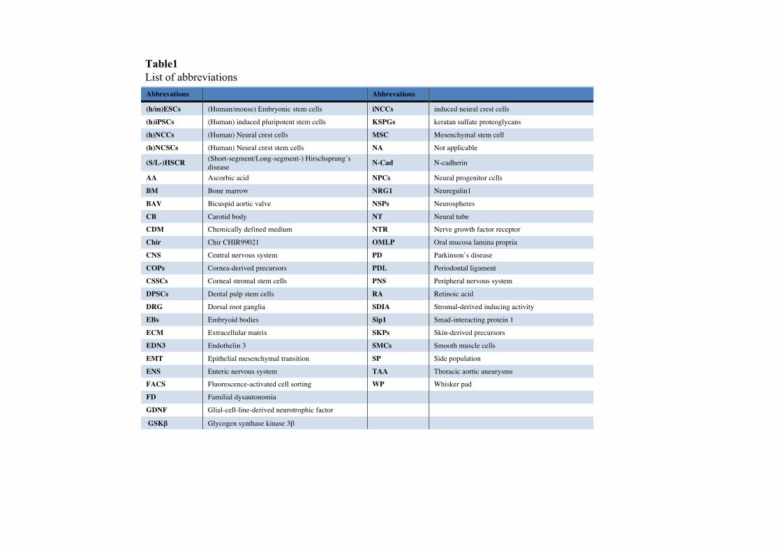
Abbrevations Abbrevations
(h/m)ESCs (Human/mouse) Embryonic stem cells iNCCs induced neural crest cells
(h)iPSCs (Human) induced pluripotent stem cells KSPGs keratan sulfate proteoglycans
(h)NCCs (Human) Neural crest cells MSC Mesenchymal stem cell
(h)NCSCs (Human) Neural crest stem cells NA Not applicable
(S/L-)HSCR (Short-segment/Long-segment-) Hirschsprung’s disease N-Cad N-cadherin
AA Ascorbic acid NPCs Neural progenitor cells
BM Bone marrow NRG1 Neuregulin1
BAV Bicuspid aortic valve NSPs Neurospheres
CB Carotid body NT Neural tube
CDM Chemically defined medium NTR Nerve growth factor receptor
Chir Chir CHIR99021 OMLP Oral mucosa lamina propria
CNS Central nervous system PD Parkinson’s disease
COPs Cornea-derived precursors PDL Periodontal ligament
CSSCs Corneal stromal stem cells PNS Peripheral nervous system
DPSCs Dental pulp stem cells RA Retinoic acid
DRG Dorsal root ganglia SDIA Stromal-derived inducing activity
EBs Embryoid bodies Sip1 Smad-interacting protein 1
ECM Extracellular matrix SKPs Skin-derived precursors
EDN3 Endothelin 3 SMCs Smooth muscle cells
EMT Epithelial mesenchymal transition SP Side population
ENS Enteric nervous system TAA Thoracic aortic aneurysms
FACS Fluorescence-activated cell sorting WP Whisker pad
FD Familial dysautonomia
GDNF Glial-cell-line-derived neurotrophic factor
GSKβ Glycogen synthase kinase 3β
Table1List of abbreviations

Pluripotent cell types Protocols for NCSCs derivation Brief description of methods Markers for
cell sorting NC markers
analyzed NC-derived cell types generated Remarks References
mESCs (EB5) SDIA PA6 as feeder layer. Treatment with BMP4 from day 4 increased NCC population No Ap2, NCAM Sensory and autonomic neurons (Brn3a+/Peri+;
TH+/Phox2b+/Peri+ ) (Mizuseki et al., 2003)
hESCs (HES-1, HUES, HUES 1)
SDIA hESCs co-cultured with PA6 induced peripheral neuronal formation. No
SOX9, dHAND, MSX1, AP2, SNAIL2
Sensory and sympathetic neurons (Brn3a+/Peri+; TH+/Peri+ )
PA6 cells induced NC-like cells before neuronal differentiation. (Pomp et al., 2005)
hESCs (H1, H9)
SDIA Mitotically inactivated MEF as feeder layer. Treatment with Wnt3a, SCF, and EDN3 induced melanocyte differentiation from hESCs-derived EBs on fibronectin
No SOX10 Melanocytes (PAX3+, TYRP1+, DCT+, MITF+) Differentiated melanocyte showed proper localization and maker expression after grafting into reconstructed human skin.
(Fang et al., 2006)
mESCs (D3)
SDIA ST2 as feeder layer. Treatment with RA promoted NCCs formation c-Kit+/CD45- Sox10,
Pax3,Snail1/2 Neuron (Tuj1+), Glial Cells (GFAP+), Melanocytes (MITF+)
C-Kit+ cells showed cell renew capacity and were able to follow the NCCs migratory pathway after in vivo transplantation.
(Motohashi et al., 2007)
hESCs (HES1, HUES9)
SDIA PA6 as feeder layer. No AP2/NCAM1 Sensory neurons (Brn3a+, Peri+) Glial cells (PMP-22+, Sox10+)
Peripheral neurons and NC were observed after 1 week of co-culture.
(Brokhman et al., 2008)
hESCs (HUES7, HUES9)
SDIA hESC co-cultured with PA6 generated NSPs expressing NCCs marker under non-adhesive conditions.
No
SNAIL1, SOX9, MSX1, PAX3 ,FOXD3,NCX/HOX11L1
Sensory neurons (Brn3a+, Peri+), Glial cells (GFAP+)
PA6-induced hESC-derived NSPs promoted peripheral neuronal differentiation. (Pomp et al., 2008)
hESCs (H9, H1)
SDIA PA6 as feeder layer. FACS-based isolation was applied for NCCs enrichment. p75NTR+
SNAIL1/2, HNK1, SOX10, AP2 and NCAM
Sensory neurons (Peri+), Glial cells( GFAP+), Myofibroblasts (SMA+)
Transplantation of isolated NCCs in chicken and mouse showed proper migration, differentiation and proliferation.
(Jiang et al., 2009)
hESCs (HES3, HES4 and ENVY)
SDIA PA6 as feeder layer. ROCK inhibitor treatment promoted NCCs formation. No HNK1, SOX8,
SOX9,SOX10 Enteric neurons (Hu+), Glial cells (S100B+)
Transplantation of isolated NCCs in chicken and mouse showed proper migration, differentiation and proliferation. (Hotta et al., 2009)
hESCs (H9 (WA-09), RUES1-eGFP48, I-8)
SDIA Neural Rosette
MS5 as feeder layer. Treatment with BMP2/FGF2/EGF promoted NCC populations. Different growth factors and culture conditions were developed for NCC and its derivatives.
p75NTR+/ HNK1+
SOX10, PAX3, AP2,ERBB3,SNAIL2
Neurons (Peri+,Tuj1+), Schwann cells (GFAP+,MBP+), Smooth muscles (SM22α+,calponin+), Chondrogenic cell (Collagen II+, Aggrecan+), Osteogenic cell (ALP+,BSP+)
Transplantation of hNCSCs into chick embryo and adult mouse hosts showed survival, migration and differentiation compatible with neural crest identity.
(Lee et al., 2007)
hESCs (H9, H1, and RU-01eGFP
SDIA Neural Rosette MS5 as feeder layer. FORSE-/
N-CAD+ SOX10,PAX3,AP2,TWIST1,SNAIL2 NA Forse1- neural rosette cells expressed NC
markers (Elkabetz et al., 2008)
hESCs and iPS cells
SDIA Neural Rosette MS5 as feeder layer. HNK1+ AP2 Autonomic and sensory neurons
(ASCL1+,Tuj1+, Brn3a+,Peri+) Generation of NCCs and Peripheral neurons using FD patient-specific iPS cell. (Lee et al., 2009)
hESCs (H9 and H14) Neural Rosette
No feeder layer. Rapid neuralization of ESCs without Noggin and limited capacity for expansion (up to 5 passages).
No SOX9 NA Committed neural progenitor cells are capable of differentiation into various CNS cell types and NC fates.
(Bajpai et al., 2009)
Table 2Derivation and characterization of NCSCs from pluripotent ESCs/iPSCs and skin fibroblasts.

NA: Not applicable. Differentiation of NCCs into different lineages was not demonstrated.
Pluripotent cell types Protocols for NCSCs derivation Brief description of methods Markers for
cell sorting NC markers
analyzed NC-derived cell types generated Rearks References
hESCs (H9)
Neural Rosette No feeder layer. Emigrating hNCCs were obtained by plating of hESCs-derived neurospheres containing rosette structure on fibronectin.
No PAX3/6,SOX9/10,p75NTR, HNK1
Autonomic neurons (TH+/Peri+), Sensory neurons (BRN3a+/Peri+), Smooth muscle (SMA+), Melanocyte (MelanA+), Glial cells (PLP+/GFAP+)
Inhibition of BMP and Wnt signaling re-specified NCSCs into CNS cells (Curchoe et al., 2010)
hESCs (H9/14) and iPSCs
SDIA Neural Rosette
PA6 as feeder layer. Schwann cell differentiation was initiated by culturing hNCSCs in MesenPRO medium with 20ng/ml heregulin-β1 for 40 days.
p75NTR+ SOX9/10, p75NTR, HNK1,CD44
Neurons (Hu+/HNK1+), Schwann cells (S100+/P75NTR+)
Pure population of Schwann cells were generated and myelinated rat dorsal root ganglia neurons in vitro.
(Liu et al., 2012)
hESCs (WA-09) and iPSC lines (iPS-14/27)
Dual-SMAD Inhibition
Feeder layer and EBs formation were not required. Both SB431542 and Noggin induced rapid and complete neural differentiation.
PAX6- AP2, HNK1,PAX7 ,p75NTR Melanocytes (HMB45+)
Manipulation of the initial hES-cell plating density skewed the ratio of PAX6+
CNS and PAX6- NC-like cells. (Chambers et al., 2009)
hESCs (WA-09) and iPSC lines (iPS-14/27)
Dual-SMAD Inhibition
Feeder layer was not required. NC differentiation from hESCs using defined NSB (KSR medium +SMAD inhibitors followed by N2) culture system
p75NTR +/ HNK1+
AP2, HNK1, PAX7,p75NTR
Autonomic Neuron (MASH1+/Peri+), Sensory neurons (Brn3a+/Peri+), Schwann cells (GFAP+,Sox10+), Smooth muscle cells (SMA+), Chondrogenic cell (Collagen II+)
The yield of p75NTR+/HNK1+ precursors using the NSB protocol is comparable with the yield using the MS5 co-culture protocol.
(Lee et al., 2010)
hESCs and iPSCs
Dual-SMAD Inhibition
Exposure of hPSCs to WNT-activator (CHIR 99021) promoted Sox10+ NC population formation by day 6. Subsequent treatment of BMP4 and EDN3 specified melanocytic lineage.
c-Kit+ /SOX10+
SOX10,HNK1 ,p75NTR
Melanocyte (SOX10+/ MITF+) ,Melanosomal (TYRP1+ /PMEL+)
Melanocytes derived from patient-specific iPSCs reproduced pigmentation defects. (Mica et al., 2013)
hESCs (H9)
Dual-SMAD Inhibition
Addition of RA promoted vagal NCCs formation.
CD49D+
/SOX10+
HOXB2-9, FOXD3,TBX2,PAX3,SOX10,TFAT2A/B,PHOX2A/B
Enteric neurons (NOS+,5-HT+,GABA+,SSST+,TH+,CHAT+)
In vivo engraftment of hESC-derived ENC progenitors rescued HSCR mice. (Fattahi et al., 2016)
hESCs (WA01, WA09) iPSCs (Y6 iPSC, RIV9 iPSCs)
Dual-SMAD Inhibition
Low-density of hESCs/iPSCs were cultured in defined serum free media under WNT activation.
No SOX10, Pax7, TFAP2A
hNCCs differentiated via an early pre-border state (Leung et al., 2016)
Sox10::eGFP reporter hESCs
SOX10-mediated reprogramming
SOX10::eGFP fibroblasts were treated with dox-inducible lentiviruses containing SOX10, ECM, epiM and Chir 99021 or BMP4
Sox10::eGFP+
CD34+HNK1, p75NTR FOXD3, AP2
Chondrocytes, osteoblasts, peripheral neurons (HuC/D+ (ELAVL3/4), TUJ1+ (TUBB3) PRPH+, ISL1+), glial precursors (S100β+ GFAP+) and melanoblasts (MITF+,SOX10+) Neurons (Tuj1+, Peri+, BRN3A+, MAP2+, Syn+), Schwann cells (GFAP+/S100B+/PRX+, MPZ+), Smooth muscle (SMA+), Melanocytes (MITF+,TYRS+), chondrocytes (Collagen II+, Aggrecan+), adipocytes (PPAR+), osteocytes (ALP+, BSP+
FD patient fibroblasts-derived iNC cells displayed defects in cellular migration and alternative mRNA splicing.
(Kim et al., 2014)

Figure Legends Figure 1. Schematic diagram showing different adult tissue sources from which NCSCs are derived. Figure 2. Schematic illustration of four different protocols for the derivation of NCSCs from pluripotent stem cells and skin fibroblasts.
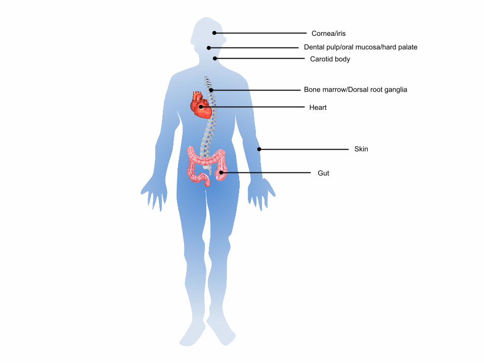
Dental pulp/oral mucosa/hard palate
Gut
Skin
Bone marrow/Dorsal root ganglia
Carotid body
Heart
Cornea/iris
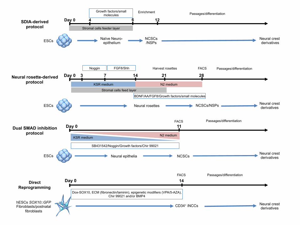
Day 0 SDIA-derived protocol Stromal cells feeder layer
ESCs
Growth factors/small molecules
4 6
Naïve Neuro- epithelium
Enrichment
NCSCs /NSPs
Neural crest derivatives
Passages/differentiation
12
Dual SMAD inhibition protocol
ESCs
Day 0
SB431542/Noggin/Growth factors/Chir 99021
Neural epithelia NCSCs
Passages/differentiation
Neural crest derivatives
KSR medium N2 medium
11 FACS
Neural rosette-derived protocol
ESCs
Stromal cells feed layer
Day 0 3 7 Noggin
14 FGF8/Shh
KSR medium
Neural rosettes
21 28 Harvest rosettes
N2 medium
NCSCs/NSPs
Passages/differentiation
BDNF/AA/FGF8/Growth factors/small molecules
Neural crest derivatives
FACS
hESCs SOX10::GFP Fibroblasts/postnatal
fibroblasts
Day 0 14
CD34+ iNCCs Neural crest derivatives
Dox-SOX10, ECM (fibronectin/laminin), epigenetic modifiers (VPA/5-AZA), Chir 99021 and/or BMP4
FACS Passages/differentiation
Direct Reprogramming