Embed Size (px)
Citation preview

Review II: The Molecules of Review II: The Molecules of LifeLife
Judy Wieber
BBSI @ Pitt 2007BBSI @ Pitt 2007
Department of Computational BiologyUniversity of Pittsburgh School of Medicine
May 24, 2007

Outline
IntroductionProteinsCarbohydratesLipidsNucleic acidsSpecial types of proteins
Proteins
Amino acidsPeptide bond formationPeptides vs. proteinsHierarchy of protein structureProtein motifsDomains
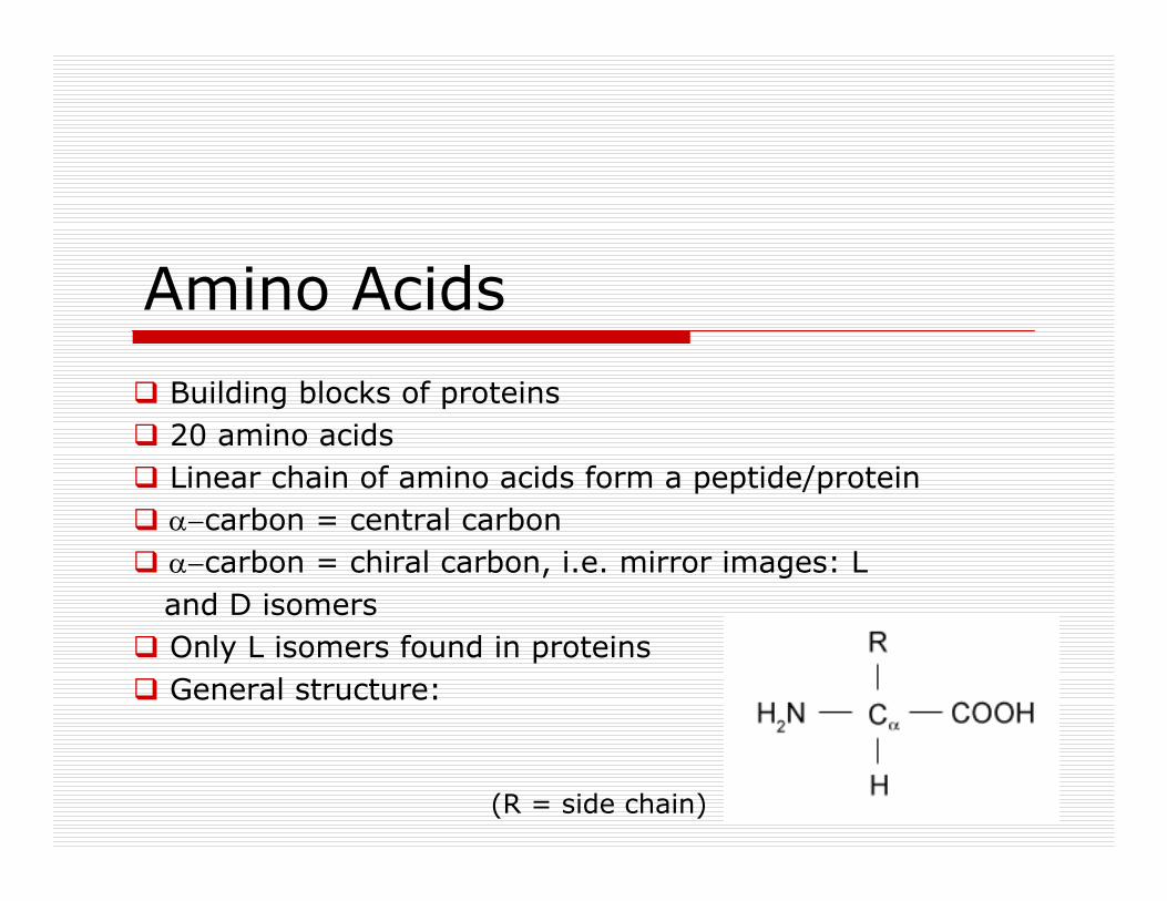
Amino Acids
Building blocks of proteins20 amino acidsLinear chain of amino acids form a peptide/proteinα−carbon = central carbonα−carbon = chiral carbon, i.e. mirror images: L and D isomersOnly L isomers found in proteinsGeneral structure:
(R = side chain)

Amino Acids (contd.)
R group variesThus, can be classified based on R groupGlycine: simplest amino acidSide chain R = HUnique because Gly α carbon is achiral
Glycine, Gly, G
H
CαH2N COOH
H

Amino Acids: Structures
orange = non-polar, hydrophobicneutral (uncharged)
blue = R (side chain)
green = polar, hydrophilic,neutral (uncharged)
magenta = polar, hydrophilic,acidic (- charged)
light blue = polar, hydrophilic,basic (+ charged)

Amino Acids: ClassificationNon-polar, hydrophobic,neutral (uncharged)Alanine, Ala, AValine, Val, VLeucine, Leu, LIsoleucine, Ile, IProline, Pro, PMethionine, Met, MPhenylalanine, Phe, FTryptophan, Trp, W
Polar, hydrophilic,neutral (uncharged)Glycine, Gly, GSerine, Ser, SThreonine, Thr, TCysteine, Cys, CAsparagine, Asn, NGlutamine, Gln, QTyrosine, Tyr, Y
Polar, hydrophilic,Acidic (negatively charged)Aspartic acid, Asp, DGlutamic acid, Glu, E
Polar, hydrophilic,basic (positively charged)Lysine, Lys, KArginine, Arg, RHistidine, His, H
(Amino acid viewer)

Peptide Bond Formation
Condensation reactionBetween –NH2 of n residue and –COOH of n+1 residueLoss of 1 water moleculeRigid, inflexible

Peptides/Proteins
Linear arrangement of n amino acid residues linked by peptide bondsn < 25, generally termed a peptiden > 25, generally termed a proteinPeptides have directionality, i.e. N terminal C-terminal
R1
CαH2N C
H
CαN COOH
HO
R2
nN terminal
C terminal
Peptide bond

Polypeptide Backbone

Binding of Ligands

Hierarchy of Protein Structure
Four levels of hierarchyPrimary, secondary, tertiary, quarternary
Primary structure: Linear sequence of residuese.g: MSNKLVLVLNCGSSSLKFAV …e.g: MCNTPTYCDLGKAAKDVFNK …
Secondary Structure: Local conformation of thepolypeptide backboneα-helix, β-strand (sheets), turns, other
(Amino acids-proteins movie)

Secondary Structure: α-helix
Most abundant; ~35% of residues in a proteinRepetitive secondary structure3.6 residues per turn; pitch (rise per turn) = 5.4 ÅC′=O of i forms H bonds with NH of residue i+4Intra-strand H bondingC′=O groups are parallel to the axis; side chains pointaway from the axisAll NH and C′O are H-bonded, except first NH and last C′OHence, polar ends; present at surfacesAmphipathic

α-helix (contd.)
N terminal
C terminal

α−helix Variations
Chain is more loosely or tightly coiled310-helix: very tightly packedπ−helix: very loosely packedBoth structures occur rarely Occur only at the ends or as single turns
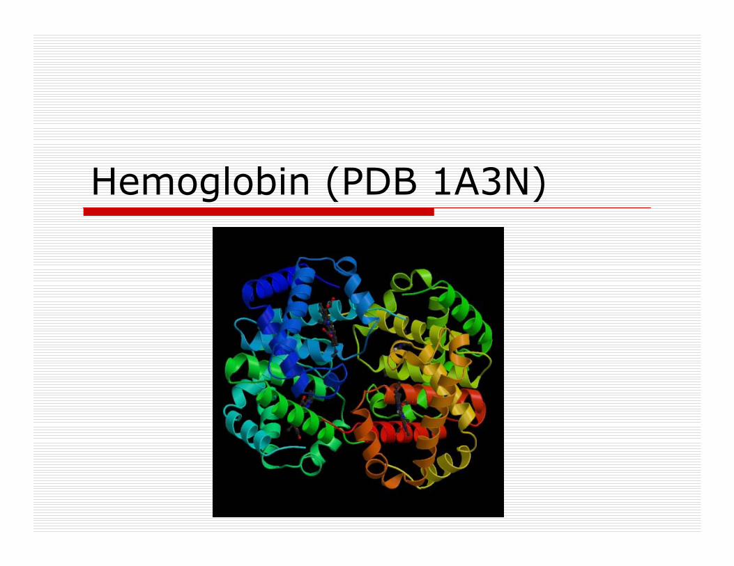
Hemoglobin (PDB 1A3N)

β−sheets
Other major structural elementBasic unit is a β-strandUsually 5-10 residuesCan be parallel or anti-parallel based on therelative directions of interacting β-strands“Pleated” appearance

β−sheetsUnlike α-helices:Are formed with different parts of the sequenceH-bonding is inter-strand (opposed to intra-strand)Side chains from adjacent residues are onopposite sides of the sheet and do not interactwith one another
Like α-helices:Repeating secondary structure (2 residues per turn)Can be amphipathic

Parallel β−sheets
The aligned amino acids in the β-strand all run in the same biochemical direction, N- to C-terminal

Anti-parallel β−sheets
The amino acids in successive strands have alternating directions, N-terminal to C-terminal

Nucleoplasmin (PDB 1K5J)

Amino Acid Preferences (1)α-helix formingThe amino acid side chain should cover and protect the backbone H-bonds in the core of the helixAla, Leu, Met, Glu, Arg, Lys: good helix formersPro, Gly, Tyr, Ser: very poor helix formers
β-strand formingAmino acids with large bulky side chains prefer to formβ-sheet structuresTyr, Trp, Ile, Val, Thr, Cys, Phe

Amino Acid Preferences (2)
Secondary structure disruptorsGly: side chain too smallPro: side chain linked to α-N, has no N-H toH-bond; rigid structure due to ringAsp, Asn, Ser: H-bonding side chains competedirectly with backbone H-bonds
Turns/Loops
Third "classical" secondary structureReverses the direction of the polypeptide chain Located primarily on protein surfaceContain polar and charged residuesThree types: I, II, III

Phosphofructokinase (PDB 4PFK)

The Torsional Angles: φ and ψ
Each amino acid in a peptide has two degreesof backbone freedomThese are defined by the φ and ψ anglesφ = angle between Cα―Nψ = angle between Cα―C’

The Ramachandran PlotPlot of allowable φ and ψ anglesφ and ψ refer to rotations of two rigid peptide units around Cα
Most combinations produce steric collisionsDisallowed regions generallyinvolve steric hindrance between side chain Cβ methylene group and main chain atoms

The Ramachandran Plot (contd.)
Theoreticallypossible;
energeticallyunstable
π-helix
310-helix
Anti-parallelβ-sheet
Parallelβ-sheet
White: sterically disallowed(except Gly)
Red: no steric clashesYellow: “allowable” steric
clashes

Protein Motifs
(Simple) combinations of secondary structure elements with a specific geometric arrangement“Super-secondary structures”Can be associated with a specific function, e.g. DNA binding, or metal ion bindingCan be part of a larger functional and/or structural assembly (“domain”)

Helix-Turn-Helix (HTH)
Simplest α-helix motif: 2 α helices joined by a loopAlso called Helix-Loop-Helix, HLHCommon structural motif forDNA binding proteinsOne helix recognizes specificsequence of nucleotides and fitsinto the groove in the DNA doublehelix; the other stabilizes the boundconfiguration

EF Hand
Specific for several differentcalcium-binding proteinsE.g. calmodulin, Troponin-C

Hairpin β-motif
Also called β-hairpin2 adjacent anti-parallel strandsjoined by a loop

Greek Key motif
4 adjacent anti-parallel β-strands

β-α-β Fold
Parallel β-strands connect by an α helixFound in most proteins with parallel β strands. E.g. Triosephosphate isomerase

Rossman Fold
Unique example of a β−α−β fold3 parallel β-sheets with 2 linkingα helicesOften seen in nucleotide-bindingproteins

Domains
Primary structureSecondary structureSuper-secondary structureDomainsFundamental unit of tertiary structure(Part of a) polypeptide chain that can fold independently into a stable tertiary structure E.g. catalytic domain of protein kinase; binding pocket of a ligand

N-terminal
C-terminal
phosphate binding
loop
catalyticloop
3D Structure of a Protein Kinase Domain

Quarternary Structure
Spatial organization of subunits to form functional proteinE.g. Hemoglobin2 α chains, 2 β chainsEach chain binds heme (Fe)Forms an α2β2 tetramer

Putting it all together
Primary Structure… KAAWGKVGAHA …
Secondary Structureα-helix, β-sheets, turns/loops
Tertiary Structurea single chain (α, β)
Quarternary Structureα2β2
Super-secondary StructureHeme-binding pocket/domain (His)

Carbohydrates
SugarsPolysaccharides

Sugars

Lipids
Fatty acidsTriglyceridesSteroidsMembranes
(Structure of lipids)

Cell Membrane

Types of Membrane Proteins

Nucleic Acids
NucleotidesDNARNA
(DNA)

Base Pairing

Biopolymers

Composition of a Cell

Special Types of Proteins
EnzymesReceptorsChannel proteinsAntibodies

Enzymes
(Ribozymes movie)
Cell Receptors
Ion-channel linked: involved in rapid synaptic signaling between excitable cells; mediated by neurotransmittersEnzyme-linked receptors: when activated, either function directly as enzymes or are associated with enzymes.G-protein coupled receptors (GPCR)

GPCRsLargest family of cell-surface receptorsBiological functions include smell, taste, vision, blood pressureneurotransmission, embryogenesis, cell growth, developmentRhodopsin is the only GPCR with a known 3D structureContains 7 membrane traversing α helices (7TM)N terminal – outside cell, C terminal – inside cellLigand binding outside cell induces conformational change detected inside cellMediating molecule is a G protein (hence the name GPCR) Heterotrimeric GTP-binding regulatory protein (α, β, γ)Activated G protein transmits signal by binding to other proteins (e.g. adenylate cyclase: converts ATP to cAMP)

GPCR Structure

GPCR Structure (contd.)

Signal effects
Regulation byreversible
phosphorylationand
dephosphorylation
Signal mediationand amplification
Signal reception
Channel Proteins
(Structure of Channel Proteins)

Antibody Structure

Additional Reading
General informationBiochemistry, 5th ed., Berg, Tymoczko, StryerBiochemistry, 3rd ed., Voet & Voet
Detailed informationProteins, 2nd ed., CreightonIntroduction to Protein Structure, 2nd ed., Branden & Tooze
InternetImages: Protein Data Bank (PDB): www.rcsb.org/pdbNumerous websites (Google protein secondary structure)