Embed Size (px)
Citation preview
1The Journal of Experimental Biology 198, 1–17 (1995)Printed in Great Britain © The Company of Biologists Limited 1995
*Au
REVIEW
FUNCTIONAL SIGNIFICANCE OF THE -SUBUNIT FOR HETERODIMERICP-TYPE ATPases
DAR C. CHOW AND JOHN G. FORTE*Department of Molecular and Cell Biology, University of California, Berkeley, CA 94720, USA
We have reviewed the structural and functional role ofthe -subunit in a subfamily of the P-ATPases known asthe / -heterodimeric, cation-exchange ATPases. Thesubfamily consists of the various isoforms of Na+/K+-ATPase and H+/K+-ATPase, both of which pump a cationout of the cell (Na+ or H+, respectively) in recycle exchangefor K+. Much of the earlier work has emphasized thefunctional activities of the -subunit, which shares manycharacteristics with the broader P-ATPase family. It is nowclear that the glycosylated -subunit is an essentialcomponent of the cation-exchange ATPase subfamily. All
-subunit isoforms have three highly conserved disulfidebonds within the extracellular domain that serve tostabilize the -subunit, / interaction and functionalactivity of the holoenzyme. Evidence strongly suggests thatthe -subunit is involved in the K+-dependent reactions ofthe enzymes, such as the E1–E2 transition and K+ occlusion,and that the extracellular domain of the -subunit plays animportant role in determining the kinetics of K+
interaction. In most vertebrate cells, the unassociated -subunit is restricted to the endoplasmic reticulum (ER),
and assembly of the / complex occurs within the ER.Signals for exiting the ER and directing the correctintracellular trafficking are primarily determined by the -subunit; Na+/K+-ATPase typically terminates in the plasmamembrane facing the basolateral membrane, whereas allisoforms of H+/K+-ATPase terminate in the apicalmembrane. The C-terminal extracellular domain of the -subunit is important for proper interaction with the -subunit and for correct intracellular trafficking.Oligosaccharides on the -subunit are not essential forenzyme function, but do serve to enhance the efficiency of
/ association by increasing the lifetime of theunassociated -subunit and the stability of the / complexto tryptic attack. We propose that highly specializedglycosylation on the -subunit of the gastric H+/K+-ATPasemay help to protect that enzyme from the harshextracellular environment of the stomach.
Key words: H+ pump, Na+ pump, glycosylation, subunit assembly,trafficking, acid secretion.
Summary
All cells possess an array of sophisticated membrane-boundenzymatic systems that perform various processes essential forlife. These processes range from regulation of the intracellularmilieu to the genesis of information transfer andcommunication between cells. A major functional class ofthese membrane-bound enzymes includes those categorized asprimary active transporters and called ATPases because theycatalyze the transport of molecules against an electrochemicalpotential by reactions directly linked to the hydrolysis of ATP.The ATPases that actively transport cations have beenextensively studied and have been categorized by Pedersen andCarafoli (1987) into three classes: F-type ATPases (F-ATPases), V-type ATPases (V-ATPases) and P-type ATPases(P-ATPases). The F-ATPases are located in bacterial plasma
Introduction
thor for correspondence.
membranes, inner mitochondrial membranes and thylakoidmembranes of chloroplasts, and they actually operate in vivoas reverse ATPases, or ATP synthases, synthesizing ATP fromADP and inorganic phosphate using energy derived fromelectrochemical gradients of protons (Amzel and Pedersen,1983). Using reaction mechanisms analogous to F-ATPases,the V-ATPases utilize ATP to create proton electrochemicalgradients; they are ubiquitously distributed in eukaryoticvacuo-lysosomal organelles and archaebacteria and are alsopresent in plasma membranes of various animal tissues(Harvey, 1992).
The P-ATPases are broadly distributed, active cationtranslocators having the distinctive feature of forming acovalent acylphosphate–enzyme intermediate (hence the P-
2 D. C. CHOW AND J. G. FORTE
designation) during the cycle of ATP hydrolysis and cationtranslocation. Among this class of ATPases are the Ca2+-ATPases of the plasma membrane, sarcoplasmic reticulum andendoplasmic reticulum, the H+-ATPase of yeast and plants, theK+-ATPase of bacteria, the Na+/K+-ATPase of animal cellplasma membranes and the H+/K+-ATPase of gastric parietalcells. Because of the formation of phosphoenzymeintermediates, the enzymatic cycle of P-ATPases can bedivided into steps that include a kinase activity, by which anaspartate residue on the enzyme is phosphorylated, and aphosphatase activity, by which the phosphoenzyme isdephosphorylated. Another common feature of these ATPasesis their inhibition by submicromolar concentrations ofvanadate, acting as a tightly binding phosphate analog (Cantleyet al. 1977; O’Neal et al. 1979; Faller et al. 1983).Furthermore, during the enzymatic cycle, P-ATPasescharacteristically exhibit two phenomenologically andstructurally distinct conformations, E1 and E2, which havedistinct kinetic variables, e.g. affinities for substrates(Jorgensen and Andersen, 1988). For this reason, the P-ATPases are also called E1–E2 ATPases. Identification andprobing of these various features have provided significantinsight into the detailed mechanistic operation of the P-ATPases (Jorgensen and Andersen, 1988; de Meis and Vianna,1979).
In addition to their functional similarities, P-ATPases havea number of structural homologies, belonging to a common
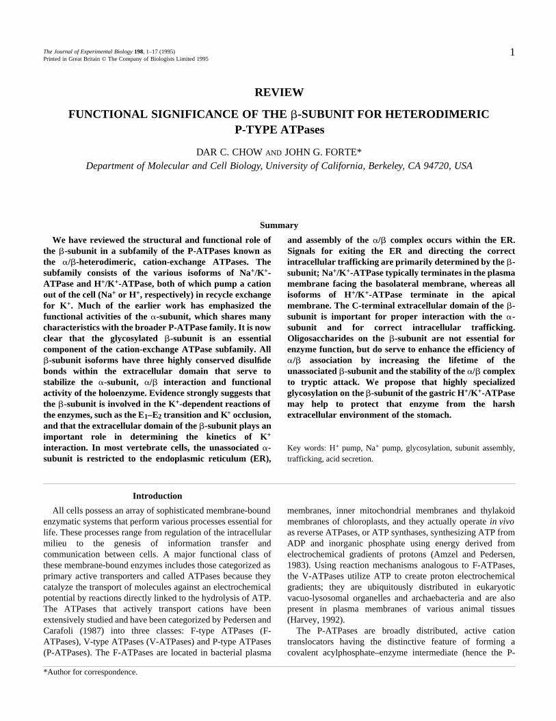
Fig. 1. Schematic representation of theorganization within a membrane of aheterodimeric cation-exchange subfamily of P-ATPase (including Na+/K+-ATPase and H+/K+-ATPase). The scheme points out the multipletransmembrane segments of the -subunit withits principal amino acid mass on thecytoplasmic side containing several importantcatalytic functions as well as the N-terminal andC-terminal segments. The -subunit has asingle transmembrane segment and most of itsmass, which is highly glycosylated and containsthree structurally important disulfide bonds, ison the extracellular side of the membrane. Thediagram also suggests that K+ binding sites maybe a structure/function cooperation between -and -subunits. An inhibitory site (O) isillustrated on the extracellular domain of the -subunit (the ouabain binding site for Na+/K+-ATPase and the omeprazole binding site forH+/K+-ATPase). The ouabain binding site hasbeen shown to involve regions within 200amino acids of the N terminus and within theC-terminal half of the Na+/K+ -subunit (Ishiiet al. 1994; Canessa et al. 1992; Feng andLingrel, 1994). Omeprazole has been shown tolabel covalently the Cys-813 and Cys-892 ofthe H+/K+ -subunit (Besancon et al. 1993).
large gene family. All members have a principal peptide ofapproximately 100kDa, designated as the catalytic subunitbecause it contains the site for ATP binding andphosphorylation. The high degree of sequence homology foramino acids within the ATP binding site and thephosphorylation site attests to the highly conserved nature ofprotein domains that interact with ATP and has been used todesign nucleic acid probes for cloning other members of thegene family (Shull and Lingrel, 1986). Amino acid sequenceanalysis suggests that all P-type catalytic subunits have similartopological and domain organization. Multiple transmembranesegments occur within both the N-terminal third and C-terminal third of the peptide. A large cytoplasmic domain inthe middle of the peptide contains the nucleotide binding andphosphorylation sites (Fig. 1). Some disagreement remains asto the exact location, and even the number, of transmembranesegments (Inesi and Kirtley, 1992); the specific locations forthe binding of cations and the paths for their transport are farfrom resolved.
Na+/K+-ATPase and H+/K+-ATPase: cation exchangersAmong the P-ATPases, the ubiquitous Na+/K+-ATPase and
the gastric H+/K+-ATPase share a number of common features;this review will primarily address these structural andfunctional homologies. Both ATPases are cation exchangers,involving the cellular uptake of K+ in exchange for the export
COO−
Transduction
+H3N
Phosphorylation
Nucleotidebinding
SS
COO−
NH3+
3Function of heterodimeric -type ATPases
of Na+ or H+. In addition to amino acid sequence homology,both Na+/K+-ATPase and gastric H+/K+-ATPase areheterodimers with the minimal functional complex containingthe approximately 100kDa -subunit and a glycosylated -subunit. The apparent molecular mass of the -subunit onSDS–PAGE is highly variable, depending upon the degree ofglycosylation, but the deglycosylated core protein is alwaysabout the same size (approximately 34kDa). Thus, manystructural and functional similarities, including K+ exchangeand the functional requirement for a -subunit, place Na+/K+-ATPase and H+/K+-ATPase within a subfamily of P-ATPases.A schematic representation of the / -heterodimericorganization for this P-ATPase subfamily is shown in Fig. 1.
The Na+/K+-ATPase is known to occur in a number ofisoforms, both for the -subunit and for the -subunit. Forexample, various -subunit isoforms have been designated ( 1,
2, 3), differing somewhat with respect to tissue distributionand affinity for ligands as well as primary structure (Lingrel etal. 1990); however, the physiological significance of theNa+/K+-ATPase isoforms is unknown. There may also beisoforms for the gastric H+/K+-ATPase. For example, a newmember of the Na+/K+-, H+/K+-ATPase subfamily has beenidentified in toad urinary bladder and has been postulated to bean H+/K+-ATPase isoform performing H+ and K+ homeostasisspecific to the urinary tract (Jaisser et al. 1993b). The -subunitof this putative isoform, designated as bl, has approximately69% amino acid identity with the -subunit of the gastricH+/K+-ATPase and approximately 67% identity with 1 ofNa+/K+-ATPase. A novel -subunit ( bl) has also been clonedfrom toad bladder epithelial cells (Jaisser et al. 1993c).Expression of bl in Xenopus oocytes requires co-expressionof bl for functional H+ and K+ exchange activity, furthersupporting the / -heterodimeric nature of the ion-exchangeATPase subfamily. A recently cloned P-ATPase, localized tosurface epithelial cells of the distal colon, may be yet anotherisoform of the H+/K+-ATPase, sharing 76% identity with rat
1 and 75% identity with bl (Jaisser et al. 1993a). Althoughthe distal colon is known to carry out active K+ absorption viaK+/H+ exchange, the colonic H+/K+-ATPase has not beenfunctionally expressed and there is no evidence yet for acorresponding -subunit.
Despite their many common features, there are some notableand instructive differences between the Na+/K+-ATPase andthe H+/K+-ATPase. Cardiac glycosides, such as ouabain, arewell-known specific inhibitors of Na+/K+-ATPase activity, butdo not inhibit gastric H+/K+-ATPase. Other compounds, suchas SCH 28080 and omeprazole (see Sachs et al. 1989),effectively inhibit H+/K+-ATPase with no effect on Na+/K+-ATPase. In fact, this latter specificity forms the basis for usingomeprazole-related compounds as gastric proton pumpinhibitors for clinical treatment of hyperacidity and peptic ulcercontrol. Functional expression of the toad bladder H+/K+-ATPase isoform, via bl bl co-expression in oocytes, revealedH+/K+ exchange transport activity that was sensitive to bothSCH 28080 and ouabain, although less sensitive than is typicalof gastric H+/K+-ATPase and Na+/K+-ATPase.
The two exchange pumps also differ in some physicalcharacteristics, e.g. generated ionic gradients and turnoverstoichiometry are markedly different. The Na+/K+-ATPasetypically transports Na+ against a 5- to 15-fold Na+
concentration gradient, whereas the gastric H+/K+-ATPase willoperate in a steady state against a proton gradient greater than106. For turnover of the Na+/K+-ATPase, the transportstoichiometry is electrogenic, 3Na+/2K+ (Post and Jolly, 1957;Clarke et al. 1989); whereas for turnover of H+/K+-ATPase thestoichiometry is electroneutral, 1H+/1K+ (Sachs et al. 1976).These physical differences may underlie a common principalof operation that is inherent in the thermodynamic efficiencyfor all P-ATPases. The work (Wp) involved in the turnover ofan ion pump is the sum of the energy requirements for all thetransported species, which is a function of the electrochemicalpotential gradient for each ion ( ¯ k) and the stoichiometricnumber of ions moved (nk):
Wp = ∑nk ¯ k ,where:
[ck]o¯ k = RTln –––– + zF ,
[ck]i
where [ck] represents the molar concentration of the ion withthe subscripts i and o referring to the inside (cytosolic) andoutside (extracellular) solutions, is the transmembraneelectrical potential difference, and R, T, z and F have theirusual meanings. The electrochemical gradients for operatingthe Na+/K+-ATPase are not very high but, taking into accountthe stoichiometry of three Na+ and two K+ per turnover (i.e.per ATP utilized), the pump demands about 42kJmol 1 perturnover. In the case of the H+/K+-ATPase, the proton gradientis huge but, since transport involves only one H+ and one K+
per turnover, the total work of the gastric H+ pump turns outto be nearly the same as that of the Na+ pump; that is, about42–46kJmol 1 per turnover. [For a general case with a ofabout 0.05V, intracellular concentrations of 11mmol l 1
Na+ and 140mmol l 1 K+, and extracellular concentrations of140mmol l 1 Na+ and 5mmol l 1 K+, the minimum energyrequirement is 11.4kJmol 1 for Na+ efflux and 3.8kJmol 1
for K + influx. From the stoichiometry of three Na+ (3 11.4)and two K+ (2 3.8) per pump turnover, a minimum ofapproximately 42kJ per Na+/K+ pump turnover is predicted.To operate the gastric H+/K+ pump, with a luminal pH of 0.8and a cell pH of 7.0, approximately 42kJmol 1 would berequired for H+ secretion; electrochemical gradient conditionspredict approximately 2.5kJmol 1 for K+ influx. Since thestoichiometry of H+/K+-ATPase is one H+ per one K+, aminimum of approximately 44kJ is required per pumpturnover.] Similar considerations applied to other P-ATPases(e.g. Ca 2+-ATPase) give about the same energy requirementsper molar turnover. The free energy available from ATPhydrolysis under typical cellular conditions is about 42 to
54kJmol 1. Thus, for the sake of energetic efficiency,evolution of the specific number of ion binding sites for a givenpump protein may have conformed to the relationship between
4 D. C. CHOW AND J. G. FORTE
the electrochemical gradient of transport and the availability ofphosphate bond energy for conformational rearrangement.
Features of -subunits of the cation exchange ATPasesubfamily
The -subunit of the Na+/K+-ATPase was initially identifiedas a glycoprotein associated with the -subunit in purifiedfunctional enzyme preparations (Brotherus et al. 1983). Theassociation between - and -subunits is relatively strong andremains stable in most non-ionic detergents. The -subunit ofthe Na+/K+-ATPase, like the -subunit, has several isoforms,designated 1, 2, 3 (see Horisberger et al. 1991b). Amongthese isoforms, there are various degrees of difference and,although all -subunits are glycosylated, the number ofglycosylations varies with the isoform.
The -subunit of the gastric H+/K+-ATPase is also a highlyglycosylated protein, so much so that it appears on SDS–PAGEas a broad 60–80kDa band that is weakly stained byCoomassie Blue. The -subunit of the H+/K+-ATPase hadeluded confirmation until it was shown that the 60–80kDaglycoprotein remained stably associated with the -subunit innon-ionic detergents and could be deglycosylated to a 34kDacore peptide similar to Na+/K+-ATPase (Okamoto et al. 1989).Amino acid sequences from several species were publishedwithin months of this finding (Canfield et al. 1990; Reuben etal. 1990; Shull, 1990; Toh et al. 1990).
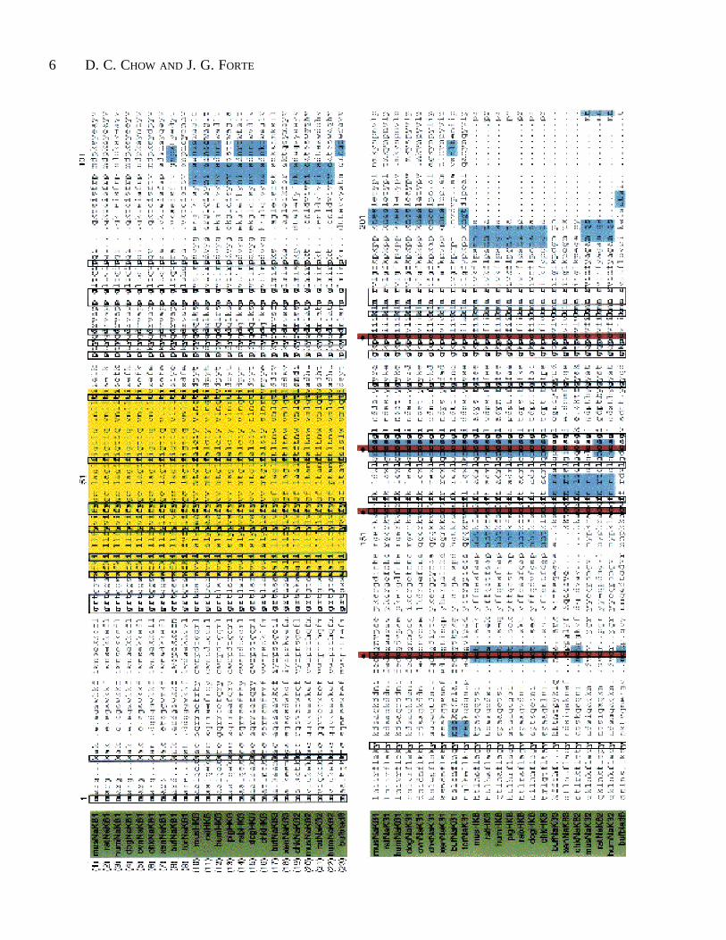
Sequences for -subunit isoforms for the subfamily ofNa+/K+- and H+/K+-ATPases are aligned in Fig. 2. Althoughdrawn from diverse animal species, at least 15% of the 300amino acids in the core are identical, along with a great dealof conservative substitution. All of the known -subunitspecies and isoforms share a common domain structure: a shortN-terminal cytoplasmic piece, a single transmembranesegment and a large extracellular C-terminal domaincontaining six extracellular cysteine residues, whose locationsare completely conserved among the isoforms. For internalconsistency, the numbering system given in Fig. 2 is usedthroughout the text.
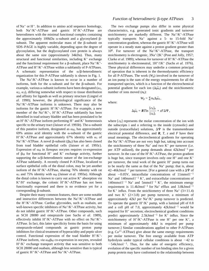
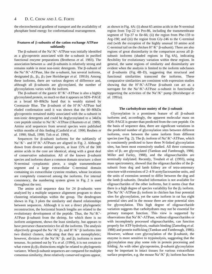
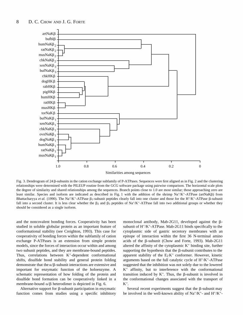
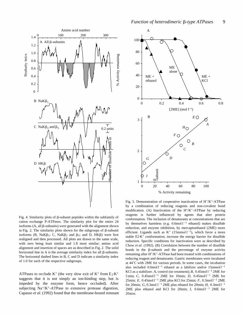
The amino acid sequence data for 24 -subunits wereanalyzed by a multiple sequence alignment program to showclustering relationships within the group. The dendrogramshown in Fig. 3 plots the similarity and shared relationshipsbetween sequences. Although it is not a direct phylogeneticreconstruction, the horizontal branch lengths are related to theevolutionary development of the peptide. Thus, the Na+/K+-ATPase -subunit from the shrimp, for which there is noisoform assignment, shows the least similarity, but appears tohave precursor characteristics for all the isoforms. The analysisobjectively grouped the Na+/K+ 1 and H+/K+ isoforms intotwo distinct clusters, indicating that they are monophyletic;however, division of the Na+/K+ 2 and 3 isoforms is moretenuous. As pointed out by Yu et al. (1994), it is not certain towhat extent 2/ 3 distinctions might be related to phylogeneticvariance. When -subunit sequences are compared for multipleconsensus similarity, three relatively conserved regions appear,
as shown in Fig. 4A: (i) about 65 amino acids in the N-terminalregion from Trp-22 to Pro-86, including the transmembranesegment of Trp-37 to Ile-66; (ii) the region from Phe-159 toArg-190; and (iii) the region from Gly-246 to the C-terminalend (with the exception of the highly unusual 10 amino acidC-terminal tail on the chicken H+/K+ -subunit). There are alsoregions of great dissimilarity in the comparison across all -subunit isoforms (shaded regions in Fig. 4A), indicatingflexibility for evolutionary variation within these regions. Ingeneral, the same regions of similarity and dissimilarity areevident when the analysis is limited to the isoform subclassesof -subunits (Fig. 4B–D), suggesting that structural andfunctional similarities transcend the isoforms. Thesecomparative similarities are consistent with expression studiesshowing that the H+/K+-ATPase -subunit can act as asurrogate for the Na+/K+-ATPase -subunit in functionallysupporting the activities of the Na+/K+ pump (Horisberger etal. 1991a).
The carbohydrate moiety of the -subunitGlycosylation is a prominent feature of all -subunit
isoforms and, accordingly, the apparent molecular mass onSDS–PAGE is greater than predicted from the core peptide. Onthe basis of sequence data, there are significant variations inthe predicted number of glycosylation sites between differentisoforms, even between the same isoform from differentspecies (see Fig. 2). The 1 isoform of Na+/K+-ATPase, whichis consistently predicted to have three N-linked glycosylationsites, has been most extensively studied. All three consensussites of 1 are glycosylated (Tamkun and Fambrough, 1986;Miller and Farley, 1988), and the oligosaccharides areterminally sialylated. Recently, Treuheit et al. (1993), usingmass spectrometry, showed that the oligosaccharides of the -subunit from dog and lamb kidney are of tetra-antennarystructure with extensions of 2–4 N-acetyllactosamine units, andthe units of extension seemed to differ between the dog andthe lamb -subunits. Detailed information is not available foroligosaccharides of the other isoforms, but it seems clear thatthere is a high degree of species variability for the 2 isoform.The Na+/K+-ATPase 2 isoform in chicken has four potentialsites for glycosylation, yet the same isoform in rat has eightpotential sites and in the mouse there are nine potential sitesfor glycosylation. This high degree of oligosaccharidevariation suggests that carbohydrates may not be essential forprimary transport function. This view is supported byobservations that Na+/K+-ATPase, without oligosaccharides orwith incompletely processed oligosaccharides, can functionproperly for ATP hydrolysis, ouabain binding (Zamofing et al.1988) and protein trafficking (Tamkun and Fambrough, 1986).However, without core glycosylation of the -subunit, theenzyme is more sensitive to trypsinolysis, suggesting that theglycosylation may play some role in protein processing andfolding. As with other glycoproteins, -subunit glycosylationmay be important in intact tissues for various extracellularsurface properties, e.g. the mouse Na+/K+ 2 isoform has been
5Function of heterodimeric -type ATPases
identified as the protein formerly known as AMOG, theadhesion molecule on glial cells (Schmalzing et al. 1992).
For the H+/K+-ATPase, all -subunits contain sevenpotential N-linked glycosylation sites, except that from the pigwhere only six such sites have been identified (see Fig. 2). Thenascent -subunit of rabbit H+/K+-ATPase is cotranslationallyglycosylated with high-mannose core oligosaccharides at allseven consensus N-linked sites (Chow and Forte, 1993). Fullymature oligosaccharides on the -subunit of H+/K+-ATPaseare relatively bulky, as shown by the high apparent molecularmass on SDS–PAGE and, like Na+/K+-ATPase, the structureis tri- or tetra-antennary (Okamoto et al. 1990; Toh et al. 1990)with an abundance of N-acetyllactosamine units (Weitzhandleret al. 1993). However, in contrast to Na+/K+-ATPase, the -subunit of H+/K+-ATPase is devoid of sialic acid(Weitzhandler et al. 1993). It has been suggested that theH+/K+ -subunit may initially be sialylated, and laterdesialylated within the highly acidic luminal space. However,it is also possible that the -subunit is never sialylated, thepost-translational pathway either lacking sialyl transferase orproviding alternative linkages, e.g. -galactose linkages orterminal fucosylation.
The function of oligosaccharides on the H+/K+-ATPase -subunit, as for the Na+/K+-ATPase, is unclear. An earlyhypothesis proposed that glycosylation on the -subunit(known then as an accessory glycoprotein for the H+/K+-ATPase) provided protection against acidic and autodigestiveextracellular conditions (Forte and Forte, 1970). A preliminarystudy indicates that oligosaccharides on the H+/K+ -subunitafford resistance against pepsinolysis in vitro (Chow et al.1993), but to what extent this operates in situ remainsuncertain.
Disulfide bonds in the -subunitThe six cysteine residues in the extracellular domain of the
-subunit are 100% conserved between all known isoforms ofboth Na+/K+-ATPase and H+/K+-ATPase. In contrast, cysteineresidues within the intracellular and transmembrane domainsvary widely. Kirley (1989) and Miller and Farley (1990)showed that the six extracellular cysteines of the Na+/K+-ATPase 1-subunit form three disulfide bonds in a sequentialpattern. For the -subunit of H+/K+-ATPase, six of the ninecysteines are in the oxidized state (Chow et al. 1992). Byanalogy with Na+/K+ 1-subunit, we suggest three extracellulardisulfide bonds in the H+/K+ -subunit in the same sequentialpattern.
Functional activities of the -subunitDespite the requirement of an / -heterodimer as the
minimal functional complex, most functional activity of theNa+/K+-ATPase, e.g. phosphorylation, ATP binding andinhibitor binding, had been identified within the -subunit.Defining a functional role for the -subunit had been hinderedby the inability to reassemble a functional / complex from
solubilized monomeric subunits. However, several recentstudies provide direct and indirect evidence of the importantfunctional properties of the -subunit, including an essentialrole in the stabilization, maturation and enzymatic activity ofboth Na+/K+-ATPase and H+/K+-ATPase.
The importance of the -subunit for functional integrity wasfirst observed for Na+/K+-ATPase by Kawamura and Nagano(1984), who showed that reduction of -subunit disulfidebonds was correlated with a loss of activity. Disulfide bondreduction required strong reducing conditions and could beattenuated by the presence of K+ or Na+, suggesting a possiblerole for the -subunit in binding these cations (Kawamura etal. 1985; Kirley, 1990). H+/K+-ATPase activity is alsoinactivated by reduction of -subunit disulfide bonds;however, the protective effect of cations in preventingreduction and sustaining enzymatic activity is specific for K+
and its congeners, Rb+ and Tl+ (Chow et al. 1992).Furthermore, the ability of K+ congeners to protect the enzyme,i.e. the measured EC50, was highly correlated with the Km ofthese cations for stimulating p-nitrophenylphosphatase(pNPPase) activity. Since pNPPase activity is a model for thephosphatase step of the ATPase cycle and stimulation ofpNPPase is related to the E2.K+ conformation, it was proposedthat the strength of disulfide bonds in the -subunit increasedas the enzyme shifted from the E1 to the E2.K+ conformation(Chow et al. 1992). Na+ is known to be antagonistic to K+, andhigh concentrations of Na+ shift the Na+/K+-ATPase or H+/K+-ATPase towards the E1 conformer (Skou, 1982). Highconcentrations of Na+ made the H+/K+-ATPase moresusceptible to reduction in the presence of K+ (Chow et al.1992). Thus, it appears that, as the conformation of the -subunit tightens (as indicated by an increase in strength of thedisulfide bonds), the enzyme shifts from the E1 to the E2.K+
state. The increase in strength of the disulfide bonds in the -subunit is correlated with increased stability of theholoenzyme; both Na+/K+-ATPase and H+/K+-ATPase are ina more stable conformation in the E2.K+ state.
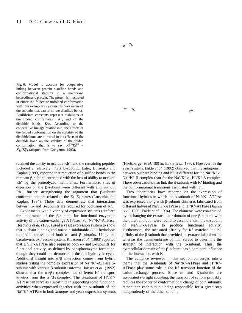
Another way to study relationships between -subunitdisulfide bond stability and general conformational stability isto monitor the strength of the disulfide bonds under theinfluence of ligands and of denaturants (Browning et al. 1992).Dose–response curves for the inactivation of H+/K+-ATPaseby 2-mercaptoethanol (2ME) under differing conformationalstates are shown in Fig. 5A. The lability of H+/K+-ATPaseactivity to 2ME is increased by ethanol (curve shifted to theleft), whereas K+ stabilizes H+/K+-ATPase against 2ME (curveshifted to the right). For all conditions, inactivation occursabruptly over a narrow range of 2ME concentration, a typicalfeature of cooperativity among various interactions stabilizinga protein (Creighton, 1993). The correlation between disulfidebond content and H+/K+-ATPase activity shown in Fig. 5Bconfirms the importance of -subunit disulfide bonds.Furthermore, the sensitivity of these disulfide bonds to agentsthat alter holoenzyme stability (stabilized by K+ and labilizedby denaturants such as organic solvents and detergents)suggests that there is cooperativity between disulfide bonds
6 D. C. CHOW AND J. G. FORTE
7Function of heterodimeric -type ATPases
8 D. C. CHOW AND J. G. FORTE
artNaKbufbl
humNaK 2
ratNaK 2musNaK 2 chkNaK 2 xenNaK 3bufNaK 3
chkHK dogHK rabHK pigHK
humHK
ratHK musHK torNaK bufNaK 1xenNaK 1
chkNaK 1 ovnNaK 1 dogNaK 1
humNaK 1 ratNaK 1
musNaK 1
1.0 0.8 0.6 0.4 0.2 0
Similarities among sequences
Fig. 3. Dendrogram of 24 -subunits in the cation exchange subfamily of P-ATPases. Sequences were first aligned as in Fig. 2 and the clusteringrelationships were determined with the PILEUP routine from the GCG software package using pairwise comparison. The horizontal scale plotsthe degree of similarity and shared relationships among the sequences. Branch points close to 1.0 are most similar; those approaching zero areleast similar. Species and isoform are indicated as described in Fig. 1 with the addition of the shrimp Na+/K+-ATPase (artNaK ) fromBhattacharyya et al. (1990). The Na+/K+-ATPase 1-subunit peptides clearly fall into one cluster and those for the H+/K+-ATPase -subunitfall into a second cluster. It is less clear whether the 2 and 3 peptides of Na+/K+-ATPase fall into two additional groups or whether theyshould be considered as a single isoform.
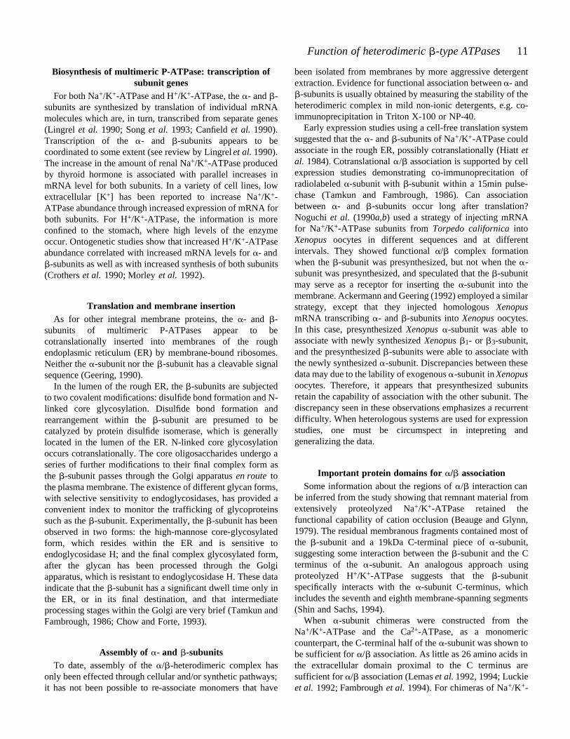
and the noncovalent bonding forces. Cooperativity has beenstudied in soluble globular protein as an important feature ofconformational stability (see Creighton, 1993). This case forcooperativity of bonding forces within the subfamily of cationexchange P-ATPases is an extension from simple proteinmodels, since the forces of interaction occur within and amongtwo subunit peptides, and they are membrane-bound peptides.Thus, correlations between K+-dependent conformationalshifts, disulfide bond stability and general protein foldingdemonstrate that the / -subunit interactions are extensive andimportant for enzymatic function of the holoenzyme. Aschematic representation of how folding of the protein anddisulfide bond formation can be cooperatively linked in amembrane-bound / heterodimer is depicted in Fig. 6.
Alternative support for -subunit participation in enzymaticfunction comes from studies using a specific inhibitory
monoclonal antibody, Mab-2G11, developed against the -subunit of H+/K+-ATPase. Mab-2G11 binds specifically to thecytoplasmic side of gastric secretory membranes with anepitope of interaction within the first 36 N-terminal aminoacids of the -subunit (Chow and Forte, 1993). Mab-2G11altered the affinity of the cytoplasmic K+ binding site, furthersupporting the hypothesis that the -subunit contributes to theapparent stability of the E2.K+ conformer. However, kineticarguments based on the full catalytic cycle of H+/K+-ATPasesuggested that the inhibition was not solely due to the loweredK+ affinity, but to interference with the conformationaltransition induced by K+. Thus, the -subunit is involved inthe conformational changes associated with the transport ofK+.
Several recent experiments suggest that the -subunit maybe involved in the well-known ability of Na+/K+- and H+/K+-
9Function of heterodimeric -type ATPases
0.80.60.40.200
20
40
60
80
100
ME +ethanol
MEalone
ME +KCl
[2ME] (mol l−1)
% A
ctiv
ity r
emai
ning
A
1008060402001
2
3
% Activity remaining
Num
ber
of d
isul
fide
bon
ds in
-sub
unit
C
A
B
D
E
F
G
H
I
B
Fig. 5. Demonstration of cooperative inactivation of H+/K+-ATPaseby a combination of reducing reagents and non-covalent bondmodification. (A) Inactivation of the H+/K+-ATPase by reducingreagents is further influenced by agents that alter proteinconformation. The inclusion of denaturants at concentrations that areby themselves harmless (e.g. 0.6mol l 1 ethanol) makes disulfidereduction, and enzyme inhibition, by mercaptoethanol (2ME) moreefficient. Ligands such as K+ (15mmol l 1), which favor a morestable E2.K+ conformation, increase the energy barrier for disulfidereduction. Specific conditions for inactivation were as described byChow et al. (1992). (B) Correlation between the number of disulfidebonds in the -subunit and the percentage of enzyme activityremaining after H+/K+-ATPase had been treated with combinations ofreducing reagent and denaturants. Gastric membranes were incubatedat 44˚C with 2ME for various periods. In some cases, the incubationalso included 0.6mol l 1 ethanol as a labilizer and/or 15mmol l 1
KCl as a stabilizer. A, control (no treatment); B, 0.45mol l 1 2ME for5 min; C, 0.45mol l 1 2ME for 10min; D, 0.45mol l 1 2ME for25min; E, 0.45mol l 1 2ME plus KCl for 25min; F, 0.3mol l 1 2MEfor 20min; G, 0.3mol l 1 2ME plus ethanol for 20min; H, 0.3mol l 1
2ME plus ethanol and KCl for 20min; I, 0.6mol l 1 2ME for20min.
0
3002001000
0.2
0.4
0.6
0.8
1.0
1.2
1.4
Amino acid number
B NaK 1
C NaK 2 and 3
D HK
A All -subunits
0.2 units
Fig. 4. Similarity plots of -subunit peptides within the subfamily ofcation exchange P-ATPases. The similarity plot for the entire 24isoforms (A, all -subunits) were generated with the alignment shownin Fig. 2. The similarity plots shown for the subgroups of -subunitisoforms (B, NaK 1; C, NaK 2 and 3; and D, HK ) were firstrealigned and then processed. All plots are drawn to the same scale,with zero being least similar and 1.8 most similar; amino acidalignment and insertion of spaces are as described in Fig. 2. The solidhorizontal line in A is the average similarity index for all -subunits.The horizontal dashed lines in B, C and D indicate a similarity indexof 1.0 for each of the respective subgroups.
ATPases to occlude K+ (the very slow exit of K+ from E2.K+
suggests that it is not simply an ion-binding step, but isimpeded by the enzyme form, hence occluded). Aftersubjecting Na+/K+-ATPase to extensive protease digestion,Capasso et al. (1992) found that the membrane-bound remnant
10 D. C. CHOW AND J. G. FORTE
Fig. 6. Model to account for cooperativelinking between protein disulfide bonds andconformational stability in a membraneheterodimeric protein. The protein is illustratedin either the folded or unfolded conformationwith four exemplary cysteine residues in one ofthe subunits that can form two disulfide bonds.Equilibrium constants represent stabilities ofthe folded conformation, KF, and of thedisulfide bonds, KSS. According to thecooperative linkage relationship, the effects ofthe folded conformation on the stability of thedisulfide bond are mirrored in the effects of thedisulfide bond on the stability of the foldedconformation, that is to say, KF
SS/KFSH =
KFSS/KU
SS (adapted from Creighton, 1993).
retained the ability to occlude Rb+, and the remaining peptidesincluded a relatively intact -subunit. Later, Lutsenko andKaplan (1993) reported that reduction of disulfide bonds in theremnant -subunit correlated with the loss of ability to occludeRb+ by the proteolyzed membranes. Furthermore, sites ofdigestion on the -subunit were different with and withoutRb+, further strengthening the argument that -subunitconformations are related to the E1–E2 states (Lutsenko andKaplan, 1994). These data demonstrate that interactionsbetween - and -subunits are required for occlusion of K+.
Experiments with a variety of expression systems reinforcethe importance of the -subunit for functional enzymaticactivity of the cation-exchange ATPases. For Na+/K+-ATPase,Horowitz et al. (1990) used a yeast expression system to showthat ouabain binding and ouabain-inhibitable ATP hydrolysisrequired expression of both - and -subunits. Using thebaculovirus expression system, Klaassen et al. (1993) reportedthat H+/K+-ATPase also required both - and -subunits forfunctional activity, as defined by phosphoenzyme formation,though they could not demonstrate the full hydrolytic cycle.Additional insight into / interaction comes from hybridstudies testing the complex expression of Na+/K+-ATPase -subunit with various -subunit isoforms. Jaisser et al. (1992)showed that the 1/ 1 complex had different K+ transportkinetics from the 1/ 3 complex. The -subunit of H+/K+-ATPase can serve as a substitute in supporting some functionalactivities when expressed together with the -subunit of theNa+/K+-ATPase in both Xenopus and yeast expression systems
(Horisberger et al. 1991a; Eakle et al. 1992). However, in theyeast system, Eakle et al. (1992) observed that the antagonismbetween ouabain binding and K+ is different for the Na+/K+ ,Na+/K+ complex than for the Na+/K+ , H+/K+ complex.These observations also link the -subunit with K+ binding andthe conformational transitions associated with K+.
Two laboratories have reported on the expression offunctional hybrids in which the -subunit of Na+/K+-ATPasewas expressed along with -subunit chimeras fabricated fromdifferent halves of Na+/K+-ATPase and H+/K+-ATPase (Jauninet al. 1993; Eakle et al. 1994). The chimeras were constructedby exchanging the extracellular domain of one -subunit withthe other, and both were found to assemble with the -subunitof Na+/K+-ATPase to produce functional activity.Furthermore, the measured affinity for K+ matched the K+
affinity of the -subunit that provided the extracellular domain,whereas the transmembrane domain served to determine thestrength of interaction with the -subunit. Thus, theextracellular domain of the -subunit has a dominant influenceon the interaction with K+.
The evidence reviewed in this section converges into atheme that the -subunits of Na+/K+-ATPase and H+/K+-ATPase play some role in the K+ transport function of thecation-exchange process. Since - and -subunits areassociated via tight coupling, the transport of cations probablyrequires the concerted conformational change of both subunits,rather than each subunit being responsible for a given stepindependently of the other subunit.
11Function of heterodimeric -type ATPases
Biosynthesis of multimeric P-ATPase: transcription ofsubunit genes
For both Na+/K+-ATPase and H+/K+-ATPase, the - and -subunits are synthesized by translation of individual mRNAmolecules which are, in turn, transcribed from separate genes(Lingrel et al. 1990; Song et al. 1993; Canfield et al. 1990).Transcription of the - and -subunits appears to becoordinated to some extent (see review by Lingrel et al. 1990).The increase in the amount of renal Na+/K+-ATPase producedby thyroid hormone is associated with parallel increases inmRNA level for both subunits. In a variety of cell lines, lowextracellular [K+] has been reported to increase Na+/K+-ATPase abundance through increased expression of mRNA forboth subunits. For H+/K+-ATPase, the information is moreconfined to the stomach, where high levels of the enzymeoccur. Ontogenetic studies show that increased H+/K+-ATPaseabundance correlated with increased mRNA levels for - and
-subunits as well as with increased synthesis of both subunits(Crothers et al. 1990; Morley et al. 1992).
Translation and membrane insertionAs for other integral membrane proteins, the - and -
subunits of multimeric P-ATPases appear to becotranslationally inserted into membranes of the roughendoplasmic reticulum (ER) by membrane-bound ribosomes.Neither the -subunit nor the -subunit has a cleavable signalsequence (Geering, 1990).
In the lumen of the rough ER, the -subunits are subjectedto two covalent modifications: disulfide bond formation and N-linked core glycosylation. Disulfide bond formation andrearrangement within the -subunit are presumed to becatalyzed by protein disulfide isomerase, which is generallylocated in the lumen of the ER. N-linked core glycosylationoccurs cotranslationally. The core oligosaccharides undergo aseries of further modifications to their final complex form asthe -subunit passes through the Golgi apparatus en route tothe plasma membrane. The existence of different glycan forms,with selective sensitivity to endoglycosidases, has provided aconvenient index to monitor the trafficking of glycoproteinssuch as the -subunit. Experimentally, the -subunit has beenobserved in two forms: the high-mannose core-glycosylatedform, which resides within the ER and is sensitive toendoglycosidase H; and the final complex glycosylated form,after the glycan has been processed through the Golgiapparatus, which is resistant to endoglycosidase H. These dataindicate that the -subunit has a significant dwell time only inthe ER, or in its final destination, and that intermediateprocessing stages within the Golgi are very brief (Tamkun andFambrough, 1986; Chow and Forte, 1993).
Assembly of - and -subunitsTo date, assembly of the / -heterodimeric complex has
only been effected through cellular and/or synthetic pathways;it has not been possible to re-associate monomers that have
been isolated from membranes by more aggressive detergentextraction. Evidence for functional association between - and
-subunits is usually obtained by measuring the stability of theheterodimeric complex in mild non-ionic detergents, e.g. co-immunoprecipitation in Triton X-100 or NP-40.
Early expression studies using a cell-free translation systemsuggested that the - and -subunits of Na+/K+-ATPase couldassociate in the rough ER, possibly cotranslationally (Hiatt etal. 1984). Cotranslational / association is supported by cellexpression studies demonstrating co-immunoprecitation ofradiolabeled -subunit with -subunit within a 15min pulse-chase (Tamkun and Fambrough, 1986). Can associationbetween - and -subunits occur long after translation?Noguchi et al. (1990a,b) used a strategy of injecting mRNAfor Na+/K+-ATPase subunits from Torpedo californica intoXenopus oocytes in different sequences and at differentintervals. They showed functional / complex formationwhen the -subunit was presynthesized, but not when the -subunit was presynthesized, and speculated that the -subunitmay serve as a receptor for inserting the -subunit into themembrane. Ackermann and Geering (1992) employed a similarstrategy, except that they injected homologous XenopusmRNA transcribing - and -subunits into Xenopus oocytes.In this case, presynthesized Xenopus -subunit was able toassociate with newly synthesized Xenopus 1- or 3-subunit,and the presynthesized -subunits were able to associate withthe newly synthesized -subunit. Discrepancies between thesedata may due to the lability of exogenous -subunit in Xenopusoocytes. Therefore, it appears that presynthesized subunitsretain the capability of association with the other subunit. Thediscrepancy seen in these observations emphasizes a recurrentdifficulty. When heterologous systems are used for expressionstudies, one must be circumspect in intepreting andgeneralizing the data.
Important protein domains for / associationSome information about the regions of / interaction can
be inferred from the study showing that remnant material fromextensively proteolyzed Na+/K+-ATPase retained thefunctional capability of cation occlusion (Beauge and Glynn,1979). The residual membranous fragments contained most ofthe -subunit and a 19kDa C-terminal piece of -subunit,suggesting some interaction between the -subunit and the Cterminus of the -subunit. An analogous approach usingproteolyzed H+/K+-ATPase suggests that the -subunitspecifically interacts with the -subunit C-terminus, whichincludes the seventh and eighth membrane-spanning segments(Shin and Sachs, 1994).
When -subunit chimeras were constructed from theNa+/K+-ATPase and the Ca2+-ATPase, as a monomericcounterpart, the C-terminal half of the -subunit was shown tobe sufficient for / association. As little as 26 amino acids inthe extracellular domain proximal to the C terminus aresufficient for / association (Lemas et al. 1992, 1994; Luckieet al. 1992; Fambrough et al. 1994). For chimeras of Na+/K+-
12 D. C. CHOW AND J. G. FORTE
ATPase and H+/K+-ATPase, Gottardi and Caplan (1993a)showed that the C-terminal half of the -subunit dictates theassembly with the respective -subunit. Furthermore, thecomplex of -chimera and -subunit appears to be functional(Blostein et al. 1993).
For deletion mutations made within the -subunit ofNa+/K+-ATPase, mutant subunits lacking the entirecytoplasmic piece, including portions of the transmembranedomain, or almost half of the extracytoplasmic C-terminaldomain, could associate with -subunits (Hamrick et al. 1993,Renaud et al. 1991). These data suggested that the extracellularsegment of about 100 amino acids of the -subunitimmediately adjacent to the membrane is responsible for /association. However, the chimera of this 100-amino-acidsegment, fused with the cytoplasmic and transmembrane pieceof a totally independent membrane protein, failed to associatewith the -subunit, whereas the analogous chimera using thewhole extracellular piece could associate with the -subunit(Hamrick et al. 1993). Intepretation of these results is furthercomplicated by results from point mutation studies. Mutationof Pro-256, or of hydrophobic amino acids near the C terminus,prevents the formation of a functional / complex on theplasma membrane (Beggah et al. 1993; Geering et al. 1993).In fact, the entire region from Leu-252 to Lys-260 is highlyconserved among all subfamily members (see Fig. 2). Noguchiet al. (1994) recently reported that mutation of the first twoextracellular cysteine residues on the -subunit, Cys-134 andCys-157, will generate an inactive / complex and thatmutation of the other four extracellular cysteines preventsassembly of the / complex. To reconcile apparentinconsistencies between the regional deletion data and pointmutation studies, one could speculate that, while there are veryspecific regions of subunit interplay, assembly of the /complex relies on multiple cooperative regional interactions,such that misfolding of one region would alter appropriatebonding within other regions. This may also explain thedifficulties of in vitro reassembly of the / complex fromseparated subunits. Chimeras of -subunit constructed fromcomplementary portions of Na+/K+- and H+/K+-ATPase -subunits have been used to evaluate relative domainparticipation in the / association (Jaunin et al. 1993; Eakleet al. 1994). These chimeras were constructed with the entirecytoplasmic domain plus the transmembrane domain from one
-subunit and the extracytoplasmic domain from the other. All-subunit chimeras were capable of assembly with the Na+/K+-
ATPase -subunit to provide functional pumps, with somedifferences in specific functional properties of the resultingcomplexes. Chimeras with the N-terminal piece from Na+/K+-ATPase -subunit had the highest efficiency in stabilizing the
/ complex. Thus, multiple regions within the -subunitparticipate in the interactive and functional activities of theholoenzyme.
Functional maturation of the nascent / complexSeveral lines of evidence suggest that the / complex is
functional soon after it is formed: (1) within 15min ofsynthesis, association between subunits can be measured(Tamkun and Fambrough, 1986); (2) within 10–15min of -subunit synthesis, the Na+/K+-ATPase appears to be capableof binding ouabain (Caplan et al. 1990); and (3) within 15minof synthesis, a detectable fraction of newly synthesizedNa+/K+-ATPase -subunit is converted from a trypsin-sensitive form to a trypsin-resistant form consistent withconformational rearrangement (Geering et al. 1987). In theabove systems, protein transport from ER to plasma membranerequires at least 40–60min; thus, Na+/K+-ATPase must befunctional before it reaches the plasma membrane.
For the H+/K+-ATPase, identification of an associatedcomplex between the -subunit and the 52kDa immature -subunit (core-glycosylated form) indicated that / assemblyof the H+/K+-ATPase commences in the ER, probablycotranslationally. Within 15min of radiolabeled amino acidincorporation, abundant radiolabeled -subunit was seenwithin cell fractions enriched in ER but was barely detectablein fractions corresponding to normal functioning parietal celltubulovesicles (Crothers et al. 1993). After 30min, the amountof newly synthesized -subunit in the mature tubulovesicularfraction was significantly increased. Thus, the pattern offunctional maturation for the H+/K+-ATPase appears to besimilar to that of the Na+/K+-ATPase.
Fate of unassociated subunitsThere appear to be conflicting data describing the fate of
unassociated subunits. An early model proposed thatunassociated - and -subunits are retained within the ER untilthey associate by collision. They then supposedly exit the ERas the heterodimer or are eventually degraded by the ERquality control mechanisms. This model was based on theobservations that (1) when -subunit was overexpressed in amouse cell line, it was almost exclusively locatedintracellularly, probably in the ER (Takeyasu et al. 1988), and(2) when the -subunit was overexpressed in Xenopus oocytes,it was maintained in the core-glycosylated form, indicative ofresidence within the ER. Exceptions to this model can befound. Hundal et al. (1992) showed, in muscle, that insulinstimulated the translocation of the 2-subunit from anintracellular pool to the plasma membrane and thetranslocation of the 1-subunit from a different intracellularpool to the plasma membrane. This exception implies thatassembly of 2/ 1 might occur outside the ER under insulinregulation, but alternative explanations are possible. Insectcells transfected with baculovirus carrying either - or -subunit genes express the subunits to their plasma membraneindependent of each other (DeTomaso et al. 1992). Animportant feature of these baculovirus-transfected insect cellsis that they can be induced to fuse and form syncytia, such thatplasma membrane proteins can redistribute but the membraneproteins within the intracellular pool cannot. DeTomaso andMercer (1993) recently exploited this property to demonstratethat assembly of functional Na+/K+-ATPase / complex on
13Function of heterodimeric -type ATPases
the plasma membrane of the fused syncytium originated fromcells independently expressing either the -subunit or the -subunit of the Na+/K+-ATPase. In contrast, assembly of hybridNa+/K+-ATPase -subunit and -subunit of the H+/K+-ATPase did not occur in a fusion experiment, but did occurwhen the cells were cotransfected, suggesting that the hybridcould assemble only within the ER. This result provides a basisfor the possibility of / assembly outside the ER. Invertebrate cells, the fact that / assembly appears to occurpredominantly, or exclusively, within the ER may simply bedue to the inability of the -subunit to exit the ER. In addition,we point out that / assembly does not guarantee appropriatetrafficking. In their study of deletion mutants, Fambrough’sgroup showed that removal of four amino acids from the Cterminus or five amino acids from the transmembrane segmentdid not prevent subunit assembly, but profoundly compromisedthe ability of the / complex to move out of the ER (Hamricket al. 1993; Renaud et al. 1991).
/ association of gastric H+/K+-ATPase, like that ofNa+/K+-ATPase, appears to begin within the ER (Chow et al.1994). Using the omeprazole-treated stomach model, Crotherset al. (1993) found that degeneration of H+/K+-ATPase wasdue to a parallel degradation of both - and -subunits. Therehas been one report that some mature -subunit, independentof -subunit, can be recovered from parietal cells (Baldwin,1990); however, this recovery may be due to the exit of someunassociated -subunit from the ER. In support of thishypothesis, for all reported expression systems, the -subunitof the H+/K+-ATPase can exit the ER without a corresponding
-subunit, whereas the -subunit of the H+/K+-ATPaseappears to require / association before exiting the ER(Gottardi and Caplan, 1993a; Jaunin et al. 1993).
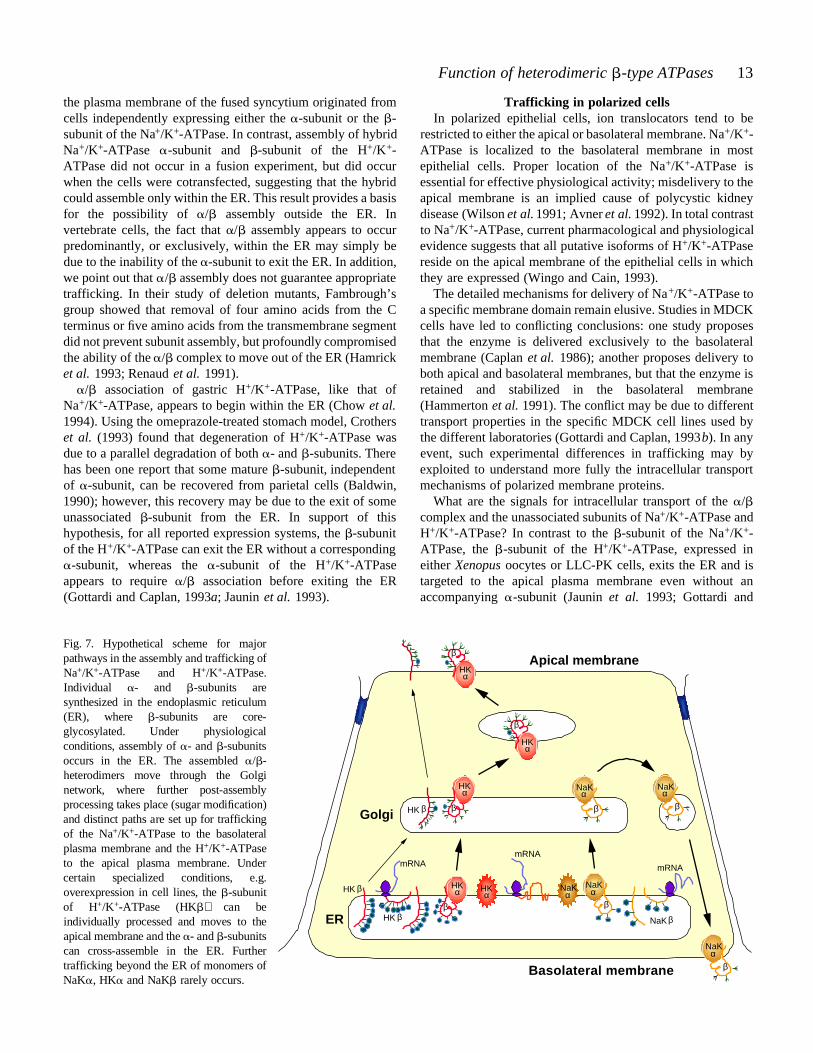
HK β
m
HK β
ER
Golgi
F i g . 7. Hypothetical scheme for majorpathways in the assembly and trafficking ofN a+/ K+-ATPase and H+/ K+- A T P a s e .Individual - and -subunits aresynthesized in the endoplasmic reticulum(ER), where -subunits are core-glycosylated. Under physiologicalconditions, assembly of - and - s u b u n i t soccurs in the ER. The assembled / -heterodimers move through the Golginetwork, where further post-assemblyprocessing takes place (sugar modific a t i o n )and distinct paths are set up for traffickingof the Na+/ K+-ATPase to the basolateralplasma membrane and the H+/ K+- A T P a s eto the apical plasma membrane. Undercertain specialized conditions, e.g.overexpression in cell lines, the - s u b u n i tof H+/ K+-ATPase (HK ) can beindividually processed and moves to theapical membrane and the - and - s u b u n i t scan cross-assemble in the ER. Furthertrafficking beyond the ER of monomers ofN a K , HK and NaK rarely occurs.
Trafficking in polarized cellsIn polarized epithelial cells, ion translocators tend to be
restricted to either the apical or basolateral membrane. Na+/K+-ATPase is localized to the basolateral membrane in mostepithelial cells. Proper location of the Na+/K+-ATPase isessential for effective physiological activity; misdelivery to theapical membrane is an implied cause of polycystic kidneydisease (Wilson et al. 1991; Avner et al. 1992). In total contrastto Na+/K+-ATPase, current pharmacological and physiologicalevidence suggests that all putative isoforms of H+/K+-ATPasereside on the apical membrane of the epithelial cells in whichthey are expressed (Wingo and Cain, 1993).
The detailed mechanisms for delivery of Na+/K+-ATPase toa specific membrane domain remain elusive. Studies in MDCKcells have led to conflicting conclusions: one study proposesthat the enzyme is delivered exclusively to the basolateralmembrane (Caplan et al. 1986); another proposes delivery toboth apical and basolateral membranes, but that the enzyme isretained and stabilized in the basolateral membrane(Hammerton et al. 1991). The conflict may be due to differenttransport properties in the specific MDCK cell lines used bythe different laboratories (Gottardi and Caplan, 1993b). In anyevent, such experimental differences in trafficking may byexploited to understand more fully the intracellular transportmechanisms of polarized membrane proteins.
What are the signals for intracellular transport of the /complex and the unassociated subunits of Na+/K+-ATPase andH+/K+-ATPase? In contrast to the -subunit of the Na+/K+-ATPase, the -subunit of the H+/K+-ATPase, expressed ineither Xenopus oocytes or LLC-PK cells, exits the ER and istargeted to the apical plasma membrane even without anaccompanying -subunit (Jaunin et al. 1993; Gottardi and
HKα
NaKα
NaKα
β
β β
βNaK
mRNARNAmRNA
βHK β
HKα
β
NaKα
β
HKα
β
NaKα
β
Apical membrane
Basolateral membrane
HKα
HKα
NaKα
14 D. C. CHOW AND J. G. FORTE
Caplan, 1993c). Furthermore, in the polarized LLC-PK cells,the H+/K+-ATPase -subunit is delivered to the apicalmembrane, whereas Na+/K+-ATPase is delivered to thebasolateral membrane. Chimeras constructed of the N-terminalhalf of the H+/K+-ATPase -subunit and the C-terminal halfof the Na+/K+-ATPase -subunit not only assemble withNa+/K+-ATPase -subunit, but the complex is also deliveredto the apical membrane. This targetting suggests that both the
- and -subunits of H+/K+-ATPase carry a signal for apicalmembrane delivery. The -subunit of H+/K+-ATPase doesrequire association with the -subunit for efficient delivery tothe apical membrane. Detection of the 52kDa core-glycosylated H+/K+-ATPase -subunit in ER-enrichedfractions from gastric homogenates is consistent with the -subunit pausing within the ER for some processing activity,such as / -subunit assembly and functional maturation. Aschematic model depicting the translation and assembly of theH+/K+-ATPase and Na+/K+-ATPase subunits and theirtrafficking to respective apical and basolateral membranes isshown in Fig. 7.
ReferencesACKERMANN, U. AND GEERING, K. (1992). 1- and 3-subunits can
associate with presynthesized -subunits of Xenopus oocyte Na,K-ATPase. J. biol. Chem . 267, 12911–12915.
AMZEL, L. M. AND PEDERSEN, P. L. (1983). Proton ATPases: structureand mechanism. A. Rev. Biochem . 52, 801–824.
AVNER, E. D., SWEENEY, W. E., JR AND NELSON, W. J. (1992).Abnormal sodium pump distribution during renal tubulogenesis incongenital murine polycystic kidney disease. Proc. natn. Acad. Sci.U.S.A. 89, 7447–7451.
BALDWIN, G. S. (1990). The -subunit of the gastric H+/K+-ATPasecan occur without the -subunit. FEBS Lett. 272, 159–162.
BEAUGE, L. A. AND GLYNN, I. M. (1979). Occlusion of K ions in theunphosphorylated sodium pump. Nature 280, 510–512.
BEGGAH, A. T., BEGUIN, P., JAUNIN, P., PEITSCH, M. C. AND GEERING,K. (1993). Hydrophobic C-terminal amino acids in the -subunitare involved in assembly with the -subunit of Na,K-ATPase.Biochemistry, N.Y. 32, 14117–14124.
BESANCON, M., SHIN, J. M., MERCIER, F., MUNSON, K., MILLER, M.,HERSEY, S. AND SACHS, G. (1993). Membrane topology andomeprazole labeling of the gastric H+,K+-adenosinetriphosphatase.Biochemistry, N.Y. 32, 2345–2355.
BHATTACHARYYA, K. K., BERGSTROM, E. E. AND HOKIN, L. E. (1990).Molecular cloning of the -subunit of the Na,K-ATPase in the brineshrimp, Artemia. The cDNA-derived amino acid sequence showslow homology with the -subunits of vertebrates except in thesingle transmembrane and the carboxy-terminal domains. FEBSLett. 269, 233–238.
BLOSTEIN, R., ZHANG, R., GOTTARDI, C. J. AND CAPLAN, M. J. (1993).Functional properties of an H,K-ATPase/Na,K-ATPase chimera. J.biol. Chem. 268, 10654–10658.
BROTHERUS, J. R., JACOBSEN, L. AND JORGENSEN, P. L.(1983). Solubleand enzymatically stable (Na++K+)-ATPase from mammaliankidney consisting predominantly of protomer -units. Biochim.biophys. Acta 731, 290–303.
BROWN, T. A., HOROWITZ, B., MILLER, R. P., MCDONOUGH, A. A. AND
FARLEY, R. A. (1987). Molecular cloning and sequence analysis of
the (Na++K+)-ATPase subunit from dog kidney. Biochim.biophys. Acta 912, 244–253.
BROWNING, C., CHOW, D. AND FORTE, J. G. (1992). Stability of theH,K-ATPase: Cooperativity between disulfide and conformationalstability in a membrane protein model. FASEB J. 6, A1187 (no.1450).
CANESSA, C. M., HORISBERGER, J. D., LOUVARD, D. AND ROSSIER, B.C. (1992). Mutation of a cysteine in the first transmembranesegment of Na,K-ATPase subunit confers ouabain resistance.EMBO J. 11, 1681–1687.
CANFIELD, V. A. AND LEVENSON, R. (1991). Structural organizationand transcription of the mouse gastric (H+, K+)-ATPase subunitgene. Proc. natn. Acad. Sci. U.S.A. 88, 8247–8251.
CANFIELD, V. A., OKAMOTO, C. T., CHOW, D., DORFMAN, J., GROS, P.,FORTE, J. G. AND LEVENSON, R.(1990). Cloning of the H,K-ATPase
subunit. Tissue-specific expression, chromosomal assignmentand relationship to Na,K-ATPase subunits. J. biol. Chem. 265,19878–19884.
CANTLEY, L. C., JR, JOSEPHSON, L., WARNER, R., YANAGISAWA, M.,LECHENE, C. AND GUIDOTTI, G.(1977). Vanadate is a potent (Na,K)-ATPase inhibitor found in ATP derived from muscle. J. biol. Chem.252, 7421–7423.
CAPASSO, J. M., HOVING, S., TAL, D. M., GOLDSHLEGER, R. AND
KARLISH, S. J. (1992). Extensive digestion of Na+,K+-ATPase byspecific and nonspecific proteases with preservation of cationocclusion sites. J. biol. Chem . 267, 1150–1158.
CAPLAN, M. J., ANDERSON, H. C., PALADE, G. E. AND JAMIESON, J. D.(1986). Intracellular sorting and polarized cell surface delivery of(Na+,K+)-ATPase, an endogenous component of MDCK cellbasolateral plasma membranes. Cell 46, 623–631.
CAPLAN, M. J., FORBUSH III, B., PALADE, G. E. AND JAMIESON, J. D.(1990). Biosynthesis of the Na,K-ATPase in Madin–Darby caninekidney cells. Activation and cell surface delivery. J. biol. Chem.265, 3528–3534.
CHOW, D. C., BROWNING, C. M. AND FORTE, J. G.(1992). Gastric H,K-ATPase activity is inhibited by reduction of disulfide bonds in the
-subunit. Am. J. Physiol . 256, C39–C46.CHOW, D. C. AND FORTE, J. G. (1993). Characterization of the -
subunit of the gastric H,K-ATPase by an inhibitory monoclonalantibody. Am. J. Physiol . 265, C1562–C1570.
CHOW, D., SCALLEY, M., SMOLKA, A. J. AND FORTE, J. G. (1994).Assembly of -subunit with -subunit of the H,K-ATPasecommences within the endoplasmic reticulum. Gastroenterology106, A63.
CHOW, D. C., USHIGOME, M. M., CROTHERS, J. M. AND FORTE, J. G.(1993). Funcitonal and protective roles for the -subunit of theH,K-ATPase. Gastroenterology 104, A55.
CLARKE, R. J., APELL, H. J. AND LAUGER, P. (1989). Pump current andNa+/K+ coupling ratio of Na+/K+-ATPase in reconstituted lipidvesicles. Biochim. biophys. Acta 981, 326–336.
CREIGHTON, T. E. (1993). Proteins – Structure and MolecularProperties (2nd edn). New York: W. H. Freeman and Company.
CROTHERS, J. M., JR, CHOW, D. C. AND FORTE, J. G. (1993).Omeprazole decreases H+–K+-ATPase protein and increasespermeability of oxyntic secretory membranes in rabbits. Am. J.Physiol. 265, G231–G241.
CROTHERS, J. M., REENSTRA, W. W. AND FORTE, J. G. (1990).Ontogeny of gastric H,K-ATPase in suckling rabbits. Am. J.Physiol. 259, G913–G921.
DE MEIS, L. AND VIANNA, A. L.(1979). Energy interconversion by the
15Function of heterodimeric -type ATPases
Ca2+-dependent ATPase of the sarcoplasmic reticulum. A. Rev.Biochem. 48, 275–292.
DETOMASO, A. W. AND MERCER, R. W. (1993). The and subunitof the Na,K-ATPase can assemble at the plasma membrane. Molec.Biol. Cell 4, 94a (no. 548).
DETOMASO, A. W., XIE, Z., LIU, G. AND MERCER, R. W. (1992).Expression, targeting and assembly of functional Na,K-ATPasepolypeptides in baculovirus-infected insect cells. J. biol. Chem.268, 1470–1478.
EAKLE, K. A., KABALIN, M. A., WANG, S. G. AND FARLEY, R. A.(1994). The influence of subunit structure on the stability ofNa+,K+-ATPase complexes and interaction with K+. J. biol. Chem.269, 6550–6557.
EAKLE, K. A., KIM, K. S., KABALIN, M. A. AND FARLEY, R. A. (1992).High-affinity ouabain binding by yeast cells expressing Na+,K+-ATPase subunits and the gastric H+, K+-ATPase subunit. Proc.natn. Acad. Sci. U.S.A. 89, 2834–2838.
FALLER, L. D., RABON, E. AND SACHS, G. (1983). Vanadate bindingto the gastric H,K-ATPase and inhibition of the enzyme’s catalyticand transport activities. Biochemistry, N.Y. 22, 4676–4685.
FAMBROUGH, D. M., LEMAS, V., HAMRICK, M., EMERICK, M., RENAUD,K. J., INMAN, E. M., HWANG, B. AND TAKEYASU, K. (1994).Analysis of subunit assembly of the Na,K-ATPase. Am. J. Physiol.266, C579–C589.
FENG, J. AND LINGREL, J. B. (1994). Analysis of amino acid residuesin the H5–H6 transmembrane and extracellular domains of Na,K-ATPase subunit identifies threonine 797 as a determinant ofouabain sensitivity. Biochemistry, N.Y. 33, 4218–4224.
FORTE, T. M. AND FORTE, J. G. (1970). Histochemical staining andcharacterization of glycoproteins in acid-secreting cells of the frogstomach. J. Cell Biol. 47, 437–452.
GEERING, K. (1990). Subunit assembly and functional maturation ofNa,K-ATPase. J. Membr. Biol. 115, 109–121.
GEERING, K., JAUNIN, P., JAISSER, F., MERILLAT, A. M., HORISBERGER,J. D., MATHEWS, P. M., LEMAS, V., FAMBROUGH, D. M. AND
ROSSIER, B. C. (1993). Mutation of a conserved proline residue inthe -subunit ectodomain prevents Na+–K+-ATPaseoligomerization. Am. J. Physiol. 265, C1169–C1174.
GEERING, K., KRAEHENBUHL, J. P. AND ROSSIER, B. C. (1987).Maturation of the catalytic -subunit of Na,K-ATPase duringintracellular transport. J. Cell Biol . 105, 2613–2619.
GLOOR, S. (1989). Cloning and nucleotide sequence of the mouseNa,K-ATPase -subunit. Nucleic Acids Res. 17, 10117.
GLOOR, S., ANTONICEK, H., SWEADNER, K. J., PAGLIUSI, S., FRANK, R.,MOOS, M. AND SCHACHNER, M. (1990). The adhesion molecule onglia (AMOG) is a homologue of the subunit of the Na,K-ATPase.J. Cell Biol . 110, 165–174.
GOOD, P. J., RICHTER, K. AND DAWID, I. B. (1990). A nervous system-specific isotype of the subunit of Na+,K+-ATPase expressedduring early development of Xenopus laevis.Proc. natn. Acad. Sci.U.S.A. 87, 9088–9092.
GOTTARDI, C. J. AND CAPLAN, M. J. (1993a). Molecular requirementsfor the cell-surface expression of multisubunit ion-transportingATPases. Identification of protein domains that participate in Na,K-ATPase and H,K-ATPase subunit assembly. J. biol. Chem. 268,14342–14347.
GOTTARDI, C. J. AND CAPLAN, M. J. (1993b). Delivery of Na+,K+-ATPase in polarized epithelial cells. Science 260, 552–554.
GOTTARDI, C. J. AND CAPLAN, M. J. (1993c). An ion-transportingATPase encodes multiple apical localization signals. J. Cell Biol.121, 283–293.
HAMMERTON, R. W., KRZEMINSKI, K. A., MAYS, R. W., RYAN, T. A.,WOLLNER, D. A. AND NELSON, W. J. (1991). Mechanism forregulating cell surface distribution of Na+,K+-ATPase in polarizedepithelial cells. Science 254, 847–850.
HAMRICK, M., RENAUD, K. J. AND FAMBROUGH, D. M. (1993).Assembly of the extracellular domain of the Na,K-ATPase subunit with the subunit. Analysis of subunit chimeras andcarboxyl-terminal deletions. J. biol. Chem. 268, 24367–24373.
HARVEY, W. R. (1992). Physiology of V-ATPases. J. exp. Biol. 172,1–17.
HIATT, A., MCDONOUGH, A. A. AND EDELMAN, I. S.(1984). Assemblyof the (Na+–K+)-adenosine triphosphatase. Post-translationalmembrane integration of the subunit. J. biol. Chem. 259,2629–2635.
HORISBERGER, J. D., JAUNIN, P., REUBEN, M. A., LASATER, L. S.,CHOW, D. C., FORTE, J. G., SACHS, G., ROSSIER, B. C. AND GEERING,K. (1991a). The H,K-ATPase -subunit can act as a surrogatefor the -subunit of Na,K-pumps. J. biol. Chem. 266,19131–19134.
HORISBERGER, J. D., LEMAS, V., KRAEHENBUHL, J. P. AND ROSSIER, B.C. (1991b). Structure–function relationship of Na,K-ATPase. A.Rev. Physiol. 53, 565–584.
HOROWITZ, B., EAKLE, K. A., S CHEINER-BOBIS, G., RANDOLPH, G. R.,CHEN, C. Y., HITZEMAN, R. A. AND FARLEY, R. A.(1990). Synthesisand assembly of functional mammalian Na,K-ATPase in yeast. J.biol. Chem. 265, 4189–4192.
HUNDAL, H. S., MARETTE, A., MITSUMOTO, Y., RAMLAL, T., BLOSTEIN,R. AND KLIP, A. (1992). Insulin induces translocation of the 2 and
1 subunits of the Na+/K+-ATPase from intracellular compartmentsto the plasma membrane in mammalian skeletal muscle. J. biol.Chem. 267, 5040–5043.
INESI, G. AND KIRTLEY, M. R. (1992). Structural features of cationtransport ATPases. J. Bioenerg. Biomembr. 24, 271–283.
ISHII, T., LEMAS, M. V. AND TAKEYASU, K. (1994). Na+-, ouabain-,Ca2+- and thapsigargin-sensitive ATPase activity expressed inchimeras between the calcium and the sodium pump subunits.Proc. natn. Acad. Sci. U.S.A. 91, 6103–6107.
JAISSER, F., CANESSA, C. M., HORISBERGER, J. D. AND ROSSIER, B. C.(1992). Primary sequence and functional expression of a novelouabain-resistant Na,K-ATPase. The subunit modulatespotassium activation of the Na,K-pump. J. biol. Chem. 267,16895–16903.
JAISSER, F., COUTRY, N., FARMAN, N., BINDER, H. J. AND ROSSIER, B.C. (1993a). A putative H+–K+-ATPase is selectively expressed insurface epithelial cells of rat distal colon. Am. J. Physiol. 265,C1080–C1089.
JAISSER, F., HORISBERGER, J. D., GEERING, K. AND ROSSIER, B. C.(1993b). Mechanisms of urinary K+ and H+ secretion: Primarystructure and functional expression of a novel H,K-ATPase. J. CellBiol. 123, 1421–1430.
JAISSER, F., HORISBERGER, J. D. AND ROSSIER, B. C. (1993c). Primarysequence and functional expression of a novel subunit of the P-ATPase gene family. Pflügers Arch . 425, 446–452.
JAUNIN, P., JAISSER, F., BEGGAH, A. T., TAKEYASU, K., MANGEAT, P.,ROSSIER, B. C., HORISBERGER, J. D. AND GEERING, K. (1993). Roleof the transmembrane and extracytoplasmic domain of subunitsin subunit assembly, intracellular transport and functionalexpression of Na,K-pumps. J. Cell Biol. 123, 1751–1759.
JORGENSEN, P. L. AND ANDERSEN, J. P. (1988). Structural basis forE1–E2 conformational transitions in Na,K-pump and Ca-pumpproteins. J. Membr. Biol . 103, 95–120.
16 D. C. CHOW AND J. G. FORTE
KAWAKAMI, K., NOJIMA, H., OHTA, T. AND NAGANO, K. (1986).Molecular cloning and sequence analysis of human Na,K-ATPase
-subunit. Nucleic Acids Res. 14, 2833–2844.KAWAMURA, M. AND NAGANO, K. (1984). Evidence for essential
disulfide bonds in the -subunit of (Na++K+)-ATPase. Biochim.biophys. Acta 774, 188–192.
KAWAMURA, M., OHMIZO, K., MOROHASHI, M. AND NAGANO, K.(1985). Protective effect of Na+ and K+ against inactivation of(Na++K+)-ATPase by high concentrations of 2-mercaptoethanol athigh temperatures. Biochim. biophys. Acta 821, 115–120.
KIRLEY, T. L. (1989). Determination of three disulfide bonds and onefree sulfhydryl in the subunit of (Na,K)-ATPase. J. biol. Chem.264, 7185–7192.
KIRLEY, T. L. (1990). Inactivation of (Na+,K+)-ATPase by -mercaptoethanol. Differential sensitivity to reduction of the three
subunit disulfide bonds. J. biol. Chem . 265, 4227–4232.KLAASSEN, C. H., VAN UEM, T. J., DE MOEL, M. P., DE CALUWE, G.
L., SWARTS, H. G. AND DE PONT, J. J.(1993). Functional expressionof gastric H,K-ATPase using the baculovirus expression system.FEBS Lett . 329, 277–282.
LEMAS, M. V. AND FAMBROUGH, D. M. (1993). Sequence analysis ofDNA encoding an avian Na+,K+-ATPase 2-subunit. Biochim.biophys. Acta 1149, 339–342.
LEMAS, M. V., H AMRICK, M., TAKEYASU, K. AND FAMBROUGH, D. M.(1994). 26 amino acids of an extracellular domain of the Na,K-ATPase -subunit are sufficient for assembly with the Na,K-ATPase -subunit. J. biol. Chem. 269, 8255–8259.
LEMAS, M. V., TAKEYASU, K. AND FAMBROUGH, D. M. (1992). Thecarboxyl-terminal 161 amino acids of the Na,K-ATPase -subunitare sufficient for assembly with the -subunit. J. biol. Chem. 267,20987–20991.
LINGREL, J. B., ORLOWSKI, J., SHULL, M. M. AND PRICE, E. M. (1990).Molecular genetics of Na,K-ATPase. Prog. Nucleic Acid Res.molec. Biol. 38, 37–89.
LUCKIE, D. B., LEMAS, V., BOYD, K. L., FAMBROUGH, D. M. AND
TAKEYASU, K. (1992). Molecular dissection of functional domainsof the E1E2-ATPase using sodium and calcium pump chimericmolecules. Biophys. J . 62, 220–226.
LUTSENKO, S. AND KAPLAN, J. H. (1993). An essential role for theextracellular domain of the Na,K-ATPase -subunit in cationocclusion. Biochemistry, N.Y. 32, 6737–6743.
LUTSENKO, S. AND KAPLAN, J. H. (1994). Molecular events in closeproximity to the membrane associated with the binding of ligandsto the Na,K-ATPase. J. biol. Chem . 269, 4555–4564.
MA, J. Y., SONG, Y.H., SJOSTRAND, S. E., RASK, L. AND MARDH, S.(1991). cDNA cloning of the -subunit of the human gastric H,K-ATPase. Biochem. biophys. Res. Commun. 180, 39–45.
MARTIN-VASALLO, P., DACKOWSKI, W., EMANUEL, J. R. AND
LEVENSON, R. (1989). Identification of a putative isoform of theNa,K-ATPase subunit. Primary structure and tissue-specificexpression. J. biol. Chem . 264, 4613–4618.
MERCER, R. W., SCHNEIDER, J. W., SAVITZ, A., EMANUEL, J., BENZ,E. J. AND LEVENSON, R. (1986). Rat-brain Na,K-ATPase -chaingene: primary structure, tissue-specific expression andamplification in ouabain-resistant HeLa C+ cells. Molec. cell. Biol.6, 3884–3890.
MILLER, R. P. AND FARLEY, R. A. (1988). All three potential N-glycosylation sites of the dog kidney (Na++K+)-ATPase -subunitcontain oligosaccharide. Biochim. biophys. Acta 954, 50–57.
MILLER, R. P. AND FARLEY, R. A. (1990). -subunit of (Na++K+)-
ATPase contains three disulfide bonds. Biochemistry, N.Y. 29,1524–1532.
MORLEY, G. P., CALLAGHAN, J. M., ROSE, J. B., TOH, B. H., GLEESON,P. A. AND VAN DRIEL, I. R. (1992). The mouse gastric H,K-ATPase
subunit. Gene structure and coordinate expression with the subunit during ontogeny. J. biol. Chem . 267, 1165–1174.
NOGUCHI, S., HIGASHI, K. AND KAWAMURA, M. (1990a). A possiblerole of the -subunit of (Na,K)-ATPase in facilitating correctassembly of the -subunit into the membrane. J. biol. Chem. 265,15991–15995.
NOGUCHI, S., HIGASHI, K. AND KAWAMURA, M. (1990b). Assembly ofthe -subunit of Torpedo californica Na+/K+-ATPase with its pre-existing -subunit in Xenopus oocytes. Biochim. biophys. Acta1023, 247–253.
NOGUCHI, S., MUTOH, Y. AND KAWAMURA, M. (1994). The functionalroles of the disulfide bonds in the -subunit of (Na,K)-ATPase asstudied by site-directed mutagenesis. FEBS Lett. 341, 233–238.
NOGUCHI, S., NODA, M., TAKAHASHI, H., KAWAKAMI, K., OHTA, T.,NAGANO, K., HIROSE, T., INAYAMA, S., KAWAMURA, M. AND NUMA,S.(1986). Primary structure of the -subunit of Torpedo californica(Na++K+)-ATPase deduced from the cDNA sequence. FEBS Lett.196, 315–320.
OKAMOTO, C. T., KARPILOW, J. M., SMOLKA, A. AND FORTE, J. G.(1990). Isolation and characterization of gastric microsomalglycoproteins. Evidence for a glycosylated b-subunit of the H,K-ATPase. Biochim. biophys. Acta 1037, 360–372.
O’NEAL, S. G., RHOADS, D. B. AND RACKER, E. (1979). Vanadateinhibition of sarcoplasmic reticulum Ca2+-ATPase and otherATPases. Biochem. biophys. Res. Commun. 89, 845–850.
PEDERSEN, P. L. AND CARAFOLI, E. (1987). Ion motive ATPase. II.Trends biochem. Sci . 12, 186–189.
POST, R. L. AND JOLLY, P. C. (1957). The linkage of sodium,potassium and ammonium active transport across the humanerythrocyte membrane. Biochim. biophys. Acta 25, 118–128.
RENAUD, K. J., INMAN, E. M. AND FAMBROUGH, D. M. (1991).Cytoplasmic and transmembrane domain deletions of Na,K-ATPase -subunit. Effects on subunit assembly and intracellulartransport. J. biol. Chem . 266, 20491–20497.
REUBEN, M. A., LASATER, L. S. AND SACHS, G. (1990).Characterization of a subunit of the gastric H+/K+-transportingATPase. Proc. natn. Acad. Sci. U.S.A. 87, 6767–6771.
SACHS, G., CHANG, H. H., RABON, E., SCHACKMAN, R., LEWIN, M. AND
SACCOMANI, G. (1976). A nonelectrogenic H+ pump in plasmamembranes of hog stomach. J. biol. Chem. 251, 7690–7698.
SACHS, G., KAUNITZ, J., MENDLEIN, J. AND WALLMARK, B. (1989).Biochemistry of gastric acid secretion: H+–K+-ATPase. InHandbook of Physiology – The Gastrointestinal System, vol. III,chapter 12 (ed. J. G. Forte), pp. 229–253. Bethesda, MD: AmericanPhysiological Society.
SCHMALZING, G., KRONER, S., SCHACHNER, M. AND GLOOR, S.(1992).The adhesion molecule on glia (AMOG/ 2) and 1 subunitsassemble to functional sodium pumps in Xenopus oocytes. J. biol.Chem. 267, 20212–20216.
SHIN, J. M. AND SACHS, G. (1994). Identification of a region of theH,K-ATPase subunit associated with the subunit. J. biol. Chem.269, 8642–8646.
SHULL, G. E. (1990). cDNA cloning of the -subunit of the rat gastricH,K-ATPase. J. biol. Chem . 265, 12123–12126.
SHULL, G. E., LANE, L. K. AND LINGREL, J. B. (1986). Amino-acidsequence of the -subunit of the (Na++K+)ATPase deduced from acDNA. Nature 321, 429–431.
17Function of heterodimeric -type ATPases
SHULL, G. E. AND LINGREL, J. B. (1986). Molecular cloning of the ratstomach (H++K+)-ATPase. J. biol. Chem . 261, 16788–16791.
SKOU, J. C. (1982). The (Na++K+)-ATPase: coupling of the reactionwith ATP to the reaction with Na+ and K+. Ann. N.Y. Acad. Sci.402, 169–84.
SONG, I., MORTELL, M. P., GANTZ, I., BROWN, D. R. AND YAMADA, T.(1993). Molecular cloning and structural analysis of canine gastricH+,K+-ATPase. Biochem. biophys. Res. Commun. 196, 1240–1247.
TAKEYASU, K., TAMKUN, M. M., SIEGEL, N. R. AND FAMBROUGH, D.M. (1987). Expression of hybrid (Na++K+)-ATPase moleculesafter transfection of mouse Ltk-cells with DNA encoding the -subunit of an avian brain sodium pump. J. biol. Chem. 262,10733–10740.
TAMKUN, M. M. AND FAMBROUGH, D. M. (1986). The (Na++K+)-ATPase of chick sensory neurons. Studies on biosynthesis andintracellular transport. J. biol. Chem . 261, 1009–1019.
TOH, B. H., GLEESON, P. A., SIMPSON, R. J., MORITZ, R. L.,CALLAGHAN, J. M., GOLDKORN, I., JONES, C. M., MARTINELLI, T.M., MU, F. T., HUMPHRIS, D. C., PETTITT, J. M., MORI, Y., MASUDA,T., SOBIESZCZUK, P., WEINSTOCK, J., MANTAMADIOTIS, T. AND
BALDWIN, G. S.(1990). The 60- to 90-kDa parietal cell autoantigenassociated with autoimmune gastritis is a subunit of the gastricH+/K+-ATPase (proton pump). Proc. natn. Acad. Sci. U.S.A. 87,6418–6422.
TREUHEIT, M. J., COSTELLO, C. E. AND KIRLEY, T. L.(1993). Structures
of the complex glycans found on the -subunit of (Na,K)-ATPase.J. biol. Chem . 268, 13914–13919.
VERREY, F., KAIROUZ, P., SCHAERER, E., FUENTES, P., GEERING, K.,ROSSIER, B. C. AND KRAEHENBUHL, J. P. (1989). Primary sequenceof Xenopus laevis Na+–K+-ATPase and its localization in A6kidney cells. Am. J. Physiol . 256, F1034–F1043.
WEITZHANDLER, M., KADLECEK, D., AVDALOVIC, N., FORTE, J. G.,CHOW, D. AND TOWNSEND, R. R. (1993). Monosaccharide andoligosaccharide analysis of proteins transferred to polyvinylidenefluoride membranes after sodium dodecyl sulfate–polyacrylamidegel electrophoresis. J. biol. Chem . 268, 5121–5130.
WILSON, P. D., SHERWOOD, A. C., PALLA, K., DU, J., WATSON, R. AND
NORMAN, J. T. (1991). Reversed polarity of Na+–K+-ATPase:mislocation to apical plasma membranes in polycystic kidneydisease epithelia. Am. J. Physiol . 260, F420–F430.
WINGO, C. S. AND CAIN, B. D. (1993). The renal H–K-ATPase:physiological significance and role in potassium homeostasis. A.Rev. Physiol. 55, 323–347.
YU, H., ISHII, T., PEARSON, W. R. AND TAKEYASU, K. (1994). Primarystructure of avian H+/K+-ATPase -subunit. Biochim. biophys.Acta 1190, 189–192.
ZAMOFING, D., ROSSIER, B. C. AND GEERING, K. (1988). Role of theNa,K-ATPase -subunit in the cellular accumulation andmaturation of the enzyme as assessed by glycosylation inhibitors.J. Membr. Biol . 104, 69–79.


![[45 ] THE QUANTITATIVE NUTRITIONAL …jeb.biologists.org/content/jexbio/33/1/45.full.pdf · Quantitative nutritional requirements of Drosophila melanogaster 47 spores, and the fluctuations](https://img.pdfslide.us/doc/110x75/5ac1f6ec7f8b9a4e7c8db233/45-the-quantitative-nutritional-jeb-nutritional-requirements-of-drosophila.jpg)