Embed Size (px)
Citation preview
Am J Cancer Res 2016;6(2):562-576www.ajcr.us /ISSN:2156-6976/ajcr0022502
Review ArticleThe life and works of S100P - from conception to cancer
Filip Prica1, Tomasz Radon1, Yuzhu Cheng2, Tatjana Crnogorac-Jurcevic1
1Centre for Molecular Oncology, Barts Cancer Institute, Queen Mary University of London, London, UK; 2Institute of Genetic Medicine, Newcastle University, Newcastle, UK
Received December 22, 2015; Accepted January 10, 2016; Epub January 15, 2016; Published February 1, 2016
Abstract: Since its discovery in 1992, the small, 10.4 kDa calcium-binding protein S100P has gained the atten-tion of researchers from different scientific fields due to its potential roles in both healthy and neoplastic tissues. Although not ubiquitously expressed, in tissues where it is present, S100P is associated with distinct changes in cellular behaviour. In this review we have summarized the evolutionary history of S100P, its expression and involve-ment in implantation and human embryonic development, as well as important functions in normal tissue and cancer. Finally, we have demonstrated its pivotal role as a potential diagnostic and therapeutic target, which opens promising avenues for further fruitful research on S100P.
Keywords: S100P, embryonic development, cancer
Introduction
S100P is a member of the large family of S100 calcium-binding proteins that mediate Ca2+ dependent signal transduction pathways [1, 2]. It was originally isolated from the placenta, (which is reflected in its name “P”) by Becker et al. in 1992 [3, 4]. S100P is also relatively novel in evolutionary terms, as it is present only in the genomes of vertebrate species. The expres-sion of this protein has been observed during the rhythmic hormonal fluctuations within the uterine wall, where it may have close associa-tion with embryonic implantation, as well as the developing embryo, and plays a functional role in a number of adult human tissues. However, a majority of the published reports describe roles of S100P in diverse human cancers, where it is increasingly recognized as a potential diagnos-tic and therapeutic target.
Here we present a comprehensive review of the multitude of S100P functions, which are implicated in almost all aspects of cellular behavior.
Ancestral origin
The S100 family (called so due to their solubility in 100% ammonium sulphate at neutral pH) of
calcium-binding proteins comprises a large number of proteins with a high degree of struc-tural similarity. Most have shown cell and tissue specific expression, however, some functional redundancy is also possible. Since their discov-ery in 1965 these proteins have been implicat-ed in a whole host of cellular functions, both intracellularly and as secreted molecules [5, 6].
S100s are considered relatively ‘young’ in evo-lutionary terms, as they are present only in ver-tebrate species [7]. Over 20 S100 proteins have been identified, but the number might still increase with the rapid accumulation of novel genomic sequences of additional vertebrate species. In the human genome, 16 S100 genes (S100A1-A16) cluster in the human epidermal differentiation complex on chromosome 1q21 [8], while S100B, S100G, S100Z and S100P are present on separate chromosomes [9]. In humans, S100P gene maps on the 4th chromo-some (4p16), with its homologs being found in the respective chromosomal locations in chim-panzee, dog, Norwegian rat, and opossum. Interestingly, despite being present in a wide number of mammals, including all primates with available genomic sequences, S100P is not expressed ubiquitously and the gene is missing in a number of species, including major-ity of rodents [9]. This can be due to either
The life and works of S100P - from conception to cancer
563 Am J Cancer Res 2016;6(2):562-576
methodological issues such as incomplete genome sequencing e.g. in the cow and the
fish, or due to the loss of the corresponding genome sequences during speciation.
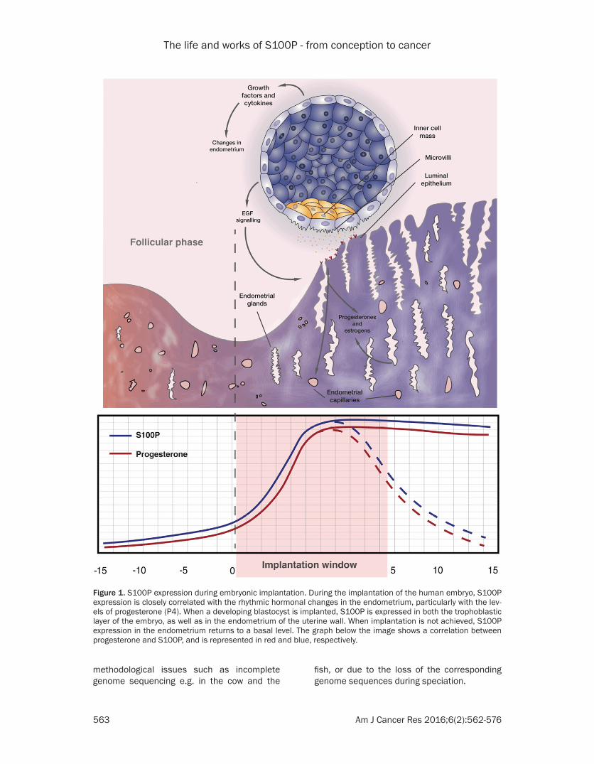
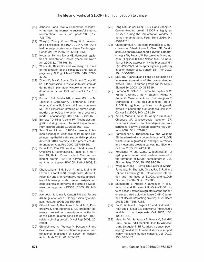
Figure 1. S100P expression during embryonic implantation. During the implantation of the human embryo, S100P expression is closely correlated with the rhythmic hormonal changes in the endometrium, particularly with the lev-els of progesterone (P4). When a developing blastocyst is implanted, S100P is expressed in both the trophoblastic layer of the embryo, as well as in the endometrium of the uterine wall. When implantation is not achieved, S100P expression in the endometrium returns to a basal level. The graph below the image shows a correlation between progesterone and S100P, and is represented in red and blue, respectively.
The life and works of S100P - from conception to cancer
564 Am J Cancer Res 2016;6(2):562-576
S100P in implantation and human embryonic development
A receptive endometrium and viable blastocyst are the two necessary conditions for success-ful implantation, continuation of progressive cell divisions and further development of a liv-ing embryo [10, 11]. Interestingly, the rhythmic changes in receptivity of the uterine endome-trium correlate with the rise of S100P levels in humans due to hormonal variation, especial-ly with marked increase in progesterone (P4). During the implantation window, which lasts approximately four days, S100P expression surges to levels that are approximately 100 times higher than in other phases of the men-strual cycle [12-14]. This suggests that S100P is potentially a unique biomarker of a recept- ive endometrium. In addition, it was shown, at least in vitro, that expression of S100P also increases significantly in stromal cells after their co-culture with trophoblast cells [15, 16], which implies that S100P may also be involved in interactions at the maternal-fetal interface (Figure 1). Clearly, further exciting work to fully elucidate how S100P may encourage or even permit implantation is still awaited for.
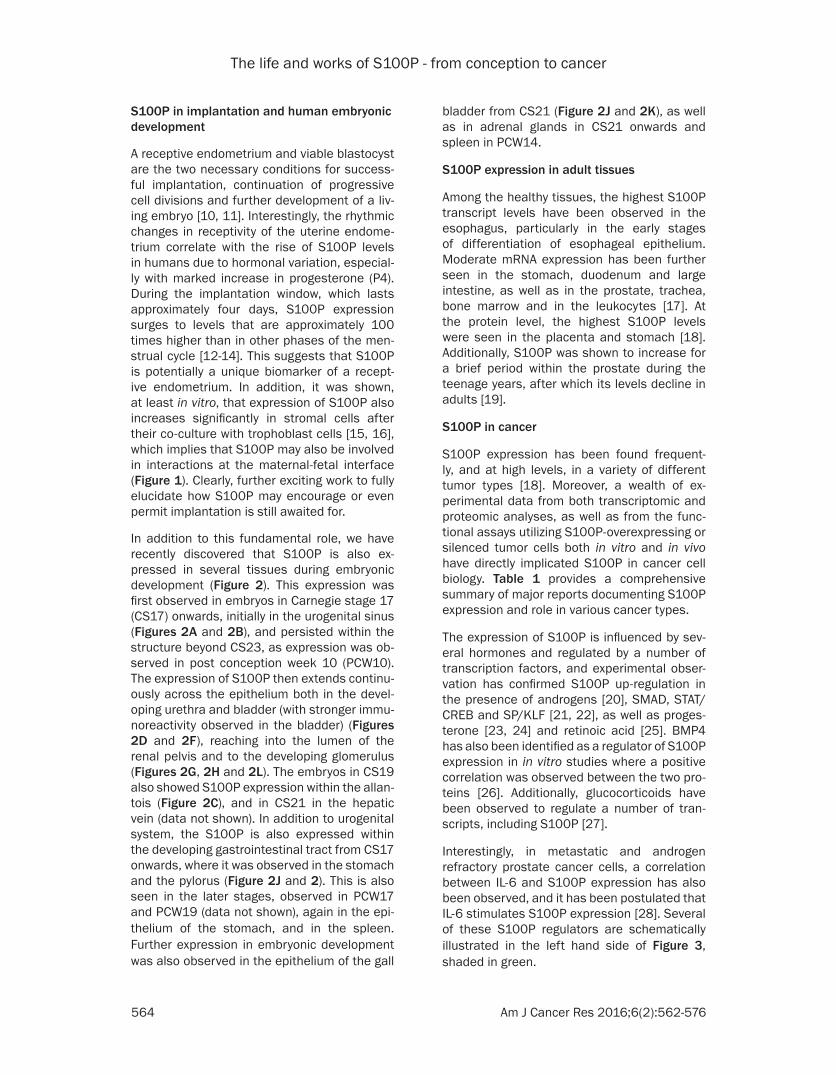
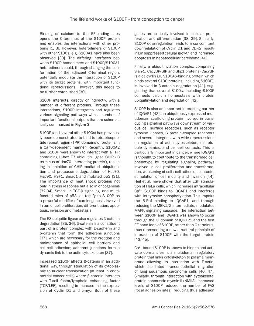
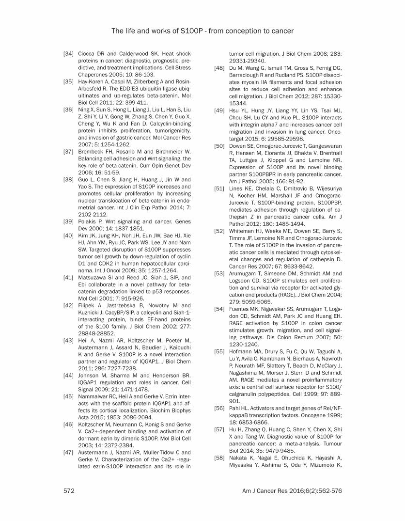
In addition to this fundamental role, we have recently discovered that S100P is also ex- pressed in several tissues during embryonic development (Figure 2). This expression was first observed in embryos in Carnegie stage 17 (CS17) onwards, initially in the urogenital sinus (Figures 2A and 2B), and persisted within the structure beyond CS23, as expression was ob- served in post conception week 10 (PCW10). The expression of S100P then extends continu-ously across the epithelium both in the devel-oping urethra and bladder (with stronger immu-noreactivity observed in the bladder) (Figures 2D and 2F), reaching into the lumen of the renal pelvis and to the developing glomerulus (Figures 2G, 2H and 2L). The embryos in CS19 also showed S100P expression within the allan-tois (Figure 2C), and in CS21 in the hepatic vein (data not shown). In addition to urogenital system, the S100P is also expressed within the developing gastrointestinal tract from CS17 onwards, where it was observed in the stomach and the pylorus (Figure 2J and 2). This is also seen in the later stages, observed in PCW17 and PCW19 (data not shown), again in the epi-thelium of the stomach, and in the spleen. Further expression in embryonic development was also observed in the epithelium of the gall
bladder from CS21 (Figure 2J and 2K), as well as in adrenal glands in CS21 onwards and spleen in PCW14.
S100P expression in adult tissues
Among the healthy tissues, the highest S100P transcript levels have been observed in the esophagus, particularly in the early stages of differentiation of esophageal epithelium. Moderate mRNA expression has been further seen in the stomach, duodenum and large intestine, as well as in the prostate, trachea, bone marrow and in the leukocytes [17]. At the protein level, the highest S100P levels were seen in the placenta and stomach [18]. Additionally, S100P was shown to increase for a brief period within the prostate during the teenage years, after which its levels decline in adults [19].
S100P in cancer
S100P expression has been found frequent- ly, and at high levels, in a variety of different tumor types [18]. Moreover, a wealth of ex- perimental data from both transcriptomic and proteomic analyses, as well as from the func-tional assays utilizing S100P-overexpressing or silenced tumor cells both in vitro and in vivo have directly implicated S100P in cancer cell biology. Table 1 provides a comprehensive summary of major reports documenting S100P expression and role in various cancer types.
The expression of S100P is influenced by sev-eral hormones and regulated by a number of transcription factors, and experimental obser-vation has confirmed S100P up-regulation in the presence of androgens [20], SMAD, STAT/CREB and SP/KLF [21, 22], as well as proges-terone [23, 24] and retinoic acid [25]. BMP4 has also been identified as a regulator of S100P expression in in vitro studies where a positive correlation was observed between the two pro-teins [26]. Additionally, glucocorticoids have been observed to regulate a number of tran-scripts, including S100P [27].
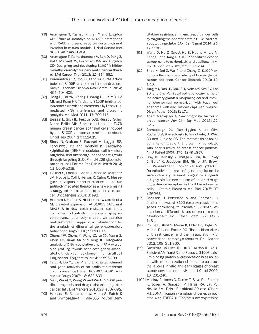
Interestingly, in metastatic and androgen refractory prostate cancer cells, a correlation between IL-6 and S100P expression has also been observed, and it has been postulated that IL-6 stimulates S100P expression [28]. Several of these S100P regulators are schematically illustrated in the left hand side of Figure 3, shaded in green.
The life and works of S100P - from conception to cancer
565 Am J Cancer Res 2016;6(2):562-576
Figure 2. S100P expression in the human embryo. S100P is expressed in the right and left horns of the urogenital sinus (RUGS and LUGS), as well as in the bladder portion of the urogenital sinus (indicated by arrow) in CS17 (A, B), as well as in the allantois (C). In CS21 (D), S100P in the urethral portion of the urogenital sinus (E) shows stronger
The life and works of S100P - from conception to cancer
566 Am J Cancer Res 2016;6(2):562-576
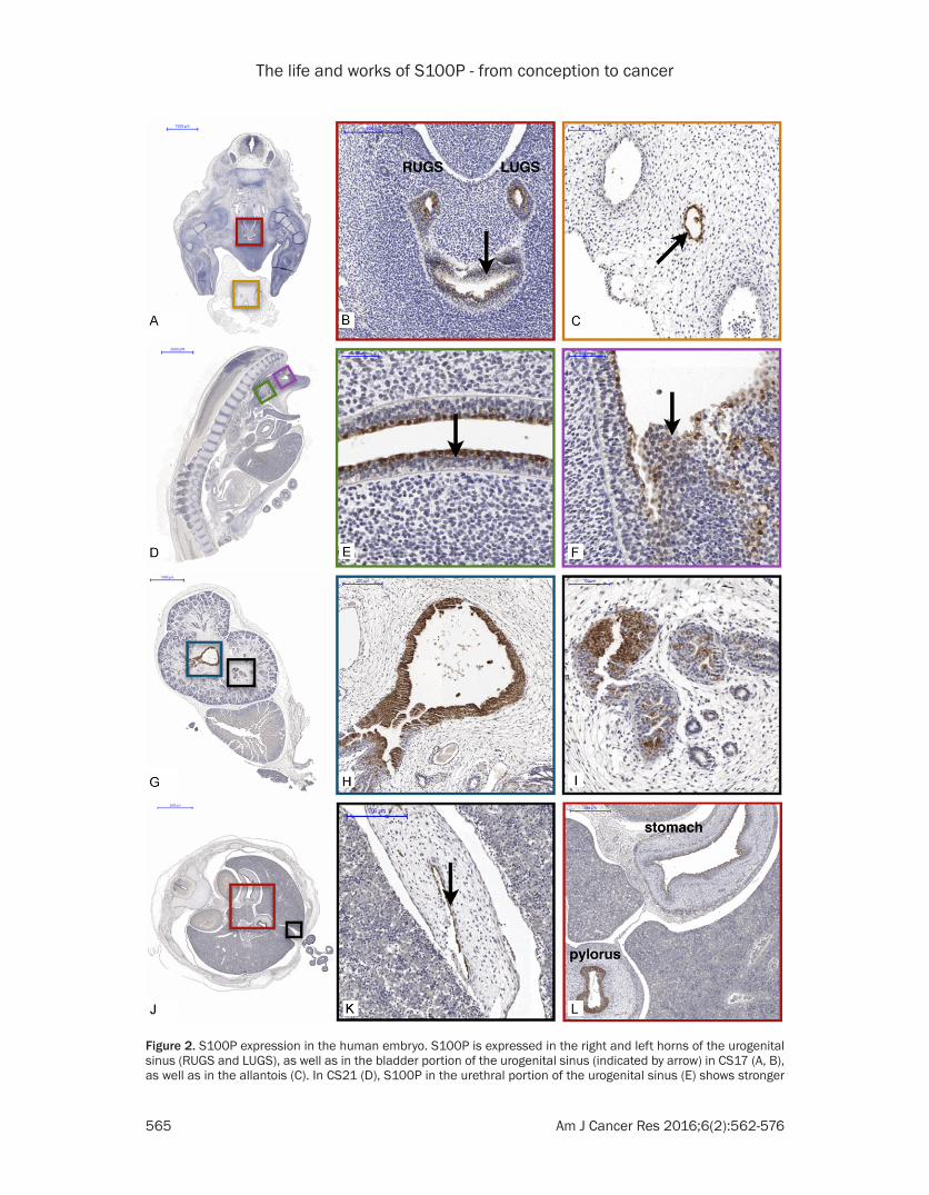
immunoreactivity than in the bladder portion (F). In the kidney, expression of S100P is seen in the renal pelvis (G, H) as well as in the glomerulus (I) in PCW10. Further expression is seen in the gastrointestinal tract (J-L), including the gallbladder (indicated with arrow) (K) as well as the stomach and pylorus (I) in CS23.
Table 1. S100P in cancerBasal cell carcinoma of the salivary gland
S100P is used for differentiating basal cell neoplasms from adenoid cystic carcinomas [93].
Breast Cancer S100P is one of the markers of cancer initiation, is expressed in ductal hyperplasia, in lesions with high-risk of progression, in situ and invasive ductal carcinomas, and is associated with poor prognosis. Its expression correlates with ERBB2/Her2/neu, ER (estradiol) and P4 expression [94-102].
Cholagiocarcinoma S100P expression is a strong indicator of the early stages of cholangiocarcinoma with increased expres-sion correlating with progression from low to high grade biliary intraepithelial neoplasia (BilIN) [103], and is a sensitive biomarker for detecting cholangicarcinoma [70].
Cervical cancer S100P is upregulated in all stages of cervical adenocarcinoma [104-107].Colon cancer S100P is highly expressed in non-dysplastic tissue from ulcerative colitis patients with high-grade
dysplasia [108], and may be used to distinguish flat adenoma from normal mucosa [109, 110]. The overexpression of S100P in colorectal cancer cells promotes metastasis [111], and acts as a potential prognostic biomarker [112].
Esophageal cancer S100P is downregulated in esophageal squamous cell carcinoma [113, 114].Endometrial Cancer S100P expression is higher in endometrial cancer than in normal endometrium and increases with
tumor grade [38].Gastric cancer Immunohistochemical analysis of tissue microarray shows S100P expression in >75% of gastric can-
cers; its downregulation in gastric cancer cell lines leads to apoptosis and inhibition of colony-formation. In contrast, low expression of S100P is linked to poor patients’ outcome [115, 116].
Hepatocellular carcinoma
S100P is a novel prognostic factor in HCC that can predict survival in patients with advanced tumor stage or early recurrences [117, 118].
Lung cancer S100P is one of five genes found consistently deregulated in meta-analysis of 12 cDNA array studies. Its expression is observed in early stages of non-small cell lung cancer (NSCLC) and lung adenocarcinoma, and with S100A2 and trypsinogens is predictive of metastatic progression and poor survival in NSCLC [119-122].
Melanoma S100P, RAGE and ezrin are significantly higher in melanomas than in benign nevus pigmentosus, and metastatic melanoma in comparison to the primary tumor [123].
Oral cancer S100P is one of the salivary biomarkers in oral squamous carcinoma that can detect cancer recurrence in patients in remission [74, 124].
Ovarian cancer High expression of S100P is correlated with shorter overall survival after chemotherapy [125, 126]; con-versely, this is also noted in clear cell adenocarcinoma of the ovary which express low levels of S100P [127].
Prostate cancer S100P is expressed in only 18.5% of prostate cancers, and its expression is significantly lower in cancer than in normal prostate and benign prostate hyperplasia [11]. However, it is one of the highest expressed genes in the androgen – independent CWR22 prostate cancer xenografts [128, 129]. Ad-ditionally, it correlates with metastatic progression of hormone refractory prostate cancer cells [130].
Pancreatic adeno-carcinoma
S100P is expressed in the precursor lesions of pancreatic ductal adenocarcinoma (PDAC), as well as throughout all stages of PDAC development and progression, and is involved in growth and invasion of cancer cells [50, 131-134].
Mucinous cystic neoplasms
S100P is expressed in pancreatic mucinous cystic neoplasms (MCN) [59, 135].
Intraductal papillary mucinous tumors
Intraductal papillary mucinous tumors (IPMTs) in the pancreas are also expressing S100P [136, 137].
Urothelial cancer S100P is a diagnostic biomarker of urothelial cancer [138, 139], and acts as a potential marker for distinguishing urothelial from squamous differentiation [140].
Structure and function of S100P
Structurally, S100P belongs to a family of small dimeric members of the large EF-hand super-family of calcium-binding proteins, although it has been shown to bind other divalent metal ions, like Mg2+, Cu2+ and Zn2+ [1, 2]. It is a 95
amino acid residue protein, comprising two EF-hands, first one with the low affinity for cal-cium binding and the second, canonical one, which binds calcium with high affinity. S100P monomers readily interact with one another with high affinity, and homodimer formation is deemed obligatory for S100P functions [29].
The life and works of S100P - from conception to cancer
567 Am J Cancer Res 2016;6(2):562-576
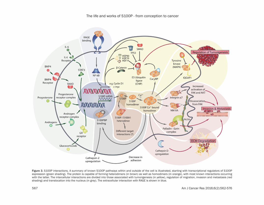
Figure 3. S100P interactions. A summary of known S100P pathways within and outside of the cell is illustrated, starting with transcriptional regulators of S100P expression (green shading). The protein is capable of forming heterodimers (in brown) as well as homodimers (in orange), with most known interactions occurring with the latter. The intercellular interactions are divided into those associated with tumorigenesis (in yellow), regulation of migration, invasion and metastasis (red shading) and translocation into the nucleus (in grey). The extracellular interaction with RAGE is shown in blue.
The life and works of S100P - from conception to cancer
568 Am J Cancer Res 2016;6(2):562-576
Binding of calcium to the EF-binding sites opens the C-terminus of the S100P protein and enables the interactions with other pro-teins [1, 3]. However, heterodimers of S100P with other S100s, e.g. S100A1 have also been observed [30]. The differing interfaces bet- ween S100P homodimers and S100P/S100A1 heterodimers could, through changing the con-formation of the adjacent C-terminal region, potentially modulate the interaction of S100P with its target proteins, with important func-tional repercussions. However, this needs to be further established [30].
S100P interacts, directly or indirectly, with a number of different proteins. Through these interactions, S100P integrates and regulates various signaling pathways with a number of important functional outputs that are schemat-ically summarized in Figure 3.
S100P (and several other S100s) has previous-ly been demonstrated to bind to tetratricopep-tide repeat region (TPR) domains of proteins in a Ca2+-dependent manner. Recently, S100A2 and S100P were shown to interact with a TPR containing U-box E3 ubiquitin ligase CHIP (‘C terminus of Hsc70- interacting protein’), result-ing in inhibition of CHIP-mediated ubiquitina-tion and proteasome degradation of Hsp70, Hsp90, HSF1, Smad1 and mutated p53 [31]. The importance of heat shock proteins not only in stress response but also in oncogenesis [32-34], Smad1 in TGF-β signaling, and multi-faceted roles of p53, all testify to S100P as a powerful modifier of carcinogenesis involved in tumor cell proliferation, differentiation, apop-tosis, invasion and metastasis.
The E3 ubiquitin ligase also regulates β-catenin degradation [35, 36]. β-catenin is a constituent part of a protein complex with E-cadherin and α-catenin that form the adherens junctions [37], which are necessary for the creation and maintenance of epithelial cell barriers and cell-cell adhesion; adherent junctions form a dynamic link to the actin cytoskeleton [37].
Increased S100P affects β-catenin in an addi-tional way, through stimulation of its cytoplas-mic to nuclear translocation (at least in endo-metrial cancer cells) where β-catenin interacts with T-cell factor/lymphoid enhancing factor (TCF/LEF), resulting in increase in the expres-sion of Cyclin D1 and c-myc. Both of these
genes are critically involved in cellular proli- feration and differentiation [38, 39]. Similarly, S100P downregulation leads to a concomitant downregulation of Cyclin D1 and CDK2, result-ing in suppressed cellular growth and increased apoptosis in hepatocellular carcinoma [40].
Finally, a ubiquitinylation complex comprising Siah-1, CacyBP/SIP and Skp1 proteins (CacyBP is a calcyclin i.e. S100A6-binding protein which binds several S100 proteins, including S100P), is involved in β-catenin degradation [41], sug-gesting that several S100s, including S100P connects calcium homeostasis with protein ubiquitinylation and degradation [42].
S100P is also an important interacting partner of IQGAP1 [43], an ubiquitously expressed mul-tidomain scaffolding protein involved in trans-ducing signaling pathways downstream of vari-ous cell surface receptors, such as receptor tyrosine kinases, G protein-coupled receptors and several integrins, with wide repercussions on regulation of actin cytoskeleton, microtu-bule dynamics, and cell-cell contacts. This is particularly important in cancer, where IQGAP1 is thought to contribute to the transformed cell phenotype by regulating signaling pathways involved in cell proliferation and transforma-tion, weakening of cell : cell adhesion contacts, stimulation of cell motility and invasion [44]. Heil et al. have shown that after EGF stimula-tion of HeLa cells, which increases intracellular Ca2+, S100P binds to IQGAP1 and interferes with its tyrosine phosphorylation. This impairs the B-Raf binding to IQGAP1, and through reducing the MEK1/2 intermediate, modulates MAPK signaling cascade. The interaction bet- ween S100P and IQGAP1 was shown to occur through the IQ domain of IQGAP1 and the first EF hand loop of S100P, rather than C-terminus, thus representing a new structural principle of interaction of S100P with the target protein [43, 45].
Ca2+-bound S100P is known to bind to and acti-vate dormant ezrin, a multidomain regulatory protein that links cytoskeleton to plasma mem-brane allowing its interaction with F-actin, which facilitated transendothelial migration of lung squamous carcinoma cells [46, 47]. Similarly, through interaction with cytoskeletal protein nonmuscle myosin II (NMIIA), increased levels of S100P reduced the number of FAS (focal adhesion sites), reducing thus adhesion
The life and works of S100P - from conception to cancer
569 Am J Cancer Res 2016;6(2):562-576
and increasing cell migration [48]. In addition, in lung cancer cells, S100P affects migration and invasion through interaction with integrin Alpha 7 (α7), which is mediated by FAK/AKT-ZEB signaling [49]. In pancreatic cancer cells, S100P was also recently shown to interact with another binding partner, S100PBP, a protein with no homology to any characterized protein, and which, through regulation of cathepsin Z and integrin αvb5 modulates cell adhesion [50, 51]. Furthermore, S100P overexpression was also shown to correlate with increased expres-sion of another S100 family member, S100A6, as well as the aspartic protease cathepsin D, both of which are involved in migration and invasion of pancreatic adenocarcinoma cells [52].
Finally, S100P can act in an autocrine manner via Receptor for Activated Glycation End Pro- ducts (RAGE) to stimulate cell proliferation and survival via the NF-kB pathway [53, 54]. S100P (along with several other S100 proteins [55]) acts as initial activator of the pathway via NF-κB/Rel complexes that translocate to the nucleus and induce the expression of a large number of diverse target genes [56]. The S100P-RAGE interaction has recently been employed as a novel therapeutic strategy and will be further discussed in the Therapeutic section of this review.
S100P and diagnostics
Due to its expression in neoplastic lesions and absence in most healthy tissues, S100P has been evaluated as a potential biomarker for detection of several cancers, most com-monly using immunohistochemistry approach-es. Several studies have highlighted S100P as a marker of pancreatic adenocarcinoma (PDAC) as its expression increases as precursor lesions PanINs (pancreatic intraepithelial neoplasias) progress [50, 57]; moreover, S100P has been identified as a possible marker of intraductal papillary mucinous neoplasms (IPMNs) [58] as well as mucinous cystic neoplasms [59], addi-tional potential precursor lesions for PDAC. As a member of a panel, e.g. with mesothelin and/or KOC, S100P showed potential in correct differentiation of true PDACs from borderline cases in cytological assessment of EUS ob- tained biopsies or surgical resections [60-62]. S100P, mesothelin and IMP3 have also been found as useful biomarkers in gallbladder ade-nocarcinoma [63], as well as in extrahepatic bile duct carcinoma [64, 65].
In cholangiocarcinomas, S100P was proposed to be an effective diagnostic marker in combi-nation with maspin, pVHL and insulin-like growth factor II mRNA-binding protein 3 in bile duct biopsies [66], in distinguishing adenocar-cinoma from benign biliary epithelium on endo-scopic bile duct biopsy specimens, as well as for distinguishing between cholangiolar- type intrahepatic from bile duct intrahepatic cholangiocarcinomas. Furthermore, bile levels of S100P were significantly higher in cholangio-carcinoma patients compared to those with cholelithiasis [26, 67-70].
Interestingly, S100P has also been identified as potential non-invasive biomarker of oral squamous carcinoma in saliva [71-74]. Finally, S100P has been observed in patients with early stage breast cancer, and its expression has been associated with poor prognosis and survival [75]; particularly important is its poten-tial value as a diagnostic marker of triple nega-tive breast cancer [76].
S100P as a therapeutic target
Because of its established functional roles in cancer, S100P has been considered a valuable therapeutic target. Several attempts have been made to inhibit either S100P, its targets, or its interactions, of which one has gained much attention recently - the interaction between S100P and RAGE [77, 78]. In vitro attempts to inhibit this interaction was first achieved with cromolyn, and more successfully with its 5-methyl analogue [79, 80], both of which bind to the C-domain of S100P, interfering thus with its binding to RAGE [81].
However, a recurring issue with cromolyn is lack of specificity for S100P as it binds to other S100 proteins, as well as its low biodistribution and bioavailability.
Alternative methods of inhibiting S100P, now intracellularly, have been conducted using anti-sense mRNA retroviral transfection in colon [82], gastric [36], breast [83], and glioblastoma [84] cancer cell lines, and have resulted in a decrease of cellular motility and metastatic potential. Finally, anti-S100P antibodies have been tested both in vitro and in vivo and have shown promising results as both single agents and in combination with chemotherapeutic drugs, such as gemcitabine in pancreatic can-cer [85].
The life and works of S100P - from conception to cancer
570 Am J Cancer Res 2016;6(2):562-576
S100P expression was found to be associated with cancer resistance to several chemothera-peutic agents, and its silencing sensitized the cancer cells in vitro to doxorubicin [86], cispla-tin [87] and oxaliplatin [88]. Furthermore, S100P has also been associated with drug resistance in gastric [89] and pancreatic can-cers [90]. Therefore, blocking S100P function might also be expected to improve responses to other therapeutic treatments. However, this needs to be carefully assessed, as some con-flicting reports exist, for example, in ovarian and gastric cancer cells, where, at least in vitro, overexpression of S100P led to sensitization of cancer cells to carboplatin and paclitaxel [91], and oxaliplatin, respectively [92].
Despite this, S100P appears to represent a potentially very effective anti-cancer target, at least for in some cancer types, and further development of anti-S100P specific therapies will likely prove to be a fruitful and productive field of investigation.
Conclusion
In this review, we have summarized the current knowledge on S100P with the addition of our own recent observations of S100P expression in human embryonic development.
Despite its relatively short evolutionary history, functions of S100P in vertebrates are vital, from involvement in the earliest steps of embry-onic implantation and subsequent embryonic development to exerting the important roles in both healthy adult and cancer tissues. It is, however, for the latter, that S100P has gained most of its existing attention, as it can be potentially utilized as both a diagnostic/prog-nostic marker and a promising therapeutic tar-get. Since its roles have far-reaching cross-dis-ciplinary implications, spanning from reproduc-tive physiology and embryonic development to inflammation and oncology, studying S100P will thus continue to be an important and fruit-ful research topic.
Acknowledgements
We would like to thank Dr Steven Lisgo from the Institute of Genetic Medicine, Newcastle University, and Dr Dianne Gerelli from the Institute of Child Health, University College London for their assistance with the human
embryonic and foetal material which was provided by the joint MRC/Wellcome Trust grant#099175/Z/12/Z human Developmental Biology Resource (http://hdbr.org).
Disclosure of conflict of interest
None.
Address correspondence to: Tatjana Crnogorac-Jurcevic, Centre for Molecular Oncology, Barts Cancer Institute, Queen Mary University of London, John Vane Science Centre, Charterhouse Square, London, EC1M 6BQ, UK. Tel: +44 (0)20 7-882-3554; Fax: +44 (0)20 7-882-3884; E-mail: [email protected]
References
[1] Donato R. S100: a multigenic family of calci-um-modulated proteins of the EF-hand type with intracellular and extracellular functional roles. Int J Biochem Cell Biol 2001; 33: 637-668.
[2] Gribenko A, Lopez MM, Richardson JM 3rd and Makhatadze GI. Cloning, overexpression, puri-fication, and spectroscopic characterization of human S100P. Protein Sci 1998; 7: 211-215.
[3] Becker T, Gerke V, Kube E and Weber K. S100P, a novel Ca(2+)-binding protein from human placenta. cDNA cloning, recombinant protein expression and Ca2+ binding properties. Eur J Biochem 1992; 207: 541-547.
[4] Zhu HY, Tong XM, Lin XN, Jiang LY, Wang JX and Zhang SY. Expression and Distribution of Calcium-Binding Protein S100P in Human Placenta during Pregnancy. Int J Fertil Steril 2015; 8: 445-452.
[5] Bresnick AR, Weber DJ and Zimmer DB. S100 proteins in cancer. Nat Rev Cancer 2015; 15: 96-109.
[6] Moore BW. A soluble protein characteristic of the nervous system. Biochem Biophys Res Commun 1965; 19: 739-744.
[7] Zimmer DB, Eubanks JO, Ramakrishnan D and Criscitiello MF. Evolution of the S100 family of calcium sensor proteins. Cell Calcium 2013; 53: 170-179.
[8] Kypriotou M, Huber M and Hohl D. The human epidermal differentiation complex: cornified envelope precursors, S100 proteins and the ‘fused genes’ family. Exp Dermatol 2012; 21: 643-649.
[9] Shang X, Cheng H and Zhou R. Chromosomal mapping, differential origin and evolution of the S100 gene family. Genet Sel Evol 2008; 40: 449-464.
The life and works of S100P - from conception to cancer
571 Am J Cancer Res 2016;6(2):562-576
[10] Achache H and Revel A. Endometrial receptivi-ty markers, the journey to successful embryo implantation. Hum Reprod Update 2006; 12: 731-746.
[11] Wang Q, Zhang JG and Wang W. Expression and significance of S100P, CD147, and OCT4 in different prostate cancer tissue TNM stages. Genet Mol Res 2015; 14: 6844-6851.
[12] Kodaman PH and Taylor HS. Hormonal regula-tion of implantation. Obstet Gynecol Clin North Am 2004; 31: 745-766, ix.
[13] Wilcox AJ, Baird DD and Weinberg CR. Time of implantation of the conceptus and loss of pregnancy. N Engl J Med 1999; 340: 1796-1799.
[14] Zhang D, Ma C, Sun X, Xia H and Zhang W. S100P expression in response to sex steroids during the implantation window in human en-dometrium. Reprod Biol Endocrinol 2012; 10: 106.
[15] Popovici RM, Betzler NK, Krause MS, Luo M, Jauckus J, Germeyer A, Bloethner S, Schlot- terer A, Kumar R, Strowitzki T and von Wolff M. Gene expression profiling of human endo-metrial-trophoblast interaction in a coculture model. Endocrinology 2006; 147: 5662-5675.
[16] Burrows TD, King A, Loke YW. Trophoblast mi-gration during human placental implantation. Hum Reprod Update 1996; 2: 307 - 321.
[17] Sato N and Hitomi J. S100P expression in hu-man esophageal epithelial cells: Human eso- phageal epithelial cells sequentially produce different S100 proteins in the process of dif-ferentiation. Anat Rec 2002; 267: 60-69.
[18] Parkkila S, Pan PW, Ward A, Gibadulinova A, Oveckova I, Pastorekova S, Pastorek J, Mart- inez AR, Helin HO and Isola J. The calcium-binding protein S100P in normal and malig-nant human tissues. BMC Clin Pathol 2008; 8: 2.
[19] Dhanasekaran SM, Dash A, Yu J, Maine IP, Laxman B, Tomlins SA, Creighton CJ, Menon A, Rubin MA and Chinnaiyan AM. Molecular profil-ing of human prostate tissues: insights into gene expression patterns of prostate develop-ment during puberty. FASEB J 2005; 19: 243-245.
[20] Averboukh L, Liang P, Kantoff PW and Pardee AB. Regulation of S100P expression by andro-gen. Prostate 1996; 29: 350-355.
[21] Gibadulinova A, Oveckova I, Parkkila S, Past- orekova S and Pastorek J. Key promoter ele-ments involved in transcriptional activation of the cancer-related gene coding for S100P calcium-binding protein. Oncol Rep 2008; 20: 391-396.
[22] Gibadulinova A, Tothova V, Pastorek J and Pastorekova S. Transcriptional regulation and functional implication of S100P in cancer. Amino Acids 2011; 41: 885-892.
[23] Tong XM, Lin XN, Song T, Liu L and Zhang SY. Calcium-binding protein S100P is highly ex-pressed during the implantation window in human endometrium. Fertil Steril 2010; 94: 1510-1518.
[24] Chandramouli A, Mercado-Pimentel ME, Hut- chinson A, Gibadulinova A, Olson ER, Dickin- son S, Shanas R, Davenport J, Owens J, Bhatta- charyya AK, Regan JW, Pastorekova S, Arumu- gam T, Logsdon CD and Nelson MA. The induc-tion of S100p expression by the Prostaglandin E(2) (PGE(2))/EP4 receptor signaling pathway in colon cancer cells. Cancer Biol Ther 2010; 10: 1056-1066.
[25] Shyu RY, Huang SL and Jiang SY. Retinoic acid increases expression of the calcium-binding protein S100P in human gastric cancer cells. J Biomed Sci 2003; 10: 313-319.
[26] Hamada S, Satoh K, Hirota M, Fujibuchi W, Kanno A, Umino J, Ito H, Satoh A, Kikuta K, Kume K, Masamune A and Shimosegawa T. Expression of the calcium-binding protein S100P is regulated by bone morphogenetic protein in pancreatic duct epithelial cell lines. Cancer Sci 2009; 100: 103-110.
[27] Kino T, Manoli I, Kelkar S, Wang Y, Su YA and Chrousos GP. Glucocorticoid receptor (GR) beta has intrinsic, GRalpha-independent tran-scriptional activity. Biochem Biophys Res Com- mun 2009; 381: 671-675.
[28] Hammacher A, Thompson EW and Williams ED. Interleukin-6 is a potent inducer of S100P, which is up-regulated in androgen-refractory and metastatic prostate cancer. Int J Biochem Cell Biol 2005; 37: 442-450.
[29] Koltzscher M and Gerke V. Identification of hydrophobic amino acid residues involved in the formation of S100P homodimers in vivo. Biochemistry 2000; 39: 9533-9539.
[30] Wang G, Zhang S, Fernig DG, Spiller D, Martin-Fernandez M, Zhang H, Ding Y, Rao Z, Rudland PS and Barraclough R. Heterodimeric interac-tion and interfaces of S100A1 and S100P. Biochem J 2004; 382: 375-383.
[31] Shimamoto S, Kubota Y, Yamaguchi F, Toku- mitsu H and Kobayashi R. Ca2+/S100 pro-teins act as upstream regulators of the chaper-one-associated ubiquitin ligase CHIP (C termi-nus of Hsc70-interacting protein). J Biol Chem 2013; 288: 7158-7168.
[32] Dai C, Whitesell L, Rogers AB and Lindquist S. Heat shock factor 1 is a powerful multifaceted modifier of carcinogenesis. Cell 2007; 130: 1005-1018.
[33] Mendillo ML, Santagata S, Koeva M, Bell GW, Hu R, Tamimi RM, Fraenkel E, Ince TA, Whitesell L and Lindquist S. HSF1 drives a transcription-al program distinct from heat shock to support highly malignant human cancers. Cell 2012; 150: 549-562.
The life and works of S100P - from conception to cancer
572 Am J Cancer Res 2016;6(2):562-576
[34] Ciocca DR and Calderwood SK. Heat shock proteins in cancer: diagnostic, prognostic, pre-dictive, and treatment implications. Cell Stress Chaperones 2005; 10: 86-103.
[35] Hay-Koren A, Caspi M, Zilberberg A and Rosin-Arbesfeld R. The EDD E3 ubiquitin ligase ubiq-uitinates and up-regulates beta-catenin. Mol Biol Cell 2011; 22: 399-411.
[36] Ning X, Sun S, Hong L, Liang J, Liu L, Han S, Liu Z, Shi Y, Li Y, Gong W, Zhang S, Chen Y, Guo X, Cheng Y, Wu K and Fan D. Calcyclin-binding protein inhibits proliferation, tumorigenicity, and invasion of gastric cancer. Mol Cancer Res 2007; 5: 1254-1262.
[37] Brembeck FH, Rosario M and Birchmeier W. Balancing cell adhesion and Wnt signaling, the key role of beta-catenin. Curr Opin Genet Dev 2006; 16: 51-59.
[38] Guo L, Chen S, Jiang H, Huang J, Jin W and Yao S. The expression of S100P increases and promotes cellular proliferation by increasing nuclear translocation of beta-catenin in endo-metrial cancer. Int J Clin Exp Pathol 2014; 7: 2102-2112.
[39] Polakis P. Wnt signaling and cancer. Genes Dev 2000; 14: 1837-1851.
[40] Kim JK, Jung KH, Noh JH, Eun JW, Bae HJ, Xie HJ, Ahn YM, Ryu JC, Park WS, Lee JY and Nam SW. Targeted disruption of S100P suppresses tumor cell growth by down-regulation of cyclin D1 and CDK2 in human hepatocellular carci-noma. Int J Oncol 2009; 35: 1257-1264.
[41] Matsuzawa SI and Reed JC. Siah-1, SIP, and Ebi collaborate in a novel pathway for beta-catenin degradation linked to p53 responses. Mol Cell 2001; 7: 915-926.
[42] Filipek A, Jastrzebska B, Nowotny M and Kuznicki J. CacyBP/SIP, a calcyclin and Siah-1-interacting protein, binds EF-hand proteins of the S100 family. J Biol Chem 2002; 277: 28848-28852.
[43] Heil A, Nazmi AR, Koltzscher M, Poeter M, Austermann J, Assard N, Baudier J, Kaibuchi K and Gerke V. S100P is a novel interaction partner and regulator of IQGAP1. J Biol Chem 2011; 286: 7227-7238.
[44] Johnson M, Sharma M and Henderson BR. IQGAP1 regulation and roles in cancer. Cell Signal 2009; 21: 1471-1478.
[45] Nammalwar RC, Heil A and Gerke V. Ezrin inter-acts with the scaffold protein IQGAP1 and af-fects its cortical localization. Biochim Biophys Acta 2015; 1853: 2086-2094.
[46] Koltzscher M, Neumann C, Konig S and Gerke V. Ca2+-dependent binding and activation of dormant ezrin by dimeric S100P. Mol Biol Cell 2003; 14: 2372-2384.
[47] Austermann J, Nazmi AR, Muller-Tidow C and Gerke V. Characterization of the Ca2+ -regu-lated ezrin-S100P interaction and its role in
tumor cell migration. J Biol Chem 2008; 283: 29331-29340.
[48] Du M, Wang G, Ismail TM, Gross S, Fernig DG, Barraclough R and Rudland PS. S100P dissoci-ates myosin IIA filaments and focal adhesion sites to reduce cell adhesion and enhance cell migration. J Biol Chem 2012; 287: 15330-15344.
[49] Hsu YL, Hung JY, Liang YY, Lin YS, Tsai MJ, Chou SH, Lu CY and Kuo PL. S100P interacts with integrin alpha7 and increases cancer cell migration and invasion in lung cancer. Onco- target 2015; 6: 29585-29598.
[50] Dowen SE, Crnogorac-Jurcevic T, Gangeswaran R, Hansen M, Eloranta JJ, Bhakta V, Brentnall TA, Luttges J, Kloppel G and Lemoine NR. Expression of S100P and its novel binding partner S100PBPR in early pancreatic cancer. Am J Pathol 2005; 166: 81-92.
[51] Lines KE, Chelala C, Dmitrovic B, Wijesuriya N, Kocher HM, Marshall JF and Crnogorac-Jurcevic T. S100P-binding protein, S100PBP, mediates adhesion through regulation of ca-thepsin Z in pancreatic cancer cells. Am J Pathol 2012; 180: 1485-1494.
[52] Whiteman HJ, Weeks ME, Dowen SE, Barry S, Timms JF, Lemoine NR and Crnogorac-Jurcevic T. The role of S100P in the invasion of pancre-atic cancer cells is mediated through cytoskel-etal changes and regulation of cathepsin D. Cancer Res 2007; 67: 8633-8642.
[53] Arumugam T, Simeone DM, Schmidt AM and Logsdon CD. S100P stimulates cell prolifera-tion and survival via receptor for activated gly-cation end products (RAGE). J Biol Chem 2004; 279: 5059-5065.
[54] Fuentes MK, Nigavekar SS, Arumugam T, Logs- don CD, Schmidt AM, Park JC and Huang EH. RAGE activation by S100P in colon cancer stimulates growth, migration, and cell signal-ing pathways. Dis Colon Rectum 2007; 50: 1230-1240.
[55] Hofmann MA, Drury S, Fu C, Qu W, Taguchi A, Lu Y, Avila C, Kambham N, Bierhaus A, Nawroth P, Neurath MF, Slattery T, Beach D, McClary J, Nagashima M, Morser J, Stern D and Schmidt AM. RAGE mediates a novel proinflammatory axis: a central cell surface receptor for S100/calgranulin polypeptides. Cell 1999; 97: 889-901.
[56] Pahl HL. Activators and target genes of Rel/NF-kappaB transcription factors. Oncogene 1999; 18: 6853-6866.
[57] Hu H, Zhang Q, Huang C, Shen Y, Chen X, Shi X and Tang W. Diagnostic value of S100P for pancreatic cancer: a meta-analysis. Tumour Biol 2014; 35: 9479-9485.
[58] Nakata K, Nagai E, Ohuchida K, Hayashi A, Miyasaka Y, Aishima S, Oda Y, Mizumoto K,

The life and works of S100P - from conception to cancer
573 Am J Cancer Res 2016;6(2):562-576
Tanaka M and Tsuneyoshi M. S100P is a novel marker to identify intraductal papillary muci-nous neoplasms. Hum Pathol 2010; 41: 824-831.
[59] Fukushima N and Fukayama M. Mucinous cys-tic neoplasms of the pancreas: pathology and molecular genetics. J Hepatobiliary Pancreat Surg 2007; 14: 238-242.
[60] Dim DC, Jiang F, Qiu Q, Li T, Darwin P, Rodgers WH and Peng HQ. The usefulness of S100P, mesothelin, fascin, prostate stem cell antigen, and 14-3-3 sigma in diagnosing pancreatic adenocarcinoma in cytological specimens ob-tained by endoscopic ultrasound guided fine-needle aspiration. Diagn Cytopathol 2014; 42: 193-199.
[61] Kato K, Kamada H, Fujimori T, Aritomo Y, Ono M and Masaki T. Molecular Biologic Approach to the Diagnosis of Pancreatic Car- cinoma Using Specimens Obtained by EUS-Guided Fine Needle Aspiration. Gastroenterol Res Pract 2012; 2012: 243524.
[62] Ali A, Brown V, Denley S, Jamieson NB, Morton JP, Nixon C, Graham JS, Sansom OJ, Carter CR, McKay CJ, Duthie FR and Oien KA. Expres- sion of KOC, S100P, mesothelin and MUC1 in pancreatico-biliary adenocarcinomas: devel-opment and utility of a potential diagnostic im-munohistochemistry panel. BMC Clin Pathol 2014; 14: 35.
[63] Shi J, Liu H, Wang HL, Prichard JW and Lin F. Diagnostic utility of von Hippel-Lindau gene product, maspin, IMP3, and S100P in adeno-carcinoma of the gallbladder. Hum Pathol 2013; 44: 503-511.
[64] Kawashima H, Itoh A, Ohno E, Miyahara R, Ohmiya N, Tanaka T, Shimoyama Y, Nakamura S, Ebata T, Nagino M, Goto H and Hirooka Y. Diagnostic and prognostic value of immunohis-tochemical expression of S100P and IMP3 in transpapillary biliary forceps biopsy samples of extrahepatic bile duct carcinoma. J Hepato- biliary Pancreat Sci 2013; 20: 441-447.
[65] Schmidt MT, Himmelfarb EA, Shafi H, Lin F, Xu H and Wang HL. Use of IMP3, S100P, and pVHL immunopanel to aid in the interpretation of bile duct biopsies with atypical histology or suspicious for malignancy. Appl Immuno- histochem Mol Morphol 2012; 20: 478-487.
[66] Chen L, Huang K, Himmelfarb EA, Zhai J, Lai JP, Lin F and Wang HL. Diagnostic value of maspin in distinguishing adenocarcinoma from benign biliary epithelium on endoscopic bile duct bi-opsy. Hum Pathol 2015; 46: 1647-54.
[67] Maeda S, Morikawa T, Takadate T, Suzuki T, Minowa T, Hanagata N, Onogawa T, Motoi F, Nishimura T and Unno M. Mass spectrometry-based proteomic analysis of formalin-fixed paraffin-embedded extrahepatic cholangiocar-
cinoma. J Hepatobiliary Pancreat Sci 2015; 22: 683-691.
[68] Liau JY, Tsai JH, Yuan RH, Chang CN, Lee HJ and Jeng YM. Morphological subclassification of intrahepatic cholangiocarcinoma: etiologi-cal, clinicopathological, and molecular fea-tures. Mod Pathol 2014; 27: 1163-1173.
[69] Sato Y, Harada K, Sasaki M and Nakanuma Y. Clinicopathological significance of S100 protein expression in cholangiocarcinoma. J Gastroenterol Hepatol 2013; 28: 1422-1429.
[70] Hamada S, Satoh K, Hirota M, Kanno A, Ishida K, Umino J, Ito H, Kikuta K, Kume K, Masamune A, Katayose Y, Unno M and Shimosegawa T. Calcium-binding protein S100P is a novel diag-nostic marker of cholangiocarcinoma. Cancer Sci 2011; 102: 150-156.
[71] Nagler RM. Saliva as a tool for oral cancer di-agnosis and prognosis. Oral Oncol 2009; 45: 1006-1010.
[72] Li Y, St John MA, Zhou X, Kim Y, Sinha U, Jordan RC, Eisele D, Abemayor E, Elashoff D, Park NH and Wong DT. Salivary transcriptome diagnos-tics for oral cancer detection. Clin Cancer Res 2004; 10: 8442-8450.
[73] Martin JL, Gottehrer N, Zalesin H, Hoff PT, Shaw M, Clarkson JH, Haan P, Vartanian M, McLeod T and Swanick SM. Evaluation of Salivary Transcriptome Markers for the Early Detection of Oral Squamous Cell Cancer in a Prospective Blinded Trial. Compend Contin Educ Dent 2015; 36: 365-373.
[74] Brinkmann O, Kastratovic DA, Dimitrijevic MV, Konstantinovic VS, Jelovac DB, Antic J, Nesic VS, Markovic SZ, Martinovic ZR, Akin D, Spiel- mann N, Zhou H and Wong DT. Oral squamous cell carcinoma detection by salivary biomark-ers in a Serbian population. Oral Oncol 2011; 47: 51-55.
[75] Maciejczyk A, Lacko A, Ekiert M, Jagoda E, Wysocka T, Matkowski R, Halon A, Gyorffy B, Lage H and Surowiak P. Elevated nuclear S100P expression is associated with poor sur-vival in early breast cancer patients. Histol Histopathol 2013; 28: 513-524.
[76] Maierthaler M, Kriegsmann M, Peng C, Jauch S, Szabo A, Wallwiener M, Rom J, Sohn C, Schneeweiss A, Sinn HP, Yang R and Burwinkel B. S100P and HYAL2 as prognostic markers for patients with triple-negative breast cancer. Exp Mol Pathol 2015; 99: 180-187.
[77] Penumutchu SR, Chou RH and Yu C. Structural insights into calcium-bound S100P and the V domain of the RAGE complex. PLoS One 2014; 9: e103947.
[78] Donato R. RAGE: a single receptor for several ligands and different cellular responses: the case of certain S100 proteins. Curr Mol Med 2007; 7: 711-724.
The life and works of S100P - from conception to cancer
574 Am J Cancer Res 2016;6(2):562-576
[79] Arumugam T, Ramachandran V and Logsdon CD. Effect of cromolyn on S100P interactions with RAGE and pancreatic cancer growth and invasion in mouse models. J Natl Cancer Inst 2006; 98: 1806-1818.
[80] Arumugam T, Ramachandran V, Sun D, Peng Z, Pal A, Maxwell DS, Bornmann WG and Logsdon CD. Designing and developing S100P inhibitor 5-methyl cromolyn for pancreatic cancer thera-py. Mol Cancer Ther 2013; 12: 654-662.
[81] Penumutchu SR, Chou RH and Yu C. Interaction between S100P and the anti-allergy drug cro-molyn. Biochem Biophys Res Commun 2014; 454: 404-409.
[82] Jiang L, Lai YK, Zhang J, Wang H, Lin MC, He ML and Kung HF. Targeting S100P inhibits co-lon cancer growth and metastasis by Lentivirus-mediated RNA interference and proteomic analysis. Mol Med 2011; 17: 709-716.
[83] Beissel B, Silva ID, Pesquero JB, Russo J, Schor N and Bellini MH. S-phase reduction in T47D human breast cancer epithelial cells induced by an S100P antisense-retroviral construct. Oncol Rep 2007; 17: 611-615.
[84] Sims JN, Graham B, Pacurari M, Leggett SS, Tchounwou PB and Ndebele K. Di-ethylhe- xylphthalate (DEHP) modulates cell invasion, migration and anchorage independent growth through targeting S100P in LN-229 glioblasto-ma cells. Int J Environ Res Public Health 2014; 11: 5006-5019.
[85] Dakhel S, Padilla L, Adan J, Masa M, Martinez JM, Roque L, Coll T, Hervas R, Calvis C, Messe- guer R, Mitjans F and Hernandez JL. S100P antibody-mediated therapy as a new promising strategy for the treatment of pancreatic can-cer. Oncogenesis 2014; 3: e92.
[86] Bertram J, Palfner K, Hiddemann W and Kneba M. Elevated expression of S100P, CAPL and MAGE 3 in doxorubicin-resistant cell lines: comparison of mRNA differential display re-verse transcription-polymerase chain reaction and subtractive suppressive hybridization for the analysis of differential gene expression. Anticancer Drugs 1998; 9: 311-317.
[87] Zhang YW, Zheng Y, Wang JZ, Lu XX, Wang Z, Chen LB, Guan XX and Tong JD. Integrated analysis of DNA methylation and mRNA expres-sion profiling reveals candidate genes associ-ated with cisplatin resistance in non-small cell lung cancer. Epigenetics 2014; 9: 896-909.
[88] Tang H, Liu YJ, Liu M and Li X. Establishment and gene analysis of an oxaliplatin-resistant colon cancer cell line THC8307/L-OHP. Anti- cancer Drugs 2007; 18: 633-639.
[89] Ge F, Wang C, Wang W and Wu B. S100P pre-dicts prognosis and drug resistance in gastric cancer. Int J Biol Markers 2013; 28: e387-392.
[90] Hamada S, Masamune A, Miura S, Satoh K and Shimosegawa T. MiR-365 induces gem-
citabine resistance in pancreatic cancer cells by targeting the adaptor protein SHC1 and pro-apoptotic regulator BAX. Cell Signal 2014; 26: 179-185.
[91] Wang Q, He Z, Gao J, Hu S, Huang M, Liu M, Zheng J and Tang H. S100P sensitizes ovarian cancer cells to carboplatin and paclitaxel in vi-tro. Cancer Lett 2008; 272: 277-284.
[92] Zhao X, Bai Z, Wu P and Zhang Z. S100P en-hances the chemosensitivity of human gastric cancer cell lines. Cancer Biomark 2013; 13: 1-10.
[93] Jung MJ, Roh JL, Choi SH, Nam SY, Kim SY, Lee SW and Cho KJ. Basal cell adenocarcinoma of the salivary gland: a morphological and immu-nohistochemical comparison with basal cell adenoma with and without capsular invasion. Diagn Pathol 2013; 8: 171.
[94] Adam Maciejczyk A. New prognostic factors in breast cancer. Adv Clin Exp Med 2013; 22: 5-15.
[95] Barraclough DL, Platt-Higgins A, de Silva Rudland S, Barraclough R, Winstanley J, West CR and Rudland PS. The metastasis-associat-ed anterior gradient 2 protein is correlated with poor survival of breast cancer patients. Am J Pathol 2009; 175: 1848-1857.
[96] Bray JD, Jelinsky S, Ghatge R, Bray JA, Tunkey C, Saraf K, Jacobsen BM, Richer JK, Brown EL, Winneker RC, Horwitz KB and Lyttle CR. Quantitative analysis of gene regulation by seven clinically relevant progestins suggests a highly similar mechanism of action through progesterone receptors in T47D breast cancer cells. J Steroid Biochem Mol Biol 2005; 97: 328-341.
[97] Carlsson H, Petersson S and Enerback C. Cluster analysis of S100 gene expression and genes correlating to psoriasin (S100A7) ex-pression at different stages of breast cancer development. Int J Oncol 2005; 27: 1473-1481.
[98] Chung L, Shibli S, Moore K, Elder EE, Boyle FM, Marsh DJ and Baxter RC. Tissue biomarkers of breast cancer and their association with conventional pathologic features. Br J Cancer 2013; 108: 351-360.
[99] Guerreiro Da Silva ID, Hu YF, Russo IH, Ao X, Salicioni AM, Yang X and Russo J. S100P calci-um-binding protein overexpression is associat-ed with immortalization of human breast epi-thelial cells in vitro and early stages of breast cancer development in vivo. Int J Oncol 2000; 16: 231-240.
[100] Mackay A, Jones C, Dexter T, Silva RL, Bulmer K, Jones A, Simpson P, Harris RA, Jat PS, Neville AM, Reis LF, Lakhani SR and O’Hare MJ. cDNA microarray analysis of genes associ-ated with ERBB2 (HER2/neu) overexpression
The life and works of S100P - from conception to cancer
575 Am J Cancer Res 2016;6(2):562-576
in human mammary luminal epithelial cells. Oncogene 2003; 22: 2680-2688.
[101] Russo J, Hu YF, Silva ID and Russo IH. Cancer risk related to mammary gland structure and development. Microsc Res Tech 2001; 52: 204-223.
[102] Schor AP, Carvalho FM, Kemp C, Silva ID and Russo J. S100P calcium-binding protein ex-pression is associated with high-risk prolifera-tive lesions of the breast. Oncol Rep 2006; 15: 3-6.
[103] Aishima S, Fujita N, Mano Y, Kubo Y, Tanaka Y, Taketomi A, Shirabe K, Maehara Y and Oda Y. Different roles of S100P overexpression in in-trahepatic cholangiocarcinoma: carcinogene-sis of perihilar type and aggressive behavior of peripheral type. Am J Surg Pathol 2011; 35: 590-598.
[104] Chao A, Wang TH and Lai CH. Overview of mi-croarray analysis of gene expression and its applications to cervical cancer investigation. Taiwan J Obstet Gynecol 2007; 46: 363-373.
[105] Chao A, Wang TH, Lee YS, Hsueh S, Chao AS, Chang TC, Kung WH, Huang SL, Chao FY, Wei ML and Lai CH. Molecular characterization of adenocarcinoma and squamous carcinoma of the uterine cervix using microarray analysis of gene expression. Int J Cancer 2006; 119: 91-98.
[106] Jakubickova L, Barathova M, Pastorekova S, Pastorek J and Gibadulinova A. Expression of S100P gene in cervical carcinoma cells is inde-pendent of E7 human papillomavirus onco-gene. Acta Virol 2005; 49: 133-137.
[107] Mills AM, Karamchandani JR, Vogel H and Longacre TA. Endocervical fibroblastic malig-nant peripheral nerve sheath tumor (neurofi-brosarcoma): report of a novel entity possibly related to endocervical CD34 fibrocytes. Am J Surg Pathol 2011; 35: 404-412.
[108] Brentnall TA, Pan S, Bronner MP, Crispin DA, Mirzaei H, Cooke K, Tamura Y, Nikolskaya T, Jebailey L, Goodlett DR, McIntosh M, Aebersold R, Rabinovitch PS and Chen R. Proteins That Underlie Neoplastic Progression of Ulcerative Colitis. Proteomics Clin Appl 2009; 3: 1326.
[109] Kita H, Hikichi Y, Hikami K, Tsuneyama K, Cui ZG, Osawa H, Ohnishi H, Mutoh H, Hoshino H, Bowlus CL, Yamamoto H and Sugano K. Differential gene expression between flat ade-noma and normal mucosa in the colon in a microarray analysis. J Gastroenterol 2006; 41: 1053-1063.
[110] Liu F, Guo JB, Shen ZY, Mu TY, Zhi PK and Li GX. [Application of genome-wide microarray for screening genes related to peritoneal metasta-sis of colorectal cancer]. Nan Fang Yi Ke Da Xue Xue Bao 2012; 32: 400-403.
[111] Dong L, Wang F, Yin X, Chen L, Li G, Lin F, Ni W, Wu J, Jin R and Jiang L. Overexpression of
S100P promotes colorectal cancer metastasis and decreases chemosensitivity to 5-FU in vi-tro. Mol Cell Biochem 2014; 389: 257-264.
[112] Wang Q, Zhang YN, Lin GL, Qiu HZ, Wu B, Wu HY, Zhao Y, Chen YJ and Lu CM. S100P, a po-tential novel prognostic marker in colorectal cancer. Oncol Rep 2012; 28: 303-310.
[113] Ji J, Zhao L, Wang X, Zhou C, Ding F, Su L, Zhang C, Mao X, Wu M and Liu Z. Differential expression of S100 gene family in human esophageal squamous cell carcinoma. J Can- cer Res Clin Oncol 2004; 130: 480-486.
[114] Zhi H, Zhang J, Hu G, Lu J, Wang X, Zhou C, Wu M and Liu Z. The deregulation of arachidonic acid metabolism-related genes in human eso- phageal squamous cell carcinoma. Int J Cancer 2003; 106: 327-333.
[115] Jia SQ, Niu ZJ, Zhang LH, Zhong XY, Shi T, Du H, Zhang GG, Hu Y, Su XL and Ji JF. Identification of prognosis-related proteins in advanced gas-tric cancer by mass spectrometry-based com-parative proteomics. J Cancer Res Clin Oncol 2009; 135: 403-411.
[116] Zhang Q, Hu H, Shi X and Tang W. Knockdown of S100P by lentiviral-mediated RNAi promotes apoptosis and suppresses the colony-forma-tion ability of gastric cancer cells. Oncol Rep 2014; 31: 2344-2350.
[117] Ko CH, Cheng CF, Lai CP, Tzu TH, Chiu CW, Lin MW, Wu SY, Sun CY, Tseng HW, Wang CC, Kuo ZK, Wang LM and Chen SF. Differential pro-teomic analysis of cancer stem cell properties in hepatocellular carcinomas by isobaric tag labeling and mass spectrometry. J Proteome Res 2013; 12: 3573-3585.
[118] Yuan RH, Chang KT, Chen YL, Hsu HC, Lee PH, Lai PL and Jeng YM. S100P expression is a novel prognostic factor in hepatocellular carci-noma and predicts survival in patients with high tumor stage or early recurrent tumors. PLoS One 2013; 8: e65501.
[119] Amelung JT, Buhrens R, Beshay M and Rey- mond MA. Key genes in lung cancer transla-tional research: a meta-analysis. Pathobiology 2010; 77: 53-63.
[120] Bartling B, Rehbein G, Schmitt WD, Hofmann HS, Silber RE and Simm A. S100A2-S100P ex-pression profile and diagnosis of non-small cell lung carcinoma: impairment by advanced tu-mour stages and neoadjuvant chemotherapy. Eur J Cancer 2007; 43: 1935-1943.
[121] Diederichs S, Bulk E, Steffen B, Ji P, Tickenbrock L, Lang K, Zanker KS, Metzger R, Schneider PM, Gerke V, Thomas M, Berdel WE, Serve H and Muller-Tidow C. S100 family members and trypsinogens are predictors of distant metas-tasis and survival in early-stage non-small cell lung cancer. Cancer Res 2004; 64: 5564-5569.
The life and works of S100P - from conception to cancer
576 Am J Cancer Res 2016;6(2):562-576
[122] Rehbein G, Simm A, Hofmann HS, Silber RE and Bartling B. Molecular regulation of S100P in human lung adenocarcinomas. Int J Mol Med 2008; 22: 69-77.
[123] Zhu L, Ito T, Nakahara T, Nagae K, Fuyuno Y, Nakao M, Akahoshi M, Nakagawa R, Tu Y, Uchi H and Furue M. Upregulation of S100P, recep-tor for advanced glycation end products and ezrin in malignant melanoma. J Dermatol 2013; 40: 973-979.
[124] Cheng YS, Jordan L, Rees T, Chen HS, Oxford L, Brinkmann O and Wong D. Levels of potential oral cancer salivary mRNA biomarkers in oral cancer patients in remission and oral lichen planus patients. Clin Oral Investig 2014; 18: 985-993.
[125] Surowiak P, Maciejczyk A, Materna V, Drag-Zalesinska M, Wojnar A, Pudelko M, Kedzia W, Spaczynski M, Dietel M, Zabel M and Lage H. Unfavourable prognostic significance of S100P expression in ovarian cancers. Histopathology 2007; 51: 125-128.
[126] Wang X, Tian T, Li X, Zhao M, Lou Y, Qian J, Liu Z, Chen H and Cui Z. High expression of S100P is associated with unfavorable prognosis and tumor progression in patients with epithelial ovarian cancer. Am J Cancer Res 2015; 5: 2409-2421.
[127] Umezaki Y, Ito M, Nakashima M, Mihara Y, Naruke Y, Kurohama H, Yatsunami N and Yasuhi I. S100P is a useful marker for differen-tiation of ovarian mucinous tumors. Eur J Gynaecol Oncol 2015; 36: 138-141.
[128] Mousses S, Bubendorf L, Wagner U, Hostetter G, Kononen J, Cornelison R, Goldberger N, Elkahloun AG, Willi N, Koivisto P, Ferhle W, Raffeld M, Sauter G and Kallioniemi OP. Clinical validation of candidate genes associ-ated with prostate cancer progression in the CWR22 model system using tissue microar-rays. Cancer Res 2002; 62: 1256-1260.
[129] Amler LC, Agus DB, LeDuc C, Sapinoso ML, Fox WD, Kern S, Lee D, Wang V, Leysens M, Higgins B, Martin J, Gerald W, Dracopoli N, Cordon-Cardo C, Scher HI and Hampton GM. Dysregulated expression of androgen-respon-sive and nonresponsive genes in the andro-gen-independent prostate cancer xenograft model CWR22-R1. Cancer Res 2000; 60: 6134-6141.
[130] Basu GD, Azorsa DO, Kiefer JA, Rojas AM, Tuzmen S, Barrett MT, Trent JM, Kallioniemi O and Mousses S. Functional evidence implicat-ing S100P in prostate cancer progression. Int J Cancer 2008; 123: 330-339.
[131] Arumugam T, Simeone DM, Van Golen K and Logsdon CD. S100P promotes pancreatic can-cer growth, survival, and invasion. Clin Cancer Res 2005; 11: 5356-5364.
[132] Crnogorac-Jurcevic T, Missiaglia E, Blaveri E, Gangeswaran R, Jones M, Terris B, Costello E, Neoptolemos JP and Lemoine NR. Molecular alterations in pancreatic carcinoma: expres-sion profiling shows that dysregulated expres-sion of S100 genes is highly prevalent. J Pathol 2003; 201: 63-74.
[133] Missiaglia E, Blaveri E, Terris B, Wang YH, Costello E, Neoptolemos JP, Crnogorac-Jur- cevic T and Lemoine NR. Analysis of gene ex-pression in cancer cell lines identifies candi-date markers for pancreatic tumorigenesis and metastasis. Int J Cancer 2004; 112: 100-112.
[134] Naidoo K, Jones R, Dmitrovic B, Wijesuriya N, Kocher H, Hart IR and Crnogorac-Jurcevic T. Proteome of formalin-fixed paraffin-embedded pancreatic ductal adenocarcinoma and lymph node metastases. J Pathol 2012; 226: 756-763.
[135] Fukushima N, Sato N, Prasad N, Leach SD, Hruban RH and Goggins M. Characterization of gene expression in mucinous cystic neo-plasms of the pancreas using oligonucleotide microarrays. Oncogene 2004; 23: 9042-9051.
[136] Terris B, Ponsot P, Paye F, Hammel P, Sauvanet A, Molas G, Bernades P, Belghiti J, Ruszniewski P and Flejou JF. Intraductal papillary mucinous tumors of the pancreas confined to secondary ducts show less aggressive pathologic fea-tures as compared with those involving the main pancreatic duct. Am J Surg Pathol 2000; 24: 1372-1377.
[137] Ohuchida K, Mizumoto K, Egami T, Yamaguchi H, Fujii K, Konomi H, Nagai E, Yamaguchi K, Tsuneyoshi M and Tanaka M. S100P is an ear-ly developmental marker of pancreatic carci-nogenesis. Clin Cancer Res 2006; 12: 5411-5416.
[138] Yao R, Lopez-Beltran A, Maclennan GT, Monti- roni R, Eble JN and Cheng L. Expression of S100 protein family members in the patho- genesis of bladder tumors. Anticancer Res 2007; 27: 3051-3058.
[139] Mohanty SK, Smith SC, Chang E, Luthringer DJ, Gown AM, Aron M and Amin MB. Evaluation of contemporary prostate and urothelial lin-eage biomarkers in a consecutive cohort of poorly differentiated bladder neck carcino-mas. Am J Clin Pathol 2014; 142: 173-183.
[140] Gulmann C, Paner GP, Parakh RS, Hansel DE, Shen SS, Ro JY, Annaiah C, Lopez-Beltran A, Rao P, Arora K, Cho Y, Herrera-Hernandez L, Alsabeh R and Amin MB. Immunohistochemi- cal profile to distinguish urothelial from squa-mous differentiation in carcinomas of urothe-lial tract. Hum Pathol 2013; 44: 164-172.


![Of ck15, s100 - termedia.pl (lichen planopilaris – LPP), LP pigmentosus and LP pigmentosus-inversus forms [2, 3]. Lichen planus is a common dermatosis characterized by pruritic,](https://img.pdfslide.us/doc/110x75/6082dd23409de75ded015edc/of-ck15-s100-lichen-planopilaris-a-lpp-lp-pigmentosus-and-lp-pigmentosus-inversus.jpg)