Embed Size (px)
Citation preview
Introduction Prostate Cancer (PCa) is the most common ma-lignancy, and the second most common cause of cancer-related deaths among men in the United States [1]. Most patients in the United States undergo radical prostatectomy or radia-tion therapy for treatment of localized PCa [2], and a majority of patients with prostate con-fined tumors show decreased levels of serum prostate specific antigen (PSA), a surrogate marker for treatment failure, once the prostate gland and tumor are removed. However, evi-dence of increasing PSA values following pri-
mary treatment occurs in approximately 15-30% of patients and relates to tumor recurrence [3, 4]. Recurrent PCa is treated with androgen with-drawal therapy (AWD), which causes hormonal suppression of androgen production and may include treatment with androgen receptor (AR) antagonists (including flutamide or bicalu-tamide) [5, 6]. Patients usually respond initially to such treatment but eventually relapse indi-cating the development of castration resistant PCa (CRPC) [7]. Despite extensive research to find a cure for CRPC, the only treatment options currently available to these patients are do-cetaxel (Taxotere) [8, 9] and the cancer vaccine
Am J Cancer Res 2011;1(4):542-561 www.ajcr.us /ISSN:2156-6976/ajcr0000049
Review Article Statins and prostate cancer: role of cholesterol inhibition vs. prevention of small GTP-binding proteins Mohana Roy1, Hsing-Jien Kung2, Paramita M. Ghosh1,2
1VA Northern California Health Care System and 2University of California Davis, Sacramento, CA, USA Received March 5, 2011; Accepted March 27, 2011; Epub March 28, 2011; Published April 1, 2011 Abstract: Prostate cancer (PCa) is initially regulated by androgens, such as testosterone and dihydrotestosterone, which regulates cell proliferation and survival by activating the androgen receptor (AR), but later progresses to an aggressive, metastatic, androgen-independent stage for which, currently, there is no cure. Here, we argue that pre-vention of PCa progression is a better strategy compared to trying to cure the disease once it has already progressed. Statins inhibit the mevalonate pathway, thus preventing the synthesis of cholesterol, geranylgeranyl pyrophosphate and farnesyl pyrophosphate. Multiple clinical studies have shown an inverse relationship between statin use and PCa risk, especially the risk for developing advanced metastatic cancer. Biochemical investigations have largely corrobo-rated the positive effect of statins on PCa risk, showing that statins inhibited cell proliferation, induced apoptosis, and decreased cell migration and invasion in PCa cells in vitro. However, investigations of the biochemical mechanism of statin action in preventing advanced/high risk PCa remains inconclusive, as statins can act through cholesterol, geranylgeranyl, or farnesyl mediated signals. This review discusses the current clinical and biochemical findings on the use of statins in preventing PCa. Evidence of statin action through cholesterol as well as geranylgeranylation and farnesylation has been discussed. As cholesterol is a precursor of androgen production, it can reduce PCa risk by decreasing the levels of circulating testosterone, which in turn reduces the levels of interprostatic dihydrotestoster-one, a strong ligand for the AR. Cholesterol was also shown to accumulate in lipid rafts and regulate the activation of the phosphatidylinositol 3-kinase/Akt pathway. However, clinical evidence from multiple studies also point to the existence of cholesterol-independent pathways mediating statin action in PCa patients. In particular, ligand-activated AR activation is seen in early stage PCa and activation of the cholesterol pathway did not indicate an effect on metas-tasis. Cell migration and invasion, on the other hand, is regulated strongly by members of the Ras superfamily of small GTPases, especially the Rho family, which is geranylgeranylated. This review, therefore, also compares the ef-fects of statins on both cholesterol and geranylgeranylated and farnesylated small GTPases regulating tumor progres-sion and metastasis in biochemical and clinical studies. Keywords: Mevalonate pathway, cholesterol, geranylgeranyl pyrophosphate, farnesyl pyrophosphate, Akt, androgen receptor, metastasis, Ras, Rac, Rho
Statins and prostate cancer
543 Am J Cancer Res 2011;1(4):542-561
Sipuleucel-T (Provenge; Dendreon) [10], which extends survival by 3 and 4 months, respec-tively. Hence, it is imperative to investigate pos-sible therapeutic agents which decrease the risk of recurrence and progression of PCa. 3-Hydroxy-3-methylglutaryl coenzyme A (HMG-CoA) reductase inhibitors, commonly known as statins, are cholesterol-lowering drugs that are the second most prescribed therapeutic drug class in the United States, after painkillers such as acetaminophen [11]. As of 2010, approxi-mately 24 million Americans use these medica-tions [12]. Statin therapy is highly recom-mended and used for the prevention of cardio-vascular disease in men forty years or older, who incidentally also have the highest risk of developing PCa [13]. Investigations revealed that, in addition to cholesterol-lowering effects, statins also have proapoptotic and antimetas-tatic effects in cancer cells [14]. As a result, several studies have investigated the possible therapeutic value of statins in PCa treatment. Many investigators assume that the effect of statins on PCa stems from their effects on cho-lesterol and point to the accumulation of choles-terol in solid tumors, and studies linking the increased risk of aggressive PCa to elevated cholesterol levels [15]. However, in vitro studies show that statins may also inhibit PCa by pre-venting the activation of small GTP-binding pro-teins of the Ras superfamily [16-19] which play important roles in the development and progres-sion of PCa. In this review, we will compare lit-erature investigating the effects of cholesterol vs Ras GTPases on PCa and seek to determine which of these mechanisms mediate the effect of statins on PCa risk reduction.
Downstream targets of statins in the mevalo-nate pathway There are a number of statins that have been produced to date: atorvastatin, (Lipitor, the best selling pharmaceutical in history), lovastatin, simvastatin, pravastatin, pitavastatin, rosuvas-tatin, mevastatin, cerivastatin and fluvastatin. It has been suggested that lipophilic (hydrophobic) statins (e.g. atorvastatin, simvas-tatin, lovastatin, fluvastatin) may be able to af-fect cancer more than the hydrophillic statins (e.g. pravastatin and rosuvastatin), since hydro-phobic statins have greater intracellular access and are able to cross biological membranes [11]. Hydrophobic statins inhibit proliferation of
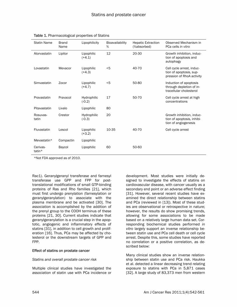
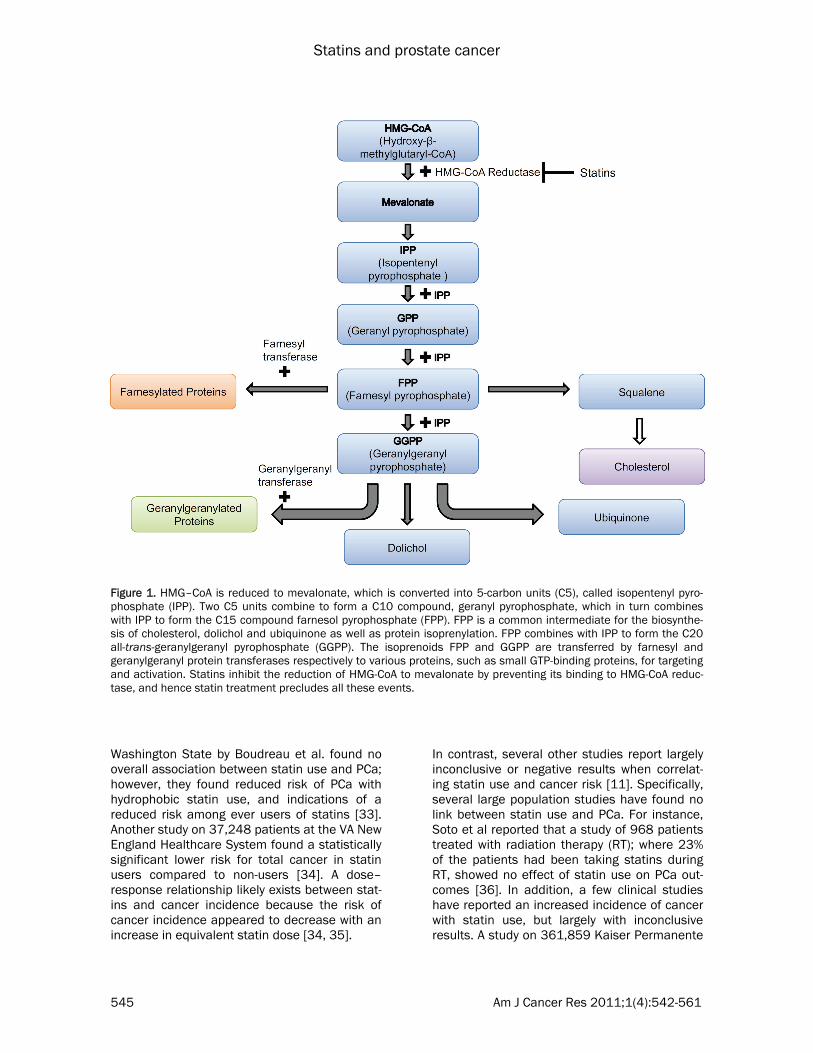
various PCa cell lines by inducing G1 cell cycle arrest. Lovastatin achieved this arrest at 0.5 mmol/L, a concentration easily achieved in the serum following oral administration [20]. The hydrophilic pravastatin, however, was less effec-tive at inhibiting HMG-CoA reductase in PC-3 cells and had to be present at 200 times higher concentrations to effect a cell cycle arrest, since it’s uptake into cells is not as efficient as the other statins [20]. In addition, the bioavailability of the HMG-CoA reductase inhibitors is limited by extensive first-pass metabolism [21]. The differences in metabolism among the various statins lead to different distributions of the drugs in the liver (via enterohepatic circulation) or peripheral tissues (via systemic circulation) at equivalent doses. Table 1 summarizes the vari-ous biological properties of statins [13, 21-24]. As shown in Figure 1, statins inhibit the synthe-sis of HMG-CoA reductase, which catalyzes a rate-limiting step in the mevalonate pathway. These inhibitors decrease hepatic cholesterol production, consequently decreasing hepatic Low Density Lipoprotein (LDL)-cholesterol up-take and ultimately cause a 20-60% decrease in plasma LDL-cholesterol level [21]. In PCa, feed-back regulation of cholesterol absorption and synthesis is lost [25], resulting in the upregula-tion of the mevalonate pathway and increased LDL receptor expression. This can affect risk of PCa in two ways – by decreased cholesterol metabolism as well as by inhibition of other products of the mevalonate pathway. Intracellu-lar depletion of cholesterol can induce apop-tosis through inhibition of the phosphatidylinosi-tol-3-kinase (PI3K)/Akt pathway, which plays a key role in mediating signals for cell growth, cell survival, and cell cycle progression [26-28]. Cholesterol, a critical component in biological membranes, accumulates in “lipid rafts”, which were shown to be important in mediating signal transduction pathways, including the PI3K-Akt pathway [15]. Further details of this pathway will be provided later in this review. In addition, since statins function at an early step in the synthesis of cholesterol, they also inhibit the production of isoprenoids such as geranylgeranyl pyrophosphate (GGPP) and far-nesyl pyrophosphate (FPP) [29] (Figure 1). These isoprenoids are involved in cellular signal transduction by activating farnesylated proteins (for example, Ras proteins) and geranylger-anylated proteins (for example, RhoA and
Statins and prostate cancer
544 Am J Cancer Res 2011;1(4):542-561
Rac1). Geranylgeranyl transferase and farnesyl transferase use GPP and FPP for post-translational modifications of small GTP-binding proteins of Ras and Rho families [21], which must first undergo prenylation (farnesylation or geranylgeranylation) to associate with the plasma membrane and be activated [30]. The association is accomplished by the addition of the prenyl group to the COOH terminus of these proteins [21, 30]. Current studies indicate that geranylgeranylation is a crucial step in the apop-totic, angiogenic and inflammatory effects of statins [31], in addition to cell growth and prolif-eration [16]. Thus, PCa may be affected by cho-lesterol or the downstream targets of GPP and FPP.
Effect of statins on prostate cancer Statins and overall prostate cancer risk Multiple clinical studies have investigated the association of statin use with PCa incidence or
development. Most studies were initially de-signed to investigate the effects of statins on cardiovascular disease, with cancer usually as a secondary end point or an adverse effect finding [31]. However, several recent studies have ex-amined the direct relationship between statins and PCa (reviewed in [13]). Most of these stud-ies are observational or retrospective in nature; however, the results do show promising trends, allowing for some associations to be made based on a relatively large human data set. Cor-responding biochemical studies performed in vitro largely support an inverse relationship be-tween statin use and PCa cell death or cell cycle arrest. Despite this, some studies have reported no correlation or a positive correlation, as de-scribed below: Many clinical studies show an inverse relation-ship between statin use and PCa risk. Haukka et al. detected a linear decreasing trend relating exposure to statins with PCa in 5,871 cases [32]. A large study of 83,373 men from western
Table 1. Pharmacological properties of Statins
Statin Name Brand Name
Lipophilicity Bioavailability %
Hepatic Extraction (%absorbed)
Observed Mechanism in PCa cells in vitro
Atorvastatin Lipitor Lipophilic (+4.1)
12 20-30 Growth inhibition, induc-tion of apoptosis and autophagy
Lovastatin Mevacor Lipophilic (+4.3)
<5 40-70 Cell cycle arrest, induc-tion of apoptosis, sup-pression of RhoA activity
Simvastatin Zocor Lipophilic (+4.7)
<5 50-80 Induction of apoptosis through depletion of in-tracellular cholesterol
Pravastatin Pravacol Hydrophilic (-0.2)
17 50-70 Cell cycle arrest at high concentrations
Pitavastatin Livalo Lipophilic 80
Rosuvas-tatin
Crestor Hydrophilic (-0.3)
20 Growth inhibition, induc-tion of apoptosis, inhibi-tion of angiogenesis
Fluvastatin Lescol Lipophilic (+3.2)
10-35 40-70 Cell cycle arrest
Mevastatin* Compactin Lipophilic
Cerivas-tatin*
Baycol Lipophilic 60 50-60
*Not FDA approved as of 2010.
Statins and prostate cancer
545 Am J Cancer Res 2011;1(4):542-561
Washington State by Boudreau et al. found no overall association between statin use and PCa; however, they found reduced risk of PCa with hydrophobic statin use, and indications of a reduced risk among ever users of statins [33]. Another study on 37,248 patients at the VA New England Healthcare System found a statistically significant lower risk for total cancer in statin users compared to non-users [34]. A dose–response relationship likely exists between stat-ins and cancer incidence because the risk of cancer incidence appeared to decrease with an increase in equivalent statin dose [34, 35].
In contrast, several other studies report largely inconclusive or negative results when correlat-ing statin use and cancer risk [11]. Specifically, several large population studies have found no link between statin use and PCa. For instance, Soto et al reported that a study of 968 patients treated with radiation therapy (RT); where 23% of the patients had been taking statins during RT, showed no effect of statin use on PCa out-comes [36]. In addition, a few clinical studies have reported an increased incidence of cancer with statin use, but largely with inconclusive results. A study on 361,859 Kaiser Permanente
Figure 1. HMG–CoA is reduced to mevalonate, which is converted into 5-carbon units (C5), called isopentenyl pyro-phosphate (IPP). Two C5 units combine to form a C10 compound, geranyl pyrophosphate, which in turn combines with IPP to form the C15 compound farnesol pyrophosphate (FPP). FPP is a common intermediate for the biosynthe-sis of cholesterol, dolichol and ubiquinone as well as protein isoprenylation. FPP combines with IPP to form the C20 all-trans-geranylgeranyl pyrophosphate (GGPP). The isoprenoids FPP and GGPP are transferred by farnesyl and geranylgeranyl protein transferases respectively to various proteins, such as small GTP-binding proteins, for targeting and activation. Statins inhibit the reduction of HMG-CoA to mevalonate by preventing its binding to HMG-CoA reduc-tase, and hence statin treatment precludes all these events.
Statins and prostate cancer
546 Am J Cancer Res 2011;1(4):542-561
patients in Northern California, found an overall increased rate of cancer in male statin users, as well as an increased risk in stage I PCa but a decreased risk in stage II PCa [37]. However, the same study notes that another Kaiser study (California Men’s Health Study) found a 28% reduction in risk of PCa in statin users [37]. Pos-sible mechanisms which could result in such a risk included increased mitotic abnormalities and/or immunosuppression caused by altera-tions in leukocyte function (discussed in [11]). Increased mitotic abnormalities can interfere with the proper function of centromeres, thus increasing the risk for mutations. Statins also may inhibit promoter function and cause bind-ing to leukocyte function antigen, which could lead to immunosuppression. However, statin concentrations used in such findings largely are based on animal models, and are higher than those used in humans for clinical treatment [11]. Statin use was also associated with a lower pro-portion of prostatectomy patients with positive surgical margins and lower tumor volume [38]. In a case control study, no overall association was found between statin use and PCa risk [39]. Despite this, risk related to statin use was modified by body mass index, and obese men who used statins had an increased risk relative to obese nonusers [39]. Taken together, it appears that there is a dis-crepancy between the studies described above, where some show a positive, others negative and yet others, no correlation between statin use and PCa risk. While there appears to be a correlation between hydrophobic statin use and a reduced risk of PCa incidence, there is no overall consensus based on the studies de-scribed so far, as to the overall effect of statins on the development of PCa. Therefore, further studies were required to delineate this relation-ship, as described below. Statins and reduced risk of advanced/aggressive prostate cancer The discrepancy between the effects of statins on PCa risk from various studies can be ex-plained by the fact that while statins may or may not have an overall benefit in preventing PCa, they specifically prevent advanced or ag-gressive PCa. As mentioned earlier, the study on Kaiser Permanente patients in Northern Califor-
nia found an increased risk in stage I PCa but a decreased risk in stage II PCa in male statin users [37]. Several studies found that statins reduced advanced PCa risk but not overall PCa risk [14, 40, 41]. Platz et al. conducted a study on 34,438 health professionals, and found no overall reduction in PCa risk with statin use. However, an analysis of the extent of the dis-ease showed a significant (46%) reduction in advanced PCa risk (compared with non-drug users), and the risk decreased with increasing duration of use [14]. The risk reduction was even stronger for metastatic and fatal disease [42, 43]. Metaanalysis of 6 randomized clinical trials and 13 observational studies revealed a statistically significant inverse relationship be-tween statin use and advanced disease, but not overall risk of developing PCa [44]. As described in the Introduction, 15-30% of pa-tients experience biochemical or clinical recur-rence within 10 years after undergoing radical prostatectomy (RP). Biochemical recurrence is marked by an increase in postoperative PSA levels to 0.4 ng/mL followed by another in-crease [45]. A study by Hamilton et al. found that 1319 men who underwent RP and took statins at the time of surgery, had a 30% lower risk for biochemical recurrence (p=0.03) than men not taking statins [46]. The reduced risk was dose dependent, with no reduction in men who took the simvastatin dose-equivalent of 20 mg or less and a 46% reduction in men who took the dose-equivalent of 20 mg or more. Time to recurrence after RP is associated with the risk of PCa progression and death; hence, these studies suggest a potential therapeutic value in statin use [46]. Overall, these studies indicate an effect of statin use on PCa progres-sion, which usually involves increased cell mi-gration or metastasis – rather than on the de-velopment of the primary tumor.
Statins and radiosensitization Inhibition of the HMG-CoA reductase pathway by statins has been associated with in vitro radio-sensitization. In a study on 512 patients treated with permanent brachytherapy (internal RT), statins (especially atorvastatin, which has one of the greatest bioavailabilities) improved the clinical presentation of PCa in an 8 year bio-chemical progression free survival [47]. Addi-tionally, a 2010 study by Gutt el al. found that statin use improved freedom from biochemical
Statins and prostate cancer
547 Am J Cancer Res 2011;1(4):542-561
failure (FFBF) and relapse-free survival (RFS) in PCa patients treated with radiotherapy (RT) [12]. This effect was seen across all RT dose ranges and was independent of AWD use. The authors of this study suggest that lowered LDL levels through statin may modify intra-prostatic hormonal levels, resulting in a differential effect of RT. Surprisingly, they also note that patients undergoing more aggressive radiation treat-ments derive less benefit from statin use [12]. This may explain the inconclusive RT results in an aforementioned study by Soto et al., which used higher radiation doses and saw no bene-fits in patients taking statins [36]. The beneficial effect of statins on RT is easy to understand. As RT kills cells by causing DNA damage, and resistance to RT is primarily caused by the ability of cells to achieve DNA repair following this damage, it is likely that stat-ins cause radiosensitization by preventing DNA repair following radiation induced DNA damage. As a result, cells located in different phases of the cell cycle have a wide variation in sensitivity to ionizing radiation. For instance, cells located in the G1 and G2-M phases of the cell cycle are most sensitive to ionizing radiation-induced cell death. Therefore, statins may sensitize these cells to radiation through G1 cell cycle arrest [21]. It has been shown that Ras overexpres-sion, which promotes cell cycle progression, may confer radiation resistance whereas that Ras-associated increase in radiation resistance can be reversed by lovastatin in osteosarcoma cells [21]. The negative effect of higher doses of radiations is harder to explain. It is likely that high radiation doses kill cells regardless of drug use and therefore, the effect of statins are not immediately obvious at these RT doses. Possible mechanisms of statin effect on pros-tate cancer cells A large number of in vitro and preclinical studies have been conducted to explain the effects of statins on PCa risk. These studies demon-strated that statins induce G1 arrest in PCa cell lines [20, 21]. This effect of statins was demon-strated not only in epithelial but also in endothe-lial cells. As a result, statins also may inhibit angiogenesis, and reduce tumor growth through this mechanism. Rosuvastatin, a relatively new statin drug, was shown to inhibit PCa cell growth and decrease tumor size in mice xenograft mod-els. Moreover, rosuvastatin inhibited angiogene-
sis, by reducing the number of blood vessels within tumors of statin-treated mice [48]. A di-rect effect of statins on angiogenesis via choles-terol inhibition vs. isoprenoid inhibition was studied in mice and showed that whereas low grade cerivastatin or atorvastain consumption (0.005 to 0.01 µmol/L) lowered cholesterol lev-els and promoted endothelial cell proliferation and angiogenesis, in keeping with its cardiopro-tective functions, at high statin concentrations (0.05 to 1 µmol/L), despite lowered cholesterol, angiogenesis was inhibited, supporting choles-terol independent effects of statins in tumor suppression [49]. The anti-angiogenic effect of statins was reversed by GGPP, clearly demon-strating a role for geranylgeranylated proteins in this effect [49]. Thus, decreased PCa risk can be attributed to statin-induced suppression of tumor growth, induction of apoptosis, and/or inhibition of angiogenesis [11]. The deleterious effect of statins on angiogenesis in tumors, of course, is at odds with its effects on promoting angiogenesis in the cardiovasculature and will be discussed later. Statins also have anti-inflammatory properties, and inflammation is an important process af-fecting PCa growth. Thus, statins may reduce PCa progression, in part by inhibiting inflamma-tion. In a study on 236 men, statin intake be-fore surgery was significantly associated with reduced risk of inflammation of surrounding malignant glands within RP specimens. The sig-nificant risk reduction for tumor inflammation among statin users was independent of demo-graphic, clinical, and pathological characteris-tics [50]. Another possible mechanism of statins’ action is the suppression of androgen production. Cho-lesterol is a required intermediate in androgen synthesis in the testis and adrenal glands. Thus, it is possible that reducing the available sub-strate for testosterone and dihydrotestosterone (DHT) production may suppress circulating an-drogen levels, thereby reducing androgen-dependent PCa tumor growth. The presence of cholesterol may promote castrate resistant PCa by conferring the ability to produce androgens despite castration. Locke et al. reported the ability of castration-resistant PCa cells to syn-thesize androgens from cholesterol precursors [51]. This effect of cholesterol has significant consequences in the detection of PCa as well. Androgens activate the AR which in turn tran-
Statins and prostate cancer
548 Am J Cancer Res 2011;1(4):542-561
scriptionally regulates PSA production. There-fore, lowering of cholesterol by statin use di-rectly reduces serum PSA levels. A study by Loeb et al. of 504 statin users at the Johns Hop-kins School of Medicine showed a decline in PSA levels with statin use [38]. The study by Gutt et al mentioned above also correlated statin use with lower PSA and clinical stage at diagnosis [12]. Even among men who were free of PCa, statin use lowered PSA levels in propor-tion to the decrease in LDL levels [35, 46]. However, Hall et al. [52] failed to demonstrate that statin use decreases circulating hormones in patients, such as free testosterone, dehy-droepiandrosterone sulfate, or luteinizing hor-mone, suggesting that they may act by inhibiting ligand-independent rather than dependent AR transcriptional activity. An explanation for this observation may be found in the results from another study, showing that statins may lower intraprostatic androgen levels, despite not low-ering serum androgen levels in patients [53]. Consequently, changes in intraprostatic andro-gen synthesis can change factors necessary for prostate growth and decrease PSA levels in men [53]. Because PSA is widely used as a screening test for PCa, the effect of statins or cholesterol on PSA levels independent of any biological in-fluence on PCa can create a detection bias [13]. In support of this hypothesis, no correlation was observed between statins and PCa incidence in a small cohort of Korean patients who nonethe-less, showed that mean PSA and PSA density were significantly lower in patients on statins [54]. Fortunately, the majority of studies re-ported here, take this bias into account – how-ever, it still has to be kept in mind for older studies when this effect of cholesterol was not known. Do statins prevent prostate cancer risk by inhib-iting cholesterol production? Because the primary site of cholesterol synthe-sis is the liver, statins that are currently avail-able have been selected for their capacity to target the liver and decrease cholesterol biosyn-thesis [55], whereas they accumulate poorly in other tissues [55, 56]. This decreases their bioavailability in other tissues such as the pros-tate. It was estimated that less than 5% of an oral dose of simvastatin reaches the general circulation as an active inhibitor [56]. This is because simvastatin and lovastatin are con-
sumed as prodrugs and require esterase-dependent conversion to the active form [57]. Humans lack high levels of serum esterases, and thus these statins remain in their pro-drug form and accumulate mainly in the liver [56]. On the other hand, statins such as pravastatin and atorvastatin do not require this enzymatic con-version; however, due to the hydrophobic nature of pravastatin, it’s bioavailability in the liver is about 20% [58], while atorvastatin (Lipitor) re-mains the most bioavailable statin currently in the market – however, it too has low bioavail-ability in peripheral tissues. Based on these results, Solomon and Freeman have argued that any anti-tumor effects within peripheral tissues are likely to arise indirectly from potent LDL low-ering effect, and not from the inhibition of iso-prenoid synthesis within tumor cells [56]. How-ever, in vitro studies showed that lovastatin achieved G1 arrest arrest at 0.5 mmol/L, a con-centration easily achieved in the serum follow-ing oral administration [20]. It may be argued that this could be due to the production of GGPP and FPP in the liver which is then systemi-cally delivered to their sites of action, such as the prostate. Indeed, a number of studies report a positive correlation between cholesterol levels and PCa risk. Mondul et al. examined the association between plasma cholesterol concentration and PCa in a large prospective cohort study con-ducted in Washington County, MD [59]. There was no overall association between cholesterol concentration and incidence of total, advanced, or organ-confined PCa. However, compared to men with high cholesterol (240 mg/dl), men with desirable (200 mg/dl) or borderline (200 to 240 mg/dl) levels were less likely to develop high-grade PCa [59]. Similar conclusions were drawn from a study in a cohort including 5112 PCa patients, where the authors investigated associations among triglycerides (TG), total cho-lesterol (TC), and PCa while taking into account glucose [60]. The results of the study showed that glucose and lipid metabolism influence PCa risk [60]. Another study showed that men with low cholesterol had a lower risk of Gleason 8 to 10 PCa than men with high cholesterol, al-though no association was present for PCa over-all [43]. However, these correlations could be coincidental and given the effect of the mevalo-nate pathway and its increased activity in PCa – upregulation of cholesterol would be accompa-nied by an upregulation of isoprenyl groups,
Statins and prostate cancer
549 Am J Cancer Res 2011;1(4):542-561
which may increase PCa risk by a mechanism independent of cholesterol metabolism. The discriminating factor, of course, would be the effect of lowering cholesterol specifically, rather than reducing all of the downstream ef-fectors of the mevalonate pathway. A 2007 Fin-nish study of 49,446 men reported that statin users had a 25% lower risk of advanced PCa when compared with men who took other cho-lesterol-lowering drugs or no cholesterol medi-cation (described in [41]). The effect was dose-dependent, with increased statin use linked to increased positive outcomes [41]. This suggests that statins have a reduced risk of developing advanced or high grade PCa but not necessarily all cholesterol lowering drugs. This and similar studies, therefore, point to a mechanism of ac-tion of statins other than the lowering of choles-terols per se.
In vitro mechanisms of cholesterol action in prostate cancer cells Direct correlation between cholesterol metabo-lism and PCa was provided in the form of in vi-tro studies that confirmed that upregulation of cholesterol increases the risk of PCa. In a series of experiments, Michael Freeman and co-investigators have shown that cholesterol deple-tion in lipid rafts on biological membranes may influence and downregulate oncogenic signals. They showed that LNCaP human PCa cells con-tain cholesterol-rich lipid rafts that mediate epi-dermal growth factor (EGF)-induced and consti-tutive signaling through the PI3K/Akt pathway [61]. Disruption of these lipid rafts inhibited, while their reconstitution restored, EGF recep-tor/PI3K/Akt signaling which regulated apop-tosis. Treatment with 20 µM simvastatin caused cholesterol depletion of lipid rafts, followed by downregulation of the PI3K-Akt pathway and apoptosis [28, 61]. Elevation of circulating cho-lesterol in SCID mice promoted tumor growth, increased Akt phosphorylation, and reduced apoptosis in the xenografts [28]. The phos-phorylation of Akt, which is an important step in intracellular oncogenic signaling, is regulated by caveolin-1, a cholesterol-regulated protein. Caveolin-1 is the main structural protein of caveolae, commonly found in lipid rafts. Studies have also shown that raft-dependent signaling events can be inhibited by altering cholesterol content in the raft sites [62]. AR was found to localize to a lipid raft compart-
ment in androgen-sensitive LNCaP prostate ade-nocarcinoma cells. AR can send rapid, non-genomic signals, activating various pathways in response to androgen stimulation. Studies also demonstrated that the treatment of AR with androgen rapidly activated Akt1 in a non-genomic manner, through the activation of up-stream PI3K. Endogenous AR interacted with endogenous Akt1 preferentially in raft areas, and androgen addition increased this interac-tion [63]. This study also showed that a complex forms between AR and Akt in the lipid raft mem-brane sections; while treatment of LNCaP cells with the AR antagonist bicalutamide prevented the complex formation, whereas LY294002 (PI3K inhibitor) and Src inhibitor PP2 only had marginal effects. Thus, the binding of AR to Akt may bypass the PI3K and Src pathways. Addi-tionally, the major effects of androgen on Akt phosphorylation activity occurred in raft frac-tions. These findings indicate that the AR com-plex may activate Akt directly in raft sections of the cell membrane [63]. Taken together, these studies hypothesize that cholesterols may regu-late PCa risk by upregulating an interaction be-tween the AR and Akt, which in turn promoted tumor cell growth. These studies, however, do not account for the greater effect of cholesterol (and statins) in high grade and advanced PCa.
Cholesterol-independent mechanisms of statin action in prostate cancer cells Despite the strong argument for cholesterol deregulation as a mediator of statin action in PCa cells, several lines of evidence point to a cholesterol independent mechanism in statin induced reduction in PCa risk. A study on 1,812 men by Hall et al. measured whether levels of circulating androgens and their carrier protein, sex hormone–binding globulin (SHBG), varied by statin use. The study found no relationship be-tween statin use and free testosterone, dehy-droepiandrosterone sulfate, or luteinizing hor-mone [52]. The study concluded that it is unlikely that statins affect circulating androgens and PCa risk through a hormonal mechanism. In a longitudinal study of 1214 PCa-free men who were prescribed statins but who had undergone routine PSA testing before and after statin in-take, serum PSA decline after statins initiation was significantly associated with statin dose and the decline in LDL-cholesterol levels [35]; however, even after adjusting for LDL changes, statin use still remained associated with the PSA decline, suggesting that there may be both
Statins and prostate cancer
550 Am J Cancer Res 2011;1(4):542-561
cholesterol and non-cholesterol-mediated mechanisms by which statin treatment influ-ences PSA [35]. A study by Mondul et al. on 2,574 men showed that statin users experienced a greater decline in PSA levels compared to non-statin users, al-though the difference was not statistically sig-nificant [64]. Detection bias was accounted for, but did not explain the inverse association be-tween statin use and lowered PSA levels. The observed inverse relationship between statin use and PSA levels could indirectly suggest that statins may influence PSA concentration through cholesterol-lowering properties. How-ever, other mechanisms are also possible, since PSA levels decreased even after controlling for cholesterol levels [64]. In contrast to in vitro and animal studies, a 1989 study in the Lancet showed that total cho-lesterol levels were reduced in patients with evidence of metastasis (n = 30) compared with those without metastasis (n = 73) [65]. LDL was cleared faster in patients with metastatic dis-ease compared to patients without metastasis and in healthy controls [65]. A study in 127 Chi-nese patients with prostate cancer with bone metastasis undergoing hormone treatment showed that patients who showed good re-sponse to the treatment (n=54) had higher cho-lesterol levels compared to those who showed poor response (n=73) [66]. These studies con-tradict a recent metabolomics study where the mean cholesterol level in PCa bone metastases was 127.30 mg/g (n=20) as compared to 81.06 and 35.85 mg/g in bone metastases of different origin (n=7) and normal bone (n=14), respectively (p=0.0002 and 0.001) [67]. The smaller number of specimen used in the latter study may therefore indicate a subset of pa-tients who contrast the former two. Overall, these studies suggest that cholesterol lowering is not the only mechanism by which statins de-crease the risk of PCa, and that other mecha-nisms may exist as well. Statin action through isoprenylation Geranygeranylated and farnesylated small GTP-binding proteins Small GTP-binding proteins of the Ras super-family are involved in such diverse cellular func-tions as cytokinesis, cell motility, cell adhesion
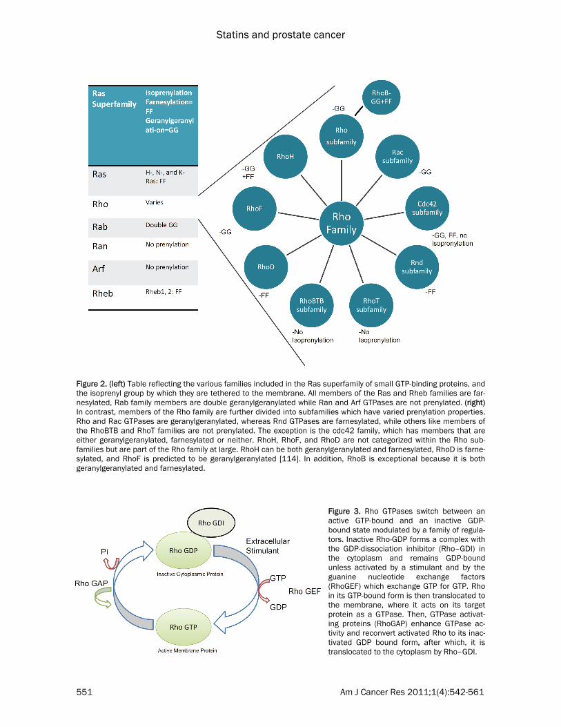
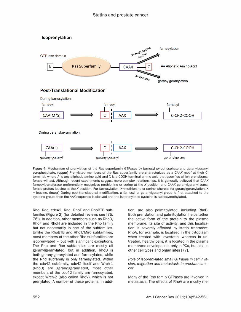
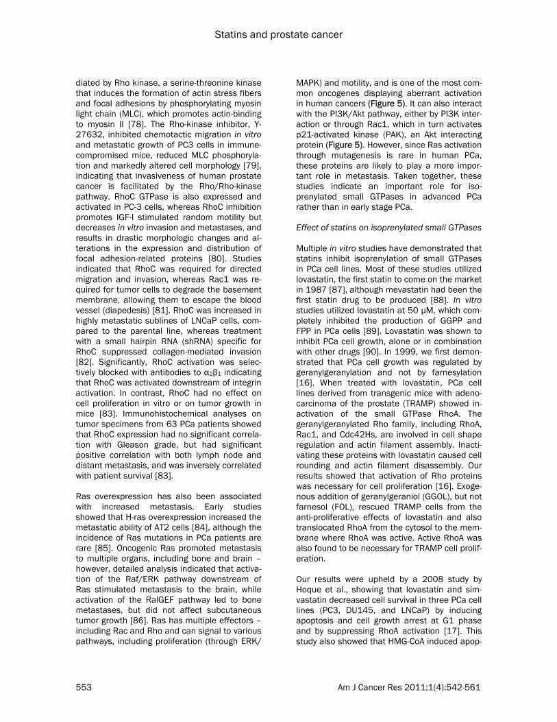
and cell proliferation [16, 31]. This superfamily consists of 5 families with a total of more than 150 members: Ras, Rho, Rab, Ran and Arf (reviewed in [68, 69]) (Figure 2). All members of this family contain an N-terminal GTPase do-main and switch between an active GTP-bound and an inactive GDP-bound state modulated by a family of regulators. RhoA, for example, forms a complex with the GDP-dissociation inhibitor (Rho–GDI) in the cytoplasm and remains GDP-bound unless activated by a stimulant and by guanine nucleotide exchange factors (GEF), which exchange GTP for GDP. Upon release of binding proteins and Rho-GDI, activated RhoA in its GTP-bound form can be translocated to the membrane, where it can bind its target protein and regulate downstream effectors. GTPase activating proteins (GAPs) enhance GTPase ac-tivity, increasing GTP hydrolysis, which recon-verts activated Rho to its inactivated Rho-GDI/GDP bound form, which is then sequestered in the cytoplasm [70] (Figure 3). Most of the 150 members of the Ras superfamily are similarly regulated, but they utilize different regulatory factors. Members of the Ras superfamily that are regu-lated by isoprenylation, include H-, N- and K-Ras, as well as Rheb-1 and -2, which are farne-sylated, whereas Rab GTPases require double geranylgeranylation [71]. Ran GTPases are, on the other hand, primarily nuclear localized and do not require prenylation for its activation [72]. The Rho family is unusual, since it has members that are geranylgeranylated, farnesylated, both or neither (Figure 2). As shown in Figure 4, all prenylated members of the Ras superfamily are characterized by a CAAX motif at their C-terminal, where A is any aliphatic amino acid and X is a COOH-terminal amino acid that speci-fies which prenyltransferase will act. Although recent experiments suggest more complex rela-tionships, it is generally believed that CAAX far-nesyltransferase preferentially recognizes me-thionine or serine at the X position, whereas CAAX geranylgeranyl transferase prefers leucine at the X position [73]. During post-translational modification, a farnesyl or geranylgeranyl group is first attached to the cysteine group, then the AAX sequence is cleaved and the isoprenylated cysteine is carboxymethylated [74]. Rho proteins are known to regulate both cell growth and motility, and consists of over 40 members which have been subclassified into
Statins and prostate cancer
551 Am J Cancer Res 2011;1(4):542-561
Figure 2. (left) Table reflecting the various families included in the Ras superfamily of small GTP-binding proteins, and the isoprenyl group by which they are tethered to the membrane. All members of the Ras and Rheb families are far-nesylated, Rab family members are double geranylgeranylated while Ran and Arf GTPases are not prenylated. (right) In contrast, members of the Rho family are further divided into subfamilies which have varied prenylation properties. Rho and Rac GTPases are geranylgeranylated, whereas Rnd GTPases are farnesylated, while others like members of the RhoBTB and RhoT families are not prenylated. The exception is the cdc42 family, which has members that are either geranylgeranylated, farnesylated or neither. RhoH, RhoF, and RhoD are not categorized within the Rho sub-families but are part of the Rho family at large. RhoH can be both geranylgeranylated and farnesylated, RhoD is farne-sylated, and RhoF is predicted to be geranylgeranylated [114]. In addition, RhoB is exceptional because it is both geranylgeranylated and farnesylated.
Figure 3. Rho GTPases switch between an active GTP-bound and an inactive GDP-bound state modulated by a family of regula-tors. Inactive Rho-GDP forms a complex with the GDP-dissociation inhibitor (Rho–GDI) in the cytoplasm and remains GDP-bound unless activated by a stimulant and by the guanine nucleotide exchange factors (RhoGEF) which exchange GTP for GTP. Rho in its GTP-bound form is then translocated to the membrane, where it acts on its target protein as a GTPase. Then, GTPase activat-ing proteins (RhoGAP) enhance GTPase ac-tivity and reconvert activated Rho to its inac-tivated GDP bound form, after which, it is translocated to the cytoplasm by Rho–GDI.
Statins and prostate cancer
552 Am J Cancer Res 2011;1(4):542-561
Rho, Rac, cdc42, Rnd, RhoT and RhoBTB sub-familes (Figure 2) (for detailed reviews see [75, 76]). In addition, other members such as RhoD, RhoF and RhoH are included in the Rho family but not necessarily in one of the subfamilies. Unlike the RhoBTB and RhoT/Miro subfamilies, most members of the other Rho subfamilies are isoprenylated – but with significant exceptions. The Rho and Rac subfamilies are mostly all geranylgeranylated, but in addition, RhoB is both geranylgeranylated and farnesylated, while the Rnd subfamily is only farnesylated. Within the cdc42 subfamily, cdc42 itself and Wrch-1 (RhoU) are geranylgeranylated, most other members of the cdc42 family are farnesylated, except Wrch-2 (also called RhoV), which is not prenylated. A number of these proteins, in addi-
tion, are also palmitoylated, including RhoB. Both prenylation and palmitoylation helps tether the active form of the protein to the plasma membrane, its site of activity, and this localiza-tion is severely affected by statin treatment. RhoA, for example, is localized in the cytoplasm when treated with lovastatin, whereas in un-treated, healthy cells, it is located in the plasma membrane envelope, not only in PCa, but also in other cell types and organ sites [77]. Role of isoprenylated small GTPases in cell inva-sion, migration and metastasis in prostate can-cer Many of the Rho family GTPases are involved in metastasis. The effects of RhoA are mostly me-
Figure 4. Mechanism of prenylation of the Ras superfamily GTPases by farnesyl pyrophosphate and geranylgeranyl pyrophosphate. (upper) Prenylated members of the Ras superfamily are characterized by a CAAX motif at their C-terminal, where A is any aliphatic amino acid and X is a COOH-terminal amino acid that specifies which prenyltrans-ferase will act. Although recent experiments suggest more complex relationships, it is generally believed that CAAX farnesyltransferase preferentially recognizes methionine or serine at the X position and CAAX geranylgeranyl trans-ferase prefers leucine at the X position. For farnesylation, X=methionine or serine whereas for geranylgeranylation, X = leucine. (lower) During post-translational modification, a farnesyl or geranylgeranyl group is first attached to the cysteine group, then the AAX sequence is cleaved and the isoprenylated cysteine is carboxymethylated.
Statins and prostate cancer
553 Am J Cancer Res 2011;1(4):542-561
diated by Rho kinase, a serine-threonine kinase that induces the formation of actin stress fibers and focal adhesions by phosphorylating myosin light chain (MLC), which promotes actin-binding to myosin II [78]. The Rho-kinase inhibitor, Y-27632, inhibited chemotactic migration in vitro and metastatic growth of PC3 cells in immune-compromised mice, reduced MLC phosphoryla-tion and markedly altered cell morphology [79], indicating that invasiveness of human prostate cancer is facilitated by the Rho/Rho-kinase pathway. RhoC GTPase is also expressed and activated in PC-3 cells, whereas RhoC inhibition promotes IGF-I stimulated random motility but decreases in vitro invasion and metastases, and results in drastic morphologic changes and al-terations in the expression and distribution of focal adhesion-related proteins [80]. Studies indicated that RhoC was required for directed migration and invasion, whereas Rac1 was re-quired for tumor cells to degrade the basement membrane, allowing them to escape the blood vessel (diapedesis) [81]. RhoC was increased in highly metastatic sublines of LNCaP cells, com-pared to the parental line, whereas treatment with a small hairpin RNA (shRNA) specific for RhoC suppressed collagen-mediated invasion [82]. Significantly, RhoC activation was selec-tively blocked with antibodies to α2β1 indicating that RhoC was activated downstream of integrin activation. In contrast, RhoC had no effect on cell proliferation in vitro or on tumor growth in mice [83]. Immunohistochemical analyses on tumor specimens from 63 PCa patients showed that RhoC expression had no significant correla-tion with Gleason grade, but had significant positive correlation with both lymph node and distant metastasis, and was inversely correlated with patient survival [83]. Ras overexpression has also been associated with increased metastasis. Early studies showed that H-ras overexpression increased the metastatic ability of AT2 cells [84], although the incidence of Ras mutations in PCa patients are rare [85]. Oncogenic Ras promoted metastasis to multiple organs, including bone and brain – however, detailed analysis indicated that activa-tion of the Raf/ERK pathway downstream of Ras stimulated metastasis to the brain, while activation of the RalGEF pathway led to bone metastases, but did not affect subcutaneous tumor growth [86]. Ras has multiple effectors – including Rac and Rho and can signal to various pathways, including proliferation (through ERK/
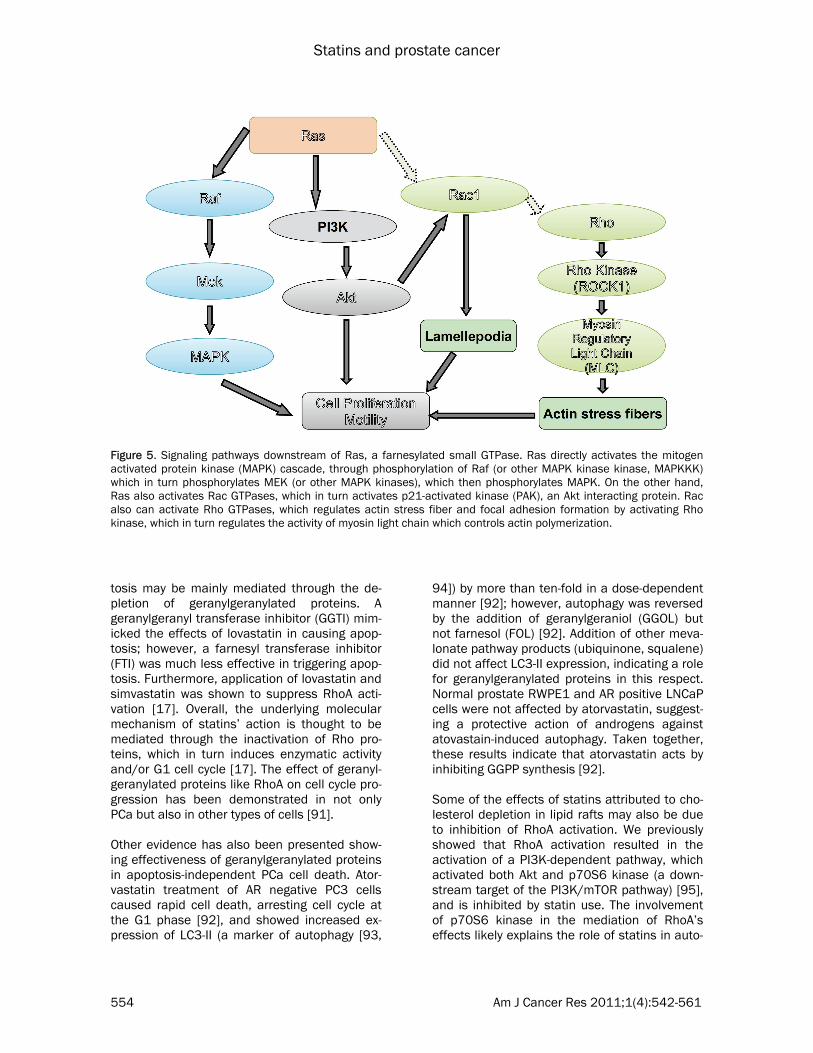
MAPK) and motility, and is one of the most com-mon oncogenes displaying aberrant activation in human cancers (Figure 5). It can also interact with the PI3K/Akt pathway, either by PI3K inter-action or through Rac1, which in turn activates p21-activated kinase (PAK), an Akt interacting protein (Figure 5). However, since Ras activation through mutagenesis is rare in human PCa, these proteins are likely to play a more impor-tant role in metastasis. Taken together, these studies indicate an important role for iso-prenylated small GTPases in advanced PCa rather than in early stage PCa.
Effect of statins on isoprenylated small GTPases Multiple in vitro studies have demonstrated that statins inhibit isoprenylation of small GTPases in PCa cell lines. Most of these studies utilized lovastatin, the first statin to come on the market in 1987 [87], although mevastatin had been the first statin drug to be produced [88]. In vitro studies utilized lovastatin at 50 µM, which com-pletely inhibited the production of GGPP and FPP in PCa cells [89]. Lovastatin was shown to inhibit PCa cell growth, alone or in combination with other drugs [90]. In 1999, we first demon-strated that PCa cell growth was regulated by geranylgeranylation and not by farnesylation [16]. When treated with lovastatin, PCa cell lines derived from transgenic mice with adeno-carcinoma of the prostate (TRAMP) showed in-activation of the small GTPase RhoA. The geranylgeranylated Rho family, including RhoA, Rac1, and Cdc42Hs, are involved in cell shape regulation and actin filament assembly. Inacti-vating these proteins with lovastatin caused cell rounding and actin filament disassembly. Our results showed that activation of Rho proteins was necessary for cell proliferation [16]. Exoge-nous addition of geranylgeraniol (GGOL), but not farnesol (FOL), rescued TRAMP cells from the anti-proliferative effects of lovastatin and also translocated RhoA from the cytosol to the mem-brane where RhoA was active. Active RhoA was also found to be necessary for TRAMP cell prolif-eration. Our results were upheld by a 2008 study by Hoque et al., showing that lovastatin and sim-vastatin decreased cell survival in three PCa cell lines (PC3, DU145, and LNCaP) by inducing apoptosis and cell growth arrest at G1 phase and by suppressing RhoA activation [17]. This study also showed that HMG-CoA induced apop-
Statins and prostate cancer
554 Am J Cancer Res 2011;1(4):542-561
tosis may be mainly mediated through the de-pletion of geranylgeranylated proteins. A geranylgeranyl transferase inhibitor (GGTI) mim-icked the effects of lovastatin in causing apop-tosis; however, a farnesyl transferase inhibitor (FTI) was much less effective in triggering apop-tosis. Furthermore, application of lovastatin and simvastatin was shown to suppress RhoA acti-vation [17]. Overall, the underlying molecular mechanism of statins’ action is thought to be mediated through the inactivation of Rho pro-teins, which in turn induces enzymatic activity and/or G1 cell cycle [17]. The effect of geranyl-geranylated proteins like RhoA on cell cycle pro-gression has been demonstrated in not only PCa but also in other types of cells [91]. Other evidence has also been presented show-ing effectiveness of geranylgeranylated proteins in apoptosis-independent PCa cell death. Ator-vastatin treatment of AR negative PC3 cells caused rapid cell death, arresting cell cycle at the G1 phase [92], and showed increased ex-pression of LC3-II (a marker of autophagy [93,
94]) by more than ten-fold in a dose-dependent manner [92]; however, autophagy was reversed by the addition of geranylgeraniol (GGOL) but not farnesol (FOL) [92]. Addition of other meva-lonate pathway products (ubiquinone, squalene) did not affect LC3-II expression, indicating a role for geranylgeranylated proteins in this respect. Normal prostate RWPE1 and AR positive LNCaP cells were not affected by atorvastatin, suggest-ing a protective action of androgens against atovastain-induced autophagy. Taken together, these results indicate that atorvastatin acts by inhibiting GGPP synthesis [92]. Some of the effects of statins attributed to cho-lesterol depletion in lipid rafts may also be due to inhibition of RhoA activation. We previously showed that RhoA activation resulted in the activation of a PI3K-dependent pathway, which activated both Akt and p70S6 kinase (a down-stream target of the PI3K/mTOR pathway) [95], and is inhibited by statin use. The involvement of p70S6 kinase in the mediation of RhoA’s effects likely explains the role of statins in auto-
Figure 5. Signaling pathways downstream of Ras, a farnesylated small GTPase. Ras directly activates the mitogen activated protein kinase (MAPK) cascade, through phosphorylation of Raf (or other MAPK kinase kinase, MAPKKK) which in turn phosphorylates MEK (or other MAPK kinases), which then phosphorylates MAPK. On the other hand, Ras also activates Rac GTPases, which in turn activates p21-activated kinase (PAK), an Akt interacting protein. Rac also can activate Rho GTPases, which regulates actin stress fiber and focal adhesion formation by activating Rho kinase, which in turn regulates the activity of myosin light chain which controls actin polymerization.
Statins and prostate cancer
555 Am J Cancer Res 2011;1(4):542-561
phagy induction, described above [92], as the mTOR pathway is known to prevent autophagy [96]. Overall, these studies indicate an impor-tant role for isoprenylated small GTPases in metastasis and suggest that the greater effect of statins in advanced PCa may be, at least partly, attributed to the inhibition of the same GTPases by these drugs.
Expression of geranylgeranylated or farnesy-lated proteins in clinical samples from prostate cancer patients Although farnesylated Ras proteins may not affect PCa cell growth, they play an important role in PCa metastasis. Unlike other solid can-cers such as pancreatic or lung cancers which express ras mutations, such activation of Ha-, Ki- or N-Ras are not found in PCa in American or French patients [85, 97], although some studies reported such mutations in Japanese PCa pa-tients [98], but not Chinese patients [99]. De-spite this, high grade PCa tissues were repeat-edly found to express high Ras expression [100, 101], especially at sites of bone metastasis [102], and H-ras expression has been sug-gested as a biomarker of advanced PCa [103]. In support of this, N-Ras expression was associ-ated with the development of hormone refrac-tory PCa, and a shorter time to relapse [104]. Other studies, however, disputed such findings and found no difference between Ras expres-sion in benign vs. malignant prostate tissues [105, 106]. Increased expression of Rheb and Rho GTPases has also been reported in pros-tate tumor tissue compared to normal prostate [107, 108]. In particular, Rac proteins were highly expressed in PIN and carcinoma com-pared to benign prostate tissue [109]. On the other hand RhoE was underexpressed in pros-tate tumor tissue compared to benign prostate [110], which underscores its role in suppressing cell cycle progression. The Rac-specific guanine nucleotide exchange factor, Tiam1, plays a ma-jor role in oncogenicity, tumour invasion and metastasis. Tiam1 was significantly overex-pressed in both PIN (p<0.001) and PCa (p<0.001) when compared to benign secretory epithelium, and was statistically significantly associated with disease recurrence (p=0.016), the presence of lymph vessel invasion (p=0.031), high Gleason scores (p=0.044) and decreased disease-free survival (p=0.03) [111]. These studies point to the diversity of the Ras superfamily of small GTPases and the variety of
functions these proteins conduct. Do statins prevent prostate cancer risk by inhib-iting isoprenoid production in prostate cancer patients? Despite strong evidence in vitro, there are argu-ments that statins may not be able to inhibit isoprenylated small GTPases in the prostate. Solomon and Freeman [56] point out that since statins have been designed to accumulate in the liver, very little of these drugs actually are released into the circulation, and may not reach the prostate in sufficient quantities to affect tumor growth. They argue that the concentra-tions of the drugs released in the plasma, (lovastatin from a single 40 mg dose is esti-mated 9.5–15 ng/ml in the plasma; for a 40 mg dose of simvastatin it is approx. 10 ng/ml; and for a single 40 mg dose of pravastatin it is approx. 50 ng/ml) are far below those required to demonstrate biological effects on cells in culture [56]. Yet it can also be argued that lu-minal epithelial cells in culture may not express the enzymes required to break up the prodrugs used: whereas the prostate expresses a number of other cells, including mainly the mesenchy-mal stromal cells, where drug metabolism may be more efficient. Further, GGPP and FPP are also produced in the liver and can be released into the circulation where it acts on various or-gans. Hence, statins may affect their production directly at the site of synthesis. The effect of statins in peripheral tissue is obvi-ous from the most common side-effect of stat-ins, skeletal muscle myopathy [112], which may be traced to a geranylgeranylation defect in-duced by statin use in muscle cells [113]. Hence it is likely that lower concentrations of statins may be required to affect the prostatic stroma in situ compared to pure cultures of prostate epithelial cells in vitro, which in turn will make the drug widely available to the neighboring epithelial cells. As of now, there is little clinical evidence that distinguishes the effects of statins on cholesterolemia vs. those on isoprenyl formation. Further studies are needed to go beyond the obvious in vitro and pre-clinical studies to determine whether statins affect PCa through inhibiting cholesterol forma-tion or by affecting the function of geranylger-anylated or farnesylated small GTP-binding pro-teins of the Ras, Rho or Rab families. Atorvas-tatin, which has a much higher bioavailability,
Statins and prostate cancer
556 Am J Cancer Res 2011;1(4):542-561
displayed biochemical effects at much lower concentrations (1 µM compared to 50 µM for lovastatin), and induced cell death and cell cy-cle arrest at doses as low as 5 µM [92]. This effect was reversed by addition of geranylgera-niol, indicating an effect mediated by iso-prenylation [92]. These studies indicate the pos-sibility of statin action being mediated by iso-prenylated small GTPases preferentially over, or in addition to, cholesterol metabolism. Conclusion Based on this and similar results in animal mod-els, we conclude that statins, especially statins with high bioavailability, inhibit cholesterol for-mation and exert cardioprotective effects at low concentrations, whereas at high concentrations would also be capable of tumor suppression by inhibition of small GTPases involved in cell pro-
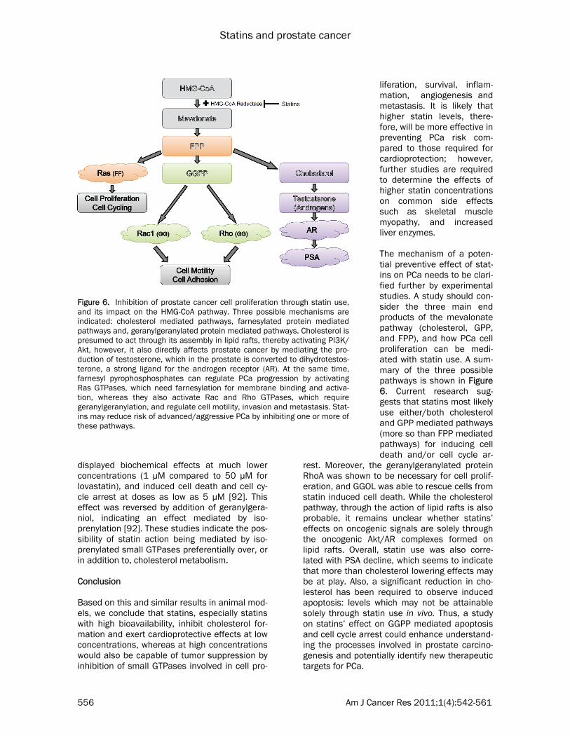
liferation, survival, inflam-mation, angiogenesis and metastasis. It is likely that higher statin levels, there-fore, will be more effective in preventing PCa risk com-pared to those required for cardioprotection; however, further studies are required to determine the effects of higher statin concentrations on common side effects such as skeletal muscle myopathy, and increased liver enzymes. The mechanism of a poten-tial preventive effect of stat-ins on PCa needs to be clari-fied further by experimental studies. A study should con-sider the three main end products of the mevalonate pathway (cholesterol, GPP, and FPP), and how PCa cell proliferation can be medi-ated with statin use. A sum-mary of the three possible pathways is shown in Figure 6. Current research sug-gests that statins most likely use either/both cholesterol and GPP mediated pathways (more so than FPP mediated pathways) for inducing cell death and/or cell cycle ar-
rest. Moreover, the geranylgeranylated protein RhoA was shown to be necessary for cell prolif-eration, and GGOL was able to rescue cells from statin induced cell death. While the cholesterol pathway, through the action of lipid rafts is also probable, it remains unclear whether statins’ effects on oncogenic signals are solely through the oncogenic Akt/AR complexes formed on lipid rafts. Overall, statin use was also corre-lated with PSA decline, which seems to indicate that more than cholesterol lowering effects may be at play. Also, a significant reduction in cho-lesterol has been required to observe induced apoptosis: levels which may not be attainable solely through statin use in vivo. Thus, a study on statins’ effect on GGPP mediated apoptosis and cell cycle arrest could enhance understand-ing the processes involved in prostate carcino-genesis and potentially identify new therapeutic targets for PCa.
Figure 6. Inhibition of prostate cancer cell proliferation through statin use, and its impact on the HMG-CoA pathway. Three possible mechanisms are indicated: cholesterol mediated pathways, farnesylated protein mediated pathways and, geranylgeranylated protein mediated pathways. Cholesterol is presumed to act through its assembly in lipid rafts, thereby activating PI3K/Akt, however, it also directly affects prostate cancer by mediating the pro-duction of testosterone, which in the prostate is converted to dihydrotestos-terone, a strong ligand for the androgen receptor (AR). At the same time, farnesyl pyrophosphosphates can regulate PCa progression by activating Ras GTPases, which need farnesylation for membrane binding and activa-tion, whereas they also activate Rac and Rho GTPases, which require geranylgeranylation, and regulate cell motility, invasion and metastasis. Stat-ins may reduce risk of advanced/aggressive PCa by inhibiting one or more of these pathways.
Statins and prostate cancer
557 Am J Cancer Res 2011;1(4):542-561
Please address correspondence to: Paramita M. Ghosh, PhD, Department of Urology, University of California Davis School of Medicine, 4860 Y Street, Suite 3500, Sacramento, CA 95817, USA. Tel: (916)843-9336, Fax: (916)364-0306, E-mail: [email protected] References [1] Williams H and Powell IJ. Epidemiology, pa-
thology, and genetics of prostate cancer among African Americans compared with other ethnicities. Methods Mol Biol 2009; 472: 439-453.
[2] Burkhardt JH, Litwin MS, Rose CM, Correa RJ, Sunshine JH, Hogan C and Hayman JA. Com-paring the costs of radiation therapy and radi-cal prostatectomy for the initial treatment of early-stage prostate cancer. J Clin Oncol 2002; 20: 2869-2875.
[3] Roehl KA, Han M, Ramos CG, Antenor JA and Catalona WJ. Cancer progression and survival rates following anatomical radical retropubic prostatectomy in 3,478 consecutive patients: long-term results. J Urol 2004; 172: 910-914.
[4] Pound CR, Partin AW, Eisenberger MA, Chan DW, Pearson JD and Walsh PC. Natural history of progression after PSA elevation following radical prostatectomy. JAMA 1999; 281: 1591-1597.
[5] Bruchovsky N, Klotz L, Crook J and Golden-berg SL. Locally advanced prostate cancer--biochemical results from a prospective phase II study of intermittent androgen suppression for men with evidence of prostate-specific antigen recurrence after radiotherapy. Cancer 2007; 109: 858-867.
[6] Schmid HP, Keuler FU and Altwein JE. Rising prostate-specific antigen after primary treat-ment of prostate cancer: sequential hormone manipulation. Urol Int 2007; 79: 95-104.
[7] Wang Y, Kreisberg JI and Ghosh PM. Cross-talk between the androgen receptor and the phosphatidylinositol 3-kinase/Akt pathway in prostate cancer. Curr Cancer Drug Targets 2007; 7: 591-604.
[8] Petrylak D. Therapeutic options in androgen-independent prostate cancer: building on do-cetaxel. BJU Int 2005; 96 Suppl 2: 41-46.
[9] Boehmer A, Anastasiadis AG, Feyerabend S, Nagele U, Kuczyk M, Schilling D, Corvin S, Merseburger AS and Stenzl A. Docetaxel, es-tramustine and prednisone for hormone-refractory prostate cancer: a single-center experience. Anticancer Res 2005; 25: 4481-4486.
[10] Small EJ, Schellhammer PF, Higano CS, Red-fern CH, Nemunaitis JJ, Valone FH, Verjee SS, Jones LA and Hershberg RM. Placebo-controlled phase III trial of immunologic ther-apy with sipuleucel-T (APC8015) in patients
with metastatic, asymptomatic hormone re-fractory prostate cancer. J Clin Oncol 2006; 24: 3089-3094.
[11] Gonyeau MJ and Yuen DW. A clinical review of statins and cancer: helpful or harmful? Phar-macotherapy 2010; 30: 177-194.
[12] Gutt R, Tonlaar N, Kunnavakkam R, Karrison T, Weichselbaum RR and Liauw SL. Statin use and risk of prostate cancer recurrence in men treated with radiation therapy. J Clin Oncol 2010; 28: 2653-2659.
[13] Murtola TJ, Visakorpi T, Lahtela J, Syvala H and Tammela T. Statins and prostate cancer prevention: where we are now, and future directions. Nat Clin Pract Urol 2008; 5: 376-387.
[14] Platz EA, Leitzmann MF, Visvanathan K, Rimm EB, Stampfer MJ, Willett WC and Giovannucci E. Statin drugs and risk of advanced prostate cancer. J Natl Cancer Inst 2006; 98: 1819-1825.
[15] Freeman MR and Solomon KR. Cholesterol and prostate cancer. J Cell Biochem 2004; 91: 54-69.
[16] Ghosh PM, Ghosh-Choudhury N, Moyer ML, Mott GE, Thomas CA, Foster BA, Greenberg NM and Kreisberg JI. Role of RhoA activation in the growth and morphology of a murine prostate tumor cell line. Oncogene 1999; 18: 4120-4130.
[17] Hoque A, Chen H and Xu XC. Statin induces apoptosis and cell growth arrest in prostate cancer cells. Cancer Epidemiol Biomarkers Prev 2008; 17: 88-94.
[18] Lee J, Lee I, Park C and Kang WK. Lovastatin-induced RhoA modulation and its effect on senescence in prostate cancer cells. Biochem Biophys Res Commun 2006; 339: 748-754.
[19] Shack S, Gorospe M, Fawcett TW, Hudgins WR and Holbrook NJ. Activation of the cholesterol pathway and Ras maturation in response to stress. Oncogene 1999; 18: 6021-6028.
[20] Sivaprasad U, Abbas T and Dutta A. Differen-tial efficacy of 3-hydroxy-3-methylglutaryl CoA reductase inhibitors on the cell cycle of pros-tate cancer cells. Mol Cancer Ther 2006; 5: 2310-2316.
[21] Chan KK, Oza AM and Siu LL. The statins as anticancer agents. Clin Cancer Res 2003; 9: 10-19.
[22] Igel M, Sudhop T and von Bergmann K. Phar-macology of 3-hydroxy-3-methylglutaryl-coenzyme A reductase inhibitors (statins), including rosuvastatin and pitavastatin. J Clin Pharmacol 2002; 42: 835-845.
[23] Shitara Y and Sugiyama Y. Pharmacokinetic and pharmacodynamic alterations of 3-hydroxy-3-methylglutaryl coenzyme A (HMG-CoA) reductase inhibitors: drug-drug interac-tions and interindividual differences in trans-porter and metabolic enzyme functions. Phar-macol Ther 2006; 112: 71-105.
Statins and prostate cancer
558 Am J Cancer Res 2011;1(4):542-561
[24] Williams D and Feely J. Pharmacokinetic-pharmacodynamic drug interactions with HMG-CoA reductase inhibitors. Clin Pharmacokinet 2002; 41: 343-370.
[25] Chen Y and Hughes-Fulford M. Human pros-tate cancer cells lack feedback regulation of low-density lipoprotein receptor and its regula-tor, SREBP2. Int J Cancer 2001; 91: 41-45.
[26] Li YC, Park MJ, Ye SK, Kim CW and Kim YN. Elevated levels of cholesterol-rich lipid rafts in cancer cells are correlated with apoptosis sensitivity induced by cholesterol-depleting agents. Am J Pathol 2006; 168: 1107-1118; quiz 1404-1105.
[27] Oh HY, Lee EJ, Yoon S, Chung BH, Cho KS and Hong SJ. Cholesterol level of lipid raft microdo-mains regulates apoptotic cell death in pros-tate cancer cells through EGFR-mediated Akt and ERK signal transduction. Prostate 2007; 67: 1061-1069.
[28] Zhuang L, Kim J, Adam RM, Solomon KR and Freeman MR. Cholesterol targeting alters lipid raft composition and cell survival in prostate cancer cells and xenografts. J Clin Invest 2005; 115: 959-968.
[29] Grunler J, Ericsson J and Dallner G. Branch-point reactions in the biosynthesis of choles-terol, dolichol, ubiquinone and prenylated proteins. Biochim Biophys Acta 1994; 1212: 259-277.
[30] Sinensky M and Lutz RJ. The prenylation of proteins. Bioessays 1992; 14: 25-31.
[31] Demierre MF, Higgins PD, Gruber SB, Hawk E and Lippman SM. Statins and cancer preven-tion. Nat Rev Cancer 2005; 5: 930-942.
[32] Haukka J, Sankila R, Klaukka T, Lonnqvist J, Niskanen L, Tanskanen A, Wahlbeck K and Tiihonen J. Incidence of cancer and statin usage--record linkage study. Int J Cancer 2010; 126: 279-284.
[33] Boudreau DM, Yu O, Buist DS and Miglioretti DL. Statin use and prostate cancer risk in a large population-based setting. Cancer Causes Control 2008; 19: 767-774.
[34] Farwell WR, Scranton RE, Lawler EV, Lew RA, Brophy MT, Fiore LD and Gaziano JM. The association between statins and cancer inci-dence in a veterans population. J Natl Cancer Inst 2008; 100: 134-139.
[35] Hamilton RJ, Goldberg KC, Platz EA and Freed-land SJ. The influence of statin medications on prostate-specific antigen levels. J Natl Can-cer Inst 2008; 100: 1511-1518.
[36] Soto DE, Daignault S, Sandler HM and Ray ME. No effect of statins on biochemical out-comes after radiotherapy for localized pros-tate cancer. Urology 2009; 73: 158-162.
[37] Friedman GD, Flick ED, Udaltsova N, Chan J, Quesenberry CP, Jr. and Habel LA. Screening statins for possible carcinogenic risk: up to 9 years of follow-up of 361,859 recipients. Phar-macoepidemiol Drug Saf 2008; 17: 27-36.
[38] Loeb S, Kan D, Helfand BT, Nadler RB and Catalona WJ. Is statin use associated with prostate cancer aggressiveness? BJU Int 2010; 105: 1222-1225.
[39] Agalliu I, Salinas CA, Hansten PD, Ostrander EA and Stanford JL. Statin use and risk of prostate cancer: results from a population-based epidemiologic study. Am J Epidemiol 2008; 168: 250-260.
[40] Jacobs EJ, Rodriguez C, Bain EB, Wang Y, Thun MJ and Calle EE. Cholesterol-lowering drugs and advanced prostate cancer inci-dence in a large U.S. cohort. Cancer Epidemiol Biomarkers Prev 2007; 16: 2213-2217.
[41] Murtola TJ, Tammela TL, Lahtela J and Au-vinen A. Cholesterol-lowering drugs and pros-tate cancer risk: a population-based case-control study. Cancer Epidemiol Biomarkers Prev 2007; 16: 2226-2232.
[42] Platz EA, Clinton SK and Giovannucci E. Asso-ciation between plasma cholesterol and pros-tate cancer in the PSA era. Int J Cancer 2008; 123: 1693-1698.
[43] Platz EA, Till C, Goodman PJ, Parnes HL, Figg WD, Albanes D, Neuhouser ML, Klein EA, Thompson IM, Jr. and Kristal AR. Men with low serum cholesterol have a lower risk of high-grade prostate cancer in the placebo arm of the prostate cancer prevention trial. Cancer Epidemiol Biomarkers Prev 2009; 18: 2807-2813.
[44] Bonovas S, Filioussi K and Sitaras NM. Statin use and the risk of prostate cancer: A metaanalysis of 6 randomized clinical trials and 13 observational studies. International Journal of Cancer 2008; 123: 899-904.
[45] Stephenson AJ, Kattan MW, Eastham JA, Do-tan ZA, Bianco FJ, Jr., Lilja H and Scardino PT. Defining biochemical recurrence of prostate cancer after radical prostatectomy: a proposal for a standardized definition. J Clin Oncol 2006; 24: 3973-3978.
[46] Hamilton RJ, Banez LL, Aronson WJ, Terris MK, Platz EA, Kane CJ, Presti JC, Jr., Amling CL and Freedland SJ. Statin medication use and the risk of biochemical recurrence after radical prostatectomy: results from the Shared Equal Access Regional Cancer Hospital (SEARCH) Database. Cancer 2010; 116: 3389-3398.
[47] Moyad MA, Merrick GS, Butler WM, Wallner KE, Galbreath RW, Kurko B and Adamovich E. Statins, especially atorvastatin, may favorably influence clinical presentation and biochemi-cal progression-free survival after brachyther-apy for clinically localized prostate cancer. Urology 2005; 66: 1150-1154.
[48] Wang C, Tao W, Wang Y, Bikow J, Lu B, Keating A, Verma S, Parker TG, Han R and Wen XY. Rosuvastatin, identified from a ze-brafish chemical genetic screen for antiangio-genic compounds, suppresses the growth of prostate cancer. Eur Urol 2010; 58: 418-426.
Statins and prostate cancer
559 Am J Cancer Res 2011;1(4):542-561
[49] Weis M, Heeschen C, Glassford AJ and Cooke JP. Statins have biphasic effects on angio-genesis. Circulation 2002; 105: 739-745.
[50] Banez LL, Klink JC, Jayachandran J, Lark AL, Gerber L, Hamilton RJ, Masko EM, Vollmer RT and Freedland SJ. Association between statins and prostate tumor inflammatory infiltrate in men undergoing radical prostatectomy. Can-cer Epidemiol Biomarkers Prev 2010; 19: 722-728.
[51] Locke JA, Guns ES, Lubik AA, Adomat HH, Hendy SC, Wood CA, Ettinger SL, Gleave ME and Nelson CC. Androgen levels increase by intratumoral de novo steroidogenesis during progression of castration-resistant prostate cancer. Cancer Res 2008; 68: 6407-6415.
[52] Hall SA, Page ST, Travison TG, Montgomery RB, Link CL and McKinlay JB. Do statins affect androgen levels in men? Results from the Boston area community health survey. Cancer Epidemiol Biomarkers Prev 2007; 16: 1587-1594.
[53] Mener DJ. Prostate specific antigen reduction following statin therapy: Mechanism of action and review of the literature. IUBMB Life 2010; 62: 584-590.
[54] Ku JH, Jeong CW, Park YH, Cho MC, Kwak C and Kim HH. Relationship of statins to clinical presentation and biochemical outcomes after radical prostatectomy in Korean patients. Prostate Cancer Prostatic Dis 2010;
[55] Garrett IR and Mundy GR. The role of statins as potential targets for bone formation. Arthri-tis Res 2002; 4: 237-240.
[56] Solomon KR and Freeman MR. Do the choles-terol-lowering properties of statins affect can-cer risk? Trends Endocrinol Metab 2008; 19: 113-121.
[57] Rao S, Porter DC, Chen X, Herliczek T, Lowe M and Keyomarsi K. Lovastatin-mediated G1 arrest is through inhibition of the proteasome, independent of hydroxymethyl glutaryl-CoA reductase. Proc Natl Acad Sci U S A 1999; 96: 7797-7802.
[58] Koitabashi Y, Kumai T, Matsumoto N, Wata-nabe M, Sekine S, Yanagida Y and Kobayashi S. Orange juice increased the bioavailability of pravastatin, 3-hydroxy-3-methylglutaryl CoA reductase inhibitor, in rats and healthy human subjects. Life Sci 2006; 78: 2852-2859.
[59] Mondul AM, Clipp SL, Helzlsouer KJ and Platz EA. Association between plasma total choles-terol concentration and incident prostate can-cer in the CLUE II cohort. Cancer Causes Con-trol 2010; 21: 61-68.
[60] Van Hemelrijck M, Garmo H, Holmberg L, Wall-dius G, Jungner I, Hammar N and Lambe M. Prostate cancer risk in the Swedish AMORIS study: the interplay among triglycerides, total cholesterol, and glucose. Cancer 2010;
[61] Zhuang L, Lin J, Lu ML, Solomon KR and Free-man MR. Cholesterol-rich lipid rafts mediate
akt-regulated survival in prostate cancer cells. Cancer Res 2002; 62: 2227-2231.
[62] Di Vizio D, Adam RM, Kim J, Kim R, Sotgia F, Williams T, Demichelis F, Solomon KR, Loda M, Rubin MA, Lisanti MP and Freeman MR. Caveolin-1 interacts with a lipid raft-associated population of fatty acid synthase. Cell Cycle 2008; 7: 2257-2267.
[63] Cinar B, Mukhopadhyay NK, Meng G and Free-man MR. Phosphoinositide 3-kinase-independent non-genomic signals transit from the androgen receptor to Akt1 in membrane raft microdomains. J Biol Chem 2007; 282: 29584-29593.
[64] Mondul AM, Selvin E, De Marzo AM, Freedland SJ and Platz EA. Statin drugs, serum choles-terol, and prostate-specific antigen in the Na-tional Health and Nutrition Examination Sur-vey 2001-2004. Cancer Causes Control 2010; 21: 671-678.
[65] Henriksson P, Eriksson M, Ericsson S, Rudling M, Stege R, Berglund L and Angelin B. Hypo-cholesterolaemia and increased elimination of low-density lipoproteins in metastatic cancer of the prostate. Lancet 1989; 2: 1178-1180.
[66] Chen SS, Chen KK, Lin AT, Chang YH, Wu HH and Chang LS. Correlation between pretreat-ment serum biochemical markers and treat-ment outcome for prostatic cancer with bony metastasis. J Chin Med Assoc 2009; 72: 301-306.
[67] Thysell E, Surowiec I, Hornberg E, Crnalic S, Widmark A, Johansson AI, Stattin P, Bergh A, Moritz T, Antti H and Wikstrom P. Metabolomic characterization of human prostate cancer bone metastases reveals increased levels of cholesterol. PLoS One 2010; 5: e14175.
[68] Wennerberg K, Rossman KL and Der CJ. The Ras superfamily at a glance. J Cell Sci 2005; 118: 843-846.
[69] Colicelli J. Human RAS superfamily proteins and related GTPases. Sci STKE 2004; 2004: RE13.
[70] Schmidt A and Hall A. Guanine nucleotide exchange factors for Rho GTPases: turning on the switch. Genes Dev 2002; 16: 1587-1609.
[71] Calero M, Chen CZ, Zhu W, Winand N, Havas KA, Gilbert PM, Burd CG and Collins RN. Dual prenylation is required for Rab protein local-ization and function. Mol Biol Cell 2003; 14: 1852-1867.
[72] Richards SA, Lounsbury KM and Macara IG. The C terminus of the nuclear RAN/TC4 GTPase stabilizes the GDP-bound state and mediates interactions with RCC1, RAN-GAP, and HTF9A/RANBP1. J Biol Chem 1995; 270: 14405-14411.
[73] Seabra MC, Reiss Y, Casey PJ, Brown MS and Goldstein JL. Protein farnesyltransferase and geranylgeranyltransferase share a common alpha subunit. Cell 1991; 65: 429-434.
[74] Adamson P, Marshall CJ, Hall A and Tilbrook
Statins and prostate cancer
560 Am J Cancer Res 2011;1(4):542-561
PA. Post-translational modifications of p21rho proteins. J Biol Chem 1992; 267: 20033-20038.
[75] Wennerberg K and Der CJ. Rho-family GTPases: it's not only Rac and Rho (and I like it). J Cell Sci 2004; 117: 1301-1312.
[76] Bustelo XR, Sauzeau V and Berenjeno IM. GTP-binding proteins of the Rho/Rac family: regu-lation, effectors and functions in vivo. Bioes-says 2007; 29: 356-370.
[77] Ghosh PM, Mott GE, Ghosh-Choudhury N, Radnik RA, Stapleton ML, Ghidoni JJ and Kreisberg JI. Lovastatin induces apoptosis by inhibiting mitotic and post-mitotic events in cultured mesangial cells. Biochim Biophys Acta 1997; 1359: 13-24.
[78] Yao L, Romero MJ, Toque HA, Yang G, Caldwell RB and Caldwell RW. The role of RhoA/Rho kinase pathway in endothelial dysfunction. J Cardiovasc Dis Res 2010; 1: 165-170.
[79] Somlyo AV, Bradshaw D, Ramos S, Murphy C, Myers CE and Somlyo AP. Rho-kinase inhibitor retards migration and in vivo dissemination of human prostate cancer cells. Biochem Bio-phys Res Commun 2000; 269: 652-659.
[80] Yao H, Dashner EJ, van Golen CM and van Golen KL. RhoC GTPase is required for PC-3 prostate cancer cell invasion but not motility. Oncogene 2006; 25: 2285-2296.
[81] Sequeira L, Dubyk CW, Riesenberger TA, Coo-per CR and van Golen KL. Rho GTPases in PC-3 prostate cancer cell morphology, invasion and tumor cell diapedesis. Clin Exp Metastasis 2008; 25: 569-579.
[82] Hall CL, Dubyk CW, Riesenberger TA, Shein D, Keller ET and van Golen KL. Type I collagen receptor (alpha2beta1) signaling promotes prostate cancer invasion through RhoC GTPase. Neoplasia 2008; 10: 797-803.
[83] Iiizumi M, Bandyopadhyay S, Pai SK, Watabe M, Hirota S, Hosobe S, Tsukada T, Miura K, Saito K, Furuta E, Liu W, Xing F, Okuda H, Ko-bayashi A and Watabe K. RhoC promotes me-tastasis via activation of the Pyk2 pathway in prostate cancer. Cancer Res 2008; 68: 7613-7620.
[84] Ichikawa T, Schalken JA, Ichikawa Y, Steinberg GD and Isaacs JT. H-ras expression, genetic instability, and acquisition of metastatic ability by rat prostatic cancer cells following v-H-ras oncogene transfection. Prostate 1991; 18: 163-172.
[85] Gumerlock PH, Poonamallee UR, Meyers FJ and deVere White RW. Activated ras alleles in human carcinoma of the prostate are rare. Cancer Res 1991; 51: 1632-1637.
[86] Yin J, Pollock C, Tracy K, Chock M, Martin P, Oberst M and Kelly K. Activation of the Ral-GEF/Ral pathway promotes prostate cancer metastasis to bone. Mol Cell Biol 2007; 27: 7538-7550.
[87] Krukemyer JJ and Talbert RL. Lovastatin: a
new cholesterol-lowering agent. Pharmaco-therapy 1987; 7: 198-210.
[88] Endo A, Kuroda M and Tanzawa K. Competi-tive inhibition of 3-hydroxy-3-methylglutaryl coenzyme A reductase by ML-236A and ML-236B fungal metabolites, having hypocholes-terolemic activity. FEBS Lett 1976; 72: 323-326.
[89] Danesi R, McLellan CA and Myers CE. Specific labeling of isoprenylated proteins: application to study inhibitors of the post-translational farnesylation and geranylgeranylation. Bio-chem Biophys Res Commun 1995; 206: 637-643.
[90] de Souza PL, Castillo M and Myers CE. En-hancement of paclitaxel activity against hor-mone-refractory prostate cancer cells in vitro and in vivo by quinacrine. Br J Cancer 1997; 75: 1593-1600.
[91] Ghosh PM, Moyer ML, Mott GE and Kreisberg JI. Effect of cyclin E overexpression on lovas-tatin-induced G1 arrest and RhoA inactivation in NIH3T3 cells. J Cell Biochem 1999; 74: 532-543.
[92] Parikh A, Childress C, Deitrick K, Lin Q, Ruk-stalis D and Yang W. Statin-induced auto-phagy by inhibition of geranylgeranyl biosyn-thesis in prostate cancer PC3 cells. Prostate 2010; 70: 971-981.
[93] Kim RH, Bold RJ and Kung HJ. ADI, autophagy and apoptosis: metabolic stress as a thera-peutic option for prostate cancer. Autophagy 2009; 5: 567-568.
[94] Kim RH, Coates JM, Bowles TL, McNerney GP, Sutcliffe J, Jung JU, Gandour-Edwards R, Chuang FY, Bold RJ and Kung HJ. Arginine deiminase as a novel therapy for prostate cancer induces autophagy and caspase-independent apoptosis. Cancer Res 2009; 69: 700-708.
[95] Ghosh PM, Bedolla R, Mikhailova M and Kreis-berg JI. RhoA-dependent murine prostate can-cer cell proliferation and apoptosis: role of protein kinase Czeta. Cancer Res 2002; 62: 2630-2636.
[96] Wu Z, Chang PC, Yang JC, Chu CY, Wang LY, Chen NT, Ma AH, Desai SJ, Lo SH, Evans CP, Lam KS and Kung HJ. Autophagy Blockade Sensitizes Prostate Cancer Cells towards Src Family Kinase Inhibitors. Genes Cancer 2010; 1: 40-49.
[97] Moyret-Lalle C, Marcais C, Jacquemier J, Moles JP, Daver A, Soret JY, Jeanteur P, Ozturk M and Theillet C. ras, p53 and HPV status in benign and malignant prostate tumors. Int J Cancer 1995; 64: 124-129.
[98] Konishi N, Hiasa Y, Matsuda H, Tao M, Tsuzuki T, Hayashi I, Kitahori Y, Shiraishi T, Yatani R, Shimazaki J and et al. Intratumor cellular het-erogeneity and alterations in ras oncogene and p53 tumor suppressor gene in human prostate carcinoma. Am J Pathol 1995; 147:
Statins and prostate cancer
561 Am J Cancer Res 2011;1(4):542-561
1112-1122. [99] Chia SJ, Tang WY, Elnatan J, Yap WM, Goh HS
and Smith DR. Prostate tumours from an Asian population: examination of bax, bcl-2, p53 and ras and identification of bax as a prognostic marker. Br J Cancer 2000; 83: 761-768.
[100] Viola MV, Fromowitz F, Oravez S, Deb S, Finkel G, Lundy J, Hand P, Thor A and Schlom J. Ex-pression of ras oncogene p21 in prostate can-cer. N Engl J Med 1986; 314: 133-137.
[101] Agnantis NJ, Constantinidou AE, Papaevagelou M and Apostolikas N. Comparative immuno-histochemical study of ras-p21 oncoprotein in adenomatous hyperplasia and adenocarci-noma of the prostate gland. Anticancer Res 1994; 14: 2135-2140.
[102] Fan K. Heterogeneous subpopulations of hu-man prostatic adenocarcinoma cells: potential usefulness of P21 protein as a predictor for bone metastasis. J Urol 1988; 139: 318-322.
[103] Idikio HA and Manickavel V. Correlation of blood group antigen expression and oncogene-related proteins in malignant prostatic tis-sues. Pathol Res Pract 1991; 187: 189-197.
[104] Traynor P, McGlynn LM, Mukhergee R, Grim-sley SJ, Bartlett JM and Edwards J. An in-crease in N-Ras expression is associated with development of hormone refractory prostate cancer in a subset of patients. Dis Markers 2008; 24: 157-165.
[105] Varma VA, Austin GE and O'Connell AC. Anti-bodies to ras oncogene p21 proteins lack immunohistochemical specificity for neoplas-tic epithelium in human prostate tissue. Arch Pathol Lab Med 1989; 113: 16-19.
[106] Bushman EC, Nayak RN and Bushman W. Immunohistochemical staining of ras p21: staining in benign and malignant prostate tissue. J Urol 1995; 153: 233-237.
[107] Muller JM, Metzger E, Greschik H, Bosserhoff AK, Mercep L, Buettner R and Schule R. The transcriptional coactivator FHL2 transmits Rho signals from the cell membrane into the nu-cleus. EMBO J 2002; 21: 736-748.
[108] Kobayashi T, Shimizu Y, Terada N, Yamasaki T, Nakamura E, Toda Y, Nishiyama H, Kamoto T, Ogawa O and Inoue T. Regulation of andro-gen receptor transactivity and mTOR-S6 kinase pathway by Rheb in prostate cancer cell proliferation. Prostate 2010; 70: 866-874.
[109] Engers R, Ziegler S, Mueller M, Walter A, Will-ers R and Gabbert HE. Prognostic relevance of increased Rac GTPase expression in prostate carcinomas. Endocr Relat Cancer 2007; 14: 245-256.
[110] Bektic J, Pfeil K, Berger AP, Ramoner R, Pelzer A, Schafer G, Kofler K, Bartsch G and Klocker H. Small G-protein RhoE is underexpressed in prostate cancer and induces cell cycle arrest and apoptosis. Prostate 2005; 64: 332-340.
[111] Engers R, Mueller M, Walter A, Collard JG, Willers R and Gabbert HE. Prognostic rele-vance of Tiam1 protein expression in prostate carcinomas. Br J Cancer 2006; 95: 1081-1086.
[112] Meador BM and Huey KA. Statin-associated myopathy and its exacerbation with exercise. Muscle Nerve 2010; 42: 469-479.
[113] Cao P, Hanai J, Tanksale P, Imamura S, Suk-hatme VP and Lecker SH. Statin-induced mus-cle damage and atrogin-1 induction is the result of a geranylgeranylation defect. FASEB J 2009; 23: 2844-2854.
[114] Roberts PJ, Mitin N, Keller PJ, Chenette EJ, Madigan JP, Currin RO, Cox AD, Wilson O, Kirschmeier P and Der CJ. Rho Family GTPase modification and dependence on CAAX motif-signaled posttranslational modification. J Biol Chem 2008; 283: 25150-25163.