Embed Size (px)
Citation preview
1. Introduction Squamous cell carcinoma (SCC) is an epithelial malignancy that occurs in organs that are nor-mally covered with squamous epithelium which includes several different anatomic sites, in-cluding the skin, lips, mouth, esophagus, urinary tract, prostate, lungs, vagina, and cervix. Of these anatomic sites, there are four which make up the majority of SCC cases: non-melanoma skin cancer, head and neck cancer, esophageal cancer, and non-small cell lung cancer. Given the range of tissues in which it arises, SCC represents the most common cancer capable of metastatic spread in the US and worldwide [1]. Despite advances in diagnostic methods and combined treatment modalities, the survival rate has not improved significantly over the last 30 years [2] due in part to a lack of reliable early diagnostic biomarkers and a limited
number of molecularly targeted therapeutic strategies. Numerous genetic alterations have been de-scribed in SCC sub-types, although the molecu-lar mechanisms contributing to tumor initiation and progression are still poorly understood. SCCs share many phenotypic and molecular characteristics with each other [3-5], thus mo-lecular insights, new markers, or drug targets discovered in individual SCCs may shed light on this type of cancer as a whole. In this article we will review SCC as a disease by describing the most common anatomic types of SCC with regard to their epidemiology, pathology, and risk factors. We will also review the current under-standing of the molecular characteristics and prognostic markers. And finally, we will focus on targeted therapy and new approaches to studying SCC.
Am J Cancer Res 2011;1(3):275-300 www.ajcr.us /ISSN:2156-6976/ajcr0000023
Review Article Squamous cell carcinoma – similarities and differences among anatomical sites Wusheng Yan1, Ignacio I. Wistuba2, Michael R. Emmert-Buck1, Heidi S. Erickson3 1Pathogenetics Unit, Laboratory of Pathology, Center for Cancer Research, National Cancer Institute, National Insti-tutes of Health, Bethesda, MD 20892, USA; 2Thoracic Molecular Pathology Lab, Departments of Pathology and Tho-racic/Head & Neck Medical Oncology, University of Texas MD Anderson Cancer Center, Houston, TX 77030, USA; 3Thoracic Molecular Pathology Lab, Department of Thoracic/Head & Neck Medical Oncology, University of Texas MD Anderson Cancer Center, Houston, TX 77030, USA. Received December 20, 2010; accepted December 31, 2010; Epub January 1, 2010; Published February 15, 2011 Abstract: Squamous cell carcinoma (SCC) is an epithelial malignancy involving many anatomical sites and is the most common cancer capable of metastatic spread. Development of early diagnosis methods and novel therapeutics are important for prevention and mortality reduction. In this effort, numerous molecular alterations have been described in SCCs. SCCs share many phenotypic and molecular characteristics, but they have not been extensively compared. This article reviews SCC as a disease, including: epidemiology, pathology, risk factors, molecular characteristics, prog-nostic markers, targeted therapy, and a new approach to studying SCCs. Through this comparison, several themes are apparent. For example, HPV infection is a common risk factor among the four major SCCs (NMSC, HNSC, ESCC, and NSCLC) and molecular abnormalities in cell-cycle regulation and signal transduction predominate. These data reveal that the molecular insights, new markers, and drug targets discovered in individual SCCs may shed light on this type of cancer as a whole. Keywords: Squamous cell carcinoma (SCC), non-melanoma skin cancer (NMSC), head and neck squamous cell carci-nomas (HNSCC), esophageal squamous cell carcinoma (ESCC), non-small cell lung cancer (NSCLC), epidemiology, risk factors, molecular characteristics, prognostic markers, targeted therapy
SCC highlights and insights
276 Am J Cancer Res 2011;1(3):275-300
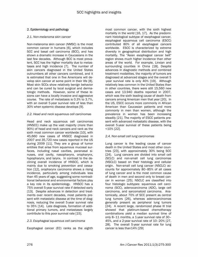
2. Epidemiology and pathology 2.1. Non-melanoma skin cancer Non-melanoma skin cancer (NMSC) is the most common cancer in humans [6], which includes SCC and basal cell carcinoma (BCC), and has shown a dramatic increase in Caucasians in the last few decades. Although BCC is most preva-lent, SCC has the higher mortality due to metas-tases and high incidence [7]. The number of skin cancers diagnosed in the United States outnumbers all other cancers combined, and it is estimated that one in five Americans will de-velop skin cancer at some point in their life [8]. Most skin SCCs show relatively benign behavior and can be cured by local surgical and derma-tologic methods. However, some of these le-sions can have a locally invasive and aggressive course. The rate of metastasis is 0.3% to 3.7%, with an overall 5-year survival rate of less than 30% when systemic disease develops [9]. 2.2. Head and neck squamous cell carcinomas Head and neck squamous cell carcinomas (HNSCC) make up the vast majority (more than 90%) of head and neck cancers and rank as the sixth most common cancer worldwide [10], with 45,660 new cases of HNSCC diagnosed in 2007 and 35,720 new cases reported in the US during 2009 [11]. They are a group of tumor entities that arise from squamous mucosal sur-faces, including nasal cavities, paranasal si-nuses, oral cavity, nasopharynx, oropharynx, hypopharynx, and larynx. In contrast to the de-clining overall incidence of HNSCC, which is mainly due to smoking prevention and cessa-tion [12], oropharynx carcinoma shows a rising incidence, particularly among individuals less than 45 years of age, suggesting some nontradi-tional behavioral and environmental factors play a key role in its epidemiology. HNSCC has a 75% overall 5-year survival rate if detected early [13]. Despite advances in detection and treat-ments over recent decades, most patients pre-sent with metastatic disease at the time of diag-nosis, reducing the overall 5-year survival rate to 35% [14]. Late diagnosis, formation of addi-tional primary tumors, and metastases largely contribute to this poor survival rate [15]. 2.3. Esophageal squamous cell carcinoma Esophageal cancer (EC) ranks as the eighth
most common cancer, with the sixth highest mortality in the world [16, 17]. As the predomi-nant histological subtype of esophageal cancer, esophageal squamous cell carcinoma (ESCC) contributed 80% of all esophageal cancers worldwide. ESCC is characterized by extreme diversity in geographical distribution and high mortality. The "Asian esophageal cancer belt" region shows much higher incidence than other areas of the world. For example, Linxian and surrounding counties in China [18]. Despite advances in diagnostic methods and combined treatment modalities, the majority of tumors are diagnosed at advanced stages and the overall 5-year survival rate is only 40% [19]. Although relatively less common in the United States than in other countries, there were still 15,560 new cases and 13.940 deaths reported in 2007, which was the sixth leading cause of death from cancers among American men that year [20]. In the US, ESCC occurs more commonly in African American than Caucasian patients and more commonly in men than women, although the prevalence in women has been increasing steadily [21]. The majority of ESCC patients pre-sent with advanced metastatic disease, with the overall 5-year survival of these patients being <10% [22]. 2.4. Non-small cell lung carcinomas Lung cancer is the leading cause of cancer death in the United States and most other coun-tries [23], with approximately 30% being SCC [24]. Lung cancers are divided into small cell (SCLC) and non-small cell lung carcinomas (NSCLC) based on their histology and cellular origin. Non-small cell lung cancer (NSCLC) ac-counts for approximately 80–85% of all cases of lung cancer and is the most common cause of death in men and second only to breast can-cer in woman [25]. NSCLC are classified into four histologic subtypes: squamous cell carci-noma (SCC), adenocarcinoma (ADC), large cell carcinoma, and sarcomatoid carcinoma. Ana-tomically, about 70% of SCC present as central lung tumors [26], whereas adenocarcinomas generally present as peripheral lung tumors [24]. A recent large, randomized phase III trial showed that platinum-based chemotherapy combinations yield a median survival time of only 8–11 months, a 1-year survival rate of 30–45%, and a 2-year survival rate of 10–20% [27, 28]. The overall 5-year survival rate for lung cancer is less than14% [29].
SCC highlights and insights
277 Am J Cancer Res 2011;1(3):275-300
2.5. Overall comparison Overall 5-year survival rates for the four major SCCs are among the lowest of the major can-cers. NMSC has an advantage over the other SCCs, as it is presented on the skin surface and not an internal organ; therefore, the chance of early detection is much greater and it is often cured by dermatologic and local surgical meth-ods. For all of the major SCCs, including NMSC, a common theme of late diagnosis, formation of additional primary tumors and metastases are associated with the poor survival rates pre-sented above. Regional recurrence after surgi-cal resection is also a contributing factor as is seen more commonly in NSCLC SCC than other histologic subtypes because SCC is able to spread by extending through periobronchial tubes which allow them to directly invade medi-astinal lymph nodes and other mediastinal
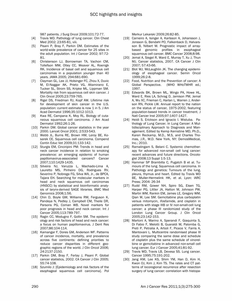
structures [26, 30]. Even though NMSC has an early detection advantage over the other SCCS, repeated exposure to risk factors will influence severity of disease progression and recurrence as equally as observed in the other major SCCs. 3. Risk factors The incidence of SCC shows marked variation in its distribution, suggesting that personal habits, environmental exposures, infections, and eth-nicity all play a role in the etiology of SCC (Table 1), with several of these risk factors influencing prognosis (Table 2). 3.1. Non-melanoma skin cancer Unlike other types of squamous cell carcinoma, NMSC is primarily caused by chronic long-term UV solar radiation exposure [31], in conjunction
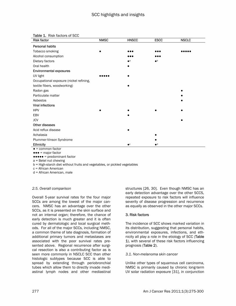
Table 1. Risk factors of SCC Risk factor NMSC HNSCC ESCC NSCLC
Personal habits Tobacco smoking ● ●●● ●●● ●●●●● Alcohol consumption ●●● ●●● Dietary factors ●a ●b Oral health ● Environmental exposures UV light ●●●●● ● Occupational exposure (nickel refining, textile fibers, woodworking) ● Radon gas ● Particulate matter ● Asbestos ● Viral infections HPV ● ● ● ● EBV ● JCV ● Other diseases Acid reflux disease ● Achalasia ● Plummer-Vinson Syndrome ● Ethnicity ●c ●d ● = common factor ●●● = major factor ●●●●● = predominant factor a = Betel nut chewing b = High-starch diet without fruits and vegetables, or pickled vegetables c = African American d = African American, male

SCC highlights and insights
278 Am J Cancer Res 2011;1(3):275-300
with the patient’s skin type. Fair-skinned indi-viduals who always burn and never tan are at a much higher risk for developing skin SCC than those with darker-skin [32], and it has been demonstrated that both sun exposure earlier in life and intense sun exposure appear to heavily predispose the populations to skin cancer [32]. Furthermore, human papilloma viruses (HPV) may be involved in the multi-step process of skin carcinogenesis as a co-factor with UV-radiation [33], especially in patients with poor immune status such as organ transplant recipi-ents [34]. And, smoking tobacco may double the risk of skin cancer [35], thus although the effect is not as great as in other SCCs, smoking plays a role in the development of NMSC. 3.2. Head and neck squamous cell carcinomas Contrary to NMSC, alcohol and tobacco use are the most common risk factors for HNSCC in the US, although they have not been associated with survival [36]. Moreover, alcohol and to-
bacco are likely synergistic in causing cancer of the head and neck [37]. Cigarette smokers have a lifetime increased risk for head and neck cancers which is 5- to 25-fold increased over the general population [38], and smoking ces-sation does not eliminate the risk of cancer de-velopment [39]. In addition, environmental ex-posure to tobacco smoke also increases the risk of developing HNSCC, even for individuals who have never actively smoked [40]. Heavy alcohol consumption is also an independent risk factor for HNSCC, particularly for cancers of the hypo-pharynx [41]. Moreover, smokers and alcohol drinkers are at risk for the development of sec-ond primary oral cancers [42]. Interestingly, even in the presence of alcohol consumption or tobacco use, a high intake of fruit and vegeta-bles may prevent the development of a quarter of HNSCC and possibly one half of oral and oralphyrengeal SCC [43]. Causation has been shown with viral infection for HNSCC and the association varies based on the site of the tu-mor. For example, human papilloma virus
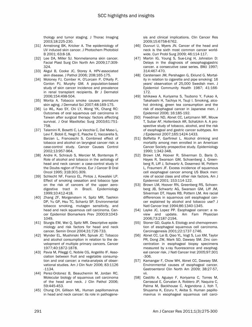
Table 2. Prognostic indicators and markers of SCC NMSC HNSCC ESCC NSCLC
Risk factors HPV ●
Molecular markers TP53/p53 ● ● EGFR ● ● ● Ki-67 ● ● ● ● p63 ● ● ● ● VEGF ● ● SOX2 ● Smad6/7 ●[229] CDH1 ● ● CD44v6 ●[230] MMPs ● ● ● Trop2 ●[231] EpCAM ●[232] HER2 ●[233] ●[234] CCND1 ● ● ● ● CCND1 + p53 mutation ● Bcl-x ●[235] Bcl-2 ● ● Bax ●[236] p16 ● CD24 ● IGFR-1 + EGFR ● RASSF1A ● miR-21 ● miR-211 ●

SCC highlights and insights
279 Am J Cancer Res 2011;1(3):275-300
(HPV), in particular HPV16, shows the highest distribution in the tonsils [44], while Epstein-Barr virus (EBV) infection is associated with na-sopharyngeal cancer. HPV is associated with 20–25% of HNSCC, and individuals with HPV-positive tumors have a better overall survival compared to those with HPV-negative tumors [45, 46]. Specifically, the presence of HPV-16 is now recognized as a highly favorable prognostic indicator for patients with HNSCC [45]. Betel quid chewing, a common habit in some regions of Asia and some Asian communities in the western world is considered a regional risk fac-tor for cancers with a poorer prognosis [36]. In addition, oral health, acid reflux disease and environmental exposures (nickel refining, textile fibers, and woodworking) are also related to HNSCC tumorigenesis. 3.3. Esophageal squamous cell carcinoma Similar to HNSCC, smoking and alcohol inges-tion are major etiologic factors for the develop-ment of ESCC [47]. Studies have shown that ESCC risk is increased approximately three- to seven-fold in current smokers [48-50] and three- to five-fold in heavy alcohol users [50-53], with additional associations between esophageal irritants such as lye ingestion, rapidly consumed high-starch diets without fruits and vegetables, and radiation therapy [47]. There also may be a causal relationship between ESCC and previous diseases such as achalasia, head and neck can-cer, and Plummer-Vinson syndrome [54]. Pick-led vegetable intake and micronutrient defi-ciency (such as zinc) may contribute to ESCC formation in some parts of China, especially in light of laboratory experiments demonstrating that high tissue zinc concentration is strongly associated with a reduced risk of developing ESCC in experimental animals [55, 56]. There are also other potential but as yet unsubstanti-ated risk factors like PAHs and acetaldehyde related to ESCC in China [57], with HVP-16 and HPV-18 reported to be risk factors [55, 58]. In the US, ESCC incidence is highest in African Americans and males. Interestingly, one cohort study of ESCC and esophageal adenocarcinoma (EA) found decreased risk of ESCC, but not EA, was associated with higher intake of both fruit and vegetables [59]. 3.4. Non-small cell lung carcinomas The causal relationship between smoking and lung cancer is well established with a 10- to 20-
fold increased risk of lung cancer in smokers compared with never smokers [60] and is the major risk factor for the development of NSCLC SCC [61, 62]. In the US, smoking is estimated to account for 87% of lung cancer cases (90% in men and 85% in women) [63]. The lifetime risk of developing lung cancer is approximately 17.2% for male smokers and 11.6% for female smokers, and this risk is significantly lower in nonsmokers: 1.3% in men and 1.4% in women [64]. Healthy ex-smokers have been shown to have a similar gene expression pattern in nor-mal bronchial epithelium as non-smokers, indi-cating that most smoking-induced gene expres-sion changes revert to normal levels after smok-ing cessation [65]. Aside from smoking, NSCLC is also related to genetic factors [66], radon gas [67], asbestos [68], and air pollution [69]; with Radon exposure reported as the second major cause of lung cancer after smoking [67]. There is a synergistic effect between tobacco smoking and asbestos exposure in the formation of lung cancer [68], and more recently HPV [70], JC virus [71] and cytomegalovirus [72] have been reported as additional potential risk factors for NSCLC. 3.5. Overall comparison Tobacco smoking and HPV infection appear to be carcinogenic causes for all four sub-types. In addition, several risk factors are shared among the major SCC types. HNSCC and ESCC share the most factors, consistent with their histologi-cal relationship, including alcohol consumption dietary factors, and ethnicity. Unlike the other SCCs, UV exposure is the major risk factor for NMSC. Although numerous etiologic factors for SCC are known, the exact roles and molecular mechanisms of action have not been fully eluci-dated.
4. Molecular characteristics and prognostic markers The development of clinically evident SCC is a multistep process involving the accumulation of multiple genetic alterations modulated by ge-netic predisposition, known risk factors, and other unknown environmental influences. The alterations are typically oncogene activation, including recessive oncogenes [73] and tumor suppressor gene (TSG) inactivation via muta-tions, loss of heterozygosity, deletions, or other mechanisms (e.g. methylation and miRNA modulation of gene expression) [74]. Molecular

SCC highlights and insights
280 Am J Cancer Res 2011;1(3):275-300
profiling studies that began with single or rela-tively small groups of genes or proteins have now progressed to large-scale and high-throughput methods using DNA-, RNA-, and pro-tein-based approaches. These large-scale methods analyze thousands of genes at one time and have led to a better understanding of the complexity of gene abnormality patterns of SCC and have accelerated the discovery of novel genes involved in SCC pathogenesis. In addition to conventional prognostic factors [75], these molecular characteristics are becoming increasingly valuable as biomarkers in adjunct prognostic tools. There are numerous molecular markers that have been identified in SCC, and in this section we compare and contrast the major molecular abnormalities and their prog-nostic value among the four major SCCs. 4.1. Molecular markers common to NMSC, HNSCC, ESCC, and NSCLC Different patterns of molecular changes and their role in prognosis have been shown be-tween the four major SCCs; however, several key similarities are present including abnormali-ties in TP53, p63, Ki-67, CCND1, EGFR, and COX2 (Table 2, Table 3). TP53 TP53 is one of the most important tumor sup-pressor genes in humans [76] and functions as a transcriptional regulator that controls the ex-pression of genes involved in the cell cycle, DNA repair, apoptosis, and senescence. Under stress conditions, p53 is activated and triggers a variety of cellular responses needed to main-tain the integrity of the genome, thus the pro-tein has been designated a guardian of the ge-nome. p53 mutations can lead to inactivation of p53 and have been found in a broad spec-trum of human cancers [77], including NMSC, HNSCC, ESCC, and NSCLC. Inactivation of p53 is considered a critical step in the development of NMSC [78]; however, TP53 mutation has not been correlated with aggressiveness of SCC, indicating the involvement of subsequent mo-lecular events that determine tumor behavior [79]. TP53 alterations and its loss of function is a characteristic early change in HNSCC [80]. In HNSCC, it has been observed that TP53 muta-tions that occur within the core domain com-pletely blocking DNA binding are linked to accel-erated tumor progression, reduced therapeutic
responsiveness, and decreased patient survival compared to tumors that harbor less disruptive TP53 mutations [81, 82]. In ESCC, p53 muta-tion has been frequently identified [83, 84] and its function positively correlated with MDM2 and p14(ARF) expression [85]. Overexpression p53 has also been significantly correlated with poorer prognosis for ESCC [86]. In NSCLCs, TP53 mutations have been detected in 40-90% of resected tumors [87-89], with disruption of the TP53 pathway frequently observed in SCC [73]. TP533 mutations can produce chemo-therapy resistance [90]; however, the specific type of mutation and sensitivity to chemother-apy agents has not been identified [88, 91]. As described, TP53/p53 abnormalities are present in all four major SCCs, but to date is only consid-ered a prognostic factor for HNSCC and ESCC. p63 p63, a member of the p53 family, is critical for the development of stratified epithelial tissues such as epidermis [92] and is usually limited to the proliferative (basal layer) compartment of the epithelium [93]. p63 expression has been reported to be a strong predictor of poorly differ-entiated NMSC [94]. Whereas, expression of p63 is frequently (>95%) observed in HNSCC and associated with increased survival [95-98]. Hence, p63 may be involved in squamous cell carcinoma formation through various paths. p63 alteration is also seen in ESCC and reduced expression of p63 has prognostic implications for patients [99]. In NSCLC SCC, p63 is used to differentiate SCC from adenocarcinoma be-cause a diffuse strong p63 and CK5/6 immuno-expression is essentially restricted to SCC [24]. Interestingly, in NSCLC high expression of CCND1 [100] and CD24 [101] are associated with a worse clinical outcome, while expression of p63 [102] and BCL-2 [103] have a positive prognostic value. In contrast to TP53, p63 ab-normalities in the four major SCCs are consid-ered prognostic for SCC. Ki-67 (MKI67) Ki-67 is a cell proliferation index marker typi-cally increased in tumors and found related to rapid growth and recurrence in NMSC [104]. In HNSCC, co-expression of p21/Ki-67 is a strong negative prognostic factor [105]. In patients with stage II and III advanced ESCC, a signifi-cant correlation has been identified between
SCC highlights and insights
281 Am J Cancer Res 2011;1(3):275-300
Ser392 phosphorylation of p53 and high levels of Ki-67, lymphatic invasion, and poorer progno-sis [106]. Similarly, one cohort study found 97% of NSCLC samples expressed Ki-67 and overexpression was associated with significantly shorter survival [107]. Similar to p63, Ki-67 abnormalities are prognostic for the four major SCCs. But in contrast to p63, Ki-67 abnormali-ties all appear to be indicative of poor prognosis in all four major SCCs. CCND1 CCND1, a cell cycle regulator, acts by phos-phorylating and inactivating the retinoblastoma protein [108]. In NMSC, CCND1 is involved in the early development of SCC via abnormal tis-sue organization and differentiation [109], with overexpression frequently seen in keratinocyte carcinogenesis [110-112]. Interestingly, CCND1 overexpression coupled with TP53 mutation has been correlated with poorer prognosis in NMSC [110-112]. In HNSCC, CCND1 polymorphism (GG) in exon 4 has been shown to be an inde-pendent prognostic indicator of disease-free interval [113]. In ESCC, CCND1 is overex-pressed in 23 to 73% of tumor samples [55, 114] and is significantly correlated with poorer prognosis [115, 116]. Contrary to CCND1 in ESCC, the absence of CCND1 immunoexpres-sion in NSCLC is associated with worse progno-sis [117]. Similarly to p63, CCND1 is associated with poorer prognosis in NMSC, ESCC, and NSCLC, but is conversely associated with im-proved prognosis in HNSCC. EGFR EGFR is present in the cell membrane as a monomer and is activated by ligand binding to the extracellular domain [118]. Mutations that lead to EGFR overexpression have been associ-ated with a number of cancers. For example, some studies identified EGFR overexpression in metastatic SCC of the skin [119-121]. EGFR was also shown to be up-regulated/overexpressed in 90% of HNSCCs and associ-ated with local recurrence and poor survival [122, 123]. Overexpression of EGFR [124, 125], is significantly correlated with poorer prognosis for ESCC. SCCs demonstrate most of the genetic abnormalities commonly present in NSCLCs, except for KRAS and EGFR gene muta-tions, which are more frequent in adenocarcino-mas [73, 126]. Overexpression of EGFR, p53
[127-129] and Her2 [130, 131] has shown con-flicting results in NSCLC and their use as prog-nostic markers require further study [132-136]. However, two prognostic proteins in NSCLC have been identified and are used together. NSCLCs having both IGFR-1- and EGFR-positive immunoreactivity represent a subpopulation capable of developing aggressive clinical behav-ior [137]. Again, similarly to p63, EGFR is asso-ciated with poorer prognosis in NSCM, HNSCC and ESCC. But due to conflicting reports, the use of p63 as prognostic marker in NSCLC needs to be investigated further. COX2 COX2 (PTGS2) is an enzyme that functions in protein metabolism by increasing prostaglandin synthesis and plays a role in tumorigenesis. The expression of COX-2 is upregulated in many cancers and its product, PGH2, is converted by prostaglandin E2 syntheses into PGE2 which in turn can stimulate cancer progression [138]. For example, in NMSC, high expression of COX-2 has been found in AK, SCC and BCC [139] and COX-2 expression increases during progression from AK to SCC [140]. Overexpression of COX2 has also been shown in HNSCC, and some ex vivo studies have demonstrated that COX-2 is overexpressed in ESCC and premalignant le-sions [141, 142]. Statistically significant COX-2 overexpression has also been found in 28.9 % of NSCLC SCC [143]. Overexpression of COX-2 and upregulation of the prostaglandin pathway plays a significant role in SCC and blockade of the process has strong potential for cancer pre-vention and therapy. However, to date, COX2 has not been identified as a prognostic marker of SCCs. 4.2. Other key molecular markers shared in several SCCs In addition to the shared cell cycle regulation (TP53, p63, Ki-67, and CCND1), signal trans-duction (EGFR), and protein metabolism (COX2) molecular abnormalities, several other molecu-lar abnormalities of gene expression, protein expression, gene mutation, and epigenetic regu-lation and their respective prognostic values have been characterized in SCCs (Table 2, Table 3). Key markers shared in two or three of the major SCCs involve several classes of genes including signal transduction (VEGF), transcrip-tion factor (SOX2), cell adhesion (CDH1), and

SCC highlights and insights
282 Am J Cancer Res 2011;1(3):275-300
extracellular matrix degradation (MMPs). Addi-tional markers affecting only one of the SCCs can be found in Table 2 and Table 3. VEGF VEGF is an important signal transduction pro-tein involved in both vasculogenesis and angio-genesis. Four subtypes have been described (A, B, C, and D). VEGF alteration is involved in HNSCC and ESCC. In oral SCC, it was reported that overexpression as measured by immuno-histochemistry of subtypes A and B were corre-lated with tumor angiogenesis and subtypes C and D with metastases and poor prognosis [144]. In ESCC, VEGF (VEGF A) expression has historically ranged between 24-93% [145], and recently elevated immunoexpression has been reported in 55% of ESSC tissues (n= 108) [146]. Overexpression of VEGF has been signifi-cantly correlated with poorer prognosis of ESCC [145]. SOX2 A recently discovered lineage-survival oncogene, SOX2, has been shown to be important in HNSCC, ESCC, and NSCLC. Lineage survival oncogenes are activated by somatic mutations and thus may play an important role in carcino-genesis. The genomic amplification of the SOX 2 embryonic stem cell transcription factor lo-cated on chromosome 3q26.33 was first re-ported in ESCC and NSCLC [147]. SOX2 has been associated with poor prognosis in ESCC [148], but its use as a prognostic marker in NSCLC has not yet been elucidated. In HNSCC (oral), high protein expression of SOX2 has been found to correspond to copy number gain in 52% of oral SCC tumors [149]. miR-145 is in-volved in regulating SOX2 [150]; however, its role as a prognostic factor in SCCs is still un-known. Our group has recently characterized SOX-2 pro-tein expression related to the pathogenesis of NSCLC SCC [151]. By assessing SOX2 mRNA expression in various published datasets against the previously characterized OCT4/SOX2/NANOG signature, we were able to effec-tively separate SCCs from adenocarcinomas. In this study, we further characterized SOX immu-noexpression of NSCLC tissues and identified SOX2 protein expression pattern in SCC devel-opment (hyperplasia, dysplasia, and carcinoma
in situ) [151]. CDH1 (E-cadherin) CDH1 belongs to the cadherin family of Ca 2+ -dependent cell-cell adhesion molecules which induce and maintain intercellular connections. CDH1 has been implicated in carcinogenesis due to reduced expression [152] and promoter hypermethylation [153]. In NMSC, down-regulation of CDH1 is linked to increased poten-tial for tumor invasiveness and distant metasta-sis and the frequencies of CDH1 promoter hy-permethylation appear to be correlated with a more advanced stage of squamous carcino-genesis in skin [153]. In ESCC, tumors with reduced CDH1 expression invade deeper, have more lymph node metastasis, and have more lymphatic invasion than tumors with preserved CDH1 expression [154]. Disorganized CDH1 expression was also reported to be a feature of advanced ESCC [152]. MMPs Matrix metalloproteinases (MMPs) are a family of zinc-containing endopeptidases that degrade various components of the extracellular matrix, and have been strongly implicated in multiple stages of cancer progression including the ac-quisition of invasive and metastatic properties. MMPs are altered in NMSC, HNSCC and ESCC. In NMSC, MMP-2 and MMP-9 immunoexpres-sion is associated with NMSC pathogenesis and is an indicator of cutaneous cancer invasion and progression [155]. Expression of MMPs has been identified in both the epithelial and stromal elements of HNSCC [156], with MMP2 and MMP9 showed an association with invasive potential [157, 158]and poor outcome [159]. Gu et al. evaluated the expression of MMP1, MMP7, MMP9, and MMP13 in ESCC tissues from 208 patients and found that MMP7, MMP9, and MMP13 may be involved with early stage ESCC, and their coexpression predicted poor outcome for ESCC patients [160]. These data are consistent with the previous finding that expression of MMP-7 and MMP-9 may be a good marker for the presence of lymphatic me-tastasis in ESCC [161]. Moreover, our group identified MMP3 and MMP10 as potential early diagnosis biomarkers for ESCC. Both enzymes showed a tumor increase at both the transcrip-tional and proteomic levels [162].
SCC highlights and insights
283 Am J Cancer Res 2011;1(3):275-300
Gene mutations Gene mutations other than those in TP53, EGFR, and CCND1 have also been shown to be important in SCCs, with a large proportion of mutations having been identified in NSCLC and to a less extent HNSCC. Functional inactivation of p16 through deletion frequently occurs in HNSCC [163]. Chromosome 3p contains nu-merous TSGs (e.g. ALS2CL, EPHA3 and CMYA1 ) and the loss of heterozygosity in this region may contribute to SCCs, including HNSCC [164] and NSCLC [165]. Activating RAS mutations occur in approximately 15% to 20% of NSCLC, with a majority of them being KRAS [166], however these mutations are rare in SCC [167]. Also, c-MET, TTF-1, LKB1, BRAF and PIK3CA are often mutated or amplified in NSCLC [167]. Methylation The epigenetic mechanism of methylation has been shown to modulate of gene expression in SCCs. Since its discovery as a cyclin-dependent kinase inhibitor in 1993, the tumor suppressor p16 (INK4A/MTS-1/CDKN2A) has gained wide-spread importance in cancer [168]. Like NMSC, gene methylation has also been identified in HNSCC, ESCC, and NSCLC to varying degrees. p16 inactivation often occurs via methylation in both HNSCC [163] and ESCC [169]. This silenc-ing of p16 in ESCC has been shown to lead to deregulation of cell proliferation and conse-quent genomic instability [169]. Different pat-terns of gene methylation have been found in the major histological types of NSCLCs, with LKB1 and RASSF1 being the most important in NSCLC SCC. Inactivation of the TSG LKB1 by mutation and deletion is a relatively frequent event in SCC (19%) of the lung [170]. But, RAS association domain family 1 gene (RASSF1) is the most frequently hypermethylated gene in NSCLC. RASSF1A methylation has been corre-lated with worse prognosis in surgically resected NSCLC patients and was confirmed as an inde-pendent prognostic factor by multivariate analy-sis [171]. miRNAs miRNAs are a class of small (~18–24 mer) nu-cleic acids that negatively regulate gene expres-sion. Through their targets, miRNAs are known to play important roles in cell differentiation, proliferation, and apoptosis, and altered miRNA
levels result in the aberrant expression of gene products that may contribute to cancer biology [172, 173]. An emerging number of studies have shown that miRNAs can act as oncogenes, as tumor suppressor genes, or sometimes as both [174]. High-throughput analyses have demonstrated that miRNA expression is com-monly dysregulated in human cancer [173]. However, considerable disagreement remains with respect to the miRNA signature for specific cancer cell types, which appears to depend largely on the analytical platform [173]. Dysregulation of miRNAs have been identified in HNSCC, ESCC, and NSCLC and not NMSC. How-ever, Drosha, an important enzyme in the miRNA machinery, has been found to be overex-pressed in NMSC; thus giving strength to the hypothesis of miRNA involvevement in NMSC carcinogenesis [175]. In HNSCC, overexpres-sion of miR-211 has been associated with inva-sive potential [176]. In addition to miR-211, miRNA profiling has revealed four upregulated miRNAs (miR-21, -31, -18, and -221) and 13 down-regulated miRNAs (miRNA-133a, -133b,-125a, -138, -139, -200c, -26b, -302b, -302c, -342, -371, -373, -375) associated with HNSCC [177]. Moreover, the ratio of miR-221:miR-375 showed a high discriminatory potential, with a sensitivity of 92% and specificity of 93% in dis-tinguishing tumor from normal tissue, suggest-ing that this simple molecular marker may hold significant clinical potential as a diagnostic tool [178]. miR-21 has also been reported in ESCC and found to induce cell proliferation and inva-sion [179]. Our group evaluated ESCC related miRNAs by comparing microdissected cells in-volved in normal differentiation and tumorigene-sis and confirmed that miR-21 was overex-pressed in tumors (Zhu, et al, submitted). miRNAs have been shown to regulate several important pathways in NSCLC and have been correlated with disease outcome in NSCLC [180-182]. Of interest, a five miRNA signature (let-7a, miR-221, miR-137, miR-372, and miR-182) has been identified in NSCLC that predicts treat-ment outcome [182]. In that study, patients with high risk scores in their miRNA signatures showed poor overall and disease-free survivals compared with patients with low risk scores [182]. Loss of expression of miRNA-128b, puta-tive regulator of EGFR, correlated with response to targeted EGFR inhibition in primary NSCLC [183]. miRNA is an area of very active research that will have an impact on pathogenesis and
SCC highlights and insights
284 Am J Cancer Res 2011;1(3):275-300
therapy as more is learned about the role of miRNAs in SCC. Numerous molecular abnormalities in gene ex-pression, protein expression, gene mutation, and epigenetic regulation have been character-ized in SCC (Table 3), with several of these markers associated with disease prognosis (Table 2). Commonalities in molecular changes present in the four major SCCs are predomi-nantly found in cell cycle regulation and signal transduction. Although not all of the described molecular abnormalities are shared in all four of the SCCs, many are shared in at least two or three of the SCCs. The comparison of molecular characteristic similarities and differences in SCC provide insight not only into the relation-ships between NMSC, HNSCC, ESCC, and NSCLC, but to SCC as a whole. And this insight into SCC can putatively be translated to im-proved disease control and treatment. Cur-rently, drugs targeting several of these gene and pathway abnormalities are being used in the treatment of SCC. 5. Targeted therapy Targeted therapy is a type of treatment that uses drugs that identify and attack specific can-cer cells without harming normal cells and has been extensively investigated in recent dec-ades, both as a single modality therapy and in combination with cytotoxic treatments such as radiotherapy or chemotherapy. With the grow-ing understanding of molecular genetics of SCC, targeted therapies now offer expanded treat-ment options for patients that have clinically significant benefits. The targets that are cur-rently considered the most relevant in SCCs fall into one of the following categories: cell-cycle regulation, signal transduction, growth factor receptors, angiogenesis, and protein degrada-tion. Similar to the fact that there are few prognostic markers for NMSC, there are currently no tar-geted drugs developed for NMSC. Although not developed as a targeted therapy, Diclofenac, a dual inhibitor of COX-1 and COX-2 with higher selectivity for COX-2, has been investigated and reported to be effective for patients with AK, a precancerous syndrome of skin [184]. Several targeted therapies are being investi-gated for HNSC, ESCC, and NSCLC with many of
the molecular targets being shared among the three SCCs (Table 4). Targeted therapies have been extensively investigated in HNSC and NSCLC, especially EGFR TKIs and their mecha-nisms of resistance. 5.1. Growth factor receptor antagonists EGFR EGFR is a member of the ERBB family of trans-membrane tyrosine kinase receptors [132]. EGFR and their receptors are involved in signal transduction and tumor growth, thus blockade of these systems provides a therapeutic ap-proach, through neutralizing ligands, inhibiting ligand binding, or blocking the tyrosine kinases of the receptors. Examples of EGFR inhibitors include monoclonal antibodies against the ex-tracellular domain of the receptor (e.g., cetuxi-mab and panitumumab) and receptor tyrosine kinase inhibitors (TKIs) that target the intracellu-lar domain (e.g., gefitinib and erlotinib). EGFR inhibitors have been applied in HNSCC, ESCC and extensively in NSCLC clinical trials. In HNSCC, patients with locally advanced disease have been shown to benefit from the addition of EGFR inhibition (for example, cetuximab) to ra-diotherapy [185]. But, EGFR targeted therapy trials conducted in HNSCC to date still show various disadvantages such as low efficacy and significant toxicity [186]. In ESCC, inhibition of EGFR-TK by erlotinib is promising through induc-ing growth inhibition and cell cycle arrest in hu-man esophageal cancer cells and enhancing the antineoplastic effects of other targeted agents [187]. Cetuximab and panitumumab are currently being investigated in NSCLC. Phase II trials showed that cetuximab improved survival in-chemo-naïve patients with advanced cancer [188, 189] and panitumumab is currently in phase II trial [190]. EGFR TKIs gefitinib and erlotinib were the first two targeted agents recently approved for the treatment of NSCLC in the United States and several markers have been identified that pre-dict response in NSCLC patients. These EGFR TKIs produce responses in approximately 10% of NSCLC patients having progressed with prior chemotherapy [191-193]. But in those patients who benefit from gefitinib or erlotinib the re-sponses can be dramatic and may last for longer than a year, with favorable response be-ing associated with activating mutations in the
SCC highlights and insights
285 Am J Cancer Res 2011;1(3):275-300
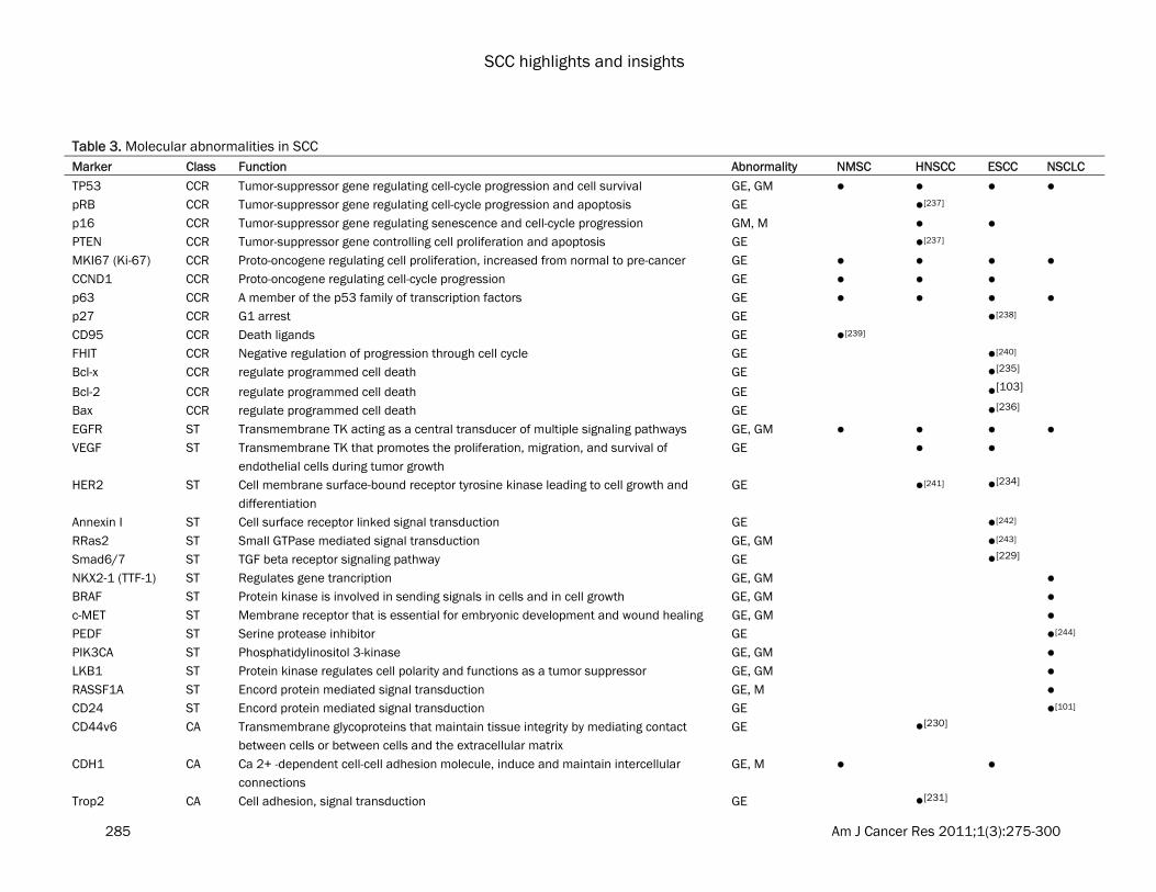
Table 3. Molecular abnormalities in SCC Marker Class Function Abnormality NMSC HNSCC ESCC NSCLC
TP53 CCR Tumor-suppressor gene regulating cell-cycle progression and cell survival GE, GM ● ● ● ● pRB CCR Tumor-suppressor gene regulating cell-cycle progression and apoptosis GE ●[237] p16 CCR Tumor-suppressor gene regulating senescence and cell-cycle progression GM, M ● ● PTEN CCR Tumor-suppressor gene controlling cell proliferation and apoptosis GE ●[237] MKI67 (Ki-67) CCR Proto-oncogene regulating cell proliferation, increased from normal to pre-cancer GE ● ● ● ● CCND1 CCR Proto-oncogene regulating cell-cycle progression GE ● ● ● p63 CCR A member of the p53 family of transcription factors GE ● ● ● ● p27 CCR G1 arrest GE ●[238] CD95 CCR Death ligands GE ●[239] FHIT CCR Negative regulation of progression through cell cycle GE ●[240] Bcl-x CCR regulate programmed cell death GE ●[235]
Bcl-2 CCR regulate programmed cell death GE ●[103] Bax CCR regulate programmed cell death GE ●[236] EGFR ST Transmembrane TK acting as a central transducer of multiple signaling pathways GE, GM ● ● ● ● VEGF ST Transmembrane TK that promotes the proliferation, migration, and survival of GE ● ● endothelial cells during tumor growth HER2 ST Cell membrane surface-bound receptor tyrosine kinase leading to cell growth and GE ●[241] ●[234] differentiation Annexin I ST Cell surface receptor linked signal transduction GE ●[242] RRas2 ST Small GTPase mediated signal transduction GE, GM ●[243] Smad6/7 ST TGF beta receptor signaling pathway GE ●[229] NKX2-1 (TTF-1) ST Regulates gene trancription GE, GM ● BRAF ST Protein kinase is involved in sending signals in cells and in cell growth GE, GM ● c-MET ST Membrane receptor that is essential for embryonic development and wound healing GE, GM ● PEDF ST Serine protease inhibitor GE ●[244] PIK3CA ST Phosphatidylinositol 3-kinase GE, GM ● LKB1 ST Protein kinase regulates cell polarity and functions as a tumor suppressor GE, GM ● RASSF1A ST Encord protein mediated signal transduction GE, M ● CD24 ST Encord protein mediated signal transduction GE ●[101] CD44v6 CA Transmembrane glycoproteins that maintain tissue integrity by mediating contact GE ●[230] between cells or between cells and the extracellular matrix CDH1 CA Ca 2+ -dependent cell-cell adhesion molecule, induce and maintain intercellular GE, M ● ● connections Trop2 CA Cell adhesion, signal transduction GE ●[231]
SCC highlights and insights
286 Am J Cancer Res 2011;1(3):275-300
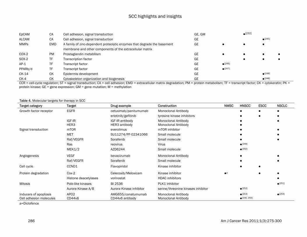
EpCAM CA Cell adhesion, signal transduction GE, GM ●[232] ALCAM CA Cell adhesion, signal transduction GE ●[245] MMPs EMD A family of zinc-dependent proteolytic enzymes that degrade the basement GE ● ● ● membrane and other components of the extracellular matrix COX-2 PM Prostaglandin metabllism GE ● ● ● ● SOX-2 TF Transcription factor GE ● ● ● AP-1 TF Transcript factor GE ●[246] PPARb/d TF Transcript factor GE ●[247] CK-14 CK Epidermis development GE ●[248] CK-4 CK Cytoskeleton organization and biogenesis GE ●[248] CCR = cell-cycle regulation; ST = signal transduction; CA = cell adhesion; EMD = extracellular matrix degradation; PM = protein metabolism; TF = transcript factor; CK = cytokeratin; PK = protein kinase; GE = gene expression; GM = gene mutation; M = methylation
Table 4. Molecular targets for therapy in SCC
Target category Target Drug example Construction NMSC HNSCC ESCC NSCLC
Growth factor receptor EGFR cetuximab/panitumumab Monoclonal Antibody ● ● ● erlotinib/gefitinib tyrosine kinase inhibitors ● ● ● IGF-IR IGF-IR antibody Monoclonal Antibody ● ● HER3 HER3 antibody Monoclonal Antibody ● Signal transduction mTOR everolimus mTOR inhibitor ● ● MET SU11274/PF-02341066 Small molecule ● ● Raf/VEGFR Sorafenib Small molecule ● ● Ras reovirus Virus ●[249] MEK1/2 AZD6244 Small molecule ●[250]
Angiogenesis VEGF bevacizumab Monoclonal Antibody ● ● Raf/VEGFR Sorafenib Small molecule ● ●
Cell cycle CCND1 Flavopiridol Kinase inhibitor ● ●
Protein degradation Cox-2 Celecoxib/Meloxicam Kinase inhibitor ●a ● ● Histone deacetylases vorinostat HDAC inhibitors ●
Mitosis Polo-like kinases BI 2536 PLK1 inhibitor ●[251] Aurora Kinase A/B Aurora Kinase inhibitor serine/threonine kinases inhibitor ●[252]
Inducers of apoptosis APO2 AMG655/conatumumab Monoclonal Antibody ●[253] ●[254] Cell adhesion molecules CD44v6 CD44v6 antibody Monoclonal Antibody ●[230, 255]
a=Diclofence

SCC highlights and insights
287 Am J Cancer Res 2011;1(3):275-300
EGFR tyrosine kinase domain (exons 18 to 21), increased gene copy number, and increased protein expression [191-194]. Even though targeting EGFR mutated NSCLCs with gefitinib or erlotinib has been effective, most of these patients acquire resistance to the EGFR TKI therapy [195, 196] in an average of 6-12 months [197]. Interestingly, smokers presenting with NSCLCs are generally resistant to EGFR-TKIs [198, 199], which may have implications in other targeted therapies of this class because SCC is the predominant histologic subtype associated with smokers. In an effort to counteract EGFR TKI resistance mechanisms, an initial study has shown TKI resistant NSCLC cell lines can be treated by administering PI3K-mTOR and MEK signaling inhibitors simultaneously with EGFR TKIs [200]; but, to date this has not been tested in NSCLC patient populations. Other growth factor receptor antagonists IGF-I acts though the IGF receptor 1 (IGF- R1) to promote cell survival and cell proliferation [201]. It plays an important role in normal growth and has anabolic effects in adults nor-mally. But IGF-I has been implicated in cancers [202] as its anti-apoptotic properties allow can-cerous cells to resist the cytotoxic effects of chemotherapeutic drugs or radiotherapy. A re-cent clinical trial reported that combining the fully humanized anti–IGF-IR monoclonal anti-body A12 with radiation to treat HNSCC resulted in more pronounced antitumor activity than ei-ther agent alone [203]. In NSCLC, early clinical trials showed an acceptable safety profile to-gether with pharmacodynamic evidence that IGF-RI can be successfully targeted [204]. And, biomarkers of the IGF-IR pathway were shown to be key elements in development and monitoring of anti-IGF-RI therapy in NSCLC [205]. Recently, phase II study data has suggested that co-administration of an anti-IGF-IR antibody with chemotherapy improves the response rate and progression-free survival [204]. HER3 (ERBB3), a gene encoding a member of the epidermal growth factor receptor (EGFR) family of receptor tyrosine kinases has emerged for HNSCC targeted therapy within the past four years [206, 207]. Several laboratories are de-signing antibodies that will block HER3 het-erodimerization with EGFR or HER2 to prevent signaling to PI3K/Akt [208].
5.2. Cell-cycle regulation Although cell-cycle regulation contains the most molecular abnormalities in SCC (Table 3) and subsequently numerous potential therapeutic biomarkers, currently only one marker, CCND1, has been exploited. Mutations, amplification and overexpression of CCND1 are frequently observed in a variety of tumors, including SCCs, and may contribute to tumorigenesis. Flavopiri-dol, the first cyclin-dependent kinase inhibitor in human clinical trials, was reported as a target-ing drug for HNSCC [209]. Flavopiridol has also been shown to decrease CCND1 expression in ESCC cell lines and was subsequently found to induce radiosensitivity [210] and may have ap-plication to the other SCCs.
5.3. Signal transduction Signal transduction was the second most com-mon group of molecular abnormalities in the four major SCCs (Table 3). Of which, both of mTOR and MET have been identified in HNSCC and NSCLC as molecular targets. mTOR is a serine/threonine protein kinase that regulates cell growth, cell proliferation, cell motility, cell survival, protein synthesis, and transcription. It is activated by Akt and blocks apoptosis to in-crease the proliferative potential of cancer cells. Several mTOR inhibitors are currently under investigation in HNSCC [211]. In an ongoing phase II trial, combined inhibition of mTOR with everolimus and gefitinib was evaluated in pa-tients with stage IIIB/IV NSCLC [212]. Borte-zomib is a small-molecule proteasome inhibitor that has shown encouraging results in a phase II trial, and a phase III trial of gemcitabine/carboplatin ± bortezomib in advanced stage NSCLC is in progress [213]. C-MET is the cell surface receptor for hepato-cyte growth factor (HGF), also known as scatter factor [214]. Binding of the receptor to its ligand, hepatocyte growth factor, induces recep-tor dimerization that triggers conformational changes that activate MET tyrosine kinase activ-ity which then have profound effects on cell growth, survival, motility, invasion and angio-genesis [215]. Dysregulation of MET signalling has been shown to contribute to tumorigenesis in a number of malignancies and hence can serve as a potential drug target. Zucali, et al. found that activated cMET appeared to be a marker of primary gefitinib resistance in NSCLC

SCC highlights and insights
288 Am J Cancer Res 2011;1(3):275-300
patients and suggested cMET may be a target for treatment [216]. In NSCLC, several phase II/III tiral with PF-02341066 either as monother-apy or in combination with EGF-R inhibitors are currently underway [217]. In addition, preliminary data from a phase II trial testing of sorafenib, a potent inhibitor of the Raf-1, B-Raf, VEGFR-2-3, and PDGFR-B pathways, in metastatic or recurrent HNSCC were recently reported [218]. Sorafenib treatment for NSCLC is being evaluated in several phase III studies [217]. 5.4. Protein degradation Cox-2 has been implicated in apoptosis resis-tance, angiogenesis, decreased host immunity and enhanced invasion and metastasis, and, thus is involved in critical aspects of carcino-genesis [219]. COX-2 selective inhibitors, a form of non-steroidal anti-inflammatory drug (NSAID) that directly targets COX-2, has been shown to reduce the occurrence of cancers and pre-cancerous growths [220] and is in clinical trials for both ESCC and NSCLC [219]. Histone deacetylase inhibitors (HDIs) have been shown to block the activation of COX2 transcription [221]. In recent years, there has been an effort to develop HDIs for cancer therapy and they have demonstrated activity in patients with ad-vanced solid tumors in phase I trials as well as in patients with relapsed NSCLC [222]. Since COX2 dysregulation was also shown in all of the four major SCCs, HDIs should be investigated for SCC as a whole. 5.5. Angiogenesis Although VEGF alteration has not yet been shown to be directly involved in NMSC and NSCLC, VEGF expression in generally is com-monly seen in tumors due to its involvement in vasculogenesis and angiogenesis. Furthermore, VEGF may cause a cell to survive, move, or fur-ther differentiate through various molecular mechanisms, thus VEGF is a potential target for the treatment of cancer, including SCCs. Anti-VEGF therapies have capitalized upon this po-tential and have proven to be important in the treatment of certain cancers using monoclonal antibodies (bevacizumab) and orally-available small molecule VEGF TKIs (sorafenib). In SCC, sorafenib has been used in clinical trials of HNSCC and NSCLC [223, 224]. Bevacizumab,
the first commercially available angiogenesis inhibitor, has been tested in HNSCC and NSSCLC [225, 226]. Interestingly, improved outcome has been shown using bevacizumab in combination with the combination chemother-apy of paclitaxel carboplatin in patients with advanced NSCLC, but is contraindicated for SCC due to safety risks [226]. This contraindication for SCC is due to grade 5 hemoptysis in SSC patients and identification via multivariate analysis of SCC as an individual significant risk factor [226]. As such, bevacizumab in combina-tion with paclitaxel carboplatin is now only used with non-squamous NSCLCs. Advances in understanding the molecular pathogenesis of SCC has provided a unique opportunity to attack SCC by targeted therapy. Examination of molecular abnormalities in tumors has become increasingly important. Similarly, the development of molecular “signatures” (e.g. mRNA expression profiles) from tumors that provide information on the prognosis and predict the response of individual patient’s tumors to specified therapy would be a major step forward. For a targeted therapy to truly be effective, we must also have biomarkers to precisely predict or monitor tumor response or resistance to cytotoxic and targeted agents [227]. Unfortunately, to date, no good clinical or biological markers to predict outcomes of targeted therapy have been identified. 6. New approach to studying molecular abnor-malities in SCC In an effort to advance the molecular profiling and subsequent understanding of SCC as a whole, new methodologies are being developed using specific anatomic site SCCs with the goal of applying these novel approaches to the char-acterization of molecular abnormalities in other SCCs. Our group utilized a microdissected nor-mal basal-tumor gene expression comparison to identify pathways and genes that could be puta-tive therapeutic targets for ESCC (Yan et al. sub-mitted). In other words, we contrasted the ex-pression profile of a normal dividing cell popula-tion against its counterpart transformed cell population in a search for growth-related genes that are unique to cancer and not part of the standard cell growth machinery per se [228]. The data showed that gene expression in nor-mal differentiated cells was markedly different from normal basal cells and tumor; whereas,
SCC highlights and insights
289 Am J Cancer Res 2011;1(3):275-300
tumor and normal basal cells were more closely related. Tumor cells showed a general de-crease in differentially expressed genes relative to normal basal cells as opposed to differenti-ated cells that exhibited the opposite trend. The results identified two highly dysregulated net-works in normal differentiation and tumorigene-sis; DNA repair pathways were involved in nor-mal and pathological growth; and some individ-ual cell differentiation related pathway and genes were uniquely expressed in basal cells compared to differentiated cells. Furthermore, using our ‘biologic filter’, 12 genes were identi-fied as being unique to the normal basal-tumor comparison and could potentially be therapeutic targets for treating ESCC. In a separate study, we have focused on charac-terizing targeted-therapy related molecular bio-markers from NSCLC ever-smokers versus never-smokers, using microdissected paired tumor/normal cells and a novel qRT-PCR with pre-amplification method developed by our group (Yan et al. submitted). The data provided potentially useful information in guiding an indi-vidual treatment approach for lung cancer. Although these strategies have been developed in ESCC, these novel methodologies can be ap-plied to other SCCs to identify potential thera-peutic targets directly related to tumorigenesis. We hope that these new approaches to studying SCC will also elucidate markers for prognosis and lead to effective therapies for SCCs of all anatomical sites. 7. Conclusion Despite improvements in diagnosis and therapy, mortality and morbidity rates for some forms of SCC remain high. Early diagnosis is of course important in preventing this cancer and reduc-ing mortality, and in parallel to improving screening and diagnostic efforts, there is a sig-nificant need to develop novel therapeutic agents for patients with advanced disease. SCCs demonstrate a wide range of epithelial tumors that vary in their anatomic sites. These tumors show varying degrees of relationship to risk factors, with HPV infection showing the greatest relationship. Modern molecular genetic analysis allows us to probe beneath the phenotypic surface to the underlying etiologic molecular abnormalities of
SCC and a number of molecules that contribute to the complex events of carcinogenesis and cancer progression in these cancers have been identified. The molecular lesions found in SCC tumors share common elements and character-istic changes, with molecular abnormalities of cell-cycle regulation and signal transduction predominating SCCs as a whole. Encouraged by the development of methodolo-gies for isolation of cells from small histologic lesions, such as laser microdissection, com-bined with techniques to perform genomic stud-ies from minute amount of DNA, RNA and pro-tein, several groups, including ours, have made substantial progress on unveiling the molecular and genetic abnormalities of SCCs. The develop-ment and application of new molecular genetic methods for analysis of SCC tissue specimens will help delineate the significant molecular ab-normalities responsible for SCC development and progression. Additional studies are needed to further improve our understanding of the similarities and differences among the various SCCs, toward improvements in diagnosis, prog-nosis and therapy. 8. Acknowledgements This work was supported by the Cohen-Reinauch BATTLE-2 Fund and the Intramural Program of the Center for Cancer Research, National Cancer Institute, NIH. Please address correspondence to: Dr. Heidi S. Erick-son, Department of Thoracic / Head & Neck Medical Oncology, UT MD Anderson Cancer Center, Houston, Texas 77030, USA. E-mail: [email protected]; Dr. Michael R. Emmert-Buck, Pathogenetics Unit, Laboratory of Pathology, Center for Cancer Research, National Cancer Institute, Na-tional Institutes of Health, Bethesda, Maryland 20892, USA. E-mail: [email protected] References [1] Marinkovich MP. Tumour microenvironment:
laminin 332 in squamous-cell carcinoma. Nat Rev Cancer 2007; 7:370-380.
[2] Agada FO, Patmore H, Alhamarneh O, Stafford ND, Greenman J. Genetic profile of head and neck squamous cell carcinoma: clinical implica-tions. J Laryngol Otol 2009; 123:266-272.
[3] Farhadieh RD, Salardini A, Yang JL, Russell P, Smee R. Diagnosis of second head and neck tumors in primary laryngeal SCC is an indicator of overall survival and not associated with poorer overall survival: a single centre study in
SCC highlights and insights
290 Am J Cancer Res 2011;1(3):275-300
987 patients. J Surg Oncol 2009;101:72-77. [4] Travis WD. Pathology of lung cancer. Clin Chest
Med 2002; 23:65-81, viii. [5] Pisani P, Bray F, Parkin DM. Estimates of the
world-wide prevalence of cancer for 25 sites in the adult population. Int J Cancer 2002; 97:72-81.
[6] Christenson LJ, Borrowman TA, Vachon CM, Tollefson MM, Otley CC, Weaver AL, Roenigk RK. Incidence of basal cell and squamous cell carcinomas in a population younger than 40 years. JAMA 2005; 294:681-690.
[7] Clayman GL, Lee JJ, Holsinger FC, Zhou X, Duvic M, El-Naggar AK, Prieto VG, Altamirano E, Tucker SL, Strom SS, Kripke ML, Lippman SM. Mortality risk from squamous cell skin cancer. J Clin Oncol 2005;23:759-765.
[8] Rigel DS, Friedman RJ, Kopf AW. Lifetime risk for development of skin cancer in the U.S. population: current estimate is now 1 in 5. J Am Acad Dermatol 1996;35:1012-1013.
[9] Kwa RE, Campana K, Moy RL. Biology of cuta-neous squamous cell carcinoma. J Am Acad Dermatol 1992;26:1-26.
[10] Parkin DM. Global cancer statistics in the year 2000. Lancet Oncol 2001; 2:533-543.
[11] Webb JL, Burns RE, Brown HM, Leroy BE, Ko-sarek CE. Squamous cell carcinoma. Compend Contin Educ Vet 2009;31:133-142.
[12] Sturgis EM, Cinciripini PM: Trends in head and neck cancer incidence in relation to smoking prevalence: an emerging epidemic of human papillomavirus-associated cancers? Cancer 2007;110:1429-1435.
[13] Silveira NJ, Varuzza L, Machado-Lima A, Lauretto MS, Pinheiro DG, Rodrigues RV, Severino P, Nobrega FG, Silva WA, Jr., de BPCA, Tajara EH. Searching for molecular markers in head and neck squamous cell carcinomas (HNSCC) by statistical and bioinformatic analy-sis of larynx-derived SAGE libraries. BMC Med Genomics 2008;1:56.
[14] Chin D, Boyle GM, Williams RM, Ferguson K, Pandeya N, Pedley J, Campbell CM, Theile DR, Parsons PG, Coman WB. Novel markers for poor prognosis in head and neck cancer. Int J Cancer 2005;113:789-797.
[15] Ragin CC, Modugno F, Gollin SM. The epidemi-ology and risk factors of head and neck cancer: a focus on human papillomavirus. J Dent Res 2007;86:104-114.
[16] Kamangar F, Dores GM, Anderson WF. Patterns of cancer incidence, mortality, and prevalence across five continents: defining priorities to reduce cancer disparities in different geo-graphic regions of the world. J Clin Oncol 2006; 24:2137-2150.
[17] Parkin DM, Bray F, Ferlay J, Pisani P. Global cancer statistics, 2002. CA Cancer J Clin 2005; 55:74-108.
[18] Szumilo J: [Epidemiology and risk factors of the esophageal squamous cell carcinoma]. Pol
Merkur Lekarski 2009;26:82-85. [19] Carneiro A, Isinger A, Karlsson A, Johansson J,
Jonsson G, Bendahl PO, Falkenback D, Halvars-son B, Nilbert M. Prognostic impact of array-based genomic profiles in esophageal squamous cell cancer. BMC Cancer 2008;8:98.
[20] Jemal A, Siegel R, Ward E, Murray T, Xu J, Thun MJ. Cancer statistics, 2007. CA Cancer J Clin 2007; 57:43-66.
[21] Blot WJ, McLaughlin JK. The changing epidemi-ology of esophageal cancer. Semin Oncol 1999;26:2-8.
[22] Food, Nutrition and the Prevention of cancer: A Global Perspective. (WHO WHoTWHR ed.; 1997.
[23] Edwards BK, Brown ML, Wingo PA, Howe HL, Ward E, Ries LA, Schrag D, Jamison PM, Jemal A, Wu XC, Frieman C, Harlan L, Warren J, Ander-son RN, Pickle LW. Annual report to the nation on the status of cancer, 1975-2002, featuring population-based trends in cancer treatment. J Natl Cancer Inst 2005;97:1407-1427.
[24] Heidi S. Erickson and Ignacio I. Wistuba. Pa-thology of Lung Cancer. In Lung Cancer: A Mul-tidisciplinary Approach to Diagnosis and Man-agement. Edited by Kemp Kernstine MD, Ph.D., Karen Reckamp, M.D., M.S, and Charles Tho-mas, J.R., M.D. New York, NY: DemosMedical, Inc.; 2010.
[25] Ramalingam S, Belani C. Systemic chemother-apy for advanced non-small cell lung cancer: recent advances and future directions. Oncolo-gist 2008;13 Suppl 1:5-13.
[26] Hammar SP Brambilla C, Pugatch B et al. Tu-mours of the lung. Squamous cell carcinoma. In Pathology and genetics: Tumours of the lung, pleura, thymus and heart. Edited by Travis WD BE, Muller-Hermelink HK, et al. Lyon: IARC Press; 2004: 26-34
[27] Rudd RM, Gower NH, Spiro SG, Eisen TG, Harper PG, Littler JA, Hatton M, Johnson PW, Martin WM, Rankin EM, James LE, Gregory WM, Qian W, Lee SM. Gemcitabine plus carboplatin versus mitomycin, ifosfamide, and cisplatin in patients with stage IIIB or IV non-small-cell lung cancer: a phase III randomized study of the London Lung Cancer Group. J Clin Oncol 2005;23:142-153.
[28] Martoni A, Marino A, Sperandi F, Giaquinta S, Di Fabio F, Melotti B, Guaraldi M, Palomba G, Preti P, Petralia A, Artioli F, Picece V, Farris A, Mantovani L. Multicentre randomised phase III study comparing the same dose and schedule of cisplatin plus the same schedule of vinorel-bine or gemcitabine in advanced non-small cell lung cancer. Eur J Cancer 2005;41:81-92.
[29] Travis WD, Travis LB, Devesa SS. Lung cancer. Cancer 1995;75:191-202.
[30] Jang KM, Lee KS, Shim YM, Han D, Kim H, Kwon OJ, Kim J, Kim TS. The rates and CT pat-terns of locoregional recurrence after resection surgery of lung cancer: correlation with histopa-
SCC highlights and insights
291 Am J Cancer Res 2011;1(3):275-300
thology and tumor staging. J Thorac Imaging 2003;18:225-230.
[31] Armstrong BK, Kricker A. The epidemiology of UV induced skin cancer. J Photochem Photobiol B 2001; 63:8-18.
[32] Lee DA, Miller SJ. Nonmelanoma skin cancer. Facial Plast Surg Clin North Am 2009;17:309-324.
[33] Akgul B, Cooke JC, Storey A. HPV-associated skin disease. J Pathol 2006; 208:165-175.
[34] Moloney FJ, Comber H, O'Lorcain P, O'Kelly P, Conlon PJ, Murphy GM. A population-based study of skin cancer incidence and prevalence in renal transplant recipients. Br J Dermatol 2006;154:498-504.
[35] Morita A. Tobacco smoke causes premature skin aging. J Dermatol Sci 2007;48:169-175.
[36] Lo WL, Kao SY, Chi LY, Wong YK, Chang RC: Outcomes of oral squamous cell carcinoma in Taiwan after surgical therapy: factors affecting survival. J Oral Maxillofac Surg 2003;61:751-758.
[37] Talamini R, Bosetti C, La Vecchia C, Dal Maso L, Levi F, Bidoli E, Negri E, Pasche C, Vaccarella S, Barzan L, Franceschi S. Combined effect of tobacco and alcohol on laryngeal cancer risk: a case-control study. Cancer Causes Control 2002;13:957-964.
[38] Andre K, Schraub S, Mercier M, Bontemps P. Role of alcohol and tobacco in the aetiology of head and neck cancer: a case-control study in the Doubs region of France. Eur J Cancer B Oral Oncol 1995; 31B:301-309.
[39] Schlecht NF, Franco EL, Pintos J, Kowalski LP. Effect of smoking cessation and tobacco type on the risk of cancers of the upper aero-digestive tract in Brazil. Epidemiology 1999;10:412-418.
[40] Zhang ZF, Morgenstern H, Spitz MR, Tashkin DP, Yu GP, Hsu TC, Schantz SP. Environmental tobacco smoking, mutagen sensitivity, and head and neck squamous cell carcinoma. Can-cer Epidemiol Biomarkers Prev 2000;9:1043-1049.
[41] Sturgis EM, Wei Q, Spitz MR. Descriptive epide-miology and risk factors for head and neck cancer. Semin Oncol 2004;31:726-733.
[42] Wynder EL, Mushinski MH, Spivak JC: Tobacco and alcohol consumption in relation to the de-velopment of multiple primary cancers. Cancer 1977;40:1872-1878.
[43] Pavia M, Pileggi C, Nobile CG, Angelillo IF. Asso-ciation between fruit and vegetable consump-tion and oral cancer: a meta-analysis of obser-vational studies. Am J Clin Nutr 2006; 83:1126-1134.
[44] Perez-Ordonez B, Beauchemin M, Jordan RC. Molecular biology of squamous cell carcinoma of the head and neck. J Clin Pathol 2006; 59:445-453.
[45] Chung CH, Gillison ML. Human papillomavirus in head and neck cancer: its role in pathogene-
sis and clinical implications. Clin Cancer Res 2009;15:6758-6762.
[46] Duvvuri U, Myers JN. Cancer of the head and neck is the sixth most common cancer world-wide. Curr Probl Surg 2009; 46:114-117.
[47] Martin IG, Young S, Sue-Ling H, Johnston D: Delays in the diagnosis of oesophagogastric cancer. a consecutive case series. BMJ 1997; 314:467-470.
[48] Carstensen JM, Pershagen G, Eklund G. Mortal-ity in relation to cigarette and pipe smoking: 16 years' observation of 25,000 Swedish men. J Epidemiol Community Health 1987; 41:166-172.
[49] Ishikawa A, Kuriyama S, Tsubono Y, Fukao A, Takahashi H, Tachiya H, Tsuji I. Smoking, alco-hol drinking, green tea consumption and the risk of esophageal cancer in Japanese men. J Epidemiol 2006; 16:185-192.
[50] Freedman ND, Abnet CC, Leitzmann MF, Mouw T, Subar AF, Hollenbeck AR, Schatzkin A. A pro-spective study of tobacco, alcohol, and the risk of esophageal and gastric cancer subtypes. Am J Epidemiol 2007;165:1424-1433.
[51] Boffetta P, Garfinkel L. Alcohol drinking and mortality among men enrolled in an American Cancer Society prospective study. Epidemiology 1990; 1:342-348.
[52] Brown LM, Hoover R, Silverman D, Baris D, Hayes R, Swanson GM, Schoenberg J, Green-berg R, Liff J, Schwartz A, Dosemeci M, Pottern L, Fraumeni JF. Excess incidence of squamous cell esophageal cancer among US Black men: role of social class and other risk factors. Am J Epidemiol 2001; 153:114-122.
[53] Brown LM, Hoover RN, Greenberg RS, Schoen-berg JB, Schwartz AG, Swanson GM, Liff JM, Silverman DT, Hayes RB, Pottern LM. Are racial differences in squamous cell esophageal can-cer explained by alcohol and tobacco use? J Natl Cancer Inst 1994;86:1340-1345.
[54] Layke JC, Lopez PP. Esophageal cancer: a re-view and update. Am Fam Physician 2006;73:2187-2194.
[55] Stoner GD, Gupta A. Etiology and chemopreven-tion of esophageal squamous cell carcinoma. Carcinogenesis 2001;22:1737-1746.
[56] Abnet CC, Lai B, Qiao YL, Vogt S, Luo XM, Taylor PR, Dong ZW, Mark SD, Dawsey SM. Zinc con-centration in esophageal biopsy specimens measured by x-ray fluorescence and esophag-eal cancer risk. J Natl Cancer Inst 2005;97:301-306.
[57] Kamangar F, Chow WH, Abnet CC, Dawsey SM. Environmental causes of esophageal cancer. Gastroenterol Clin North Am 2009; 38:27-57, vii.
[58] Castillo A, Aguayo F, Koriyama C, Torres M, Carrascal E, Corvalan A, Roblero JP, Naquira C, Palma M, Backhouse C, Argandona J, Itoh T, Shuyama K, Eizuru Y, Akiba S. Human papillo-mavirus in esophageal squamous cell carci-
SCC highlights and insights
292 Am J Cancer Res 2011;1(3):275-300
noma in Colombia and Chile. World J Gastroen-terol 2006;12:6188-6192.
[59] Freedman ND, Park Y, Subar AF, Hollenbeck AR, Leitzmann MF, Schatzkin A, Abnet CC. Fruit and vegetable intake and esophageal cancer in a large prospective cohort study. Int J Cancer 2007;121:2753-2760.
[60] Brownson RC, Alavanja MC, Caporaso N, Si-moes EJ, Chang JC. Epidemiology and preven-tion of lung cancer in nonsmokers. Epidemiol Rev 1998;20:218-236.
[61] Wu-Williams AH SJ. Epidemiology of Lung Can-cer. Lung Biology in Health and Disease. New York: Dekker; 1994.
[62] Wistuba II. Genetics of preneoplasia: lessons from lung cancer. Curr Mol Med 2007;7:3-14.
[63] Samet JM, Wiggins CL, Humble CG, Pathak DR:.Cigarette smoking and lung cancer in New Mexico. Am Rev Respir Dis 1988;137:1110-1113.
[64] Villeneuve PMY. Lifetime probability of develop-ing lung cancer, by smoking status, Canada. Canadian Journal of Public Health 1994;85:4.
[65] Spira A, Beane J, Shah V, Liu G, Schembri F, Yang X, Palma J, Brody JS. Effects of cigarette smoke on the human airway epithelial cell tran-scriptome. Proc Natl Acad Sci U S A 2004; 101:10143-10148.
[66] Gorlova OY, Weng SF, Zhang Y, Amos CI, Spitz MR. Aggregation of cancer among relatives of never-smoking lung cancer patients. Int J Can-cer 2007;121:111-118.
[67] Catelinois O, Rogel A, Laurier D, Billon S, Hemon D, Verger P, Tirmarche M. Lung cancer attributable to indoor radon exposure in france: impact of the risk models and uncertainty analysis. Environ Health Perspect 2006; 114:1361-1366.
[68] O'Reilly KM, McLaughlin AM, Beckett WS, Sime PJ. Asbestos-related lung disease. Am Fam Physician 2007;75:683-688.
[69] Coyle YM, Minahjuddin AT, Hynan LS, Minna JD. An ecological study of the association of metal air pollutants with lung cancer incidence in Texas. J Thorac Oncol 2006; 1:654-661.
[70] Cheng YW, Chiou HL, Sheu GT, Hsieh LL, Chen JT, Chen CY, Su JM, Lee H. The association of human papillomavirus 16/18 infection with lung cancer among nonsmoking Taiwanese women. Cancer Res 2001;61:2799-2803.
[71] Zheng H, Abdel Aziz HO, Nakanishi Y, Masuda S, Saito H, Tsuneyama K, Takano Y. Oncogenic role of JC virus in lung cancer. J Pathol 2007;212:306-315.
[72] Giuliani L, Jaxmar T, Casadio C, Gariglio M, Manna A, D'Antonio D, Syrjanen K, Favalli C, Ciotti M. Detection of oncogenic viruses SV40, BKV, JCV, HCMV, HPV and p53 codon 72 poly-morphism in lung carcinoma. Lung Cancer 2007; 57:273-281.
[73] Minna JD, Roth JA, Gazdar AF. Focus on lung cancer. Cancer Cell 2002;1:49-52.
[74] Fearon ER, Vogelstein B: A genetic model for colorectal tumorigenesis. Cell 1990;61:759-767.
[75] Chansky K, Sculier JP, Crowley JJ, Giroux D, Van Meerbeeck J, Goldstraw P. The International Association for the Study of Lung Cancer Stag-ing Project: prognostic factors and pathologic TNM stage in surgically managed non-small cell lung cancer. J Thorac Oncol 2009;4:792-801.
[76] Kozomara RJ, Brankovic-Magic MV, Jovic NR, Stosic SM, Magic ZM. Prognostic significance of TP53 mutations in oral squamous cell carci-noma with human papilloma virus infection. Int J Biol Markers 2007;22:252-257.
[77] Harris SL, Levine AJ. The p53 pathway: positive and negative feedback loops. Oncogene 2005; 24:2899-2908.
[78] Rodust PM, Stockfleth E, Ulrich C, Leverkus M, Eberle J. UV-induced squamous cell carcinoma--a role for antiapoptotic signalling pathways. Br J Dermatol 2009; 161 Suppl 3:107-115.
[79] El-Deiry WS. Targeting mutant p53 shows prom-ise for sunscreens and skin cancer. J Clin In-vest 2007;117:3658-3660.
[80] Bonner JA, Harari PM, Giralt J, Azarnia N, Shin DM, Cohen RB, Jones CU, Sur R, Raben D, Jas-sem J, Ove R, Kies MS, Baselga J, Youssoufian H, Amellal N, Rowinsky EK, Ang KK. Radiother-apy plus cetuximab for squamous-cell carci-noma of the head and neck. N Engl J Med 2006;354:567-578.
[81] Delfino V, Casartelli G, Garzoglio B, Scala M, Mereu P, Bonatti S, Margarino G, Abbondan-dolo A. Micronuclei and p53 accumulation in preneoplastic and malignant lesions of the head and neck. Mutagenesis 2002;17:73-77.
[82] Shin DM, Charuruks N, Lippman SM, Lee JJ, Ro JY, Hong WK, Hittelman WN. p53 protein accu-mulation and genomic instability in head and neck multistep tumorigenesis. Cancer Epide-miol Biomarkers Prev 2001;10:603-609.
[83] Hollstein M, Sidransky D, Vogelstein B, Harris CC. p53 mutations in human cancers. Science 1991;253:49-53.
[84] Reid BJ, Blount PL, Rabinovitch PS. Biomarkers in Barrett's esophagus. Gastrointest Endosc Clin N Am 2003;13:369-397.
[85] Cheng TH, Hsu PK, Li AF, Hung IC, Huang MH, Hsu HS. Correlation of p53, MDM2 and p14(ARF) protein expression in human esophageal squamous cell carcinoma. J Cancer Res Clin Oncol 2009;135:1577-1582.
[86] Ikeda G, Isaji S, Chandra B, Watanabe M, Kawarada Y. Prognostic significance of biologic factors in squamous cell carcinoma of the esophagus. Cancer 1999;86:1396-1405.
[87] Brattstrom D, Bergqvist M, Lamberg K, Kraaz W, Scheibenflug L, Gustafsson G, Inganas M, Wagenius G, Brodin O. Complete sequence of p53 gene in 20 patients with lung cancer: com-parison with chemosensitivity and immunohis-tochemistry. Med Oncol 1998; 15:255-261.

SCC highlights and insights
293 Am J Cancer Res 2011;1(3):275-300
[88] Tsai CM, Chang KT, Wu LH, Chen JY, Gazdar AF, Mitsudomi T, Chen MH, Perng RP. Correlations between intrinsic chemoresistance and HER-2/neu gene expression, p53 gene mutations, and cell proliferation characteristics in non-small cell lung cancer cell lines. Cancer Res 1996; 56:206-209.
[89] Safran H, King T, Choy H, Gollerkeri A, Kwakwa H, Lopez F, Cole B, Myers J, Tarpey J, Rosmarin A. p53 mutations do not predict response to paclitaxel/radiation for nonsmall cell lung carci-noma. Cancer 1996;78:1203-1210.
[90] Blandino G, Levine AJ, Oren M. Mutant p53 gain of function: differential effects of different p53 mutants on resistance of cultured cells to chemotherapy. Oncogene 1999; 18:477-485.
[91] Bergqvist M, Brattstrom D, Gullbo J, Hesselius P, Brodin O, Wagenius G. p53 status and its in vitro relationship to radiosensitivity and chemo-sensitivity in lung cancer. Anticancer Res 2003; 23:1207-1212.
[92] Barbieri CE, Pietenpol JA. p63 and epithelial biology. Exp Cell Res 2006;312:695-706.
[93] Koster MI, Kim S, Roop DR. P63 deficiency: a failure of lineage commitment or stem cell maintenance? J Investig Dermatol Symp Proc 2005; 10:118-123.
[94] Zhang H, Liu J, Cagle PT, Allen TC, Laga AC, Zander DS. Distinction of pulmonary small cell carcinoma from poorly differentiated squamous cell carcinoma: an immunohistochemical ap-proach. Mod Pathol 2005;18:111-118.
[95] Takeda T, Sugihara K, Hirayama Y, Hirano M, Tanuma JI, Semba I. Immunohistological evaluation of Ki-67, p63, CK19 and p53 ex-pression in oral epithelial dysplasias. J Oral Pathol Med 2006;35:369-375.
[96] Bortoluzzi MC, Yurgel LS, Dekker NP, Jordan RC, Regezi JA. Assessment of p63 expression in oral squamous cell carcinomas and dysplasias. Oral Surg Oral Med Oral Pathol Oral Radiol En-dod 2004; 98:698-704.
[97] Zangen R, Ratovitski E, Sidransky D. DeltaN-p63alpha levels correlate with clinical tumor response to cisplatin. Cell Cycle 2005;4:1313-1315.
[98] Rocco JW, Leong CO, Kuperwasser N, DeYoung MP, Ellisen LW. p63 mediates survival in squamous cell carcinoma by suppression of p73-dependent apoptosis. Cancer Cell 2006; 9:45-56.
[99] Takahashi Y, Noguchi T, Takeno S, Kimura Y, Okubo M, Kawahara K. Reduced expression of p63 has prognostic implications for patients with esophageal squamous cell carcinoma. Oncol Rep 2006;15:323-328.
[100] Caputi M, Groeger AM, Esposito V, Dean C, De Luca A, Pacilio C, Muller MR, Giordano GG, Baldi F, Wolner E, Giordano A. Prognostic role of cyclin D1 in lung cancer. Relationship to proliferating cell nuclear antigen. Am J Respir Cell Mol Biol 1999;20:746-750.
[101] Lee HJ, Choe G, Jheon S, Sung SW, Lee CT, Chung JH. CD24, a novel cancer biomarker, predicting disease-free survival of non-small cell lung carcinomas: a retrospective study of prognostic factor analysis from the viewpoint of forthcoming (seventh) new TNM classification. J Thorac Oncol 2010;5:649-657.
[102] Massion PP, Taflan PM, Jamshedur Rahman SM, Yildiz P, Shyr Y, Edgerton ME, Westfall MD, Roberts JR, Pietenpol JA, Carbone DP, Gonzalez AL. Significance of p63 amplification and over-expression in lung cancer development and prognosis. Cancer Res 2003;63:7113-7121.
[103] Martin B, Paesmans M, Berghmans T, Branle F, Ghisdal L, Mascaux C, Meert AP, Steels E, Vallot F, Verdebout JM, Lafitte JJ, Sculier JP. Role of Bcl-2 as a prognostic factor for survival in lung cancer: a systematic review of the literature with meta-analysis. Br J Cancer 2003; 89:55-64.
[104] Skalova A, Michal M. Patterns of cell prolifera-tion in actinic keratoacanthomas and squamous cell carcinomas of the skin. Immu-nohistochemical study using the MIB 1 anti-body in formalin-fixed paraffin sections. Am J Dermatopathol 1995;17.332-334.
[105] Fischer CA, Jung M, Zlobec I, Green E, Storck C, Tornilo L, Lugli A, Wolfensbergg M, Terracciano LM: Co-overexpression of p21 and Ki-67 in head and neck squamous cell carcinoma rela-tive to a significantly poor prognosis. Head & Neck 2011 (in press).
[106] Matsumoto M, Furihata M, Kurabayashi A, Sa-saguri S, Araki K, Hayashi H, Ohtsuki Y. Prog-nostic significance of serine 392 phosphoryla-tion in overexpressed p53 protein in human esophageal squamous cell carcinoma. Oncol-ogy 2004;67:143-150.
[107] Meert AP, Martin B, Verdebout JM, Feoli F, Mas-caux C, Ninane V, Sculier JP. EGFR, c-erbB-2 and ki-67 in NSCLC and preneoplastic bron-chial lesions. Anticancer Res 2006; 26:135-138.
[108] Alao JP. The regulation of cyclin D1 degrada-tion: roles in cancer development and the po-tential for therapeutic invention. Mol Cancer 2007;6:24.
[109] Burnworth B, Popp S, Stark HJ, Steinkraus V, Brocker EB, Hartschuh W, Birek C, Boukamp P. Gain of 11q/cyclin D1 overexpression is an essential early step in skin cancer development and causes abnormal tissue organization and differentiation. Oncogene 2006; 25:4399-4412.
[110] Mansoor A, McKee PH, Simpson JA, McGuire B, Hobbs C. Prognostic significance of Ki-67 and p53 immunoreactivity in cutaneous squamous cell carcinomas. Am J Dermatopathol 1996; 18:351-357.
[111] Jensen V, Prasad AR, Smith A, Raju M, Wendel CS, Schmelz M, Leyva W, Warneke J, Krouse RS. Prognostic criteria for squamous cell can-
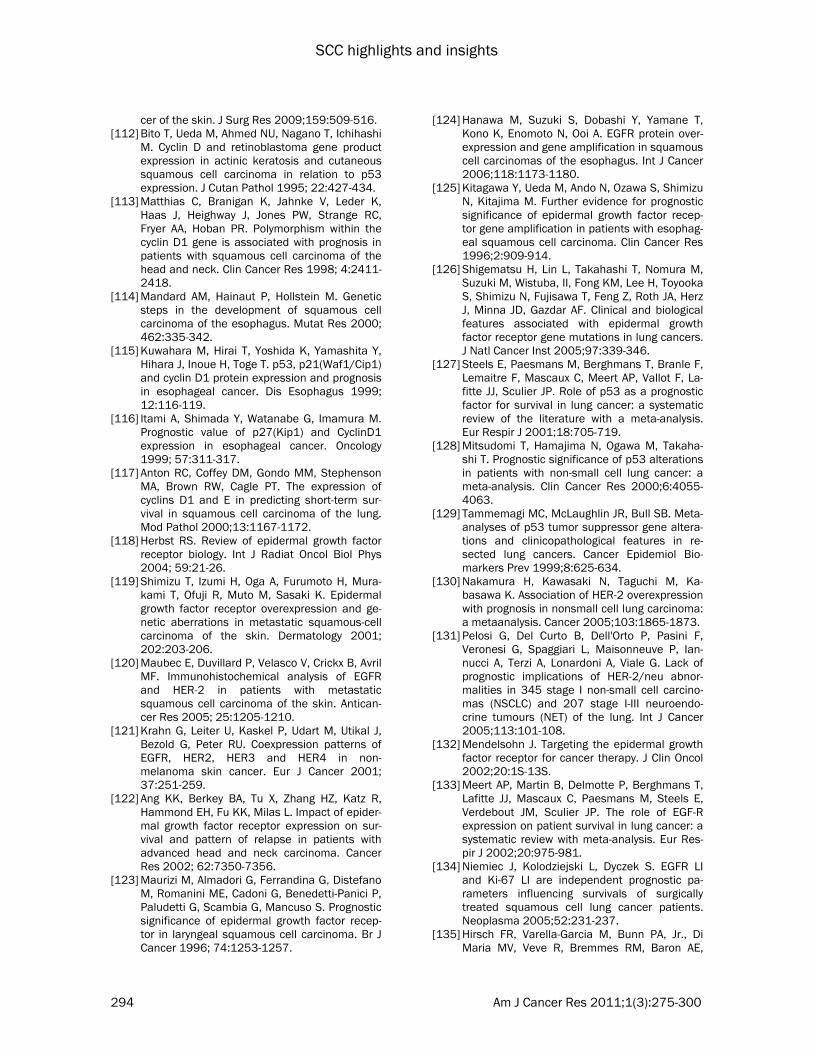
SCC highlights and insights
294 Am J Cancer Res 2011;1(3):275-300
cer of the skin. J Surg Res 2009;159:509-516. [112] Bito T, Ueda M, Ahmed NU, Nagano T, Ichihashi
M. Cyclin D and retinoblastoma gene product expression in actinic keratosis and cutaneous squamous cell carcinoma in relation to p53 expression. J Cutan Pathol 1995; 22:427-434.
[113] Matthias C, Branigan K, Jahnke V, Leder K, Haas J, Heighway J, Jones PW, Strange RC, Fryer AA, Hoban PR. Polymorphism within the cyclin D1 gene is associated with prognosis in patients with squamous cell carcinoma of the head and neck. Clin Cancer Res 1998; 4:2411-2418.
[114] Mandard AM, Hainaut P, Hollstein M. Genetic steps in the development of squamous cell carcinoma of the esophagus. Mutat Res 2000; 462:335-342.
[115] Kuwahara M, Hirai T, Yoshida K, Yamashita Y, Hihara J, Inoue H, Toge T. p53, p21(Waf1/Cip1) and cyclin D1 protein expression and prognosis in esophageal cancer. Dis Esophagus 1999; 12:116-119.
[116] Itami A, Shimada Y, Watanabe G, Imamura M. Prognostic value of p27(Kip1) and CyclinD1 expression in esophageal cancer. Oncology 1999; 57:311-317.
[117] Anton RC, Coffey DM, Gondo MM, Stephenson MA, Brown RW, Cagle PT. The expression of cyclins D1 and E in predicting short-term sur-vival in squamous cell carcinoma of the lung. Mod Pathol 2000;13:1167-1172.
[118] Herbst RS. Review of epidermal growth factor receptor biology. Int J Radiat Oncol Biol Phys 2004; 59:21-26.
[119] Shimizu T, Izumi H, Oga A, Furumoto H, Mura-kami T, Ofuji R, Muto M, Sasaki K. Epidermal growth factor receptor overexpression and ge-netic aberrations in metastatic squamous-cell carcinoma of the skin. Dermatology 2001; 202:203-206.
[120] Maubec E, Duvillard P, Velasco V, Crickx B, Avril MF. Immunohistochemical analysis of EGFR and HER-2 in patients with metastatic squamous cell carcinoma of the skin. Antican-cer Res 2005; 25:1205-1210.
[121] Krahn G, Leiter U, Kaskel P, Udart M, Utikal J, Bezold G, Peter RU. Coexpression patterns of EGFR, HER2, HER3 and HER4 in non-melanoma skin cancer. Eur J Cancer 2001; 37:251-259.
[122] Ang KK, Berkey BA, Tu X, Zhang HZ, Katz R, Hammond EH, Fu KK, Milas L. Impact of epider-mal growth factor receptor expression on sur-vival and pattern of relapse in patients with advanced head and neck carcinoma. Cancer Res 2002; 62:7350-7356.
[123] Maurizi M, Almadori G, Ferrandina G, Distefano M, Romanini ME, Cadoni G, Benedetti-Panici P, Paludetti G, Scambia G, Mancuso S. Prognostic significance of epidermal growth factor recep-tor in laryngeal squamous cell carcinoma. Br J Cancer 1996; 74:1253-1257.
[124] Hanawa M, Suzuki S, Dobashi Y, Yamane T, Kono K, Enomoto N, Ooi A. EGFR protein over-expression and gene amplification in squamous cell carcinomas of the esophagus. Int J Cancer 2006;118:1173-1180.
[125] Kitagawa Y, Ueda M, Ando N, Ozawa S, Shimizu N, Kitajima M. Further evidence for prognostic significance of epidermal growth factor recep-tor gene amplification in patients with esophag-eal squamous cell carcinoma. Clin Cancer Res 1996;2:909-914.
[126] Shigematsu H, Lin L, Takahashi T, Nomura M, Suzuki M, Wistuba, II, Fong KM, Lee H, Toyooka S, Shimizu N, Fujisawa T, Feng Z, Roth JA, Herz J, Minna JD, Gazdar AF. Clinical and biological features associated with epidermal growth factor receptor gene mutations in lung cancers. J Natl Cancer Inst 2005;97:339-346.
[127] Steels E, Paesmans M, Berghmans T, Branle F, Lemaitre F, Mascaux C, Meert AP, Vallot F, La-fitte JJ, Sculier JP. Role of p53 as a prognostic factor for survival in lung cancer: a systematic review of the literature with a meta-analysis. Eur Respir J 2001;18:705-719.
[128] Mitsudomi T, Hamajima N, Ogawa M, Takaha-shi T. Prognostic significance of p53 alterations in patients with non-small cell lung cancer: a meta-analysis. Clin Cancer Res 2000;6:4055-4063.
[129] Tammemagi MC, McLaughlin JR, Bull SB. Meta-analyses of p53 tumor suppressor gene altera-tions and clinicopathological features in re-sected lung cancers. Cancer Epidemiol Bio-markers Prev 1999;8:625-634.
[130] Nakamura H, Kawasaki N, Taguchi M, Ka-basawa K. Association of HER-2 overexpression with prognosis in nonsmall cell lung carcinoma: a metaanalysis. Cancer 2005;103:1865-1873.
[131] Pelosi G, Del Curto B, Dell'Orto P, Pasini F, Veronesi G, Spaggiari L, Maisonneuve P, Ian-nucci A, Terzi A, Lonardoni A, Viale G. Lack of prognostic implications of HER-2/neu abnor-malities in 345 stage I non-small cell carcino-mas (NSCLC) and 207 stage I-III neuroendo-crine tumours (NET) of the lung. Int J Cancer 2005;113:101-108.
[132] Mendelsohn J. Targeting the epidermal growth factor receptor for cancer therapy. J Clin Oncol 2002;20:1S-13S.
[133] Meert AP, Martin B, Delmotte P, Berghmans T, Lafitte JJ, Mascaux C, Paesmans M, Steels E, Verdebout JM, Sculier JP. The role of EGF-R expression on patient survival in lung cancer: a systematic review with meta-analysis. Eur Res-pir J 2002;20:975-981.
[134] Niemiec J, Kolodziejski L, Dyczek S. EGFR LI and Ki-67 LI are independent prognostic pa-rameters influencing survivals of surgically treated squamous cell lung cancer patients. Neoplasma 2005;52:231-237.
[135] Hirsch FR, Varella-Garcia M, Bunn PA, Jr., Di Maria MV, Veve R, Bremmes RM, Baron AE,

SCC highlights and insights
295 Am J Cancer Res 2011;1(3):275-300
Zeng C, Franklin WA. Epidermal growth factor receptor in non-small-cell lung carcinomas: correlation between gene copy number and protein expression and impact on prognosis. J Clin Oncol 2003;21:3798-3807.
[136] Nakamura H, Kawasaki N, Taguchi M, Ka-basawa K. Survival impact of epidermal growth factor receptor overexpression in patients with non-small cell lung cancer: a meta-analysis. Thorax 2006;61:140-145.
[137] Ludovini V, Bellezza G, Pistola L, Bianconi F, Di Carlo L, Sidoni A, Semeraro A, Del Sordo R, Tofanetti FR, Mameli MG, Daddi G, Cavaliere A, Tonato M, Crino L. High coexpression of both insulin-like growth factor receptor-1 (IGFR-1) and epidermal growth factor receptor (EGFR) is associated with shorter disease-free survival in resected non-small-cell lung cancer patients. Ann Oncol 2009; 20:842-849.
[138] Menter DG, Schilsky RL, DuBois RN. Cyclooxy-genase-2 and cancer treatment: understanding the risk should be worth the reward. Clin Can-cer Res;16:1384-1390.
[139] Higashi Y, Kanekura T, Kanzaki T. Enhanced expression of cyclooxygenase (COX)-2 in human skin epidermal cancer cells: evidence for growth suppression by inhibiting COX-2 expres-sion. Int J Cancer 2000;86:667-671.
[140] An KP, Athar M, Tang X, Katiyar SK, Russo J, Beech J, Aszterbaum M, Kopelovich L, Epstein EH, Jr., Mukhtar H, Bickers DR. Cyclooxygenase-2 expression in murine and human nonmela-noma skin cancers: implications for therapeutic approaches. Photochem Photobiol 2002; 76:73-80.
[141] Zimmermann KC, Sarbia M, Weber AA, Bor-chard F, Gabbert HE, Schror K. Cyclooxygenase-2 expression in human esophageal carcinoma. Cancer Res 1999;59:198-204.
[142] Xu XC. COX-2 inhibitors in cancer treatment and prevention, a recent development. Anticancer Drugs 2002;13:127-137.
[143] Ko YHI, Soon Ja; Kim, Jeong Oh; Byun, Jae Ho; Jung, Chan Kwon; Lee, Myung Ah; Hong, Yeong Seon; Park, Jae Kil; Wang, Young Pil; Kang, Jin Hyoung: Correlation of CD44, VEGF-C and COX-2 with clinicopathologic parameters and clinical outcomes. Journal of Thoracic Oncology 2007; 2:ps779.
[144] Uehara M, Sano K, Ikeda H, Sekine J, Irie A, Yokota T, Tobita T, Ohba S, Inokuchi T. Expres-sion of vascular endothelial growth factor and prognosis of oral squamous cell carcinoma. Oral Oncol 2004; 40:321-325.
[145] Kleespies A, Guba M, Jauch KW, Bruns CJ. Vas-cular endothelial growth factor in esophageal cancer. J Surg Oncol 2004;87:95-104.
[146] Boone J, van Hillegersberg R, Offerhaus GJ, van Diest PJ, Borel Rinkes IH, Ten Kate FJ. Targets for molecular therapy in esophageal squamous cell carcinoma: an immunohistochemical analy-sis. Dis Esophagus 2009;22:496-504.
[147] Bass AJ, Watanabe H, Mermel CH, Yu S, Perner S, Verhaak RG, Kim SY, Wardwell L, Tamayo P, Gat-Viks I, Ramos AH, Woo MS, Weir BA, Getz G, Beroukhim R, O'Kelly M, Dutt A, Rozenblatt-Rosen O, Dziunycz P, Komisarof J, Chirieac LR, Lafague CJ, Scheble V, Wilbertz T, Ma C, Rao S, Nakagawa H, Stairs DB, Lin L, Giordano TJ, Wagner P, Minna JD, Gazdar AF, Zhu CQ, Brose MS, Cecconello L, Jr UR, Marie SK, Dahl O, Shivdasani RA, Tsao MS, Rubin MA, Wong KK, Regev A, Hahn WC, Beer DG, Rustgi AK, Meyer-son M. SOX2 is an amplified lineage-survival oncogene in lung and esophageal squamous cell carcinomas. Nat Genet 2009;41:1238-1242.
[148] Wang Q, He W, Lu C, Wang Z, Wang J, Giercksky KE, Nesland JM, Suo Z. Oct3/4 and Sox2 are significantly associated with an unfavorable clinical outcome in human esophageal squamous cell carcinoma. Anticancer Res 2009; 29:1233-1241.
[149] Freier K, Knoepfle K, Flechtenmacher C, Pungs S, Devens F, Toedt G, Hofele C, Joos S, Lichter P, Radlwimmer B. Recurrent copy number gain of transcription factor SOX2 and corresponding high protein expression in oral squamous cell carcinoma. Genes Chromosomes Cancer 2010; 49:9-16.
[150] Xu N, Papagiannakopoulos T, Pan G, Thomson JA, Kosik KS. MicroRNA-145 regulates OCT4, SOX2, and KLF4 and represses pluripotency in human embryonic stem cells. Cell 2009; 137:647-658.
[151] Yuan P, Kadara H, Behrens C, Tang X, Woods D, Solis LM, Huang J, Spinola M, Dong W, Yin G, Fujimoto J, Kim E, Xie Y, Girard L, Moran C, Hong WK, Minna JD, Wistuba II. Sex determin-ing region Y-Box 2 (SOX2) is a potential cell-lineage gene highly expressed in the patho-genesis of squamous cell carcinomas of the lung. PLoS One 2010;5:e9112.
[152] Bongiorno PF, al-Kasspooles M, Lee SW, Rach-wal WJ, Moore JH, Whyte RI, Orringer MB, Beer DG. E-cadherin expression in primary and me-tastatic thoracic neoplasms and in Barrett's oesophagus. Br J Cancer 1995;71:166-172.
[153] Chiles MC, Ai L, Zuo C, Fan CY, Smoller BR. E-cadherin promoter hypermethylation in preneo-plastic and neoplastic skin lesions. Mod Pathol 2003;16:1014-1018.
[154] Uchikado Y, Natsugoe S, Okumura H, Setoyama T, Matsumoto M, Ishigami S, Aikou T. Slug Ex-pression in the E-cadherin preserved tumors is related to prognosis in patients with esophag-eal squamous cell carcinoma. Clin Cancer Res 2005;11:1174-1180.
[155] O'Grady A, Dunne C, O'Kelly P, Murphy GM, Leader M, Kay E. Differential expression of matrix metalloproteinase (MMP)-2, MMP-9 and tissue inhibitor of metalloproteinase (TIMP)-1 and TIMP-2 in non-melanoma skin cancer: im-plications for tumour progression. Histopathol-

SCC highlights and insights
296 Am J Cancer Res 2011;1(3):275-300
ogy 2007; 51:793-804. [156] Kurahara S, Shinohara M, Ikebe T, Nakamura
S, Beppu M, Hiraki A, Takeuchi H, Shirasuna K. Expression of MMPS, MT-MMP, and TIMPs in squamous cell carcinoma of the oral cavity: correlations with tumor invasion and metasta-sis. Head Neck 1999;21:627-638.
[157] Gorogh T, Beier UH, Baumken J, Meyer JE, Hoff-mann M, Gottschlich S, Maune S. Metallopro-teinases and their inhibitors: influence on tu-mor invasiveness and metastasis formation in head and neck squamous cell carcinomas. Head Neck 2006; 28:31-39.
[158] Jeon YK, Lee BY, Kim JE, Lee SS, Kim CW. Mo-lecular characterization of Epstein-Barr virus and oncoprotein expression in nasopharyngeal carcinoma in Korea. Head Neck 2004;26:573-583.
[159] Ondruschka C, Buhtz P, Motsch C, Freigang B, Schneider-Stock R, Roessner A, Boltze C. Prog-nostic value of MMP-2, -9 and TIMP-1,-2 immu-noreactive protein at the invasive front in ad-vanced head and neck squamous cell carcino-mas. Pathol Res Pract 2002;198:509-515.
[160] Gu ZD, Li JY, Li M, Gu J, Shi XT, Ke Y, Chen KN. Matrix metalloproteinases expression corre-lates with survival in patients with esophageal squamous cell carcinoma. Am J Gastroenterol 2005;100:1835-1843.
[161] Tanioka Y, Yoshida T, Yagawa T, Saiki Y, Takeo S, Harada T, Okazawa T, Yanai H, Okita K. Ma-trix metalloproteinase-7 and matrix metallopro-teinase-9 are associated with unfavourable prognosis in superficial oesophageal cancer. Br J Cancer 2003;89:2116-2121.
[162] Mukherjee S, Roth MJ, Dawsey SM, Yan W, Rodriguez-Canales J, Erickson HS, Hu N, Gold-stein AM, Taylor PR, Richardson AM, Tangrea MA, Chuaqui RF, Emmert-Buck MR. Increased matrix metalloproteinase activation in esophag-eal squamous cell carcinoma. J Transl Med 2010;8:91.
[163] O'Regan EM, Toner ME, Finn SP, Fan CY, Ring M, Hagmar B, Timon C, Smyth P, Cahill S, Flavin R, Sheils OM, O'Leary JJ. p16(INK4A) genetic and epigenetic profiles differ in relation to age and site in head and neck squamous cell carci-nomas. Hum Pathol 2008;39:452-458.
[164] Lee DJ SF, Banuchi VE, Qiu W, Close LG, As-saad AM, Su GH. Multiple tumor-suppressor genes on chromosome 3p contribute to head and neck squamous cell carcinoma tumori-genesis. Cancer Biol Ther 2010;10:689-93.
[165] Wistuba, II, Behrens C, Virmani AK, Mele G, Milchgrub S, Girard L, Fondon JW, 3rd, Garner HR, McKay B, Latif F, Lerman MI, Lam S, Gaz-dar AF, Minna JD. High resolution chromosome 3p allelotyping of human lung cancer and pre-neoplastic/preinvasive bronchial epithelium reveals multiple, discontinuous sites of 3p al-lele loss and three regions of frequent break-points. Cancer Res 2000; 60:1949-1960.
[166] Aviel-Ronen S, Blackhall FH, Shepherd FA, Tsao MS. K-ras mutations in non-small-cell lung car-cinoma: a review. Clin Lung Cancer 2006;8:30-38.
[167] Herbst RS, Heymach JV, Lippman SM. Lung cancer. N Engl J Med 2008;359:1367-1380.
[168] Liggett WH, Jr., Sidransky D. Role of the p16 tumor suppressor gene in cancer. J Clin Oncol 1998;16:1197-1206.
[169] Bian YS, Osterheld MC, Fontolliet C, Bosman FT, Benhattar J. p16 inactivation by methylation of the CDKN2A promoter occurs early during neoplastic progression in Barrett's esophagus. Gastroenterology 2002;122:1113-1121.
[170] Ji H, Ramsey MR, Hayes DN, Fan C, McNamara K, Kozlowski P, Torrice C, Wu MC, Shimamura T, Perera SA, Liang MC, Cai D, Naumov GN, Bao L, Contreras CM, Li D, Chen L, Krishnamurthy J, Koivunen J, Chirieac LR, Padera RF, Bronson RT, Lindeman NI, Christiani DC, Lin X, Shapiro GI, Janne PA, Johnson BE, Meyerson M, Kwiat-kowski DJ, Castrillon DH, Bardeesy N, Shar-pless NE, Wong KK. LKB1 modulates lung can-cer differentiation and metastasis. Nature 2007;448:807-810.
[171] Yanagawa N, Tamura G, Oizumi H, Kanauchi N, Endoh M, Sadahiro M, Motoyama T. Promoter hypermethylation of RASSF1A and RUNX3 genes as an independent prognostic prediction marker in surgically resected non-small cell lung cancers. Lung Cancer 2007;58:131-138.
[172] Miska EA. How microRNAs control cell division, differentiation and death. Curr Opin Genet Dev 2005;15:563-568.
[173] Jay C, Nemunaitis J, Chen P, Fulgham P, Tong AW. miRNA profiling for diagnosis and progno-sis of human cancer. DNA Cell Biol 2007;26:293-300.
[174] Fabbri M. MicroRNAs and cancer epigenetics. Curr Opin Investig Drugs 2008;9:583-590.
[175] Sand M, Gambichler T, Skrygan M, Sand D, Scola N, Altmeyer P, Bechara FG. Expression levels of the microRNA processing enzymes Drosha and dicer in epithelial skin cancer. Can-cer Invest, 2010;28:649-653.
[176] Chang KW, Liu CJ, Chu TH, Cheng HW, Hung PS, Hu WY, Lin SC. Association between high miR-211 microRNA expression and the poor progno-sis of oral carcinoma. J Dent Res 2008; 87:1063-1068.
[177] Shiiba M, Ku HT. MicroRNAs in Head and Neck Squamous Cell Carcinoma (HNSCC) and Oral Squamous Cell Carcinoma (OSCC). Cancers 2010; 2:16.
[178] Avissar M, Christensen BC, Kelsey KT, Marsit CJ. MicroRNA expression ratio is predictive of head and neck squamous cell carcinoma. Clin Cancer Res 2009; 15:2850-2855.
[179] Hiyoshi Y, Kamohara H, Karashima R, Sato N, Imamura Y, Nagai Y, Yoshida N, Toyama E, Ha-yashi N, Watanabe M, Baba H. MicroRNA-21 regulates the proliferation and invasion in eso-
SCC highlights and insights
297 Am J Cancer Res 2011;1(3):275-300
phageal squamous cell carcinoma. Clin Cancer Res 2009; 15:1915-1922.
[180] Yanaihara N, Caplen N, Bowman E, Seike M, Kumamoto K, Yi M, Stephens RM, Okamoto A, Yokota J, Tanaka T, Calin GA, Liu CG, Croce CM, Harris CC. Unique microRNA molecular profiles in lung cancer diagnosis and prognosis. Cancer Cell 2006; 9:189-198.
[181] Volinia S, Calin GA, Liu CG, Ambs S, Cimmino A, Petrocca F, Visone R, Iorio M, Roldo C, Ferracin M, Prueitt RL, Yanaihara N, Lanza G, Scarpa A, Vecchione A, Negrini M, Harris CC, Croce CM. A microRNA expression signature of human solid tumors defines cancer gene targets. Proc Natl Acad Sci U S A 2006; 103:2257-2261.
[182] Yu SL, Chen HY, Chang GC, Chen CY, Chen HW, Singh S, Cheng CL, Yu CJ, Lee YC, Chen HS, Su TJ, Chiang CC, Li HN, Hong QS, Su HY, Chen CC, Chen WJ, Liu CC, Chan WK, Li KC, Chen JJ, Yang PC. MicroRNA signature predicts survival and relapse in lung cancer. Cancer Cell 2008; 13:48-57.
[183] Weiss GJ, Bemis LT, Nakajima E, Sugita M, Birks DK, Robinson WA, Varella-Garcia M, Bunn PA, Jr., Haney J, Helfrich BA, Kato H, Hirsch FR, Franklin WA. EGFR regulation by microRNA in lung cancer: correlation with clinical response and survival to gefitinib and EGFR expression in cell lines. Ann Oncol 2008; 19:1053-1059.
[184] Stockfleth E, Kerl H. Guidelines for the manage-ment of actinic keratoses. Eur J Dermatol 2006; 16:599-606.
[185] Bernier J, Bentzen SM, Vermorken JB. Molecu-lar therapy in head and neck oncology. Nat Rev Clin Oncol 2009;6:266-277.
[186] Razak AR, Siu LL, Le Tourneau C. Molecular targeted therapies in all histologies of head and neck cancers: an update. Curr Opin Oncol; 22:212-220.
[187] Sutter AP, Hopfner M, Huether A, Maaser K, Scherubl H. Targeting the epidermal growth factor receptor by erlotinib (Tarceva) for the treatment of esophageal cancer. Int J Cancer 2006;118:1814-1822.
[188] Butts CA, Bodkin D, Middleman EL, Englund CW, Ellison D, Alam Y, Kreisman H, Graze P, Maher J, Ross HJ, Elis PM, McNulty W, Kaplan E, Pautret V, Weber MR, Shepherd FA. Random-ized phase II study of gemcitabine plus cisplatin or carboplatin [corrected], with or without cetuximab, as first-line therapy for patients with advanced or metastatic non small-cell lung cancer. J Clin Oncol 2007;25:5777-5784.
[189] Rosell R, Robinet G, Szczesna A, Ramlau R, Constenla M, Mennecier BC, Pfeifer W, O'Byrne KJ, Welte T, Kolb R, Pirker R, Chemaissani A, Perol M, Ranson MR, Ellis PA, Pilz K, Reck M. Randomized phase II study of cetuximab plus cisplatin/vinorelbine compared with cisplatin/vinorelbine alone as first-line therapy in EGFR-expressing advanced non-small-cell lung can-cer. Ann Oncol 2008;19:362-369.
[190] Crawford J, Sandler AB, Hammond LA. ABX-EGF in combination with paclitaxel and carboplatin for advanced non-small lung cancer (NSCLC). J Clin Oncol 2004;22:7083 (abstr).
[191] Shepherd FA, Rosell R. Weighing tumor biology in treatment decisions for patients with non-small cell lung cancer. J Thorac Oncol 2007;2 Suppl 2:S68-76.
[192] Sequist LV, Lynch LT. EGFR tyrosine kinase inhibitors in lung cancer: An evolving story. Annu Rev Med 2008;59:429-42.
[193] Sequist LV, Bell DW, Lynch TJ, Haber DA. Mo-lecular predictors of response to epidermal growth factor receptor antagonists in non-small-cell lung cancer. J Clin Oncol 2007;25:587-595.
[194] John T, Liu G, Tsao MS. Overview of molecular testing in non-small-cell lung cancer: muta-tional analysis, gene copy number, protein ex-pression and other biomarkers of EGFR for the prediction of response to tyrosine kinase inhibi-tors. Oncogene 2009; 28 Suppl 1:S14-23.
[195] Jackman D, Pao W, Riely GJ, Engelman JA, Kris MG, Janne PA, Lynch T, Johnson BE, Miller VA. Clinical definition of acquired resistance to epidermal growth factor receptor tyrosine kinase inhibitors in non-small-cell lung cancer. J Clin Oncol 2009; 28:357-360.
[196] Linardou H, Dahabreh IJ, Bafaloukos D, Kos-midis P, Murray S. Somatic EGFR mutations and efficacy of tyrosine kinase inhibitors in NSCLC. Nat Rev Clin Oncol 2009;6:352-366.
[197] Nguyen KS, Kobayashi S, Costa DB. Acquired resistance to epidermal growth factor receptor tyrosine kinase inhibitors in non-small-cell lung cancers dependent on the epidermal growth factor receptor pathway. Clin Lung Cancer 2009;10:281-289.
[198] Pao W, Miller V, Zakowski M, Doherty J, Politi K, Sarkaria I, Singh B, Heelan R, Rusch V, Fulton L, Mardis E, Kupfer D, Wilson R, Kris M, Varmus H. EGF receptor gene mutations are common in lung cancers from "never smokers" and are associated with sensitivity of tumors to gefitinib and erlotinib. Proc Natl Acad Sci U S A 2004; 101:13306-13311.
[199] Kobayashi K, Nishioka M, Kohno T, Nakamoto M, Maeshima A, Aoyagi K, Sasaki H, Ta-kenoshita S, Sugimura H, Yokota J. Identifica-tion of genes whose expression is upregulated in lung adenocarcinoma cells in comparison with type II alveolar cells and bronchiolar epithelial cells in vivo. Oncogene 2004; 23:3089-3096.
[200] Faber AC, Li D, Song Y, Liang MC, Yeap BY, Bronson RT, Lifshits E, Chen Z, Maira SM, Gar-cia-Echeverria C, Wong KK, Engelman JA. Differ-ential induction of apoptosis in HER2 and EGFR addicted cancers following PI3K inhibition. Proc Natl Acad Sci U S A 2009;106:19503-19508.
[201] LeRoith D, Werner H, Beitner-Johnson D, Rob-erts CT, Jr.. Molecular and cellular aspects of
SCC highlights and insights
298 Am J Cancer Res 2011;1(3):275-300
the insulin-like growth factor I receptor. Endocr Rev 1995, 16:143-163.
[202] Warshamana-Greene GS, Litz J, Buchdunger E, Garcia-Echeverria C, Hofmann F, Krystal GW. The insulin-like growth factor-I receptor kinase inhibitor, NVP-ADW742, sensitizes small cell lung cancer cell lines to the effects of chemo-therapy. Clin Cancer Res 2005;11:1563-1571.
[203] Allen GW, Saba C, Armstrong EA, Huang SM, Benavente S, Ludwig DL, Hicklin DJ, Harari PM. Insulin-like growth factor-I receptor signaling blockade combined with radiation. Cancer Res 2007; 67:1155-1162.
[204] Gualberto A, Pollak M. Emerging role of insulin-like growth factor receptor inhibitors in oncol-ogy: early clinical trial results and future direc-tions. Oncogene 2009;28:3009-3021.
[205] A. Gualberto CLM, Dean A, Ang AL, J. Reynolds JM, Lee AV, Terstappen LW, Haluska P, Lipton A and Karp DD. Characterization of NSCLC pa-tients responding to anti-IGF-IR therapy. Journal of Clinical Oncology, 2008 ASCO Annual Meet-ing Proceedings 2008; 26.
[206] Wheeler DL, Huang S, Kruser TJ, Nechrebecki MM, Armstrong EA, Benavente S, Gondi V, Hsu KT, Harari PM. Mechanisms of acquired resis-tance to cetuximab: role of HER (ErbB) family members. Oncogene 2008; 27:3944-3956.
[207] Sergina NV, Rausch M, Wang D, Blair J, Hann B, Shokat KM, Moasser MM. Escape from HER-family tyrosine kinase inhibitor therapy by the kinase-inactive HER3. Nature 2007;445:437-441.
[208] Harari PM, Wheeler DL, Grandis JR. Molecular target approaches in head and neck cancer: epidermal growth factor receptor and beyond. Semin Radiat Oncol 2009;19:63-68.
[209] Patel V, Senderowicz AM, Pinto D, Jr., Igishi T, Raffeld M, Quintanilla-Martinez L, Ensley JF, Sausville EA, Gutkind JS. Flavopiridol, a novel cyclin-dependent kinase inhibitor, suppresses the growth of head and neck squamous cell carcinomas by inducing apoptosis. J Clin Invest 1998;102:1674-1681.
[210] Sato S, Kajiyama Y, Sugano M, Iwanuma Y, Tsurumaru M. Flavopiridol as a radio-sensitizer for esophageal cancer cell lines. Dis Esophagus 2004; 17:338-344.
[211] Hay N, Sonenberg N. Upstream and down-stream of mTOR. Genes Dev 2004, 18:1926-1945.
[212] Kris MG, Riely GJ, Azzoli CG, Heelan RT, Krug LM, Pao W. Combined inhibition of mTOR and EGFR with everolimus (RAD001) and gefitinib in patients with non-small cell lung cancer who have smoked cigarettes: A phase II trial. J Clin Oncol 2007; 25:abstr 7575.
[213] Davies AM, McCoy J, Lara PN, Gumerlock PH, Crowley J, Gandara DR. Bortezomib + gemcit-abine (Gem)/carboplatin (Carbo) results in en-couraging survival in advanced non-small cell lung cance (NSCLC): Results of a phase II
Southwest Oncology Group (SWOG) trial (S0339). J Clin Oncol 2006;24:abstr 7017.
[214] Eder JP, Vande Woude GF, Boerner SA, Lo-Russo PM. Novel therapeutic inhibitors of the c-Met signaling pathway in cancer. Clin Cancer Res 2009;15:2207-2214.
[215] Puri N, Salgia R. Synergism of EGFR and c-Met pathways, cross-talk and inhibition, in non-small cell lung cancer. J Carcinog 2008;7:9.
[216] Zucali PA, Ruiz MG, Giovannetti E, Destro A, Varella-Garcia M, Floor K, Ceresoli GL, Rodri-guez JA, Garassino I, Comoglio P, Roncalli M, Santoro A, Giaccone G. Role of cMET expres-sion in non-small-cell lung cancer patients treated with EGFR tyrosine kinase inhibitors. Ann Oncol 2008;19:1605-1612.
[217] Dempke WC, Suto T, Reck M. Targeted thera-pies for non-small cell lung cancer. Lung Can-cer, 2010;67:257-274.
[218] Thariat J, Milas L, Ang KK. Integrating radiother-apy with epidermal growth factor receptor an-tagonists and other molecular therapeutics for the treatment of head and neck cancer. Int J Radiat Oncol Biol Phys 2007;69:974-984.
[219] Lee JM, Mao JT, Krysan K, Dubinett SM. Signifi-cance of cyclooxygenase-2 in prognosis, tar-geted therapy and chemoprevention of NSCLC. Future Oncol 2007; 3:149-153.
[220] Chow LW, Loo WT, Toi M. Current directions for COX-2 inhibition in breast cancer. Biomed Phar-macother 2005, 59 Suppl 2:S281-284.
[221] Kentaro Yamaguchi AL, Andrew J. Dannenberg, and Kotha Subbaramaiah1: Histone Deacety-lase Inhibitors Suppress the Induction of c-Jun and Its Target Genes Including COX-2* THE JOURNAL OF BIOLOGICAL CHEMISTRY VOL 2005;280:9.
[222] Traynor AM, Dubey S, Eickhoff JC, Kolesar JM, Schell K, Huie MS, Groteluschen DL, Marcotte SM, Hallahan CM, Weeks HR, Wilding G, Espinoza-Delgado I, Schiller JH. Vorinostat (NSC# 701852) in patients with relapsed non-small cell lung cancer: a Wisconsin Oncology Network phase II study. J Thorac Oncol 2009;4:522-526.
[223] Williamson JM, Huang CH, Guaglianone P, Wolf GT and Urba SG. A phase II trial of sorafenib in patients with recurrent and/or metastatic head and neck squamous cell carcinoma (HNSCC): A Southwest Oncology Group (SWOG) trial. Jour-nal of Clinical Oncology, 2008 ASCO Annual Meeting Proceedings 2007;25:1.
[224] Gridelli C, Maione P, Del Gaizo F, Colantuoni G, Guerriero C, Ferrara C, Nicolella D, Comunale D, De Vita A, Rossi A. Sorafenib and sunitinib in the treatment of advanced non-small cell lung cancer. Oncologist 2007;12:191-200.
[225] Karamouzis MV, Johnson R, Rajasenan K, Bran-stetter B and Argiris A. Phase II trial of pe-metrexed (P) and bevacizumab (B) in patients (pts) with recurrent or metastatic head and neck squamous cell carcinoma (HNSCC): An
SCC highlights and insights
299 Am J Cancer Res 2011;1(3):275-300
interim analysis. Journal of Clinical Oncology, 2008 ASCO Annual Meeting Proceedings 2007; 25:1.
[226] Johnson DH, Fehrenbacher L, Novotny WF, Herbst RS, Nemunaitis JJ, Jablons DM, Langer CJ, DeVore RF, 3rd, Gaudreault J, Damico LA, Holmgren E, Kabbinavar F. Randomized phase II trial comparing bevacizumab plus carboplatin and paclitaxel with carboplatin and paclitaxel alone in previously untreated locally advanced or metastatic non-small-cell lung cancer. J Clin Oncol 2004; 22:2184-2191.
[227] Fong KM, Sekido Y, Gazdar AF, Minna JD. Lung cancer. 9: Molecular biology of lung cancer: clinical implications. Thorax 2003, 58:892-900.
[228] Cole KA, Krizman DB, Emmert-Buck MR. The genetics of cancer--a 3D model. Nat Genet 1999;21:38-41.
[229] Osawa H, Nakajima M, Kato H, Fukuchi M, Ku-wano H. Prognostic value of the expression of Smad6 and Smad7, as inhibitory Smads of the TGF-beta superfamily, in esophageal squamous cell carcinoma. Anticancer Res 2004;24:3703-3709.
[230] Orian-Rousseau V. CD44, a therapeutic target for metastasising tumours. Eur J Cancer 2010; 46:1271-1277.
[231] Fong D, Spizzo G, Gostner JM, Gastl G, Moser P, Krammel C, Gerhard S, Rasse M, Laimer K. TROP2: a novel prognostic marker in squamous cell carcinoma of the oral cavity. Mod Pathol 2008;21:186-191.
[232] Yanamoto S, Kawasaki G, Yoshitomi I, Iwamoto T, Hirata K, Mizuno A. Clinicopathologic signifi-cance of EpCAM expression in squamous cell carcinoma of the tongue and its possibility as a potential target for tongue cancer gene therapy. Oral Oncol 2007;43:869-877.
[233] Tse GM, Yu KH, Chan AW, King AD, Chen GG, Wong KT, Tsang RK, Chan AB. HER2 expression predicts improved survival in patients with cer-vical node-positive head and neck squamous cell carcinoma. Otolaryngol Head Neck Surg 2009;141:467-473.
[234] Polkowski W, van Sandick JW, Offerhaus GJ, ten Kate FJ, Mulder J, Obertop H, van Lanschot JJ. Prognostic value of Lauren classification and c-erbB-2 oncogene overexpression in adenocar-cinoma of the esophagus and gastroesophag-eal junction. Ann Surg Oncol 1999;6:290-297.
[235] Takayama T, Nagao M, Sawada H, Yamada Y, Emoto K, Fujimoto H, Ueno M, Hirao S, Naka-jima Y. Bcl-X expression in esophageal squamous cell carcinoma: association with tumor progression and prognosis. J Surg Oncol 2001;78:116-123.
[236] Ikeguchi M, Maeta M, Kaibara N. Bax expres-sion as a prognostic marker of postoperative chemoradiotherapy for patients with esophag-eal cancer. Int J Mol Med 2001; 7:413-417.
[237] Pai SI, Westra WH. Molecular pathology of head and neck cancer: implications for diagnosis,
prognosis, and treatment. Annu Rev Pathol 2009; 4:49-70.
[238] Ohashi Y, Sasano H, Yamaki H, Shizawa S, Shi-neha R, Akaishi T, Satomi S, Nagura H. Cell cycle inhibitory protein p27 in esophageal squamous cell carcinoma. Anticancer Res 1999; 19:1843-1848.
[239] Filipowicz E, Adegboyega P, Sanchez RL, Ga-talica Z. Expression of CD95 (Fas) in sun-exposed human skin and cutaneous carcino-mas. Cancer 2002;94:814-819.
[240] Kitamura A, Yashima K, Okamoto E, Andachi H, Hosoda A, Kishimoto Y, Shiota G, Ito H, Kaibara N, Kawasaki H. Reduced Fhit expression occurs in the early stage of esophageal tumorigenesis: no correlation with p53 expression and apop-tosis. Oncology 2001;61:205-211.
[241] Khademi B, Shirazi FM, Vasei M, Doroudchi M, Gandomi B, Modjtahedi H, Pezeshki AM, Ghaderi A. The expression of p53, c-erbB-1 and c-erbB-2 molecules and their correlation with prognostic markers in patients with head and neck tumors. Cancer Lett 2002; 184:223-230.
[242] Paweletz CP, Ornstein DK, Roth MJ, Bichsel VE, Gillespie JW, Calvert VS, Vocke CD, Hewitt SM, Duray PH, Herring J, Wang QH, Hu N, Linehan WM, Taylor PR, Liotta LA, Emmert-Buck, MR, Petricoin EF,3rd: Loss of annexin 1 correlates with early onset of tumorigenesis in esophageal and prostate carcinoma. Cancer Res 2000; 60:6293-6297.
[243] Sharma R, Sud N, Chattopadhyay TK, Ralhan R. TC21/R-Ras2 upregulation in esophageal tumorigenesis: potential diagnostic implica-tions. Oncology 2005; 69:10-18.
[244] Rodriguez-Pineiro AM, Blanco-Prieto S, Sanchez-Otero N, Rodriguez-Berrocal FJ, de la Cadena MP. On the identification of biomarkers for non-small cell lung cancer in serum and pleural effusion. J Proteomics 2010;73:1511-1522.
[245] Verma A, Shukla NK, Deo SV, Gupta SD, Ralhan R. MEMD/ALCAM: a potential marker for tumor invasion and nodal metastasis in esophageal squamous cell carcinoma. Oncology 2005;68:462-470.
[246] Cooper SJ, Bowden GT. Ultraviolet B regulation of transcription factor families: roles of nuclear factor-kappa B (NF-kappaB) and activator pro-tein-1 (AP-1) in UVB-induced skin carcinogene-sis. Curr Cancer Drug Targets 2007;7:325-334.
[247] Kim DJ, Murray IA, Burns AM, Gonzalez FJ, Per-dew GH, Peters JM. Peroxisome proliferator-activated receptor-beta/delta inhibits epider-mal cell proliferation by down-regulation of kinase activity. J Biol Chem 2005;280:9519-9527.
[248] Chung JY, Braunschweig T, Hu N, Roth M, Trai-coff JL, Wang QH, Knezevic V, Taylor PR, Hewitt SM. A multiplex tissue immunoblotting assay for proteomic profiling: a pilot study of the nor-mal to tumor transition of esophageal squamous cell carcinoma. Cancer Epidemiol
SCC highlights and insights
300 Am J Cancer Res 2011;1(3):275-300
Biomarkers Prev 2006;15:1403-1408. [249] Ridge JA, Glisson BS, Lango MN, et al. "Head
and Neck Tumors" In Cancer Management: A Multidisciplinary Approach. Edited by Pazdur R WL, Camphausen KA, Hoskins WJ 2008.[11 (Series Editor)
[250] Garon EB, Finn RS, Hosmer W, Dering J, Ginther C, Adhami S, Kamranpour N, Pitts S, Desai A, Elashoff D, French T, Smith P, Slamon DJ. Iden-tification of common predictive markers of in vitro response to the Mek inhibitor selumetinib (AZD6244; ARRY-142886) in human breast cancer and non-small cell lung cancer cell lines. Mol Cancer Ther 2010;9:1985-1994.
[251] Von Pawel J, Reck M, Digel W, Kortsik C, Tho-mas M, Frickhofen N. 1 inhibitor, in patients with relapsed advanced or metastatic non-small cell lung cancer (NSCLC). J Clin Oncol 2008;26 (Suppl):8030.
[252] Lapenna S, Giordano A. Cell cycle kinases as therapeutic targets for cancer. Nat Rev Drug Discov 2009; 8:547-566.
[253] Ashkenazi A, Herbst RS. To kill a tumor cell: the potential of proapoptotic receptor agonists. J Clin Invest 2008;118:1979-1990.
[254] Paz-Ares L Sanchez Torres JM, Diaz-Padilla I. Safety and efficacy of AMG 655 in combination with paclitaxel and carboplatin (PC) in patients with advanced non-small cell lung cancer. J Clin Oncol 2009;27:19048
[255] Heider KH, Kuthan H, Stehle G, Munzert G. CD44v6: a target for antibody-based cancer therapy. Cancer Immunol Immunother 2004; 53:567-579.


![Squamous Cell Carcinoma of the Middle Ear …temporal bone malignancy, especially squamous cell carcinoma[14]. The early symptoms of temporal bone carcinoma closely resemble those](https://img.pdfslide.us/doc/110x75/5f027ff47e708231d4049179/squamous-cell-carcinoma-of-the-middle-ear-temporal-bone-malignancy-especially-squamous.jpg)