Embed Size (px)
Citation preview
ANESTH ANALG 303 1990:70:30>15
Review Article
Pro- and Anticonvulsant Effects of Anesthetics (Part I)
Paul A. Modica, MD, Rene Tempelhoff, MD, and Paul F. White, rhD, MD
Key Words: ANTICONVULSANTS. BRAIN, PRO- AND ANTICONWLSANTS. COMPLICATIONS, CONVULSIONS. TOXICITY, CONVULSIONS.
Part I Introduction Inhalation anesthetics
Volatile agents Enflurane Halothane Isoflurane Investigational volatile agents
Nitrous oxide
Opioid (narcotic) analgesics Intravenous analgesics
Meperidine Morphine Fentanyl and its analogues
Summary
Introduction Intravenous anesthetics
Sedative-hypnotics
Part I1
Barbiturates Etomidate Benzodiazepines Ketamine Propofol
Local anesthetics Anesthetic adjuncts
Muscle relaxants Anticholinesterases Anticholinergics
Summary
Part 11 of this review article will appear in the following issue of the journal.
Received from the Department of Anesthesiology, Washington University school of Medicine, St. Louis, Missouri. Accepted for publication October 10,1989.
Address correspondence to Dr. White, Department of Anesthe- siology, Box 8054, Washington University School of Medicine, 660 South Euclid Avenue, St. Louis, MO 63110.
An epileptic seizure has been defined as a sudden alteration of central nervous system (CNS) function resulting from a high-voltage electrical discharge. This discharge may arise from an assemblage of neurons in either cortical or subcortical tissues. The spread of this excitatory activity to the subcortical, thalamic, and brainstem centers corresponds to the tonic phase of the seizure and loss of consciousness (1). In contrast, myoclonic activity refers to a series of rhythmic or arrhythmic muscular contractions (2). Depending on the electroencephalographic (EEG) findings, myoclonus is divided into epileptic and nonepileptic activity (3). Nonepileptic myoclonus originates from the brainstem or spinal cord and is due to either loss of cortical inhibition (4) or to im- paired function of spinal interneurons (3,5). Without EEG monitoring, it is extremely difficult to determine whether abnormal-appearing seizurelike muscle movements are due to epileptiform activity or non- epileptic myoclonia.
Many anesthetic and analgesic drugs have been reported to cause seizure activity clinically (Table 1, A). Interestingly, many of these same drugs have also been shown to possess anticonvulsant properties (Table 1, B). In an effort to explain deficiencies with Guedel's original stages of anesthesia, Winters and colleagues (67 ) proposed a multidirectional contin- uum of anesthetic states (Figure 1). For example, some agents (e.g., diethyl ether) traverse both CNS excitation (stages I, 11) and depression (stage 111). Others (e.g., halothane, ultrashort acting barbitu- rates) progress directly from stage I to 111, whereas still others (e.g., nitrous oxide [N,O], enflurane, ketamine, and narcotics [S]) induce a stage I1 catalep- toid CNS excitation, which on occasion progresses to myoclonia or generalized convulsions. Based on EEG studies in cats, Winters suggested that both exces- sively disorganized (stage 11) and decreased reticular- formation activity (stage 111) result in unresponsive- ness to painful stimuli and amnesia, consistent with
01990 by the International Anesthesia Research Society

ANESTH ANALG 304 1990;7030>15
MODICA ET AL.
Table 1. Anesthetics and Analgesics Reported to Cause and/or Suppress Seizure Activity in Humans
~
A. Proconvulsants 8. Anticonvulsants
Nitrous oxide Halothane Halothane Enflurance Enflurane Isoflurane Isoflurane Thiopental Morphine E tomida te Meperidine Diazepam Fentanyl Lorazepam Sufentanil Midazolam Methohexital Ketamine Etomidate Diazepam Local anesthetics Ketamine Propofol Local anesthetics
Prop o f o 1
ME TRAZOL, KETAMINE PHENCYCLIOINE f-7
Y HYDROXYBUTYRATE SE, ZUR ES
ETHER(
MESCAL IN& N20
Figure 1. Winter's proposed scheme of reversible progression of CNS states induced by various anesthetic and excitant agents. Stages I, 11, A, B, C, myoclonus, and seizures indicate CNS excitation, whereas stages Ill and IV indicate CNS depression. (From Winters WD, Ferrer-Allado T, Guzman-Flores C, Alcaraz M. The cataleptic state induced by ketamine: a review of the neuro- pharmacology of anesthesia. Neuropharmacology 1972;11:30>15, with permission.)
the anesthetic state. Although both epileptic and anesthetic states possess similar features regarding arousal and memory, the categorization proposed by Winters fails to adequately explain the actions of those anesthetics that appear to possess both procon- vulsant and anticonvulsant properties (Table 1). For example, ketamine, an alleged stage I1 anesthetic which Winters believed to be contraindicated in epi- lepsy, has been successfully used to terminate status epilepticus (9, lo), and conversely, methohexital, a
stage 111 agent, has been used in the diagnostic activation of epileptogenic foci (11,12).
To properly categorize the various anesthetic agents with respect to their effects on the seizure threshold, there are several important factors to con- sider. The first consideration is the patient population studied. For example, methohexital, in its current formulation (Brevital; Eli Lilly, Indianapolis, Ind.), will only produce epileptiform activity in patients with known seizure disorders (11,12). A second fac- tor to consider is the method of proconvulsant and anticonvulsant documentation. This had led to con- fusion in the literature regarding the effects of some anesthetics and analgesics on CNS activity. Fentanyl- induced seizures have been described clinically with- out the support of EEG documentation (13-15). In some instances, these convulsivelike muscle move- ments may be due to nonepileptic myoclonus.
For most drugs used in anesthesia, subsequent EEG evaluation during their administration has helped to clarify whether or not a particular agent is truly epileptogenic in patients. However, for certain anesthetics, such as ketamine (16), the origin of epileptiform activity involves subcortical neuronal pathways. In humans, subcortical seizures are de- tected only with implanted EEG depth electrodes, not by standard surface leads. Thus, a third important factor to consider in determining whether or not an anesthetic possesses proconvulsant or anticonvulsant properties is the type of EEG recording electrodes (surface or depth) used during its evaluation.
In this review article, we have attempted to ana- lyze the evidence for proconvulsant and anticonvul- sant activity of anesthetics and analgesics. This infor- mation has been evaluated with respect to (a) patient population (epileptic or nonepileptic); (b) documen- tation of pro- and anticonvulsant activity (EEG study or clinical report); and (c) method of EEG analysis (surface or depth electrodes).
Inhala tion Anesthetics Volatile Agents
Enflurune. Abnormal movements consisting of twitching of individual muscle groups, and even tonic-clonic activity, were frequently observed during the early clinical evaluation of enflurane (17,18). Subsequent EEG recordings in normal patients dem- onstrated epileptiform activity (19,20) and grand ma1 seizure patterns (21) (Table 2). These findings with enflurane have also been confirmed in patients with temporal lobe epilepsy during both electrocortico-
ANESTHETICS AND SEIZURES ANESTH ANALC 305 1990:70:30>15
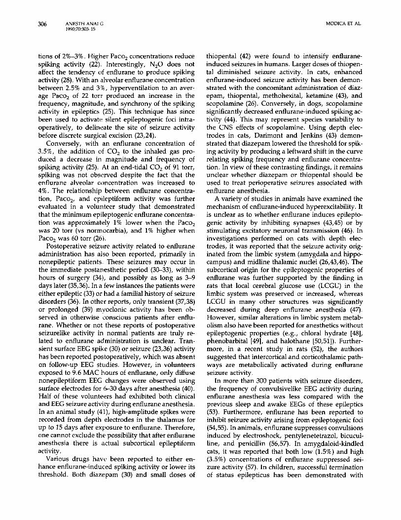
Table 2. Proconvulsant Effects of Inhalation Anesthetics in Humans Seizure
documentation Type of EEG
Clinical EEG electrodes Agent Population report study used in study References
58, 64, 96 Nitrous oxide Nonepileptic + - Surface Epileptic - - Depth 16
Halothane
Enflurance
lsoflurane
Sevoflurane
Nonepileptic - - Epileptic - -
Surface 6 M 2 Surface 63
Nonepileptic + i Surface 17-21, 26, 28 Epileptic + i Surfaceldepth 22-25 Nonepileptic - - Surface 75-78 Epileptic NIA N/A
Nonepileptic - - Surface 86 Epileptic NIA NIA
Desflurane (1-653) Nonepileptic NIA N/A Epileptic N/A NIA
+, presence of seizures; -, absence of seizures; EEC, electroencephalographic; N/A, information not available.
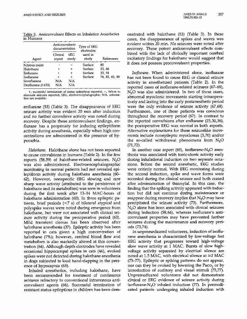
owdm
LO m
Figure 2 . Electroencephalographic patterns with increasing concentrations of enflurane. (From Neigh JL, Garman JK, Harp JR. The electroencephalographic pattern during anes- thesia with Ethrane: effects of depth of anes- thesia, Paco,, and nitrous oxide. Anesthesiol-
0
6 &
ogy 1971;35:482-7, with permission.) 15
graphic (22-24) and depth electrode recordings (25). In ten healthy volunteers, grand ma1 seizure patterns were precipitated by auditory, visual, and tactile stimulation at end-tidal enflurane concentrations of 3%-6% (26). This has also been reported during otic microsurgery (27). In both normal (28) and epileptic (U) patients, increasing depth of enflurane anesthe- sia is characterized by the appearance of high-voltage spikes, with the subsequent development of spike
waves-spike and dome activity with intermittent periods of burst suppression (Figure 2). In contrast to other volatile anesthetics, these burst suppression patterns are thought to represent an excitatory event
The ability of enflurane to produce seizure or EEG spiking activity is influenced by both its concentra- tion and the Paco, (25,28). At a normal Paco, level, spiking is maximal at inspired enflurane concentra-
(29).
ANESTH ANALG 1990;70303-15
306 MODICA ET AL.
tions of 2%-3%. Higher Paco, concentrations reduce spiking activity (22). Interestingly, N,O does not affect the tendency of enflurane to produce spiking activity (28). With an alveolar enflurane concentration between 2.5% and 3%, hyperventilation to an aver- age Paco, of 22 torr produced an increase in the frequency, magnitude, and synchrony of the spiking activity in epileptics (25). This technique has since been used to activate silent epileptogenic foci intra- operatively, to delineate the site of seizure activity before discrete surgical excision (23,24).
Conversely, with an enflurane concentration of 3.5%, the addition of CO, to the inhaled gas pro- duced a decrease in magnitude and frequency of spiking activity (25). At an end-tidal CO, of 91 torr, spiking was not observed despite the fact that the enflurane alveolar concentration was increased to 4%. The rela tionship between enflurane concentra- tion, Paco,, and epileptiform activity was further evaluated in a volunteer study that demonstrated that the minimum epileptogenic enflurane concentra- tion was approximately 1% lower when the Paco, was 20 torr (vs normocarbia), and 1% higher when Paco, was 60 torr (26).
Postoperative seizure activity related to enflurane administration has also been reported, primarily in nonepileptic patients. These seizures may occur in the immediate postanesthetic period (30-33), within hours of surgery (34), and possibly as long as 3-9 days later (35,36). In a few instances the patients were either epileptic (33) or had a familial history of seizure disorders (36). In other reports, only transient (37,38) or prolonged (39) mvoclonic activity has been ob- served in otherwise conscious patients after enflu- rane. Whether or not these reports of postoperative seizurelike activity in normal patients are truly re- lated to enflurane administration is unclear. Tran- sient surface EEG spike (30) or seizure (23,36) activity has been reported postoperatively, which was absent on follow-up EEG studies. However, in volunteers exposed to 9.6 MAC hours of enflurane, only diffuse nonepileptiform EEG changes were observed using surface electrodes for 6-30 days after anesthesia (40). Half of these volunteers had exhibited both clinical and EEG seizure activity during enflurane anesthesia. In an animal study (41), high-amplitude spikes were recorded from depth electrodes in the thalamus for up to 15 days after exposure to enflurane. Therefore, one cannot exclude the possibility that after enflurane anesthesia there is actual subcortical epileptiform activity.
Various drugs have been reported to either en- hance enflurane-induced spiking activity or lower its threshold. Both diazepam (30) and small doses of
thiopental (42) were found to intensify enflurane- induced seizures in humans. Larger doses of thiopen- tal diminished seizure activity. In cats, enhanced enflurane-induced seizure activity has been demon- strated with the concomitant administration of diaz- epam, thiopental, methohexital, ketamine (43), and scopolamine (26). Conversely, in dogs, scopolamine significantly decreased enflurane-induced spiking ac- tivity (44). This may represent species variability to the CNS effects of scopolamine. Using depth elec- trodes in cats, Darimont and Jenkins (43) demon- strated that diazepam lowered the threshold for spik- ing activity by producing a leftward shift in the curve relating spiking frequency and enflurane concentra- tion. In view of these contrasting findings, it remains unclear whether diazepam or thiopental should be used to treat perioperative seizures associated with enflurane anesthesia.
A variety of studies in animals have examined the mechanism of enflurane-induced hyperexcitability . It is unclear as to whether enflurane induces epilepto- genic activity by inhibiting synapses (43,45) or by stimulating excitatory neuronal transmission (46). In investigations performed on cats with depth elec- trodes, it was reported that the seizure activity orig- inated from the limbic system (amygdala and hippo- campus) and midline thalamic nuclei (26,43,46). The subcortical origin for the epileptogenic properties of enflurane was further supported by the finding in rats that local cerebral glucose use (LCGU) in the limbic system was preserved or increased, whereas LCGU in many other structures was significantly decreased during deep enflurane anesthesia (47). However, similar alterations in limbic system metab- olism also have been reported for anesthetics without epileptogenic properties (e.g., chloral hydrate [48], phenobarbital [49], and halothane [50,51]). Further- more, in a recent study in rats (52), the authors suggested that intercortical and corticothalamic path- ways are metabolically activated during enflurane seizure activity.
In more than 300 patients with seizure disorders, the frequency of convulsivelike EEG activity during enflurane anesthesia was less compared with the previous sleep and awake EEGs of these epileptics (53). Furthermore, enflurane has been reported to inhibit seizure activity arising from epileptogenic foci (54,55). In animals, enflurane suppresses convulsions induced by electroshock, pentylenetetrazol, bicucul- line, and penicillin (56,57). In amygdaloid-kindled cats, it was reported that both low (1.5%) and high (3.5%) concentrations of enflurane suppressed sei- zure activity (57). In children, successful termination of status epilepticus has been demonstrated with
ANESTHETICS AND SEIZURES ANESTH ANALG 307 1990;70303-15
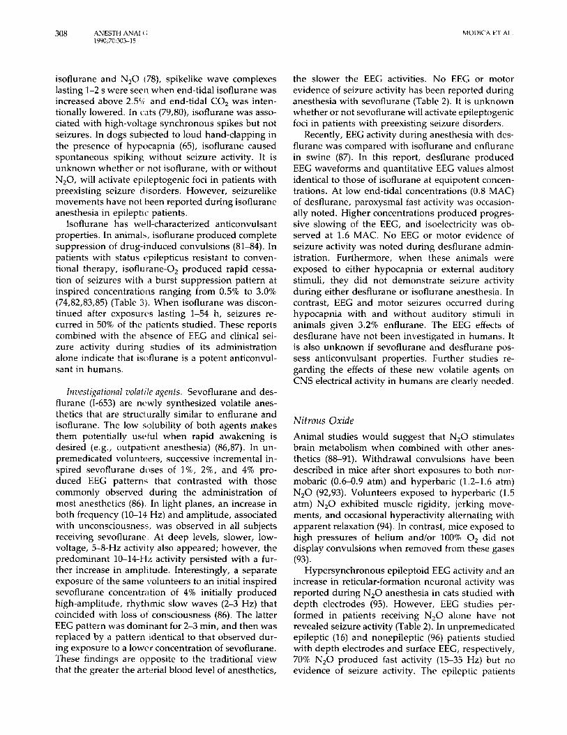
Table 3 . Anticonvulsant Effects on Inhalation Anesthetics in Humans
Anticonvulsant documentation Type of EEG
electrodes Clinical EEG used in
Agent report study study References
Nitrous oxide - - Surface 85 Halothane + + Surface 53, 66 Enflurane + + Surface 53, 54 Isoflurane + + Surface 74, 82, 83, 85 Sevoflurane N/A N/A Desflurane (1-653) N/A N/A
+, successful termination of status epilepticus reported; -, failure to eliminate seizures reported; EEG, electroencephalographic; NIA, informa- tion not available.
enflurane (53) (Table 3). The disappearance of EEG seizure activity was evident 20 min after induction and no further convulsive activity was noted during recovery. Despite these anticonvulsant findings, en- flurane has a propensity for inducing epileptiform activity during anesthesia, especially when high con- centrations are administered in the presence of hy- pocarbia.
Halothane. Halothane alone has not been reported to cause convulsions in humans (Table 2). In the few reports (58,59) of halothane-related seizures, N20 was also administered. Electroencephalographic monitoring in normal patients had not revealed epi- leptiform activity during halothane anesthesia (60- 62). However, nonspecific EEG slowing and rare sharp wave activity (attributed to the persistence of halothane and its metabolites) was seen in volunteers during the first week after 13-14 MAC-hours of halothane administration (60). In three epileptic pa- tients, brief periods (<7 s) of bilateral atypical and polyspike waves were noted during emergence from halothane, but were not associated with clinical sei- zure activity during the perioperative period (63). Mild transient clonus has been observed after halothane anesthesia (37). Epileptic activity has been reported in cats given a high concentration of halothane (7%); however, cerebral blood flow and metabolism is also markedly altered at this concen- tration (64). Although depth electrodes have revealed occasional hippocampal spikes in cats (46), evoked spikes were not detected during halothane anesthesia in dogs subjected to loud hand-clapping in the pres- ence of hypocapnia (65).
Inhaled anesthetics, including halothane, have been recommended for treatment of continuous seizures refractory to conventional intravenous anti- convulsant agents (66). Successful termination of resistant status epilepticus in children has been dem-
onstrated with halothane (53) (Table 3). In these cases, the disappearance of spikes and waves was evident within 20 min. No seizures were noted after recovery. These potent anticonvulsant effects com- bined with the lack of clinically important cerebral excitatory findings for halothane would suggest that it does not possess proconvulsant properties.
Isoflurune. When administered alone, isoflurane has not been found to cause EEG or clinical seizure activity in anesthetized patients (Table 2). In the reported cases of isoflurane-related seizures (67-69), N,O was also administered. In two of these cases, abnormal myoclonic movements starting intraopera- tively and lasting into the early postanesthetic period were the only evidence of seizure activity (67,68). Furthermore, one of these patients was conscious throughout the recovery period (67). In contrast to the reported convulsions after enflurane (23,30,36), the postoperative EEG was normal in both reports. Alternative explanations for these seizurelike move- ments include nonepileptic myoclonus (3,70) and/or the so-called withdrawal phenomena from N20 (71,72).
In another case report (69), isoflurane-N,O anes- thesia was associated with tonic-clonic activity early during inhalational induction on two separate occa- sions. Before the second anesthetic, EEG studies were entirely normal. With EEG monitoring during the second induction, spike and wave forms were recorded during the clinical seizure and both ceased after administration of thiamylal. In this case, the finding that the spiking activity appeared with induc- tion but did not continue during maintenance or reappear during recovery implies that N,O may have precipitated the seizure activity (73). Furthermore, N 2 0 alone has been associated with clinical seizures during induction (58,64), whereas isoflurane’s anti- convulsant properties may have prevented further seizures during the maintenance and recovery peri- ods (73,74).
In unpremedicated volunteers, induction of isoflu- rane anesthesia is characterized by low-voltage fast EEG activity that progresses toward high-voltage slow wave activity at 1 MAC. Bursts of slow high- voltage activity separated by electrical silence are noted at 1.5 MAC, with electrical silence at 22 MAC (75-77). Epileptic or spiking patterns do not appear, nor can they be evoked by lowering the Paco, or by introduction of auditory and visual stimuli (75,77). unpremedicated volunteers did not demonstrate clinical or EEG evidence of seizure activity during isoflurane-N20 inhaled induction (77). In premedi- cated patients undergoing inhaled induction with
308 ANESTH ANAI (; 1990;70:30>15
MODICA ET AL.
isoflurane and N,O (78), spikelike wave complexes lasting 1-2 s were seen when end-tidal isoflurane was increased above 2.5% and end-tidal CO, was inten- tionally lowered. In c'its (79,80), isoflurane was asso- ciated with high-voltage synchronous spikes but not seizures. In dogs subjected to loud hand-clapping in the presence of hypocapnia (65), isoflurane caused spontaneous spiking without seizure activity. It is unknown whether or not isoflurane, with or without N,O, will activate epileptogenic foci in patients with preexisting seizure disorders. However, seizurelike movements have not been reported during isoflurane anesthesia in epileptic patients.
Isoflurane has well-characterized anticonvulsant properties. In animals, isoflurane produced complete suppression of drug-induced convulsions (81-84). In patients with status epilepticus resistant to conven- tional therapy, isoflurane-0, produced rapid cessa- tion of seizures with a burst suppression pattern at inspired concentrations ranging from 0.5% to 3.0% (74,82,83,85) (Table 3). When isoflurane was discon- tinued after exposures lasting 1-54 h, seizures re- curred in 50% of the patients studied. These reports combined with the absence of EEG and clinical sei- zure activity during studies of its administration alone indicate that isoflurane is a potent anticonvul- sant in humans.
lnvestigatioiial volatile agents. Sevoflurane and des- flurane (1-653) are newly synthesized volatile anes- thetics that are structurally similar to enflurane and isoflurane. The low solubility of both agents makes them potentially usetul when rapid awakening is desired (e.g., outpatient anesthesia) (86,87). In un- premeditated volunteers, successive incremental in- spired sevoflurane doses of 1%, 2%, and 4% pro- duced EEG patterns that contrasted with those commonly observed during the administration of most anesthetics (86). In light planes, an increase in both frequency (10-14 Hz) and amplitude, associated with unconsciousness, was observed in all subjects receiving sevoflurane. At deep levels, slower, low- voltage, 5-8-Hz activity also appeared; however, the predominant 10-14-Hz activity persisted with a fur- ther increase in amplitude. Interestingly, a separate exposure of the same volunteers to an initial inspired sevoflurane concentr'i tion of 4% initially produced high-amplitude, rhythmic slow waves (2-3 Hz) that coincided with loss ot consciousness (86). The latter EEG pattern was dominant for 2-3 min, and then was replaced by a pattern identical to that observed dur- ing exposure to a lower concentration of sevoflurane. These findings are opposite to the traditional view that the greater the arterial blood level of anesthetics,
the slower the EEG activities. No EEG or motor evidence of seizure activity has been reported during anesthesia with sevoflurane (Table 2). It is unknown whether or not sevoflurane will activate epileptogenic foci in patients with preexisting seizure disorders.
Recently, EEG activity during anesthesia with des- flurane was compared with isoflurane and enflurane in swine (87). In this report, desflurane produced EEG waveforms and quantitative EEG values almost identical to those of isoflurane at equipotent concen- trations. At low end-tidal concentrations (0.8 MAC) of desflurane, paroxysmal fast activity was occasion- ally noted. Higher concentrations produced progres- sive slowing of the EEG, and isoelectricity was ob- served at 1.6 MAC. No EEG or motor evidence of seizure activity was noted during desflurane admin- istration. Furthermore, when these animals were exposed to either hypocapnia or external auditory stimuli, they did not demonstrate seizure activity during either desflurane or isoflurane anesthesia. In contrast, EEG and motor seizures occurred during hypocapnia with and without auditory stimuli in animals given 3.2% enflurane. The EEG effects of desflurane have not been investigated in humans. It is also unknown if sevoflurane and desflurane pos- sess anticonvulsant properties. Further studies re- garding the effects of these new volatile agents on CNS electrical activity in humans are clearly needed.
Nitrous Oxide Animal studies would suggest that N,O stimulates brain metabolism when combined with other anes- thetics (88-91). Withdrawal convulsions have been described in mice after short exposures to both nor- mobaric (0.6-0.9 atm) and hyperbaric (1.2-1.6 atm) N,O (92,93). Volunteers exposed to hyperbaric (1.5 atm) N,O exhibited muscle rigidity, jerking move- ments, and occasional hyperactivity alternating with apparent relaxation (94). In contrast, mice exposed to high pressures of helium and/or 100% 0, did not display convulsions when removed from these gases (93).
Hypersynchronous epileptoid EEG activity and an increase in reticular-formation neuronal activity was reported during N,O anesthesia in cats studied with depth electrodes (95). However, EEG studies per- formed in patients receiving N,O alone have not revealed seizure activity (Table 2). In unpremedicated epileptic (16) and nonepileptic (96) patients studied with depth electrodes and surface EEG, respectively, 70% N,O produced fast activity (15-35 Hz) but no evidence of seizure activity. The epileptic patients
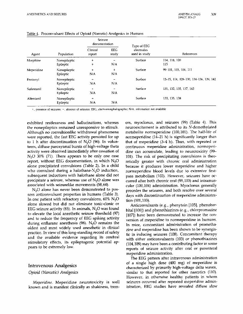
ANESTHETICS AND SEIZURES ANESTH ANALG 309 19Y13;7(1~3O,L1 i
Table 4. Proconvulsant Effects of Opioid (Narcotic) Analgesics in Humans Seizure
documentation Type of EEG
Clinical EEG electrodes Agent Population report study used in study References
Morphine Nonepileptic
Meperidine Nonepileptic
Fentanyl Nonepileptic
Sufentanil Nonepileptic
Alfen tanil Nonepileptic
Epileptic
Epileptic
Epileptic
Epileptic
Epileptic
+ + +
NIA +
NIA +
NIA
+ NIA
- Surface 114, 118, 120
f Surface 99-101, 103, 104, 111
- Surface 1 ~ 1 5 , 114, 128-130. 1 3 . ~ 3 6 , 139. 142
NIA 115
NIA
N/A
Surface 131, 132, 135, 137, 143 - NIA - Surface 133, 135, 138
N/A
+, presence of seizures, -, absence of seizures, EEG, electroencephalographlc. N/A, informahon not available
exhibited restlessness and hallucinations, whereas the nonepileptics remained unresponsive to stimuli. Although no convulsionlike withdrawal phenomena were reported, the fast EEG activity persisted for up to 1 h after discontinuation of N 2 0 (96). In volun- teers, diffuse paroxysmal bursts of high-voltage theta activity were observed immediately after cessation of N,O 30% (71). There appears to be only one case report, without EEG documentation, in which N 2 0 alone precipitated convulsions (Table 2). In a child who convulsed during a halothane-N,O induction, subsequent inductions with halothane alone did not precipitate a seizure, whereas use of N,O alone was associated with seizurelike movements (58,64).
N,O alone has never been demonstrated to pos- sess anticonvulsant properties in humans (Table 3). In one patient with refractory convulsions, 60% N 2 0 alone slowed but did not eliminate tonic-clonic or EEG seizure activity (85). In animals, N,O was found to elevate the local anesthetic seizure threshold (97) and to reduce the frequency of EEG spiking activity during enflurane anesthesia (98). N 2 0 remains the oldest and most widely used anesthetic in clinical practice. In view of this long-standing record of safety and the available evidence regarding its cerebral stirnulatory effects, its epileptogenic potential ap- pears to be extremely low.
Intravenous Analgesics Opioid (Narcotic) Analgesics
Meperidine. Meperidine neurotoxicity is well known and is manifest clinically as shakiness, trem-
ors, myoclonus, and seizures (99) (Table 4). This neuroexcitement is attributed to its N-demethylated metabolite normeperidine (100,101). The half-life of normeperidine (14-21 h) is significantly longer than that of meperidine ( 3 4 h). Thus, with repeated or continuous meperidine administration, normeperi- dine can accumulate, leading to neurotoxicity (99- 101). The risk of precipitating convulsions is theo- retically greater with chronic oral administration because it produces lower meperidine and higher normeperidine blood levels due to extensive first- pass metabolism (102). However, seizures have oc- curred after both chronic oral (99,103) and intramus- cular (100,104) administration. Myoclonus generally precedes the seizures, and both resolve over several days with discontinuation of meperidine administra- tion (101,103).
Anticonvulsants (e.g., phenytoin [ 1051, phenobar- bital [106]) and phenothiazines (e.g., chlorpromazine [107]) have been demonstrated to increase the con- version of meperidine to normeperidine in humans. In mice, concomitant administration of prometha- zine and meperidine has been shown to be synergis- tic in inducing seizures (108). Concomitant therapy with either anticonvulsants (103) or phenothiazines (104,109) may have been a contributing factor in some reports of seizure activity after oral or parenteral meperidine administration.
The EEG pattern after intravenous administration of a single high dose (400 mg) of meperidine is characterized by primarily high-voltage delta waves, similar to that reported for other narcotics (110). However, in otherwise healthy patients in whom seizures occurred after repeated meperidine admin- istration, EEG studies have revealed diffuse slow
310 ANESTH ANAl G 1 W0;70: 30% 15
MODICA ET AL.
activity and epileptiform discharges (99,111) (Ta- ble 4).
Patient subgroups that appear to be more suscep- tible to the neurotoxicity associated with chronic meperidine therapy include (a) patients with renal failure in whom the normeperidine half-life is pro- longed (34 h) due to decreased metabolism and excretion (100,103), and (b) patients with advanced malignancy (100,101) or sickle cell anemia (112), both of whom receive progressively larger doses of mepe- ridine due to the development of tolerance. Seizure- like movements have not been reported in epileptic patients during either acute or chronic meperidine administration. Furthermore, it is unknown whether or not acute or chronic meperidine use will activate epileptogenic EEG foci in patients with preexisting seizure disorders.
Meperidine has demonstrated the lowest safety margin for convulsions of all narcotics studied. In dogs, EEG seizure activity was induced with an average intravenous rneperidine dose (20 mg/kg) that was only 2.2 times greater than the mean effective intravenous dose (9 mg/kg) for surgical analgesia (8). In mice, naloxone can only partially block the convul- sions induced by normeperidine, whereas meperi- dine-induced seizures are completely blocked by prior administration of naloxone (113). Neither me- peridine nor normeperidine has ever been shown to possess anticonvulsant properties. Within the range of meperidine doses used clinically to supplement general or regional anesthetic techniques, the procon- vulsant effects of the drug appear to be of little concern. However, seizures may result from norme- peridine accumulation after prolonged meperidine administration (e.g., patient-controlled analgesia), especially in patients with renal failure, sickle cell, and cancer.
Morphine. Morphine alone has never been demon- strated to produce stlizure activity in humans after intravenous administration. High-dose morphine (1- 2 mg/kg, IV) during open-heart surgery was associ- ated with progressive slowing of EEG activity and an increase in low-frequency amplitude. No epilepti- form activity was noted (114). Electroencephalo- graphic investigations have not been performed in epileptic patients during high-dose morphine admin- istration. However, A tonic-clonic seizure was re- ported after administration of epidural morphine to a known epileptic (115) (Table 4). There was a 6-h delay between epidural administration and the onset of seizure, consistent with the known kinetics of mor- phine in the cerebrospinal fluid (116,117). A convul- sion has also occurred after intrathecal morphine in a
nonepileptic cancer patient (118). These reports sug- gest a subcortical origin for morphine-induced sei- zures. Electrophysiologic studies in rats support this concept as morphine causes disinhibition of the py- ramidal cells of the hippocampus, leading to seizure discharges (119). A grand ma1 seizure in a 14-yr-old boy occurred 90 min after 20 mg of intramuscular morphine and 0.4 mg of scopolamine were adminis- tered (120). A prodrome of intense itching was expe- rienced by the patient, indicating that morphine probably contributed to the seizure activity, although the well-known excitatory CNS effects of scopola- mine may have been an additional contributing factor in this case. Withdrawal seizures have been reported clinically, predominantly in neonates born to heroin- addicted mothers (121).
DeCastro et al. (8) found that an average morphine dose of 180 mg/kg, IV, was required to precipitate EEG seizure activity in dogs. This was 72 times greater than the mean effective intravenous dose (2.5 mg/kg) for surgical analgesia. In rats, higher systemic doses of morphine produce epileptiform patterns and behavioral convulsions that are not reversed by opi- ate antagonists (122). Seizures have also been dem- onstrated after intracerebroventricular administration of morphine in rats (123,124) and after intrathecal administration of morphine in dogs (125) and rats (126). Although the cerebroventricular-induced sei- zures are naloxone reversible (124), the intrathecal morphine-induced tonic-clonic movements were not naloxone reversible (126). These findings suggest that the proconvulsant actions of high doses of morphine in animals are mediated by both opiate (e.g., mu) and nonopiate (e.g., y-aminobutyric acid) receptor mech- anisms (122,127).
Morphine alone has never been demonstrated to possess anticonvulsant properties in humans. In rab- bits, intravenous morphine terminated bicuculline- and picrotoxin-induced EEG and behavioral seizures (127). In rats, intravenous morphine delayed the onset of pentylenetetrazol- and flurothyl-induced sei- zures (122). Although intravenous morphine in high doses has both pro- and anticonvulsant properties in animals, the doses of the drug used in clinical practice appear to have little effect on the seizure threshold. This is especially true with noncerebrospinal fluid routes of morphine administration.
Fenfanyl and its analogues. There have been several reports of grand ma1 seizure-like motor behavior in patients after administration of low (1OC-200 pg) (14,15,128,129) to moderate (2250-2500 pg) (13,130) doses of intravenous fentanyl. This phenomena has also occurred after sufentanil (40-150 pg, IV [131,
ANESTHETICS AND SEIZURES ANESTH ANALG 31 1 1990;7@:30>15
1321) and alfentanil (1500 pg, IV [133]) administration (Table 4). None of the patients had a history of seizure disorder and their EEGs after surgery were unremarkable. Unfortunately, in these case reports, the authors did not have the benefit of EEG recording during the seizurelike event.
Surface EEG recordings in patients treated with high intravenous doses of fentanyl (60-150 pg/kg [114,134-136]), sufentanil (15 pgkg [135,137]), or alfentanil (>50 p g k g [135,138]) are characterized by high-voltage slow delta waves. Similar EEG findings were also demonstrated in patients receiving low-to- moderate doses of fentanyl (139). Except for sporadic isolated sharp waves noted in two of the high-dose fentanyl studies (134,136), these EEG investigations did not detect any epileptic spike waves or other abnormal patterns (Table 4). The significance of these sharp waves is not known as they were nonepilepti- form in appearance and never became generalized (134). Although they have not been related to tonic- clonic movements, similar sharp waves also have been noted during the administration of other drugs known to be epileptogenic (e.g., phencyclidine [140], intrathecal metrizamide [ 1411). Whether sharp waves represent an EEG equivalent of a CNS stimulatory action or merely EEG artifact remains unclear. Al- though tonic-clonic movements associated with their administration have not been reported in epileptic patients, EEG studies have never been performed in this population during anesthesia with fentanyl or its analogues alone.
Abnormal motor activity resembling epileptic con- vulsions has occurred in the absence of cortical sei- zure activity on simultaneous EEG recordings made during low-dose (500-600 pg, IV) fentanyl (132,142) and sufentanil (1.3 pglkg, IV) (143) administration. Yet, seizurelike movements were not observed dur- ing EEG studies in patients treated with high doses of fentanyl and its analogues. There are several possible explanations for these observations. The abnormal movements observed after low-to-moderate doses of fentanyl or sufentanil could be due to nonepileptic myoclonus produced by the interaction of these nar- cotics with opiate receptors leading to blockade of cortical inhibitory pathways. This would allow lower CNS centers in the brainstem and/or spinal cord to display altered excitability (4,142). Patients treated with higher doses of fentanyl and its analogues may have failed to exhibit these seizurelike myoclonic movements because the plasma opioid levels were high enough to depress both higher and lower CNS centers. In one report, nonepileptic myoclonus was probably responsible for the seizurelike motor activ- ity observed after recovery from low-dose fentanyl
administration (128). In addition, this patient also received etomidate, an anesthetic that produces non- epileptic myoclonic activity (144), which occasionally persists into the postoperative period (145,146).
An alternative explanation is that these move- ments represent an exaggerated form of narcotic- induced rigidity (142). Rigidity can occur after low doses of fentanyl (147) and high-dose alfentanil in- ductions have produced rigidity involving all extrem- ities that closely resembles seizures in volunteers (148). Recent studies have suggested that rigidity may involve neurochemical mechanisms in the stria- tonigral pathways similar to Parkinson’s disease (149-151). Thus, exaggerated rigidity could have been responsible for the seizurelike movements observed after low-dose sufentanil administration (131). In these two cases, one patient had a history of Parkin- son’s disease, and the other patient was receiving chronic metoclopramide therapy. Metoclopramide in- hibits cerebral dopaminergic pathways and can pro- duce extrapyramidal signs (152,153).
The tonic-clonic movements reported with admin- istration of low-to-moderate doses of fentanyl and sufentanil could also be due to subcortical seizure activity. Electroencephalograph-documented cortical seizures have been induced by high-dose fentanyl (8,154,155) and sufentanil (156) in animals. The EEG seizures in rats were accompanied by decreases in LCGU in cortical structures and relative increases in LCGU in the subcortical limbic system (hippocam- pus, amygdala, claustrum) (154,155), an area rich in opioid receptors. In humans, subcortical seizures are not detected by surface EEG leads, and EEG studies with depth electrodes are required to confirm or reject the hypothesis that fentanyl and sufentanil cause seizures in humans (155).
Fentanyl and its analogues alone have never been demonstrated to possess anticonvulsant properties in either humans or animals. However, during fentanyl administration with droperidol and N,O (neurolep- tanesthesia), EEG monitoring in epileptic patients did not demonstrate an increase in spiking activity com- pared with their baseline EEG studies (53). In a study of 104 patients receiving neuroleptanesthesia, no EEG or clinical signs of seizures were observed dur- ing or after neuroradiologic examinations with in- trathecal metrizamide, a known convulsant (141). As suggested by DeCastro et al. (8), these findings appear to indicate that droperidol may offer some protection against narcotic-induced neuroexcitation.
Whether the seizurelike movements reported dur- ing clinical administration of fentanyl and its ana- logues originate from epileptogenic or nonepilepto- genic mechanisms is unclear. All currently available
ANESTH ANAL C; 1990:70:30515
312 MODICA ET AL.
clinical and EEG evidence appears to favor either nonepileptic myoclonus or exaggerated rigidity as the most likely explanation. However, further studies regarding the neurochemical mechanisms of narcotic- induced rigidity and the effects of progressively higher doses of fentanyl and its analogues on electri- cal activity in the limbic system are needed.
Summary Many inhaled anesthetics and intravenous analgesics have been alleged to produce both proconvulsant and anticonvulsant activity in humans (Table 1). The reasons for these contrasting actions on the CNS are poorly understood at the present time. However, biologic variability plays an important role in deter- mining individual patient’s responses to anesthetic and analgesic drugs. In addition, variations in the responsiveness of inhibitory and excitatory neurons to the central depressant effects of these drugs could also explain these apparently conflicting data. De- pending on the brain concentration, centrally active drugs may produce differing effects on the CNS inhibitory and excitatory neurotransmitter systems. The availability of increasingly powerful magnetic resonance imaging techniques to provide noninva- sive information about tissue chemistry (e.g., neuro- transmitters and citric acid cycle metabolites) and positron emission tomography to noninvasively eval- uate CNS drug-receptor interactions should lead to a more in-depth understanding of the in vivo effects of anesthetics and analgesics on the CNS.
In the second part of this review article, we discuss the pro- and anticonvulsant effects of the sedative- hypnotics, local anesthetics, and other anesthetic adjuvant drugs.
References 1. Adams RD, Victor hl. Principles of neurology. New York:
McGraw-Hill, 1985:23?-54. 2. Halliday AM. The neurophysiology of myoclonic jerking-
a reappraisal. In: Charlton MH, ed. Myoclonic seizures. Amsterdam: Excerpta Medica, 1975:l-29.
3. Marsden CD, Hallett M, Fahn S. The nosology and patho- physiology of myoclonus. In: Marsden CD, Fahn s, eds. Movement disorders London: Butterworth, 1982:19&248.
4. Swanson PD, Luttrell CN, Magladery JW. Myoclonus--a report of 67 cases and review of the literature. Medicine
5. Penry JK, Hoefnagel L), Vanden Noort S. Muscle spasm and abnormal posture resulting from damage to interneurones in the spinal cord. Arch Neurol 1960;34:5604.
6. Winters WD, Ferrer-Allado T, Guzman-Flores C, Alcaraz M. The cataleptic state induced by ketarnine: a review of the
1962;41:339-56.
neuropharmacology of anesthesia. Neuropharmacology 1972; 11:30?-15.
7. Winters WD. Epilepsy or anesthesia with ketamine (editori- al). Anesthesiology 1972;36:309-12.
8. DeCastro J, van de Water A, Wouters L, Xhonneux R, Reneman R, Kay B. Comparative study of cardiovascular, neurological, and metabolic side effects of eight narcotics in dogs. Acta Anaesthesiol Belg 1979;30:6-99.
9. Fisher MMcD. Use of ketamine hydrochloride in the treat- ment of convulsions. Anaesth Intensive Care 1974;2:266-8.
10. Davis RW, Tolstoshev GC. Ketamine use in severe febrile convulsions (letter). Med J Aust 1976;2:465-6.
11. Gumpert J, Paul R. Activation of the electroencephalogram with intravenous Brietal (methohexitone): the findings in 100 cases. J Neurol Neurosurg Psychiatry 1971;34:646-8.
12. Paul R, Harris R. A comparison of methohexitone and thio- pentone in electrocorticography. J Neurol Neurosurg Psychi- atry 1970;33:10W.
13. Rao TLK, Mummaneni N, El-Etr AA. Convulsions: an un- usual response to intravenous fentanyl administration. Anesth Analg 1982;61:102C-l.
14. Safwat AM, Daniel D. Grand ma1 seizure after fentanyl administration (letter). Anesthesiology 1983;59:78.
15. Hoien AO. Another case of grand ma1 seizure after fentanyl administration (letter). Anesthesiology 1984;60:387-8.
16. Ferrer-Allado T, Brechner VL, Dymond A, Cozen H, Crandall P. Ketamine-induced electroconvulsive phenomena in the human limbic and thalamic regions. Anesthesiology 1973;38: 333-44.
17. Virtue RW, Lund LO, Phelps M Jr, Vogel JHK, Beckwitt H, Heron M. Difluoro-methyl 1,1,2-trifluoro-2-chloroethyl ether as an anesthetic agent: results with dogs, and a preliminary note on observations in man. Can Anaesth SOC J 1966;13:233- 41.
18. Botty C, Brown B, Stanley V, Stephan CR. Clinical experi- ences with compound 347, a halogenated anesthetic agent. Anesth Analg 1968;47:477-505.
19. Lebowitz MH, Blitt CD, Dillon JB, Clinical investigation of compound 347 (Ethrane). Anesth Analg 1970;49:1-10.
20. Bart A], Homi J, Linde HW. Changes in power spectra of electroencephalograms during anesthesia with fluroxene, methoxyflurane and Ethrane. Anesth Analg 1971;50:5343.
21. Wollman H, Smith AL, Hoffman JC. Cerebral blood flow and oxygen consumption in man during electroencephalographic seizure patterns induced by anesthesia with Ethrane. Fed Proc 1969;28:356.
22. Niejadlik K, Galindo A. Electrocorticographic seizure activity during enflurane anesthesia. Anesth Analg 1975;54:7224.
23. Fariello RG. Epileptogenic properties of enflurane and their clinical interpretation. Electroencephalogr Clin Neurophysiol 1980;48:595-8.
24. Flemming DC, Fitzpatrick J, Fariello RG, Duff T, Hellman D, Hoff BH. Diagnostic activation of epileptogenic foci by enflu- rane. Anesthesiology 1980;52:431-3.
25. Lebowitz MH, Blitt CD, Dillon JB. Enflurane-induced central nervous system excitation and its relation to carbon dioxide tension. Anesth Analg 1972;51:355-63.
26. Burchiel KJ, Stockard JJ, Myers RR, Smith NT, Calverly RK, Bickford RG. Metabolic and electrophysiologic mechanisms in the initiation and termination of enflurane-induced seizures in man and cats. Electroencephalogr Clin Neurophysiol 1975; 38:555.
27. DeWolf AM, Chang JL, Larson CE, Caparosa RJ. Enflurane- induced grand ma1 seizures during otic microsurgery. Anesth Prog 1984;31:13&7.
ANESTHETICS AND SEIZURES ANESTH ANALG 313 1990;70:30>15
28.
29.
30.
31.
32.
33.
34.
35.
36.
37.
38.
39 I
40,
41,
42.
43
44.
45.
46.
47.
48.
49.
50.
51.
Neigh JL, Garman JK, Harp JR. The electroencephalographic pattern during anesthesia with Ethrane: effects of depth of anesthesia, Paco,, and nitrous oxide. Anesthesiology 1971;
Stockard JJ, Bickford RG. The neurophysiology of anesthesia. In: Amsterdam GE, ed. A basis and practice of neuroanaes- thesia. New York Elsevier, 1981349. Kruczek M, Albin MS, Wolf S, Bertoni JM. Postoperative seizure activity following enflurane anesthesia. Anesthesiol- ogy 1980;53:1754. Allan NS. Convulsions after enflurane. Anesthesia 1984;39 605-6. Yazji NS, Seed RF. Convulsive reaction following enflurane anesthesia. Anesthesia 19&4;391249. Opitz A, Brecht S, Stenzel E. Enflurane anesthesia for epilep- tic patients. Anaesthesist 1977;26329-32. Nicoll JMV. Status epilepticus following enflurane anaesthe- sia. Anaesthesia 1986;41:92730. Grant IS. Delayed convulsions following enflurane anaesthe- sia. Anaesthesia 1986;41:1024-5. Ohm WW, Cullen BF, Amory DW, Kennedy RD. Delayed seizure activity following enflurane anesthesia. Anesthesiol- ogy 1975;42:367-8. Rosenberg H, Clofine R, Bialik 0. Neurological changes during awakening from anesthesia. Anesthesiology 1981;W 125-30. Jenkins J, Milne AC. Convulsive reaction following enflurane anaesthesia. Anaesthesia 1984;39:445. Ng ATH. Prolonged myodonic contractions after enflurane anaesthesia4 case report. Can Anaesth Soc J 1980;27502-3. Burchiel KJ, Stockard JJ, Calverly RK, Smith NT. Relationship of pre- and postanesthetic EEG abnormalities to enflurane- induced seizure activity. Anesth Analg 1977;56:509-14. Julien RM, Kavan EM. Electrographic studies of a new volatile anesthetic agent: enflurane (Ethrane). J Pharmacol Exp Ther 1972;183:393-403. Furgang FA, Sohn JJ. The effect of thiopentone on enflurane- induced cortical seizures. Br J Anaesth 1977;49127-32. Darimont PC, Jenkins LC. The influence of intravenous anaesthetics on enflurane-induced central nervous system seizure activity. Can Anaesth Soc J 1977;2442-56. Moorthy SS, Reddy RV, Paradise RR, Losasso AM, Gibbs PS. Reduction of enflurane-induced spike activity by scopola- mine. Anesth Analg 1980;59:417-20. Bostem F, Hanquet M, Gallex JP. Enflurane (Ethrane) and EEG. Acta Anaesthesiol Belg 1971;2523?-45. Kavan EM, Julien RM, Lucero JJ. Electrographic alterations induced in limbic and sensory systems during induction of anaesthesia with halothane, methoxyflurane, diethyl ether, and enflurane (Ethrane). Br J Anaesth 1972;44.1234-9. Myers RR, Shapiro HM. Local cerebral metabolism during enflurane anesthesia: identification of epileptogenic foci. Elec- troencephalogr Clin Neurophysiol 1979;47153-62. Herkenham M. Anesthetics and the habenulo-interpeduncu- lar system: selective sparing of metabolic activity. Brain Res 1981;210461-6. Hodes JE, Soncrant TT, Larson DM, Carlson SG, Rapoport SI. Selective changes in local cerebral glucose utilization induced by phenobarbital in the rat. Anesthesiology 1985;63:633-9. Shapiro HM, Greenberg JH, Reivich M, Ashmead G, Sokoloff L. Local cerebral glucose uptake in awake and halothane- anesthetized primates. Anesthesiology 1978;48:97-103. Savaki HE, Desban M, Glowinski J, Besson MJ. Local cerebral glucose consumption in the rat. I. Effects of halothane anes- thesia. J Comp Neurol1983;2133645.
35~482-7.
52.
53.
54.
55.
56.
57.
58.
59.
60.
61.
62.
63.
64.
65.
66.
67.
68.
69.
70,
71.
72,
73,
Nakakimura K, Sakabe T, Funatsu N, Maekawa T, Takeshita H. Metabolic activation of intercortical and corticothalamic pathways during enflurane anesthesia in rats. Anesthesiol- ogy 1988;68.777-82. Opitz A, Marschall M, Degan R, Koch D. General anesthesia in patients with epilepsy and status epilepticus. In: Delgado- Escueta AV, Wasterlain CG, Treiman DM, Porter RJ, eds. Status epilepticus: mechanisms of brain damage and treat- ment. New York Raven, 1983:531-5. Opih A, Obenvetter WD. Edurane or halothane anaesthesia for patients with cerebral convulsive disorders? Acta Anes- thesiol Scand 1979;71(Suppl):43-7. Gallagher TJ, Galindo A, Richey ET. Inhibition of seizure activity during enflurane anesthesia. Anesth Analg 1978;57 130-2. Buzello W, Jantzen K, Scholler KL. The influence of Ethrane on the electro- and pentylenetetrazol-convulsions in mice. Anaesthesist 1975;2411&9. Oshima E, Urabe N, Shingu K, Mori K. Anticonvulsant actions of enflurane on epilepsy models in cats. Anesthesiol- ogy 1985;63294. Krenn J, Porges P, Steinbereithner K. Case of anesthesia convulsions under nitrous oxide-halothane anesthesia. An- aesthesist 1967;16:83-5. Smith PA, McDonald TR, Jones CS. Convulsions associated with halothane anaesthesia. Anaesthesia 1966;21:229-33. Burchiel KJ, Stockard JJ, Calverley RK, Smith NT, Scholl ML, Mazze RI. Electroencephalographic abnormalities following halothane anesthesia. Anesth Analg 1978;5724451. Badanan LE, Lofstrom B, Widen V. Electroencephalography in halothane anesthesia. Acta Anaesthesiol Scand 1964;8115 30. Findeiss JC, Kien GA, Huse KOW, Linde HW. Power spectral density of electroencephalogram during halothane and cyclo- propane anesthesia in man. Anesth Analg 1969;48:101%23. Bennett DR, Madsen JA, Jordan WS, Wiser WC. Ketamine anesthesia in brain-damaged epileptics: electroencephalo- graphic and clinical observations. Neurology 1973;23449-60. Steen PA, Michenfelder J . Neurotoxicity of anesthetics. An- esthesiology 1979;50:437-53. Joas TA, Stevens WC, Eger EI 11. Electroencephalographic seizure activity in dogs during anaesthesia. Br J Anaesth 1971;43:73945. Delgado-Escueta AV, Wasterlain CG, Treiman DM, Porter RJ. Management of status epilepticus. N Engl J Med 1982;306 13374. Hymes JA. Seizure activity during isoflurane anesthesia. Anesth Analg 1985;64:367-8. Harrison JL. Postoperative seizures after isoflurane anesthe- sia. Anesth Analg 1986;65:1235-6. Poulton TJ, Ellingson RJ. Seizure associated with induction of anesthesia with isoflurane. Anesthesiology 1984;61:471-6. Durrani Z. Perioperative myoclonia or seizures (letter). Anesth Analg 1987;66.5W. Henrie JR, Parkhouse J, Bickford RG. Alteration of human consciousness by nitrous oxide as assessed by electroenceph- alography and psychological tests. Anesthesiology 1961;22: 247-59. Frost EAM. Seizures after anesthesia: identifying the causes (letter). Anesth Analg 1987;66:10534. Eger EI 11. Are seizures caused by nitrous oxide or isoflurane (letter)? Anesthesiology 1985;62697-8.
74. Kofke WA, Snider MT, Young RSK, Ramer JC. Prolonged low flow isoflurane anesthesia for status epilepticus. Anesthesiol- ogy 1985;62653-6.
ANESTH ANALC 1990;70303-15
314 MODICA ET AL.
75. Eger EI 11, Stevens WC, Cromwell TH. The electroencepha- logram in man anesthetized with Forane. Anesthesiology 1971;35:504-8.
76. Homi J, Konchigeri HN, Eckenhoff JE, Linde HW. A new anesthetic agent-Forane: preliminary observations in man. Anesth Analg 1972;51:43%47.
77. Clark DL, Hosick EC, Adam N, Castro AD, Rosner BS, Neigh JL. Neural effects of isoflurane (Forane) in man. Anesthesiol-
78. Pauca AL, Dripps RD. Clinical experience with isoflurane (Forane). Br J Anaesth 1973;45:697-703.
79. Kavan EM, Julien RM. Central nervous system effects of isoflurane (Forane). Can Anaesth SOC J 1974;21:390402.
80. Julien RM, Kavan EM. Electrographic studies of isoflurane (Forane). Neuropharmacology 1974;13:677-81.
81. Koblin DD, Eger EI 11, Johnson BH, Collins BA, Terrell RC, Speers L. Are convulsant gases also anesthetics? Anesth Analg 1981;60:464-70.
82. Kofke WA, Snider MT, OConnell BK, et al. Isoflurane stops refractory seizures. Anesthesiol Rev 1987;1558-9.
83. Kofke WA, Ropper A, Young RSK, et al. Isoflurane for status epilepticus. Neurology 1987;37(Suppl 1):103.
84. Kofke WA, Towfighi J, OConnell BK, Derr J, Hawkins RA. Neuropathological effects of anesthetics used to stop status epilepticus in rats. Anesthesiol Rev 1987;15:27-8.
85. Ropper AH, Kofke WA, Bromfield EB, Kennedy SK. Com- parison of isoflurane, halothane, and nitrous oxide in status epilepticus (letter). AM Neurol 1986;1998-9.
86. Avramov MN, Shingu K, Omatsu Y, Osawa M, Mori K. Effects of different speeds of induction with sevoflurane on the EEG in man. J Anesth 1987;l:l-7.
87. Rampil IJ, Weiskopf RB, Brown JG, et al. 1-653 and isoflurane produce similar dose-related changes in the electroencepha- logram of pigs. Anesthesiology 1988;69:298-302.
88. Theye RA, Michenfelder JD. The effect of nitrous oxide on canine cerebral metabolism. Anesthesiology 1976;29:1119-24.
89. Pelligrino DA, Miletich DJ, Hoffman WE, Albrecht RA. Ni- trous oxide markedly increases cerebral cortical metabolic rate and blood flow in the goat. Anesthesiology 1984;60:405-12.
90. Sakabe T, Kuramoto T, Inoue S. Cerebral effects of nitrous oxide in the dog. Anesthesiology 1978;48:195-200.
91. Sakabe T, Tsutsui T, Maekawa R, Ishikawa T, Takeshita H. Local cerebral glucose utilization during nitrous oxide and pentobarbital anesthesia in rats. Anesthesiology 1985;63 262-6.
92. Smith RA, Winter I'M, Smith M, Eger EI 11. Convulsions in mice after anesthesia. Anesthesiology 1979;50:5014.
93. Harper MH, Winter PM, Johnson BH, Koblin DD, Eger EI 11. Withdrawal convulsions in mice following nitrous oxide. Anesth Analg 1979;59:19-21.
94. Hombein TF, Eger EI 11, Winter PM, Smith G, Weststone D, Smith KH. The minimum alveolar concentration of nitrous oxide in man. Anesth Analg 1982;61:553-6.
95. Mori K, Winters WD, Spooner CE. Comparison of reticular and cochlear multiple unit activity with auditory evoked response during various stages induced by anesthetic agents. Electroencephalogr Clin Neurophysiol 1968;24:242-8.
96. Yamamura T, Fukuda M, Takeya H, Goto T, Furukawa K. Fast oscillatory EEG activity induced by analgesic concentra- tions of nitrous oxide in man. Anesth Analg 1981;60:283-8.
97. DeJong RH, Heavner JE, DeOlivers LF. Nitrous oxide elevates local anesthetic seizure threshold. Exp Neurol1972;35:558-64.
98. Stevens JE, Oshima E, Mori K. Effects of nitrous oxide on the epileptogenic property of enflurane in cats. Br J Anaesth 1983;55:145-54.
ogy 1973;39:261-70.
99. Goetting MG, Thirmam MJ. Neurotoxicity of meperidine. Ann Emerg Med 1985;141007-9.
100. Szeto HH, Intunisi CE, Houde R, Saal S, Cheigh J, Reiden- berg MM. Accumulation of normeperidine, an active metab- olite of meperidine in patients with renal failure and cancer. Ann Intern Med 1977;86738-41.
101. Kaiko RF, Foley KM, Grabinski PY. Central nervous system excitatory effects of meperidine in cancer patients. Ann Neu- rol 1983;13180-5.
102. Mather LE, Tucker GT. Systemic availability of orally admin- istered meperidine. Clin Pharmacol Ther 1976;20535-40.
103. Hochman MS. Meperidine-associated myoclonus and sei- zures in long-term hemodialysis patients (letter). Ann Neurol 1983;14:593.
104. Mauro VF, Bonfiglio MF, Spunt AL. Meperidme-induced seizure in a patient without renal dysfunction or sickle cell anemia. Clin Pharm 1986;5:837.
105. Pond SM, Kretschzmar KM. Effect of phenytoin on meperi- dine clearance and normeperidine formation. Clin Pharmacol Ther 1981;30:680-6.
106. Stambaugh JE, Wainer IW, Schwartz I. The effect of phe- nobarbital on the metabolism of meperidine in normal volun- teers. J Clin Pharmacol 1978;18:482-90.
107. Stambaugh JE, Wainer IW. Drug interaction. Meperidine and chlorpromazine, a toxic combination. J Clin Pharmacol 1981; 21:140-6.
108. Smudski JW, Sprecher RL, Elliott HW. Convulsive interaction of promethazine, meperidine, and lidocaine. Arch Oral Biol 1964;9:595400.
109. Waterhouse RG. Epileptiform convulsions in children follow- ing prernedication with Pamergan SP100. Br J Anaesth 1967; 39:268-70.
110. Pearcy WC, Knott JR, Bjurstrom RO. Studies on nitrous oxide, meperidine and levallorphan with unipolar electroen- cephalography. Anesthesiology 1957;18:310-5.
111. Andrews HL. Cortical effects of DemerolR. J Pharmacol Exp Ther 1942;768%94.
112. Tang R, Shimomura SK, Rotblatt M. Meperidine-induced seizures in sickle cell patients. Hosp Formul 1980;15:76472.
113. Gilbert PE, Martin WR. The antagonism of the convulsant effects of heroin, d-propoxyphene, meperidine, normeperi- dine, and thebaine by naloxone in mice. J Pharmacol Exp Ther 1975;192:538-41.
114. Smith NT, Dec-Silver H, Sanford TJ, et al. EEGs during high dose fentanyl-, sufentanil-, or morphine-oxygen anesthesia. Anesth Analg 1984;63:38&93.
115. Borgeat A, Biollaz J, Depierraz B, Neff R. Grand ma1 seizure after extradural morphine analgesia. Br J Anaesth 1988;60: 733-5.
116. Bromage PR, Camporesi EM, Durant PAC, Nielsen CH. Rostra1 spread of epidural morphine. Anesthesiology 1982;W 431-6.
117. Jacobson L. Intrathecal and extradural narcotics. Adv Pain Res Ther 19&4;7199-236.
118. Landow L. An apparent seizure following inadvertent in- trathecal morphine. Anesthesiology 1985;62:545-6.
119. Linseman MA, Comgall WA. Effects of morphine on CA1 versus dentate hippocampal field potentials following sys- temic administration in freely moving rats. Neuropharmacol- ogy 1982;21:361-6.
120. Holmes RP. Convulsions following pre-operative medication (letter). Br J Anaesth 1968;40:633.
121. Rosen TS, Pippenger CE. Pharmacologic observations on the neonatal withdrawal syndrome. J Pediatr 1976;88:1044-8.
122. Frenk H. Pro- and anticonvulsant actions of morphine and the endogenous opioids: involvement and interactions of
ANESTHETICS AND SEIZURES ANESTH ANALG 315 1990;70:303-1j
multiple opiate and non-opiate systems. Brain Res Rev 1983;
123. Urca G, Frenk H, Liebeskind JC, Taylor AN. Morphine and 6:197-210.
enkephalin: analgesic and epileptogenic properties. Science 1977;19783-6.
124. Frenk H, Urca G, Liebeskind JC. Epileptic properties of leucine- and methionine-enkephalin: comparison with mor- phine and reversibility by naloxone. Brain Res 1978;147327- 37.
125. McGuigan N, Ross EL. Intraspinal administration of mor- phine. JAMA 1915;64:1494-5.
126. Frenk H, Watkins L, Mayer D. Differential behavioral effects induced by intrathecal microinjection of opiates: comparison of convulsive and cataleptic effects produced by morphine, methadone and d-ala 2p-methionine-enkephalinanide. Brain Res 1984;299:3142.
127. Sagratella S, Massotti M. Convulsant and anticonvulsant effects of opioids: relationship to GABA-mediated transmis- sion. Neuropharmacology 198221 91-1 OOO .
128. Goroszeniuk T, Albin M, Jones RM. Generalized grand ma1 seizure after recovery from uncomplicated fentanyl-etomidate anesthesia. Anesth Analg 1986;65:979-81.
129. Rosenberg M, Lisman SR. Major seizure after fentanyl admin- istration: two case reports. J Oral Maxillofac Surg 1986;44: 577-9.
130. Bailey PL, Wilbrink J, Zwanikken P, Pace NL, Stanley TH. Anesthetic induction with fentanyl. Anesth Analg 1985;64:48- 53.
131. Molbegott LP, Flashburg MH, Karasic L, Karlin BL. Probable seizures after sufentanil. Anesth Analg 1987;66:91-3.
132. Katz Rl, Eide TR, Hartman A, Poppers PJ. Two instances of seizure-like activity in the same patient associated with two different narcotics. Anesth Analg 1988;67289-90.
133. Strong WE, Matson M. Probable seizure after alfentanil. Anesth Analg 1989;68:692-3.
134. Sebel PS, B o d JG, Wauquier A, Rog P. Effects of high-dose fentanyl anesthesia on the electroencephalogram. Anesthesi- ology 1982;55:203-11.
135. Wauquier A, Bovill JG, Sebel PS. Electroencephalographic effects of fentanyl-, sufentanil-, and alfentanil anesthesia in man. Neuropsychobiology 1984;11:202-6.
136. Murkin JM, Moldenhauer CC, Hug CC Jr, Epstein CM. Absence of seizures during induction of anesthesia with high-dose fentanyl. Anesth Analg 1984;63:489-94.
137. Bovill JG, Sebel PS, Wauquier A, Rog P. Electroencephalo- graphic effects of sufentanil anesthesia in man. Br J Anaesth 1982;54:4552.
138. Bovill JG, Sebel PS, Wauquier A, Rog P, Schuyt HC. The influence of high-dose alfentanil anesthesia on the electroen- cephalogram: correlation with plasma levels. Br J Anaesth 1983;5519!35-2005.
139. Pichlmayr I, Lips U, Kunkel H. Intravenous anesthetics. In: Pichlmayr 1, Lips U, Kunkel H, eds. The electrwncephalo- gram in anesthesia. Berlin: Springer-Verlag, 198490-123.
140. Schwartz MS, Virden S, Scott DF. Effects of ketamine on the electroencephalograph. Anaesthesia 1974;29:135-40.
141. Rudehill A, Gordon E, Grepe A, Widen L. The epileptoge- nicity of neurolept anesthesia in patients during and after neuroradiological examinations with metrizimide. Acta An- aesthesiol Scand 1983;27285-8.
142. Scott JC, Samquist FH. Seizure-like movements during a fentanyl infusion with absence of seizure activity in a simul- taneous EEG recording. Anesthesiology 1985;628124.
143. Bowdle TA. Myoclonus following sufentanil without EEG seizure activity. Anesthesiology 1987;67593-5.
144. Meinck H, Molenhof 0, Kettler D. Neurophysiologic effects of etomidate, a new short acting hypnotic. Electroencepha- logr Clin Neurophysioll980;50:515-22.
145. Laughlin TP, Newberg LA. Prolonged myoclonus after etomi- date anesthesia. Anesth Analg 1985;64:80-2.
146. Mclntosh BM, Lumley J, Morgan M, Stradling P. Methohex- itone and etomidate for bronchoscopy. Anaesthesia 1979;34: 239-44.
147. Corssen G, Domino EF, Sweet RB. Neuroleptanalgesia and anesthesia. Anesth Analg 1964;47748-63.
148. Benthuysen JL, Smith NT, Sanford 'IJ, Head N, Dec-Silver H. Physiology of alfentanil-induced rigidity. Anesthesiology 1 9 8 6 ; M W .
149. Freye E, Kuschinsky K. Effects of fentanyl and droperidol on the dopamine metabolism of the rat striatum. Pharmacology
150. Havemann U, Winkler M, Kuschinsky K. Opioid receptors in the caudate nudeus can mediate EMG-recorded rigidity in rats. Naunyn Schmiedebergs Arch Pharmacol 1980;313:139- 44.
151. Havemann U, Kuschinsky K. Further characterization of opioid receptors in the striatum mediating muscular rigidity in rats. Naunyn Schmiedebergs Arch Pharmacol 1981;317
152. Pinder RM, Brogden RN, Sawyer RR, Speight TM, Avery GS. Metoclopramide: a review of its pharmacologic properties and clinical use. Drugs 1976;12:81-131.
153. Kris MG, Tyson LB, Gralla RJ, Clark RA, Allen JC, Reilly LK. Extrapyramidal reactions with high-dose metoclopramide. N Engl J Med 1983;309:4334.
154. Carlsson C, Smith DS, Keykhah MM, Englebach I, Harp JR. The effects of high-dose fentanyl on cerebral circulation and metabolism in rats. Anesthesiology 1982;5737580.
155. Tommasino C, Maekawa Y, Shapiro HM, Keifer-Goodman J, Kohlenberger RW. Fentanyl-induced seizures activate subcor- tical brain metabolism. Anesthesiology 1984;60:283-90.
156. Young ML, Smith DS, Greenberg J, Relvich M, Harp JR. Effects of sufentanil on regional cerebral glucose utilization in rats. Anesthesiology 19&1;61:5644.
1976;141-7.
321-5.