Embed Size (px)
Citation preview
Review ArticleObesity and Cancer Progression: Is There a Role ofFatty Acid Metabolism?
Seher Balaban,1,2 Lisa S. Lee,1,2 Mark Schreuder,1 and Andrew J. Hoy1,2,3
1Discipline of Physiology, School of Medical Sciences and Bosch Institute, The University of Sydney, Sydney, NSW 2006, Australia2Charles Perkins Centre, The University of Sydney, Sydney, NSW 2006, Australia3Boden Institute of Obesity, Nutrition, Exercise & Eating Disorders, The University of Sydney, Sydney, NSW 2006, Australia
Correspondence should be addressed to Andrew J. Hoy; [email protected]
Received 30 July 2014; Accepted 24 November 2014
Academic Editor: James McManaman
Copyright © 2015 Seher Balaban et al. This is an open access article distributed under the Creative Commons Attribution License,which permits unrestricted use, distribution, and reproduction in any medium, provided the original work is properly cited.
Currently, there is renewed interest in elucidating the metabolic characteristics of cancer and how these characteristics may beexploited as therapeutic targets. Much attention has centered on glucose, glutamine and de novo lipogenesis, yet the metabolism offatty acids that arise from extracellular, as well as intracellular, stores as triacylglycerol has received much less attention.This reviewfocuses on the key pathways of fatty acid metabolism, including uptake, esterification, lipolysis, and mitochondrial oxidation, andhow the regulators of these pathways are altered in cancer. Additionally, we discuss the potential link that fatty acid metabolismmay serve between obesity and changes in cancer progression.
1. Introduction
Obesity has long been known to be associated with thedevelopment of type 2 diabetes and cardiovascular disease[1]. More recently there is a growing acceptance for a linkbetween obesity and cancer [2]. However, the nature of thisrelationship remains to be fully elucidated. On one handobesity increases the risk of many types of cancer, includingesophageal, endometrial, thyroid, colon, renal, liver, andbreast [3, 4].The other aspect is that obesity is also associatedwith changes in the progression of many cancers. Theseinclude higher grade disease in prostate and breast cancer[5, 6] and poorer outcomes in endometrial, kidney, pancreas,esophageal, and thyroid cancers [7–9].
Obesity is defined by increased adiposemass arising fromenergy imbalance. The predominant cell type in adiposetissue is the adipocyte, which is the professional lipid storagecell. Alongside the adipocyte there are a number of othercell types in adipose including preadipocytes, endothelialcells, and immune cells such as resident macrophages. Thiscollective results in a highly complex organ that is centralto energy homeostasis and its biology is dramatically altered
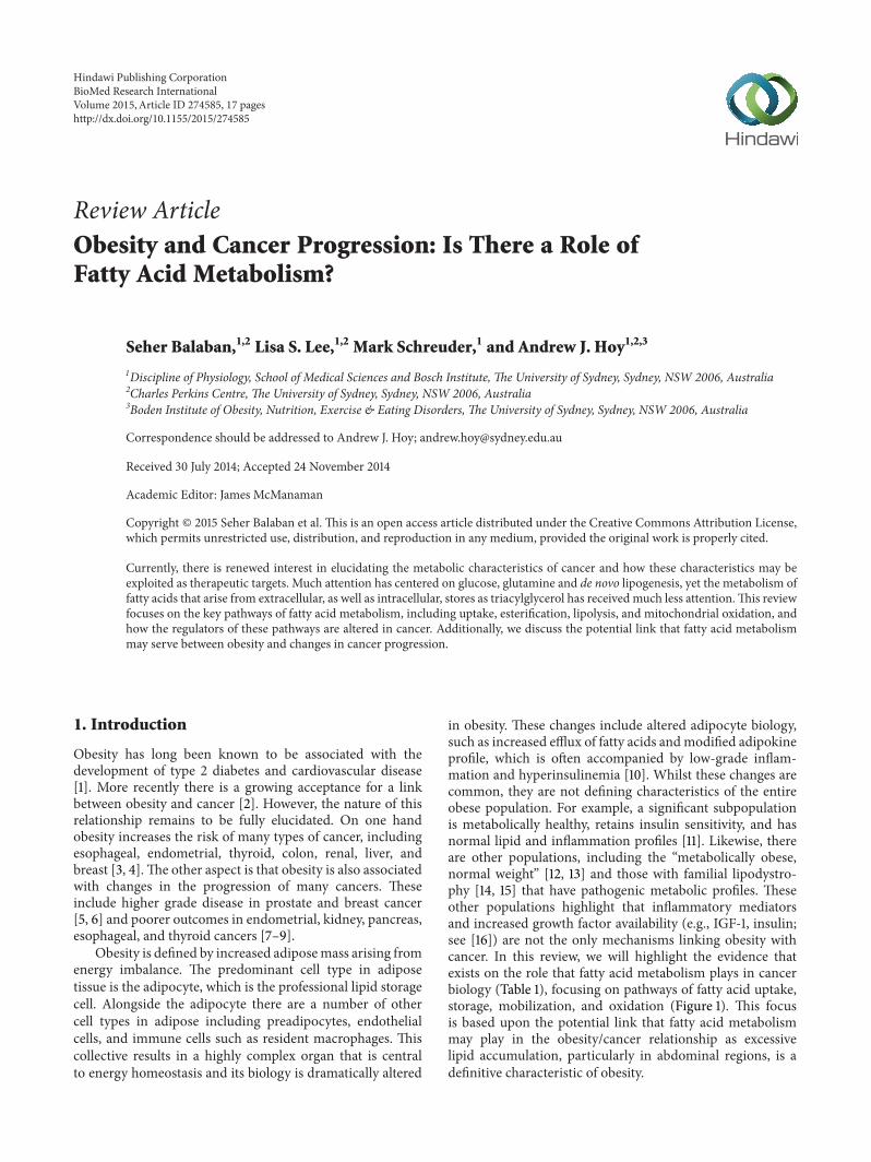
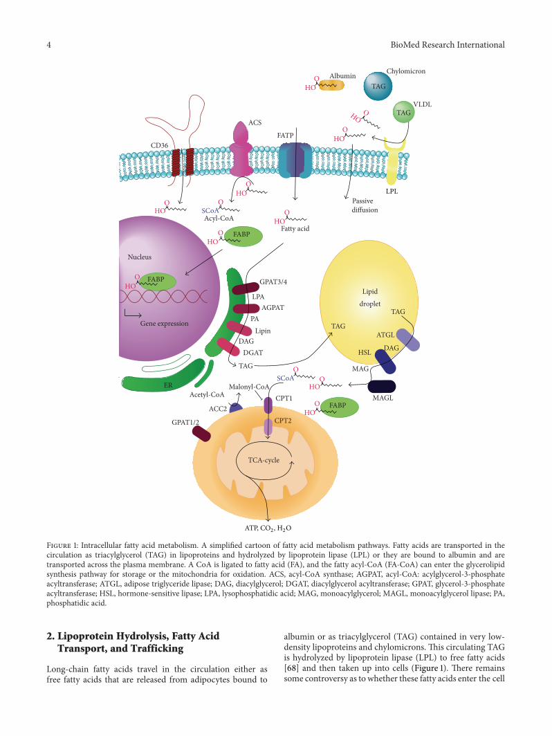
in obesity. These changes include altered adipocyte biology,such as increased efflux of fatty acids andmodified adipokineprofile, which is often accompanied by low-grade inflam-mation and hyperinsulinemia [10]. Whilst these changes arecommon, they are not defining characteristics of the entireobese population. For example, a significant subpopulationis metabolically healthy, retains insulin sensitivity, and hasnormal lipid and inflammation profiles [11]. Likewise, thereare other populations, including the “metabolically obese,normal weight” [12, 13] and those with familial lipodystro-phy [14, 15] that have pathogenic metabolic profiles. Theseother populations highlight that inflammatory mediatorsand increased growth factor availability (e.g., IGF-1, insulin;see [16]) are not the only mechanisms linking obesity withcancer. In this review, we will highlight the evidence thatexists on the role that fatty acid metabolism plays in cancerbiology (Table 1), focusing on pathways of fatty acid uptake,storage, mobilization, and oxidation (Figure 1). This focusis based upon the potential link that fatty acid metabolismmay play in the obesity/cancer relationship as excessivelipid accumulation, particularly in abdominal regions, is adefinitive characteristic of obesity.
Hindawi Publishing CorporationBioMed Research InternationalVolume 2015, Article ID 274585, 17 pageshttp://dx.doi.org/10.1155/2015/274585
2 BioMed Research International
Table1:Summaryof
regu
latorsof
fatty
acid
metabolism
andtheire
ffectso
ncancer
cellbiolog
y.
Regu
latoro
fCa
ncer
type
Alteratio
nAs
sociated
outcom
eRe
ference(s)
FAmetabolism
LPL
Prostate
Increasedactiv
ityIncreasedsusceptib
ility
[17]
Cervical
Enhanced
proteinexpressio
nIncreasedinvasio
ncapacity
[18]
Rectalandskin
Increasedactiv
ityIncreasedtumor
grow
th[19
]Lu
ngIncreasedactiv
ityLo
wer
overallsurvival
[20,21]
CD36/FAT
Colon
andovarian
Decreased
gene
expressio
nHigherm
etastatic
capacity
[22]
Breast
Decreased
gene
expressio
nHigherm
etastatic
capacity
[23]
FATP
Liver
Increasedgene
expressio
nEn
hanced
progression
[24]
FABP
4
Breast
Decreased
gene
expressio
nN/A
[25]
Bladder
Lowgene
expressio
nIncreasedtumor
progressionandinvasio
ncapacity
[26–
29]
Prostate
Increasedproteinexpressio
nIncreasedmigratio
nandinvasio
ncapacitie
s[30]
Ovaria
nIncreasedproteinexpressio
nIncreasedmigratio
nandinvasio
ncapacitie
s[31]
FABP
5
Breast
Increasedgene
expressio
nHigherm
etastatic
capacityandlower
recurrence-fr
eeandoverallsurvival
[32,33]
Endo
metria
lIncreasedgene
expressio
nNocorrelated
clinicaloutcome
[34]
Livera
ndpancreatic
Increasedproteinexpressio
nN/A
[35,36]
Prostate
Increasedgene
expressio
nIncreasedtumor
progression
[37]
Decreased
gene
expressio
nIncreasedinvasio
ncapacityandtumor
grow
th[26,38,39]
FABP
7Breast
Increasedgene
expressio
nLo
wer
recurrence
rate,improved
survival
[40,41]
Increasednu
clear
localization
Increasedproliferatio
n,pleomorph
ism,and
tumor
stage
[42]
Prim
arymelanom
aIncreasedgene
expressio
nN/A
[43]
Renal
Increasedgene
expressio
nNocorrelated
clinicaloutcome
[44,45]
ACSL3
Glio
blastoma
Increasedproteinexpressio
nIncreasedmalignant
phenotype
[46]
Colon
Increasedgene
andproteinexpressio
nN/A
[47]
ACSL4
Liver
Increasedgene
expressio
nIncreasedproliferatio
n[48]
ACSL5
Colon
Increasedgene
expressio
nIncreasedproliferatio
n[49]
AGPA
T2Ovaria
nIncreasedproteinandgene
expressio
nRe
ducedoverallsurvivaland
high
ertumor
grade,
mito
ticindex,andtumor
stage
[50–
52]
AGPA
T11
Breast,
cervical,and
colon
Increasedgene
expressio
nHighertum
orgrade
[53]
AGPA
T9Colorectal
Increasedgene
andproteinexpressio
nIncreasedcellgrow
th[54]
ATGL
Lung
andskin
IncreasedAT
GLactiv
ityIncreasedtumor
grow
thandcancer-associatedcachexia
[55]
HSL
Gastro
intestinal
Increasedgene
andproteinexpressio
nCa
ncer-associatedcachexia
[56]
Colorectal,pancreatic,stomach,and
esop
hageal
Increasedgene
expressio
nN/A
[57]
MAG
LColorectal
Increasedgene
andproteinexpressio
nN/A
[58]
Ovaria
n,breast,
melanom
a,andprostate
Increasedgene
expressio
nAggressiveness
[59]
CPT1A
Ovaria
nIncreasedgene
expressio
nIncreasedtumor
grow
th[31]
Breast
Increasedgene
andproteinexpressio
nandactiv
ityN/A
[60]
Glio
blastoma
Increasedgene
expressio
nHighertum
orgrade
[61]
CPT1C
Lung
Increasedgene
expressio
nN/A
[62]
Glio
blastoma
Increasedgene
expressio
nHighertum
orgrade
[61]
BioMed Research International 3
Table1:Con
tinued.
Regu
latoro
fCa
ncer
type
Alteratio
nAs
sociated
outcom
eRe
ference(s)
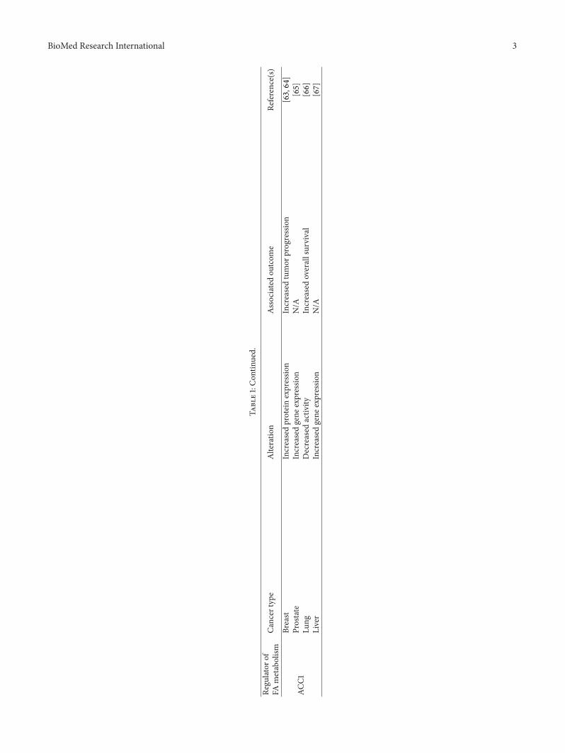
FAmetabolism
ACC1
Breast
Increasedproteinexpressio
nIncreasedtumor
progression
[63,64
]Prostate
Increasedgene
expressio
nN/A
[65]
Lung
Decreased
activ
ityIncreasedoverallsurvival
[66]
Liver
Increasedgene
expressio
nN/A
[67]
4 BioMed Research International
TCA-cycle
Lipiddroplet
Nucleus
FABP
Gene expression
CPT1ACC2
Malonyl-CoAAcetyl-CoA
FABP
LPL
FATPCD36
Passivediffusion
VLDL
Chylomicron
Fatty acid
ACS
Acyl-CoA
Albumin
HSL
MAGL
ATGL
TAG
TAG
OSCoA
ER
GPAT3/4
AGPAT
DGAT
CPT2
SCoA
FABP
MAG
DAG
TAG
TAGPA
LPA
TAG
LipinDAG
GPAT1/2
HO
HO
HO
HO
HO
HO
HO
HO
HO
HOO
O
O
OO
O
O
O
O
O
O
ATP, CO2, H2O
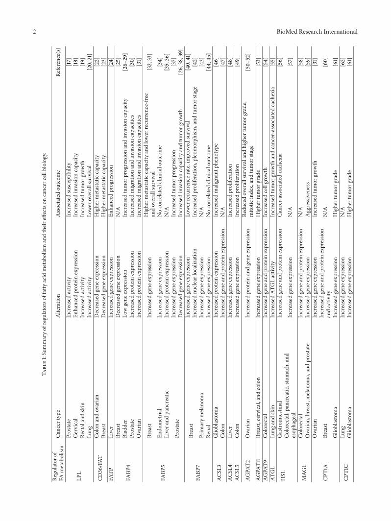
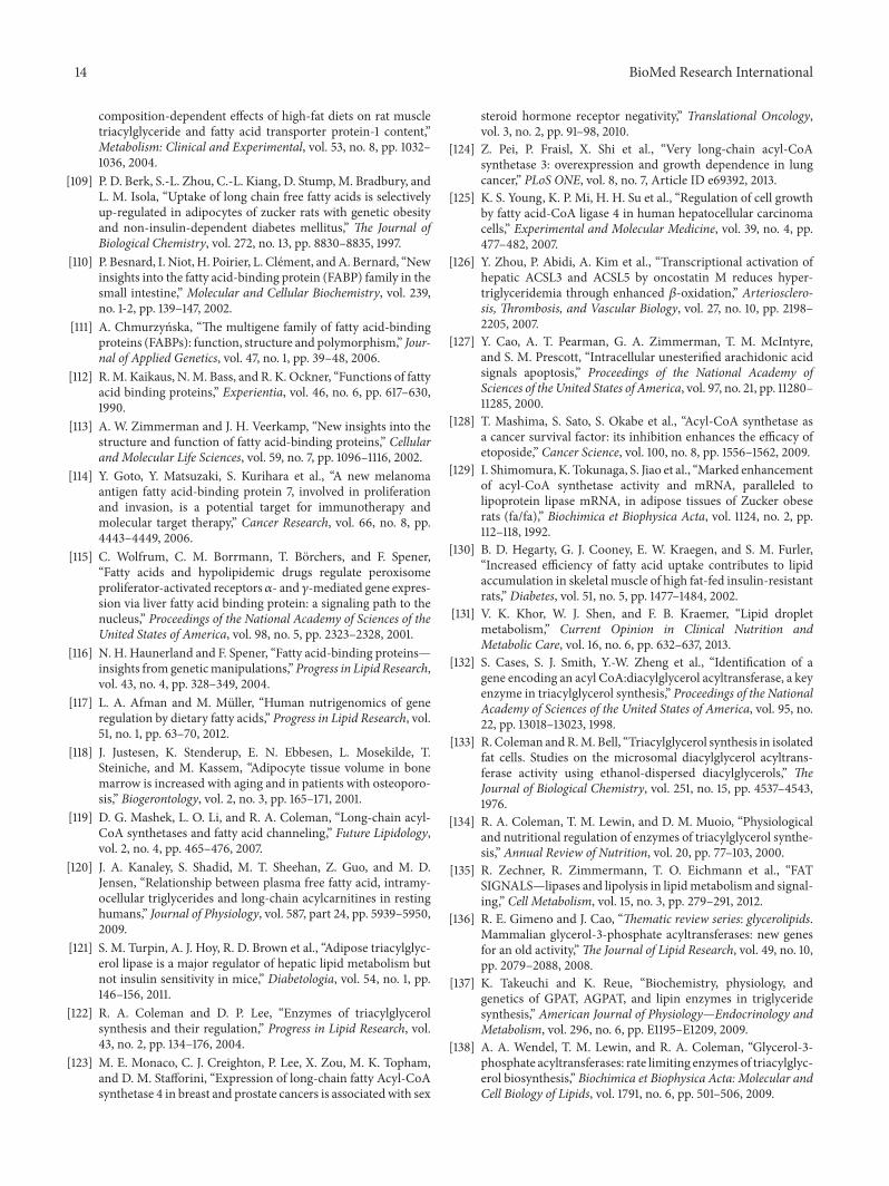
Figure 1: Intracellular fatty acid metabolism. A simplified cartoon of fatty acid metabolism pathways. Fatty acids are transported in thecirculation as triacylglycerol (TAG) in lipoproteins and hydrolyzed by lipoprotein lipase (LPL) or they are bound to albumin and aretransported across the plasma membrane. A CoA is ligated to fatty acid (FA), and the fatty acyl-CoA (FA-CoA) can enter the glycerolipidsynthesis pathway for storage or the mitochondria for oxidation. ACS, acyl-CoA synthase; AGPAT, acyl-CoA: acylglycerol-3-phosphateacyltransferase; ATGL, adipose triglyceride lipase; DAG, diacylglycerol; DGAT, diacylglycerol acyltransferase; GPAT, glycerol-3-phosphateacyltransferase; HSL, hormone-sensitive lipase; LPA, lysophosphatidic acid; MAG, monoacylglycerol; MAGL, monoacylglycerol lipase; PA,phosphatidic acid.
2. Lipoprotein Hydrolysis, Fatty AcidTransport, and Trafficking
Long-chain fatty acids travel in the circulation either asfree fatty acids that are released from adipocytes bound to
albumin or as triacylglycerol (TAG) contained in very low-density lipoproteins and chylomicrons. This circulating TAGis hydrolyzed by lipoprotein lipase (LPL) to free fatty acids[68] and then taken up into cells (Figure 1). There remainssome controversy as to whether these fatty acids enter the cell
BioMed Research International 5
by passive diffusion or by protein mediated transport. As willbe discussed below, it is clear that the latter process doescontribute to fatty acid uptake.
2.1. Lipoprotein Hydrolysis. Altered expression of LPL hasbeen reported in many cancers. For example, Narita andcolleagues [17] reported a significant association betweenincreased hydrolytic activity of LPL due to the LPL polymor-phism (Ser447stop) and the susceptibility to prostate cancer.This associationwas even stronger in patients with high gradetumors or metastasis. Similarly, this pattern was observedin cervical cancer where LPL is frequently overexpressedin cervical squamous cell carcinomas and associated withan increased invasion capacity [18]. LPL activity has beenreported in gastric and rectal cancers, malignant fibroushistiocytomas, and osteosarcomas,with the high proliferatingouter area of rectal tumors and fibrous histiocytomas havingan enhanced expression of LPL compared with the center[19]. Interestingly, the increased LPL activity in cancer tissue,compared with healthy lung tissue, predicts lower overallsurvival in non-small-cell lung cancer [20, 21]. The locationof tumor LPL is somewhat controversial as a recent studyobserved that increased LPL expression was in a subgroupof macrophages and not in cancer cells [69]. These studiesmostly report gene expression and therefore future studieslinking functional changes in cancer cell LPL activity drivingFA release from circulating TAG are required, especially asLPL activity is regulated by a variety of physiological stimuli(see review [70]).
2.2. Fatty Acid Transport. Several proteins have been iden-tified to facilitate the uptake of fatty acids into cells. Theseinclude CD36/fatty acid translocase, the fatty acid bindingprotein (FABP) family, and the fatty acid transport proteins(FATP) [71]. Many of these transporters are ubiquitouslyexpressed, while some display tissue-specific expression [72,73]. Interestingly, most tissues have coexpression of differentfatty acid transporters [74]. The reason for this remainsunknown. Possibilities may include differences in uptakecapacity and substrate specificity, sensitivity to hormonalstimuli such as insulin [75], or preferences in partitioning intodownstream pathway, for example, fatty acid esterification(storage) or oxidation [74].
2.2.1. CD36/Fatty Acid Translocase. CD36, also known asfatty acid translocase (FAT), is a multifunctional transmem-brane glycoprotein which is abundantly expressed in celltypes active in fatty acid metabolism, including adipocytes,skeletal muscles cells, cardiomyocytes, intestinal enterocytes,monocytes, and hepatocytes [76]. It was originally isolatedfrom platelet membranes as a thrombospondin receptor [77]but has also been shown as a receptor for collagen [78],oxidized lipoproteins [79], and, of greatest interest to thisreview, long-chain fatty acids [80].
CD36 has been implicated in contributing to cancer pro-gression. Low CD36 gene expression correlates with a highermetastasis grade in colon and ovarian cancers and withlow recurrence-free survival [22]. Conversely, CD36 mRNAexpression in breast cancer is inversely correlated with the
metastatic potential of five breast cancer cell lines [23], whereits expression is relatively higher in less aggressive cell lines(T47-D and MCF-7) and almost absent in highly aggressivelines (ZR-75 andMDA-MB-231). This inconsistency betweencancer types may be explained by the multifunctionality ofCD36. While it functions as a fatty acid transporter, CD36is also involved in collagen adhesion and, therefore, lessCD36may reduce cell adhesion, providing cancer cells with ahigher metastatic potential. That said, the above studies havereported gene or protein expression and not the rates of fattyacid uptake.
Fatty acid transporter abundance is not the only factorregulating FA uptake. An aspect that is often overlooked isthat FA uptake is increased by insulin stimulation [81, 82].This is thought to bemediated by translocation ofCD36 to theplasma membrane which has been observed in hepatocytesof obese Zucker rats [83], skeletal muscle [84, 85], cardiomy-ocytes [86, 87], and ovary cells [85]. This is analogous tothe translocation of the insulin sensitive glucose transporterGLUT4 [88, 89].
So far, no studies have investigated the influence of obe-sity on fatty acid transporters in cancer cells. It is clear fromstudies in other model systems that CD36 expression andfatty acid uptake are influenced by the microenvironment.For example, CD36 gene expression and protein levels areincreased in steatotic hepatocytes [90] and liver biopsies ofobese patients, correlating with the circulating free fatty acidslevels [91]. In subcutaneous adipose tissue, CD36 proteinexpression is upregulated in both obese patients and type 2diabetics [92]. Furthermore, CD36 mRNA expression levelsare greatly enhanced in liver and adipose tissue of ob/obmice,a monogenic model of obesity [93]. Interestingly, incubationof human skeletal muscle cells with adipocyte conditionedmedia increased both fatty acid uptake and CD36 proteinlevels [94]. Similar changes in CD36 expression by adipocytefactors, such as adipokines and fatty acids, have been reportedin vascular smooth muscle cells [95], cardiomyocytes [86,96], and adipocytes [97, 98]. Collectively this suggests thatchanges in adipocyte biology, especially in the context ofobesity, can alter CD36 expression in nonadipose cells suchas cancer cells that may influence the inherent role that CD36plays in cancer biology.
2.2.2. Fatty Acid Transport Protein. Fatty acid transportproteins form a highly conserved family of six transportersnamed FATP1–6 [99]. FATPs are integral membrane proteinsand are differentially expressed in a wide variety of cells[100]. These transporters are unique as they can express fattyacyl-CoA synthetase activity [101] as well as an endoplasmicreticulum localization signal domain, at least for FATP4[102]. Alongside CD36, FATPs regulate long-chain fatty acidand very long-chain fatty acid uptake [103] although thefunctional differences between CD36 and FATPs are yet tobe resolved. A recent study in Madin-Darby Canine cellsreported that CD36 is 30-fold more effective in fatty aciduptake compared with FATP4 or the acyl-CoA synthetaseACSL1 [104]. However, cooverexpression of CD36with eitherFATP4 or ACSL1 results in an enhanced fatty acid uptake rategreater than expected from the combined individual capacity
6 BioMed Research International
suggesting a synergistic relationship between CD36, FATP4,and ACSL1 to facilitate fatty acid uptake.
To date, only one study has described a possible rolefor FATPs in tumor metabolism. In this study, FATP mRNAexpression is increased in rat hepatomas compared withnormal liver tissue which correlated with fatty acid uptakerates [24]. Similar to CD36, FATP expression is influenced bythemicroenvironment, especially in obesity. FATP expressionis elevated in adipose tissue of obese patients [105, 106] andin heart [107], skeletal muscle [108], and adipose tissue [109]of rodent models of obesity. Overall, FATPs are importantplayers in lipid uptake andmetabolism.However, their role incancer, especially in the context of obesity-sensitive cancers,is far from understood and further research is needed toelucidate this role.
2.3. Intracellular Trafficking. Fatty acid binding proteins(FABPs) are a family of transport proteins with high affinityfor long-chain fatty acids, bile acids, and retinoids [110].Twelve FABP isoforms have been identified, each with itsown tissue and substrate specificity [111]. Although theirphysiological functions are not fully understood, they appearto facilitate the transport of fatty acids intracellularly andthereby regulate substrate availability for complex lipid syn-thesis (esterification) and oxidation [112, 113]. Changes inFABP expression have been associated with various diseasesincluding several forms of cancer [113] with FABP5 beingthe most well characterized FABP isoform in cancer cellbiology. For example, prostate [32], endometrial [34], liver[35], pancreatic [36], and breast [32] cancers have increasedFABP5 gene or protein expression. However, the observa-tions in prostate are controversial as other studies reportreduced expression in multiple prostate cancer lines [26,38]. Despite this, increased expression of FABP5 in prostatecancer cells increased fatty acid uptake and peroxisomeproliferator-activated receptor gamma (PPAR𝛾) expressionwhich enhanced tumor progression [37]. Additionally, over-expression of FABP5 in the benign breast cancer cell line,Rama 37, increased metastatic capacity in rats [32]. Inter-estingly, expression is higher in estrogen and progesteronenegative breast cancer cells, with the highest expression foundin triple-negative breast cancer [33]. Furthermore, patientswith higher FABP5 mRNA levels had lower recurrence-freeand overall survival probabilities [33]. Conversely, invasioncapacity and tumor growth were significantly reduced inprostate cancer cells with reduced FABP5 expression [39].
FABP7 has emerged as another participant of intracellularFA metabolism that may contribute to cancer cell biology. Itsgene expression is elevated in triple-negative breast cancercells [40], primary melanomas [43], and renal cell carci-nomas [44, 45]. Interestingly and in contrast to FABP5,FABP7 positive basal-like breast tumors had a significantlower recurrence rate and improved survival rate [41]. Incell culture studies, siRNA knockdown of FABP7 reducedproliferation and invasion in melanoma cells whilst the con-traobservation was reported with overexpression enhancingproliferation and invasion [114]. Furthermore, investigationsof the organelle-specific roles of FABP7 demonstrated thatincreased nuclear, but not cytoplasmic, FABP7 is associated
with increased proliferation, pleomorphism, and tumor stagein breast cancer suggesting that nuclear FABP7 drives amore aggressive phenotype [42]. However, themechanism bywhich FABP7 influences gene expression is yet to be resolved.FABPs may act as coactivators for transcription factors likePPARs [115] or simply function as transporters to carry FAinto the nucleus to modulate gene expression [116] via themany intranuclear targets including sterol regulatory bindingprotein, PPARs, and liver X receptors [117].
FABP4 has also been implicated in cancer biology. FABP4mRNA levels are downregulated in breast cancer cells [25].Conversely, FABP4 expression is inversely correlated withtumor progression and invasiveness in bladder cancer [26–29]. FAPB4 is also susceptible to the extracellular milieu asthere is growing evidence that adipocytes increase FABP4mRNA and protein expression in cancer cells. An ele-gant study in ovarian cancer demonstrated that coculturewith adipocytes increases FABP4 protein expression andpromotes migration and invasion of ovarian cancer cells,while FABP4 deficiency ameliorated the adipocyte-derivedmetastatic potential [31]. A similar observation of adipocyte-induced increase in FABP4 expression has been reportedin PC3 prostate cancer cells [30]. The same study alsoreported an increased expression of FABP4 in prostate cancerbone metastasis from high-fat diet mice and prostate cancerpatients [30]. The fact that bone marrow is adipocyte-rich[118] suggests a role for adipocytes in enhancing FABP4expression and thereby playing an important role in can-cer progression. Overall, FABPs are emerging as importantfactors in cancer cell lipid metabolism but more research isneeded to fully elucidate the roles of FABPs in healthy tissueand tumor cells and how these are altered by obesity.
3. Fatty Acid Activation, Esterification,and Mobilization
Once FAs are taken up by cells, they are activated by theaddition of coenzyme A (CoA) to the fatty acid moleculeby the actions of long-chain acyl-CoA synthetase (ACSL).From here, evidence suggests that fatty acyl-CoAs can bepartitioned into the esterification pathway in the endoplasmicreticulum or the mitochondria for oxidation [119]. Recently,this notion has been challenged by studies in human skeletalmuscle [120] and isolated hepatocytes frommice lacking adi-pose triglyceride lipase (ATGL) [121]. These studies suggestthat extracellular FAs enter the esterification pathway to bestored in lipid droplets prior to mitochondrial oxidation.Irrespective of the precise pathways, fatty acids have amultitude of intracellular fates, but at themost basic level FAscan be either oxidized or stored as complex lipids.
3.1. Fatty Acid Activation. ACSLs are a family of enzymesthat catalyze the addition of a CoA to a free fatty acid anddiffer in their preference to the chain length of their fattyacids substrates (short, medium, long, and very long). ACSL1,ACSL3, ACSL4, ACSL5, and ACSL6 are members of thelong-chain family that vary in both subcellular localizationand substrate specificity [122]. Along with FABPs, individual
BioMed Research International 7
ACSL isoforms have been proposed to channel fatty acids tospecific metabolic pathways.
Significant evidence suggests an important role forACSLsin cancer biology including increased expression of ACSLsin many types of cancer such as colon, liver, lung, brain,and colorectal cancers and estrogen receptor negative breasttumors and androgen receptor negative prostate tumors [46,47, 49, 123–125]. More specifically, ACSL5 gene expressionis consistently elevated in the colon cancer tissue comparedto normal colon tissue [49], so are ACSL4 gene expressionand protein levels in colon adenocarcinoma compared withadjacent normal tissue [47] and in hepatocellular carcinomatissues compared to the adjacent noncancerous liver tissue[48]. Finally, ACSL3 expression is elevated in the highlytumorigenic U87 human glioblastoma cell line and cellsderived from tumorigenic primary glioblastoma xenografts(Mayo 22) compared with the less tumorigenic U373 gliomacells [46].
Collectively, the results from these studies suggest thatexpression of ACSLs is related to tumorigenesis and tumorprogression. Cell culture loss and gain of function studiesprovide insight into the relationship between altered intracel-lular fatty acid metabolism and cancer cell biology. In termsof fatty acidmetabolism, bothACSL3 andACSL5 overexpres-sion in HepG2 cells increase fatty acid oxidation and reduceTAG levels [126]. Supporting the gene expression observa-tions, altered ASCL expression in cancer cells is linked withsurvival, proliferation, and chemoresistance. For example,overexpressed ACSL4 in human epithelial cells reduced thelevel of arachidonic acid-induced apoptosis [127], whereassiRNA-mediated ACSL3 knockdown reduced growth rates oflung cancer cell lines and colony formation [124]. Similarly,ACSL4 knockdown inhibited growth rates of the humanhepatocellular carcinoma cell line Hep3B [48]. Addition-ally, pharmacological inhibition of ACS activity by triacsin-C induced apoptosis in HEK293 cells [127] and gliomacells, which was completely suppressed by overexpression ofACSL5 [128].
The impact of ASCL expression and function in can-cer biology in the obese setting has not been reported.Interestingly, ACSL activity and Acsl1 gene expression areupregulated in liver and adipose tissues in genetic obesemodels, including ob/obmice and Zucker fatty rat (fa/fa) [93,129] and high-fat fed rats [130].This suggests that the elevatedexpression reported may be exacerbated in obesity andtherefore may accelerate cancer progression. How changes inASCL-mediated fatty acid metabolism are linked to alteredcancer progression is yet to be fully elucidated. However, Caoand colleagues [127] proposed that changes in proapoptoticarachidonic acid levels may play a role yet other bioactivelipids such as sphingolipids, including ceramides, or changesin fatty acyl-CoA availability for mitochondrial oxidation arepotential contributors.
3.2. Fatty Acid Esterification. FAs are the building blocksfor many complex lipids including phospholipids, sphin-golipids, and glycerolipids. We will focus on the synthesisof glycerolipids, such as TAG, as this is a major pool thatis susceptible to the obese environment. The storage of fatty
acids as TAG involves several condensation reactions. Thefirst step involves esterifying a fatty acyl-CoA with glycerol-3-phosphate to generate lysophosphatidic acid (LPA) by theenzyme glycerol-3-phosphate acyltransferase (GPAT). LPA isthen condensed into phosphatidic acid (PA) by 1-acylglyc-erol-3-phosphate-O-acyltransferase (AGPAT). The subse-quent reaction is catalyzed by lipin, which dephosphorylatesPA to produce diacylglycerol (DAG). The final step involvesthe addition of a third fatty acyl-CoA to DAG by diacylglyc-erol acyltransferase (DGAT) to generate TAG. This processoccurs in the endoplasmic reticulum where TAG is packagedinto lipid droplets [131]. Alongside the endoplasmic reticulumpathway, there is evidence that DGAT can also catalyze theconversion of DAG to TAG at the lipid droplet [132–134].
The lipid intermediates of the esterification pathway aresubstrates for the generation of other complex lipids, such asphospholipids in membrane synthesis, and can also act aslipid signals that modify membrane structures and promotegene transcription for cell growth, proliferation, and differen-tiation [135].
To date, gene or protein expression profiling of GPAT incancer cells has not been reported. However, it is known thatfour isoforms of GPATs are expressed in mammals; GPAT1andGPAT2 are localized in themitochondria andGPAT3 andGPAT4 in the endoplasmic reticulum [136]. As rate-limitingenzymes of fatty acid esterification, GPATs are key regulatorsof TAG synthesis [137, 138].
Similarly, little is known about the expression of lipin andDGAT in cancer patients. Mammals have three lipin pro-teins and two isoforms of DGAT that regulate phospholipidsynthesis and lipid storage [139, 140]. Consequently, theseproteins modulate the availability of fatty acid substrates forlipid signaling and metabolism, which may influence cancerprogression [141].
Themost studied enzyme of lipid esterification in relationto cancer is 1-acylglycerol-3-phosphate-O-acyltransferase(AGPAT). There are 11 known isoforms of AGPAT, whichdiffer by tissue expression and enzymatic activity [137].Thereis consistent evidence suggesting a role for AGPATs in cancercells. For example, AGPAT2 expression is elevated in ovariancancer patients with aggressive ovarian cancers and associ-ated with reduced overall survival [50–52]. Gene expressionof AGPAT11 is also increased in breast and cervical cancers,as well as colorectal cancer [53]. Interestingly, transcriptionalexpression of AGPAT9, which is highly homologous withAGPAT11, is upregulated in colorectal cancer, but not inbreast and cervical cancers [54].
Obesity is characterized by increased levels of TAG storedin tissues such as skeletal muscle, liver, and cardiac muscle[142], which is a consequence of increased esterification rates[130, 143–145]. Loss and gain of function studies in varioustissues provide an insight into the complex regulation ofthe intracellular lipid environment. For example, AGPAT6knockout mice have reduced TAG content in brown andwhite adipose tissue and interestingly altered fatty acid profileof complex lipids, such asDAGand phospholipids with a shifttowards the polyunsaturatedmore than themonounsaturatedfatty acids [146]. Similarly, adipose tissue TAG levels aredecreased in mice lacking DGAT [147] and both lipin1 and
8 BioMed Research International
lipin3 [148] and protection fromhigh-fat diet induced obesityand associated metabolic perturbations [139, 149]. Finally,GPAT-deficient mice have lower levels of liver and plasmaTAG [150]. On the other hand, overexpressions of GPAT1[151], GPAT4 [152], AGPAT1 [153], lipin1 [154], and DGAT1[155] all result in increased TAG levels. From this, it is evidentthat enzymes involved in esterification significantly influenceintracellular and extracellular lipid homeostasis. How thistranslates to pathogenic changes in cancer cells is yet to bedescribed.
3.3. Lipolysis. TAGs, along with cholesterol esters, are storedin lipid droplets to serve as a readily available source of energyfor ATP generation in the mitochondria, as well as providingbuilding blocks for phospholipids and other complex lipids.In terms of metabolic energy capacity, an average nonobeseperson stores up to 2,500 kJ of metabolic energy in glycogen,but >500,000 kJ as TAGs [156]. Whilst most of this TAG isstored in adipocytes, all cells have the capacity to synthesizeand breakdown TAGs. Interestingly, intracellular lipid stores,or lipid droplet size and/or number, are elevated in variousmalignant cells, such as breast [157], prostate [158], cervical[159], liver [160], and colon cancer cells [161]. Furthermore,biochemical assessment of lipid droplets in breast cancer cellshas shown that the TAG content is increased [162]. Not onlythat, TAG levels are higher in more aggressive breast cancercells and are associated with long-term breast cancer cellsurvival [157, 162]. These findings suggest that intracellularTAG may play a critical, yet unexplored, role in supportingboth substrates for complex lipid synthesis [163] as well asenergy production in cancer cells that collectively promotecell growth and proliferation. To do this, TAGs need to bebroken down to FAs and glycerol by a process called lipolysis.
ATGL, otherwise known as desnutrin [164], is the pre-dominant TAG lipase that is thought to be rate-limiting [165].It catalyzes the conversion of TAG to DAG and releasesa free fatty acid from the sn-2 position [166]. Hormone-sensitive lipase (HSL) catalyzes the hydrolysis of DAG intomonoacylglycerol (MAG) and a fatty acid [167]. HSL hasbroad substrate specificity, including TAG, DAG, MAG, andcholesterol ester lipid classes, but has the highest affinity forDAG [168]. MAG is then broken down by monoacylglycerollipase (MAGL) resulting in the metabolic end-product, glyc-erol, and the liberation of the final fatty acid. This processis highly conserved across species and highly regulated withmost insight arising from studies in adipocytes (see review[169]).
Adipose neutral lipase expression in various cancerpatients has been reported. Compared to normal individualswithout cancer, HSLmRNA expression is elevated in adiposetissue of colorectal, pancreatic, esophageal, and stomachcancer patients [57].This was also observed in ovarian cancerpatients, where adipocyte lipid depots which contain TAGwere reduced, while the lipolytic products, MAG and freefatty acids, were increased, collectively suggesting elevatedlipolytic activity [170]. Similarly, transcriptional and proteinexpression of HSL are increased in the adipose tissue of late-stage cancer patients exhibiting uncontrolled loss of adiposeand muscle tissue, known as cachexia [55]. Interestingly,
upregulated ATGL activity in adipose tissue was found to beresponsible for this tissue-wasting syndrome [56].
MAGL is currently the most well-documented neutrallipase and its transcriptional expression is altered in sev-eral different cancers. For example, high mRNA expressionof MAGL has been reported in ovarian [59], colorectal[58], breast, and melanoma cancer cells and particularly inaggressive prostate cancer cell lines [59]. Interestingly, invitro studies overexpressing MAGL in nonaggressive ovariancancer cells raised free FA levels and increased tumor growthrate, migration, and invasion [171]. Alternatively, pharmaco-logical inhibition attenuated MAGL-induced aggressivenessof prostate cancer cells, even in a high-lipid environment[59, 171]. Similar observations have been made in colorectalcancer cells [58].
There are conflicting observations regarding the expres-sion patterns of lipolytic enzymes in obesity [172]. HSL andATGL gene expression are reduced in the adipose tissue ofobese humans [173–175] and insulin resistant high-fat fedrats [176]. Conversely, a study by de Naeyer and colleagues[172] has reported that HSL and ATGL mRNA expressionare increased in visceral adipose tissue of morbidly obesemen; however, this pattern did not translate to changes inprotein or lipase activity. This is not surprising consideringthat these neutral lipases are predominantly regulated byposttranslational modifications, translocation, and protein-protein interactions [135]. Interestingly, lipolytic enzymeexpression, particularly ATGL, appears to bemore associatedwith insulin sensitivity rather than obesity [177]. In order toelucidate the role lipolysis plays in cancer cell biology, futurestudies need to assess pathway of lipid metabolism and fattyacid flux, rather than gene expression, and investigate howthese are altered with obesity.
4. Mitochondrial Fatty Acid Oxidation
4.1. Fatty Acid Entry into the Mitochondria. The other majorfate for extracellular fatty acids is oxidation for the gener-ation of ATP in the mitochondria. Alongside glucose andglutamine, fatty acids are a major energy source catabolizedthrough the 𝛽-oxidation pathway to generate acetyl-CoA forentry into the TCA cycle as well as FADH
2and NADH
reducing equivalents for use by the electron transport chain(ETC).
Changes in cancer cell fatty acid oxidation have beenreported.Theprimary example is observed in prostate cancer.Rather than being secreted as it is in normal prostate cells,citrate is catabolized in the TCA cycle resulting in fatty acidoxidation being the dominant bioenergetic pathway [178].Interestingly, high-fat feeding of the p48-Kras mouse modelof pancreatic cancer accelerated tumor growth and increasedenergy expenditure and whole body fatty acid oxidationthrough increased gene expression of CPT1A, ACC, andAOXenzymes, key regulators of fatty acid oxidation [179].
Fewother studies have investigated the effect of obesity oncancer fatty acid oxidation. Although there is significant con-troversy as to the effect that obesity has on fatty acid oxidationin type 2 diabetes, the increased availability of circulatingand intracellular fatty acids is thought to drive an increased
BioMed Research International 9
oxidative capacity. Evidence for this arises from studies inrodents fed a high-fat diet [124, 180] and obese type II diabeticpatients [181]. Conversely, a number of studies reported areduced capacity to oxidize fatty acids in overweight/obesehumans [182, 183]. Considering the high metabolic flexibilityof cancer cells, it is conceivable that cancer cells benefit fromhigh lipid availability that characterizes obesity through beta-oxidation either to fulfill increased energy demand or toprevent the lipotoxic effects of high level of fatty acids.
4.2. Carnitine Palmitoyltransferase 1. Unlike short-chain fattyacids, which can freely diffuse into mitochondria, long-chainfatty acids enter themitochondria by the carnitine shuttle sys-tem. First, carnitine palmitoyltransferase 1 (CPT1) catalyzesthe transfer of the fatty acid moiety from acyl-coenzyme A(CoA) to a long-chain acyl-carnitine.This is then transportedinto the mitochondrial matrix by the carnitine acyl-carnitinetranslocase (CACT) [184]. CPT2 then catalyzes the conver-sion of acyl-carnitine to carnitine and fatty acyl-CoA whichthen enters the 𝛽-oxidation pathway. CPT1 is regulated bya cytosolic pool of malonyl-CoA produced by acetyl-CoAcarboxylase 2 (ACC2) at the mitochondrial membrane [185].
The rate of mitochondrial fatty acid oxidation is regulatedby CPT1, which is an integral membrane protein located onthemitochondrial outermembrane [186]. CPT1 has three iso-forms with tissue-specific expressions and sensitivity to theallosteric-inhibitory action of malonyl-CoA: CPT1A (liver),CPT1B (muscle), and CPT1C (brain) [187, 188]. Changes inCPT1 expression have been observed in several types of can-cer including breast, lung, brain, and liver cancers [60, 62, 189,190]. A study by Linher-Melville and colleagues [60] reportedthat CPT1A mRNA levels are significantly elevated in bothMCF-7 and MDA-MB-231 cells compared to 184B5 humanmammary epithelial cells. In another study, CPT1C geneexpression is upregulated in non-small-cell lung carcinomatumor tissue comparedwithmatched normal lung tissue [62].Furthermore, high grade glioblastoma is associated withincreasedmRNA levels of bothCPT1A andCPT1C [61].Thesestudies clearly show that CPT1 expression levels are relatedto not only tumorigenesis but also tumor progression. Incontrast, CPT1 expression has been reported to be higherin the low metastatic potential, androgen receptor negativeLNCaP prostate cancer cell line compared to the highmetastatic potential, androgen receptor positive PC3 andDU145 prostate cancer cell lines [59]. Overexpression ofCPT1C in MCF-7 cell line elevated fatty acid oxidation andATP production to support resistance to glucose depriva-tion and siRNA-mediated CPT1C knockdown suppressedxenograft tumor growth [62]. Further evidence for a role forCPT1 in cancer biology has been generated from pharma-cological studies. Inhibition of CPT1 with either genetic orpharmacological manipulation has been shown to reducetotal ATP levels and the rate of ATP production in PC3prostate cancer cells [62], Burkitt’s lymphoma cells [191],and human glioblastoma cells [192] to impair proliferation.Additionally, etomoxir sensitizes human leukemia cells toapoptosis [193]. Collectively, these studies suggest a role foraltered CPT1 expression in various cancers but interestinglyCPT1 expression is sensitive to the microenvironment. For
example, CPT1A mRNA expression and fatty acid oxidationare increased in SKOV3ip1 ovarian cancer cells coculturedwith adipocytes [31]. However, it must be highlighted thatfatty acid oxidation is regulated at a number of levelsincluding CPT1 gene expression, allosterically by malonyl-CoA and fatty acid availability.
4.3. Acetyl-CoA Carboxylase. Acetyl-CoA carboxylase is abiotin-dependent enzyme that catalyzes the conversion ofacetyl-CoA into malonyl-CoA. In mammals, two isoformsof ACC are expressed: ACC1 (also known as ACC𝛼) andACC2 (also known as ACC𝛽) [194]. ACC1 is primarilyexpressed in the cytosol of hepatocytes, adipocytes, and otherlipogenic cells, while ACC2 is an enzyme associated with theouter mitochondrial membrane and is mainly expressed incardiomyocytes, skeletal muscles, and hepatocytes [195–198].Whereas the malonyl-CoA generated by ACC1 is primarilyused for de novo lipogenesis, the malonyl-CoA productof ACC2 is a potent regulator of fatty acid oxidation byinhibiting CPT1 [199, 200]. Upstream of ACC2 is AMP-activated protein kinase (AMPK), which phosphorylates andinactivates ACC2 to reduce malonyl-CoA levels and therebyincrease fatty acid oxidation.
Upregulation of ACC1 and increased de novo lipogenesisare observed in several types of cancer including breast [63,64], prostate [65], lung [66], and liver cancers [67]. Chemicaland genetic inhibition studies have identified a role for ACC1in cell survival. For example, apoptotic cell death results fromchemical inhibition of ACC1 by TOFA (5-tetradecyloxy-2-furoic acid) in lung and colon cancer cells [201] and bysoraphen A in prostate cancer cells [202]. In addition, RNAinterference- (RNAi-)mediated knockdown ofACC1 inducesapoptosis in breast [203] and prostate cancer cells [204].
To date, studies have focused on ACC1, yet few studieshave been conducted into the role of ACC2 in cancer devel-opment or progression. One of these studies demonstratedthat knockdown of ACC2 increased fatty acid oxidationand inhibited cell death in A549 human lung carcinomacells [205]. Similarly, pharmacological inhibition of malonyl-CoA decarboxylase (MCD), which increased the malonyl-CoA pool, suppresses human breast cancer cell proliferation[206]. Therefore, decreasing fatty acid oxidation rates bythe modulation of the malonyl-CoA pool by ACC2 andMCD suggests a potential role for these enzymes in cancermetabolism. However, ACC2 functions in other cancer typesremain to be elucidated.
The role of ACC2 in obesity is more established. Skeletalmuscle ACC2 phosphorylation and activity are reduced inobese patients, as a consequence of reduced AMPK activity[207, 208]. Additionally, the mRNA levels of ACC2 inwhite adipose tissue are lower in Zucker fatty rats than inlean rats [209]. Interestingly, the AMPK-ACC2-CPT1 axis ismodulated by several adipokines, whose levels are alteredin obesity. These include leptin [210], adiponectin [211], andCTRP1 [212].Moreover, recent evidence in liver demonstratesthat metformin’s actions to suppress de novo lipogenesisand increase fatty acid oxidation require AMPK-mediatedphosphorylation of ACC1 and ACC2. Thus, the significantinterest in the clinical use of metformin as the therapeutic in
10 BioMed Research International
many cancers will further contribute to the understanding ofthe role that ACC1/2 plays in cancer biology [213].
5. Conclusions
Thecurrent interest in cancermetabolism has the potential toidentify commonperturbations arising fromdiffering geneticorigins that may serve as therapeutic targets. As the currentobesity epidemic continues to grow, there is a need to notonly define cancer metabolism but also investigate how it isinfluenced by the obese microenvironment. It is clear thatcancer fatty acid metabolism plays a significant role in cancerbiology and that opportunities exist to further define this role,especially in the context of obesity-induced changes in cancerprogression.
Abbreviations
CD36/FAT: Cluster of differentiation 36/fatty acidtranslocation
LPL: Lipoprotein lipaseVLDL: Very-low density lipoproteinFABP: Fatty acid binding proteinPPAR: Peroxisome proliferator-activated receptorFATPs: Fatty acid transport proteinsTAG: TriacylglycerolLPA: Lysophosphatidic acidGPAT: Glycerol-3-phosphate acyltransferaseAGPAT: 1-Acylglycerol-3-phosphate-O-
acyltransferaseDAG: DiacylglycerolHSL: Hormone-sensitive lipaseMAG: MonoacylglycerolMAGL: Monoacylglycerol lipaseATGL: Adipose triglyceride lipaseACS: Acyl-CoA synthetaseCPT1: Carnitine palmitoyltransferase 1CoA: Coenzyme AETC: Electron transport chainACSL: Acyl-CoA synthetase long-chain family.
Conflict of Interests
The authors declare that there is no conflict of interestsregarding the publication of this paper.
Acknowledgments
Andrew J. Hoy is supported by Helen and Robert EllisPostdoctoral Research Fellowship and funding from theUniversity of Sydney. Seher Balaban is a recipient of aUniversity of Sydney Australian Postgraduate Award. MarkSchreuder is supported by funding from the Dutch CancerInstitute KWF.
References
[1] S. M. Grundy, “Obesity, metabolic syndrome, and cardiovascu-lar disease,” Journal of Clinical Endocrinology and Metabolism,vol. 89, no. 6, pp. 2595–2600, 2004.
[2] S. M. Louie, L. S. Roberts, and D. K. Nomura, “Mechanismslinking obesity and cancer,” Biochimica et Biophysica Acta:Molecular and Cell Biology of Lipids, vol. 1831, no. 10, pp. 1499–1508, 2013.
[3] K. Y.Wolin, K. Carson, and G. A. Colditz, “Obesity and cancer,”Oncologist, vol. 15, no. 6, pp. 556–565, 2010.
[4] G. de Pergola and F. Silvestris, “Obesity as amajor risk factor forcancer,” Journal of Obesity, vol. 2013, Article ID 291546, 11 pages,2013.
[5] W.C. Buschemeyer III and S. J. Freedland, “Obesity and prostatecancer: epidemiology and clinical implications,” EuropeanUrol-ogy, vol. 52, no. 2, pp. 331–343, 2007.
[6] B. Majed, T. Moreau, K. Senouci, R. J. Salmon, A. Fourquet,and B. Asselain, “Is obesity an independent prognosis factor inwoman breast cancer?” Breast Cancer Research and Treatment,vol. 111, no. 2, pp. 329–342, 2008.
[7] Y. Zhu, H.-K. Wang, H.-L. Zhang et al., “Visceral obesity andrisk of high grade disease in clinical T1a renal cell carcinoma,”The Journal of Urology, vol. 189, no. 2, pp. 447–453, 2013.
[8] A. Ansary-Moghaddam, R. Huxley, F. Barzi et al., “The effectof modifiable risk factors on pancreatic cancer mortality inpopulations of the Asia-Pacific region,” Cancer EpidemiologyBiomarkers and Prevention, vol. 15, no. 12, pp. 2435–2440, 2006.
[9] C. A. Gilbert and J. M. Slingerland, “Cytokines, obesity, andcancer: new insights on mechanisms linking obesity to cancerrisk and progression,” Annual Review of Medicine, vol. 64, pp.45–57, 2013.
[10] J. K. Sethi and A. J. Vidal-Puig, “Thematic review series: adipo-cyte Biology. Adipose tissue function and plasticity orchestratenutritional adaptation,” Journal of Lipid Research, vol. 48, no. 6,pp. 1253–1262, 2007.
[11] D. Samocha-Bonet, D. J. Chisholm, K. Tonks, L. V. Campbell,and J. R. Greenfield, “Insulin-sensitive obesity in humans—a “favorable fat” phenotype?” Trends in Endocrinology &Metabolism, vol. 23, no. 3, pp. 116–124, 2012.
[12] N. Ruderman, D. Chisholm, X. Pi-Sunyer, and S. Schneider,“The metabolically obese, normal-weight individual revisited,”Diabetes, vol. 47, no. 5, pp. 699–713, 1998.
[13] R. Kelishadi, S. R. Cook, M. E. Motlagh et al., “Metabolicallyobese normal weight and phenotypically obese metabolicallynormal youths: the CASPIAN Study,” Journal of the AmericanDietetic Association, vol. 108, no. 1, pp. 82–90, 2008.
[14] T. Nolis, “Exploring the pathophysiology behind the morecommon genetic and acquired lipodystrophies,” Journal ofHuman Genetics, vol. 59, no. 1, pp. 16–23, 2014.
[15] J. Capeau, J. Magre, M. Caron-Debarle et al., “Human lipodys-trophies: genetic and acquired diseases of adipose tissue,”Endocrine Development, vol. 19, pp. 1–20, 2010.
[16] M. J. Khandekar, P. Cohen, and B. M. Spiegelman, “Molecularmechanisms of cancer development in obesity,” Nature ReviewsCancer, vol. 11, no. 12, pp. 886–895, 2011.
[17] S. Narita, N. Tsuchiya, L.Wang et al., “Association of lipoproteinlipase gene polymorphism with risk of prostate cancer in aJapanese population,” International Journal of Cancer, vol. 112,no. 5, pp. 872–876, 2004.
[18] J. C. Carter and F. C. Church, “Mature breast adipocytes pro-mote breast cancer cell motility,” Experimental and MolecularPathology, vol. 92, no. 3, pp. 312–317, 2012.
[19] K. Sakayama, H. Masuno, T. Miyazaki, H. Okumura, T. Shibata,and H. Okuda, “Existence of lipoprotein lipase in humansarcomas and carcinomas,” Japanese Journal of Cancer Research,vol. 85, no. 5, pp. 515–521, 1994.
BioMed Research International 11
[20] D. Cerne, I. P. Zitnik, andM. Sok, “Increased fatty acid synthaseactivity in non-small cell lung cancer tissue is aweaker predictorof shorter patient survival than increased lipoprotein lipaseactivity,” Archives of Medical Research, vol. 41, no. 6, pp. 405–409, 2010.
[21] Z. Trost, M. Sok, J. Marc, and D. Cerne, “Increased lipoproteinlipase activity in non-small cell lung cancer tissue predictsshorter patient survival,” Archives of Medical Research, vol. 40,no. 5, pp. 364–368, 2009.
[22] S. M. Rachidi, T. Qin, S. Sun, W. J. Zheng, and Z. Li, “Molecularprofiling of multiple human cancers defines an inflammatorycancer-associated molecular pattern and uncovers KPNA2 as auniform poor prognostic cancer marker,” PLoS ONE, vol. 8, no.3, Article ID e57911, 2013.
[23] I. P. Uray, Y. Liang, and S. M. Hyder, “Estradiol down-regulatesCD36 expression in human breast cancer cells,” Cancer Letters,vol. 207, no. 1, pp. 101–107, 2004.
[24] D. E. Blask, L. A. Sauer, R. T. Dauchy, E. W. Holowachuk, M. S.Ruhoff, andH. S. Kopff, “Melatonin inhibition of cancer growthin vivo involves suppression of tumor fatty acid metabolismvia melatonin receptor-mediated signal transduction events,”Cancer Research, vol. 59, no. 18, pp. 4693–4701, 1999.
[25] R. Hammamieh, N. Chakraborty, M. Barmada, R. Das, andM. Jett, “Expression patterns of fatty acid binding proteins inbreast cancer cells,” Journal of Experimental Therapeutics andOncology, vol. 5, no. 2, pp. 133–143, 2005.
[26] A. Tolle, S. Suhail, M. Jung, K. Jung, and C. Stephan, “Fattyacid binding proteins (FABPs) in prostate, bladder and kidneycancer cell lines and the use of IL-FABP as survival predictor inpatients with renal cell carcinoma,” BMC cancer, vol. 11, article302, 2011.
[27] J. E. Celis, M. Østergaard, B. Basse et al., “Loss of adipocyte-type fatty acid binding protein and other protein biomarkers isassociated with progression of human bladder transitional cellcarcinomas,” Cancer Research, vol. 56, no. 20, pp. 4782–4790,1996.
[28] G. Ohlsson, J. M. A. Moreira, P. Gromov, G. Sauter, and J.E. Celis, “Loss of expression of the adipocyte-type fatty acid-binding protein (A-FABP) is associated with progression ofhumanurothelial carcinomas,”Molecular&Cellular Proteomics,vol. 4, no. 4, pp. 570–581, 2005.
[29] P. J. Wild, A. Herr, C. Wissmann et al., “Gene expressionprofiling of progressive papillary noninvasive carcinomas of theurinary bladder,” Clinical Cancer Research, vol. 11, no. 12, pp.4415–4429, 2005.
[30] M. K. Herroon, E. Rajagurubandara, A. L. Hardaway et al.,“Bone marrow adipocytes promote tumor growth in bone viaFABP4-dependent mechanisms,” Oncotarget, vol. 4, no. 11, pp.2108–2123, 2013.
[31] K. M. Nieman, H. A. Kenny, C. V. Penicka et al., “Adipocytespromote ovarian cancermetastasis and provide energy for rapidtumor growth,” Nature Medicine, vol. 17, no. 11, pp. 1498–1503,2011.
[32] C. Jing, C. Beesley, C. S. Foster et al., “Identification ofthe messenger RNA for human cutaneous fatty acid-bindingprotein as a metastasis inducer,” Cancer Research, vol. 60, no.9, pp. 2390–2398, 2000.
[33] R.-Z. Liu, K. Graham, D. D. Glubrecht, D. R. Germain, J. R.Mackey, and R. Godbout, “Association of FABP5 expressionwith poor survival in triple-negative breast cancer: implicationfor retinoic acid therapy,” The American Journal of Pathology,vol. 178, no. 3, pp. 997–1008, 2011.
[34] Z. Li, C. Huang, S. Bai et al., “Prognostic evaluation of epider-mal fatty acid-binding protein and calcyphosine, two proteinsimplicated in endometrial cancer using a proteomic approach,”International Journal of Cancer, vol. 123, no. 10, pp. 2377–2383,2008.
[35] K. Fujii, T. Kondo, H. Yokoo, T. Yamada, K. Iwatsuki, andS. Hirohashi, “Proteomic study of human hepatocellular car-cinoma using two-dimensional difference gel electrophoresiswith saturation cysteine dye,” Proteomics, vol. 5, no. 5, pp. 1411–1422, 2005.
[36] P. Sinha, G. Hutter, E. Kottgen, M. Dietel, D. Schadendorf,and H. Lage, “Increased expression of epidermal fatty acidbinding protein, cofilin, and 14-3-3-sigma (stratifin) detectedby two-dimensional gel electrophoresis, mass spectrometry andmicrosequencing of drug-resistant human adenocarcinoma ofthe pancreas,” Electrophoresis, vol. 20, no. 14, pp. 2952–2960,1999.
[37] Z. Bao, M. I. Malki, S. S. Forootan et al., “A novel cutaneousFatty Acid-binding protein-related signaling pathway leadingto malignant progression in prostate cancer cells,” Genes andCancer, vol. 4, no. 7-8, pp. 297–314, 2013.
[38] R. Das, R. Hammamieh, R. Neill, M. Melhem, and M. Jett,“Expression pattern of fatty acid-binding proteins in humannormal and cancer prostate cells and tissues,” Clinical CancerResearch, vol. 7, no. 6, pp. 1706–1715, 2001.
[39] J. Adamson, E. A. Morgan, C. Beesley et al., “High-levelexpression of cutaneous fatty acid-binding protein in prostaticcarcinomas and its effect on tumorigenicity,” Oncogene, vol. 22,no. 18, pp. 2739–2749, 2003.
[40] X. Y. Tang, S. Umemura, H. Tsukamoto, N. Kumaki, Y. Tokuda,and R. Y. Osamura, “Overexpression of fatty acid bindingprotein-7 correlates with basal-like subtype of breast cancer,”Pathology Research and Practice, vol. 206, no. 2, pp. 98–101, 2010.
[41] H. Zhang, E. A. Rakha, G. R. Ball et al., “The proteins FABP7andOATP2 are associated with the basal phenotype and patientoutcome in human breast cancer,” Breast Cancer Research andTreatment, vol. 121, no. 1, pp. 41–51, 2010.
[42] A. T. Alshareeda, E. A. Rakha, C. C. Nolan, I. O. Ellis, and A.R. Green, “Fatty acid binding protein 7 expression and its sub-cellular localization in breast cancer,” Breast Cancer Researchand Treatment, vol. 134, no. 2, pp. 519–529, 2012.
[43] Y. Goto, K. Koyanagi, N. Narita et al., “Aberrant fatty acid-binding protein-7 gene expression in cutaneous malignantmelanoma,” Journal of Investigative Dermatology, vol. 130, no.1, pp. 221–229, 2010.
[44] C. Tan, T. Takayama, N. Takaoka et al., “Impact of genderin renal cell carcinoma: the relationship of FABP7 and BRN2expression with overall survival,” Clinical Medicine Insights:Oncology, vol. 8, pp. 21–27, 2014.
[45] T. Teratani, T. Domoto, K. Kuriki et al., “Detection of transcriptfor brain-type fatty acid-binding protein in tumor and urine ofpatients with renal cell carcinoma,” Urology, vol. 69, no. 2, pp.236–240, 2007.
[46] Z. Pei, P. Sun, P. Huang, B. Lal, J. Laterra, and P. A. Watkins,“Acyl-CoA synthetase VL3 knockdown inhibits human gliomacell proliferation and tumorigenicity,” Cancer Research, vol. 69,no. 24, pp. 9175–9182, 2009.
[47] Y. Cao, K. B. Dave, T. P. Doan, and S. M. Prescott, “Fatty acidCoA ligase 4 is up-regulated in colon adenocarcinoma,” CancerResearch, vol. 61, no. 23, pp. 8429–8434, 2001.
[48] Y.-C. Liang, C.-H. Wu, J.-S. Chu et al., “Involvement of fattyacid-CoA ligase 4 in hepatocellular carcinoma growth: roles of
12 BioMed Research International
cyclic AMP and p38 mitogen-activated protein kinase,” WorldJournal of Gastroenterology, vol. 11, no. 17, pp. 2557–2563, 2005.
[49] C.-S. Yeh, J.-Y.Wang, T.-L. Cheng,C.-H. Juan,C.-H.Wu, and S.-R. Lin, “Fatty acid metabolism pathway play an important rolein carcinogenesis of human colorectal cancers by Microarray-Bioinformatics analysis,” Cancer Letters, vol. 233, no. 2, pp. 297–308, 2006.
[50] C. S. M. Diefenbach, R. A. Soslow, A. Iasonos et al., “Lysophos-phatidic acid acyltransferase-𝛽 (LPAAT-𝛽) is highly expressedin advanced ovarian cancer and is associated with aggressivehistology and poor survival,”Cancer, vol. 107, no. 7, pp. 1511–1519,2006.
[51] S. Niesporek, C. Denkert, W. Weichert et al., “Expressionof lysophosphatidic acid acyltransferase beta (LPAAT-beta)in ovarian carcinoma: correlation with tumour grading andprognosis,” British Journal of Cancer, vol. 92, no. 9, pp. 1729–1736, 2005.
[52] G. M. Springett, L. Bonham, A. Hummer et al., “Lysophospha-tidic acid acyltransferase-beta is a prognostic marker and ther-apeutic target in gynecologic malignancies,” Cancer Research,vol. 65, no. 20, pp. 9415–9425, 2005.
[53] A. K. Agarwal and A. Garg, “Enzymatic activity of the human 1-acylglycerol-3-phosphate-𝑂-acyltransferase isoform 11: upreg-ulated in breast and cervical cancers,” Journal of Lipid Research,vol. 51, no. 8, pp. 2143–2152, 2010.
[54] F. Mansilla, K.-A. Da Costa, S. Wang et al., “Lysophosphatidyl-choline acyltransferase 1 (LPCAT1) overexpression in humancolorectal cancer,” Journal of Molecular Medicine, vol. 87, no. 1,pp. 85–97, 2009.
[55] T. Agustsson, M. Ryden, J. Hoffstedt et al., “Mechanism ofincreased lipolysis in cancer cachexia,” Cancer Research, vol. 67,no. 11, pp. 5531–5537, 2007.
[56] S. K. Das, S. Eder, S. Schauer et al., “Adipose triglyceride lipasecontributes to cancer-associated cachexia,” Science, vol. 333, no.6039, pp. 233–238, 2011.
[57] M. P. Thompson, S. T. Cooper, B. R. Parry, and J. A. Tuckey,“Increased expression of the mRNA for hormone-sensitivelipase in adipose tissue of cancer patients,” Biochimica etBiophysica Acta, vol. 1180, no. 3, pp. 236–242, 1993.
[58] L. Ye, B. Zhang, E. G. Seviour et al., “Monoacylglycerol lipase(MAGL) knockdown inhibits tumor cells growth in colorectalcancer,” Cancer Letters, vol. 307, no. 1, pp. 6–17, 2011.
[59] D. K. Nomura, D. P. Lombardi, J. W. Chang et al., “Monoacyl-glycerol lipase exerts dual control over endocannabinoid andfatty acid pathways to support prostate cancer,” Chemistry andBiology, vol. 18, no. 7, pp. 846–856, 2011.
[60] K. Linher-Melville, S. Zantinge, T. Sanli, H. Gerstein, T.Tsakiridis, and G. Singh, “Establishing a relationship betweenprolactin and altered fatty acid 𝛽-oxidation via carnitine palmi-toyl transferase 1 in breast cancer cells,” BMC Cancer, vol. 11,article 56, 2011.
[61] A. Cirillo, A. Di Salle, O. Petillo et al., “High grade glioblastomais associated with aberrant expression of ZFP57, a proteininvolved in gene imprinting, and of CPT1A and CPT1C thatregulate fatty acid metabolism,” Cancer Biology and Therapy,vol. 15, no. 6, pp. 735–741, 2014.
[62] K. Zaugg, Y. Yao, P. T. Reilly et al., “Carnitine palmitoyl-transferase 1C promotes cell survival and tumor growth underconditions of metabolic stress,” Genes & Development, vol. 25,no. 10, pp. 1041–1051, 2011.
[63] L. Z. Milgraum, L. A. Witters, G. R. Pasternack, and F. P.Kuhajda, “Enzymes of the fatty acid synthesis pathway are
highly expressed in in situ breast carcinoma,” Clinical CancerResearch, vol. 3, no. 11, pp. 2115–2120, 1997.
[64] J. T. Moncur, J. P. Park, V. A. Memoli, T. K. Mohandas, and W.B. Kinlaw, “The “Spot 14” gene resides on the telomeric end ofthe 11q13 amplicon and is expressed in lipogenic breast cancers:implications for control of tumor metabolism,” Proceedings ofthe National Academy of Sciences of the United States of America,vol. 95, no. 12, pp. 6989–6994, 1998.
[65] J. V. Swinnen, F. Vanderhoydonc, A. A. Elgamal et al., “Selectiveactivation of the fatty acid synthesis pathway in human prostatecancer,” International Journal of Cancer, vol. 88, no. 2, pp. 176–179, 2000.
[66] E. Conde, A. Suarez-Gauthier, E. Garcıa-Garcıa et al., “Specificpattern of LKB1 and phospho-acetyl-CoA carboxylase proteinimmunostaining in human normal tissues and lung carcino-mas,” Human Pathology, vol. 38, no. 9, pp. 1351–1360, 2007.
[67] N. Yahagi, H. Shimano, K. Hasegawa et al., “Co-ordinateactivation of lipogenic enzymes in hepatocellular carcinoma,”European Journal of Cancer, vol. 41, no. 9, pp. 1316–1322, 2005.
[68] H. Wong and M. C. Schotz, “The lipase gene family,” Journal ofLipid Research, vol. 43, no. 7, pp. 993–999, 2002.
[69] H. Podgornik, M. Sok, I. Kern, J. Marc, and D. Cerne, “Lipopro-tein lipase in non-small cell lung cancer tissue is highly ex-pressed in a subpopulation of tumor-associated macrophages,”Pathology—Research and Practice, vol. 209, no. 8, pp. 516–520,2013.
[70] S. Kersten, “Physiological regulation of lipoprotein lipase,”Biochimica et Biophysica Acta: Molecular and Cell Biology ofLipids, vol. 1841, no. 7, pp. 919–933, 2014.
[71] J. F. F. Brinkmann, N. A. Abumrad, A. Ibrahimi, G. J. Vander Vusse, and J. F. C. Glatz, “New insights into long-chainfatty acid uptake by heart muscle: a crucial role for fatty acidtranslocase/CD36,” Biochemical Journal, vol. 367, no. 3, pp. 561–570, 2002.
[72] A. Bonen, A. Chabowski, J. J. F. P. Luiken, and J. F. C. Glatz,“Is membrane transport of FFA mediated by lipid, protein, orboth? Mechanisms and regulation of protein-mediated cellularfatty acid uptake: molecular, biochemical, and physiologicalevidence,” Physiology, vol. 22, pp. 15–29, 2007.
[73] D. Hirsch, A. Stahl, and H. F. Lodish, “A family of fattyacid transporters conserved from mycobacterium to man,”Proceedings of the National Academy of Sciences of the UnitedStates of America, vol. 95, no. 15, pp. 8625–8629, 1998.
[74] J. G. Nickerson, H. Alkhateeb, C. R. Benton et al., “Greatertransport efficiencies of the membrane fatty acid transportersFAT/CD36 and FATP4 comparedwith FABPpm and FATP1 anddifferential effects on fatty acid esterification and oxidation inrat skeletalmuscle,”The Journal of Biological Chemistry, vol. 284,no. 24, pp. 16522–16530, 2009.
[75] Q. Wu, A. M. Ortegon, B. Tsang, H. Doege, K. R. Feingold, andA. Stahl, “FATP1 is an insulin-sensitive fatty acid transporterinvolved in diet-induced obesity,” Molecular and Cellular Biol-ogy, vol. 26, no. 9, pp. 3455–3467, 2006.
[76] L. Love-Gregory and N. A. Abumrad, “CD36 genetics and themetabolic complications of obesity,”Current Opinion in ClinicalNutrition and Metabolic Care, vol. 14, no. 6, pp. 527–534, 2011.
[77] A. S. Asch, J. Barnwell, R. L. Silverstein, and R. L. Nachman,“Isolation of the thrombospondin membrane receptor,” TheJournal of Clinical Investigation, vol. 79, no. 4, pp. 1054–1061,1987.
BioMed Research International 13
[78] N. N. Tandon, U. Kralisz, and G. A. Jamieson, “Identificationof glycoprotein IV (CD36) as a primary receptor for platelet-collagen adhesion,”The Journal of Biological Chemistry, vol. 264,no. 13, pp. 7576–7583, 1989.
[79] G. Endemann, L. W. Stanton, K. S. Madden, C. M. Bryant, R.T. White, and A. A. Protter, “CD36 is a receptor for oxidizedlow density lipoprotein,” The Journal of Biological Chemistry,vol. 268, no. 16, pp. 11811–11816, 1993.
[80] A. G. S. Baillie, C. T. Coburn, and N. A. Abumrad, “Reversiblebinding of long-chain fatty acids to purified FAT, the adiposeCD36 homolog,”The Journal of Membrane Biology, vol. 153, no.1, pp. 75–81, 1996.
[81] D. M. Muoio, G. L. Dohm, E. B. Tapscott, and R. A. Coleman,“Leptin opposes insulin’s effects on fatty acid partitioning inmuscles isolated from obese ob/ob mice,”TheAmerican Journalof Physiology—Endocrinology and Metabolism, vol. 276, no. 5,part 1, pp. E913–E921, 1999.
[82] J. F. C. Glatz, A. Bonen, and J. J. F. P. Luiken, “Exercise andinsulin increase muscle fatty acid uptake by recruiting putativefatty acid transporters to the sarcolemma,” Current Opinion inClinical Nutrition andMetabolic Care, vol. 5, no. 4, pp. 365–370,2002.
[83] X. Buque, M. J. Martınez, A. Cano et al., “A subset of dysregu-lated metabolic and survival genes is associated with severity ofhepatic steatosis in obese Zucker rats,” Journal of Lipid Research,vol. 51, no. 3, pp. 500–513, 2010.
[84] A. Bonen, C. R. Benton, S. E. Campbell et al., “Plasmalemmalfatty acid transport is regulated in heart and skeletal muscle bycontraction, insulin and leptin, and in obesity and diabetes,”Acta Physiologica Scandinavica, vol. 178, no. 4, pp. 347–356,2003.
[85] J. Smith, X. Su, R. El-Maghrabi, P. D. Stahl, and N. A. Abumrad,“Opposite regulation of CD36 ubiquitination by fatty acids andinsulin: effects on fatty acid uptake,” The Journal of BiologicalChemistry, vol. 283, no. 20, pp. 13578–13585, 2008.
[86] Y. Angin, L. K. M. Steinbusch, P. J. Simons et al., “CD36 inhi-bition prevents lipid accumulation and contractile dysfunctionin rat cardiomyocytes,” Biochemical Journal, vol. 448, no. 1, pp.43–53, 2012.
[87] A. Chabowski, S. L. M. Coort, J. Calles-Escandon et al., “Insulinstimulates fatty acid transport by regulating expression ofFAT/CD36 but not FABPpm,” American Journal of Physiology:Endocrinology and Metabolism, vol. 287, no. 4, pp. E781–E789,2004.
[88] N. J. Bryant, R. Govers, andD. E. James, “Regulated transport ofthe glucose transporter GLUT4,”Nature Reviews Molecular CellBiology, vol. 3, no. 4, pp. 267–277, 2002.
[89] J. W. Slot, H. J. Geuze, S. Gigengack, D. E. James, and G. E.Lienhard, “Translocation of the glucose transporter GLUT4in cardiac myocytes of the rat,” Proceedings of the NationalAcademy of Sciences of the United States of America, vol. 88, no.17, pp. 7815–7819, 1991.
[90] I. N. Hines, H. J. Hartwell, Y. Feng et al., “Insulin resistance andmetabolic hepatocarcinogenesis with parent-of-origin effects inA×B mice,” The American Journal of Pathology, vol. 179, no. 6,pp. 2855–2865, 2011.
[91] L. P. Bechmann, R. K. Gieseler, J.-P. Sowa et al., “Apoptosisis associated with CD36/fatty acid translocase upregulation innon-alcoholic steatohepatitis,” Liver International, vol. 30, no.6, pp. 850–859, 2010.
[92] A. Bonen, N. N. Tandon, J. F. C. Glatz, J. J. F. P. Luiken, andG. J. F. Heigenhauser, “The fatty acid transporter FAT/CD36
is upregulated in subcutaneous and visceral adipose tissues inhuman obesity and type 2 diabetes,” International Journal ofObesity, vol. 30, no. 6, pp. 877–883, 2006.
[93] R. A.Memon, J. Fuller, A.H.Moser, P. J. Smith, C. Grunfeld, andK. R. Feingold, “Regulation of putative fatty acid transportersand Acyl-CoA synthetase in liver and adipose tissue in ob/obmice,” Diabetes, vol. 48, no. 1, pp. 121–127, 1999.
[94] A. Taube, S. Lambernd, G. van Echten-Deckert, K. Eckardt, andJ. Eckel, “Adipokines promote lipotoxicity in human skeletalmuscle cells,” Archives of Physiology and Biochemistry, vol. 118,no. 3, pp. 92–101, 2012.
[95] S. El Akoum, I. Cloutier, and J.-F. Tanguay, “Vascular smoothmuscle cell alterations triggered by mice adipocytes: role ofhigh-fat diet,” Journal of Atherosclerosis andThrombosis, vol. 19,no. 12, pp. 1128–1141, 2012.
[96] M. Anan, K. Uchihashi, S. Aoki et al., “A promising culturemodel for analyzing the interaction between adipose tissue andcardiomyocytes,” Endocrinology, vol. 152, no. 4, pp. 1599–1605,2011.
[97] R. Gandhi, M. Takahashi, C. Virtanen, K. Syed, J. R. Davey,and N. N. Mahomed, “Microarray analysis of the infrapatellarfat pad in knee osteoarthritis: relationship with joint inflam-mation,” Journal of Rheumatology, vol. 38, no. 9, pp. 1966–1972,2011.
[98] C. W. Wu, E. S. H. Chu, C. N. Y. Lam et al., “PPAR𝛾 is essen-tial for protection against nonalcoholic steatohepatitis,” GeneTherapy, vol. 17, no. 6, pp. 790–798, 2010.
[99] A. Stahl, “A current review of fatty acid transport proteins(SLC27),” Pflugers Archiv European Journal of Physiology, vol.447, no. 5, pp. 722–727, 2004.
[100] J. Pohl, A. Ring, T. Hermann, and W. Stremmel, “Role of FATPin parenchymal cell fatty acid uptake,” Biochimica et BiophysicaActa, vol. 1686, no. 1-2, pp. 1–6, 2004.
[101] R. E. Gimeno, “Fatty acid transport proteins,” Current Opinionin Lipidology, vol. 18, no. 3, pp. 271–266, 2007.
[102] C. M. Anderson and A. Stahl, “SLC27 fatty acid transportproteins,” Molecular Aspects of Medicine, vol. 34, no. 2-3, pp.516–528, 2013.
[103] H. Doege and A. Stah, “Protein-mediated fatty acid uptake:novel insights from in vivo models,” Physiology, vol. 21, no. 4,pp. 259–268, 2006.
[104] H. Schneider, S. Staudacher, M. Poppelreuther, W. Stremmel, R.Ehehalt, and J. Fullekrug, “Protein mediated fatty acid uptake:synergy between CD36/FAT-facilitated transport and acyl-CoAsynthetase-driven metabolism,” Archives of Biochemistry andBiophysics, vol. 546, pp. 8–18, 2014.
[105] J. F. Bower, J. M. Davis, E. Hao, and H. A. Barakat, “Differ-ences in transport of fatty acids and expression of fatty acidtransporting proteins in adipose tissue of obese black and whitewomen,” The American Journal of Physiology—Endocrinologyand Metabolism, vol. 290, no. 1, pp. E87–E91, 2006.
[106] K. Gertow, K. H. Pietilainen, H. Yki-Jarvinen et al., “Expressionof fatty-acid-handling proteins in human adipose tissue inrelation to obesity and insulin resistance,” Diabetologia, vol. 47,no. 6, pp. 1118–1125, 2004.
[107] H.-C. Chiu, A. Kovacs, R. M. Blanton et al., “Transgenicexpression of fatty acid transport protein 1 in the heart causeslipotoxic cardiomyopathy,” Circulation Research, vol. 96, no. 2,pp. 225–233, 2005.
[108] M. Marotta, A. Ferrer-Martınez, J. Parnau, M. Turini, K.MacE, and A. M. Gomez Foix, “Fiber type- and fatty acid
14 BioMed Research International
composition-dependent effects of high-fat diets on rat muscletriacylglyceride and fatty acid transporter protein-1 content,”Metabolism: Clinical and Experimental, vol. 53, no. 8, pp. 1032–1036, 2004.
[109] P. D. Berk, S.-L. Zhou, C.-L. Kiang, D. Stump, M. Bradbury, andL. M. Isola, “Uptake of long chain free fatty acids is selectivelyup-regulated in adipocytes of zucker rats with genetic obesityand non-insulin-dependent diabetes mellitus,” The Journal ofBiological Chemistry, vol. 272, no. 13, pp. 8830–8835, 1997.
[110] P. Besnard, I. Niot, H. Poirier, L. Clement, andA. Bernard, “Newinsights into the fatty acid-binding protein (FABP) family in thesmall intestine,” Molecular and Cellular Biochemistry, vol. 239,no. 1-2, pp. 139–147, 2002.
[111] A. Chmurzynska, “The multigene family of fatty acid-bindingproteins (FABPs): function, structure and polymorphism,” Jour-nal of Applied Genetics, vol. 47, no. 1, pp. 39–48, 2006.
[112] R.M. Kaikaus, N.M. Bass, and R. K. Ockner, “Functions of fattyacid binding proteins,” Experientia, vol. 46, no. 6, pp. 617–630,1990.
[113] A. W. Zimmerman and J. H. Veerkamp, “New insights into thestructure and function of fatty acid-binding proteins,” Cellularand Molecular Life Sciences, vol. 59, no. 7, pp. 1096–1116, 2002.
[114] Y. Goto, Y. Matsuzaki, S. Kurihara et al., “A new melanomaantigen fatty acid-binding protein 7, involved in proliferationand invasion, is a potential target for immunotherapy andmolecular target therapy,” Cancer Research, vol. 66, no. 8, pp.4443–4449, 2006.
[115] C. Wolfrum, C. M. Borrmann, T. Borchers, and F. Spener,“Fatty acids and hypolipidemic drugs regulate peroxisomeproliferator-activated receptors 𝛼- and 𝛾-mediated gene expres-sion via liver fatty acid binding protein: a signaling path to thenucleus,” Proceedings of the National Academy of Sciences of theUnited States of America, vol. 98, no. 5, pp. 2323–2328, 2001.
[116] N. H. Haunerland and F. Spener, “Fatty acid-binding proteins—insights fromgeneticmanipulations,”Progress in Lipid Research,vol. 43, no. 4, pp. 328–349, 2004.
[117] L. A. Afman and M. Muller, “Human nutrigenomics of generegulation by dietary fatty acids,” Progress in Lipid Research, vol.51, no. 1, pp. 63–70, 2012.
[118] J. Justesen, K. Stenderup, E. N. Ebbesen, L. Mosekilde, T.Steiniche, and M. Kassem, “Adipocyte tissue volume in bonemarrow is increased with aging and in patients with osteoporo-sis,” Biogerontology, vol. 2, no. 3, pp. 165–171, 2001.
[119] D. G. Mashek, L. O. Li, and R. A. Coleman, “Long-chain acyl-CoA synthetases and fatty acid channeling,” Future Lipidology,vol. 2, no. 4, pp. 465–476, 2007.
[120] J. A. Kanaley, S. Shadid, M. T. Sheehan, Z. Guo, and M. D.Jensen, “Relationship between plasma free fatty acid, intramy-ocellular triglycerides and long-chain acylcarnitines in restinghumans,” Journal of Physiology, vol. 587, part 24, pp. 5939–5950,2009.
[121] S. M. Turpin, A. J. Hoy, R. D. Brown et al., “Adipose triacylglyc-erol lipase is a major regulator of hepatic lipid metabolism butnot insulin sensitivity in mice,” Diabetologia, vol. 54, no. 1, pp.146–156, 2011.
[122] R. A. Coleman and D. P. Lee, “Enzymes of triacylglycerolsynthesis and their regulation,” Progress in Lipid Research, vol.43, no. 2, pp. 134–176, 2004.
[123] M. E. Monaco, C. J. Creighton, P. Lee, X. Zou, M. K. Topham,and D. M. Stafforini, “Expression of long-chain fatty Acyl-CoAsynthetase 4 in breast and prostate cancers is associatedwith sex
steroid hormone receptor negativity,” Translational Oncology,vol. 3, no. 2, pp. 91–98, 2010.
[124] Z. Pei, P. Fraisl, X. Shi et al., “Very long-chain acyl-CoAsynthetase 3: overexpression and growth dependence in lungcancer,” PLoS ONE, vol. 8, no. 7, Article ID e69392, 2013.
[125] K. S. Young, K. P. Mi, H. H. Su et al., “Regulation of cell growthby fatty acid-CoA ligase 4 in human hepatocellular carcinomacells,” Experimental and Molecular Medicine, vol. 39, no. 4, pp.477–482, 2007.
[126] Y. Zhou, P. Abidi, A. Kim et al., “Transcriptional activation ofhepatic ACSL3 and ACSL5 by oncostatin M reduces hyper-triglyceridemia through enhanced 𝛽-oxidation,” Arteriosclero-sis, Thrombosis, and Vascular Biology, vol. 27, no. 10, pp. 2198–2205, 2007.
[127] Y. Cao, A. T. Pearman, G. A. Zimmerman, T. M. McIntyre,and S. M. Prescott, “Intracellular unesterified arachidonic acidsignals apoptosis,” Proceedings of the National Academy ofSciences of the United States of America, vol. 97, no. 21, pp. 11280–11285, 2000.
[128] T. Mashima, S. Sato, S. Okabe et al., “Acyl-CoA synthetase asa cancer survival factor: its inhibition enhances the efficacy ofetoposide,” Cancer Science, vol. 100, no. 8, pp. 1556–1562, 2009.
[129] I. Shimomura, K. Tokunaga, S. Jiao et al., “Marked enhancementof acyl-CoA synthetase activity and mRNA, paralleled tolipoprotein lipase mRNA, in adipose tissues of Zucker obeserats (fa/fa),” Biochimica et Biophysica Acta, vol. 1124, no. 2, pp.112–118, 1992.
[130] B. D. Hegarty, G. J. Cooney, E. W. Kraegen, and S. M. Furler,“Increased efficiency of fatty acid uptake contributes to lipidaccumulation in skeletal muscle of high fat-fed insulin-resistantrats,” Diabetes, vol. 51, no. 5, pp. 1477–1484, 2002.
[131] V. K. Khor, W. J. Shen, and F. B. Kraemer, “Lipid dropletmetabolism,” Current Opinion in Clinical Nutrition andMetabolic Care, vol. 16, no. 6, pp. 632–637, 2013.
[132] S. Cases, S. J. Smith, Y.-W. Zheng et al., “Identification of agene encoding an acyl CoA:diacylglycerol acyltransferase, a keyenzyme in triacylglycerol synthesis,” Proceedings of the NationalAcademy of Sciences of the United States of America, vol. 95, no.22, pp. 13018–13023, 1998.
[133] R.Coleman andR.M. Bell, “Triacylglycerol synthesis in isolatedfat cells. Studies on the microsomal diacylglycerol acyltrans-ferase activity using ethanol-dispersed diacylglycerols,” TheJournal of Biological Chemistry, vol. 251, no. 15, pp. 4537–4543,1976.
[134] R. A. Coleman, T. M. Lewin, and D. M. Muoio, “Physiologicaland nutritional regulation of enzymes of triacylglycerol synthe-sis,” Annual Review of Nutrition, vol. 20, pp. 77–103, 2000.
[135] R. Zechner, R. Zimmermann, T. O. Eichmann et al., “FATSIGNALS—lipases and lipolysis in lipidmetabolism and signal-ing,” Cell Metabolism, vol. 15, no. 3, pp. 279–291, 2012.
[136] R. E. Gimeno and J. Cao, “Thematic review series: glycerolipids.Mammalian glycerol-3-phosphate acyltransferases: new genesfor an old activity,”The Journal of Lipid Research, vol. 49, no. 10,pp. 2079–2088, 2008.
[137] K. Takeuchi and K. Reue, “Biochemistry, physiology, andgenetics of GPAT, AGPAT, and lipin enzymes in triglyceridesynthesis,” American Journal of Physiology—Endocrinology andMetabolism, vol. 296, no. 6, pp. E1195–E1209, 2009.
[138] A. A. Wendel, T. M. Lewin, and R. A. Coleman, “Glycerol-3-phosphate acyltransferases: rate limiting enzymes of triacylglyc-erol biosynthesis,” Biochimica et Biophysica Acta: Molecular andCell Biology of Lipids, vol. 1791, no. 6, pp. 501–506, 2009.
BioMed Research International 15
[139] L. S. Csaki and K. Reue, “Lipins: multifunctional lipid metab-olism proteins,” Annual Review of Nutrition, vol. 30, pp. 257–272, 2010.
[140] R. Stienstra and S. Kersten, “Fight fat with DGAT,” Journal ofLipid Research, vol. 52, no. 4, pp. 591–592, 2011.
[141] E. Currie, A. Schulze, R. Zechner, T. C.Walther, and R. V. FareseJr., “Cellular fatty acidmetabolism and cancer,”CellMetabolism,vol. 18, no. 2, pp. 153–161, 2013.
[142] G. P. Holloway, L. A. Snook, R. J. Harris, J. F. C. Glatz, J. J. F. P.Luiken, and A. Bonen, “In obese Zucker rats, lipids accumulatein the heart despite normalmitochondrial content,morphologyand long-chain fatty acid oxidation,” The Journal of Physiology,vol. 589, no. 1, pp. 169–180, 2011.
[143] Z. K. Guo and M. D. Jensen, “Accelerated intramyocellular tri-glyceride synthesis in skeletal muscle of high-fat-induced obeserats,” International Journal of Obesity, vol. 27, no. 9, pp. 1014–1019, 2003.
[144] X.-J. Zhang, D. L. Chinkes, Z. Wu, D. N. Herndon, and R.R. Wolfe, “The synthetic rate of muscle triglyceride but notphospholipid is increased in obese rabbits,”Metabolism: Clinicaland Experimental, vol. 58, no. 11, pp. 1649–1656, 2009.
[145] A. Bonen, M. L. Parolin, G. R. Steinberg et al., “Triacylglycerolaccumulation in humanobesity and type 2 diabetes is associatedwith increased rates of skeletal muscle fatty acid transportincreased sarcolemmal FAT/CD36,” FASEB Journal, vol. 18, no.10, pp. 1144–1146, 2004.
[146] L. Vergnes, A. P. Beigneux, R. Davis, S.M.Watkins, S. G. Young,and K. Reue, “Agpat6 deficiency causes subdermal lipodystro-phy and resistance to obesity,” Journal of Lipid Research, vol. 47,no. 4, pp. 745–754, 2006.
[147] S. J. Smith, S. Cases, D. R. Jensen et al., “Obesity resistance andmultiple mechanisms of triglyceride synthesis in mice lackingDgat,” Nature Genetics, vol. 25, no. 1, pp. 87–90, 2000.
[148] L. S. Csaki, J. R. Dwyer, X. Li et al., “Lipin-1 and lipin-3 togetherdetermine adiposity in vivo,” Molecular Metabolism, vol. 3, no.2, pp. 145–154, 2014.
[149] H. C. Chen and R. V. Farese Jr., “Inhibition of triglyceridesynthesis as a treatment strategy for obesity: lessons fromDGAT1-deficient mice,” Arteriosclerosis, Thrombosis, and Vas-cular Biology, vol. 25, no. 3, pp. 482–486, 2005.
[150] L. E. Hammond, S. Neschen, A. J. Romanelli et al., “Mito-chondrial glycerol-3-phosphate acyltransferase-1 is essential inliver for the metabolism of excess acyl-CoAs,” The Journal ofBiological Chemistry, vol. 280, no. 27, pp. 25629–25636, 2005.
[151] D. Linden, L. William-Olsson, M. Rhedin, A.-K. Asztely, J. C.Clapham, and S. Schreyer, “Overexpression of mitochondrialGPAT in rat hepatocytes leads to decreased fatty acid oxidationand increased glycerolipid biosynthesis,” The Journal of LipidResearch, vol. 45, no. 7, pp. 1279–1288, 2004.
[152] C. A. Nagle, L. Vergnes, H. Dejong et al., “Identification of anovel sn-glycerol-3-phosphate acyltransferase isoform, GPAT4,as the enzyme deficient in Agpat6−/− mice,” Journal of LipidResearch, vol. 49, no. 4, pp. 823–831, 2008.
[153] H. Ruan and H. J. Pownall, “Overexpression of 1-acyl-glycerol-3-phosphate acyltransferase-alpha enhances lipid storage incellular models of adipose tissue and skeletal muscle,” Diabetes,vol. 50, no. 2, pp. 233–240, 2001.
[154] J. Phan and K. Reue, “Lipin, a lipodystrophy and obesity gene,”Cell Metabolism, vol. 1, no. 1, pp. 73–83, 2005.
[155] C. Bagnato and R. A. Igal, “Overexpression of diacylglycerolacyltransferase-1 reduces phospholipid synthesis, proliferation,
and invasiveness in simian virus 40-transformed human lungfibroblasts,”The Journal of Biological Chemistry, vol. 278, no. 52,pp. 52203–52211, 2003.
[156] T. C. Walther and R. V. Farese, “Lipid droplets and cellular lipidmetabolism,” Annual Review of Biochemistry, vol. 81, pp. 687–714, 2012.
[157] C. J. Antalis, T. Arnold, T. Rasool, B. Lee, K. K. Buhman, andR. A. Siddiqui, “High ACAT1 expression in estrogen receptornegative basal-like breast cancer cells is associated with LDL-induced proliferation,” Breast Cancer Research and Treatment,vol. 122, no. 3, pp. 661–670, 2010.
[158] J. V. Swinnen, P. P. van Veldhoven, M. Esquenet, W. Heyns, andG. Verhoeven, “Androgens markedly stimulate the accumula-tion of neutral lipids in the human prostatic adenocarcinomacell line LNCaP,” Endocrinology, vol. 137, no. 10, pp. 4468–4474,1996.
[159] N. G. Than, B. Sumegi, S. Bellyei et al., “Lipid droplet andmilk lipid globule membrane associated placental protein 17b(PP17b) is involved in apoptotic and differentiation processesof human epithelial cervical carcinoma cells,” European Journalof Biochemistry, vol. 270, no. 6, pp. 1176–1188, 2003.
[160] A. M. Dvorak, P. F. Weller, V. S. Harvey, E. S. Morgan, andH. F. Dvorak, “Ultrastructural localization of prostaglandinendoperoxide synthase (cyclooxygenase) to isolated, purifiedfractions of guinea pig peritoneal macrophage and line 10hepatocarcinoma cell lipid bodies,” International Archives ofAllergy and Immunology, vol. 101, no. 2, pp. 136–142, 1993.
[161] M. T. Accioly, P. Pacheco, C. M. Maya-Monteiro et al., “Lipidbodies are reservoirs of cyclooxygenase-2 and sites of prostagl-andin-E2 synthesis in colon cancer cells,” Cancer Research, vol.68, no. 6, pp. 1732–1740, 2008.
[162] E. Przybytkowski, E. Joly, C. J. Nolan et al., “Upregulationof cellular triacylglycerol—free fatty acid cycling by oleate isassociated with long-term serum-free survival of human breastcancer cells,” Biochemistry and Cell Biology, vol. 85, no. 3, pp.301–310, 2007.
[163] R. J. DeBerardinis, J. J. Lum, G. Hatzivassiliou, and C. B.Thompson, “The biology of cancer: metabolic reprogrammingfuels cell growth and proliferation,” Cell Metabolism, vol. 7, no.1, pp. 11–20, 2008.
[164] J. A. Villena, B. Viollet, F. Andreelli, A. Kahn, S. Vaulont, andH. S. Sul, “Induced adiposity and adipocyte hypertrophy inmice lacking theAMP-activated protein kinase-alpha2 subunit,”Diabetes, vol. 53, no. 9, pp. 2242–2249, 2004.
[165] R. Zimmermann, J. G. Strauss, G. Haemmerle et al., “Fat mobi-lization in adipose tissue is promoted by adipose triglyceridelipase,” Science, vol. 306, no. 5700, pp. 1383–1386, 2004.
[166] T. O. Eichmann, M. Kumari, J. T. Haas et al., “Studies onthe substrate and stereo/regioselectivity of adipose triglyc-eride lipase, hormone-sensitive lipase, and diacylglycerol-𝑂-acyltransferases,” The Journal of Biological Chemistry, vol. 287,no. 49, pp. 41446–41457, 2012.
[167] G. Haemmerle, R. Zimmermann, M. Hayn et al., “Hormone-sensitive lipase deficiency in mice causes diglyceride accumula-tion in adipose tissue, muscle, and testis,” Journal of BiologicalChemistry, vol. 277, no. 7, pp. 4806–4815, 2002.
[168] G. Fredrikson, P. Stralfors, N. O. Nilsson, and P. Belfrage,“Hormone-sensitive lipase of rat adipose tissue. Purificationand some properties,” The Journal of Biological Chemistry, vol.256, no. 12, pp. 6311–6320, 1981.
[169] R. Zechner, P. C. Kienesberger, G. Haemmerle, R. Zimmer-mann, and A. Lass, “Adipose triglyceride lipase and the lipolytic
16 BioMed Research International
catabolism of cellular fat stores,” Journal of Lipid Research, vol.50, no. 1, pp. 3–21, 2009.
[170] C. Gercel-Taylor, D. L. Doering, F. B. Kraemer, and D. D. Taylor,“Aberrations in normal systemic lipid metabolism in ovariancancer patients,” Gynecologic Oncology, vol. 60, no. 1, pp. 35–41,1996.
[171] D. K. Nomura, J. Z. Long, S. Niessen, H. S. Hoover, S.-W. Ng,and B. F. Cravatt, “Monoacylglycerol lipase regulates a fatty acidnetwork that promotes cancer pathogenesis,” Cell, vol. 140, no.1, pp. 49–61, 2010.
[172] H. de Naeyer, D. M. Ouwens, Y. van Nieuwenhove et al.,“Combined gene and protein expression of hormone-sensitivelipase and adipose triglyceride lipase, mitochondrial content,and adipocyte size in subcutaneous and visceral adipose tissueof morbidly obesemen,”Obesity Facts, vol. 4, no. 5, pp. 407–416,2011.
[173] D. Langin, A. Dicker, G. Tavernier et al., “Adipocyte lipases anddefect of lipolysis in human obesity,”Diabetes, vol. 54, no. 11, pp.3190–3197, 2005.
[174] G. R. Steinberg, B. E. Kemp, and M. J. Watt, “Adipocytetriglyceride lipase expression in human obesity,” AmericanJournal of Physiology—Endocrinology and Metabolism, vol. 293,no. 4, pp. E958–E964, 2007.
[175] J. W. E. Jocken, D. Langin, E. Smit et al., “Adipose triglyc-eride lipase and hormone-sensitive lipase protein expression isdecreased in the obese insulin-resistant state,” The Journal ofClinical Endocrinology andMetabolism, vol. 92, no. 6, pp. 2292–2299, 2007.
[176] P. Oliver, A. Caimari, R. Dıaz-Rua, and A. Palou, “Diet-induced obesity affects expression of adiponutrin/PNPLA3 andadipose triglyceride lipase, two members of the same family,”International Journal of Obesity, vol. 36, no. 2, pp. 225–232, 2012.
[177] R. A. Coleman and D. G. Mashek, “Mammalian triacylglyc-erol metabolism: synthesis, lipolysis, and signaling,” ChemicalReviews, vol. 111, no. 10, pp. 6359–6386, 2011.
[178] Y. Liu, “Fatty acid oxidation is a dominant bioenergetic pathwayin prostate cancer,” Prostate Cancer and Prostatic Diseases, vol.9, no. 3, pp. 230–234, 2006.
[179] J. Khasawneh, M. D. Schulz, A. Walch et al., “Inflammation andmitochondrial fatty acid 𝛽-oxidation link obesity to early tumorpromotion,” Proceedings of the National Academy of Sciences ofthe United States of America, vol. 106, no. 9, pp. 3354–3359, 2009.
[180] N. Turner, C. R. Bruce, S. M. Beale et al., “Excess lipid avail-ability increases mitochondrial fatty acid oxidative capacity inmuscle: evidence against a role for reduced fatty acid oxidationin lipid-induced insulin resistance in rodents,”Diabetes, vol. 56,no. 8, pp. 2085–2092, 2007.
[181] G. P. Holloway, A. Bonen, and L. L. Spriet, “Regulation ofskeletal musclemitochondrial fatty acidmetabolism in lean andobese individuals,” American Journal of Clinical Nutrition, vol.89, no. 1, pp. 455S–462S, 2009.
[182] M. W. Hulver, J. R. Berggren, R. N. Cortright et al., “Skeletalmuscle lipid metabolism with obesity,”The American Journal ofPhysiology—Endocrinology and Metabolism, vol. 284, no. 4, pp.E741–E747, 2003.
[183] J.-Y. Kim, R. C. Hickner, R. L. Cortright, G. L. Dohm, and J. A.Houmard, “Lipid oxidation is reduced in obese human skeletalmuscle,” American Journal of Physiology—Endocrinology andMetabolism, vol. 279, no. 5, pp. E1039–E1044, 2000.
[184] J. D. McGarry and N. F. Brown, “The mitochondrial carnitinepalmitoyltransferase system. From concept to molecular analy-sis,” European Journal of Biochemistry, vol. 244, no. 1, pp. 1–14,1997.
[185] L. Abu-Elheiga,M.M.Matzuk, K. A. H. Abo-Hashema, and S. J.Wakil, “Continuous fatty acid oxidation and reduced fat storagein mice lacking acetyl-CoA carboxylase 2,” Science, vol. 291, no.5513, pp. 2613–2616, 2001.
[186] G. Peluso, R. Nicolai, E. Reda, P. Benatti, A. Barbarisi, andM. Calvani, “Cancer and anticancer therapy-induced modifica-tions on metabolism mediated by carnitine system,” Journal ofCellular Physiology, vol. 182, no. 3, pp. 339–350, 2000.
[187] N. T. Price, F. R. van der Leij, V. N. Jackson et al., “A novel brain-expressed protein related to carnitine palmitoyltransferase I,”Genomics, vol. 80, no. 4, pp. 433–442, 2002.
[188] F. R. van der Leij, A. M. Kram, B. Bartelds et al., “Cytologicalevidence that the C-terminus of carnitine palmitoyltransferaseI is on the cytosolic face of themitochondrial outer membrane,”Biochemical Journal, vol. 341, no. 3, pp. 777–784, 1999.
[189] P. Mazzarelli, S. Pucci, E. Bonanno, F. Sesti, M. Calvani, andL. G. Spagnoli, “Carnitine palmitoyltransferase I in humancarcinomas: a novel role in histone deacetylation?” CancerBiology andTherapy, vol. 6, no. 10, pp. 1606–1613, 2007.
[190] P. T. Reilly and T. W. Mak, “Molecular pathways: tumor cellsCo-opt the brain-specific metabolism gene CPT1C to promotesurvival,”Clinical Cancer Research, vol. 18, no. 21, pp. 5850–5855,2012.
[191] A. Pacilli, M. Calienni, S. Margarucci et al., “Carnitine-acyl-transferase system inhibition, cancer cell death, and preventionof myc-induced lymphomagenesis,” Journal of the NationalCancer Institute, vol. 105, no. 7, pp. 489–498, 2013.
[192] L. S. Pike, A. L. Smift, N. J. Croteau, D. A. Ferrick, and M. Wu,“Inhibition of fatty acid oxidation by etomoxir impairs NADPHproduction and increases reactive oxygen species resulting inATP depletion and cell death in human glioblastoma cells,”Biochimica et Biophysica Acta, vol. 1807, no. 6, pp. 726–734, 2011.
[193] I. Samudio, R. Harmancey, M. Fiegl et al., “Pharmacologic inhi-bition of fatty acid oxidation sensitizes human leukemia cells toapoptosis induction,” The Journal of Clinical Investigation, vol.120, no. 1, pp. 142–156, 2010.
[194] S. J. Wakil, E. B. Titchener, and D. M. Gibson, “Evidence for theparticipation of biotin in the enzymic synthesis of fatty acids,”Biochimica et Biophysica Acta, vol. 29, no. 1, pp. 225–226, 1958.
[195] L. Abu-Elheiga, W. Oh, P. Kordari, and S. J. Wakil, “Acetyl-CoA carboxylase 2 mutant mice are protected against obesityand diabetes induced by high-fat/high-carbohydrate diets,”Proceedings of the National Academy of Sciences of the UnitedStates of America, vol. 100, no. 18, pp. 10207–10212, 2003.
[196] A. J. Iverson, A. Bianchi, A.-C. Nordlund, and L. A. Witters,“Immunological analysis of acetyl-CoA carboxylase mass, tis-sue distribution and subunit composition,” Biochemical Journal,vol. 269, no. 2, pp. 365–371, 1990.
[197] K. G.Thampy, “Formation of malonyl coenzyme A in rat heart.Identification andpurification of an isozymeof acetyl coenzymeA carboxylase from rat heart,” The Journal of Biological Chem-istry, vol. 264, no. 30, pp. 17631–17634, 1989.
[198] K. G. Thampy and S. J. Wakil, “Regulation of acetyl-coenzymeA carboxylase. I. Purification and properties of two forms ofacetyl-coenzyme A carboxylase from rat liver,” The Journal ofBiological Chemistry, vol. 263, no. 13, pp. 6447–6453, 1988.
BioMed Research International 17
[199] H. J. Harwood Jr., “Treating the meytabolic syndrome: acetyl-CoA carboxylase inhibition,” Expert Opinion on TherapeuticTargets, vol. 9, no. 2, pp. 267–281, 2005.
[200] L. Tong, “Structure and function of biotin-dependent carboxy-lases,” Cellular and Molecular Life Sciences, vol. 70, no. 5, pp.863–891, 2013.
[201] C. Wang, C. Xu, M. Sun, D. Luo, D.-F. Liao, and D. Cao,“Acetyl-CoA carboxylase-alpha inhibitor TOFA induces humancancer cell apoptosis,” Biochemical and Biophysical ResearchCommunications, vol. 385, no. 3, pp. 302–306, 2009.
[202] A. Beckers, S. Organe, L. Timmermans et al., “Chemicalinhibition of acetyl-CoA carboxylase induces growth arrest andcytotoxicity selectively in cancer cells,” Cancer Research, vol. 67,no. 17, pp. 8180–8187, 2007.
[203] V. Chajes, M. Cambot, K. Moreau, G. M. Lenoir, and V. Joulin,“Acetyl-CoA carboxylase 𝛼 is essential to breast cancer cellsurvival,” Cancer Research, vol. 66, no. 10, pp. 5287–5294, 2006.
[204] K. Brusselmans, E. de Schrijver, G. Verhoeven, and J. V. Swin-nen, “RNA interference—mediated silencing of the acetyl-Coa-carboxylase-𝛼 gene induces growth inhibition and apoptosis ofprostate cancer cells,” Cancer Research, vol. 65, no. 15, pp. 6719–6725, 2005.
[205] S.-M. Jeon, N. S. Chandel, and N. Hay, “AMPK regulatesNADPH homeostasis to promote tumour cell survival duringenergy stress,” Nature, vol. 485, no. 7400, pp. 661–665, 2012.
[206] W. Zhou, Y. Tu, P. J. Simpson, and F. P. Kuhajda, “Malonyl-CoA decarboxylase inhibition is selectively cytotoxic to humanbreast cancer cells,” Oncogene, vol. 28, no. 33, pp. 2979–2987,2009.
[207] G. K. Bandyopadhyay, J. G. Yu, J. Ofrecio, and J. M. Olefsky,“Increased malonyl-CoA levels in muscle from obese andtype 2 diabetic subjects lead to decreased fatty acid oxidationand increased lipogenesis; thiazolidinedione treatment reversesthese defects,” Diabetes, vol. 55, no. 8, pp. 2277–2285, 2006.
[208] G. R. Steinberg, B. J. Michell, B. J. W. van Denderen et al.,“Tumor necrosis factor𝛼-induced skeletalmuscle insulin resist-ance involves suppression of AMP-kinase signaling,” CellMetabolism, vol. 4, no. 6, pp. 465–474, 2006.
[209] M. Yamasaki, S. Hasegawa, T. Kitani, K. Hidai, and T. Fukui,“Differential effects of obesity on acetoacetyl-CoA synthetasegene in rat adipose tissues,” European Journal of Lipid Scienceand Technology, vol. 109, no. 6, pp. 617–622, 2007.
[210] A. Janovska, G. Hatzinikolas, V. Staikopoulos, J. McInerney, M.Mano, and G. A. Wittert, “AMPK and ACC phosphorylation:effect of leptin, muscle fibre type and obesity,” Molecular andCellular Endocrinology, vol. 284, no. 1-2, pp. 1–10, 2008.
[211] K. L. Mullen, J. Pritchard, I. Ritchie et al., “Adiponectin resis-tance precedes the accumulation of skeletal muscle lipids andinsulin resistance in high-fat-fed rats,” American Journal ofPhysiology: Regulatory Integrative and Comparative Physiology,vol. 296, no. 2, pp. R243–R251, 2009.
[212] J. M. Peterson, S. Aja, Z. Wei, and G. W.Wong, “CTRP1 proteinenhances fatty acid oxidation via AMP-activated protein kinase(AMPK) activation and acetyl-CoA carboxylase (ACC) inhibi-tion,” Journal of Biological Chemistry, vol. 287, no. 2, pp. 1576–1587, 2012.
[213] M. D. Fullerton, S. Galic, K.Marcinko et al., “Single phosphory-lation sites in Acc1 and Acc2 regulate lipid homeostasis and theinsulin-sensitizing effects of metformin,” Nature Medicine, vol.19, no. 12, pp. 1649–1654, 2013.

Submit your manuscripts athttp://www.hindawi.com
Stem CellsInternational
Hindawi Publishing Corporationhttp://www.hindawi.com Volume 2014
Hindawi Publishing Corporationhttp://www.hindawi.com Volume 2014
MEDIATORSINFLAMMATION
of
Hindawi Publishing Corporationhttp://www.hindawi.com Volume 2014
Behavioural Neurology
EndocrinologyInternational Journal of
Hindawi Publishing Corporationhttp://www.hindawi.com Volume 2014
Hindawi Publishing Corporationhttp://www.hindawi.com Volume 2014
Disease Markers
Hindawi Publishing Corporationhttp://www.hindawi.com Volume 2014
BioMed Research International
OncologyJournal of
Hindawi Publishing Corporationhttp://www.hindawi.com Volume 2014
Hindawi Publishing Corporationhttp://www.hindawi.com Volume 2014
Oxidative Medicine and Cellular Longevity
Hindawi Publishing Corporationhttp://www.hindawi.com Volume 2014
PPAR Research
The Scientific World JournalHindawi Publishing Corporation http://www.hindawi.com Volume 2014
Immunology ResearchHindawi Publishing Corporationhttp://www.hindawi.com Volume 2014
Journal of
ObesityJournal of
Hindawi Publishing Corporationhttp://www.hindawi.com Volume 2014
Hindawi Publishing Corporationhttp://www.hindawi.com Volume 2014
Computational and Mathematical Methods in Medicine
OphthalmologyJournal of
Hindawi Publishing Corporationhttp://www.hindawi.com Volume 2014
Diabetes ResearchJournal of
Hindawi Publishing Corporationhttp://www.hindawi.com Volume 2014
Hindawi Publishing Corporationhttp://www.hindawi.com Volume 2014
Research and TreatmentAIDS
Hindawi Publishing Corporationhttp://www.hindawi.com Volume 2014
Gastroenterology Research and Practice
Hindawi Publishing Corporationhttp://www.hindawi.com Volume 2014
Parkinson’s Disease
Evidence-Based Complementary and Alternative Medicine
Volume 2014Hindawi Publishing Corporationhttp://www.hindawi.com