Embed Size (px)
Citation preview
Review
Obesity, Energy Balance, and Cancer: New Opportunitiesfor Prevention
Stephen D. Hursting1, John DiGiovanni1, Andrew J. Dannenberg2, Maria Azrad4, Derek LeRoith3,Wendy Demark-Wahnefried4, Madhuri Kakarala5, Angela Brodie6, and Nathan A. Berger7
AbstractObesity is associated with increased risk and poor prognosis for many types of cancer. The mechanisms
underlying the obesity-cancer link are becoming increasingly clear and provide multiple opportunities for
primary to tertiary prevention. Several obesity-related host factors can influence tumor initiation, progres-
sion and/or response to therapy, and these have been implicated as key contributors to the complex effects of
obesity on cancer incidence and outcomes. These host factors include insulin, insulin-like growth factor-I,
leptin, adiponectin, steroid hormones, cytokines, and inflammation-related molecules. Each of these host
factors is considered in the context of energy balance and as potential targets for cancer prevention. The
possibility of prevention at the systems level, including energy restriction, dietary composition, and exercise
is considered as is the importance of the newly emerging field of stem cell research as a model for studying
energy balance and cancer prevention. Cancer Prev Res; 5(11); 1260–72. �2012 AACR.
IntroductionThe prevalence of obesity has doubled globally, reaching
pandemic proportions over the past 30 years. Today, 65%ofthe world’s population lives in countries where obesity killsfarmore people thanunderweight (1).Obesity increases therisk for colon, endometrial, esophageal, renal, pancreatic,and postmenopausal breast cancer, and the list continues togrow (2). More recent epidemiologic studies confirm theassociations indicated above and provide probable evi-dence for associations of obesity with gallbladder andhepatocellular carcinomas (3, 4), as well as suggestiveevidence for associations with ovarian and thyroid cancers.Obesity is also associated with increased risk for severalhematologic malignancies including plasma cell myeloma,Hodgkin and non-Hodgkin lymphoma, and leukemia (5,6). Moreover, obesity at the time of diagnosis is acknowl-edged to be a poor prognostic factor for several tumor types(7). Although difficult to investigate or implement inhumans, caloric restriction studies in multiple organismshave provided significant insights into themechanistic links
between energy balance and cancer and suggest novelapproaches for interventional targets (8).
Many energy balance–related physiologic processes,including appetite, energy expenditure, body temperaturecontrol, and nutrient and energy metabolism are regulatedby hormones, cytokines, and other host factors. There isincreasing evidence that alterations in, and cross-talkbetween cytokines and growth and inflammatory factors,for example, insulin, insulin-like growth factor (IGF)-I,leptin, and adiponectin, mediate many of the antiprolifera-tive, proapoptotic, and anticancer effects of caloric restric-tion or negative energy balance (8, 9). Given the universalneed for energy, multiple pathways with ample cross-talkand redundancies have evolved to assure cell survival,regardless of whether cells are healthy, transformed, orcancerous. These pathways may be even more highlyevolved in the preneoplastic or neoplastic cell to supportincreased energy needs for enhanced proliferation anduncontrolled cell growth (10).
Selected proposed mechanisms that undergird energybalance effects on cancer were addressed by experts in arecent workshop, entitled "The Role of Obesity in CancerSurvival and Recurrence," convened by the Institute ofMedicine’s (IOM) National Cancer Policy Forum inWashington, DC (October 31–November 1, 2011). Thisarticle provides a summary of the mechanisms that wereaddressed and reframes this information to address theirpotential to serve as targets for cancer prevention; a detailedsummary of this original workshop and accompanyingslides are available online (11).
While controversy remains, the preponderance of clinicalevidence supports a role for obesity’s influence on theincidence and course of many cancers (2–6) with strongsupport for potential mediators derived from preclinical
Authors' Affiliations: 1Department of Nutritional Sciences, University ofTexas, Austin, Texas; 2Weill Cornell Cancer Center, Weill Cornell MedicalCollege; 3Metabolism Institute, Division of Endocrinology, Diabetes andBone Disease, Mt Sinai School of Medicine, New York; 4Department ofNutrition Sciences, University of Alabama at Birmingham, Birmingham,Alabama; 5Department of Internal Medicine, University of Michigan, AnnArbor, Michigan; 6Department of Pharmacology and Experimental Thera-peutics, University of Maryland, Baltimore, Maryland; and 7Case Compre-hensive Cancer Center, CaseWestern Reserve University, Cleveland, Ohio
Corresponding Author: Nathan A. Berger, Case Western Reserve Uni-versity, 10900EuclidAvenue,Cleveland,OH44106.Phone: 216-368-4084;Fax: 216-368-3244; E-mail: [email protected]
doi: 10.1158/1940-6207.CAPR-12-0140
�2012 American Association for Cancer Research.
CancerPreventionResearch
Cancer Prev Res; 5(11) November 20121260
Research. on July 11, 2018. © 2012 American Association for Cancercancerpreventionresearch.aacrjournals.org Downloaded from
Published OnlineFirst October 3, 2012; DOI: 10.1158/1940-6207.CAPR-12-0140
mechanism-based studies, which are further supported byclinicoepidemiologic observations. These preclinical,mechanistic studies are the major focus of this report withrelated clinicoepidemiologic research noted to providetranslational relevance.
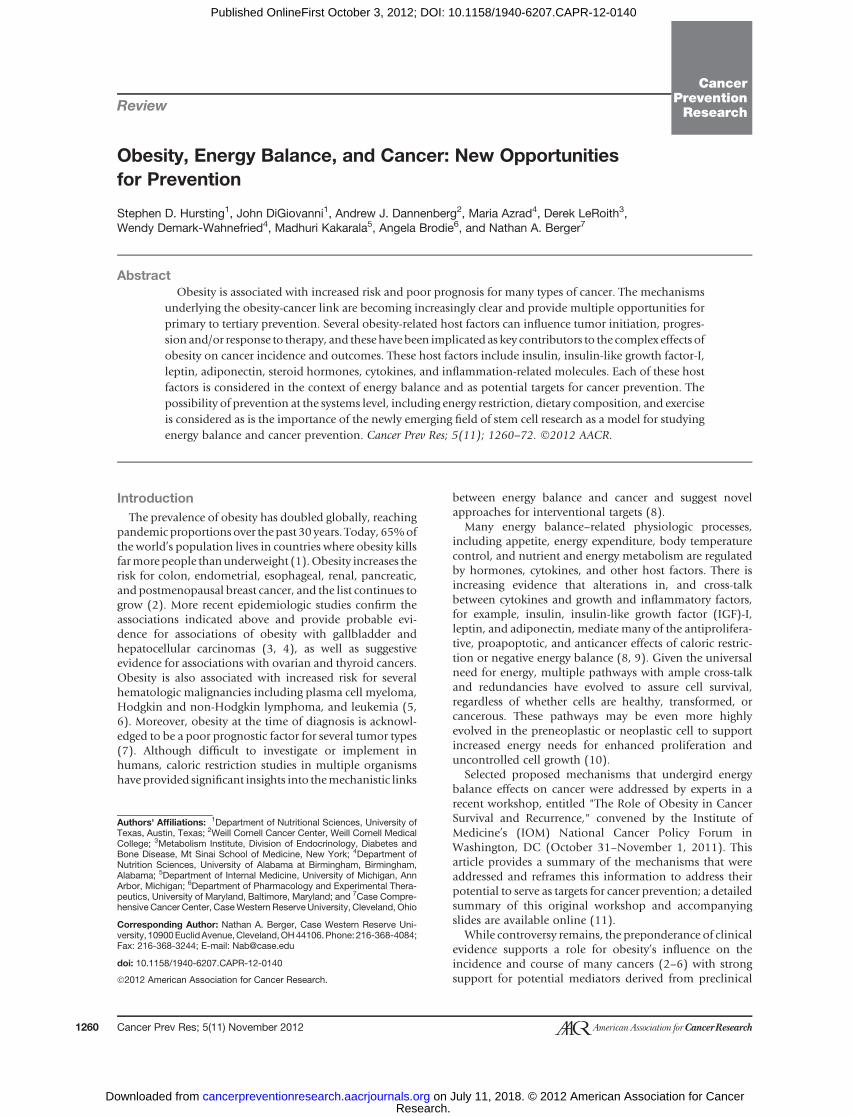
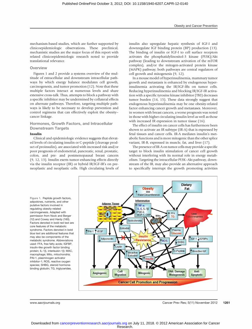
OverviewFigures 1 and 2 provide a systems overview of the mul-
titude of extracellular and downstream intracellular path-ways by which energy balance modulates cell growth,carcinogenesis, and tumor promotion (12). Note that thesemultiple factors interact at numerous levels and shareextensive cross-talk. Thus, attempts to block a pathway witha specific inhibitor may be undermined by collateral effectson alternate pathways. Therefore, targeting multiple path-ways is likely to be necessary to develop prevention andcontrol regimens that can effectively exploit the obesity–cancer linkage.
Hormones, Growth Factors, and IntracellularDownstream TargetsInsulinClinical and epidemiologic evidence suggests that elevat-
ed levels of circulating insulin or C-peptide (cleavage prod-uct of proinsulin), are associated with increased risk and/orpoor prognosis of endometrial, pancreatic, renal, prostatic,colon, and pre- and postmenopausal breast cancers(9, 12, 13). Insulin exerts tumor-enhancing effects directlyvia the insulin receptor (IR) or hybrid IR/IGF-IR’s on pre-neoplastic and neoplastic cells. High circulating levels of
insulin also upregulate hepatic synthesis of IGF-I anddownregulate IGF binding protein (BP) production (13).The binding of insulin or IGF-I to cell surface receptorsactivates the phosphatidylinositol-3 kinase (PI3K)/Aktpathway (leading to downstream activation of the mTORcomplex), and/or the mitogen-activated protein kinase(MAPK) pathway; both pathways are central regulators ofcell growth and mitogenesis (9, 12).
In amousemodel of hyperinsulinemia,mammary tumorgrowth and metastasis is enhanced by endogenous hyper-insulinemia activating the IR/IGF-IRs on tumor cells.Reducing hyperinsulinemia and blocking IR/IGF-IR activa-tion with a specific tyrosine kinase inhibitor (TKI) decreasestumor burden (14, 15). These data strongly suggest thatendogenous hyperinsulinemia may be one obesity-relatedfactor enhancing cancer growth and metastases. Moreover,in women with breast cancers, a worse prognosis was notedin those with higher circulating insulin level as well as thosewith increased IR expression in tumor tissue (16).
The effect of insulin on cancer cells has furthermore beenshown to activate an IR subtype (IR-A) that is expressed byfetal tissues and cancer cells. IR-A mediates insulin’s met-abolic functions and ismoremitogenic than the other splicevariant, IR-B, expressed in muscle, fat, and liver (17).
The presence of IR-Aon tumor cellsmayprovide a specifictarget to block insulin stimulation of cancer cell growthwithout interfering with its normal role in energy metab-olism. Targeting the intracellular PI3K–Akt pathway, down-stream of the IR, may also provide an alternative approachto specifically interrupt the growth promoting activities
Figure 1. Peptide growth factors,adipokines, nutrients, and otherputative factors involved inregulating obesity-relatedcarcinogenesis. Adapted withpermission from Nock and Berger(12) and Cowey and Hardy (160).Factors denoted in bold red text arecore features of the metabolicsyndrome. Factors denoted in boldblue text are additional features thatmay also be components of themetabolic syndrome. Abbreviationsused: FFA, free fatty acids; IGFBP,insulin-like growth factor bindingprotein; IL-1b, interleukin-1b; MAC,macrophage; Mito, mitochondria;PAI-1, plasminogen activatorinhibitor-1; ROS, reactive oxygenspecies; SHBG, steroid hormone-binding globulin; TG, triglycerides.
Obesity and Cancer Prevention
www.aacrjournals.org Cancer Prev Res; 5(11) November 2012 1261
Research. on July 11, 2018. © 2012 American Association for Cancercancerpreventionresearch.aacrjournals.org Downloaded from
Published OnlineFirst October 3, 2012; DOI: 10.1158/1940-6207.CAPR-12-0140
of insulin associated with hyperinsulinemia and insulinresistance.
Insulin-like growth factor-IIGF-I is a major endocrine and paracrine regulator of
tissue growth and metabolism as it both suppresses apo-ptosis and initiates cell-cycle progression from G1 to S-phase by activating PI3K/Akt andMAPK signal transductionpathways and modulating cyclin-dependent kinases (13).Epidemiologic evidence also supports the hypothesis thatincreased circulating IGF-I is associated with increased riskand/or worse prognosis for several types of human cancers(8, 18). IGF-I may act either directly on cells via IGF-IRs orIRs (or even IGF/IR hybrid receptors), or indirectly throughinteraction with other cancer-related molecules, for exam-ple, the tumor suppressor, p53 (13). In numerous animalmodels, obesity increases, while caloric restriction decreasescirculating IGF-I and tumor development and progression(8, 9, 19), whereas exogenous IGF-I infusion rescues tumorgrowth in caloric restricted mice (8, 18). Similar to caloricrestriction, genetic reduction of circulating IGF-I in liver-specific IGF-I–deficient mice decreases tumor progressionin models of colon, skin, mammary, and pancreatic cancer(20, 21). Interestingly, IGF-I–deficient mice, which areresistant to growth of several tumor types, have markedlyelevated insulin and adipokine levels, but a 65% reductionin IGF-I (22), suggesting that IGF-I may be a central deter-minant of energy balance modulation of cancer in exper-imental models (8). However, the obesity-IGF-I relation-ship is more complex in humans, as total circulating IGF-I
levels are often not elevated in obese individuals (23, 24).The hyperinsulinemia associated with obesity can decreaseproduction of IGF BPs (particularly BP1 and 2), and thusincrease the levels of biovailable IGF-I to enhance signalingthrough the IGF-IR (13). Agents that block the IGF-IR todecrease IGF-I signaling, and to some extent insulin signal-ing are under clinical development andmay serve as cancerprevention and control agents; however, selectivity fortumor cells and unwanted metabolic effects remain a chal-lenge for these agents (25).
VEGFVEGF is induced by insulin and IGF-I (26), and mediates
cancer cell proliferation and tumor growth by inducingangiogenesis. Produced by both adipocytes and tumor cells,higher circulating levels of VEGF are seen in obese animals(27) and humans (28), and decrease uponweight loss (29).A recent and growing body of evidence in humans suggestsstrong associations between VEGF levels and aggressivecancers (30, 31). Several TKIs that interfere with VEGFactivity have been developed and could play a role in cancerprevention.
Steroid HormonesEstrogen
Estrogen is produced in large amounts by the ovary viaconversion of androgens (testosterone and androstenedi-one) in a reaction mediated by aromatase. After meno-pause, when the ovary ceases to function, estrogens con-tinue to be produced by other tissues, with a major
LEP EGF IGF-1 INS TNF-αα
HIF1α
ADIPO
ADIPORTNFRINSRIGF-IREGFRLEPR
IRS
PIP3
PI (4,5)P2 Glucose
PDK1
PI3KSrc
Stat3
Ras GRB2
JAK2
SHP-2/
PTPN11
Raf
MEK 1/2
Erk1/2/
MAPK
C-Myc
Cyclin D1
Cell proliferation Cell growth Cell survival
Angiogenesis
Hypoxia
ROS
ROS
AKT TSC2/1
Protein translation
G6P
PEPCK
AMPKK/
STK11
IKK
Exercise
TRAF2
AMPATP
FOX01
Amino
acids
LKB1
AMPK
GSK3B mTOR
4EBp1
elF4E elF4B VEGF
p70S6K
Tumor Promotion/Progression
Autophagy
S6
Figure 2. Intracellular pathways ofgrowth factors involved in obesity-associated cancer promotion andprogression. Adapted withpermission from Nock and Berger(12) and Moore and colleagues(118).
Hursting et al.
Cancer Prev Res; 5(11) November 2012 Cancer Prevention Research1262
Research. on July 11, 2018. © 2012 American Association for Cancercancerpreventionresearch.aacrjournals.org Downloaded from
Published OnlineFirst October 3, 2012; DOI: 10.1158/1940-6207.CAPR-12-0140
contributor being adipose tissue. Multiple lines of evidencesuggest a key role for estrogen in explaining the increasedrisk of hormone receptor–positive breast cancer in obesepostmenopausalwomen. Estrogenhas alsobeen implicatedin the pathogenesis of a subset of endometrial cancersarising in obese women (32). Total and free estrogen levelsare increased in the plasma of obese as compared withnormal weight postmenopausal women (33). Estrogenbinds to estrogen receptor-a and thereby stimulates cellproliferation and inhibits apoptosis (34). Estrogens can alsoinduce VEGF and angiogenesis (35). In addition to drivingtumor formation via estrogen receptor-a–dependenteffects, estrogen can be metabolized into DNA-reactivemetabolites that potentially inducemutagenesis (34). Thereis clear evidence of the growth promoting effects of estro-gens from studies in animals (36). The importance oftargeting estrogen as a preventive intervention is under-scored by clinical data. Tamoxifen and raloxifene, function-ing as selective estrogen receptor modulators, have beenshown to significantly reduce the development of postmen-opausal breast cancer (37), and these agents are effectivesecondary preventive agents in women who have under-gone primary treatment of breast cancer (38). Raloxifenehas also been shown to have a preventive effect on thedevelopment of uterine cancer (39). Recently, treatmentwith exemestane, an aromatase inhibitor, was found todecrease the relative risk of invasive breast cancer by 65%(40). Notably, many of the women enrolled in this trialwere overweight or obese.A major unanswered question concerns the relative
importance of peripheral versus breast adipose tissue as theprimary source of the estrogen that drives tumor formationand progression in obese postmenopausal women. Untilrecently, it was assumed that the mildly elevated levels ofestrogen in venous blood of obese versus lean postmeno-pausal women could account, in part, for the observedincreased risk of breast cancer. However, 2 recent findingshave challenged this explanation. First results from theWomen’s Health Initiative Estrogen-Alone Trial indicatethat administration of estrogen only hormone replacementtherapy was associated with a lower incidence of invasivebreast cancer in postmenopausal women among those whohave had previous hysterectomy (41). Second, as detailedlater, obesity was recently found to cause breast inflamma-tion, elevated aromatase levels, and activation of estrogenreceptor-a–dependent gene expression (42–44). Collec-tively, these recent findings highlight the complexity ofestrogen biology but offer clues to the development offuture preventive strategies.
Testosterone and other androgensThe role of androgens in prostate cancer is significant as
estimates indicate that 80% to 90% of prostate cancer isdependent on circulating androgens for growth (45) andandrogen deprivation directly or through administration ofLHRH antagonists, is a highly successful mainstay of anti-prostate cancer therapy (46). However, it is difficult todescribe how obesity, androgen exposure, and prostate
cancer risk are interrelated as obesity is associated withlower circulating levels of androgens inmen (47), and thereis no strong association betweenobesity andprostate cancerrisk overall (48). However, there is a significant associationbetween obesity and aggressive prostate cancer (49), whichmay relate to the cross-talk between androgens and circu-lating cytokines [e.g., interleukin-6 (IL-6)] and growthfactors (e.g., IGF-I and EGF), which can activate the andro-gen receptor and stimulate JAK/STAT and the PI3K/Akt/mTOR pathways, respectively (50), or the overexpressionand loss of specificity of the androgen receptor, which thenpromiscuously binds ligands driving cell survival and pro-liferation (50).
AdipokinesLeptin and adiponectin are pleiotropic adipocytokines
produced and secreted by adipose tissue. As body fat storesincrease, circulating leptin concentrations increase, whereasadiponectin levels decrease. The obesity-driven imbalancein adiponectin and leptin are considered key factors linkingobesity and cancer. Both mediate energy intake by func-tioning as neuroendocrine signaling hormones that regu-late dietary intake (51, 52), metabolism, insulin sensitivity,and inflammation (53–55). Adiponectin and leptin havedirect tumor effects regulating both cell proliferation andapoptosis (56).
LeptinSix leptin receptors have been identified (ObRa–ObRf);
however, only ObRb has a functioning intracellular signal-ing domain. In vitro studies have shown that activation ofObRb by leptin stimulates cell proliferation and survival incolon (57, 58), breast (59), endometrial (60), and andro-gen-independent prostate cancer cells (61). Leptin signalingis executed throughactivationof several pathways includingJAK/STAT3, PI3K/Akt, and ERK 1/2 (62), and these path-ways all serve as potential targets for cancer prevention andcontrol. Leptin also transactivates the EGFR, Notch, andSurvivin pathways, and stimulates tumor invasion andmigration (63). Leptin can modulate tumor growth byincreasing expression of VEGF, a key driver of angiogenesis(64). In animal models, leptin deficiency inhibits mamma-ry tumor growth (65), whereas higher levels are promo-tional (66). Using azoxymethane to induce colon carcino-genesis in mice, Endo and colleagues showed that leptinsignaling through STAT3 resulted in significantly highertumor proliferation and growth, whereas leptin deficientmice had significantly lower proliferation and smallertumors despite being more obese (67). In contrast, Ribeiroand colleagues inoculated mice with RM-1 murine andro-gen-independent prostate cancer cells and found thathigher leptin concentrations did not increase prostate can-cer tumor growth (68). Thus, disparate effects have beenobserved across cancer types.
In epidemiologic studies, the link between leptin andcancer has also proven inconsistent. In a nested case–con-trol study in Japanese women, leptin was significantlyassociated with colorectal cancer after adjusting for several
Obesity and Cancer Prevention
www.aacrjournals.org Cancer Prev Res; 5(11) November 2012 1263
Research. on July 11, 2018. © 2012 American Association for Cancercancerpreventionresearch.aacrjournals.org Downloaded from
Published OnlineFirst October 3, 2012; DOI: 10.1158/1940-6207.CAPR-12-0140
risk factors including body mass index (BMI) (69). Incontrast, a case–control study in the United States did notfind an association between leptin and colorectal adeno-ma risk in women, but did observe a 3-fold increased riskamong men when comparing the highest tertile againstthe lowest (70). Gender differences in leptin-related riskfor colorectal cancer also have been observed (71). Like-wise, the data on leptin and risk for breast cancer areconflicting with one study reporting a positive association(72), whereas others reported no association (73, 74), oran inverse association (confined to premenopausal breastcancer; ref. 75). In endometrial cancer, a positive associ-ation with leptin was found for 2 studies, although inone, this relationship disappeared after adjusting for BMI(76, 77). For prostate cancer, leptin does not seem toincrease overall risk (78–80), however, it may be linkedwith more aggressive disease (81). In addition, recentstudies suggest that absolute levels of leptin may not bethe driving force behind neoplasia, but rather it is theratio of higher leptin in the presence of low adiponectinthat confers risk (82).
AdiponectinThere are 2 receptors for adiponectin, AdipoR1 and
AdipoR2, which are expressed ubiquitously. Binding ofadiponectin to its receptors stimulates phosphorylation of50-adenosine monophosphate–activated protein kinase(AMPK), a nutrient-sensing enzyme,which regulates severalkey pathways involved in cellular energy metabolism andprotein synthesis (83). In vitro, adiponectin induces apo-ptosis (84) and inhibits growth and proliferation of breast(85–87), colon (88), endometrial (84), and androgen-dependent and -independent prostate cancer (89). Adipo-nectin also sequesters several circulating growth factors (90)and inhibits angiogenesis by inducing apoptosis of endo-thelial cells (91). Studies using preneoplastic murine coloncells have shown that adiponectin inhibits leptin and IL-6–induced cell proliferation by blocking activation of NF-kBand STAT3 (92). Similarly, in late-stage colon cancer cells,adiponectin inhibits IL-6–induced cell proliferation (93).Many, but not all (94) studies using animal models haveshown that lower adiponectin results in increased colontumor growth (95). Interestingly, in the mouse mammarytumor virus (MMTV)-polyoma middle T antigen (PyMT)mammary tumor model, adiponectin-deficient mice havereduced onset and size of mammary tumors as a result ofreduced angiogenesis compared with wild-type mice (96,97).
Data from epidemiologic investigations suggest a rolefor adiponectin in reducing the risk of several cancers.Consistent with in vitro studies, but in contrast to animalmodels, a consistent association between higher adipo-nectin concentrations and decreased risk for postmeno-pausal breast cancer, as well as disease recurrence andmortality has been found repeatedly (98–102). Moststudies have also reported that higher adiponectinlevels are associated with lowered risk for endometrialcancer (102–105). A recent meta-analysis of 13 studies
examined the association between adiponectin and colo-rectal cancer and adenomas and found a significantreduction in risk for men but not for women (106). Forprostate cancer, the data are conflicting and may reflectdiscrepant associations in aggressive versus indolent dis-ease (107, 78).
Intracellular PathwaysPI3K/Akt/mTORC1
Rapamycin, a caloric restriction mimetic and potentialchemopreventive agent, effectively blocks mTORC1 com-plex formation, thus inhibitingmTORC1-mediated cellulargrowth and proliferation (108, 109) and has been shown toextend lifespan in mice (110). Furthermore, rapamycin hasbeen shown to suppress tumorigenesis in several animalmodels (111–115) and inhibits tumor promotionbyblock-ing mTORC1 signaling through p70S6K and cell-cycleproteins PCNA and cyclin D1 (116). Rapamycin also exertsanti-inflammatory effects, and reduces 12-O-tetradecanoyl-phorbol-l3-acetate (TPA)–induced infiltration of severaltypes of inflammatory cells (117). Collectively, these find-ings suggest that rapamycin may function as a potentcompound in cancer prevention. EGFR activation, likewise,is modulated by caloric intake and may also influence Aktand mTORC1 signaling and EGFR activation via IGF-IR/EGFR cross-talk (118).
50-Adenosine monophosphate–activated proteinkinase
Under conditions of caloric restriction, mTORC1 signal-ing is inhibited, leading to cell-growth arrest, inhibition ofprotein translation, and autophagy (118, 119). AMPK andthe upstream kinase, LKB1, function to repress mTORC1 inthe presence of negative energy balance through activationof TSC1/2. Low ATP/AMP ratios activate AMPK, and phos-phorylation ismaintained by LKB1 (120). Activation of thispathway not only reduces cellular energy expenditure, but italso protects against stress-induced apoptosis. Studies usingin vivo models provide evidence that the antidiabetic drug,metformin, which inhibits gluconeogenesis through indi-rect activation of AMPK, can inhibit tumor formation.Administration of metformin suppresses polyp formationin Apc/Min mice (121). Using paired isogenic colon cancercell lines (HCT116 p53þ/þ or P53�/�), Buzzai and collea-gues (122) were able to show that metformin inhibited thegrowth of xenograft tumors derived from the p53 null cellline. Also, daily exposure to metformin attenuated tumor-igenesis (lymphoma; intestinal polyps) in PTEN-deficientmice (123).
Epidemiologic studies have suggested that diabeticpatients receiving metformin have significantly reducedcancer burden compared with diabetic patients receivingother therapies (124). Recent studies have also suggestedthat metformin may be more effective in overweight/obeseindividuals or in individuals with elevated insulin levels(12), and clinical trials are now underway to evaluate theeffect of metformin for the prevention of recurrence ofbreast cancer.
Hursting et al.
Cancer Prev Res; 5(11) November 2012 Cancer Prevention Research1264
Research. on July 11, 2018. © 2012 American Association for Cancercancerpreventionresearch.aacrjournals.org Downloaded from
Published OnlineFirst October 3, 2012; DOI: 10.1158/1940-6207.CAPR-12-0140
Immune Cells and Inflammatory FactorsMacrophage infiltrationChronic inflammation has long been associated with
cancer development and progression and increases the riskof multiple tumor types (9). Obesity leads to subclinicalinflammation in visceral and subcutaneous white adiposetissue (WAT), characterized by macrophages surroundingnecrotic adipocytes and forming crown-like structures(CLS) (125–127). Increased numbers of CLS were shownwithin themammary glandsof obesemice, accompaniedbyactivation of NF-kB, increased levels of proinflammatorymediators, and higher levels and activity of aromatase andits activity, thus driving the synthesis of estrogen (andperhaps ERþ breast cancer) (42). These findings supportthe possibility that the obesity ! inflammation axis isimportant for breast carcinogenesis.Inwomen,CLSof the breast (CLS-B)were found innearly
50% of patient samples (43). The severity of breast inflam-mation, defined as theCLS-B index, correlatedwithBMI andadipocyte size, and may serve as a biomarker of increasedbreast cancer risk or poor prognosis. Consistent with thepreclinical findings, increased NF-kB binding activity,increased levels of proinflammatory mediators, and elevat-ed aromatase expression and activity were found in theinflamed breast tissue of overweight and obese women (43,44). The discovery of the connection between obesity,breast inflammation, and changes in the expressionof geneslinked to breast cancer suggests the possibility that inter-ventions, which reduce breast inflammation may decreasethe increased risk of breast cancer in obese postmenopausalwomen.
CytokinesIncreasing adiposity has been shown to be positively
associated with inflammation in both rodents and humans(128). The increased adipose tissue associated with obesity,especially WAT, produces many inflammatory cytokines,including TNF-a, IL-6, IL-1b, monocyte chemoattractantprotein (MCP-1), and C-reactive protein (CRP), which actboth locally (tissue level) and globally (circulating inserum) (129). Increased adipose-derived cytokine produc-tion, particularly increased levels of MCP-1, enhances localmacrophage infiltration, leading to further increases in thelevels of secreted inflammatory cytokines as well as height-ened recruitment of other related immune cells (129–131).These inflammatory cytokines modulate inflammation viaincreased intracellular signaling through NF-kB-, STAT3-,and c-jun-NH2 terminal protein-kinase (JNK)–related path-ways (129, 132), which are inhibited by caloric restriction(133). These findings suggest the possibility that diet-induced changes in inflammation may modulate tumordevelopment and progression.
NF-kBAt the intracellular level, inflammation is mediated
through multiple pathways. NF-kB is a transcription factoractivated in response to various stimuli including growthfactors and inflammatory molecules, and is responsible for
inducing gene expression associated with cell proliferation,apoptosis, angiogenesis, and inflammation (134). Activa-tion of NF-kB has been observed in many tumor types andhas emerged as an important target for cancer drug devel-opment (135).Obesity and caloric restrictionmodulateNF-kBactivation, possibly through alterations in growth factorsand Akt signaling (134, 136). Activation of NF-kB by Aktcan lead to the translocation of the active NF-kB subunit,p65, from the cytoplasm to the nucleus, inducing multiplegenes associated with inflammation and cancer, includingIL-6, COX-2, and IL-1b (134). Thus, NF-kB represents anattractive drug target for attempting to reduce the risk orprogression of cancer.
Systems Level ConsiderationsDiet composition
While there is a clear association between obesity andcancer in both humans and animal models, it is frequentlydifficult to distinguish the consequences of diet composi-tion from those of obesity. Studies in humans, largely basedon observational research, especially those on internationaldifferences in dietary fat consumption and cancer inci-dence, as well as, on a limited number of case–controlstudies, are suggestive of an association between dietary fatand increased risk of breast, colorectal, andprostate cancers.However, results of these studies aremixed and confoundedby body weight status (137–139). In addition, 2 largerandomized control trials, The Women’s Healthy Eatingand Living (WHEL) study (140) and The Women’s Inter-vention Nutrition Study (WINS; ref. 141) evaluated theeffect of dietary modification on cancer recurrence andsurvival in womenwith early-stage breast cancer. TheWINSstudy reported borderline significance for an associationbetween dietary fat and "breast cancer events," whereas theWHEL study found no significant association between fatand breast cancer recurrence (140–142). Comparison ofthese studies is confounded bymultiple differences, amongwhich is the ability to separate the reduction in dietary fatfrom weight loss (140–142).
To investigate the individual contribution and to bypassthe confounding issues associated with genetics, dietary fat,and obesity, experiments were carried out capitalizing onthe observation that C57BL/6 mice fed a high-fat dietbecome obese, whereas A/J mice fed the same diet remainlean. Taking advantage of these genetic differences, a seriesof crosses were conducted between the B6 and A/J mice togenerate chromosome substitution strains (CSS) of mice inwhich each pair of homozygous A/J chromosomes weresubstituted on an otherwise B6 genetic background (143).These CSSs provide a series of B6 strains that are susceptibleor resistant to diet-induced obesity based on a single pair ofA/J chromosomes. Further research has focused on sepa-rating the effect of dietary fat from obesity, using CSSs incombination with B6. ApcMin/þmouse models of intestinalcancer were used to generate congenic-consomic strains (C-CS). Using C-CSs that were susceptible to ApcMin/þ intes-tinal tumors and were either susceptible or resistant todiet-induced obesity depending on the substituted A/J
Obesity and Cancer Prevention
www.aacrjournals.org Cancer Prev Res; 5(11) November 2012 1265
Research. on July 11, 2018. © 2012 American Association for Cancercancerpreventionresearch.aacrjournals.org Downloaded from
Published OnlineFirst October 3, 2012; DOI: 10.1158/1940-6207.CAPR-12-0140
chromosome, it was shown that a high-fat diet versus low-fat diet (58% vs. 10%) resulted in significant increases inintestinal polyp numbers, tumor burden, and shorter sur-vival time independent of obesity. Moreover, mice fed thehigh-fat diets showed increases in inflammatory cytokinesin the sera (IL-6 and IL-1b) and in intestinal tissue (TNF-a,Cox-2, IL-1b, and IL-6). These studies clearly show that ahigh-fat diet, independent of obesity, can upregulate intes-tinal and circulating inflammatory mediators, and increaseintestinal polyp growth and tumor burden leading toshorter survival (144). These studies support the use oflow-fat diets as a cancer prevention strategy, at least forsome cancer types, and suggest that anti-inflammatoryagents may also be useful in preventing high-fat diet–induced carcinogenesis. In view of the WINS and WHELtrials, these studies with mouse CSSs point to the need forrandomized controlled trials of dietary fat modification forprimary prevention, especially as the WHI study showedthat the low-fat diet resulted in a reduction in primary breastcancer of borderline significance (145), and significantlyreduced ovarian cancer (146).Moreover, diet studies duringcritical windows of susceptibility along the lifecourse areneeded. In particular, studies occurring during the criticalperiods associatedwith breast bud development in the earlyyears of life also are needed.
ExerciseAs recently reviewed, exercise interventions have been
associated with reduced risk of some types of cancer inhumans, with convincing evidence that the exercise–cancerlink is independent of body weight status for colon, breast,and endometrial cancers; weak evidence for prostate, lung,and ovarian; and either null or insufficient data for othercancer types (147). Studies examining the effects of exerciseon carcinogenesis have used a variety of animalmodels andmany, but not all, studies report some evidence of a pro-tective effect of either voluntary or involuntary exercise oncarcinogenesis. The strength and direction of the associa-tion depend on cancer type, intensity of the exerciseregimen, and whether food intake was held constant orprovided ad libitum. Zhu and colleagues recently reportedthat plasmamarkers with the greatest predictive value of theanticancer effects of both moderate caloric restriction andmoderate exercise in a carcinogen-induced rat mammarymodel were adiponectin, bioavailable IGF-I, and leptin(148). In other models, exhaustive exercise has been linkedto increased reactive oxidative stress and inflammation, andincreased tumor development (149), whereas moderateexercise can often be anti-inflammatory (150). Given theinherent links between exercise and energy balance, theeffects of exercise per se, independent of decreased energybalance remain unclear. A study of gene expression profilesin normal mammary tissues of 9-week-old C57BL/6 micethat were randomized to caloric restriction and/or exercise,however, suggests that pathways may differ considerably ascaloric restriction modified gene expression in 425 genes,whereas physical activity modified expression in just 45,with overlap noted in only 3 genes (151). Results of recent
clinical trials in healthy volunteers suggest that weight lossmay exert a more powerful effect on biomarkers associatedwith inflammation and sex steroid pathways (152, 153).Recent studies in mice show that exercise reduces systemicinsulin resistance by an autophagy-inducing process. Thelatter could provide a mechanism for exercise to reducetumor growth through a process of reduced levels of circu-lating insulin (154).
Cancer stem cellsCancer stem cells, their role in carcinogenesis and pro-
gression, and their potential as targets for cancer preventionand therapy, has become a major focus of cancer research.Moreover, as described later, cancer stem cells may providetargets for some of the proliferation-stimulating effects ofadipocytokines elevated in obesity. Tissue stem cells are apopulation of cells with the capacity to undergo self-renew-al and multilineage differentiation into the normal cellpopulation that constitute tissues and organs. The cancerstem cell hypothesis postulates that tumors originatethrough dysregulation of the normal self-renewal processresulting in the aberrant replication and differentiationcharacteristic of a variety of tumors. Clonal expansion ofthese aberrant cells has recently been confirmed by lineagetracing in mouse intestinal adenomas (155) and is an earlystep in carcinogenesis. In addition to being responsible forprimary tumorigenesis, the cancer stem cells may be resis-tant to chemotherapeutic agents and responsible for tumorrecurrence andmetastasis. Thus, strategies aimed at limitingproliferation of these stem cells may be useful for bothprimary and secondary cancer prevention.
A new linkage between obesity and tumor stem cells hasrecently been identified by Zheng and colleagues (65) whoshowed that spontaneous tumors derived from murinemammary tumor virus–Wnt-1 transgenicmice, when trans-planted, were highly leptin-dependent for growth. Thus,transplantation of these tumors into obese, leptin receptor–deficient mice (db/db) with high leptin concentrations,grew to 8 times the volume of those tumors transplantedinto wild-type mice, whereas in leptin-deficient (ob/obmice), tumor growth and overall tumor burden wasreduced. The residual tumors in ob/ob mice were foundto have fewer "stem cells" and these cells were characterizedby flow cytometry to express LepRb (65). When isolated byLepRb expression, these cells exhibited stem cell propertiesof tumorsphere formation in vitro, and their survival wasregulated by leptin. Dunlap, and colleagues report that M-Wnt mammary tumor cells derived from Wnt-1 tumorprofile with human claudin-low breast tumors, are mesen-chymal and stably enriched in breast cancer cell markers,and exhibit stem cell properties (156). In addition, M-Wntcells orthotopically injected into B6 mice rapidly formclaudin-low tumors that are highly responsive to thetumor-enhancing effects of obesity, as well as the anticancereffects of calorie restriction (156). Relationships betweenobesity, adipose tissue cytokines, and stem cell biology alsocan be readily studied in vitro directly from human breasttissue excised during elective reduction mammoplasty
Hursting et al.
Cancer Prev Res; 5(11) November 2012 Cancer Prevention Research1266
Research. on July 11, 2018. © 2012 American Association for Cancercancerpreventionresearch.aacrjournals.org Downloaded from
Published OnlineFirst October 3, 2012; DOI: 10.1158/1940-6207.CAPR-12-0140
(157).However, at present, human clinical trials specificallytargeting stem cells are hampered by the need for largevolumes of breast tissue (e.g., from mammoplasty or mas-tectomy) for stem cell isolation and dynamic assays, such asin vitro tumor sphere formation as biomarker endpoints(158). Therefore, current efforts are focuseduponusing corebiopsy samples for stem cell isolation and characterizationusing advanced in situ technologies, such as multipleximaging or profiling technologies.
Conclusions and Future DirectionsWhile much current research focuses on selected signal-
ing pathways, this review emphasizes the numerous med-iators and pathways by which obesity impacts cancer. Manyof the circulating signaling molecules bind to cell surfacereceptors where they activate intracellular pathways, whichundergo cross-talk and activate intracellular downstreampathways that both converge and diverge to promote cancercell growth and metastasis.These observations suggest several strategies for preven-
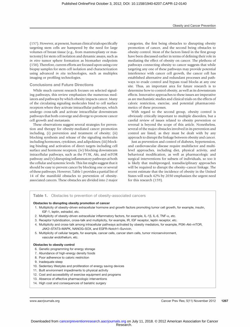
tion and therapy for obesity-mediated cancer promotionincluding, (i) prevention and treatment of obesity; (ii)blocking synthesis and release of the signaling moleculesincluding hormones, cytokines, and adipokines; (iii) block-ing binding and activation of direct targets including cellsurface and hormone receptors; (iv) blocking downstreamintracellular pathways, such as the P13K, Akt, and mTORpathway; and (v)disrupting inflammatory pathways at boththe cellular and systemic levels. This listmight suggest that itshould be easy to prevent cancer by blocking one or severalof these pathways.However, Table 1 provides a partial list of14 of the manifold obstacles to prevention of obesity-associated cancers. These obstacles are divided into 2major
categories, the first being obstacles to disrupting obesitypromotion of cancer, and the second being obstacles toobesity control. Most of the factors listed in the first grouphave been discussed earlier in terms of defining their role inmediating the effect of obesity on cancer. The plethora ofpathways connecting obesity to cancer suggests that whiletargeting any one of these pathways may provide potentialinterference with cancer cell growth, the cancer cell hasestablished alternative and redundant processes and path-ways to evade control and bypass road blocks at any onesite. Thus, an important area for future research is todetermine how to control obesity, as well as its downstreameffects. Innovative approaches to these issues are imperativeas aremechanistic studies and clinical trials on the effects ofcaloric restriction, exercise, and potential pharmacomi-metics of these processes.
With regard to the second group, obesity control isobviously critically important to multiple disorders, but acareful review of issues related to obesity prevention orreversal is beyond the scope of this article. Nonetheless,several of themajor obstacles involved in its prevention andcontrol are listed, as they must be dealt with by anyapproach to disrupt the linkage betweenobesity and cancer.
Just as prevention and control of diabetes, hypertension,and cardiovascular disease require multifactor and multi-level approaches, including diet, physical activity, andbehavioral modification, as well as pharmacologic andsurgical interventions for subsets of individuals, so too itis likely that multipronged, transdisciplinary approacheswill be required to disrupt the obesity–cancer linkage. Therecent estimate that the incidence of obesity in the UnitedStates will reach 42% by 2030 emphasizes the urgent needfor this research (159).
Table 1. Obstacles to prevention of obesity-associated cancers
Obstacles to disrupting obesity promotion of cancer1. Multiplicity of obesity-driven extracellular hormone and growth factors promoting tumor cell growth, for example, insulin,
IGF-1, leptin, estradiol, etc.2. Multiplicity of obesity-driven extracellular inflammatory factors, for example, IL-1b, IL-6, TNF-a, etc.3. Receptor hybridization, cross-talk and multiplicity, for example, IR, IGF receptor, leptin receptor, etc.4. Multiplicity and cross-talk among intracellular pathways activated by obesity mediators, for example, PI3K–Akt–mTOR,
JAK2–STAT3–MAPK, NANOG–SOX, and EGFR–Notch1–Survivin.5. Multiplicity of cellular targets, for example, cancer cells, cancer stem cells, tumor microenvironment,
vascular endothelium, etc.
Obstacles to obesity control6. Genetic programming for energy storage7. Abundance of high-energy density foods8. Poor adherence to caloric restriction9. Inadequate sleep
10. Sedentary lifestyles and proliferation of energy saving devices11. Built environment impediments to physical activity12. Cost and accessibility of exercise equipment and programs13. Absence of effective pharmacologic interventions14. High cost and consequences of bariatric surgery
Obesity and Cancer Prevention
www.aacrjournals.org Cancer Prev Res; 5(11) November 2012 1267
Research. on July 11, 2018. © 2012 American Association for Cancercancerpreventionresearch.aacrjournals.org Downloaded from
Published OnlineFirst October 3, 2012; DOI: 10.1158/1940-6207.CAPR-12-0140
Disclosure of Potential Conflicts of InterestNo potential conflicts of interest were disclosed.
Grant SupportResearch reported in this review was supported by grants P30ES007784
and RO1CA129409 to S.D. Hursting; RO1CA037111 to J. DiGiovanni;RO1CA154481, Breast Cancer Research Foundation, the Botwinick-Wolfen-sohn Foundation (in memory of Mr. and Mrs. Benjamin Botwinick) to A.J.Dannenberg; RO1CA128700 to D. LeRoith; RO1CA062483 to A. Brodie;KO7CA128884 to M. Kakarala; P30 CA13148 to M. Azrad and W. Demark-Wahnefried; and U54 CA116867 and P40 RR012305 to N.A. Berger. Theresponsibility for the content of this article rests with the authors and does
not necessarily represent the views of the Institute of Medicine (IOM), itscommittees, or its convening activities. The activities of the IOM’s NCPF aresupported by its sponsoring members, which currently include the NationalCancer Institute, the Centers for Disease Control and Prevention, theAmerican Association for Cancer Research, American Cancer Society, theAmerican Society of Clinical Oncology, the Association of American CancerInstitutes, Bristol–Myers Squibb, C-Change, theCEORoundtable onCancer,GlaxoSmithKline Oncology, Novartis Oncology, the Oncology NursingSociety, and Sanofi Oncology.
ReceivedMarch28, 2012; revisedAugust 28, 2012; accepted September 10,2012; published OnlineFirst October 3, 2012.
References1. WHO fact sheet 311. Obesity and overweight; 2011 [accessed 2011
Feb 6]. Available from: http://www.who.int/mediacentre/factsheets/fs311/en/.
2. Calle EE, Kaaks R. Overweight, obesity and cancer: epidemiologicalevidence and proposed mechanisms. Nat Rev Cancer 2004;4:579–91.
3. Ballard-Barbash R, Berrigan D, Potischman N, Dowling E. Obesityand cancer epidemiology. In: Berger NA editor. Energy balance andcancer. New York: Springer; 2010. p. 1–44.
4. Nock NL. Obesity and gastrointestinal cancers: epidemiology. In:Markowitz SD, Berger NA editors. Energy balance and gastrointes-tinal cancer. New York: Springer; 2012. p. 1–22.
5. Teras LR, Patel AV. The epidemiology of obesity and hematologicmalignancies. In: Mittelman SD, Berger NA editors. Energy balanceand hematologic malignancies. New York: Springer; 2012. p. 1–30.
6. Beason T, Colditz G. Obesity and multiple myeloma. In: MittelmanSD, Berger NA editors. Energy balance and hematologic malignan-cies. New York: Springer; 2012. p. 71–95.
7. Calle EE, Rodriguez C, Walker-Thurmond K, Thun MJ. Overweight,obesity, and mortality from cancer in a prospective studied cohort ofU.S. adults. N Engl J Med 2003;48:1625–38.
8. Hursting SD, Smith SM, Lashinger LM, Harvey AE, Perkins SN.Calories and carcinogenesis: lessons learned from30 years of calorierestriction research. Carcinogenesis 2010;31:83–9.
9. Hursting SD, Berger NA. Energy balance, host-related factors, andcancer progression. J Clin Oncol 2010;28:4058–65.
10. Vander Heiden MG, Cantley LC, Thompson CB. Understanding theWarburg effect: the metabolic requirements of cell proliferation.Science 2009;324:1029–33.
11. Institute ofMedicine of the National Academies. The role of obesity incancer survival and recurrence—workshop summary. [cited 2012Apr 3]. Available from: http://www.iom.edu/Activities/Disease/NCPF/2011-OCT-31.aspx.
12. NockN, Berger NA. Obesity and cancer, overview ofmechanisms. In:Berger NA editor. Energy balance and cancer. New York: Springer;2010. p. 129–79.
13. Pollak M. Insulin and insulin-like growth factor signaling in neoplasia.Nat Rev Cancer 2008;8:915–28.
14. Dool CJ, Mashhedi H, Zakikhani M, David S, Zhao Y, Birman E, et al.IGF1/insulin receptor kinase inhibition by BMS-536924 is bettertolerated than alloxan-induced hypoinsulinemia and more effectivethan metformin in the treatment of experimental insulin-responsivebreast cancer. Endocr Relat Cancer 2011;18:699–709.
15. Fierz Y, Novosyadlyy R, Vijayakumar A, Yakar S, LeRoith D. Insulin-sensitizing therapy attenuates type 2 diabetes-mediated mammarytumor progression. Diabetes 2010;59:686–93.
16. Goodwin PJ, Ennis M, Pritchard KI, TrudeauME, Koo J, Madarnas Y,et al. Fasting insulin andoutcome in early-stagebreast cancer: resultsof a prospective cohort study. J Clin Oncol 2002;20:42–51.
17. Ferguson RD, Novosyadlyy R, Fierz Y, Alikhani N, Sun H, Yakar S,et al. Hyperinsulinemia enhances c-Myc-mediated mammarytumor development and advances metastatic progression to thelung in a mouse model of type 2 diabetes. Breast Cancer Res2012;14:R8.
18. Hursting SD, Lavigne JA, Berrigan D, Perkins SN, Barrett JC. Calorierestriction, aging, and cancer prevention: mechanisms of action andapplicability to humans. Annu Rev Med 2003;54:131–52.
19. Gallagher EJ, Fierz Y, Ferguson RD, LeRoith D. The pathway fromdiabetes and obesity to cancer, on the route to targeted therapy.Endocr Pract 2010;16:864–73.
20. Moore T, Carbajal S, Beltran L, Perkins SN, Yakar S, Leroith D, et al.Reduced susceptibility to two-stage skin carcinogenesis inmicewithlow circulating insulin-like growth factor I levels. Cancer Res 2008;68:3680–8.
21. Harvey AE, Lashinger LM, Hursting SD. The growing challenge ofobesity and cancer: an inflammatory issue. Ann N Y Acad Sci 2011;1229:45–52.
22. Yakar S, Liu JL, Fernandez AM, Wu Y, Schally AV, Frystyk J, et al.Liver-specific igf-1 gene deletion leads tomuscle insulin insensitivity.Diabetes 2001;50:1110–8.
23. Alderete TL, Byrd-Williams CE, Toledo-Corral CM, Conti DV, Wei-gensberg MJ, Goran MI. Relationships between IGF-1 and IGFBP-1and adiposity in obese African-American and Latino adolescents.Obesity 2011;19:933–8.
24. Berrigan D, Potischman N, Dodd KW, Hursting SD, Lavigne J, BarrettJC, et al. Race/ethnic variation in serum levels of IGF-I and IGFBP-3 inUS adults. Growth Horm IGF Res 2009;19:146–55.
25. Weroha SJ, Haluska P. IGF-1 receptor inhibitors in clinical trials—early lessons. J Mammary Gland Biol Neoplasia 2008;13:471–83.
26. Miele C, Rochford JJ, Filippa N, Giorgetti-Peraldi S, Van ObberghenE. Insulin and insulin-like growth factor-I induce vascular endothelialgrowth factor mRNA expression via different signaling pathways.J Biol Chem 2000;275:21695–702.
27. Miyazawa-Hoshimoto S, Takahashi K, BujoH,HashimotoN, Yagui K,Saito Y. Roles of degree of fat deposition and its localization on VEGFexpression in adipocytes. Am J Physiol Endocrinol Metab 2005;288:E1128–36.
28. Loebig M, Klement J, Schmoller A, Betz S, Heuck N, Schweiger U,et al. Evidence for a relationship betweenVEGFandBMI independentof insulin sensitivity by glucose clamp procedure in a homogenousgroup healthy young men. PLoS ONE 2010;5:e12610.
29. Heymach JV, Shackleford TJ, Tran HT, Yoo SY, Do KA, Wergin M,et al. Effect of low-fat diets on plasma levels of NF-kB-regulatedinflammatory cytokines and angiogenic factors in men with prostatecancer. Cancer Prev Res 2011;4:1590–8.
30. Liu Y, Tamimi RM, Collins LC, Schnitt SJ, Gilmore HL, Connolly JL,et al. The association between vascular endothelial growth factorexpression in invasive breast cancer and survival varies with intrinsicsubtypes and use of adjuvant systemic therapy: results from theNurses' Health Study. Breast Cancer Res Treat 2011;129:175–84.
31. CaoD,HouM,GuanYS, JiangM,YangY,GouHF. Expression ofHIF-1alpha and VEGF in colorectal cancer: association with clinical out-comes and prognostic implications. BMC Cancer 2009;9:432.
32. Ulrich LSG. Endometrial cancer, types, prognosis, female hormonesand antihormones. Climacteric 2011;14:418–25.
33. KeyTJ, ApplebyPN,ReevesGK,RoddamA,Dorgan JF, LongcopeC,et al. Body mass index, serum sex hormones, and breast cancer riskin postmenopausal women. J Natl Cancer Inst 2003;95:1218–26.
Hursting et al.
Cancer Prev Res; 5(11) November 2012 Cancer Prevention Research1268
Research. on July 11, 2018. © 2012 American Association for Cancercancerpreventionresearch.aacrjournals.org Downloaded from
Published OnlineFirst October 3, 2012; DOI: 10.1158/1940-6207.CAPR-12-0140
34. Yager JD, Davidson NE. Estrogen carcinogenesis in breast cancer.N Engl J Med 2006;354:270–82.
35. Pequeux C, Raymond-Letron I, Blacher S, Boudou F, Adlanmerini M,Fouque MJ, et al. Stromal estrogen receptor-a promotes tumorgrowth by normalizing an increased angiogenesis. Cancer Res2012;72:1–10.
36. Brodie AM,Marsh D, Brodie HJ. Aromatase inhibitors–IV. Regressionof hormone-dependent, mammary tumors in the rat with 4-acetoxy-4-androstene-3,17-dione. J Steroid Biochem 1979;10:423–9.
37. Vogel VG, Costantino JP, Wickerham DL, Cronin WM, Cecchini RS,Atkins JN. Effects of tamoxifen vs raloxifene on the risk of developinginvasive breast cancer and other disease outcomes: the NSABPStudy of Tamoxifen and Raloxifene (STAR) P-2 trial. JAMA 2006;295:2727–4124.
38. Early Breast Cancer Trialists' Collaborative Group (EBCTCG). Effectsof chemotherapy and hormonal therapy for early breast cancer onrecurrence and 15-year survival: an overviewof the randomised trials.Lancet 2005;365:1687–717.
39. DeMichele A, Troxel AB, Berlin JA, Weber AL, Bunin GR, Turzo E,et al. Impact of raloxifene or tamoxifen use on endometrial cancerrisk: a population-based case-control study. J Clin Oncol 2008;26:4151–9.
40. Goss PE, Ingle JN, Ales-Martinez JE, Cheung AM, Chlebowski RT,Wactawski-Wende J, et al. Exemestane for breast-cancer preventionin postmenopausal women. N Engl J Med 2011;364:2381–91.
41. LaCrois AZ, Chlebowski RT, Manson JE, Aragaki AK, Johnson KC,Martin L, et al. Health outcomes after stopping conjugated equineestrogens among postmenopausal women with prior hysterectomy.JAMA 2011;305;1305–14.
42. Subbaramaiah K, Howe LR, Bhardwaj P, Du B, Gravaghi C, YantissRK, et al. Obesity is associated with inflammation and elevatedaromatase expression in the mouse mammary gland. Cancer PrevRes 2011;4:329–46.
43. Morris PG, Hudis CA, Giri D, Morrow M, Falcone DJ, Zhou XK, et al.Inflammation and increased aromatase expression occur in thebreast tissue of obese women with breast cancer. Cancer Prev Res2011;4:1021–9.
44. Subbaramaiah K, Morris PG, Zhou ZK, Morrow M, Du B, Giri D, et al.Increased levels of COX-2 and prostaglandin E2 contribute to ele-vated aromatase expression in inflamed breast tissue of obesewomen. Cancer Discov 2012;2:356–65.
45. Heinlein CA, Chang C. Androgen receptor in prostate cancer. EndocrRev 2004;25:276–308.
46. Kawakami J, Cowan JE, Elkin EP, Latini DM, DuChane J, Carroll PR,et al. Androgen-deprivation therapy asprimary treatment for localizedprostate cancer: data from Cancer of the Prostate Strategic UrologicResearch Endeavor (CaPSURE). Cancer 2006;106:1708–14.
47. Dhindsa S, Miller MG, McWhirter CL, Mager DE, Ghanim H, Chaud-huri A, et al. Testosterone concentrations in diabetic and nondiabeticobese men. Diabetes Care 2010;33:1186–92.
48. Giovannucci E, Rimm EB, Liu Y, Leitzmann M, Wu K, Stampfer MJ,et al. Body mass index and risk of prostate cancer in U.S. healthprofessionals. J Natl Cancer Inst 2003;95:1240–4.
49. Jayachandran J, Ba~nez LL, Aronson WJ, Terris MK, Presti JC Jr,Amling CL, et al. Obesity as a predictor of adverse outcome acrossblack and white race: results from the Shared Equal Access RegionalCancer Hospital (SEARCH) database. Cancer 2009;115:5263–71.
50. Lonergan PE, Tindall DJ. Androgen receptor signaling in prostatecancer development and progression. J Carcinog 2011;10:20.
51. Farooqi IS, O'Rahilly S. Leptin: a pivotal regulator of human energyhomeostasis. Am J Clin Nutr 2009;89:980S–4S.
52. Dridi S, Taouis M. Adiponectin and energy homeostasis: consensusand controversy. J Nutr Biochem 2009;20:831–9.
53. Weyer C, Funahashi T, Tanaka S, Hotta K, Matsuzawa Y, Pratley RE.Hypoadiponectinemia in obesity and type 2 diabetes: close associ-ation with insulin resistance. J Clin Endocrinol Metab 2001;86:1930–5.
54. Stefan N, Vozarova B, Funahashi T, Matsuzawa Y, Weyer C, LindsayRS, et al. Plasma adiponectin concentration is associated withskeletal muscle insulin receptor tyrosine phosphorylation, and low
plasma concentration precedes a decrease in whole-body insulnsensitivity in human. Diabetes 2002;50:1884–8.
55. Stofkova A. Leptin and adiponectin: from energy and metabolicdysbalance to inflammation and autoimmunity. Endocr Regul2009;43:157–68.
56. Housa D, Housov�a J, Vernerov�a Z, Haluzík M. Adipocytokines andcancer. Physiol Res 2006;55:233–44.
57. Hoda MR, Keely SJ, Bertelsen LS, Junger WG, Dharmasena D,Barrett KE. Leptin acts as a mitogenic and antiapoptotic factor forcolonic cancer cells. Br J Surg 2007;94:346–54.
58. Ogunwobi OO, Beales IL. The anti-apoptotic and growth stimulatoryactions of leptin in human colon cancer cells involves activation ofJNKmitogen activated protein kinase, JAK2 and PI3 kinase/Akt. Int JColorectal Dis 2007;22:401–9.
59. Saxena NK, Vertino PM, Anania FA, Sharma D. Leptin-inducedgrowth stimulation of breast cancer cells involves recruitment ofhistone acetyltransferases and mediator complex to cyclin d1 pro-moter via activation of stat3. J Biol Chem 2007;282:13316–25.
60. Sharma D, Saxena NK, Vertino PM, Anania FA. Leptin promotes theproliferation responseand invasiveness in humanendometrial cancercells by activating multiple signal-transduction pathways. EndocrRelat Cancer 2006;13:629–40.
61. Onuma M, Bub JD, Rummel TL, Iwamoto Y. Prostate cancer cell-adipocyte interaction: leptin mediates androgen-independent pros-tate cancer cell proliferation through c-Jun NH2-terminal kinase.J Biol Chem 2003;278:42660–7.
62. Garofalo C, Surmacz E. Leptin and cancer. J Cell Physiol 2006;207:12–22.
63. Knight BB, Oprea-Ilies GM, Nagalingam A, Yang L, Cohen C, SaxenaNK, et al. Survivin upregulation, dependent on leptin-EGFR-Notch1axis, is essential for leptin-induced migration of breast carcinomacells. Endocr Relat Cancer 2011;18:413–28.
64. Rene Gonzalez R, Watters A, Xu Y, Singh UP, Mann DR, Rueda BR,et al. Leptin-signaling inhibition results in efficient anti-tumor activityin estrogen receptor positive or negative breast cancer. BreastCancer Res 2009;11:R36.
65. ZhengQ,DunlapSM,Zhu J,Downs-Kelly E,Rich J,HurstingSD, et al.Leptin deficiency suppresses MMTV-Wnt-1 mammary tumor growthin obese mice and abrogates tumor initiating cell survival. EndocrRelat Cancer 2011;18:491–503.
66. Lautenbach A, Budde A, Wrann CD, Teichmann B, Vieten G, Karl T,et al. Obesity and the associatedmediators leptin, estrogen and IGF-Ienhance the cell proliferation and early tumorigenesis of breastcancer cells. Nutr Cancer 2009;61:484–91.
67. Endo H, Hosono K, Uchiyama T, Sakai E, Sugiyama M, Takahashi H,et al. Leptin acts as a growth factor for colorectal tumours at stagessubsequent to tumour initiation in murine colon carcinogenesis. Gut2011;60:1363–71.
68. RibeiroAM,AndradeS,PinhoF,Monteiro JD,CostaM, LopesC, et al.Prostate cancer cell proliferation and angiogenesis in different obesemice models. Int J Exp Pathol 2010;91:374–86.
69. Tamakoshi K, ToyoshimaH,Wakai K, KojimaM, Suzuki K,WatanabeY, et al. Leptin is associated with an increased female colorectalcancer risk: a nested case-control study in Japan. Oncology2005;68:454–61.
70. Chia VM, Newcomb PA, Lampe JW, White E, Mandelson MT,McTiernanA, et al. Leptin concentrations, leptin receptor polymorph-isms, and colorectal adenoma risk. Cancer Epidemiol BiomarkersPrev 2007;16:2697–703.
71. Stattin P, Lukanova A, Biessy C, S€oderberg S, Palmqvist R, Kaaks R,et al. Obesity and colon cancer: does leptin provide a link? IntJ Cancer 2004;109:149–52.
72. Wu MH, Chou YC, Chou WY, Hsu GC, Chu CH, Yu CP, et al.Circulating levels of leptin, adiposity and breast cancer risk. BrJ Cancer 2009;100:578–82.
73. Mantzoros CS, Bolhke K, Moschos S, Cramer DW. Leptin in relationto carcinoma in situ of the breast: a study of pre-menopausal casesand controls. Int J Cancer 1999;80:523–6.
74. Stattin P, Soderberg S, Biessy C, Lenner P, Hallmans G, Kaaks R,et al. Plasma leptin and breast cancer risk: a prospective
Obesity and Cancer Prevention
www.aacrjournals.org Cancer Prev Res; 5(11) November 2012 1269
Research. on July 11, 2018. © 2012 American Association for Cancercancerpreventionresearch.aacrjournals.org Downloaded from
Published OnlineFirst October 3, 2012; DOI: 10.1158/1940-6207.CAPR-12-0140
study in northern Sweden. Breast Cancer Res Treat 2004;86:191–6.
75. Harris HR, Tworoger SS, Hankinson SE, Rosner BA, Michels KB.Plasma leptin levels and risk of breast cancer in premenopausalwomen. Cancer Prev Res 2011;4:1449–56.
76. Petridou E, Belechri M, Dessypris N, Koukoulomatis P, DiakomanolisE, Spanos E, et al. Leptin and body mass index in relation toendometrial cancer risk. Ann Nutri Metab 2002;46:147–51.
77. Cymbaluk A, Chudecka-Gøaz A, Rzepka-G�orska I. Leptin levels inserum depending on Body Mass Index in patients with endometrialhyperplasia and cancer. Eur J Obstet Gynecol Reprod Biol 2008;136:74–7.
78. Li H, Stampfer MJ, Mucci L, Rifai N, Qiu W, Kurth T, et al. A 25-yearprospective study of plasma adiponectin and leptin concentra-tions and prostate cancer risk and survival. Clin Chem 2010;56:34–43.
79. Baillargeon J, Platz EA, Rose DP, Pollock BH, Ankerst DP, Haffner S,et al. Obesity, adipokines, and prostate cancer in a prospectivepopulation-based study. Cancer Epidemiol Biomarkers Prev2006;15:1331–5.
80. Hsing AW, Chua S Jr, Gao YT, Gentzschein E, Chang L, Deng J, et al.Prostate cancer risk and serum levels of insulin and leptin: a popu-lation-based study. J Natl Cancer Inst 2001;93:783–9.
81. L�opez Fontana CM, Maselli ME, P�erez Elizalde RF, Di Milta M�onacoNA, Uvilla Recupero AL, L�opez Laur JD. Leptin increases prostatecancer aggressiveness. J Physiol Biochem 2011;67:531–8.
82. Yamaji T, Iwasaki M, Sasazuki S, Tsugane S. Interaction betweenadiponectin and leptin influences the risk of colorectal adenoma.Cancer Res 2010;70:5430–7.
83. Kelesidis I, Kelesidis T, Mantzoros CS. Adiponectin and cancer: asystemic review. Br J Cancer 2006;94:1221–5.
84. Cong L, Gasser J, Zhao J, Yang B, Li F, Zhao AZ. Human adiponectininhibits cell growth and induces apoptosis in human endometrialcarcinoma cells, HEC-1-A and RL95 2. Endocr Relat Cancer2007;14:713–20.
85. K€orner A, Pazaitou-Panayiotou K, Kelesidis T, Kelesidis I, WilliamsCJ, Kaprara A, et al. Total and high-molecular-weight adiponectin inbreast cancer: in vitro and in vivo studies. J Clin Endocrinol Metab2007;92:1041–8.
86. Nakayama S, Miyoshi Y, Ishihara H, Noguchi S. Growth-inhibitoryeffect of adiponectin via adiponectin receptor 1 on human breastcancer cells through inhibition of S-phase entry without inducingapoptosis. Breast Cancer Res Treat 2008;112:405–10.
87. Arditi JD, Venihaki M, Karalis KP, Chrousos GP. Antiproliferativeeffect of adiponectin on MCF7 breast cancer cells: a potentialhormonal link between obesity and cancer. Horm Metab Res2007;39:9–13.
88. KimAY, Lee YS, KimKH, Lee JH, LeeHK, Jang SH, et al. Adiponectinrepresses colon cancer cell proliferation via AdipoR1- and -R2-mediated AMPK activation. Mol Endocrinol 2010;24:1441–52.
89. Bub JD, Miyazaki T, Iwamoto Y. Adiponectin as a growth inhibitor inprostate cancer cells. Biochem Biophys Res Commun 2006;340:1158–66.
90. Wang Y, Lam KS, Xu JY, Lu G, Xu LY, Cooper GJ, et al. Adiponectininhibits cell proliferation by interacting with several growth factors inan oligomerization-dependent manner. J Biol Chem 2005;280:18341–7.
91. Bra�kenhielm E, Veitonm€aki N, Cao R, Kihara S, Matsuzawa Y,
Zhivotovsky B, et al. Adiponectin-induced antiangiogenesis andantitumor activity involve caspase-mediated endothelial cell apopto-sis. Proc Natl Acad Sci U S A 2004;101:2476–81.
92. Fenton JI, Birmingham JM, Hursting SD, Hord NG. Adiponectinblocks multiple signaling cascades associated with leptin-inducedcell proliferation in Apc Min/þcolon epithelial cells. Int J Cancer2008;122:2437–45.
93. Fenton JI, Birmingham JM. Adipokine regulation of colon cancer:adiponectin attenuates interleukin-6-induced colon carcinoma cellproliferation via STAT-3. Mol Carcinog 2010;49:700–9.
94. Ealey KN, Archer MC. Elevated circulating adiponectin and elevatedinsulin sensitivity in adiponectin transgenic mice are not associated
with reduced susceptibility to colon carcinogenesis. Int J Cancer2009;124:2226–30.
95. Nishihara T, BabaM, Matsuda M, InoueM, Nishizawa Y, Fukuhara A,et al. Adiponectin deficiency enhances colorectal carcinogenesis andliver tumor formation induced by azoxymethane in mice. World JGastroenterol 2008;14:6473–80.
96. Landskroner-Eiger S, Qian B, Muise ES, Nawrocki AR, Berger JP,Fine EJ, et al. Proangiogenic contribution of adiponectin towardmammary tumor growth in vivo. Clin Cancer Res 2009;15:3265–76.
97. Denzel MS, Hebbard LW, Shostak G, Shapiro L, Cardiff RD, RanschtB. Adiponectin deficiency limits tumor vascularization in the MMTV-PyV-mT mouse model of mammary cancer. Clin Cancer Res 2009;15:3256–64.
98. Tian YF, Chu CH, Wu MH, Chang CL, Yang T, Chou YC, et al.Anthropometric measures, plasma adiponectin, and breast cancerrisk. Endocr Relat Cancer 2007;14:669–77.
99. Miyoshi Y, Funahashi T, KiharaS, Taguchi T, Tamaki Y,MatsuzawaY,et al. Association of serum adiponectin levels with breast cancer risk.Clin Cancer Res 2003;9:5699–704.
100. Mantzoros C, Petridou E, Dessypris N, Chavelas C, Dalamaga M,Alexe DM, et al. Adiponectin and breast cancer risk. J Clin EndocrinolMetab 2004;89:1102–7.
101. Tworoger SS, Eliassen AH, Kelesidis T, Colditz GA, Willett WC,Mantzoros CS, et al. Plasma adiponectin concentrations and riskof incident breast cancer. J Clin Endocrinol Metab 2007;92:1510–6.
102. Duggan C, Irwin ML, Xiao L, Henderson KD, Smith AW, BaumgartnerRN, et al. Associations of insulin resistance and adiponectin withmortality in women with breast cancer. J Clin Oncol 2011;29:32–9.
103. Cust AE, Kaaks R, Friedenreich C, Bonnet F, Laville M, Lukanova A,et al. Plasma adiponectin levels and endometrial cancer risk in pre-and postmenopausal women. J Clin Endocrinol Metab 2007;92:255–63.
104. Petridou E, Mantzoros C, Dessypris N, Koukoulomatis P, Addy C,Voulgaris Z, et al. Plasma adiponectin concentrations in relation toendometrial cancer: a case-control study inGreece. JClin EndocrinolMetab 2003;88:993–7.
105. Soliman PT, Wu D, Tortolero-Luna G, Schmeler KM, Slomovitz BM,Bray MS, et al. Association between adiponectin, insulin resistance,and endometrial cancer. Cancer 2006;106:2376–81.
106. Xu XT, Xu Q, Tong JL, Zhu MM, Huang ML, Ran ZH, et al. Meta-analysis: circulating adiponectin levels and risk of colorectal cancerand adenoma. J Dig Dis 2011;12:234–44.
107. Michalakis K, Williams CJ, Mitsiades N, Blakeman J, Balafouta-Tselenis S, Giannopoulos A, et al. Serum adiponectin concentrationsand tissue expression of adiponectin receptors are reduced inpatientswith prostate cancer: a case control study. Cancer EpidemiolBiomarkers Prev 2007;16:308–13.
108. Garcia JA, Danielpour D.Mammalian target of rapamycin inhibition asa therapeutic strategy in the management of urologic malignancies.Mol Cancer Ther 2008;7:1347–54.
109. Kopelovich L, Fay JR, SigmanCC, Crowell JA. Themammalian targetof rapamycin pathway as a potential target for cancer chemopre-vention. Cancer Epidemiol Biomarkers Prev 2007;16:1330–40.
110. Anisimov VN, Zabezhinski MA, Popovich IG, Piskunova TS,Semenchenko AV, Tyndyk ML, et al. Rapamycin extends maximallifespan in cancer-prone mice. Am J Pathol 2010;176:2092–7.
111. Granville CA, Warfel N, Tsurutani J, Hollander MC, Robertson M, FoxSD, et al. Identification of a highly effective rapamycin schedule thatmarkedly reduces the size, multiplicity, and phenotypic progressionof tobacco carcinogen-inducedmurine lung tumors. Clin Cancer Res2007;13:2281–9.
112. Yan Y, Wang Y, Tan Q, Hara Y, Yun TK, Lubet RA, et al. Efficacy ofpolyphenon E, red ginseng, and rapamycin on benzo(a)pyrene-induced lung tumorigenesis in A/J mice. Neoplasia 2006;8:52–8.
113. Raimondi AR, Molinolo A, Gutkind JS. Rapamycin prevents earlyonset of tumorigenesis in an oral-specific K-ras and p53 two-hitcarcinogenesis model. Cancer Res 2009;69:4159–66.
Hursting et al.
Cancer Prev Res; 5(11) November 2012 Cancer Prevention Research1270
Research. on July 11, 2018. © 2012 American Association for Cancercancerpreventionresearch.aacrjournals.org Downloaded from
Published OnlineFirst October 3, 2012; DOI: 10.1158/1940-6207.CAPR-12-0140
114. Liu M, Howes A, Lesperance J, Stallcup WB, Hauser CA, Kadoya K,et al. Antitumor activity of rapamycin in a transgenic mouse model ofErbB2-dependent human breast cancer. Cancer Res 2005;65:5325–36.
115. Blando J, Portis M, Benavides F, Alexander A, Mills G, Dave B, et al.PTEN deficiency is fully penetrant for prostate adenocarcinoma inC57BL/6 mice via mTOR dependent growth. Am J Pathol 2009;174:1869–79.
116. Amornphimoltham P, Leelahavanichkul K, Molinolo A, Patel V, Gut-kind JS. Inhibition of mammalian target of rapamycin by rapamycincauses the regression of carcinogen-induced skin tumor lesions. ClinCancer Res 2008;14:8094–101.
117. Checkley LA,RhoO,Moore T,HurstingS,DiGiovanni J.Rapamycin isa potent inhibitor of skin tumor promotion by 12-O-tetradecanoyl-phorbol-13-acetate. Cancer Prev Res 2011;4:1011–20.
118. Moore T, Beltran L, Carbajal S, Strom S, Traag J, Hursting SD, et al.Dietary energy balance modulates signaling through the Akt/mam-malian target of rapamycin pathways in multiple epithelial tissues.Cancer Prev Res 2008 1:65–76.
119. Memmott RM, Dennis PA. Akt-dependent and independentmechanism of mTOR regulation in cancer. Cell Signal 2009;21:656–64.
120. Lindsley JE, Rutter J. Nutrient sensing and metabolic decisions.Comp Biochem Physiol B Biochem Mol Biol 2004;139:543–59.
121. Tomimoto A, EndoH, SugiyamaM, Fujisawa T, Hosono K, TakahashiH, et al. Metformin suppresses intestinal polyp growth in ApcMin/þmice. Cancer Sci 2008;99:2136–41.
122. Buzzai M, Jones RG, Amaravadi RK, Lum JJ, DeBerardinis RJ, ZhaoF, et al. Systemic treatment with the antidiabetic drug metforminselectively impairs p53-deficient tumor cell growth. Cancer Res2007;67:6745–52.
123. Huang X, Wullschleger S, Shpiro N, McGuire VA, Sakamoto K,Woods YL, et al. Important role of the LKB1-AMPK pathway insuppressing tumorigenesis in PTEN-deficient mice. Biochem J2008;412:211–21.
124. Gallagher EJ, Leroith D. Diabetes, cancer, and metformin: connec-tions of metabolism and cell proliferation. Ann N Y Acad Sci2011;1243:54–68.
125. Weisberg SP, McCann D, Desai M, Rosenbaum M, Leibel RL, Fer-rante AW Jr. Obesity is associated with macrophage accumulation inadipose tissue. J Clin Invest 2003;112:1796–808.
126. Cinti S, Mitchell G, Barbatelli G, Murano I, Ceresi E, Faloia E, et al.Adipocyte death defines macrophage localization and function inadipose tissue of obese mice and humans. J Lipid Res 2005;46:2347–55.
127. Olefsky JM, Glass CK. Macrophages, inflammation, and insulinresistance. Annu Rev Physiol 2010;72:219–46.
128. Park HS, Park JY, YuR. Relationship of obesity and visceral adipositywith serumconcentrations ofCRP, TNF-alpha and IL-6.DiabetesResClin Pract 2005;69:29–35.
129. Shoelson SE, Herrero L, Naaz A. Obesity, inflammation, and insulinresistance. Gastroenterol 2007;132:2169–80.
130. Park EJ, Lee JH, Yu GY, He G, Ali SR, Holzer RG, et al. Dietaryand genetic obesity promote liver inflammation and tumorigenesisby enhancing IL-6 and TNF expression. Cell 2010;140:197–208.
131. Wu Y, Shang X, Sarkissyan M, Slamon D, Vadgama JV. FOXO1A is atarget for HER2-overexpressing breast tumors. Cancer Res2010;70:5475–85.
132. Grivennikov SI, Karin M. Dangerous liaisons: STAT3 and NF-kappaBcollaboration and crosstalk in cancer. Cytokine Growth Factor Rev2010;21:11–9.
133. Ye J, Keller JN. Regulation of energy metabolism by inflammation: afeedback response in obesity and calorie restriction. Aging2010;2:361–8.
134. BakerRG,HaydenMS,GhoshS.NF-kB, inflammation, andmetabolicdisease. Cell Metab 2011;13:11–22.
135. Dolcet X, Llobet D, Pallares J, Matias-Guiu X. NF-kB in develop-ment and progression of human cancer. Virchows Arch 2005;446:475–82.
136. Kim DH, Kim JY, Yu BP, Chung HY. The activation of NF-kappaBthrough Akt-induced FOXO1 phosphorylation during aging andits modulation by calorie restriction. Biogerontology 2008;9:33–47.
137. Carroll KK. Dietary fats and cancer. Am J Clin Nutr 1991;53:1064S–7S.
138. Rose DP. Dietary fatty acids and cancer. Am J Clin Nutr 1997;66(Suppl):998S–1003S.
139. Kushi L, Giovannucci E. Dietary fat and cancer. Am J Med 2002;113(Suppl 9B):63S–70S.
140. Pierce JP, Natarajan L, Caan BJ, Parker BA, Greenberg R, Flatt SW,et al. Influence of a diet very high in vegetables, fruit, and fiber and lowin fat on prognosis following treatment for breast cancer. JAMA2007;298:289–98.
141. Chlebowski RT, Blackburn GL, Thomson CA, Nixon DW, Shapiro A,Hoy MK, et al. Dietary fat reduction and breast cancer outcomes:interim efficacy results from the Women's Intervention NutritionStudy. J Natl Cancer Inst 2006;98:1767–76.
142. Pierce JP. Diet and breast cancer prognosis: making sense of theWHEL and WINS trials. Curr Opin Obstet Gynecol 2009;21:86–91.
143. Singer JB, Hill AE, Burrage LC, Olszens KR, Song J, Justice M, et al.Genetic dissection of complex traits with chromosome substitutionstrains of mice. Science 2004;304:445–48.
144. Doerner SK, Leung ES, Berger NA, Nadeau JH. High dietary fatpromotes inflammation and intestinal neoplasia, independently ofdiet-induced obesity, in B6.ApcMin/þcongenic-consomic mousestrains [abstract]. In: Proceedings of the 102nd Annual Meeting ofthe American Association for Cancer Research; 2011 Apr 2–6;Orlando, Florida. Philadelphia (PA): AARC; Cancer Research 2011;71. Abstract nr 910.
145. Prentice RL, Caan B, Chlebowski RT, Patterson R, Kuller LH, OckeneJK, et al. Low-fat dietary pattern and risk of invasivebreast cancer: theWomen's Health Initiative Randomized Controlled Dietary Modifica-tion Trial. JAMA 2006;295:629–42.
146. Prentice RL, Thomson CA, Caan B, Hubbell FA, Anderson GL,Beresford SA, et al. Low-fat dietary pattern and cancer incidence intheWomen's Health Initiative DietaryModification RandomizedCon-trolled Trial. J Natl Cancer Inst 2007;99:1534–43.
147. FriedenreichCM,NeilsonHK, LynchBM.State of the epidemiologicalevidence on physical activity and cancer prevention. Eur J Cancer2010;46:2593–604.
148. Zhu Z, Jiang W, Zacher JH, Neil ES, McGinley JN, Thompson HJ.Effects of energy restriction and wheel running on mammary carci-nogenesis and host systemic factors in a rat model. Cancer Prev Res2012;5:414–22.
149. Silveira EM, Rodrigues MF, Krause MS, Vianna DR, Almeida BS,Rossato JS, et al. Acute exercise and oxidative stress: acute exercisestimulates macrophage function: possible role of NF-kappaB Path-ways. Cell Biochem Funct 2007;25:63–73.
150. Walsh NP, Gleeson M, Shephard JR, Gleeson M, Woods JA, BishopNC, et al. Position statement. Part one: immune function and exer-cise. Exerc Immunol Rev 2011;17:6–63.
151. Padovani M, Lavigne JA, Chandramouli GV, Perkins SN, Barrett JC,Hursting SD, et al. Distinct effects of calorie restriction and exerciseon mammary gland gene expression in C57BL/6 mice. Cancer PrevRes 2009;2:1076–87.
152. Imayama I, Ulrich CM, Alfano CM, Wang C, Xiao L, Wener MH, et al.Effects of a caloric restriction weight loss diet and exercise oninflammatory biomarkers in overweight/obese postmenopausalwomen: a randomized controlled trial. Cancer Res 2012;72:2314–26.
153. Campbell KL, Foster-Schubert KE, Alfano CM, Wang CC, Wang CY,Duggan CR, et al. Reduced-calorie dietary weight loss, exercise, andsex hormones in postmenopausal women: randomized controlledtrial. J Clin Oncol 2012;30:2314–26.
154. He C, Bassik MC, Moresi V, Sun K, Wei Y, Zou Z, et al. Exercise-induced BCL2-regulated autophagy is required for muscle glucosehomeostasis. Nature 2012;481:511–5.
155. Schepers AG, Snippert HJ, Stange DE, van den Born M, van Es JH,van de Wetering M, et al. Lineage tracing reveals Lgr5þ stemcell activity in mouse intestinal adenomas. Science 2012;337:730–5.
Obesity and Cancer Prevention
www.aacrjournals.org Cancer Prev Res; 5(11) November 2012 1271
Research. on July 11, 2018. © 2012 American Association for Cancercancerpreventionresearch.aacrjournals.org Downloaded from
Published OnlineFirst October 3, 2012; DOI: 10.1158/1940-6207.CAPR-12-0140
156. Dunlap SM, Chao LJ, Nogueira L, Usary J, Perou CM, Varticovski L,et al. Obesity enhances epithelial-to-mesenchymal transition andtumor progression in murine claudin-low and basal-like mammarytumor models. Cancer Prev Res 2012:5;930–42.
157. Kakarala M, Brenner DE, Korkaya H, Cheng C, Tazi K, Ginestier C,et al. Targeting breast stem cells with the cancer preventive com-pounds curcumin and piperine. Breast Cancer Res Treat 2010;122:777–85.
158. Kakarala M, Wicha MS. Implications of the cancer stem cell hypoth-esis for breast cancer prevention and therapy. J Clin Oncol2008;26:2813–20.
159. Finkelstein EA, KhavjouOA, ThompsonH, Trogdon JG, Pan L, SherryB, et al. Obesity and severe obesity forecasts through 2030. AmJ Prev Med 2012;42:563–70.
160. Cowey S, Hardy RW. The metabolic syndrome: a high-risk state forcancer? Am J Pathol 2006;169:1505–22.
Cancer Prev Res; 5(11) November 2012 Cancer Prevention Research1272
Hursting et al.
Research. on July 11, 2018. © 2012 American Association for Cancercancerpreventionresearch.aacrjournals.org Downloaded from
Published OnlineFirst October 3, 2012; DOI: 10.1158/1940-6207.CAPR-12-0140
2012;5:1260-1272. Published OnlineFirst October 3, 2012.Cancer Prev Res Stephen D. Hursting, John DiGiovanni, Andrew J. Dannenberg, et al. PreventionObesity, Energy Balance, and Cancer: New Opportunities for
Updated version
10.1158/1940-6207.CAPR-12-0140doi:
Access the most recent version of this article at:
Cited articles
http://cancerpreventionresearch.aacrjournals.org/content/5/11/1260.full#ref-list-1
This article cites 152 articles, 58 of which you can access for free at:
Citing articles
http://cancerpreventionresearch.aacrjournals.org/content/5/11/1260.full#related-urls
This article has been cited by 13 HighWire-hosted articles. Access the articles at:
E-mail alerts related to this article or journal.Sign up to receive free email-alerts
Subscriptions
Reprints and
To order reprints of this article or to subscribe to the journal, contact the AACR Publications Department at
Permissions
Rightslink site. Click on "Request Permissions" which will take you to the Copyright Clearance Center's (CCC)
.http://cancerpreventionresearch.aacrjournals.org/content/5/11/1260To request permission to re-use all or part of this article, use this link
Research. on July 11, 2018. © 2012 American Association for Cancercancerpreventionresearch.aacrjournals.org Downloaded from
Published OnlineFirst October 3, 2012; DOI: 10.1158/1940-6207.CAPR-12-0140