Embed Size (px)
Citation preview
Visual Neuroscience (2017), 34, e012, 14 pages.Copyright © Cambridge University Press, 2017 0952-5238/17 doi:10.1017/S0952523817000098
1
SPECIAL ISSUE
Visual Thalamus
Introduction
Thalamus, from the Greek word thalamos meaning “inner chamber”, is a multifunctional diencephalic brain structure that plays impor-tant roles in receiving, processing, and relaying sensory informa-tion. The multiple and diverse functional roles of the thalamus may be best exemplified by those thalamic regions associated with light-derived visual stimuli. These regions receive, process, and relay not only classical image-forming visual information, which is a fundamental building block of vision, but also the less-well-studied nonimage-forming visual information.
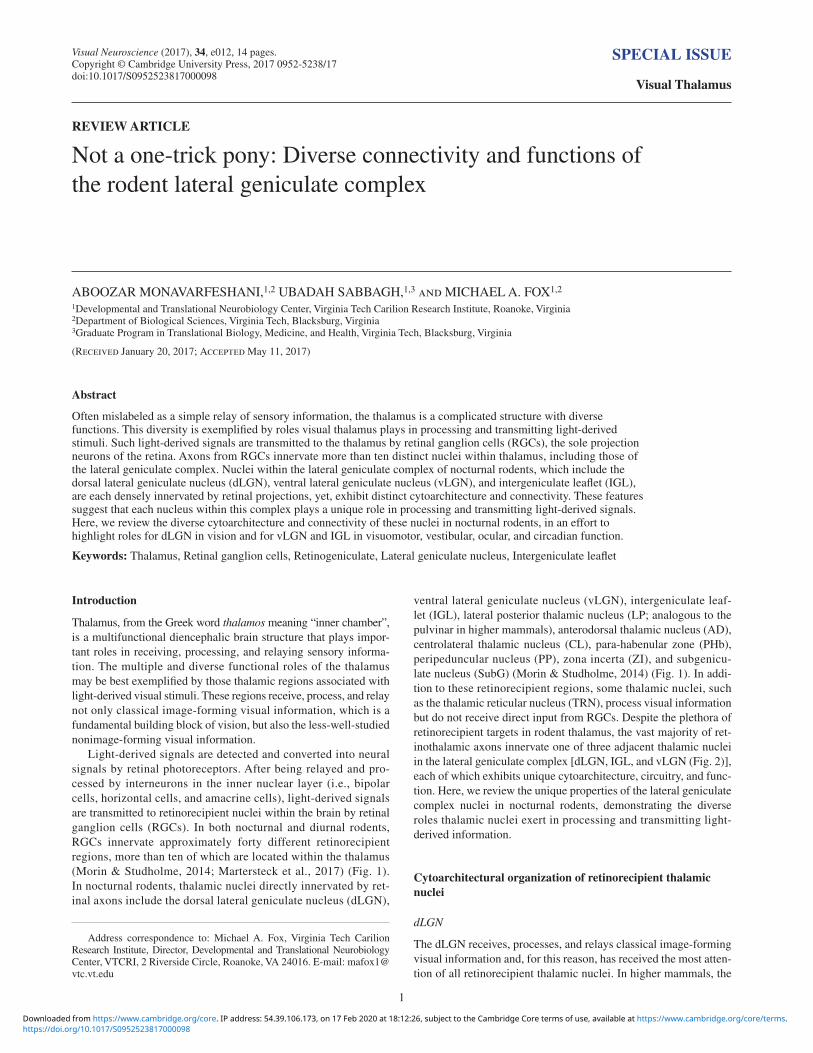
Light-derived signals are detected and converted into neural signals by retinal photoreceptors. After being relayed and pro-cessed by interneurons in the inner nuclear layer (i.e., bipolar cells, horizontal cells, and amacrine cells), light-derived signals are transmitted to retinorecipient nuclei within the brain by retinal ganglion cells (RGCs). In both nocturnal and diurnal rodents, RGCs innervate approximately forty different retinorecipient regions, more than ten of which are located within the thalamus (Morin & Studholme, 2014; Martersteck et al., 2017) (Fig. 1). In nocturnal rodents, thalamic nuclei directly innervated by ret-inal axons include the dorsal lateral geniculate nucleus (dLGN),
ventral lateral geniculate nucleus (vLGN), intergeniculate leaf-let (IGL), lateral posterior thalamic nucleus (LP; analogous to the pulvinar in higher mammals), anterodorsal thalamic nucleus (AD), centrolateral thalamic nucleus (CL), para-habenular zone (PHb), peripeduncular nucleus (PP), zona incerta (ZI), and subgenicu-late nucleus (SubG) (Morin & Studholme, 2014) (Fig. 1). In addi-tion to these retinorecipient regions, some thalamic nuclei, such as the thalamic reticular nucleus (TRN), process visual information but do not receive direct input from RGCs. Despite the plethora of retinorecipient targets in rodent thalamus, the vast majority of ret-inothalamic axons innervate one of three adjacent thalamic nuclei in the lateral geniculate complex [dLGN, IGL, and vLGN (Fig. 2)], each of which exhibits unique cytoarchitecture, circuitry, and func-tion. Here, we review the unique properties of the lateral geniculate complex nuclei in nocturnal rodents, demonstrating the diverse roles thalamic nuclei exert in processing and transmitting light-derived information.
Cytoarchitectural organization of retinorecipient thalamic nuclei
dLGN
The dLGN receives, processes, and relays classical image-forming visual information and, for this reason, has received the most atten-tion of all retinorecipient thalamic nuclei. In higher mammals, the
REVIEW ARTICLE
Not a one-trick pony: Diverse connectivity and functions of the rodent lateral geniculate complex
ABOOZAR MONAVARFESHANI,1,2 UBADAH SABBAGH,1,3 and MICHAEL A. FOX1,2
1Developmental and Translational Neurobiology Center, Virginia Tech Carilion Research Institute, Roanoke, Virginia2Department of Biological Sciences, Virginia Tech, Blacksburg, Virginia3Graduate Program in Translational Biology, Medicine, and Health, Virginia Tech, Blacksburg, Virginia
(Received January 20, 2017; Accepted May 11, 2017)
Abstract
Often mislabeled as a simple relay of sensory information, the thalamus is a complicated structure with diverse functions. This diversity is exemplified by roles visual thalamus plays in processing and transmitting light-derived stimuli. Such light-derived signals are transmitted to the thalamus by retinal ganglion cells (RGCs), the sole projection neurons of the retina. Axons from RGCs innervate more than ten distinct nuclei within thalamus, including those of the lateral geniculate complex. Nuclei within the lateral geniculate complex of nocturnal rodents, which include the dorsal lateral geniculate nucleus (dLGN), ventral lateral geniculate nucleus (vLGN), and intergeniculate leaflet (IGL), are each densely innervated by retinal projections, yet, exhibit distinct cytoarchitecture and connectivity. These features suggest that each nucleus within this complex plays a unique role in processing and transmitting light-derived signals. Here, we review the diverse cytoarchitecture and connectivity of these nuclei in nocturnal rodents, in an effort to highlight roles for dLGN in vision and for vLGN and IGL in visuomotor, vestibular, ocular, and circadian function.
Keywords: Thalamus, Retinal ganglion cells, Retinogeniculate, Lateral geniculate nucleus, Intergeniculate leaflet
Address correspondence to: Michael A. Fox, Virginia Tech Carilion Research Institute, Director, Developmental and Translational Neurobiology Center, VTCRI, 2 Riverside Circle, Roanoke, VA 24016. E-mail: [email protected]
https://doi.org/10.1017/S0952523817000098Downloaded from https://www.cambridge.org/core. IP address: 54.39.106.173, on 17 Feb 2020 at 18:12:26, subject to the Cambridge Core terms of use, available at https://www.cambridge.org/core/terms.
Monavarfeshani et al.2
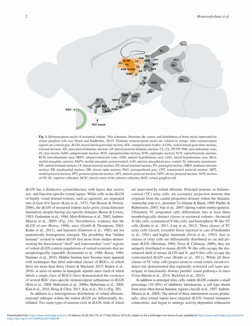
dLGN has a distinctive cytoarchitecture with layers that receive eye- and function-specific retinal inputs. While cells in the dLGN of highly visual diurnal rodents, such as squirrels, are separated into at least five layers (Kaas et al., 1972; Van Hooser & Nelson, 2006), the dLGN of nocturnal rodents lacks gross cytoarchitecture lamination, despite having eye-specific domains (Reese & Cowey, 1983; Godement et al., 1984; Muir-Robinson et al., 2002; Jaubert-Miazza et al., 2005) (Fig. 2A). Nevertheless, evidence that the dLGN of rats (Reese, 1988), mice (Grubb & Thompson, 2004; Krahe et al., 2011), and hamsters (Emerson et al., 1982) are not anatomically homogenous emerged. The possibility that “hidden laminae” existed in rodent dLGN first arose from studies demon-strating the dorsolateral “shell” and ventromedial “core” regions of rodent dLGN contain populations of retinal terminals that are morphologically separable (Erzurumlu et al., 1988; Reese, 1988; Hammer et al., 2015). Hidden laminae have become more apparent with techniques that label individual classes of RGCs, of which there are more than thirty (Sanes & Masland, 2015; Baden et al., 2016). A series of studies in transgenic reporter mice (each of which labels a single class of RGCs) have demonstrated the existence of several RGC class-specific retinorecipient sublaminae in dLGN (Kim et al., 2008; Huberman et al., 2008a; Huberman et al., 2009; Kim et al., 2010; Hong & Chen, 2011; Kay et al., 2011) (Fig. 2D).
In addition to a heterogeneous distribution of retinal afferents, neuronal subtypes within the rodent dLGN are differentially dis-tributed. Two main types of neurons exist in dLGN, both of which
are innervated by retinal afferents. Principal neurons, or thalamo-cortical (TC) relay cells, are excitatory projection neurons that originate from the caudal progenitor domain within the thalamic ventricular zone (i.e., prosomer 2) (Altman & Bayer, 1989; Puelles & Rubenstein, 2003; Vue et al., 2007) during rodent embryogenesis. Ultimately TC progenitor cells differentiate into at least three morphologically distinct classes in nocturnal rodents—biconical X-like cells, symmetrical Y-like cells, and hemispheric W-like TC cells (Krahe et al., 2011; Ling et al., 2012). These classes of TC relay cells closely resemble those reported in cats (Friedlander et al., 1981) and higher mammals (Irvin et al., 1993). Just as classes of relay cells are differentially distributed in cat and pri-mate dLGN (Sherman, 1985; Nassi & Callaway, 2009), they are uniquely distributed in mouse dLGN: W-like cells occupy the dor-solateral shell of mouse dLGN and X- and Y-like cells occupy the ventromedial dLGN core (Krahe et al., 2011). While all three classes of TC relay cells project axons to visual cortex, recent evi-dence has demonstrated that regionally restricted cell types par-ticipate in functionally distinct parallel visual pathways in mice (Cruz-Martín et al., 2014; Bickford et al., 2015).
In addition to principal relay cells, rodent dLGN contains a small percentage (10–20%) of inhibitory interneurons, a cell type absent from most other dorsal thalamic regions (Arcelli et al., 1997; Jaubert-Miazza et al., 2005). The arrival of these interneurons occurs postna-tally, after retinal inputs have targeted dLGN, formed immature connections, and begun to undergo activity-dependent refinement
Fig. 1. Retinorecipient nuclei of nocturnal rodents. This schematic illustrates the variety and distribution of brain nuclei innervated by retinal ganglion cells (see Morin and Studholme, 2014). Thalamic retinorecipient nuclei are colored in orange; other retinorecipient regions are colored gray. dLGN, dorsal lateral geniculate nucleus; IGL, intergeniculate leaflet; vLGNe, ventral lateral geniculate nucleus, external division; AD, anterodorsal thalamic nucleus; LP, lateral posterior thalamic nucleus; CL, CL; PP, PP; PHb, para-habenular zone; ZI, zona incerta; SubG, subgeniculate nucleus; SGN, suprageniculate nucleus; SON, supraoptic nucleus; SCN, suprachiasmatic nucleus; RCH, retrochiasmatic area; SBPV, subparaventricular zone; AHN, anterior hypothalamic area; LHA, lateral hypothalamic area; MeA, medial amygdala, anterior; MePV, medial amygdala, posteroventral; AAV, anterior amygdaloid area, ventral; SI, substantia innominata; MT, medial terminal nucleus; LT, lateral terminal nucleus; DT, dorsal terminal nucleus; PN, paranigral nucleus; MRN, midbrain reticular nucleus; PB, parabrachial nucleus; DR, dorsal raphe nucleus; PAG, periaqueductal gray; CPT, commissural pretectal nucleus; MPT, medial pretectal nucleus; PPT, posterior pretectal nucleus; APT, anterior pretectal nucleus; OPN, olivary pretectal nucleus; NOT, nucleus of OT; SC, superior colliculus; DCIC, dorsal cortex of the inferior colliculus; RGC, retinal ganglion cell.
https://doi.org/10.1017/S0952523817000098Downloaded from https://www.cambridge.org/core. IP address: 54.39.106.173, on 17 Feb 2020 at 18:12:26, subject to the Cambridge Core terms of use, available at https://www.cambridge.org/core/terms.
Connectivity of the lateral geniculate complex 3
Fig. 2. Organization of retinal projections in nuclei of the lateral geniculate complex. (A) Coronal view of a Nissl-stained mouse brain. Arrows indicate the location of dLGN, IGL, and vLGN. Image is from the Allen Brain Atlas (http://www.brain-map.org). (B–D) Schematic representation of coronal section through the lateral geniculate complex of nocturnal rodents. (B) depicts eye-specific segregation of retinal projections in dLGN, IGL, and vLGN. Terminals of ipsilateral retinal projections are depicted as green dots; terminals of contralateral retinal projections are depicted as orange dots. RGCs from which these projections arise are shown in the retinal cross sections. Dotted line in dLGN depicts the approximate boundary separating the dorsolateral shell (s) from the ventromedial core (c). The dotted line in vLGN depicts the boundary separating the external layer (e) from the internal layer (i). (C) depicts topographic mapping of retinal arbors in dLGN, vLGN, and IGL. Colors represent temporal (T) to nasal (N) location of RGCs in the retina (Feldheim et al., 1998; Pfeiffenberger et al., 2006; Huberman et al., 2008a). (D) depicts class-specific target-ing of RGC axons to distinct sublamina of dLGN, vLGN, and IGL. Colors represent some classes of RGCs studied with transgenic reporter mice. Names of these reporter mouse lines are indicated in parentheses (see Hattar et al. 2006; Kim et al. 2008; Huberman et al. 2009; Kim et al. 2010; Osterhout et al. 2011; Rivlin-Etzion et al. 2011). Color-filled dots in the dLGN, IGL, and vLGN represent retinal terminals (and are not meant to indicate that these terminals innervate distinct cells). dLGN, dorsal lateral genic-ulate nucleus; IGL, intergeniculate leaflet; vLGN, ventral lateral geniculate nucleus; dsRGC, direction-selective retinal ganglion cell; ipRGC, intrinsically photosensitive retinal ganglion cell.
https://doi.org/10.1017/S0952523817000098Downloaded from https://www.cambridge.org/core. IP address: 54.39.106.173, on 17 Feb 2020 at 18:12:26, subject to the Cambridge Core terms of use, available at https://www.cambridge.org/core/terms.
Monavarfeshani et al.4
(Jones & Rubenstein, 2004; Singh et al., 2012; Golding et al., 2014; Jager et al., 2016). The precise origin of these interneurons is cur-rently under debate, with studies suggesting they arise from a rostral progenitor domain within the thalamus (i.e., prosomer 3) or from tectum (Virolainen et al., 2012; Golding et al., 2014; Jager et al., 2016). Evidence is also emerging that dLGN interneurons are not a homogeneous population in mice, and can instead be divided into at least two classes based on soma size, membrane capacitance, and neu-ronal nitric oxide synthase (nNOS) expression (Leist et al., 2016). Similar interneuron diversity has been reported in rats (Gabbott & Bacon, 1994b), cats (Montera & Zempel, 1985; Montero & Singer, 1985), and primates (Braak & Bachmann, 1985). At present, however, it remains unclear whether classes of local inhibitory interneurons exhibit regional preferences in the nocturnal rodent dLGN.
vLGN and IGL
Unlike dLGN, the vLGN of nocturnal rodents is organized into at least two easily identifiable laminae—the magnocellular external vLGN (vLGNe), which contains large cells and receives dense innervation from retina, and the parvocellular internal vLGN (vLGNi), which receives little, if any, retinal input (Niimi et al., 1963; Hickey & Spear, 1976; Gabbott & Bacon, 1994a; Harrington, 1997). These laminae are separated by a small neuron-free, fiber-rich neuropil. Cell types within vLGN are vastly different than those in dLGN resulting in a significant difference in the transcrip-tome of each region (Su et al., 2011; Yuge et al., 2011). Moreover, the vLGN lacks stereotypic TC relay cells, and has only a limited number of vesicular glutamate transporter-expressing glutamater-gic neurons (Fremeau et al., 2001; Yuge et al., 2011). Thus, in con-trast to dLGN where glutamatergic neurons are the major cell type, vLGN contains a vastly higher population of GABAergic neurons (Gabbott & Bacon, 1994b; Harrington, 1997; Inamura et al., 2011). These major cellular differences reflect distinct embryonic origins of cells in vLGN, which are derived from progenitors in the most caudal prethalamus, a rostral domain of the thalamic ventricular zone, and the zona limitans interthalamica (ZLI) (Vue et al., 2007; Delaunay et al., 2009; Nakagawa & Shimogori, 2012; Virolainen et al., 2012). Although detailed studies of vLGN neurons lag behind similar characterizations of glutamatergic TC relay cells and local GABAergic interneurons in dLGN, expression studies and trans-genic reporter mice strongly suggest that distinct classes of cells are distributed in a laminar arrangement in vLGNe, even if it is not apparent at the gross cytoarchitectural level (Moore & Card, 1994; http://www.brain-map.org).
Although vLGN and IGL likely serve different functions in nocturnal rodents and are anatomically distinguishable (Hickey & Spear, 1976; Moore & Card, 1994; Morin, 2013), we have grouped them together throughout this review because of shared features that will be discussed. Like those in vLGNe, neurons in the IGL originate from a rostral region of the thalamic progenitor zone and the ZLI (Vue et al., 2007; Delaunay et al., 2009). A large fraction of neurons in IGL generate GABA (Moore & Speh, 1993), few are glutamatergic, and none project axons to visual cortex (Harrington, 1997). However, it is important to highlight that some classes of IGL neurons are absent from vLGN. This includes NPY-expressing neurons in rodent IGL that project axons to hypothalamic nuclei (Card & Moore, 1989; Moore & Card, 1994; Harrington, 1997). Based on available data, classes of morphologically and neuro-chemically distinct neurons do not appear to cluster into distinct layers or regions of IGL, except, perhaps, for coarse differences in
their distribution between rostral and caudal regions of the IGL (Brauer et al., 1983; Moore & Card, 1994; Morin, 2013).
Afferent projections of retinorecipient thalamic nuclei
dLGN: Retinal afferents
In mammals, the primary excitatory drive onto TC relay cells is provided by retinal inputs (Sherman, 2005; Petrof & Sherman, 2013). Anatomically, retinal projections to dLGN are spatially organized in (at least) three fundamental ways. First, retinal affer-ents are segregated into nonoverlapping eye-specific domains in an activity-dependent manner (Huberman et al., 2008a; Zhang et al., 2012). The dLGN of nocturnal rodents receives a relatively small contribution (5–10%) of retinal afferents from the ipsilateral retina and these projections are confined to a ventromedial core region of the dLGN (Jaubert-Miazza et al., 2005; Gaillard et al., 2013; Morin & Studholme, 2014) (Fig. 2B). Second, retinal projections to dLGN are organized topographically-so that neighboring RGCs provide input to neighboring TC relay cells and provide a continuous and faithful representation of spatial information from retina to brain (Feldheim et al., 1998; Pfeiffenberger et al., 2006; Huberman et al., 2008a; Cang & Feldheim, 2013) (Fig. 2C). Third, and perhaps most remarkably, retinal projections undergo class-specific segre-gation in rodent dLGN (Fig. 2D). Although more than 30 classes of RGCs exist in nocturnal rodents only a subset innervate dLGN (Sanes & Masland, 2015; Baden et al., 2016; Ellis et al., 2016). This suggests that targeting mechanisms exist that guide some classes of RGC axons into dLGN and exclude others. Once appro-priate classes of retinal axons enter dLGN, they are further segre-gated into a newly-appreciated laminar organization (Hong & Chen, 2011; Dhande & Huberman, 2014; Sanes & Masland, 2015). The presence of these stereotyped class-specific retinal projections has been elegantly revealed by transgenic reporter mice in which individual RGC classes are labeled with reporter proteins (Hattar et al., 2006; Kim et al., 2008; Huberman et al., 2008b; Huberman et al., 2009; Kim et al., 2010; Kay et al., 2011; Rivlin-Etzion et al., 2011; Dhande et al., 2013; Triplett et al., 2014). While only a small set of individual RGC projections have been mapped with this approach, some rules are beginning to emerge. First, projections of direction-selective classes of RGCs arborize in more dorsolateral regions of dLGN, including the shell of dLGN (Kim et al., 2008; Huberman et al., 2009; Rivlin-Etzion et al., 2011; Cruz-Martín et al., 2014). Second, there is considerable overlap in the laminar termination zones of RGC axons (Fig. 2D), and taken in the context of recent ultrastructural and circuit tracing experiments in dLGN (Morgan et al., 2016; Rompani et al., 2017), this raises the possi-bility that individual TC relay cells may receive inputs from mul-tiple classes of RGCs.
In addition to being segregated based on eye of origin, topogra-phy, and RGC class, retinal inputs in dLGN are structurally and functionally distinct from retinal inputs in other retinorecipient nuclei, even other thalamic nuclei (Sherman, 2005; Hammer et al., 2014). Specifically, retinal terminals onto dLGN TC relay cells are significantly larger than all other terminals in dLGN (and larger than retinal terminals in all other retinorecipient nuclei), and exhibit unique ultrastructural morphology and function (Guillery, 1969; Lund & Cunningham, 1972; Sherman, 2004; Guido, 2008; Bickford et al., 2010; Hong & Chen, 2011). It is worth pointing out, however, that at least two distinct types of RG synapses have been identified in rodent dLGN: “simple encapsulated” RG synapses, in which a single
https://doi.org/10.1017/S0952523817000098Downloaded from https://www.cambridge.org/core. IP address: 54.39.106.173, on 17 Feb 2020 at 18:12:26, subject to the Cambridge Core terms of use, available at https://www.cambridge.org/core/terms.
Connectivity of the lateral geniculate complex 5
retinal terminal synapses onto a TC relay cell dendrite, and “complex encapsulated” RG synapses in which axons from numerous RGCs converge to innervate adjacent regions of a TC relay cell dendrite (Lund & Cunningham, 1972; Hammer et al., 2015; Morgan et al., 2016). Finally, it is important to point out that retinal projections not only innervate TC relay cells, but also local interneurons in nocturnal rodents (Sherman, 2004; Seabrook et al., 2013b).
dLGN: Nonretinal afferents
While retinal inputs provide the excitatory drive to TC relay cells, they account for only 5–10% of the total inputs onto a relay cell and are far outnumbered by nonretinal inputs (Sherman & Guillery, 2002; Bickford et al., 2010; Cetin & Callaway, 2014). A summary of the main inputs to rodent dLGN is depicted in Fig. 3.
While many nonretinal inputs onto dLGN TC relay cells have modulatory or inhibitory roles, a recent study identified a novel glutamatergic nonretinal source of “driver-like” input onto dLGN TC relay cells (Bickford et al., 2015). These inputs arise from the ipsilateral superior colliculus (SC) and terminate onto W-like TC relay cells in the dorsolateral shell of dLGN (Harting et al., 1991a; Bickford et al., 2015). Circuit tracing experiments indicate these excitatory tectogeniculate connections contribute to the processing and transmission of direction-selective visual information (Bickford et al., 2015).
While tectogeniculate inputs represent a minor source of inputs to dLGN, a more significant portion of nonretinal glutamatergic inputs arise from cortical projection neurons in layer VI of primary visual cortex (Sherman, 2016). Corticothalamic inputs are small, located on distal portions of TC relay cell dendrites, and generate weak excitatory postsynaptic potentials (EPSPs) in relay cells (Sherman & Guillery, 2002; Petrof & Sherman, 2013). For this rea-son, it is likely that these inputs are insufficient for the relay of information alone and are, therefore, modulatory in nature (Petrof & Sherman, 2013). Despite these features, corticothalamic inputs do significantly influence RG transmission by affecting the gain of signal transmission and sharpening of receptive field properties of TC relay cells (Sherman & Guillery, 2002; Briggs & Usrey, 2008; Olsen et al., 2012; Bickford, 2015).
In addition to tectal and cortical glutamatergic inputs, TC relay cells in higher mammals receive modulatory cholinergic, sero-tonergic, noradrenergic, and dopaminergic inputs from a variety of sources in the brainstem including parabigeminal nucleus, pedun-culopontine region, locus coeruleus, dorsal raphe nucleus of the midbrain, and the midbrain reticular formation (Mackay-Sim et al., 1983; de Lima et al., 1985; De Lima & Singer, 1987; Papadopoulos & Parnavelas, 1990a, b; McCormick, 1992; Jones, 2012). At pre-sent, some of these afferent projections have been demonstrated in nocturnal rodents (Hallanger et al., 1987; Harting et al., 1991b), but additional studies are needed to map specific sources of these inputs and to understand their role in signal processing in rodents.
Finally, the last significant source of nonretinal inputs to dLGN are inhibitory GABAergic inputs that arise from both local inhibitory neurons and projection neurons in the TRN, a region that forms a lateral shell around dorsal thalamus in nocturnal rodents (Hale et al., 1982; Guillery & Harting, 2003; Pinault, 2004). Inhibitory neurons in the ipsilateral pretectum also project to dLGN (Born & Schmidt, 2007), however evidence suggests that these projections innervate dLGN interneurons not TC relay cells (Wang et al., 2002; Born & Schmidt, 2007), adding further complexity to dLGN circuitry.
An interesting facet of the convergence of retinal and nonretinal inputs in dLGN is that their development appears tightly coordinated.
Retinal axons target and innervate dLGN prior to the arrival of non-retinal inputs and play instructive roles in the establishment of nonretinal circuitry (Brooks et al., 2013; Seabrook et al., 2013a; Golding et al., 2014; Grant et al., 2016). Likewise, nonretinal inputs contribute to the development and function of retinogenicu-late synapses. For example, the presence of corticothalamic axons and corticogeniculate synapses play essential roles in the establish-ment, refinement, and maintenance of retinal inputs (Shanks et al., 2016; Thompson et al., 2016).
vLGN and IGL: Retinal afferents
Several features of retinal projections to vLGNe are similar to those in dLGN: retinal afferents provide a main excitatory drive to vLGNe; the arrival of retinal inputs in vLGNe occurs neonatally and precedes nonretinal inputs (Su et al., 2011); retinal inputs are mapped topographically in vLGNe and these inputs are segregated into nonoverlapping eye-specific domains (Holcombe & Guillery, 1984; Hammer et al., 2014; Morin & Studholme, 2014). On this last point, it warrants mention that ipsilateral retinal projections occupy a region of vLGN that is more complex and less stereo-typed than its counterparts in dLGN. For this reason, activity-dependent refinement of eye-specific retinal projections has not been thoroughly characterized in vLGNe, nor has it served as an anatomical readout of activity-dependent refinement as has been the case in dLGN (Jaubert-Miazza et al., 2005; Demas et al., 2006; Stevens et al., 2007; Huberman et al., 2008b; Rebsam et al., 2009; Xu et al., 2011; Rebsam et al., 2012; Dilger et al., 2015).
There are, however, several dramatic differences between ret-inal projections in dLGN and vLGNe. First, the majority of dLGN-projecting classes of RGCs fail to send collateral axon branches into vLGNe despite having to pass by it (or through it) on the way to dLGN (Huberman et al., 2008b; Kim et al., 2008; Huberman et al., 2009; but see also; Rivlin-Etzion et al., 2011). Instead, sets of nonimage-forming classes of RGCs, including the M1 class of melanopsin-expressing intrinsically photosensitive RGCs (ipRGCs; labeled in Opn4taulacz/taulacz mice) and RGCs labeled in Cdh3-GFP mice, innervate vLGNe (Hattar et al., 2006; Osterhout et al., 2011). It is worth noting that a small subset of ipRGC termi-nals have been reported in the medial-most region of dLGN in Opn4taulacz/taulacz mice. While these may reflect projections from M1 ipRGCs, it is also possible that they belong to axons from other ipRGCs, such as M3 ipRGCs (Schmidt et al., 2011). Indeed other classes of ipRGCs innervate dLGN and mediate image-forming visual signals in nocturnal rodents (Ecker et al., 2010; Estevez et al., 2012). Projections of M1 ipRGCs and Cdh3-GFP RGCs arborize broadly across all regions of vLGNe, raising questions as to whether hidden lamina exist in vLGNe. However, it is important to point out that studies in diurnal rats identified distinct lamination of retinal projections in this region (Gaillard et al., 2013). Moreover, only a small set of the RGCs that likely innervate rodent vLGNe have been identified and studied to date, leaving open the possibility that additional RGC classes will be identified whose projections are regionally restricted to specific sublamina of vLGNe. In support of this, there is a small region of vLGNe underlying the optic tract (OT) that contains a region with morphologically distinct retinal arbors (Hammer et al., 2014), much like that observed in the dorsolateral shell of dLGN (Reese, 1988; Grubb & Thompson, 2004).
Significant differences also exist in the anatomy and physiology of retinal synapses in vLGN compared to dLGN. Retinal terminals in rodent vLGNe are remarkably smaller and less morphologically
https://doi.org/10.1017/S0952523817000098Downloaded from https://www.cambridge.org/core. IP address: 54.39.106.173, on 17 Feb 2020 at 18:12:26, subject to the Cambridge Core terms of use, available at https://www.cambridge.org/core/terms.

Monavarfeshani et al.6
complex than those in dLGN, and this difference is reflected in their ability to elicit considerably weaker EPSPs (Mize & Horner, 1984; Hammer et al., 2014). While several features of retinogeniculate synapses in vLGNe more closely resemble features of modula-tory glutamatergic inputs (including their size, synaptic strength, and relative level of convergence on postsynaptic neurons), retinal inputs onto vLGN principal neurons exhibit paired-pulse depres-sion and are likely to be “driver” inputs (Hammer et al., 2014). Ultrastructural analysis further suggests that retinal terminals in vLGNe do not form “complex encapsulated” RG synapses and are
not typically ensheathed by glial processes (Stelzner et al., 1976; Hammer et al., 2014), both of which are features of RG synapses in rodent dLGN (Hammer et al., 2014; Hammer et al., 2015). These differences in retinal input type, synaptic morphology, and synap-tic physiology suggest that light-derived information is processed differently in vLGNe.
Characteristics of retinal afferents in the nocturnal rodent IGL diverge even farther from their analogues in dLGN. Retinal projections to IGL are not segregated into nonoverlapping eye-specific domains (Su et al., 2013; Hammer et al., 2014; Morin & Studholme, 2014)
Fig. 3. Afferent and efferent projections of rodent dLGN. (A) Sources of afferent projections to dLGN are colored green. (B) Brain regions innervated by dLGN efferents are colored in orange. dLGN, dorsal lateral geniculate nucleus; vLGN, ventral lateral geniculate nucleus; TRN, thalamic reticular nucleus; SC, superior colliculus; PRT, pretectal region; MRN, midbrain reticular nucleus; DR, dorsal raphe nucleus; PPN, pedunculopontine nucleus; PBG, parabigeminal nucleus; PB, parabrachial nucleus; LC, locus coeruleus; VIS1, visual cortex, layer I; VIS4, visual cortex, layer IV; VIS6, visual cortex, layer VI; RGC, retinal ganglion cell.
https://doi.org/10.1017/S0952523817000098Downloaded from https://www.cambridge.org/core. IP address: 54.39.106.173, on 17 Feb 2020 at 18:12:26, subject to the Cambridge Core terms of use, available at https://www.cambridge.org/core/terms.
Connectivity of the lateral geniculate complex 7
nor are they topographically mapped (Harrington, 1997) (Fig. 2). These two features of retinal inputs distinguish IGL from vLGNe, which is surprising given that the only known classes of RGCs that project to IGL, M1 ipRGCs, and Cdh6-expressing RGCs, also pro-ject to vLGNe (Hattar et al., 2006; Osterhout et al., 2011). It is possible that subsets of RGCs in these classes differentially target IGL and vLGNe, or that each retinorecipient region has unique target-derived signals that allow for differential nonimage-forming RGC axon targeting mechanisms (Fox & Guido, 2011).
Retinal terminals in IGL do share ultrastructural similarities with retinal terminals in dLGN and vLGNe, such as the presence of pale mitochondria and round synaptic vesicles (Moore & Card, 1994). In contrast to retinal terminals in dLGN, which reside on large proximal dendrites (Rafols & Valverde, 1973; Wilson et al., 1984), retinal inputs appear to contact small diameter, distal den-drites in IGL (Moore & Card, 1994). Analysis of retinal terminal size by anterograde labeling suggests that these terminals are sim-ilar in size to those in vLGNe, but are much less densely distributed than those in other major retinorecipient nuclei (Hammer et al., 2014). Some peculiar features of retinal terminals in IGL have been noted in the literature and warrant mention here. For example, while retinal terminals in vLGNe, dLGN, and all other retinore-cipient nuclei contain Vesicular Glutamate Transporter 2 (VGluT2), little, if any, of this transporter (or its closely related family mem-ber VGluT1) is present in IGL (Fujiyama et al., 2003; Su et al., 2013; Hammer et al., 2014). The limited level of VGluT expression in IGL appears unchanged following surgical or genetic enucle-ation (Fujiyama et al., 2003; Hammer et al., 2014), further sug-gesting retinal terminals in IGL may lack machinery for glutamate release. Despite this apparent lack of machinery for glutamate packaging and release, Blasiak et al. (2009) showed that gluta-mate receptor antagonists impair excitatory responses to OT stim-ulation in IGL neurons. How do we reconcile such differences? It is possible that VGluT2 is present in retinal terminals in IGL but is below the limit of detection by immunostaining. Would such low levels of vGluT2 allow faithful transmission of signals at retinal synapses in IGL? Perhaps. Retinal terminals in IGL contain pituitary adenylate cyclase-activating peptide (PACAP) (Engelund et al., 2010), a neurotransmitter that can be co-released with glutamate and amplifies glutamatergic signaling (Kopp et al., 2001; Michel et al., 2006). Co-release of PACAP may reduce the necessity of retinal terminals to package and release large quan-tities of glutamate and therefore retinal terminals may require min-imal levels of VGluTs.
In addition to differences in synapse size and neurotransmitters released, a final asymmetry between retinal connections in IGL and other retinorecipient nuclei is that a considerable fraction of prin-ciple projection neurons in IGL (such as NPY + neurons) are not directly innervated by retinal inputs. Instead, the ability of these projection neurons to propagate light-derived signals requires yet-to-be identified interneurons (Thankachan & Rusak, 2005; Morin, 2013), and suggests the existence of a unique circuitry for the transmission of sensory information in IGL.
vLGN and IGL: Nonretinal afferents
Despite some differences in retinal connectivity with vLGNe and IGL, there appears to be a high degree of similarity in the nonreti-nal afferents innervating these structures. It is worth noting three distinctions between sources of nonretinal afferents to vLGNe and IGL when compared with dLGN. First, the number of nuclei that project afferents to rodent vLGNe and IGL far exceed and are far
more diverse than those to dLGN (Figs. 4 and 5). An incomplete list of these sources includes superior colliculus, visual cortex, oli-vary pretectal nucleus, anterior pretectal nucleus, posterior pretec-tal nucleus, locus coeruleus, dorsal raphe nucleus, subparafasicular thalamic nucleus, mesencephalic nucleus, lateral dorsal tegmental nucleus, medial and lateral terminal nuclei, supraoculomotor periaqueductal gray, retrorubral nucleus, pontine reticular nucleus, pararubral nucleus, and medial vestibular nucleus (Cosenza & Moore, 1984; Moore & Card, 1994; Moore et al., 2000; Vrang et al., 2003; Horowitz et al., 2004) (Figs. 4 and 5). In addition, neurons in rodent IGL (but not vLGNe) receive afferents from a variety of sources which include (but are not limited to) prefrontal cortex, ZI, suprachiasmatic nucleus, cuneiform nucleus, and superior and lateral vestibular nuclei (Morin & Blanchard, 1998, 1999; Vrang et al., 2003; Horowitz et al., 2004) (Fig. 5). Second, subcortical projections to vLGNe and IGL arise from both ipsilateral and con-tralateral sources in nocturnal rodents (Moore & Card, 1994; Vrang et al., 2003; Horowitz et al., 2004). For example, IGL neurons receive bilateral input from the olivary pretectal nucleus (OPN) and suprachiasmatic nucleus (SCN), and contralateral inputs from the other IGL (Vrang et al., 2003); likewise, vLGNe neurons receive input from the contralateral vLGNe (Cosenza & Moore, 1984). Third, few sources of afferents are shared with dLGN, strongly inferring diverse roles of these thalamic regions in processing light-derived information (Horowitz et al., 2004). Even in cases when projections to all three regions of the lateral geniculate complex arise from a single brain region, they originate from different cel-lular sources. For example while dLGN, vLGNe, and IGL all receive input from corticothalamic cells in primary visual cortex, inputs to dLGN arise from layer VI cells whereas those innervating vLGNe and IGL arise from layer V (Cosenza & Moore, 1984; Bourassa & Deschênes, 1995; Jacobs et al., 2007; Seabrook et al., 2013a; Hammer et al., 2014).
Efferent projections of retinorecipient thalamic nuclei
dLGN efferents
Of the retinorecipient thalamic nuclei discussed here, the dLGN has by far the fewest targets of efferent projections. In fact, the relative simplicity of efferent projections from dLGN is striking and empha-sizes a singular function of dLGN in processing and transferring image-forming visual information. TC relay cells project axons to only two ipsilateral regions in nocturnal rodents: primary visual cor-tex and TRN (Rafols & Valverde, 1973; Towns et al., 1982; Reese & Cowey, 1983; Crabtree & Killackey, 1989; López-Bendito & Molnár, 2003; Jurgens et al., 2012). Recent studies in mice have demon-strated that projections to visual cortex exhibit class-specificity, with W-like relay cells in the dorsolateral shell of dLGN, conveying direction-selective visual information to layer I and Y-like and X-like relay cells projecting to layer IV of primary visual cortex (Cruz-Martín et al., 2014; Bickford et al., 2015). At present, it remains unclear whether these three classes of TC relay cells make unique connections with TRN neurons in nocturnal rodents. It is worth mentioning that, in cat, Y-cells provide the predominant dLGN input to the perigeniculate nucleus, the visual sector of cat TRN which overlies dLGN (Dubin & Cleland, 1977; Friedlander et al., 1980).
vLGN and IGL efferents
Efferents from vLGNe and IGL target many of the same brain regions and, in contrast to the simplicity of the dLGN efferents,
https://doi.org/10.1017/S0952523817000098Downloaded from https://www.cambridge.org/core. IP address: 54.39.106.173, on 17 Feb 2020 at 18:12:26, subject to the Cambridge Core terms of use, available at https://www.cambridge.org/core/terms.
Monavarfeshani et al.8
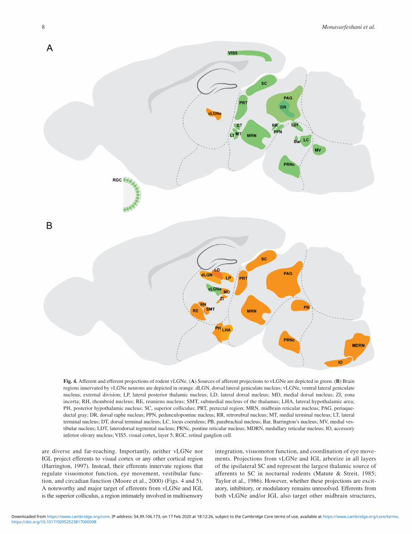
are diverse and far-reaching. Importantly, neither vLGNe nor IGL project efferents to visual cortex or any other cortical region (Harrington, 1997). Instead, their efferents innervate regions that regulate visuomotor function, eye movement, vestibular func-tion, and circadian function (Moore et al., 2000) (Figs. 4 and 5). A noteworthy and major target of efferents from vLGNe and IGL is the superior colliculus, a region intimately involved in multisensory
integration, visuomotor function, and coordination of eye move-ments. Projections from vLGNe and IGL arborize in all layers of the ipsilateral SC and represent the largest thalamic source of afferents to SC in nocturnal rodents (Matute & Streit, 1985; Taylor et al., 1986). However, whether these projections are excit-atory, inhibitory, or modulatory remains unresolved. Efferents from both vLGNe and/or IGL also target other midbrain structures,
Fig. 4. Afferent and efferent projections of rodent vLGNe. (A) Sources of afferent projections to vLGNe are depicted in green. (B) Brain regions innervated by vLGNe neurons are depicted in orange. dLGN, dorsal lateral geniculate nucleus; vLGNe, ventral lateral geniculate nucleus, external division; LP, lateral posterior thalamic nucleus; LD, lateral dorsal nucleus; MD, medial dorsal nucleus; ZI, zona incerta; RH, rhomboid nucleus; RE, reuniens nucleus; SMT, submedial nucleus of the thalamus; LHA, lateral hypothalamic area; PH, posterior hypothalamic nucleus; SC, superior colliculus; PRT, pretectal region; MRN, midbrain reticular nucleus; PAG, periaque-ductal gray; DR, dorsal raphe nucleus; PPN, pedunculopontine nucleus; RR, retrorubral nucleus; MT, medial terminal nucleus; LT, lateral terminal nucleus; DT, dorsal terminal nucleus; LC, locus coeruleus; PB, parabrachial nucleus; Bar, Barrington’s nucleus; MV, medial ves-tibular nucleus; LDT, laterodorsal tegmental nucleus; PRNc, pontine reticular nucleus; MDRN, medullary reticular nucleus; IO, accessory inferior olivary nucleus; VIS5, visual cortex, layer 5; RGC, retinal ganglion cell.
https://doi.org/10.1017/S0952523817000098Downloaded from https://www.cambridge.org/core. IP address: 54.39.106.173, on 17 Feb 2020 at 18:12:26, subject to the Cambridge Core terms of use, available at https://www.cambridge.org/core/terms.

Connectivity of the lateral geniculate complex 9
including the ipsilateral OPN, the nucleus of the OT, and the anterior pretectal nucleus (Cadusseau & Roger, 1991; Harrington, 1997; Moore et al., 2000). Other regions associated with eye move-ments, visuomotor function, and attention innervated by vLGNe and IGL include two of the accessory optic system nuclei (lateral terminal nucleus and medial terminal nucleus; Swanson et al., 1974;
Ribak & Peters, 1975), zona incerta (Brauer & Schober, 1982), and nuclei within the pons (Harrington, 1997).
In addition to visuomotor functions, vLGNe and IGL efferents project to both hypothalamic and thalamic regions (Moore et al., 2000). The largest of these projections innervates the SCN, and is referred to as the geniculohypothalamic (GH) tract. GH projections
Fig. 5. Afferent and efferent projections of rodent IGL. (A) Sources of afferent projections to IGL are colored green. (B) Brain regions innervated by IGL neurons are depicted in orange. IGL, intergeniculate leaflet; LP, lateral posterior thalamic nucleus; PP, PP; ZI, zona incerta; SPF, subparafascicular nucleus; RH, rhomboid nucleus; RE, reuniens nucleus; PVT, paraventricular nucleus; SCN, suprachias-matic nucleus; RCH, retrochiasmatic area; SBPV, subparaventricular zone; AHN, anterior hypothalamic area; PH, posterior hypotha-lamic nucleus; VMH, ventromedial hypothalamic nucleus; DMH, dorsomedial nucleus of the hypothalamus; SC, superior colliculus; PRT, pretectal region; MT, medial terminal nucleus; LT, lateral terminal nucleus; DT, dorsal terminal nucleus; DR, dorsal raphe nucleus; PAG, periaqueductal gray; CUN, cuneiform nucleus; PPN, pedunculopontine nucleus; RR, retrorubral nucleus; LC, locus coeruleus; LDT, laterodorsal tegmental nucleus; Bar, Barrington’s nucleus; MV, medial vestibular nucleus; LAV, lateral vestibular nucleus; SUV, superior vestibular nucleus; PRNc, pontine reticular nucleus; VIS5, visual cortex, layer 5; ACA5/6, anterior cingulate area, layer 5 and 6; RGC, retinal ganglion cell.
https://doi.org/10.1017/S0952523817000098Downloaded from https://www.cambridge.org/core. IP address: 54.39.106.173, on 17 Feb 2020 at 18:12:26, subject to the Cambridge Core terms of use, available at https://www.cambridge.org/core/terms.

Monavarfeshani et al.10
are GABAergic, are important for modulating circadian function (Harrington, 1997), and originate from NPY+ or Enk+ neurons in the IGL (Card & Moore, 1982; Harrington et al., 1987; Card & Moore, 1989). A separate set of neurons in vLGNe and IGL inner-vate contralateral vLGNe and IGL (Harrington, 1997), contralat-eral dLGN (Mikkelsen, 1992; Kolmac et al., 2000), and, at least in higher mammals, pulvinar (Nakamura & Kawamura, 1988).
Diverse connectivity leads to diverse functions of retinorecipient thalamic nuclei
Retinorecipient nuclei within the thalamus exemplify the diverse roles these brain regions play in sensory processing. Despite residing adjacent to each other in the lateral geniculate complex and receiving light-derived signals directly from the retina, there are few similarities in their cytoarchitecture, connectivity, or func-tion of dLGN, vLGN, and IGL.
Based upon its efferent projections to cortex and early studies showing near unitary matching of retinal afferents to TC relay cells (Glees & le Gros Clark, 1941), the dLGN was initially character-ized as a simple relay of visual information. Certainly, the relative simplicity of its efferent projections suggests a near singular role in transmitting image-forming visual information to visual cortex. However, describing the dLGN as a simple relay underestimates its role in processing image-forming visual information. Modulatory feedback from cortex, cholinergic inputs from a subset of brain-stem nuclei, and inhibition from interneurons, endows the dLGN with the ability to shape visual information before transmitting it to higher cortical centers (Piscopo et al., 2013; Roth et al., 2016; Weyand, 2016; Rompani et al., 2017). For example, top-down feedback from corticothalamic inputs is thought to increase the selectivity of TC relay cells, sharpen TC relay cell receptive field properties, enhance synchronicity among TC relay cells, and influ-ence the gain of retinogeniculate transmission (Sherman & Guillery, 2002; Briggs & Usrey, 2008; Weyand, 2016). In nocturnal rodents (and higher mammals) the manner in which retinal inputs innervate dLGN offers the possibility of modifying light-derived signals: feed forward inhibition of retinal inputs through local interneurons can either enhance temporal specificity of RG transmission or can enhance lateral inhibition (Martinez et al., 2014; Weyand, 2016), single retinal axons innervating multiple TC relay cells can amplify visual signals (Weyand, 2016), and the convergence of numerous retinal axons on single relay cell dendrites can produce TC recep-tive fields that are not present in retina (Hammer et al., 2015; Morgan et al., 2016; Weyand, 2016; Rompani et al., 2017). Thus, dLGN is more likely an active component of the machinery required to transform image-forming information into vision and not a passive relay.
In contrast, vLGNe and IGL have little (if any) role in vision. Despite not innervating visual cortex, neurons in these thalamic regions do innervate visual system centers upstream of dLGN (e.g., SC) and even provide inputs to dLGN. Lesion experiments also suggest a potential role for vLGNe and IGL in visual intensity discrimination (Horel, 1968; Legg & Cowey, 1977a, b; Harrington, 1997), however, these studies (and, in fact, all lesion studies of these regions) need to be interpreted cautiously as the lesions dis-rupt the overlying OT and may have secondary effects on other retinorecipient nuclei. Based upon input from classes of RGCs that convey nonimage-forming visual information, and projections to a variety of subcortical structures, roles for vLGNe and IGL in modu-lating circadian rhythms, visuomotor function, eye movements, and vestibular function seem likely. While lesion studies have addressed
some of these possibilities in nocturnal rodents (Harrington & Rusak, 1986; Lewandowski & Usarek, 2002) more elegant and specific genetic approaches to lesion or silence activity in vLGNe and IGL have yet-to-be applied. Such studies are needed to defini-tively identify the functions of vLGNe and IGL, and to identify any potential differences in these two nuclei.
Acknowledgments
We apologize to those whose work we excluded from this review due to space constraints. Work in the Fox laboratory is supported by the National Institutes of Health (EY021222, EY024712, AI124677) and by a Brain and Behavior Research Foundation NARSAD Independent Investigator Award. A.M. is supported by a Virginia Tech Carilion Research Institute Medical Research Scholar fellowship.
References
Altman, J. & Bayer, S.A. (1989). Development of the rat thalamus: VI. The posterior lobule of the thalamic neuroepithelium and the time and site of origin and settling pattern of neurons of the lateral geniculate and lateral posterior nuclei. Journal of Comparative Neurology 284, 581–601.
Arcelli, P., Frassoni, C., Regondi, M., Biasi, S. & Spreafico, R. (1997). GABAergic neurons in mammalian thalamus: A marker of thalamic complexity? Brain Research Bulletin 42, 27–37.
Baden, T., Berens, P., Franke, K., Rosón, M.R., Bethge, M. & Euler, T. (2016). The functional diversity of retinal ganglion cells in the mouse. Nature 529, 345–350.
Bickford, M.E. (2015). Thalamic circuit diversity: Modulation of the driver/modulator framework. Frontiers in Neural Circuits 9, 86.
Bickford, M.E., Slusarczyk, A., Dilger, E.K., Krahe, T.E., Kucuk, C. & Guido, W. (2010). Synaptic development of the mouse dorsal lateral geniculate nucleus. The Journal of Comparative Neurology 518, 622–635.
Bickford, M.E., Zhou, N., Krahe, T.E., Govindaiah, G. & Guido, W. (2015). Retinal and tectal “driver-like” inputs converge in the shell of the mouse dorsal lateral geniculate nucleus. The Journal of Neuroscience 35, 10523–10534.
Blasiak, A., Blasiak, T. & Lewandowski, M. (2009). Electrophysiology and pharmacology of the optic input to the rat intergeniculate leaflet in vitro. Acta Physiologica Polonica 60, 171.
Born, G. & Schmidt, M. (2007). GABAergic pathways in the rat subcor-tical visual system: A comparative study in vivo and in vitro. European Journal of Neuroscience 26, 1183–1192.
Bourassa, J. & Deschênes, M. (1995). Corticothalamic projections from the primary visual cortex in rats: A single fiber study using biocytin as an anterograde tracer. Neuroscience 66, 253–263.
Braak, H. & Bachmann, A. (1985). The percentage of projection neurons and interneurons in the human lateral geniculate nucleus. Human Neurobiology 4, 91–95.
Brauer, K. & Schober, W. (1982). Identification of geniculo-tectal relay neurons in the rat’s ventral lateral geniculate nucleus. Experimental Brain Research 45, 84–88.
Brauer, K., Schober, W., Leibnitz, L., Werner, L., Lüth, H. & Winkelmann, E. (1983). The ventral lateral geniculate nucleus of the albino rat morphological and histochemical observations. Journal fur Hirnforschung 25, 205–236.
Briggs, F. & Usrey, W.M. (2008). Emerging views of corticothalamic function. Current Opinion in Neurobiology 18, 403–407.
Brooks, J.M., Su, J., Levy, C., Wang, J.S., Seabrook, T.A., Guido, W. & Fox, M.A. (2013). A molecular mechanism regulating the timing of corticogeniculate innervation. Cell Reports 5, 573–581.
Cadusseau, J. & Roger, M. (1991). Cortical and subcortical connections of the pars compacta of the anterior pretectal nucleus in the rat. Neuroscience Research 12, 83–100.
Cang, J. & Feldheim, D.A. (2013). Developmental mechanisms of topo-graphic map formation and alignment. Annual Review of Neuroscience 36, 51–77.
Card, J.P. & Moore, R.Y. (1982). Ventral lateral geniculate nucleus efferents to the rat suprachiasmatic nucleus exhibit avian pancreatic polypeptide-like immunoreactivity. Journal of Comparative Neurology 206, 390–396.
https://doi.org/10.1017/S0952523817000098Downloaded from https://www.cambridge.org/core. IP address: 54.39.106.173, on 17 Feb 2020 at 18:12:26, subject to the Cambridge Core terms of use, available at https://www.cambridge.org/core/terms.
Connectivity of the lateral geniculate complex 11
Card, J.P. & Moore, R.Y. (1989). Organization of lateral geniculate-hypothalamic connections in the rat. Journal of Comparative Neurology 284, 135–147.
Cetin, A. & Callaway, E.M. (2014). Optical control of retrogradely infected neurons using drug-regulated “TLoop” lentiviral vectors. Journal of Neurophysiology 111, 2150–2159.
Cosenza, R.M. & Moore, R.Y. (1984). Afferent connections of the ventral lateral geniculate nucleus in the rat: An HRP study. Brain Research 310, 367–370.
Crabtree, J.W. & Killackey, H.P. (1989). The topographic organiza-tion and axis of projection within the visual sector of the rabbit’s thalamic reticular nucleus. European Journal of Neuroscience 1, 94–109.
Cruz-Martín, A., El-Danaf, R.N., Osakada, F., Sriram, B., Dhande, O.S., Nguyen, P.L., Callaway, E.M., Ghosh, A. & Huberman, A.D. (2014). A dedicated circuit links direction-selective retinal ganglion cells to the primary visual cortex. Nature 507, 358–361.
De Lima, A.D. & Singer, W. (1987). The serotoninergic fibers in the dorsal lateral geniculate nucleus of the cat: Distribution and synaptic connections demonstrated with immunocytochemistry. Journal of Comparative Neurology 258, 339–351.
de Lima, D.A., Montero, V. & Singer, W. (1985). The cholinergic inner-vation of the visual thalamus: An EM immunocytochemical study. Experimental Brain Research 59, 206–212.
Delaunay, D., Heydon, K., Miguez, A., Schwab, M., Nave, K.A., Thomas, J.L., Spassky, N., Martinez, S. & Zalc, B. (2009). Genetic tracing of subpopulation neurons in the prethalamus of mice (Mus musculus). Journal of Comparative Neurology 512, 74–83.
Demas, J., Sagdullaev, B.T., Green, E., Jaubert-Miazza, L., McCall, M.A., Gregg, R.G., Wong, R.O. & Guido, W. (2006). Failure to main-tain eye-specific segregation in nob, a mutant with abnormally pat-terned retinal activity. Neuron 50, 247–259.
Dhande, O.S., Estevez, M.E., Quattrochi, L.E., El-Danaf, R.N., Nguyen, P.L., Berson, D.M. & Huberman, A.D. (2013). Genetic dis-section of retinal inputs to brainstem nuclei controlling image stabiliza-tion. The Journal of Neuroscience 33, 17797–17813.
Dhande, O.S. & Huberman, A.D. (2014). Retinal ganglion cell maps in the brain: Implications for visual processing. Current Opinion in Neurobiology 24, 133–142.
Dilger, E.K., Krahe, T.E., Morhardt, D.R., Seabrook, T.A., Shin, H-S. & Guido, W. (2015). Absence of plateau potentials in dLGN cells leads to a breakdown in retinogeniculate refinement. Journal of Neuroscience 35, 3652–3662.
Dubin, M.W. & Cleland, B.G. (1977). Organization of visual inputs to interneurons of lateral geniculate nucleus of the cat. Journal of Neurophysiology 40, 410–427.
Ecker, J.L., Dumitrescu, O.N., Wong, K.Y., Alam, N.M., Chen, S-K., LeGates, T., Renna, J.M., Prusky, G.T., Berson, D.M. & Hattar, S. (2010). Melanopsin-expressing retinal ganglion-cell photoreceptors: Cellular diversity and role in pattern vision. Neuron 67, 49–60.
Ellis, E.M., Gauvain, G., Sivyer, B. & Murphy, G.J. (2016). Shared and distinct retinal input to the mouse superior colliculus and dorsal lateral geniculate nucleus. Journal of Neurophysiology 116, 602–610.
Emerson, V., Chalupa, L., Thompson, I. & Talbot, R. (1982). Behavioural, physiological, and anatomical consequences of monocular deprivation in the golden hamster (Mesocricetus auratus). Experimental Brain Research 45, 168–178.
Engelund, A., Fahrenkrug, J., Harrison, A. & Hannibal, J. (2010). Vesicular glutamate transporter 2 (VGLUT2) is co-stored with PACAP in projections from the rat melanopsin-containing retinal ganglion cells. Cell and Tissue Research 340, 243–255.
Erzurumlu, R.S., Jhaveri, S. & Schneider, G.E. (1988). Distribution of morphologically different retinal axon terminals in the hamster dorsal lateral geniculate nucleus. Brain Research 461, 175–181.
Estevez, M.E., Fogerson, P.M., Ilardi, M.C., Borghuis, B.G., Chan, E., Weng, S., Auferkorte, O.N., Demb, J.B. & Berson, D.M. (2012). Form and function of the M4 cell, an intrinsically photosensitive retinal ganglion cell type contributing to geniculocortical vision. Journal of Neuroscience 32, 13608–13620.
Feldheim, D.A., Vanderhaeghen, P., Hansen, M.J., Frisén, J., Lu, Q., Barbacid, M. & Flanagan, J.G. (1998). Topographic guidance labels in a sensory projection to the forebrain. Neuron 21, 1303–1313.
Fox, M.A. & Guido, W. (2011). Shedding light on class-specific wiring: Development of intrinsically photosensitive retinal ganglion cell cir-cuitry. Molecular Neurobiology 44, 321–329.
Fremeau, R.T., Troyer, M.D., Pahner, I., Nygaard, G.O., Tran, C.H., Reimer, R.J., Bellocchio, E.E., Fortin, D., Storm-Mathisen, J. & Edwards, R.H. (2001). The expression of vesicular glutamate trans-porters defines two classes of excitatory synapse. Neuron 31, 247–260.
Friedlander, M., Lin, C. & Sherman, S. (1980). Dendritic and axonal morphology of physiological classes of geniculo-cortical relay cells. In Experimental Brain Research (Vol. 41, No. 1, pp. A3-A3). 175 Springer Verlag, New York, US.
Friedlander, M., Lin, C., Stanford, L. & Sherman, S.M. (1981). Morphology of functionally identified neurons in lateral geniculate nucleus of the cat. Journal of Neurophysiology 46, 80–129.
Fujiyama, F., Hioki, H., Tomioka, R., Taki, K., Tamamaki, N., Nomura, S., Okamoto, K. & Kaneko, T. (2003). Changes of immunocytochemical localization of vesicular glutamate transporters in the rat visual system after the retinofugal denervation. Journal of Comparative Neurology 465, 234–249.
Gabbott, P. & Bacon, S. (1994a). An oriented framework of neuronal pro-cesses in the ventral lateral geniculate nucleus of the rat demonstrated by NADPH diaphorase histochemistry and GABA immunocytochem-istry. Neuroscience 60, 417–440.
Gabbott, P.L. & Bacon, S.J. (1994b). Two types of interneuron in the dorsal lateral geniculate nucleus of the rat: A combined NADPH diaph-orase histochemical and GABA immunocytochemical study. Journal of Comparative Neurology 350, 281–301.
Gaillard, F., Karten, H.J. & Sauvé, Y. (2013). Retinorecipient areas in the diurnal murine rodent Arvicanthis niloticus: A disproportionally large superior colliculus. Journal of Comparative Neurology 521, 1699–1726.
Glees, P. & le Gros Clark, W. (1941). The termination of optic fibres in the lateral geniculate body of the monkey. Journal of Anatomy 75, 295.
Godement, P., Salaün, J. & Imbert, M. (1984). Prenatal and postnatal development of retinogeniculate and retinocollicular projections in the mouse. Journal of Comparative Neurology 230, 552–575.
Golding, B., Pouchelon, G., Bellone, C., Murthy, S., Di Nardo, A.A., Govindan, S., Ogawa, M., Shimogori, T., Lüscher, C. & Dayer, A. (2014). Retinal input directs the recruitment of inhibitory interneurons into thalamic visual circuits. Neuron 81, 1057–1069.
Grant, E., Hoerder-Suabedissen, A. & Molnár, Z. (2016). The regula-tion of corticofugal fiber targeting by retinal inputs. Cerebral Cortex 26, 1336–1348.
Grubb, M.S. & Thompson, I.D. (2004). Biochemical and anatomical sub-division of the dorsal lateral geniculate nucleus in normal mice and in mice lacking the β2 subunit of the nicotinic acetylcholine receptor. Vision Research 44, 3365–3376.
Guido, W. (2008). Refinement of the retinogeniculate pathway. The Journal of Physiology 586, 4357–4362.
Guillery, R. (1969). The organization of synaptic interconnections in the laminae of the dorsal lateral geniculate nucleus of the cat. Zeitschrift für Zellforschung und mikroskopische Anatomie 96, 1–38.
Guillery, R. & Harting, J.K. (2003). Structure and connections of the thalamic reticular nucleus: Advancing views over half a century. Journal of Comparative Neurology 463, 360–371.
Hale, P., Sefton, A.J., Baur, L. & Cottee, L. (1982). Interrelations of the rat’s thalamic reticular and dorsal lateral geniculate nuclei. Experimental Brain Research 45, 217–229.
Hallanger, A.E., Levey, A.I., Lee, H.J., Rye, D.B. & Wainer, B.H. (1987). The origins of cholinergic and other subcortical afferents to the thalamus in the rat. Journal of Comparative Neurology 262, 105–124.
Hammer, S., Carrillo, G.L., Govindaiah, G., Monavarfeshani, A., Bircher, J.S., Su, J., Guido, W. & Fox, M.A. (2014). Nuclei-specific differences in nerve terminal distribution, morphology, and develop-ment in mouse visual thalamus. Neural Development 9, 1.
Hammer, S., Monavarfeshani, A., Lemon, T., Su, J. & Fox, M.A. (2015). Multiple retinal axons converge onto relay cells in the adult mouse thal-amus. Cell Reports 12, 1575–1583.
Harrington, M., DeCoursey, P., Bruce, D. & Buggy, J. (1987). Circadian pacemaker (SCN) transplants into lateral ventricles fail to restore locomotor rhythmicity in arrhythmic hamsters. Soc Neurosci Abstr 13, 465–472.
Harrington, M.E. (1997). The ventral lateral geniculate nucleus and the intergeniculate leaflet: Interrelated structures in the visual and circadian systems. Neuroscience & Biobehavioral Reviews 21, 705–727.
Harrington, M.E. & Rusak, B. (1986). Lesions of the thalamic interge-niculate leaflet alter hamster circadian rhythms. Journal of Biological Rhythms 1, 309–325.
https://doi.org/10.1017/S0952523817000098Downloaded from https://www.cambridge.org/core. IP address: 54.39.106.173, on 17 Feb 2020 at 18:12:26, subject to the Cambridge Core terms of use, available at https://www.cambridge.org/core/terms.

Monavarfeshani et al.12
Harting, J.K., Huerta, M.F., Hashikawa, T. & van Lieshout, D.P. (1991a). Projection of the mammalian superior colliculus upon the dor-sal lateral geniculate nucleus: Organization of tectogeniculate pathways in nineteen species. Journal of Comparative Neurology 304, 275–306.
Harting, J.K., Van Lieshout, D., Hashikawa, T. & Weber, J. (1991b). The parabigeminogeniculate projection: Connectional studies in eight mammals. Journal of Comparative Neurology 305, 559–581.
Hattar, S., Kumar, M., Park, A., Tong, P., Tung, J., Yau, K.W. & Berson, D.M. (2006). Central projections of melanopsin-expressing retinal ganglion cells in the mouse. Journal of Comparative Neurology 497, 326–349.
Hickey, T. & Spear, P. (1976). Retinogeniculate projections in hooded and albino rats: An autoradiographic study. Experimental Brain Research 24, 523–529.
Holcombe, V. & Guillery, R. (1984). The organization of retinal maps within the dorsal and ventral lateral geniculate nuclei of the rabbit. Journal of Comparative Neurology 225, 469–491.
Hong, Y.K. & Chen, C. (2011). Wiring and rewiring of the retinogenicu-late synapse. Current Opinion in Neurobiology 21, 228–237.
Horel, J.A. (1968). Effects of subcortical lesions on brightness discrim-ination acquired by rats without visual cortex. Journal of Comparative and Physiological Psychology 65, 103.
Horowitz, S.S., Blanchard, J.H. & Morin, L.P. (2004). Intergeniculate leaflet and ventral lateral geniculate nucleus afferent connections: An anatomical substrate for functional input from the vestibulo-visuomotor system. Journal of Comparative Neurology 474, 227–245.
Huberman, A.D., Feller, M.B. & Chapman, B. (2008a). Mechanisms underlying development of visual maps and receptive fields. Annual Review of Neuroscience 31, 479–509.
Huberman, A.D., Manu, M., Koch, S.M., Susman, M.W., Lutz, A.B., Ullian, E.M., Baccus, S.A. & Barres, B.A. (2008b). Architecture and activity-mediated refinement of axonal projections from a mosaic of genetically identified retinal ganglion cells. Neuron 59, 425–438.
Huberman, A.D., Wei, W., Elstrott, J., Stafford, B.K., Feller, M.B. & Barres, B.A. (2009). Genetic identification of an on–off direction-selective retinal ganglion cell subtype reveals a layer-specific subcor-tical map of posterior motion. Neuron 62, 327–334.
Inamura, N., Ono, K., Takebayashi, H., Zalc, B. & Ikenaka, K. (2011). Olig2 lineage cells generate GABAergic neurons in the prethalamic nuclei, including the zona incerta, ventral lateral geniculate nucleus and reticular thalamic nucleus. Developmental Neuroscience 33, 118–129.
Irvin, G.E., Casagrande, V.A. & Norton, T.T. (1993). Center/surround relationships of magnocellular, parvocellular, and koniocellular relay cells in primate lateral geniculate nucleus. Visual Neuroscience 10, 363–373.
Jacobs, E.C., Campagnoni, C., Kampf, K., Reyes, S.D., Kalra, V., Handley, V., Xie, Y.Y., Hong-Hu, Y., Spreur, V. & Fisher, R.S. (2007). Visualization of corticofugal projections during early cortical development in a τ-GFP-transgenic mouse. European Journal of Neuroscience 25, 17–30.
Jager, P., Ye, Z., Yu, X., Zagoraiou, L., Prekop, H-T., Partanen, J., Jessell, T.M., Wisden, W., Brickley, S.G. & Delogu, A. (2016). Tectal-derived interneurons contribute to phasic and tonic inhibition in the visual thalamus. Nature Communications 7, 13579.
Jaubert-Miazza, L., Green, E., Lo, F., Bui, K., Mills, J. & Guido, W. (2005). Structural and functional composition of the developing retino-geniculate pathway in the mouse. Visual Neuroscience 22, 661.
Jones, E.G. (2012). The Thalamus. Springer Science & Business Media, Berlin, Germany.
Jones, E.G. & Rubenstein, J.L. (2004). Expression of regulatory genes during differentiation of thalamic nuclei in mouse and monkey. Journal of Comparative Neurology 477, 55–80.
Jurgens, C.W., Bell, K.A., McQuiston, A.R. & Guido, W. (2012). Optogenetic stimulation of the corticothalamic pathway affects relay cells and GABAergic neurons differently in the mouse visual thalamus. PLoS One 7, e45717.
Kaas, J., Guillery, R. & Allman, J. (1972). Some principles of organization in the dorsal lateral geniculate nucleus. Brain, Behavior and Evolution 6, 283–299.
Kay, J.N., De la Huerta, I., Kim, I-J., Zhang, Y., Yamagata, M., Chu, M.W., Meister, M. & Sanes, J.R. (2011). Retinal ganglion cells with distinct directional preferences differ in molecular identity, structure, and central projections. The Journal of Neuroscience 31, 7753–7762.
Kim, I-J., Zhang, Y., Meister, M. & Sanes, J.R. (2010). Laminar restric-tion of retinal ganglion cell dendrites and axons: Subtype-specific
developmental patterns revealed with transgenic markers. The Journal of Neuroscience 30, 1452–1462.
Kim, I-J., Zhang, Y., Yamagata, M., Meister, M. & Sanes, J.R. (2008). Molecular identification of a retinal cell type that responds to upward motion. Nature 452, 478–482.
Kolmac, C.I., Power, B.D. & Mitrofanis, J. (2000). Dorsal thalamic connections of the ventral lateral geniculate nucleus of rats. Journal of Neurocytology 29, 31–41.
Kopp, M.D., Meissl, H., Dehghani, F. & Korf, H.W. (2001). The pituitary adenylate cyclase-activating polypeptide modulates glutamatergic cal-cium signalling: Investigations on rat suprachiasmatic nucleus neurons. Journal of Neurochemistry 79, 161–171.
Krahe, T.E., El-Danaf, R.N., Dilger, E.K., Henderson, S.C. & Guido, W. (2011). Morphologically distinct classes of relay cells exhibit regional pref-erences in the dorsal lateral geniculate nucleus of the mouse. The Journal of Neuroscience 31, 17437–17448.
Legg, C. & Cowey, A. (1977a). Effects of subcortical lesions on visual intensity discriminations in rats. Physiology & Behavior 19, 635–646.
Legg, C. & Cowey, A. (1977b). The role of the ventral lateral geniculate nucleus and posterior thalamus in intensity discrimination in rats. Brain Research 123, 261–273.
Leist, M., Datunashvilli, M., Kanyshkova, T., Zobeiri, M., Aissaoui, A., Cerina, M., Romanelli, M.N., Pape, H-C. & Budde, T. (2016). Two types of interneurons in the mouse lateral geniculate nucleus are char-acterized by different h-current density. Scientific Reports 6, 24904.
Lewandowski, M.H. & Usarek, A. (2002). Effects of intergeniculate leaflet lesions on circadian rhythms in the mouse. Behavioural Brain Research 128, 13–17.
Ling, C., Hendrickson, M.L. & Kalil, R.E. (2012). Morphology, classification, and distribution of the projection neurons in the dorsal lateral geniculate nucleus of the rat. PloS One 7, e49161.
López-Bendito, G. & Molnár, Z. (2003). Thalamocortical development: How are we going to get there? Nature Reviews Neuroscience 4, 276–289.
Lund, R. & Cunningham, T. (1972). Aspects of synaptic and laminar organization of the mammalian lateral geniculate body. Investigative Ophthalmology 11, 291–302.
Mackay-Sim, A., Sefton, A.J. & Martin, P.R. (1983). Subcortical projections to lateral geniculate and thalamic reticular nuclei in the hooded rat. Journal of Comparative Neurology 213, 24–35.
Martersteck, E.M., Hirokawa, K.E., Evarts, M., Bernard, A., Duan, X., Li, Y., Ng, L., Oh, S.W., Ouellette, B. & Royall, J.J. (2017). Diverse central projection patterns of retinal ganglion cells. Cell Reports 18, 2058–2072.
Martinez, L.M., Molano-Mazón, M., Wang, X., Sommer, F.T. & Hirsch, J.A. (2014). Statistical wiring of thalamic receptive fields opti-mizes spatial sampling of the retinal image. Neuron 81, 943–956.
Matute, C. & Streit, P. (1985). Selective retrograde labeling with D-[3H]-aspartate in afferents to the mammalian superior colliculus. Journal of Comparative Neurology 241, 34–49.
McCormick, D.A. (1992). Neurotransmitter actions in the thalamus and cerebral cortex and their role in neuromodulation of thalamocortical activity. Progress in Neurobiology 39, 337–388.
Michel, S., Itri, J., Han, J.H., Gniotczynski, K. & Colwell, C.S. (2006). Regulation of glutamatergic signalling by PACAP in the mam-malian suprachiasmatic nucleus. BMC Neuroscience 7, 15.
Mikkelsen, J. (1992). The organization of the crossed geniculogenicu-late pathway of the rat: A Phaseolus vulgaris-leucoagglutinin study. Neuroscience 48, 953–962.
Mize, R.R. & Horner, L.H. (1984). Retinal synapses of the cat medial interlaminar nucleus and ventral lateral geniculate nucleus differ in size and synaptic organization. Journal of Comparative Neurology 224, 579–590.
Montera, V. & Zempel, J. (1985). Evidence for two types of GABA-containing interneurons in the a-laminae of the cat lateral geniculate nucleus: A double-label HRP and GABA-immunocytochemical study. Experimental Brain Research 60, 603–609.
Montero, V. & Singer, W. (1985). Ultrastructural identification of somata and neural processes immunoreactive to antibodies against glutamic acid decarboxylase (GAD) in the dorsal lateral geniculate nucleus of the cat. Experimental Brain Research 59, 151–165.
Moore, R.Y. & Card, J.P. (1994). Intergeniculate leaflet: An anatomically and functionally distinct subdivision of the lateral geniculate complex. Journal of Comparative Neurology 344, 403–430.
https://doi.org/10.1017/S0952523817000098Downloaded from https://www.cambridge.org/core. IP address: 54.39.106.173, on 17 Feb 2020 at 18:12:26, subject to the Cambridge Core terms of use, available at https://www.cambridge.org/core/terms.
Connectivity of the lateral geniculate complex 13
Moore, R.Y. & Speh, J.C. (1993). GABA is the principal neurotransmitter of the circadian system. Neuroscience Letters 150, 112–116.
Moore, R.Y., Weis, R. & Moga, M.M. (2000). Efferent projections of the intergeniculate leaflet and the ventral lateral geniculate nucleus in the rat. Journal of Comparative Neurology 420, 398–418.
Morgan, J.L., Berger, D.R., Wetzel, A.W. & Lichtman, J.W. (2016). The fuzzy logic of network connectivity in mouse visual thalamus. Cell 165, 192–206.
Morin, L. & Blanchard, J. (1998). Interconnections among nuclei of the subcortical visual shell: The intergeniculate leaflet is a major constit-uent of the hamster subcortical visual system. Journal of Comparative Neurology 396, 288–309.
Morin, L. & Blanchard, J. (1999). Forebrain connections of the hamster intergeniculate leaflet: Comparison with those of ventral lateral genicu-late nucleus and retina. Visual Neuroscience 16, 1037–1054.
Morin, L.P. (2013). Neuroanatomy of the extended circadian rhythm system. Experimental Neurology 243, 4–20.
Morin, L.P. & Studholme, K.M. (2014). Retinofugal projections in the mouse. Journal of Comparative Neurology 522, 3733–3753.
Muir-Robinson, G., Hwang, B.J. & Feller, M.B. (2002). Retinogeniculate axons undergo eye-specific segregation in the absence of eye-specific layers. The Journal of Neuroscience 22, 5259–5264.
Nakagawa, Y. & Shimogori, T. (2012). Diversity of thalamic progenitor cells and postmitotic neurons. European Journal of Neuroscience 35, 1554–1562.
Nakamura, H. & Kawamura, S. (1988). The ventral lateral geniculate nucleus in the cat: Thalamic and commissural connections revealed by the use of WGA-HRP transport. Journal of Comparative Neurology 277, 509–528.
Nassi, J.J. & Callaway, E.M. (2009). Parallel processing strategies of the primate visual system. Nature Reviews Neuroscience 10, 360–372.
Niimi, K., Kanaseki, T. & Takimoto, T. (1963). The comparative anatomy of the ventral nucleus of the lateral geniculate body in mammals. Journal of Comparative Neurology 121, 313–323.
Olsen, S.R., Bortone, D.S., Adesnik, H. & Scanziani, M. (2012). Gain control by layer six in cortical circuits of vision. Nature 483, 47–52.
Osterhout, J.A., Josten, N., Yamada, J., Pan, F., Wu, S-w., Nguyen, P.L., Panagiotakos, G., Inoue, Y.U., Egusa, S.F. & Volgyi, B. (2011). Cadherin-6 mediates axon-target matching in a non-image-forming visual circuit. Neuron 71, 632–639.
Papadopoulos, G.C. & Parnavelas, J.G. (1990a). Distribution and syn-aptic organization of dopaminergic axons in the lateral geniculate nucleus of the rat. Journal of Comparative Neurology 294, 356–361.
Papadopoulos, G.C. & Parnavelas, J.G. (1990b). Distribution and syn-aptic organization of serotoninergic and noradrenergic axons in the lateral geniculate nucleus of the rat. Journal of Comparative Neurology 294, 345–355.
Petrof, I. & Sherman, S.M. (2013). Functional significance of synaptic terminal size in glutamatergic sensory pathways in thalamus and cortex. The Journal of physiology 591, 3125–3131.
Pfeiffenberger, C., Yamada, J. & Feldheim, D.A. (2006). Ephrin-As and patterned retinal activity act together in the development of topo-graphic maps in the primary visual system. The Journal of Neuroscience 26, 12873–12884.
Pinault, D. (2004). The thalamic reticular nucleus: Structure, function and concept. Brain Research Reviews 46, 1–31.
Piscopo, D.M., El-Danaf, R.N., Huberman, A.D. & Niell, C.M. (2013). Diverse visual features encoded in mouse lateral geniculate nucleus. The Journal of Neuroscience 33, 4642–4656.
Puelles, L. & Rubenstein, J.L. (2003). Forebrain gene expression domains and the evolving prosomeric model. Trends in Neurosciences 26, 469–476.
Rafols, J.A. & Valverde, F. (1973). The structure of the dorsal lateral geniculate nucleus in the mouse. A golgi and electron microscopic study. Journal of Comparative Neurology 150, 303–331.
Rebsam, A., Bhansali, P. & Mason, C.A. (2012). Eye-specific projections of retinogeniculate axons are altered in albino mice. Journal of Neuroscience 32, 4821–4826.
Rebsam, A., Petros, T.J. & Mason, C.A. (2009). Switching retinogenicu-late axon laterality leads to normal targeting but abnormal eye-specific segregation that is activity dependent. Journal of Neuroscience 29, 14855–14863.
Reese, B. (1988). ‘Hidden lamination’ in the dorsal lateral geniculate nucleus: The functional organization of this thalamic region in the rat. Brain Research Reviews 13, 119–137.
Reese, B. & Cowey, A. (1983). Projection lines and the ipsilateral retino-geniculate pathway in the hooded rat. Neuroscience 10, 1233–1247.
Ribak, C.E. & Peters, A. (1975). An autoradiographic study of the projections from the lateral geniculate body of the rat. Brain Research 92, 341–368.
Rivlin-Etzion, M., Zhou, K., Wei, W., Elstrott, J., Nguyen, P.L., Barres, B.A., Huberman, A.D. & Feller, M.B. (2011). Transgenic mice reveal unexpected diversity of on-off direction-selective retinal ganglion cell subtypes and brain structures involved in motion processing. The Journal of Neuroscience 31, 8760–8769.
Rompani, S.B., Müllner, F.E., Wanner, A., Zhang, C., Roth, C.N., Yonehara, K. & Roska, B. (2017). Different modes of visual integra-tion in the lateral geniculate nucleus revealed by single-cell-initiated transsynaptic tracing. Neuron 93, 767–776. e766.
Roth, M.M., Dahmen, J.C., Muir, D.R., Imhof, F., Martini, F.J. & Hofer, S.B. (2016). Thalamic nuclei convey diverse contextual infor-mation to layer 1 of visual cortex. Nature Neuroscience 19, 299–307.
Sanes, J.R. & Masland, R.H. (2015). The types of retinal ganglion cells: Current status and implications for neuronal classification. Annual Review of Neuroscience 38, 221–246.
Schmidt, T.M., Chen, S-K. & Hattar, S. (2011). Intrinsically photosen-sitive retinal ganglion cells: Many subtypes, diverse functions. Trends in Neurosciences 34, 572–580.
Seabrook, T.A., El-Danaf, R.N., Krahe, T.E., Fox, M.A. & Guido, W. (2013a). Retinal input regulates the timing of corticogeniculate innerva-tion. The Journal of Neuroscience 33, 10085–10097.
Seabrook, T.A., Krahe, T.E., Govindaiah, G. & Guido, W. (2013b). Interneurons in the mouse visual thalamus maintain a high degree of retinal convergence throughout postnatal development. Neural Development 8, 1.
Shanks, J.A., Ito, S., Schaevitz, L., Yamada, J., Chen, B., Litke, A. & Feldheim, D.A. (2016). Corticothalamic axons are essential for retinal ganglion cell axon targeting to the mouse dorsal lateral geniculate nucleus. The Journal of Neuroscience 36, 5252–5263.
Sherman, S.M. (1985). Functional organization of the W-, X-, and Y-cell pathways in the cat: A review and hypothesis. Progress in Psychobiology and Physiological Psychology 11, 233–314.
Sherman, S.M. (2004). Interneurons and triadic circuitry of the thalamus. Trends in Neurosciences 27, 670–675.
Sherman, S.M. (2005). Thalamic relays and cortical functioning. Progress in Brain Research 149, 107–126.
Sherman, S.M. (2016). Thalamus plays a central role in ongoing cortical functioning. Nature Neuroscience 16, 533–541.
Sherman, S.M. & Guillery, R. (2002). The role of the thalamus in the flow of information to the cortex. Philosophical Transactions of the Royal Society of London Series B: Biological Sciences 357, 1695–1708.
Singh, R., Su, J., Brooks, J., Terauchi, A., Umemori, H. & Fox, M.A. (2012). Fibroblast growth factor 22 contributes to the development of retinal nerve terminals in the dorsal lateral geniculate nucleus. Frontiers in molecular neuroscience 4, 61.
Stelzner, D.J., Baisden, R.H. & Goodman, D.C. (1976). The ventral lateral geniculate nucleus, pars lateralis of the rat. Cell and Tissue Research 170, 435–454.
Stevens, B., Allen, N.J., Vazquez, L.E., Howell, G.R., Christopherson, K.S., Nouri, N., Micheva, K.D., Mehalow, A.K., Huberman, A.D. & Stafford, B. (2007). The classical complement cascade mediates CNS synapse elimination. Cell 131, 1164–1178.
Su, J., Haner, C.V., Imbery, T.E., Brooks, J.M., Morhardt, D.R., Gorse, K., Guido, W. & Fox, M.A. (2011). Reelin is required for class-specific retinogeniculate targeting. The Journal of Neuroscience 31, 575–586.
Su, J., Klemm, M.A., Josephson, A.M. & Fox, M.A. (2013). Contributions of VLDLR and LRP8 in the establishment of retinogeniculate projections. Neural Development 8, 1.
Swanson, L., Cowan, W. & Jones, E. (1974). An autoradiographic study of the efferent connections of the ventral lateral geniculate nu-cleus in the albino rat and the cat. Journal of Comparative Neurology 156, 143–163.
Taylor, A., Jeffery, G. & Lieberman, A. (1986). Subcortical afferent and efferent connections of the superior colliculus in the rat and compar-isons between albino and pigmented strains. Experimental Brain Research 62, 131–142.
Thankachan, S. & Rusak, B. (2005). Juxtacellular recording/labeling analysis of physiological and anatomical characteristics of rat interge-niculate leaflet neurons. The Journal of Neuroscience 25, 9195–9204.
https://doi.org/10.1017/S0952523817000098Downloaded from https://www.cambridge.org/core. IP address: 54.39.106.173, on 17 Feb 2020 at 18:12:26, subject to the Cambridge Core terms of use, available at https://www.cambridge.org/core/terms.
Monavarfeshani et al.14
Thompson, A.D., Picard, N., Min, L., Fagiolini, M. & Chen, C. (2016). Cortical feedback regulates feedforward retinogeniculate refinement. Neuron 91, 1021–1033.
Towns, L.C., Burton, S., Kimberly, C. & Fetterman, M. (1982). Projections of the dorsal lateral geniculate and lateral posterior nuclei to visual cortex in the rabbit. Journal of Comparative Neurology 210, 87–98.
Triplett, J.W., Wei, W., Gonzalez, C., Sweeney, N.T., Huberman, A.D., Feller, M.B. & Feldheim, D.A. (2014). Dendritic and axonal targeting patterns of a genetically-specified class of retinal ganglion cells that participate in image-forming circuits. Neural Development 9, 2.
Van Hooser, S.D. & Nelson, S.B. (2006). The squirrel as a rodent model of the human visual system. Visual Neuroscience 23, 765–778.
Virolainen, S-M., Achim, K., Peltopuro, P., Salminen, M. & Partanen, J. (2012). Transcriptional regulatory mechanisms under-lying the GABAergic neuron fate in different diencephalic prosomeres. Development 139, 3795–3805.
Vrang, N., Mrosovsky, N. & Mikkelsen, J.D. (2003). Afferent projections to the hamster intergeniculate leaflet demonstrated by retrograde and anterograde tracing. Brain Research Bulletin 59, 267–288.
Vue, T.Y., Aaker, J., Taniguchi, A., Kazemzadeh, C., Skidmore, J.M., Martin, D.M., Martin, J.F., Treier, M. & Nakagawa, Y. (2007).
Characterization of progenitor domains in the developing mouse thal-amus. Journal of Comparative Neurology 505, 73–91.
Wang, S., Eisenback, M., Datskovskaia, A., Boyce, M. & Bickford, M.E. (2002). GABAergic pretectal terminals contact GABAergic inter-neurons in the cat dorsal lateral geniculate nucleus. Neuroscience Letters 323, 141–145.
Weyand, T.G. (2016). The multifunctional lateral geniculate nucleus. Reviews in the neurosciences 27, 135–157.
Wilson, J., Friedlander, M. & Sherman, S. (1984). Fine structural morphology of identified X- and Y-cells in the cat’s lateral geniculate nucleus. Proceedings of the Royal Society of London Series B, Biological Sciences 221, 411–436.
Xu, H-p., Furman, M., Mineur, Y.S., Chen, H., King, S.L., Zenisek, D., Zhou, Z.J., Butts, D.A., Tian, N., Picciotto, M.R. & Crair, M.C. (2011). An instructive role for patterned spontaneous retinal activity in mouse visual map development. Neuron 70, 1115–1127.
Yuge, K., Kataoka, A., Yoshida, A.C., Itoh, D., Aggarwal, M., Mori, S., Blackshaw, S. & Shimogori, T. (2011). Region-specific gene expres-sion in early postnatal mouse thalamus. Journal of Comparative Neurology 519, 544–561.
Zhang, J., Ackman, J.B., Xu, H-P. & Crair, M.C. (2012). Visual map development depends on the temporal pattern of binocular activity in mice. Nature Neuroscience 15, 298–307.
https://doi.org/10.1017/S0952523817000098Downloaded from https://www.cambridge.org/core. IP address: 54.39.106.173, on 17 Feb 2020 at 18:12:26, subject to the Cambridge Core terms of use, available at https://www.cambridge.org/core/terms.


![Journal Wrist Ganglion[1]](https://img.pdfslide.us/doc/110x75/577cc6881a28aba7119e84ab/journal-wrist-ganglion1.jpg)