Embed Size (px)
Citation preview

Proc. Natl. Acad. Sci. USAVol. 86, pp. 7223-7227, September 1989Neurobiology
Expression of an intrinsic growth strategy by mammalianretinal neurons
(retinal ganglion cell/cell culture/Hausdorff dimension)
P. READ MONTAGUE*t AND MICHAEL J. FRIEDLANDER*t#*Neurobiology Research Center and tDepartment of Physiology and Biophysics, University of Alabama at Birmingham, Birmingham, AL 35294
Communicated by Torsten N. Wiesel, May 30, 1989 (received for review April 25, 1989)
ABSTRACT Postnatal cat retinal ganglion cells (RGCs)were retrogradely labeled with fluorescent microspheres, dis-sociated from the retina using a peeling procedure, and mon-itored in cell culture with a time-lapse video microscopy system.The spatial patterns formed by the growing neurites wereanalyzed using conventional and fractal measures (Hausdorffdimension, H) of their extent and complexity. The resultspresented were obtained from the arborizations formed by theneurites of 48 labeled and isolated ganglion cells growingseparate from each other and separate from a feeder layer ofastrocytes. Cells were obtained from animals when the RGCswere postmitotic and after dendritic differentiation in vivo atage 0-1 week (4/48), 2-5 weeks (35/48), or 6-8 weeks (9/48).By 48 hr after plating, the number of surviving labeled RGCswas reduced to 22-28% of its initial value. After removal of allprocesses and isolation in vitro, these RGCs expressed neuritepatterns strikingly similar to those seen in the intact retina,although the RGCs had been deprived of potential cues fromthe intact retina and target tissue. Self crossings of the growingneurites were rare (<0.5%, 20 cells, n = 2500 neurites).Calculation of the Hausdorff dimension, a metric for thespace-filling capacity ofthe neurite patterns, revealed that after3-day culture 77% (n = 56) of the RGCs achieved relativelyuniform coverage of territory (1.6 < H < 1.9). This coveragewas independent of the number of interbranchpoint segmentsand/or the total neurite length of a particular neurite pattern.A sample of dendritic arbors from RGCs in intact retinayielded similar values for the Hausdorff dimension (H = 1.73,SD = 0.12, n = 18, range 1.54-1.94). These results reveal thata mammalian central nervous system neuron, for at least 8postnatal weeks, has the intrinsic capacity for reexpression ofin vivo structure characteristic of that cell type in the absenceof interaction with neighboring neurons, afferent input, andtarget tissue. These neurons exhibit stereotyped growth result-ing in uniform coverage ofa restricted territory by the strategicselection ofthe length, location, and orientation of interbranch-point segments.
Neuronal morphology, as an emergent cellular trait, dependson a host of epigenetic infuences as well as a variety ofprocesses that are intrinsic to the neuron (1, 2). The intrinsiccontrol of neuronal form has always been a fundamentalproblem of biological development, and recently, the intrin-sic capacity for neurons to develop the complex dendriticpatterns characteristic ofcertain central nervous system siteshas been demonstrated in cell culture (3-9). This developmentoccurs in spite of isolation from epigenetic influences fromother neurons and is most robust in cells derived fromprenatal animals after commitment to the neuronal lineageand yet before extensive dendritic differentiation (9, 10).However, the degree to which this intrinsic capacity is
retained after the expression of mature dendritic structureand function is unknown. Moreover, the existence of localand/or global strategies for the elaboration of overall mor-phology, territorial coverage, and morphological varianceremains unclear (but see refs. 11 and 12).By virtue of their essentially planar dendritic arborizations
in vivo certain kinds of vertebrate neurons, such as retinalganglion cells (RGCs) or cerebellar Purkinje cells, provide asimple system for studying the intrinsic nature of dendriticstructure and the possible existence of growth strategies in acell culture environment. For a variety of reasons, includingthose listed below, mammalian RGCs represent an attractivemodel system for addressing the role ofintrinsic determinantsof cellular morphology and physiology for these neurons: (i)In the intact retina, RGCs elaborate their dendritic arbors inwhat is essentially a two-dimensional space; therefore, atwo-dimensional cell culture environment does not necessar-ily force a RGC to assume aberrant morphology (13, 14). (ii)Injection of tracers into the targets of RGC axons results inthe retrograde labeling ofRGC somata, thus allowing for thepositive identification of the RGCs after dissociation of theretina (15). (iii) The exquisite lamination of the retina enablesone to dissect away primarily the ganglion cell layer, thusproviding primary cultures enriched in RGCs (16-18). (iv)The elaboration of individual RGC dendritic patterns iscrucial for the functioning of the retina because the dendritesof these cells form biplanar dendritic sheets that subserve avariety of functions including the anatomical segregation ofthe well-described on and off channels in the retina (13, 14).(v) There is a well-described relationship between RGCdendritic structure and physiological function so that a studyof the expression and intrinsic nature of RGC dendriticpatterns has potential functional significance in the intactretina (19-23).
In the intact retina, the dendrites of RGCs aborize inessentially planar dendritic sheets that independently coverthe retina. The formation of these dendritic sheets composedof individual RGC arbors depends on the ability of the indi-vidual arbors to fill in a restricted region of space (11). Apowerful tool for assessing how well any geometrical structurefills in an available space is the Hausdorffdimension (H) of thestructure (24, 25). This measure provides a metric for how wellthe RGC neurites fill in a bounded region of space and is scaleindependent. For the planar RGC neurite patterns elaboratedin cell culture, the Hausdorffdimension is a number that variescontinuously between 1 and 2. As the space-filling capacity ofa neurite pattern increases, the Hausdorffdimension increasesfrom a lower bound of 1 and approaches 2. To evaluate theintrinsic capacity for the expression of neuritic structure andthe strategy used by postnatal RGCs for coverage ofa spatiallyrestricted region, we identified, isolated, and grew RGCs incell culture. In addition, we applied this metric to the neuritearborizations of these cells during the reexpression of neuritic
Abbreviation: RGC, retinal ganglion cell.tTo whom reprint requests should be sent at the * address.
7223
The publication costs of this article were defrayed in part by page chargepayment. This article must therefore be hereby marked "advertisement"in accordance with 18 U.S.C. §1734 solely to indicate this fact.

7224 Neurobiology: Montague and Friedlander
structure lacking interaction with other neurons and/or targettissue.
MATERIALS AND METHODSRGCs in this study were obtained from kittens (postnatal age1-8 weeks) used in ongoing electrophysiological experimentsor from our closed breeding colony. Anesthesia was inducedwith a 35-40 mg/kg i.p. injection of sodium pentobarbitol(Nembutal). Multiple 1-pLI injections of rhodamine-labeledlatex microspheres (26) (Luma-Flor) were made into eitherthe optic tract or the A layers of the dorsal lateral geniculatenucleus (15). The optic tract and the A layers of the dorsallateral geniculate nucleus were located by electrophysiolog-ical recording before the injection of the microspheres. Aftera survival period of 48-72 hr, anesthesia was reinduced withvaporized methoxyfluothane (Fluothane) (0.6%-1.3%) inN2O/O2 (1:1). A femoral vein was cannulated, the eyeballwas carefully removed, and a lethal dose of Nembutal (120mg/kg) was administered into the femoral catheter. Theretina was gently removed from the eyeball in a sterilecalcium- and magnesium-free buffer solution and placed intoan enzymatic solution (0.1% trypsin/0.5 mM EDTA in cal-cium- and magnesium-free Earle's balanced salt solution) topartially digest the connective tissue elements. After 12-17min, the RGC layer was peeled from the retina as described(16-18). The layer was disrupted by gentle trituration fol-lowed by centrifugation at 40 x g for 5 min. Although theRGCs had achieved extensive dendritic branching beforetheir removal from the retina (15, 27-30), centrifugationremoved all processes from the RGCs before plating. TheRGCs were resuspended in Hanks' balanced salt solution andplated onto glass coverslips coated with either poly(L-lysine),poly(D-lysine), or Cell-Tak (Biopolymers, Farmington, CT).Other substrates, such as uncoated glass, uncoated plastic,and fibronectin-coated glass, were tried, but the RGCs didnot successfully attach and grow under these conditions. Amitotic inhibitor (cytosine arabinofuranoside at 10 ,uM) wasadded 12-24 hr after initial plating.
After cells attached to the coverslips, the coverslips wereintroduced into culture vessels containing a preestablishedlayer of rat astrocytes as a feeder layer (31, 32). The coverslipswere equipped with pedestals isolating the retinal neuronsfrom the rat astrocytes (32). The astrocytes were maintainedin basal Eagle's medium with 10% fetal bovine serum until 1.5days before introduction of the retinal neurons. At that time,the medium was changed to HB101 (DuPont) supplementedwith 0.5-1% fetal bovine serum, 20 mM D-glucose, L-glutamine at 50 mg/100 ml, 26mM bicarbonate, and 20-25mMK+. The elevated potassium levels and astrocytes were re-quired to support the neuron growth for >3 days (33-36). Theculture vessel was placed in an incubator that surrounded thestage of an inverted microscope. The environment was main-tained at 36.0-36.5°C and 5% CO2. The microscope wasequipped with phase-contrast optics and a Dage MTI series 68video camera that provided 1024 horizontal scan lines. Outputof the camera was recorded with a Panasonic AG6010 time-lapse video cassette recorder that provided 300 horizontal scanlines. Video frames were collected at rates between 1 frameevery 4 sec and 1 frame every 1.8 sec.Two approaches were used to follow RGC growth: (i) Single
labeled cells were followed longitudinally by continuous re-cording on the time-lapse video (six cells), or (ii) at a given timeafter plating, every coverslip from a particular dissociationwas fixed (5% acetic acid/95% ethanol or 2% paraformalde-hyde/0.5 M phosphate buffer), and cells on the coverslipswere videotaped. This second method was preceded by sys-tematically videotaping the labeled cells before fixation toobserve short-term growth and record relative positions of thelabeled cells before fixation. Video images were captured from
the video tape with a frame grabber, and overlay drawings ofthe RGCs were made directly from the video images andstored in a computer. Lack of interaction with other retinalcells was possible because the cultures were extremely sparse(<4 cells per mm2), and the pedestals kept RGCs completelyisolated from the rat astrocyte monolayer.
All quantitative analysis, including the estimate of theHausdorff dimension (H), was done automatically by usingthe stored overlay drawings (see Fig. 5 legend for details). Insome analyses, multiple neurite patterns from a single ac-tively growing RGC were used because the patterns showedsignificantly different structure as measured by the Hausdorffdimension, the total neurite length, and/or the number ofinterbranchpoint segments. This approach yielded a total of75 neurite patterns for analysis. Video images of 18 cameralucida drawings of RGCs (in vivo) taken from publishedliterature (23, 27, 30) were used to produce overlay drawingsfor these cells. These overlays were drawn in the samemanner as the overlays produced for the cultured cells. Thiscell group from intact retinal preparations included oneembryonic day-60 cell, four postnatal day-0 cells, threepostnatal day-5 cells, three postnatal day-15 cells, and sevenadult cells. The Hausdorffdimension was then calculated forthis group of in vivo cells to compare with the cultured RGCs.
RESULTSA freshly plated, fluorescently labeled RGC is illustrated inFig. 1 a and b. Approximately 5-6 hr after the RGCs wereinitially plated, a subset (10-15%) of the labeled cells showedevidence of flattened lamellipodia about their perimeter (Fig.1 c and d show a labeled RGC after 7 hr in culture). This stagewas followed by coalescence of the lamellipodia into phase-dark processes that extended centrifugally in an intermittentfashion at -10-15 ,um per day. Within 3-7 days, the cellsdeveloped characteristic branching patterns reminiscent ofRGCs seen in vivo in postnatal cats (Figs. 1 e and g; 2 a-d).In control experiments where neurons from other sites (e.g.,cerebral cortex and thalamus) were grown under identicalconditions, the neurons formed neurite patterns radicallydifferent from those of the RGCs as assessed by a variety ofcriteria (Fig. 3 and see below). This example is typical ofmore than two-thirds of the cortical neurons from controlexperiments. Although cells with stellate morphology werealso seen in the cortical cultures, no RGC was ever seen toexpress pyramidal structure. Additionally, the cortical neu-rons (especially those with stellate-like structure) did notsurvive well in the low-density and low-serum conditions ofthe RGC culture system.The strategy by which the RGCs developed their neuritic
patterns and established territory was evaluated by assessinghow the RGCs increased and decreased the total neuritelength of their arbors and the contribution of these changesto the space-filling capacity of the neurite patterns. In time-lapse analysis of individual RGCs (n = 6), the total neuritelength paralleled the change in the number of interbranch-point segments formed. The parallel trend between the totalneurite length and the number of interbranchpoint segmentswas corroborated by quantifying the relationship betweentotal neurite length and number of neuritic branches (Fig. 4a,r = 0.92, slope = 7.17) for a larger sample of RGCs (n = 48)independent of culture time (culture time varied from 0 to 18days after plating). The mean interbranchpoint segmentlength for any given cell was stable after =3-day culture (x =11.72 ,um, SD = 2.9 ,um, n = 67). This stability is reflectedin the linear relationship illustrated in Fig. 4 A. The onlyinstability in this unimodal linear relationship is exhibited byRGCs in culture for <2 days (see legend for Fig. 4A). Theseobservations indicate that changes in the RGC total neuritelength resulted predominantly from changes in the number of
Proc. Natl. Acad. Sci. USA 86 (1989)
Proc. Natl. Acad. Sci. USA 86 (1989) 7225
I
FIG. 1. Representative structure of retinal ganglion cells inculture. (a, c, e, and g) Phase-contrast photomicrographs of identi-fied RGCs (taken from retinae ofcats of postnatal age 2-8 weeks) thathave redeveloped in cell culture; photomicrographs were taken at 2.5hr, 7 hr, 8 days, and 10 days after plating, respectively. (b, d, f, andh) Fluorescence photomicrographs of the same cells. Cells in a andc on poly(L-lysine)-coated plastic culture dishes between circularislands of astrocytes, and cells in e and g are growing on Cell-Tak(Bioplymers)-coated 16-mm glass coverslips equipped with paraffinpedestals and resting on a confluent feeder layer of astrocytesderived from rat cerebral cortex (32). A small subset of cells (n = 8)included in this study was grown on poly(L-lysine)-coated Falconprimaria culture dishes between circular islands of rat astrocytes thatwere surrounded by Vaseline. Notice the unlabeled nonneuronalcells in c. g and h were taken directly off the video screen. (Bar =40/Lm.)
interbranchpoint segments of some stable mean length ratherthan from elongation and retraction of existing segments.To test whether an intrinsic strategy existed for neurite
coverage of a spatially restricted region, we assessed thecontributions of total neurite length (T) and number ofinterbranchpoint segments (N) to the Hausdorff dimension(H) for all neurite patterns (n = 75) generated by the RGCs(Figs. 4B and 5). The relationship revealed an intrinsicstrategy for covering a two-dimensional territory. Neuritepatterns that efficiently fill space (as assessed by havinghigher H values--e.g., 1.60-1.88) can vary considerably intheir total number of interbranchpoint segments or total
FIG. 2. Range of structure after significant branching has oc-curred. (a-d) Images of RGCs, all postively identified by retrogradelabeling, growing on coverslips above the rat astrocyte layer; a, c,and d are 10-day cultures, whereas b is an 8-day culture. b and d weregrown on coverslips coated with poly(L-lysine) (Mr = 70,000, Sigma),and a and c were grown on coverslips coated with 3 ,tg of Cell-Tak.a and b are photomicrographs, and c and d are photographs takendirectly from a video monitor. Long axon-like processes werepresent on the cells in a (double arrowheads) and b (arrowhead atbottom). The smaller arrowheads in a and b point to cells growing onthe opposite side of the coverslip; likewise, the arrowhead in dindicates a cell growing on the opposite side of the coverslip. RGCsdid not grow on uncoated glass coverslips, and there was nosignificant effect of substrate [Cell-Tak vs. poly(L-lysine)] on theHausdorff dimension of the neurite patterns (two-tailed Mann-Whitney U test: U = 318, n1 = 24, n2 = 44; P = 0.007).
neurite length. Thus, the RGC neurites do not fill spacesimply by adding new segments of some stable mean lengthor by simply increasing the total neurite length of the neuritepattern. Instead, they achieve a relatively uniform coverageof a restricted territory by the strategic selection of length,location, and orientation of individual interbranchpoint seg-
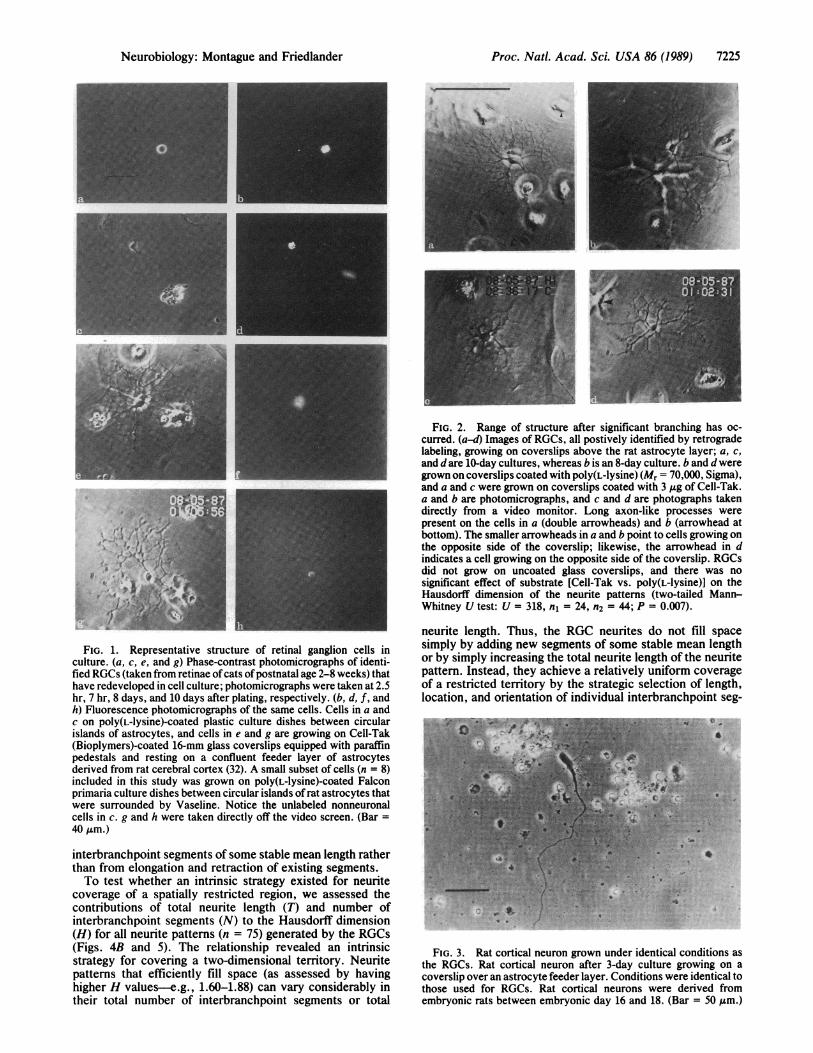
FIG. 3. Rat cortical neuron grown under identical conditions asthe RGCs. Rat cortical neuron after 3-day culture growing on acoverslip over an astrocyte feeder layer. Conditions were identical tothose used for RGCs. Rat cortical neurons were derived fromembryonic rats between embryonic day 16 and 18. (Bar = 50 Am.)
Neurobiology: Montague and Friedlander
I I IN,i"'I ':' 1#1 .
k..L

7226 Neurobiology: Montague and Friedlander
soo50 A
tt 4000
0 3000.2
i 2000
3 1000
00
2.0
1.8
1.6H
1.4
1.2
1.006
0 .* *g.0 *0
0
0* 0
. 00
100 200 300 400
Number of segments
B
ISio0 600
b*11 1 , l
_ f|-E||- ii i_ ___ ____ __ ___ ___ XXL--__- _I
_ __ ___ _-- __
0 0
* K @ 000*00 oT&~~~~~~~~~~~Tow0 ON
a
0.2 0.4 0.6
TorN
0.8 1.0
FIG. 4. (A) Scatter plot of the total neurite length of the neuriticarbors versus the number of segments belonging to each arborindependent of time in culture. Although total neurite length de-creases rapidly as number of segments drops below -55-65, a lineartrend between these two parameters is exhibited (r = 0.92, slope =7.17). The nonlinearity can be partially accounted for by observingthat most (7/12) cells that possess <60 segments belong to the 0- to48-hr group-a time when the neurites are not as consistentlysuccessful in extending processes as later. During this period one ora few segments commonly extend much farther than average and,hence, represent a greater proportion of total segment length.Nonlinearity in this range can be attributed to the establishment orloss of one or more of these longer-than-average segments. Withincreasing culture time, this effect was not as dominant. (B) Plot ofthe HausdorffdimensionHversus the normalized total neurite lengthTand the normalized number of segments Nfor every neurite patternanalyzed. All three of these variables were normalized to the largestvalue obtained in the population of 75 neurite patterns. Number ofsegments ranged from 8 to 506; total neurite length ranged from 56to 3234 Atm. The Hausdorff dimensions ranged from 1.04 to 1.88.After 3-day culture, the Hausdorff dimension ranged from 1.32 to1.88 (x = 1.66, SD = 0.10, n = 56).
ments. After 3-day culture, most neurite patterns (77%, n =56 neurite patterns) achieve relatively uniform coverage (1.60< H < 1.88) of a territory independent of the amount of"working material" (total neurite length or number of inter-branchpoint segments) available. During this same period, aminority (23%) ofneurite patterns haveH values from 1.32 to1.59 and appear sparser in their territorial coverage. Theneurites ofany individual RGC exhibited a high degree of selfavoidance with self intersections occurring at a frequency of-0.5% (20 cells, n = 2500 interbranchpoint segments). Allcells in culture for >3 days had neuritic arbors with diametersbetween 110 and 270 ,um. For the in vivo cells, H ranged from1.54 to 1.94 with an average of 1.73 (n = 18, SD = 0.12) ascompared with a range of 1.32 to 1.88 and an average of 1.66(n = 56, SD = 0.10) for RGCs in culture for >3 days. Thesample ofRGCs from intact preparations included a range ofmorphological classes (10 (, 6 a, 2 y). For direct comparisonof the morphology and coverage of a RGC grown in cultureto one studied in vivo, note the similarity between the cell inFig. 5 to the P3 cell in Figure 8A in Ramoa et al. (15). The Hvalues for these RGCs were 1.66 and 1.59, respectively. Incontrol experiments, neurons derived from other regions of
10 -
c 82.-I
ax 6
4~2
n
c
-1.66
In (esh size)d
5 6
FIG. 5. Method of calculating the Hausdorff dimension of theneurite patterns of the RGCs. (a) Line drawing of a representativeRGC after 10-day culture (same cell as Fig. 2c). This line drawing wasmade directly from a stored overlay drawing of a digitized videoimage of the RGC. The stored overlay drawing was used forcalculating the Hausdorff dimension of the RGC neurite pattern(somata were not used in this calculation). (b and c) Illustration of thebox counting method used to estimate the Hausdorff dimension. Aninteractive computer program covered the stored overlay drawingwith a sequence (n > 20) of grids composed of square boxes of sideequal to m. For each grid (characterized by the box or mesh size), thenumber ofboxes intersected by the overlay drawing was counted andstored in the computer. To avoid biasing box count due to grid(square mesh) geometry, position and orientation of the overlaydrawing were randomly varied relative to the grid. At each particularmesh size, this procedure was done 10 times, and the number ofboxes intersected was averaged. Thus, a relationship between meshsize and average number of boxes intersected was established. Thelogarithm of the number of boxes intersected [In (# boxes int)] wasplotted against the logarithm ofmesh size; negative ofthe slope of theregression line for this plot yields the Hausdorff dimension of theneurite pattern (1.66 in this example). In b and c, the intersectedboxes were filled to illustrate the method. This method for estimatingHausdorff dimension follows from the definition of Hausdorff di-mension (24, 25). (d) Plot oflogarithm ofnumber ofboxes intersectedversus logarithm of mesh size for the cell in a. Because we had noa priori knowledge that the processes producing the neurite patternswould generate structures that yield logarithmic normally distributedvariations in the average box count as a function ofmesh size, we didnot include confidence-interval estimates for the regression-lineslope.
the central nervous system (see above) followed a differentpattern of growth. These neurons exhibited frequent self-crossings, and their growth strategy was dominated by neu-rite extension. In addition, these neurons filled space muchless efficiently than RGCs with H values consistently <1.3independent of the time in culture.
Proc. Natl. Acad Sci. USA 86 (1989)
I
0 0
00
0
AD
Proc. Natl. Acad. Sci. USA 86 (1989) 7227
DISCUSSION
These experiments show that postmitotic (37, 38) mammalianneurons that have already achieved extensive dendritic dif-ferentiation in vivo (15, 27-30) can reexpress emergent struc-tural features in vitro characteristic and reminiscent of the invivo condition. This reexpression is independent of factorsfrom other cells normally present in the immediate locale oftheRGCs in the intact retina and is independent of interactionswith target tissue. The most striking feature of the expressionof the neuritic structure of these cells is the existence of an
apparent growth strategy that operated more or less indepen-dently of number (or amount) of neurites expressed. Modu-lation of the number of neurite segments having a stable meanlength and the strategic selection of their position, orientation,and length produced relatively uniform territorial coverage, as
assessed by the Hausdorff dimension, and yet generated richmorphological variance (Figs. 1 e and g; 2 a-d).
Existence of this morphological variance is obvious upon
casual inspection ofthe cells and is implicit in the observationthat the space-filling capacity was independent of the numberof interbranchpoint segments and the total neurite length(Fig. 2). Hence, once a RGC has survived for >3 days inculture and established neuritic branches, its space-fillingcapacity (H) will probably (77%) fall between 1.6 and 1.9 andbe independent ofthe amount of"working material" available.The fraction ofRGCs with lowH values (<1.6) declines withtime in culture from 50% at <72 hr to 23% for cells in culturefor >72 hr. The first 72 hr in culture is an exceptional periodin that rapid changes in the space-filing capacity (H) of theneurites occur because the RGCs are not as consistentlysuccessful at extending and maintaining neurites as they are
at later times (see legend for Fig. 4A). The observation thatuniform territorial coverage is more or less independent ofthe amount of "working material" is consistent with the highdegree of self avoidance of the neurites and the limiteddiameters of the neurite patterns. However, the mere exis-tence of these two phenomena do not establish a causalrelationship with the strategy of uniform coverage. A varietyof growth schemes employing only self-avoidance and/or a
limited period of growth could allow for the emergence ofuniform coverage of a 2-dimensional territory.
This study provides evidence for the intrinsic capabilities ofindividual RGCs to elaborate an emergent cellular trait. How-ever, extrinsic factors clearly play a role in the developmentand maintenance of the dendritic arbors of RGCs in themammalian retina. The structure ofRGC dendritic arbors fromcat and rat has been shown in vivo to exhibit a sensitivity andplasticity to the proximity and branching patterns of neigh-boring ganglion cells after perturbations that significantlydisrupt these neighbor-dependent relationships (11, 12, 39, 40).It has recently been suggested that transient morphologicalfeatures of developing RGCs subserve some form of dendriticcompetition (12, 15, 28). In view of the demonstrated sensi-tivity to external perturbations, it is noteworthy that thecultured RGCs expressed a range of structure in such ahomogeneous environment. This intrinsic structural varianceconstrained by a common mode of covering territory mayallow for the emergence of the diverse morphological andfunctional classes of RGCs in the retina that are later shapedby epigenetic influences, such as the presence and activity ofafferent input. How the intrinsic capacity and strategy forelaborating neuritic structure depends on developmental ageand epigenetic influences from neighboring RGCs and targettissue is an interesting question awaiting a more preciseanswer.
We thank the University ofAlabama at Birmingham NeurobiologyResearch Center Cell Reconstruction Facility, Drs. Lee Ann Cole-man and Yves Fregnac for comments and suggestions on themanuscript, Kevin Ramer for software development, and WandaSmith for word processing. This work was supported by NationalScience Foundation Grant BNS 8720069.
1. Ramon y Cajal, S., Degeneration and Regeneration of theNervous System [May, R. M., trans. (1928) (Oxford Univ.Press, London)].
2. Cowan, W. M. (1978) Int. Rev. Physiol. 17, 150-191.3. Scott, B. E., Engelbert, V. E. & Fisher, K. C. (1969) Exp.
Neurol. 23, 230-248.4. Fishbach, G. (1970) Science 169, 1331-1333.5. Banker, G. A. & Cowan, W. M. (1977) Brain Res. 126, 397-
425,6. Dichter, M. A. (1978) Brain Res. 149, 279-293.7. Banker, G. A. & Cowan, W. M. (1979) J. Comp. Neurol. 187,
469-494.8. Kriegstein, A. R. & Dichter, M. A. (1983) J. Neurosci. 3,
1634-1647.9. Banker, G. A. & Waxman, A. B. (1988) in Intrinsic Determi-
nants of Neuronal Form and Function, eds. Lasek, R. J. &Black, M. M. (Liss, New York), pp. 61-82.
10. Lasek, R. J. (1988) in Intrinsic Determinants ofNeuronal Formand Function, eds. Lasek, R. J. & Black, M. M. (Liss, NewYork), pp. 1-58.
11. Wassle, H., Peichl, L. & Boycott, B. B. (1981) Nature (Lon-don) 292, 344-345.
12. Perry, V. H. & Linden, R. (1982) Nature (London) 297, 683-685.
13. Dowling, J. (1987) The Retina (Belknap-Harvard Univ. Press,Cambridge, MA).
14. Famiglietti, E. V. & Kolb, H. (1976) Science 194, 193-195.15. Ramoa, A. S., Campbell, G. A. & Shatz, C. J. (1988) J. Neu-
rosci. 8, 4239-4261.16. Shiosaka, S., Kiyama, K. & Tohyama, M. (1984) J. Neurosci.
Methods 10, 229-235.17. Montague, P. R. & Friedlander, M. J. (1987) Soc. Neurosci.
Abstr. 13, 1299.18. Montague, P. R. & Friedlander, M. J. (1988) Soc. Neurosci.
Abstr. 14, 459.19. Saito, H.-A. (1983) J. Comp. Neurol. 221, 279-288.20. Amthor, F. (1984) Brain Res. 298, 187-190.21. Fukuda, Y., Hasaio, C. F. & Watanabe, M. (1984) J. Neuro-
physiol. 52, 999-1013.22. Stanford, L. R. & Sherman, S. M. (1984) Brain Res. 297,
381-386.23. Stanford, L. R. (1987) J. Neurophysiol. 58, 940-964.24. Mandelbrot, B. B. (1982) The Fractal Geometry of Nature
(Freeman, San Francisco).25. Falconer, K. J. (1986) Geometry of Fractal Sets (Cambridge
Univ. Press, Cambridge, MA).26. Katz, L. C., Burkhalter, A. & Dreyer, W. J. (1984) Nature
(London) 310, 498-500.27. Maslim, J., Webster, M. & Stone, J. (1986) J. Comp. Neurol.
254, 382-402.28. Ramoa, A. S., Campbell, G. A. & Shatz, C. J. (1987) Science
237, 522-525.29. Dann, J. F., Buhl, E. H. & Peichl, L. (1987) Neurosci. Lett. 80,
21-26.30. Dann, J. F., Buhl, E. H. & Peichl, L. (1988) J. Neurosci. 8,
1485-1499.31. McCarthy, K. D. & deVellis, J. (1980) J. Cell Biol. 85, 890-902.32. Banker, G. A. (1980) Science 209, 809-810.33. Scott, B. S. & Fisher, K. C. (1970) Exp. Neurol. 27, 16-22.34. Scott, B. S. & Fisher, K. C. (1971) Exp. Neurol. 31, 183-188.35. Scott, B. S. (1976) J. Cell. Physiol. 91, 305-316.36. Bennett, M. R. & White, W. (1979) Brain Res. 173, 549-553.37. Johns, P. R., Rusoff, A. C. & Dubin, M. W. (1979) J. Comp.
Neurol. 187, 545-556.38. Walsh, C., Polley, E. H., Hickey, T. L. & Guillery, R. W.
(1983) Nature (London) 302, 611-613.39. Eysel, U. L., Peichl, L. & Wassle, H. (1985) J. Comp. Neurol.
242, 134-145.40. Leventhal, A. G., Schall, J. D. & Ault, S. J. (1988) J. Neurosci.
8, 2028-2038.
Neurobiology: Montague and Friedlander