Embed Size (px)
Citation preview

Am J Cardiovasc Dis 2013;3(1):17-26www.AJCD.us /ISSN:2160-200X/AJCD1211005
Review ArticleInteraction between sphingomyelin and oxysterols contributes to atherosclerosis and sudden death
Fred A Kummerow
Burnsides Research Laboratory, Department of Comparative Biosciences, College of Veterinary Medicine, Univer-sity of Illinois, 1208 W. Pennsylvania Avenue, Urbana, IL 61801, USA
Received November 26, 2012; Accepted January 23, 2013; Epub February 17, 2013; Published February 27, 2013
Abstract: Despite major public health efforts, coronary heart disease continues to be the leading cause of death in the United States. Oxidized lipids contribute to heart disease both by increasing deposition of calcium on the arte-rial wall, a major hallmark of atherosclerosis, and by interrupting blood flow, a major contributor to heart attack and sudden death. Oxidized cholesterol (oxysterols) enhances the production of sphingomyelin, a phospholipid found in the cellular membranes of the coronary artery. This increases the sphingomyelin content in the cell membrane, which in turn enhances the interaction between the membrane and ionic calcium (Ca2+), thereby increasing the risk of arterial calcification. Patients undergoing bypass surgery had greater concentrations of oxysterols in their plasma than cardiac catheterized controls with no stenosis, and had five times more sphingomyelin in their arteries than in the artery of the placenta of a newborn. The oxysterols found in the plasma of these patients were also found in the plasma of rabbits that had been fed oxidized cholesterol and in frying fats and powdered egg yolk intended for human consumption. Together these findings suggest that oxysterols found in the diet are absorbed and contribute to arterial calcification. Oxidized low-density lipoprotein (OxLDL) further contributes to heart disease by increasing the synthesis of thromboxane in platelets, which increases blood clotting. Cigarette smoke and trans fatty acids, found in partially hydrogenated soybean oil, both inhibit the synthesis of prostacyclin, which inhibits blood clotting. By increasing the ratio of thromboxane to prostacyclin, these factors interact to interrupt blood flow, thereby contrib-uting to heart attack and sudden death. Levels of oxysterols and OxLDL increase primarily as a result of three diet or lifestyle factors: the consumption of oxysterols from commercially fried foods such as fried chicken, fish, and french fries; oxidation of cholesterol in vivo driven by consumption of excess polyunsaturated fatty acids from vegetable oils; and cigarette smoking. Along with the consumption of trans fatty acids from partially hydrogenated vegetable oil, these diet and lifestyle factors likely underlie the persistent national burden of heart disease.
Keywords: Sphingomyelin, oxysterols, thromboxane, prostacyclin, trans fatty acids, calcium, stenosis
Introduction
Pohjantahti et. al. [1] summarized 50 studies and concluded that the elevation of oxidized low-density lipoprotein (OxLDL) is a key event in the development of atherosclerosis. Diets enriched in oxidized fatty acids increase fatty streak lesions in the aorta of cholesterol-fed rabbits. Staprans et al. [2] fed rabbits a chow diet to which 0.33% cholesterol was added. The control group received cholesterol that had been stored at -70°C under N2 to prevent oxida-tion. A second group received the same diet except approximately 5% of the total added cholesterol was oxidized. These rabbits
received 25 mg oxidized cholesterol per day. Five oxysterols were found in the plasma of these rabbits: 7alpha-hydroxycholesterol, 7b-hydroxycholesterol beta-epoxycholesterol, alpha-epoxycholesterol, and 7-ketocholesterol. The percentage of aortic area covered by fatty streaks was twice as great in the rabbits receiv-ing oxidized cholesterol as in the controls. In my laboratory, these five oxysterols were also found in elevated concentrations in the plasma of human patients who had undergone coronary artery bypass grafting (CABG) surgery, suggest-ing that they are important in the development of atherosclerosis in both animals and humans. Staprans, et al. demonstrated that oxidized

Sphingomyelin, oxysterols, atherosclerosis and sudden death
18 Am J Cardiovasc Dis 2013;3(1):17-26
cholesterol in the serum of rabbits is both syn-thesized endogenously and derived from food. Oxysterols are synthesized endogenously via enzymatic or radical-mediated oxidation. Enzymatic oxidation mainly occurs in the liver and steroidogenic tissues. The radical species responsible for cholesterol oxidation are derived from activated oxygen, which could occur in a variety of tissues [3], including the artery cell wall [4-6]. It was also demonstrated that the absorption of dietary oxidized choles-terol occurred within 24 hours, after feeding trace amounts of radiolabeled oxidized choles-terol, in the livers of rabbits. Similar absorption of oxidized cholesterol has also been described in humans [7]. Thus, oxidized cholesterol in the diet as well as dietary and lifestyle factors that promote the endogenous oxidation of choles-terol likely play important roles in the develop-ment of atherosclerosis. This review describes research from my laboratory suggesting that these dietary and lifestyle factors contribute to
atherosclerosis through two mechanisms: sphingomyelin-mediated arterial calcification and thromboxane-mediated interruption of blood flow.
The role of sphingomyelin and calcium in the pathogenesis of atherosclerosis
In a review article entitled “The pathogenesis of coronary heart disease: Perspectives for the 1990s” [8], Ross stated: “Coronary heart dis-ease of the extremities is most apparent at branching points of the arterial tree where blood flow is irregular with current and back currents. The cellular events that occur during the progression of lesions in hypercholesterol-emic animals are almost exactly mirrored by those observed in human atherosclerotic coro-nary arteries in hearts removed in transplant operations”. These lesions are shown in (Figure 1) of a patient needing CABG surgery. The right coronary artery is 75% and 60% occluded, cir-
Figure 1. Diagnostic summary of the cardiac catheterized heart of a patient needing a coronary heart bypass graft-ing (CABG) surgery. The right coronary artery is 75% occluded. Circumflex artery is 100% occluded. The left one is 75% occluded and the right. (This figure ss courtesy of Dr. Bruce Handler).

Sphingomyelin, oxysterols, atherosclerosis and sudden death
19 Am J Cardiovasc Dis 2013;3(1):17-26
cumflex is 100% and 65% occluded and the left one is 75% occluded.
DeBakey et. al. [9] had noted thickening at branching and bifurcations during CABG sur-gery. Kummerow et. al. [10] also noted thicken-ing in the branching arteries in aging pigs on a cholesterol-free diet. It did not differ significant-ly in sphingomyelin composition (Figure 2) from that of the non-branching adjacent tissue of pigs at 6 months of age. By 18 and 48 months of age, however, the sphingomyelin content was significantly higher at the thickened branching areas than at the non-thickened segment of the arteries. This indicated that during aging of the arteries, there was a striking increase in the amount of sphingomyelin in the branching points. Lipids extracted from both pig and human arteries indicated that aging is a factor that increases sphingomyelin. The non-branch-ing segment of the aorta, obtained on autopsy from six men 21-27 years of age, contained four times more sphingomyelin than arteries isolated from human placenta, indicating that the sphingomyelin content of arteries increas-es with age. Aging is not the only factor that increased the sphingomyelin composition of arterial cells. Women and men under 40 years of age who had been subjected to CABG sur-gery contained the same high percentage of sphingomyelin in their non-athermatous arteri-al cells as those over 40 years of age. Therefore, the disease itself caused a premature increase in sphingomyelin in non-athermatous arterial
cells in CABG patients, pointing to a fundamen-tal disturbance in phospholipid metabolism in their arterial cells.
Kummerow et. al. [11] found patients who had CABG surgery sometimes needed a second CABG surgery because the vein used in the first surgery had become occluded. During this sec-ond surgery, an unoccluded vein from the same patient was used to replace the occluded vein. Approval for collection of discarded veins was provided by the University of Illinois and the Carle Foundation Institutional Review Boards. The occluded veins contained significantly more sphingomyelin and Ca2+ than the unoc-cluded veins. The unoccluded veins contained 24% sphingomyelin and 182 ppm of Ca2+ as compared to 48% of sphingomyelin and 6,345 ppm of Ca2+ in the occluded veins that had been used in the first CABG surgery. The increased sphingomyelin and Ca2+ concentra-tions in the occluded veins were responsible for the need of the initial CABG surgery.
In humans, excess oxysterols stimulated the synthesis of sphingomyelin and inhibited sphin-gomyelin metabolism [12]. By using a radiola-beled methyl-3H choline, the time- and dose-dependent effects of 27-hydroxycholesterol on sphingomyelin synthesis could be observed. The increased radioactivity in sphingomyelin was accompanied by a decreased radioactivity in phosphatidylcholine. This result indicated that 27-hydroxycholesterol increased the trans-
Figure 2. Composition of sphingomyelin in a non-branching seg-ment compared to the branching segment of the same artery at bi-furcations of 6, 18 and 48 month old swine kept on a cholesterol/saturated fat free diet. Means with a letter “a” are statistically differ-ent at P<0.05, com-pare to the data from non-branching at the same age, Means with a symbol * or ** are statistically different at P<0.05, compare to the data from the same ar-tery at 6 or 18 months, respectively.
Sphingomyelin, oxysterols, atherosclerosis and sudden death
20 Am J Cardiovasc Dis 2013;3(1):17-26
fer of choline from phosphatidylcholine to sphingomyelin. It is known that oxysterols could enhance 45Ca2+ uptake and cytosolic free Ca2+.
The enhanced Ca2+ uptake has been thought to come from greater membrane permeability by oxysterols [13, 14]. By measuring parallel cyto-solic 45Ca2+uptake, cytosolic free Ca2+, choline label in sphingomyelin and cytotoxicity in the presence of 27-hydroxycholesterol we observed that enhanced 45Ca2+uptake and cytosolic free Ca2+ level occurred earlier than cytotoxicity, but later than increased radioactivity in sphingomy-elin. This finding suggests that enhanced 45Ca2+uptake may result from an increase in sphingomyelin synthesis and not from the cyto-toxicity of 27-hydroxycholesterol. This is further supported by evidence from previous studies [15-17]. Deposition of calcium in the coronary artery has been found to be directly proportion-al to an elevated sphingomyelin concentration [16]. Just as with 27-hydroxycholesterol, 25-hydroxycholesterol increased both 45Ca2+uptake and sphingomyelin composition
[15]. Since sphingomyelin is located on the exterior of the plasma membrane [17] and has an exposed polar head group accessible to the aqueous environment, the negative charge on the exterior side on sphingomyelin would be accessible for ionic bonding with Ca2+.
27-Hydroxycholesterol at a level of 0.1μg/mL, which is within the range of its plasma concen-tration in healthy adults [18] had no obviously stimulating effect on the incorporation of cho-line label into sphingomyelin from phosphatidyl-choline during 15d of treatment. When the level was increased to 0.5μg/mL, however, it took only 3d for 27-hydroxycholesterol to increase radioactivity in sphingomyelin. These results indicate that 27-hydroxycholesterol only increases transfer of choline from phosphati-dylcholine into sphingomyelin when it is present in concentrations higher than those found in healthy adults. This was consistent with the reports that both 27-hydroxycholesterol and sphingomyelin increase in atherosclerosis [19].
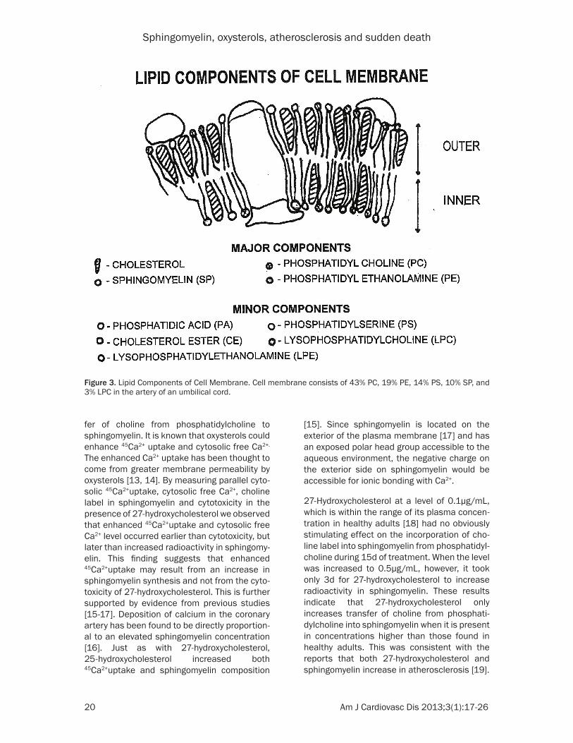
Figure 3. Lipid Components of Cell Membrane. Cell membrane consists of 43% PC, 19% PE, 14% PS, 10% SP, and 3% LPC in the artery of an umbilical cord.
Sphingomyelin, oxysterols, atherosclerosis and sudden death
21 Am J Cardiovasc Dis 2013;3(1):17-26
Lipid analysis of plasma and arterial tissue obtained from CABG patients
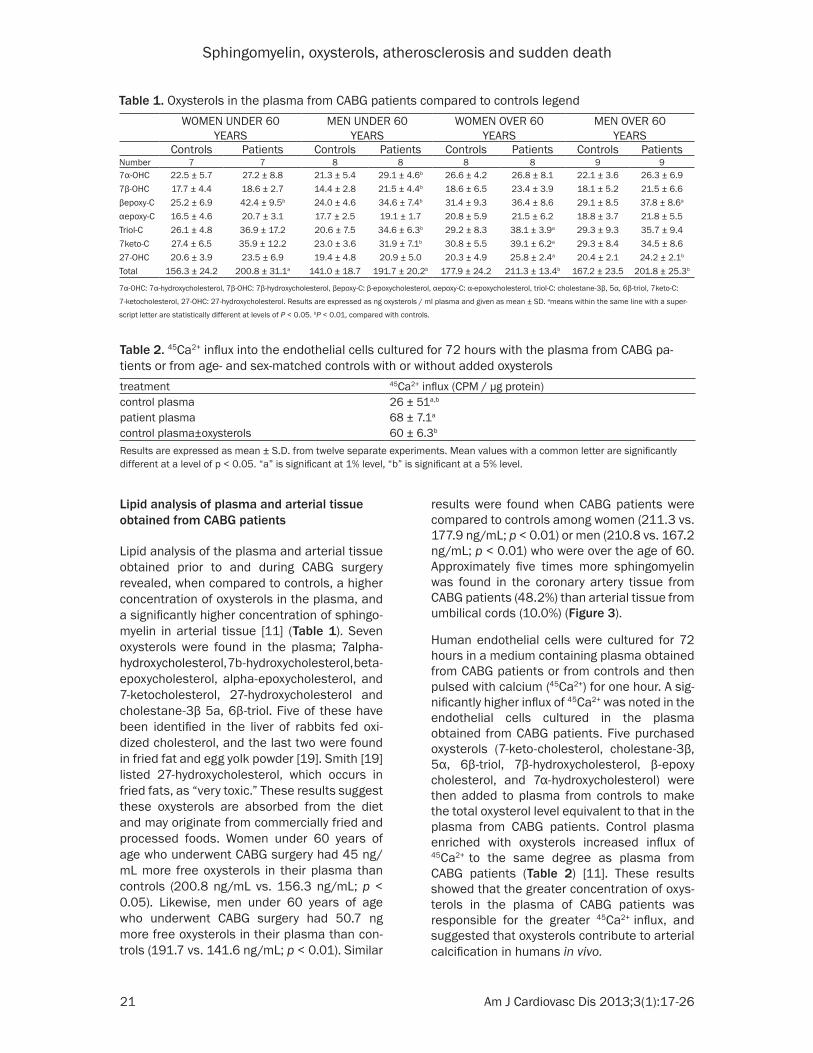
Lipid analysis of the plasma and arterial tissue obtained prior to and during CABG surgery revealed, when compared to controls, a higher concentration of oxysterols in the plasma, and a significantly higher concentration of sphingo-myelin in arterial tissue [11] (Table 1). Seven oxysterols were found in the plasma; 7alpha-hydroxycholesterol, 7b-hydroxycholesterol, beta- epoxycholesterol, alpha-epoxycholesterol, and 7-ketocholesterol, 27-hydroxycholesterol and cholestane-3β 5a, 6β-triol. Five of these have been identified in the liver of rabbits fed oxi-dized cholesterol, and the last two were found in fried fat and egg yolk powder [19]. Smith [19] listed 27-hydroxycholesterol, which occurs in fried fats, as “very toxic.” These results suggest these oxysterols are absorbed from the diet and may originate from commercially fried and processed foods. Women under 60 years of age who underwent CABG surgery had 45 ng/mL more free oxysterols in their plasma than controls (200.8 ng/mL vs. 156.3 ng/mL; p < 0.05). Likewise, men under 60 years of age who underwent CABG surgery had 50.7 ng more free oxysterols in their plasma than con-trols (191.7 vs. 141.6 ng/mL; p < 0.01). Similar
results were found when CABG patients were compared to controls among women (211.3 vs. 177.9 ng/mL; p < 0.01) or men (210.8 vs. 167.2 ng/mL; p < 0.01) who were over the age of 60. Approximately five times more sphingomyelin was found in the coronary artery tissue from CABG patients (48.2%) than arterial tissue from umbilical cords (10.0%) (Figure 3).
Human endothelial cells were cultured for 72 hours in a medium containing plasma obtained from CABG patients or from controls and then pulsed with calcium (45Ca2+) for one hour. A sig-nificantly higher influx of 45Ca2+ was noted in the endothelial cells cultured in the plasma obtained from CABG patients. Five purchased oxysterols (7-keto-cholesterol, cholestane-3β, 5α, 6β-triol, 7β-hydroxycholesterol, β-epoxy cholesterol, and 7α-hydroxycholesterol) were then added to plasma from controls to make the total oxysterol level equivalent to that in the plasma from CABG patients. Control plasma enriched with oxysterols increased influx of 45Ca2+ to the same degree as plasma from CABG patients (Table 2) [11]. These results showed that the greater concentration of oxys-terols in the plasma of CABG patients was responsible for the greater 45Ca2+ influx, and suggested that oxysterols contribute to arterial calcification in humans in vivo.
Table 1. Oxysterols in the plasma from CABG patients compared to controls legendWOMEN UNDER 60
YEARSMEN UNDER 60
YEARSWOMEN OVER 60
YEARSMEN OVER 60
YEARSControls Patients Controls Patients Controls Patients Controls Patients
Number 7 7 8 8 8 8 9 97α-OHC 22.5 ± 5.7 27.2 ± 8.8 21.3 ± 5.4 29.1 ± 4.6b 26.6 ± 4.2 26.8 ± 8.1 22.1 ± 3.6 26.3 ± 6.97β-OHC 17.7 ± 4.4 18.6 ± 2.7 14.4 ± 2.8 21.5 ± 4.4b 18.6 ± 6.5 23.4 ± 3.9 18.1 ± 5.2 21.5 ± 6.6βepoxy-C 25.2 ± 6.9 42.4 ± 9.5b 24.0 ± 4.6 34.6 ± 7.4b 31.4 ± 9.3 36.4 ± 8.6 29.1 ± 8.5 37.8 ± 8.6a
αepoxy-C 16.5 ± 4.6 20.7 ± 3.1 17.7 ± 2.5 19.1 ± 1.7 20.8 ± 5.9 21.5 ± 6.2 18.8 ± 3.7 21.8 ± 5.5Triol-C 26.1 ± 4.8 36.9 ± 17.2 20.6 ± 7.5 34.6 ± 6.3b 29.2 ± 8.3 38.1 ± 3.9a 29.3 ± 9.3 35.7 ± 9.47keto-C 27.4 ± 6.5 35.9 ± 12.2 23.0 ± 3.6 31.9 ± 7.1b 30.8 ± 5.5 39.1 ± 6.2a 29.3 ± 8.4 34.5 ± 8.627-OHC 20.6 ± 3.9 23.5 ± 6.9 19.4 ± 4.8 20.9 ± 5.0 20.3 ± 4.9 25.8 ± 2.4a 20.4 ± 2.1 24.2 ± 2.1b
Total 156.3 ± 24.2 200.8 ± 31.1a 141.0 ± 18.7 191.7 ± 20.2b 177.9 ± 24.2 211.3 ± 13.4b 167.2 ± 23.5 201.8 ± 25.3b
7α-OHC: 7α-hydroxycholesterol, 7β-OHC: 7β-hydroxycholesterol, βepoxy-C: β-epoxycholesterol, αepoxy-C: α-epoxycholesterol, triol-C: cholestane-3β, 5α, 6β-triol, 7keto-C:
7-ketocholesterol, 27-OHC: 27-hydroxycholesterol. Results are expressed as ng oxysterols / ml plasma and given as mean ± SD. ameans within the same line with a super-
script letter are statistically different at levels of P < 0.05. bP < 0.01, compared with controls.
Table 2. 45Ca2+ influx into the endothelial cells cultured for 72 hours with the plasma from CABG pa-tients or from age- and sex-matched controls with or without added oxysterolstreatment 45Ca2+ influx (CPM / µg protein)control plasma 26 ± 51a,b
patient plasma 68 ± 7.1a
control plasma±oxysterols 60 ± 6.3b
Results are expressed as mean ± S.D. from twelve separate experiments. Mean values with a common letter are significantly different at a level of p < 0.05. “a” is significant at 1% level, “b” is significant at a 5% level.
Sphingomyelin, oxysterols, atherosclerosis and sudden death
22 Am J Cardiovasc Dis 2013;3(1):17-26
The concentration of cholesterol and lipid oxidation products in the plasma of cardiac catheterized patients
The concentration of cholesterol, lipid oxidation products, and total antioxidant capacity in the plasma of 2,000 cardiac catheterized patients with 0, 10–69, and 70–100% stenosis of their arteries were analyzed [20]. The results showed that lipid oxidation products increased with the severity of stenosis. The total antioxidant capacity decreased with the severity of steno-sis. The plasma cholesterol concentration, however, was not significantly different between these groups of patients. Therefore, the con-centration of oxidation products rather than the concentration of cholesterol in the plasma increased with the severity of coronary heart disease. All of the women and men, in all age groups, with cardiovascular coronary heart dis-ease also had increased individual and total oxysterol levels in their plasma compared with the controls.
Lipid composition and calcium concentration in plaque tissue of the carotid and coronary arteries
The lipid composition and calcium concentra-tion in plaque tissue of the carotid and coro-nary arteries were analyzed. The total phospho-
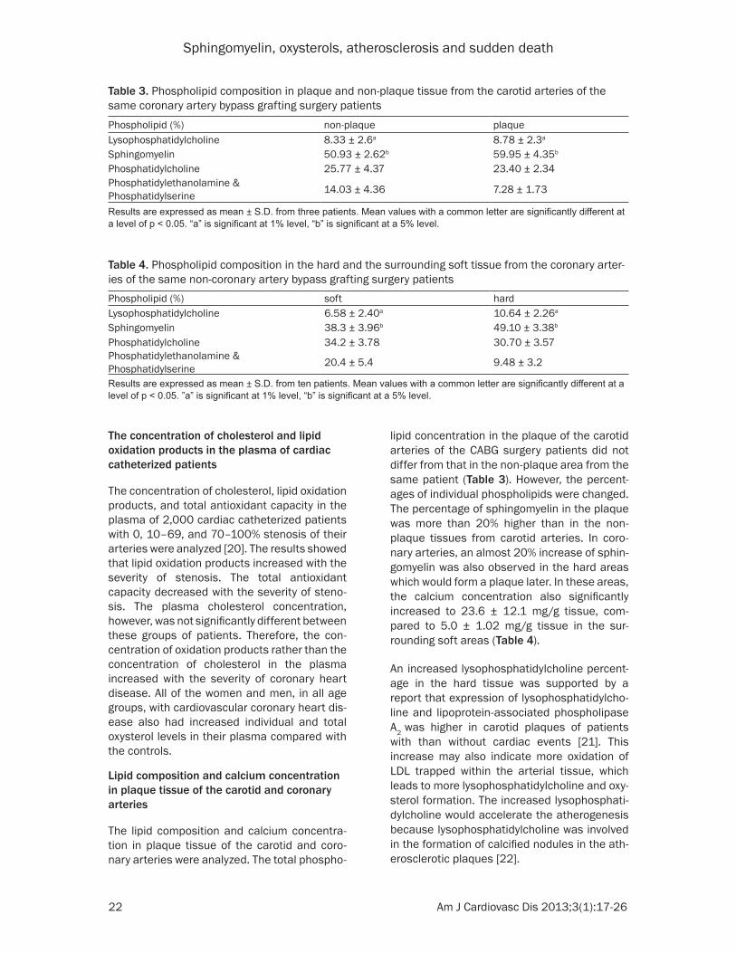
lipid concentration in the plaque of the carotid arteries of the CABG surgery patients did not differ from that in the non-plaque area from the same patient (Table 3). However, the percent-ages of individual phospholipids were changed. The percentage of sphingomyelin in the plaque was more than 20% higher than in the non-plaque tissues from carotid arteries. In coro-nary arteries, an almost 20% increase of sphin-gomyelin was also observed in the hard areas which would form a plaque later. In these areas, the calcium concentration also significantly increased to 23.6 ± 12.1 mg/g tissue, com-pared to 5.0 ± 1.02 mg/g tissue in the sur-rounding soft areas (Table 4).
An increased lysophosphatidylcholine percent-age in the hard tissue was supported by a report that expression of lysophosphatidylcho-line and lipoprotein-associated phospholipase A2 was higher in carotid plaques of patients with than without cardiac events [21]. This increase may also indicate more oxidation of LDL trapped within the arterial tissue, which leads to more lysophosphatidylcholine and oxy-sterol formation. The increased lysophosphati-dylcholine would accelerate the atherogenesis because lysophosphatidylcholine was involved in the formation of calcified nodules in the ath-erosclerotic plaques [22].
Table 3. Phospholipid composition in plaque and non-plaque tissue from the carotid arteries of the same coronary artery bypass grafting surgery patients Phospholipid (%) non-plaque plaqueLysophosphatidylcholine 8.33 ± 2.6a 8.78 ± 2.3a
Sphingomyelin 50.93 ± 2.62b 59.95 ± 4.35b
Phosphatidylcholine 25.77 ± 4.37 23.40 ± 2.34Phosphatidylethanolamine & Phosphatidylserine 14.03 ± 4.36 7.28 ± 1.73
Results are expressed as mean ± S.D. from three patients. Mean values with a common letter are significantly different at a level of p < 0.05. “a” is significant at 1% level, “b” is significant at a 5% level.
Table 4. Phospholipid composition in the hard and the surrounding soft tissue from the coronary arter-ies of the same non-coronary artery bypass grafting surgery patientsPhospholipid (%) soft hardLysophosphatidylcholine 6.58 ± 2.40a 10.64 ± 2.26a
Sphingomyelin 38.3 ± 3.96b 49.10 ± 3.38b
Phosphatidylcholine 34.2 ± 3.78 30.70 ± 3.57Phosphatidylethanolamine & Phosphatidylserine 20.4 ± 5.4 9.48 ± 3.2
Results are expressed as mean ± S.D. from ten patients. Mean values with a common letter are significantly different at a level of p < 0.05. ”a” is significant at 1% level, “b” is significant at a 5% level.

Sphingomyelin, oxysterols, atherosclerosis and sudden death
23 Am J Cardiovasc Dis 2013;3(1):17-26
The influence of thromboxane and prostacy-clin in blood flow
The presence of thromboxane, a potent inducer of vasoconstriction and platelet adhesion, in the arteries is responsible for the interruption of blood flow, causing the clogging of the arter-ies and therefore sudden death [23]. Mahfouz and Kummerow showed that the components of oxidized low-density lipoprotein (OxLDL) were responsible for platelet sensitization to throm-bin and the increase of thromboxane release. More significant, we found that OxLDL at low concentrations is more potent in enhancing the platelet response to thrombin and to increasing thromboxane release than native LDL [24]. In contrast, prostacyclin is vasoprotective and keeps blood flowing. It is a dominant prosta-glandin produced by endothelial cells and is a potent vasodilator and inhibitor of platelet aggregation and leukocyte adhesion [25]. However, smoking cigarettes and consuming trans fatty acids in partially hydrogenated fats in the diet inhibits prostacyclin generation by the coronary arteries [26, 27].
Cigarette smoke is a major risk factor for ath-erosclerosis and is associated with coronary, cerebral, and peripheral vascular disease. Cigarette smoke is a mixture of gases and par-ticulate matter. Each puff has been reported to contain 1014 free radicals in each phase (solu-ble and particulate) [28]. Both phases contain high concentrations of reactive oxygen species, nitrogen oxide, peroxynitrate, and free radicals
of organic compounds which cause two major processes: the oxidation of LDL and the inhibi-tion of prostacyclin synthesis, both dangerous for life [25]. According to Maddox, sudden car-diac death is the largest cause of natural death in the U.S., causing about 325,000 adult deaths in the United States each year [29].
Antioxidants
Feeding rabbits a high-cholesterol diet supple-mented with antioxidants prevented the intimal thickening of aortas, even though their blood continued to have a cholesterol level 40 times higher than control rabbits, but their plasma oxysterol levels decreased significantly com-pared to the rabbits fed without antioxidants [30]. The same results occurred in hyperlipid-emic chickens fed vitamin E, where lipid peroxi-dation and coronary heart disease lesions sig-nificantly decreased while hyperlipidemia remained high [31].
Data obtained from the US. department of agriculture and U.S. census bureau
Data from the U.S. Department of Agriculture indicated that from 1912 through 2011, the consumption of polyunsaturated fat has increased from 11.3 pound per capita to 64.5 pounds per capita and saturated fat has decreased from 28 pounds per capita to 13.4 pounds per capita in the same period (Table 5). [32] http://usda.mannlib.cornell.edu/MannUs- da/viewDocumentInfo.do?documentID=1290,
Table 5. Fats and vegetable oils consumption in US since 1912 per capita (in pounds)Years 1912 1950 1999 2011Items Total* Per cap Total* Per cap Total* Per cap. Total* Per capCorn oil 53.0 0.6 223.0 1.47 1416.9 5.2 1620.0 5.2Cottonseed oil 950.0 10.0 1445.0 9.51 832.8 3.1 620.0 2.0Olive oil 43.0 0.5 76.0 0.50 329.8 1.2 650.1 2.1Palm oil 0.0 0.0 26.0 0.17 416 1.5 2525.2 8.1Palm kernel oil 0.0 0.0 26.0 0.17 233.2 0.9 778.0 2.5Peanut oil 8.0 0.1 103.0 0.68 1524.7 5.6 202.7 0.6Canola oil 0.0 0.0 0.0 0.00 111.2 0.4 4249.0 13.6Safflower oil 0.0 0.0 5.1 0.03 15.8 0.1 60.9 0.2Sesame oil 0.0 0.0 5.0 0.03 15.8 0.1 27.2 0.1Soybean oil 16.0 0.2 1446.0 9.51 8029.6 29.4 9000.0 28.8Sunflower oil 0.0 0.0 0.5 0.00 393.7 1.4 395 1.3Total unsaturated oils 1070.0 11.3 3355.6 22.1 13319.5 48.8 20128.1 64.5Lard 1069.0 11.2 1891.0 12.60 202.0 0.7 480.0 1.5Butter 1579.0 16.6 1648.0 10.70 1307.0 4.8 1510.0 4.8Tallow 22.0 0.2 69.0 0.45 996.0 3.6 1050.0 3.4Coconut 0.0 0.0 69.0 0.45 927.0 3.4 1155.1 3.7Total saturated fats 2670.0 28.0 3677.0 24.2 3432.0 12.6 4195.1 13.4US Population in millions 95 152 273 312
(*) Totals in millions of pounds. Courtesy Mark Ash, of U.S. Department of Agriculture. http://factfinder2.census.gov/faces/tableservices/jsf/pages/productview.
xhtml?pid=ACS_11_1YR_DP05&prodType=table. http://www.npg.org/facts/us_historical_pops.htm. http://www.census.gov/population/estimates/state/st-99-1.txt.

Sphingomyelin, oxysterols, atherosclerosis and sudden death
24 Am J Cardiovasc Dis 2013;3(1):17-26
A report from the Inter-Society Commission for Heart Disease Resources [33] stated: “Use salad and cooking oils, new soft margarines and shortenings low in saturated fat” and “avoid butter, margarine and shortenings high in saturated fats;” which I did not agree with, because the consumption of polyunsaturated fats increase the production of oxysterols [34-36].
Conclusion
My hypothesis for the cause of heart disease and sudden coronary death is based on the composition, structure, and biochemistry of the coronary arteries. The composition of the cel-lular membrane has five different phospholip-ids which change in composition during the life time regardless of whether you have heart dis-ease or not. One of these phospholipids, sphin-gomyelin, increases from 10% at birth to 48% in someone who had a bypass surgery CABG and to 60% in patients who have plaques in their coronary arteries.
The sphingomyelin and calcium that occluded the veins and arteries of these patients was possible because of the binding of calcium to sphingomyelin. According to Shah and Schulman [37], the binding of Ca2+ to monolay-ers of phospholipids such as sphingomyelin happens because of the hydrogenation of sphingomyelin in the presence of water. This hydrogenation turns a normally neutral sphin-gomyelin into an anion with two negative charg-
es and therefore Ca2+ is attract-ed to sphingomyelin and binding occurs Figure 4. Direct evidence showing that metal ions do bind to fatty acid monolayers has been given by Langmuir and Schaefer [38] as well as by Schulman and Dogan [39], who removed the monolayers from the interface and subjected them to chemical analysis. In the case of phospholipids the interaction of Ca2+ can be indicated as 2PO4
- + Ca2+ E PO4
- -- Ca2+ -- PO4-. At
high salt concentration the amount of bound Ca2+ indicates an interaction between metal ions and the monolayer. The main generator of atherosclero-
Figure 4. Schematic representation of the interaction of calcium ion with sphingomyelin monolayer. By Shah and Schulman with permission [37].
sis and sudden death are the oxysterols in excess. Normal levels of oxysterols in the plas-ma will not cause phosphatidylcholine to con-vert into sphingomyelin and therefore less cal-cium will bind to it resulting in less artery blockage.
Data from the U.S. Center of Disease Control, http://www.cdc.gov/nchs/data/dvs/lead- 1900_98.pdf, http://www.cdc.gov/nchs/data/nvsr/nvsr60/nvsr60_04, shows that death due to heart disease decreased from 1970 to 2010 by 19%. It also shows that smoking habits decreased significantly by 43% in the same period. Since cigarette smoking also causes heart disease, the 19% death decrease due to heart disease in the last 40 years may be because of the major decrease in smoking hab-its. In 2010 the total number of deaths was 600,000, with half caused by sudden death. Therefore the death rate due to coronary heart disease and sudden death remains high. According to the U.S. Department of Agriculture data [32] we have switched from the consump-tion of saturated fats to polyunsaturated fats, which now are in almost everything that is con-sumed. Vegetables oils, partially hydrogenated fats, and fried foods are responsible for the persistently high rate of heart disease. The most effective way to prevent coronary heart disease and sudden death according to these conclusions is to eat fewer commercially fried foods, fewer polyunsaturated fats and to avoid partially hydrogenated fats. Conversely, we should eat more vegetables and fruit as a source of antioxidants.
Sphingomyelin, oxysterols, atherosclerosis and sudden death
25 Am J Cardiovasc Dis 2013;3(1):17-26
Acknowledgements
I wish to thank Dr. Chris Masterjohn, Ph.D. for his helpful insight and editing of the text. I like to also thank Dr. MM.Mahfouz Ph.D., Dr. Qi Zhou MD. Drs. L. Cook and T. Connelly for the tissue and Dr. B. Handler for the blood, cardiol-ogist at Carle Hospital, Urbana, Illinois. This work was supported by funding from The Weston A. Price Foundation and the Verna L. and John R. Hildebrand Foundation.
Address correspondence to: Dr. Fred A Kummerow, Burnsides Research Laboratory, 1208 W. Pennsylvania Avenue, Urbana, IL 61801, USA. Tel: 217-344-6380; Fax: 217-333-7370; E-mail: [email protected]
References
[1] Pohjantahti-Maaroos H, Palomaki A, Kank-kunen P, Laitinen R, Husgafvel S, Oksanen K. Circulating oxidized low-density lipoproteins and arterial elasticity: comparison between men with metabolic syndrome and physically active counterparts. Cardiovasc Diabetol 2010 Aug 20; 9: 41.
[2] Staprans I, Pan XM, Rapp JH, Feingold KR. Oxi-dized Cholesterol in the Diet Accelerates the Development of Aortic Atherosclerosis in Cho-lesterol-Fed Rabbits. Arterioscler Thromb Vasc Biol 1998 Jun; 18: 977-83.
[3] Morin RJ, Peng SK. Cholesterol oxides in plas-ma and tissues. In: Peng SK, Morin RJ, editors. Biological Effects of Cholesterol Oxides. Boca Raton, FL: CRC Press 1992; pp: 89-101.
[4] Sevanian A, McLeod LL. Cholesterol autoxida-tion in phospholipid membrane bilayers. Lipids 1987; 22: 627-36.
[5] Morel DW, DiCorleto PE, Chisolm GM. Endothe-lial and smooth muscle cells alter low density lipoprotein in vitro by free radical oxidation. Ar-teriosclerosis 1984; 4: 357-64.
[6] Parthasarathy S, Printz DJ, Boyd D, Joy L, Stein-berg D. Macrophage oxidation of low density lipoprotein generates a modified form recog-nized by the scavenger receptor. Arteriosclero-sis 1986; 6: 505-10.
[7] Emanuel HA, Hassel CA, Addis PB, Bergmann SD, Zavoral JH. Plasma cholesterol oxidation products (oxysterols) in human subjects fed a meal rich in oxysterols. Journal of food science 1991; 56: 843-7.
[8] Ross R. The pathogenesis of atherosclerosis: a perspective for the 1990s. Nature 1993; 362: 801-9.
[9] De Bakey ME, Diethrich EB, Garrett HE, Mc-Cutchen JJ. Surgical treatment of cerebrovas-
cular disease. Postgraduate medicine 1967; 42: 218-30.
[10] Kummerow FA, Przybylski R, Wasowicz E. Changes in arterial membrane lipid composi-tion may precede growth factor influence in the pathogenesis of atherosclerosis. Artery 1994; 21: 63-75.
[11] Kummerow FA, Cook LS, Wasowicz E, Jelen H. Changes in the phospholipid composition of the arterial cell can result in severe atheroscle-rotic lesions. J Nutr Biochem 2001 Oct; 12: 602-607.
[12] Zhou Q, Kummerow FA. Effect of 27-hydroxy-cholesterol on cellular sphingomyelin synthe-sis and Ca++ content in cultured smooth mus-cle cells. Biomed Environ Sci 1997 Dec; 10: 369-76.
[13] Kou IL, Pikul J, Kummerow FA. Influence of 26-hydroxycholesterol on the composition and function of gel-filtered platelets. J Am Coll Nutr 1991 Apr; 10: 114-22.
[14] Boissonneault GA, Heiniger HJ. 25-Hydroxy-cholesterol-induced elevations in 45Ca up-take: permeability changes in P815 cells. J Cell Physiol 1985 Dec; 125: 471-5.
[15] Zhou Q, Jimi S, Smith TL, Kummerow FA. The effect of 25-hydroxycholesterol on accumula-tion of intracellular calcium. Cell calcium 1991; 12: 467-76.
[16] Yla-Herttuala S, Sumuvuori H, Karkola K, Mot-tonen M, Nikkari T. Atherosclerosis and bio-chemical composition of coronary arteries in Finnish men. Comparison of two populations with different incidences of coronary heart dis-ease. Atherosclerosis 1987; 65: 109-15.
[17] Van Deenen LL. Topology and Dynamics of Phospholipids in Membranes. FEBS letters 1981; 123: 3-15.
[18] Javitt NB. 26-Hydroxycholesterol: synthesis, metabolism, and biologic activities. Biochim Biophys Acta 2000 Dec 15; 1529: 136-41.
[19] Smith LL. Cholesterol autoxidation. New York: Plenum 1981.
[20] Kummerow FA, Olinescu RM, Fleischer L, Han-dler B, Shinkareva SV. The relationship of oxi-dized lipids to coronary artery stenosis. Athero-sclerosis 2000; 149: 181-90.
[21] Herrmann J, Mannheim D, Wohlert C, Versari D, Meyer FB, McConnell JP, Gössl M, Lerman LO, Lerman A. Expression of lipoprotein-associ-ated phospholipase A(2) in carotid artery plaques predicts long-term cardiac outcome. Eur Heart J 2009 Dec; 30: 2930-8.
[22] Camejo G. Lysophospholipids: effectors medi-ating the contribution of dyslipidemia to calcifi-cation associated with atherosclerosis. Athero-sclerosis 2010; 211: 36-7.
[23] Mahfouz MM, Kummerow FA. Oxysterols and TBARS are among the LDL oxidation products
Sphingomyelin, oxysterols, atherosclerosis and sudden death
26 Am J Cardiovasc Dis 2013;3(1):17-26
which enhance thromboxane A2 synthesis by platelets. Prostaglandins Other Lipid Mediat 1998 Jul; 56: 197-217.
[24] Mahfouz MM, Kummerow FA. Oxidized low-density lipoprotein (LDL) enhances thrombox-ane A(2) synthesis by platelets, but lysolecithin as a product of LDL oxidation has an inhibitory effect. Prostaglandins Other Lipid Mediat 2000 Jul; 62: 183-200.
[25] Mahfouz M, Qi Z, Kummerow FA. Inhibition of prostacyclin release by cigarette smoke extract in endothelial cells is not related to enhanced superoxide generation and NADPH-oxidase ac-tivation. J Environ Pathol Toxicol Oncol 2006; 25: 585-95.
[26] Kummerow FA, Mahfouz MM, Zhou Q. Trans fatty acids in partially hydrogenated soybean oil inhibit prostacyclin release by endothelial cells in presence of high level of linoleic acid. Prostaglandins Other Lipid Mediat 2007 Nov; 84: 138-53.
[27] Kummerow FA. The negative effects of hydro-genated trans fats and what to do about them. Atherosclerosis 2009; 205: 458-65.
[28] Pryor WA. The role of free radical reactions in biologicalo systems. New York: Academic Press 1977.
[29] Maddox TM. Heart disease and sudden cardi-ac death. Heart Disease Health Center: Web-MD 2012.
[30] Mahfouz MM, Kawano H, Kummerow FA. Ef-fect of cholesterol-rich diets with and without added vitamins E and C on the severity of ath-erosclerosis in rabbits. Am J Clin Nutr 1997 Nov; 66: 1240-9.
[31] Smith TL, Kummerow FA. Effect of dietary vita-min E on plasma lipids and atherogenesis in restricted ovulator chickens. Atherosclerosis 1989; 75: 105-9.
[32] Ash M. Vegetables oils: supply, disposition and utilization tables, 1912-1965. In: Agriculture USDo, editor. Report of Inter-Society Commis-sion for Heart Disease Resources. Washing-ton, DC: 2012.
[33] Circulation. Report of Inter-Society Commis-sion for Heart Disease Resources. American Heart Association 1970. pp: A87.
[34] Kummerow FA. Nutrition imbalance and angio-toxins as dietary risk factors in coronary heart disease. The American journal of clinical nutri-tion 1979; 32: 58-83.
[35] Kummerow FA. Dietary recommendations to reduce cholesterol consumption may have un-desirable consequences. Paroi arterielle 1981; 7: 3-5.
[36] Kummerow FA. Viewpoint on the Report of the National Cholesterol Education Program Ex-pert Panel on Detection, Evaluation and Treat-ment of High Blood Cholesterol in Adults. J Am Coll Nutr 1993 Feb; 12: 2-13.
[37] Shah DO, Schulman JH. Interaction of calcium ions with lecithin and sphingomyelin monolay-ers. Lipids 1967; 2: 21-7.
[38] Langmuir I, Schaefer VJ. Composition of Fatty Acid Films on Water Containing Calcium or Barium Salts. J Am Chem Soc 1936; 58: 284-7.
[39] Schulman JH, Dogan MZ. Tanning of fatty acid and protein monolayers by metal ions (Cr, Al, Fe and Cu). Nature 1952; 170: 31-2.
![Sphingomyelin Liposomes Containing Porphyrin phospholipid ...phosphoethanolamine-N-[methoxy(polyethylene glycol)-2000] (DSPE-PEG-2K, Avanti #880120P), and Sphingomyelin (SPM, # Coatsome](https://img.pdfslide.us/doc/110x75/5f3f9b782f336f6958157d47/sphingomyelin-liposomes-containing-porphyrin-phospholipid-phosphoethanolamine-n-methoxypolyethylene.jpg)