Embed Size (px)
Citation preview
Hindawi Publishing CorporationInternational Journal of BiomaterialsVolume 2012, Article ID 641430, 10 pagesdoi:10.1155/2012/641430
Review Article
Porous Biodegradable Metals for Hard Tissue Scaffolds: A Review
A. H. Yusop, A. A. Bakir, N. A. Shaharom, M. R. Abdul Kadir, and H. Hermawan
Medical Implant Technology Group (MediTeg), Faculty of Health Science and Biomedical Engineering, Universiti Teknologi Malaysia,81310 Johor Bahru, Malaysia
Correspondence should be addressed to H. Hermawan, [email protected]
Received 16 March 2012; Accepted 5 June 2012
Academic Editor: Giovanni Vozzi
Copyright © 2012 A. H. Yusop et al. This is an open access article distributed under the Creative Commons Attribution License,which permits unrestricted use, distribution, and reproduction in any medium, provided the original work is properly cited.
Scaffolds have been utilized in tissue regeneration to facilitate the formation and maturation of new tissues or organs where abalance between temporary mechanical support and mass transport (degradation and cell growth) is ideally achieved. Polymershave been widely chosen as tissue scaffolding material having a good combination of biodegradability, biocompatibility, andporous structure. Metals that can degrade in physiological environment, namely, biodegradable metals, are proposed as potentialmaterials for hard tissue scaffolding where biodegradable polymers are often considered as having poor mechanical properties.Biodegradable metal scaffolds have showed interesting mechanical property that was close to that of human bone with tailoreddegradation behaviour. The current promising fabrication technique for making scaffolds, such as computation-aided solid free-form method, can be easily applied to metals. With further optimization in topologically ordered porosity design exploitingmaterial property and fabrication technique, porous biodegradable metals could be the potential materials for making hard tissuescaffolds.
1. Introduction
One of the most attractive subjects in tissue engineering is thedevelopment of a scaffold, a three-dimensional porous solidstructure that plays a key role in assisting tissue regeneration[1]. Ideally, a scaffold must be porous, bioactive, andbiodegradable and possess adequate mechanical propertiessuited to the biological site. Sufficient porosity is needed toaccommodate cell proliferation and differentiation, whichwill eventually enhance tissue formation [2, 3]. It is alsodesirable for a scaffold to have high interconnectivitiesbetween pores for uniform cell seeding and distribution,and for the nutrients and metabolites exchange at thecell/scaffold construct [4–6]. A bioactive scaffold promotescell-biomaterial interactions, cell proliferation, adhesiongrowth, migration, and differentiation. It also promotesextracellular matrix (ECM) deposition and permits trans-portation for nutrient and gases and waste removal for cellsurvival [2]. A biodegradable scaffold allows the replacementof biological tissues via physiological extracellular compo-nents without leaving toxic degradation products. Its degra-dation rate should match the rate of new tissue regeneration
in order to maintain the structural integrity and to provide asmooth transition of the load transfer from the scaffold tothe tissue [3]. Finally, as a mechanical support, a scaffoldmust possess adequate mechanical stability to withstandboth the implantation procedure and the mechanical forcesthat are typically experienced at the scaffold-tissue interfaceand does not collapse during patient’s normal activities [3].Mechanically, the major challenge is to achieve adequateinitial strength and stiffness and to maintain them duringthe stage of healing or neotissues generation throughout thescaffold degradation process [3, 7, 8].
Biodegradable polymers have been widely used andaccepted as the most suitable materials for scaffolds due totheir degradability, biocompatibility, and ease of process-ability [9–11]. Synthetic biodegradable polymers such aspoly(lactic acid) (PLA), poly(glycolic acid) (PGA), and theircopolymers have been used in many clinical applications[12–16]. Biodegradable polymers degrade through hydroly-sis process and are gradually absorbed by the human bodythus allowing the supported tissue to gradually recover itsfunctionality [8, 17]. Biodegradability can be imparted intopolymers through molecular design with a controlled rate
2 International Journal of Biomaterials
0
20
40
60
80
100
0 2 4 6 8 10 12 14 16
Mas
s lo
ss (
%)
Degradation period (weeks)
PLGA5050PGAPLLA
0
5
10
15
20
0 2 4 6 8 10 12 14
Com
pres
sive
mod
ulu
s (M
Pa)
Degradation period (weeks)
PLGA7525
PDLLAPGA (×0.001)
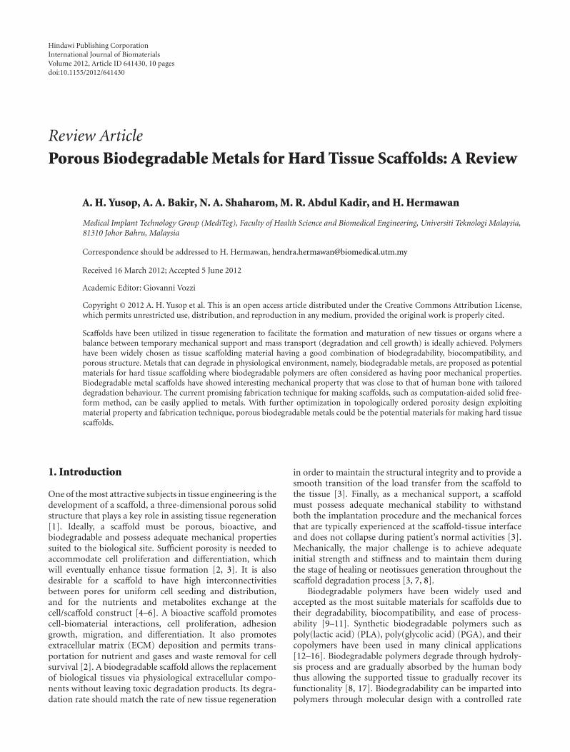
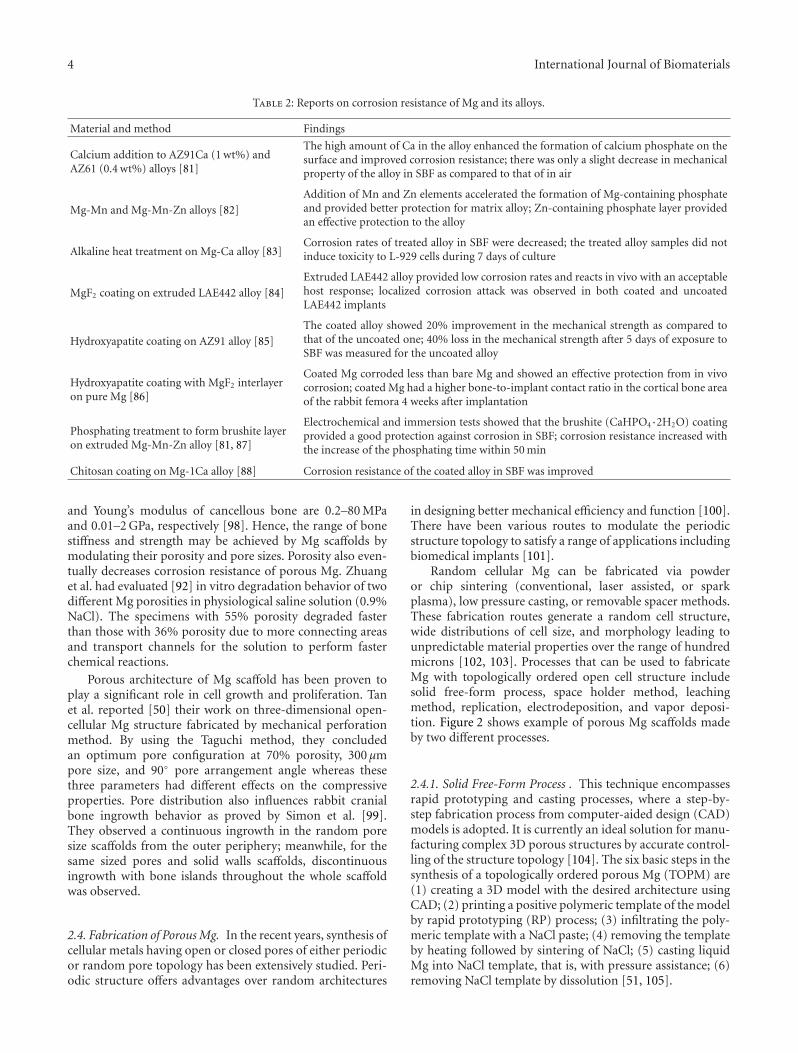
Figure 1: Mass loss and strength retention of some polymers used for scaffolds. Data compiled from [42–49].
in concert with tissue regeneration [18–21]. For instance,PLA could be combined with PGA to form poly(lactic-co-glycolic acid) (PLGA), which has degradation rate tailoredwith the tissue healing period and has been shown to supportosteoblast cells attachment and growth in vitro and in vivo[22–24]. Beside copolymerization, polymer composites havebeen explored in order to improve mechanical propertyand biocompatibility. Zhang and Ma have developed [25]a highly porous biodegradable polymer/apatite compositescaffold (95% porosity) through a thermally induced phaseseparation technique, which resulted in significant improve-ment in mechanical properties compared to polymer-onlyscaffold. The work by Ma et al. [26] has shown that osteoblastsurvival and growth were significantly enhanced in thePLLA/HA composite scaffolds compared to the plain PLLAscaffolds.
One of the major concerns regarding the use ofbiodegradable polymers as scaffold is their poor mechanicalproperties [27]. For hard tissue applications such as bone,a scaffold that possesses adequate strength and Young’smodulus is desirable. However, porous polymeric structuresare relatively weak and may not achieve sufficient level ofthe required strength [8, 27]. During degradation, polymerscould suddenly lose their mass and mechanical integrity.Figure 1 illustrates mass loss and strength retention as thefunction of degradation period for some biodegradablepolymers used for scaffold.
There is a recent and fast-growing interest in the useof biodegradable metals for biomedical applications [28].The inherent strength and ductility owned by metals arethe key features that make them appealing for hard tissueapplications. Magnesium- (Mg-) based and iron- (Fe-) basedmetals have been used, which include Mg-RE (rare earthelements) [29–33], Mg-Ca- [34, 35], pure Fe [36, 37], Fe-Mnalloys [38, 39], and Fe foam for bone replacement scaffold[40]. Mg and its alloys have been proposed for orthopaedic
implants due to their supportive physical properties tohuman bones. It has a density closer to that of naturalbones (1.8–2 g/cm3) and has been reported to support theactivation of bone cells [41]. Mg degrades in vivo throughelectrochemical process, which produces Mg hydroxideand hydrogen gas. Combining their excellent mechanicalproperties and degradability, Mg and its alloys are nowviewed as a potential alternative for making scaffold fortissue regeneration application. Therefore, this paper aimsto review the potentiality of porous biodegradable metalsas material for hard tissue scaffold. Elaborations to theirrationale, structure, mechanical properties, degradation, andfabrication method are presented.
2. Porous Mg as Scaffold Material
2.1. Rationale. Mg is largely found in bone tissue, it isan essential element to human body, and its presence isbeneficial to bone growth and strength [52–54]. It is acofactor for many enzymes and serves as stabilizer of DNAand RNA structures [55]. With approximately half of thetotal estimated 25 g content stored in bone tissue, Mg is thefourth most abundant cation in the human body [56, 57].In the extracellular fluid, the level of Mg ranges between 0.7and 1.05 mmol/L, and its homeostasis is maintained by theintestine and kidneys [52, 53]. The incidence of hyper-Mg israre due to the efficient excretion of the element in the urine[52, 56].
Mg can be considered as osteoconductive and bonegrowth stimulator material as suggested by many studies. Asignificant increase of bone area has been observed in Mg-based implants compared to those based on PLA [41, 58].The corrosion layer around Mg implants has been observedto contain calcium phosphates, which appeared to be indirect contact with the surrounding bone [41]. Xu et al.
International Journal of Biomaterials 3
have shown [59] new bone formation around Mg-Mn-Znimplants in their in vivo degradation in rats. Witte et al.observed [60] that 3 months postoperatively, open porousMg scaffolds implanted in rabbits were largely degraded,foreign body giant cells phagocytizing the remaining corro-sion products were rarely found, and no osteolytic changeswere found around the implant site. It has been shownthat porous Mg has better degradation behavior in termsof lower pH change, slower hydrogen evolution, and slowerdecrement of compressive yield strength in simulated bodyfluid (SBF) immersion tests [61]. Zreiqat et al. reported [62]an increasing bone cell adhesion on Mg-enriched aluminaas expressed by enhanced level of a5b1 integrin receptorand collagen extracellular matrix protein. Two studies usingMg-enriched apatites or collagen materials showed goodbiocompatibility on bone cell attachment and tissue growth[63, 64].
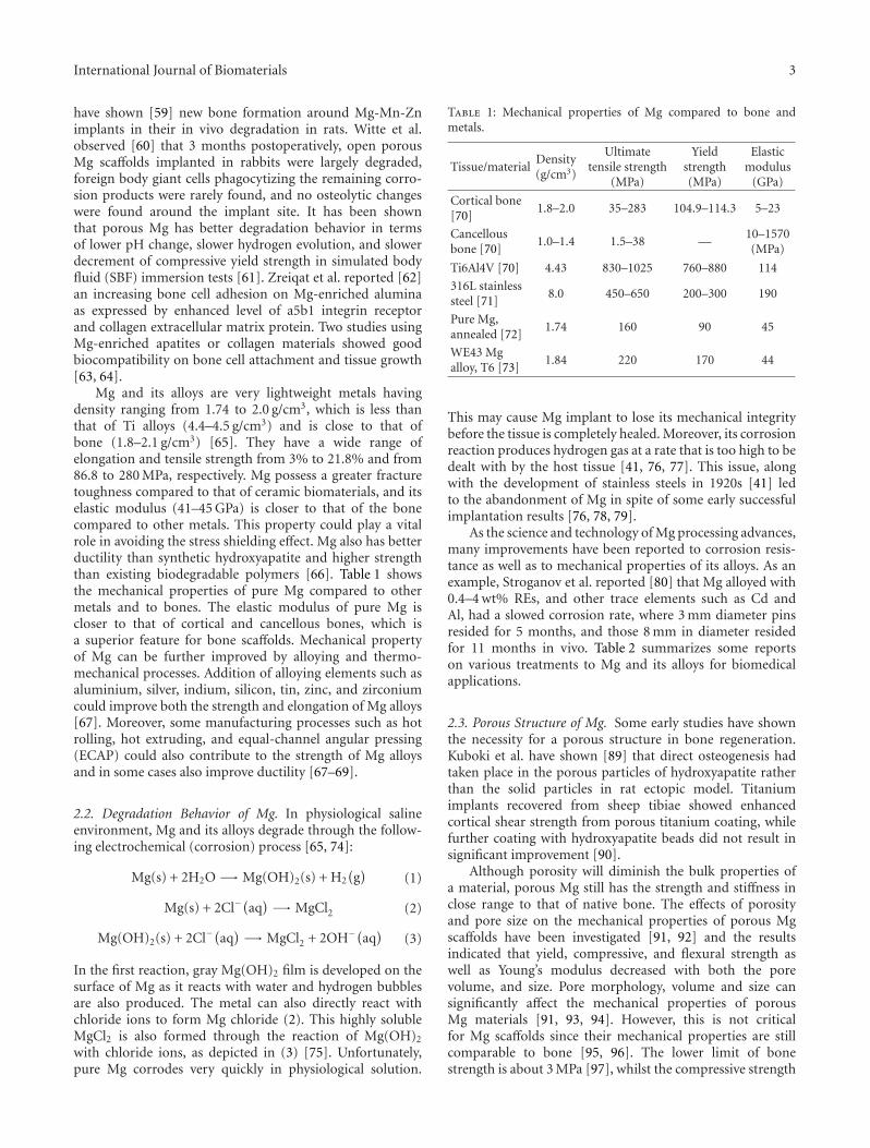
Mg and its alloys are very lightweight metals havingdensity ranging from 1.74 to 2.0 g/cm3, which is less thanthat of Ti alloys (4.4–4.5 g/cm3) and is close to that ofbone (1.8–2.1 g/cm3) [65]. They have a wide range ofelongation and tensile strength from 3% to 21.8% and from86.8 to 280 MPa, respectively. Mg possess a greater fracturetoughness compared to that of ceramic biomaterials, and itselastic modulus (41–45 GPa) is closer to that of the bonecompared to other metals. This property could play a vitalrole in avoiding the stress shielding effect. Mg also has betterductility than synthetic hydroxyapatite and higher strengththan existing biodegradable polymers [66]. Table 1 showsthe mechanical properties of pure Mg compared to othermetals and to bones. The elastic modulus of pure Mg iscloser to that of cortical and cancellous bones, which isa superior feature for bone scaffolds. Mechanical propertyof Mg can be further improved by alloying and thermo-mechanical processes. Addition of alloying elements such asaluminium, silver, indium, silicon, tin, zinc, and zirconiumcould improve both the strength and elongation of Mg alloys[67]. Moreover, some manufacturing processes such as hotrolling, hot extruding, and equal-channel angular pressing(ECAP) could also contribute to the strength of Mg alloysand in some cases also improve ductility [67–69].
2.2. Degradation Behavior of Mg. In physiological salineenvironment, Mg and its alloys degrade through the follow-ing electrochemical (corrosion) process [65, 74]:
Mg(s) + 2H2O −→ Mg(OH)2(s) + H2(g)
(1)
Mg(s) + 2Cl−(aq) −→ MgCl2 (2)
Mg(OH)2(s) + 2Cl−(aq) −→ MgCl2 + 2OH−(aq
)(3)
In the first reaction, gray Mg(OH)2 film is developed on thesurface of Mg as it reacts with water and hydrogen bubblesare also produced. The metal can also directly react withchloride ions to form Mg chloride (2). This highly solubleMgCl2 is also formed through the reaction of Mg(OH)2
with chloride ions, as depicted in (3) [75]. Unfortunately,pure Mg corrodes very quickly in physiological solution.
Table 1: Mechanical properties of Mg compared to bone andmetals.
Tissue/materialDensity(g/cm3)
Ultimatetensile strength
(MPa)
Yieldstrength(MPa)
Elasticmodulus
(GPa)
Cortical bone[70]
1.8–2.0 35–283 104.9–114.3 5–23
Cancellousbone [70]
1.0–1.4 1.5–38 —10–1570(MPa)
Ti6Al4V [70] 4.43 830–1025 760–880 114
316L stainlesssteel [71]
8.0 450–650 200–300 190
Pure Mg,annealed [72]
1.74 160 90 45
WE43 Mgalloy, T6 [73]
1.84 220 170 44
This may cause Mg implant to lose its mechanical integritybefore the tissue is completely healed. Moreover, its corrosionreaction produces hydrogen gas at a rate that is too high to bedealt with by the host tissue [41, 76, 77]. This issue, alongwith the development of stainless steels in 1920s [41] ledto the abandonment of Mg in spite of some early successfulimplantation results [76, 78, 79].
As the science and technology of Mg processing advances,many improvements have been reported to corrosion resis-tance as well as to mechanical properties of its alloys. As anexample, Stroganov et al. reported [80] that Mg alloyed with0.4–4 wt% REs, and other trace elements such as Cd andAl, had a slowed corrosion rate, where 3 mm diameter pinsresided for 5 months, and those 8 mm in diameter residedfor 11 months in vivo. Table 2 summarizes some reportson various treatments to Mg and its alloys for biomedicalapplications.
2.3. Porous Structure of Mg. Some early studies have shownthe necessity for a porous structure in bone regeneration.Kuboki et al. have shown [89] that direct osteogenesis hadtaken place in the porous particles of hydroxyapatite ratherthan the solid particles in rat ectopic model. Titaniumimplants recovered from sheep tibiae showed enhancedcortical shear strength from porous titanium coating, whilefurther coating with hydroxyapatite beads did not result insignificant improvement [90].
Although porosity will diminish the bulk properties ofa material, porous Mg still has the strength and stiffness inclose range to that of native bone. The effects of porosityand pore size on the mechanical properties of porous Mgscaffolds have been investigated [91, 92] and the resultsindicated that yield, compressive, and flexural strength aswell as Young’s modulus decreased with both the porevolume, and size. Pore morphology, volume and size cansignificantly affect the mechanical properties of porousMg materials [91, 93, 94]. However, this is not criticalfor Mg scaffolds since their mechanical properties are stillcomparable to bone [95, 96]. The lower limit of bonestrength is about 3 MPa [97], whilst the compressive strength
4 International Journal of Biomaterials
Table 2: Reports on corrosion resistance of Mg and its alloys.
Material and method Findings
Calcium addition to AZ91Ca (1 wt%) andAZ61 (0.4 wt%) alloys [81]
The high amount of Ca in the alloy enhanced the formation of calcium phosphate on thesurface and improved corrosion resistance; there was only a slight decrease in mechanicalproperty of the alloy in SBF as compared to that of in air
Mg-Mn and Mg-Mn-Zn alloys [82]
Addition of Mn and Zn elements accelerated the formation of Mg-containing phosphateand provided better protection for matrix alloy; Zn-containing phosphate layer providedan effective protection to the alloy
Alkaline heat treatment on Mg-Ca alloy [83]Corrosion rates of treated alloy in SBF were decreased; the treated alloy samples did notinduce toxicity to L-929 cells during 7 days of culture
MgF2 coating on extruded LAE442 alloy [84]
Extruded LAE442 alloy provided low corrosion rates and reacts in vivo with an acceptablehost response; localized corrosion attack was observed in both coated and uncoatedLAE442 implants
Hydroxyapatite coating on AZ91 alloy [85]
The coated alloy showed 20% improvement in the mechanical strength as compared tothat of the uncoated one; 40% loss in the mechanical strength after 5 days of exposure toSBF was measured for the uncoated alloy
Hydroxyapatite coating with MgF2 interlayeron pure Mg [86]
Coated Mg corroded less than bare Mg and showed an effective protection from in vivocorrosion; coated Mg had a higher bone-to-implant contact ratio in the cortical bone areaof the rabbit femora 4 weeks after implantation
Phosphating treatment to form brushite layeron extruded Mg-Mn-Zn alloy [81, 87]
Electrochemical and immersion tests showed that the brushite (CaHPO4·2H2O) coatingprovided a good protection against corrosion in SBF; corrosion resistance increased withthe increase of the phosphating time within 50 min
Chitosan coating on Mg-1Ca alloy [88] Corrosion resistance of the coated alloy in SBF was improved
and Young’s modulus of cancellous bone are 0.2–80 MPaand 0.01–2 GPa, respectively [98]. Hence, the range of bonestiffness and strength may be achieved by Mg scaffolds bymodulating their porosity and pore sizes. Porosity also even-tually decreases corrosion resistance of porous Mg. Zhuanget al. had evaluated [92] in vitro degradation behavior of twodifferent Mg porosities in physiological saline solution (0.9%NaCl). The specimens with 55% porosity degraded fasterthan those with 36% porosity due to more connecting areasand transport channels for the solution to perform fasterchemical reactions.
Porous architecture of Mg scaffold has been proven toplay a significant role in cell growth and proliferation. Tanet al. reported [50] their work on three-dimensional open-cellular Mg structure fabricated by mechanical perforationmethod. By using the Taguchi method, they concludedan optimum pore configuration at 70% porosity, 300 µmpore size, and 90◦ pore arrangement angle whereas thesethree parameters had different effects on the compressiveproperties. Pore distribution also influences rabbit cranialbone ingrowth behavior as proved by Simon et al. [99].They observed a continuous ingrowth in the random poresize scaffolds from the outer periphery; meanwhile, for thesame sized pores and solid walls scaffolds, discontinuousingrowth with bone islands throughout the whole scaffoldwas observed.
2.4. Fabrication of Porous Mg. In the recent years, synthesis ofcellular metals having open or closed pores of either periodicor random pore topology has been extensively studied. Peri-odic structure offers advantages over random architectures
in designing better mechanical efficiency and function [100].There have been various routes to modulate the periodicstructure topology to satisfy a range of applications includingbiomedical implants [101].
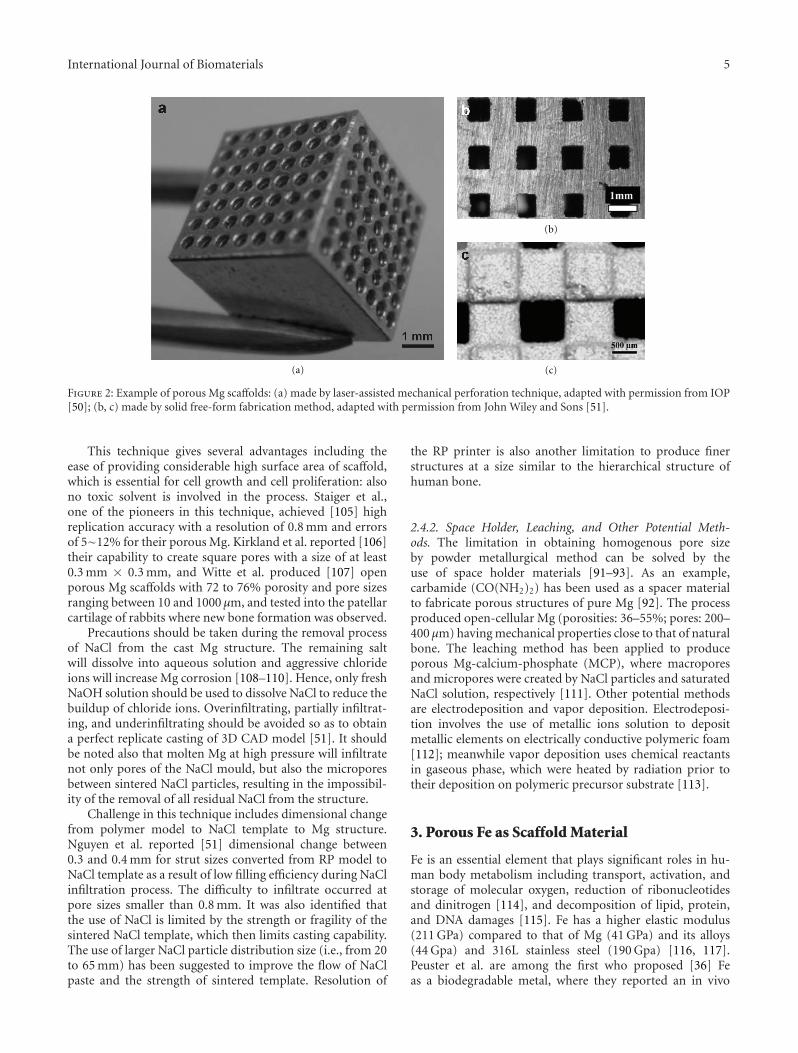
Random cellular Mg can be fabricated via powderor chip sintering (conventional, laser assisted, or sparkplasma), low pressure casting, or removable spacer methods.These fabrication routes generate a random cell structure,wide distributions of cell size, and morphology leading tounpredictable material properties over the range of hundredmicrons [102, 103]. Processes that can be used to fabricateMg with topologically ordered open cell structure includesolid free-form process, space holder method, leachingmethod, replication, electrodeposition, and vapor deposi-tion. Figure 2 shows example of porous Mg scaffolds madeby two different processes.
2.4.1. Solid Free-Form Process . This technique encompassesrapid prototyping and casting processes, where a step-by-step fabrication process from computer-aided design (CAD)models is adopted. It is currently an ideal solution for manu-facturing complex 3D porous structures by accurate control-ling of the structure topology [104]. The six basic steps in thesynthesis of a topologically ordered porous Mg (TOPM) are(1) creating a 3D model with the desired architecture usingCAD; (2) printing a positive polymeric template of the modelby rapid prototyping (RP) process; (3) infiltrating the poly-meric template with a NaCl paste; (4) removing the templateby heating followed by sintering of NaCl; (5) casting liquidMg into NaCl template, that is, with pressure assistance; (6)removing NaCl template by dissolution [51, 105].
International Journal of Biomaterials 5
(a)
(b)
(c)
Figure 2: Example of porous Mg scaffolds: (a) made by laser-assisted mechanical perforation technique, adapted with permission from IOP[50]; (b, c) made by solid free-form fabrication method, adapted with permission from John Wiley and Sons [51].
This technique gives several advantages including theease of providing considerable high surface area of scaffold,which is essential for cell growth and cell proliferation: alsono toxic solvent is involved in the process. Staiger et al.,one of the pioneers in this technique, achieved [105] highreplication accuracy with a resolution of 0.8 mm and errorsof 5∼12% for their porous Mg. Kirkland et al. reported [106]their capability to create square pores with a size of at least0.3 mm × 0.3 mm, and Witte et al. produced [107] openporous Mg scaffolds with 72 to 76% porosity and pore sizesranging between 10 and 1000 µm, and tested into the patellarcartilage of rabbits where new bone formation was observed.
Precautions should be taken during the removal processof NaCl from the cast Mg structure. The remaining saltwill dissolve into aqueous solution and aggressive chlorideions will increase Mg corrosion [108–110]. Hence, only freshNaOH solution should be used to dissolve NaCl to reduce thebuildup of chloride ions. Overinfiltrating, partially infiltrat-ing, and underinfiltrating should be avoided so as to obtaina perfect replicate casting of 3D CAD model [51]. It shouldbe noted also that molten Mg at high pressure will infiltratenot only pores of the NaCl mould, but also the microporesbetween sintered NaCl particles, resulting in the impossibil-ity of the removal of all residual NaCl from the structure.
Challenge in this technique includes dimensional changefrom polymer model to NaCl template to Mg structure.Nguyen et al. reported [51] dimensional change between0.3 and 0.4 mm for strut sizes converted from RP model toNaCl template as a result of low filling efficiency during NaClinfiltration process. The difficulty to infiltrate occurred atpore sizes smaller than 0.8 mm. It was also identified thatthe use of NaCl is limited by the strength or fragility of thesintered NaCl template, which then limits casting capability.The use of larger NaCl particle distribution size (i.e., from 20to 65 mm) has been suggested to improve the flow of NaClpaste and the strength of sintered template. Resolution of
the RP printer is also another limitation to produce finerstructures at a size similar to the hierarchical structure ofhuman bone.
2.4.2. Space Holder, Leaching, and Other Potential Meth-ods. The limitation in obtaining homogenous pore sizeby powder metallurgical method can be solved by theuse of space holder materials [91–93]. As an example,carbamide (CO(NH2)2) has been used as a spacer materialto fabricate porous structures of pure Mg [92]. The processproduced open-cellular Mg (porosities: 36–55%; pores: 200–400 µm) having mechanical properties close to that of naturalbone. The leaching method has been applied to produceporous Mg-calcium-phosphate (MCP), where macroporesand micropores were created by NaCl particles and saturatedNaCl solution, respectively [111]. Other potential methodsare electrodeposition and vapor deposition. Electrodeposi-tion involves the use of metallic ions solution to depositmetallic elements on electrically conductive polymeric foam[112]; meanwhile vapor deposition uses chemical reactantsin gaseous phase, which were heated by radiation prior totheir deposition on polymeric precursor substrate [113].
3. Porous Fe as Scaffold Material
Fe is an essential element that plays significant roles in hu-man body metabolism including transport, activation, andstorage of molecular oxygen, reduction of ribonucleotidesand dinitrogen [114], and decomposition of lipid, protein,and DNA damages [115]. Fe has a higher elastic modulus(211 GPa) compared to that of Mg (41 GPa) and its alloys(44 Gpa) and 316L stainless steel (190 Gpa) [116, 117].Peuster et al. are among the first who proposed [36] Feas a biodegradable metal, where they reported an in vivo
6 International Journal of Biomaterials
implantation test of Fe stents in the descending aorta of rab-bits. They showed evidence that pronounced inflammatoryresponse and systemic toxicity were not observed up to 18months of the study.
Currently, there is limited literature on Fe as a scaffoldmaterial. Very recently, Farack et al. have studied [40] Fefoam coated with calcium-phosphate for bone replacementscaffold where human mesenchymal stem cells prolifer-ated and differentiated more on coated Fe foams thanon uncoated ones. The coating gave enhanced bioactivityand inhibited degradation of Fe foams; however, the latestis actually questionable since Fe was generally viewed ashaving too slow degradation for implant applications [118].The open porous Fe and Fe-phosphorous alloys have beeninvestigated as biodegradable bone replacement [119], andthe results showed that addition of phosphorus increasedcompressive yield up to 11 MPa, higher than that of pure Feof 2.4 MPa, and resulted in a Young’s modulus of 2.3 GPawhich is comparable to that of typical bone. The alloysshowed also faster in vitro degradation than pure Fe but stillconsidered slow as large fraction of material was observedduring 12 months in vivo study [119]. Nevertheless, alloyingFe with phosphorous seems to be a promising way tooptimize both mechanical and degradation properties of Feespecially for bone scaffold.
Porous Fe has been fabricated via several methodsincluding solid-gas eutectic solidification process [120, 121],CO-CO2 gas foaming powder metallurgy process [122], orpowder metallurgy with the use of polymer foaming agent[119, 123], or even using wood as template [124]. However,those techniques hardly provide the topologically orderedporous as desired for bone scaffold. Moreover, owing to itsvery high melting temperature, the solid free-form methodas applied to Mg seems to be nonapplicable for Fe where theexcessive heat might destruct the NaCl template.
4. Perspective
Biodegradable metals as tissue scaffolding materials havebeen viewed as alternative to polymers for hard tissueregeneration exploiting mostly their superior mechanicalproperties over biodegradable polymers. Biodegradable met-als such as Mg and its alloys possess mechanical properties inclose range to those of native human bone and have shownencouraging results when used as tissue scaffolds. PorousFe could also be viewed as a potential scaffold material butavailable data is scarce especially in its relation to bone tissue.Among many promising techniques to fabricate metalscaffolds, solid free-form is currently viewed as the mostpotential method to fabricate biodegradable metal scaffoldshaving optimized pore morphology for cell growth andcell proliferation. This technique permitted the design andrealization of topologically ordered porous Mg with periodicstructure for enhanced mechanical efficiency and function ofa porous scaffold. Further investigations are needed in thesolid free-form fabrication method to develop a scaffold withproperties specifically tailored for cell regeneration and tissuegrowth.
Overall, application of biodegradable metals for tissueengineering scaffold is just in the beginning. Limited workhas been done and much has still is to be done. Thedirections could be in finding suitable process for mak-ing porous structure from all prospective biodegradablemetals, understanding the influence of porous structure tomechanical and degradation properties, and understandingthe cell regeneration and degradation product transport inthe porous structure. Integrating biodegradable polymers orceramics and drugs could be another interesting direction toexplore.
Acknowledgments
This work was supported by the Malaysian Ministry ofHigher Education (MOHE) and Universiti Teknologi Malay-sia (UTM) through research grants Q.J130000.7136.00H54(Tier-1) and J.130000.7836.4L019 (ERGS).
References
[1] D. Brahatheeswaran, Y. Yasuhiko, M. Toru, and K. D. Sakthi,“Polymeric scaffolds in tissue engineering application: areview,” International Journal of Polymer Science, vol. 2011,Article ID 290602, 19 pages, 2011.
[2] C. H. Chang, F. H. Lin, T. F. Kuo, and H. C. Liu, “Cartilagetissue engineering,” Biomedical Engineering: Applications,Basis and Communications, vol. 17, pp. 1–11, 2005.
[3] M. V. Risbud and M. Sittinger, “Tissue engineering: advancesin in vitro cartilage generation,” Trends in Biotechnology, vol.20, no. 8, pp. 351–356, 2002.
[4] M. Gravel, T. Gross, R. Vago, and M. Tabrizian, “Responsesof mesenchymal stem cell to chitosan-coralline compositesmicrostructured using coralline as gas forming agent,” Bio-materials, vol. 27, no. 9, pp. 1899–1906, 2006.
[5] D. W. Hutmacher, M. Sittinger, and M. V. Risbud, “Scaffold-based tissue engineering: rationale for computer-aideddesign and solid free-form fabrication systems,” Trends inBiotechnology, vol. 22, no. 7, pp. 354–362, 2004.
[6] D. W. Hutmacher, T. Schantz, I. Zein, K. W. Ng, S. H.Teoh, and K. C. Tan, “Mechanical properties and cellcultural response of polycaprolactone scaffolds designedand fabricated via fused deposition modeling,” Journal ofBiomedical Materials Research, vol. 55, pp. 203–216, 2001.
[7] J. Bonadio, E. Smiley, P. Patil, and S. Goldstein, “Localized,direct plasmid gene delivery in vivo: prolonged therapyresults in reproducible tissue regeneration,” Nature Medicine,vol. 5, no. 7, pp. 753–759, 1999.
[8] H. Y. Cheung, K. T. Lau, T. P. Lu, and D. Hui, “Acritical review on polymer-based bio-engineered materialsfor scaffold development,” Composites Part B, vol. 38, no. 3,pp. 291–300, 2007.
[9] G. Chen, T. Ushida, and T. Tateishi, “Scaffold design for tissueengineering,” Macromolecular Bioscience, vol. 2, pp. 67–77,2002.
[10] Y. Ji, K. Ghosh, X. Z. Shu et al., “Electrospun three-dimensional hyaluronic acid nanofibrous scaffolds,” Bioma-terials, vol. 27, no. 20, pp. 3782–3792, 2006.
[11] E. Piskin, “Biodegradable polymers as biomaterials,” Journalof Biomaterials Science Polymer Edition, vol. 6, pp. 75–870,1995.
International Journal of Biomaterials 7
[12] N. Ashammakhi and P. Rokkanen, “Absorbable polyglycolidedevices in trauma and bone surgery,” Biomaterials, vol. 18,no. 1, pp. 3–9, 1997.
[13] S. W. Shalaby, “Bioabsorbable polymers,” in Encyclopedia ofPharmceutical Technology, J. Swarbrick and J. C. Boylan, Eds.,vol. 1, pp. 465–476, 1988.
[14] S. J. Holland and B. J. Tighe, “Biodegradable polymers,”in Advances in Pharmaceutical Science, vol. 6, pp. 101–164,Academic Press, London, UK, 1992.
[15] T. Hayashi, “Biodegradable polymers for biomedical uses,”Progress in Polymer Science (Oxford), vol. 19, no. 4, pp. 663–702, 1994.
[16] J. Kohn and R. Langer, “Bioresorbable and bioerodiblematerials,” in An Introduction To Materials in Medicine, B. D.Ratner, A. S. Hoffman, F. J. Schoen, and J. E. Lemon, Eds., pp.65–73, Academic Press, San Diego, Calif, USA, 1997.
[17] J. S. Temenoff and A. G. Mikos, “Injectable biodegradablematerials for orthopedic tissue engineering,” Biomaterials,vol. 21, no. 23, pp. 2405–2412, 2000.
[18] K. A. Athanasiou, G. G. Niederauer, and C. M. Agrawal,“Sterilization, toxicity, biocompatibility and clinical appli-cations of polylactic acid/polyglycolic acid copolymers,”Biomaterials, vol. 17, no. 2, pp. 93–102, 1996.
[19] L. L. Hench and J. M. Polak, “Third-generation biomedicalmaterials,” Science, vol. 295, no. 5557, pp. 1014–1017, 2002.
[20] G. D. Prestwich and H. Matthew, “Hybrid, composite, andcomplex biomaterials,” Annals of the New York Academy ofSciences, vol. 961, pp. 106–108, 2002.
[21] X. Liu and P. X. Ma, “Polymeric scaffolds for bone tissueengineering,” Annals of Biomedical Engineering, vol. 32, no.3, pp. 477–486, 2004.
[22] S. L. Ishaug, G. M. Crane, M. J. Miller, A. W. Yasko, M.J. Yaszemski, and A. G. Mikos, “Bone formation by three-dimensional stromal osteoblast culture in biodegradablepolymer scaffolds,” Journal of Biomedial Materials Research,vol. 36, pp. 17–28, 1997.
[23] S. L. Ishaug-Riley, G. M. Crane, A. Gurlek et al., “Ectopicbone formation by marrow stromal osteoblast transplanta-tion using poly(DL-lactic-co-glycolic acid) foams implantedinto the rat mesentery,” Journal of Biomedial MaterialsResearch, vol. 36, pp. 1–8, 1997.
[24] C. A. Vacanti and J. P. Vacanti, “Bone and cartilage recon-struction with tissue engineering approaches,” Otolaryngo-logic Clinics of North America, vol. 27, no. 1, pp. 263–276,1994.
[25] R. Y. Zhang and P. X. Ma, “Poly(alpha-hydroxyl acids)hydroxyapatite porous composites for bone-tissue engineer-ing. I. Preparation and morphology,” Journal of BiomedialMaterials Research, vol. 44, pp. 446–455, 1999.
[26] P. X. Ma, R. Y. Zhang, G. Z. Xiao, and R. Franceschi,“Engineering new bone tissue in vitro on highly porouspoly(alpha-hydroxyl acids)/hydroxyapatite composite scaf-folds,” Journal of Biomedial Materials Research, vol. 54, pp.284–293, 2001.
[27] P. K. D. V. Yarlagadda, M. Chandrasekharan, and J. Y. M.Shyan, “Recent advances and current developments in tissuescaffolding,” Bio-Medical Materials and Engineering, vol. 15,no. 3, pp. 159–177, 2005.
[28] H. Hermawan and D. Mantovani, “Degradable metallicbiomaterials: the concept, current developments and futuredirections,” Minerva Biotecnologica, vol. 21, no. 4, pp. 207–216, 2009.
[29] C. Di Mario, H. Griffiths, O. Goktekin et al., “Drug-elutingbioabsorbable magnesium stent,” Journal of InterventionalCardiology, vol. 17, no. 6, pp. 391–395, 2004.
[30] P. Peeters, M. Bosiers, J. Verbist, K. Deloose, and B. Heublein,“Preliminary results after application of absorbable metalstents in patients with critical limb ischemia,” Journal ofEndovascular Therapy, vol. 12, no. 1, pp. 1–5, 2005.
[31] F. Witte, V. Kaese, H. Haferkamp et al., “In vivo corrosionof four magnesium alloys and the associated bone response,”Biomaterials, vol. 26, no. 17, pp. 3557–3563, 2005.
[32] R. Waksman, R. Pakala, P. K. Kuchulakanti et al., “Safety andefficacy of bioabsorbable magnesium alloy stents in porcinecoronary arteries,” Catheterization and Cardiovascular Inter-ventions, vol. 68, no. 4, pp. 607–617, 2006.
[33] A. C. Hanzi, A. S. Sologubenko, and P. J. Uggowitzer, “Designstrategy for microalloyed ultra-ductile magnesium alloys formedical applications,” Materials Science Forum, vol. 618-619,pp. 75–82, 2009.
[34] E. Zhang and L. Yang, “Microstructure, mechanical proper-ties and bio-corrosion properties of Mg–Zn–Mn–Ca alloy forbiomedical application,” Materials Science and Engineering A,vol. 497, no. 1-2, pp. 111–118, 2008.
[35] Z. Li, X. Gu, S. Lou, and Y. Zheng, “The developmentof binary Mg–Ca alloys for use as biodegradable materialswithin bone,” Biomaterials, vol. 29, no. 10, pp. 1329–1344,2008.
[36] M. Peuster, P. Wohlsein, M. Brugmann et al., “A novelapproach to temporary stenting: degradable cardiovascularstents produced from corrodible metal—results 6–18 monthsafter implantation into New Zealand white rabbits,” Heart,vol. 86, no. 5, pp. 563–569, 2001.
[37] M. Peuster, C. Hesse, T. Schloo, C. Fink, P. Beerbaum, andC. von Schnakenburg, “Long-term biocompatibility of acorrodible peripheral iron stent in the porcine descendingaorta,” Biomaterials, vol. 27, no. 28, pp. 4955–4962, 2006.
[38] H. Hermawan, H. Alamdari, D. Mantovani, and D. Dube,“Iron-manganese: new class of metallic degradable biomate-rials prepared by powder metallurgy,” Powder Metallurgy, vol.51, no. 1, pp. 38–45, 2008.
[39] M. Schinhammer, A. C. Hanzi, J. F. Loffler, and P. J.Uggowitzer, “Design strategy for biodegradable Fe-basedalloys for medical applications,” Acta Biomaterialia, vol. 6, no.5, pp. 1705–1713, 2010.
[40] J. Farack, C. Wolf-Brandstetter, S. Glorius et al., “The effectof perfusion culture on proliferation and differentiationof human mesenchymal stem cells on biocorrodible bonereplacement material,” Materials Science and Engineering B,vol. 176, pp. 1767–1772, 2011.
[41] F. Witte, V. Kaese, H. Haferkamp et al., “In vivo corrosionof four magnesium alloys and the associated bone response,”Biomaterials, vol. 26, no. 17, pp. 3557–3563, 2005.
[42] R. M. Ginde and R. K. Gupta, “In vitro chemical degradationof poly(glycolic acid) pellets and fibers,” Journal of AppliedPolymer Science, vol. 33, no. 7, pp. 2411–2429, 1987.
[43] Y. Gong, Q. Zhou, C. Gao, and J. Shen, “In vitro and in vivodegradability and cytocompatibility of poly(l-lactic acid)scaffold fabricated by a gelatin particle leaching method,”Acta Biomaterialia, vol. 3, no. 4, pp. 531–540, 2007.
[44] A. M. Reed and D. K. Gilding, “Biodegradable polymers foruse in surgery—poly(glycolic)/poly(Iactic acid) homo andcopolymers: 2. In vitro degradation,” Polymer, vol. 22, no. 4,pp. 494–498, 1981.
8 International Journal of Biomaterials
[45] J. J. Yoon and T. G. Park, “Degradation behaviors ofbiodegradable macroporous scaffolds prepared by gas foam-ing of effervescent salts,” Journal of Biomedial MaterialsResearch., vol. 55, pp. 401–408, 2001.
[46] T. Yoshioka, F. Kamada, N. Kawazoe, T. Tateishi, and G.Chen, “Structural changes and biodegradation of PLLA,PCL, and PLGA sponges during in vitro incubation,” PolymerEngineering and Science, vol. 50, no. 10, pp. 1895–1903, 2010.
[47] A. W. T. Shum and A. F. T. Mak, “Morphological and biome-chanical characterization of poly(glycolic acid) scaffolds afterin vitro degradation,” Polymer Degradation and Stability, vol.81, no. 1, pp. 141–149, 2003.
[48] L. Wu and J. Ding, “In vitro degradation of three-dimensional porous poly(D,L-lactide-co- glycolide) scaffoldsfor tissue engineering,” Biomaterials, vol. 25, no. 27, pp.5821–5830, 2004.
[49] L. Xu, Z. C. Xiong, D. Yang, L. F. Zhang, J. Chang, andC. D. Xiong, “Preparation and in vitro degradation ofnovel bioactive polylactide/wollastonite scaffolds,” Journal ofApplied Polymer Science, vol. 114, no. 6, pp. 3396–3406, 2009.
[50] L. Tan, M. Gong, F. Zheng, B. Zhang, and K. Yang, “Studyon compression behavior of porous magnesium used as bonetissue engineering scaffolds,” Biomedical Materials, vol. 4, no.1, Article ID 015016, 2009.
[51] T. L. Nguyen, M. P. Staiger, G. J. Dia, and s T. B.F. Woodfield, “A novel manufacturing route for fabrica-tion of topologically-ordered porous magnesium scaffolds,”Advanced Engineering Materials, vol. 13, pp. 872–881, 2011.
[52] N. E. L. Saris, E. Mervaala, H. Karppanen, J. A. Khawaja,and A. Lewenstam, “Magnesium: an update on physiological,clinical and analytical aspects,” Clinica Chimica Acta, vol. 294,no. 1-2, pp. 1–26, 2000.
[53] J. Vormann, “Magnesium: nutrition and metabolism,” Molec-ular Aspects of Medicine, vol. 24, no. 1–3, pp. 27–37, 2003.
[54] T. Okuma, “Magnesium and bone strength,” Nutrition, vol.17, no. 7-8, pp. 679–680, 2001.
[55] A. Hartwig, “Role of magnesium in genomic stability,”Mutation Research, vol. 475, pp. 113–134, 2001.
[56] F. I. Wolf and A. Cittadini, “Chemistry and biochemistry ofmagnesium,” Molecular Aspects of Medicine, vol. 24, no. 1–3,pp. 3–9, 2003.
[57] Merk, “Water, electrolyte mineral and acid/base metabolism,”in Section 2. Endocrine & Metabolic Disorders. Merk Manualof Diagnosis and Therapy, chapter 12, 2012, http://www.newtreatments.org/loadlocal.php?hid=998.
[58] F. Witte, H. A. Crostack, J. Nellesen, and F. Beckmann,“Characterization of degradable magnesium alloys asorthopaedic implant material by synchrotron-radiation-based microtomography,” 2012, http://www-hasylab.desy.de/science/annual reports/2001 report/part1/contrib/47/5461.pdf.
[59] L. Xu, G. Yu, E. Zhang, F. Pan, and K. Yang, “In vivo corrosionbehavior of Mg–Mn–Zn alloy for bone implant application,”Journal of Biomedical Materials Research—Part A, vol. 83, no.3, pp. 703–711, 2007.
[60] F. Witte, H. Ulrich, M. Rudert, and E. Willbold, “Biodegrad-able magnesium scaffolds: part I: appropriate inflammatoryresponse,” Journal of Biomedical Materials Research—Part A,vol. 81, no. 3, pp. 748–756, 2007.
[61] X. N. Gu, W. R. Zhou, Y. F. Zheng, Y. Liu, and Y. X. Li,“Degradation and cytotoxicity of lotus-type porous puremagnesium as potential tissue engineering scaffold material,”Materials Letters, vol. 64, no. 17, pp. 1871–1874, 2010.
[62] H. Zreiqat, C. R. Howlett, A. Zannettino et al., “Mechanismsof magnesium-stimulated adhesion of osteoblastic cells tocommonly used orthopaedic implants,” Journal of BiomedicalMaterials Research, vol. 62, no. 2, pp. 175–184, 2002.
[63] Y. Yamasaki, Y. Yoshida, M. Okazaki et al., “Synthesis offunctionally graded MgCO3 apatite accelerating osteoblastadhesion,” Journal of Biomedical Materials Research, vol. 62,no. 1, pp. 99–105, 2002.
[64] Y. Yamasaki, Y. Yoshida, M. Okazaki et al., “Action ofFGMgCO3Ap-collagen composite in promoting bone forma-tion,” Biomaterials, vol. 24, no. 27, pp. 4913–4920, 2003.
[65] M. P. Staiger, A. M. Pietak, J. Huadmai, and G. Dias,“Magnesium and its alloys as orthopedic biomaterials: areview,” Biomaterials, vol. 27, no. 9, pp. 1728–1734, 2006.
[66] X. N. Gu and Y. F. Zheng, “A review on magnesium alloysas biodegradable materials,” Frontiers of Materials Science inChina, vol. 4, no. 2, pp. 111–115, 2010.
[67] X. Gu, Y. Zheng, Y. Cheng, S. Zhong, and T. Xi, “In vitrocorrosion and biocompatibility of binary magnesium alloys,”Biomaterials, vol. 30, no. 4, pp. 484–498, 2009.
[68] H. Wang, Y. Estrin, and Z. Zuberova, “Bio-corrosion ofa magnesium alloy with different processing histories,”Materials Letters, vol. 62, no. 16, pp. 2476–2479, 2008.
[69] Z. Li, X. Gu, S. Lou, and Y. Zheng, “The developmentof binary Mg–Ca alloys for use as biodegradable materialswithin bone,” Biomaterials, vol. 29, no. 10, pp. 1329–1344,2008.
[70] J. Black and G. Hastings, Handbook of Biomaterial Properties,Chapman & Hall, London, UK, 1998.
[71] ASTM, “Standard specification for wrought 18chromium-14nickel-2.5molybdenum stainless steel bar and wire forsurgical implants (UNS S31673),” Tech. Rep. F138, ASTMInternational, West Conshohocken, Pa, USA, 2003.
[72] ASM, ASM Handbook: Properties and Selection: NonferrousAlloys & Special Purpose Materials, Vol. 2, ASM International,Materials Park, Ohio, USA, 2005.
[73] ASTM, “Standard specification for magnesium-alloy sandcastings,” Tech. Rep. B80, ASTM International, West Con-shohocken, Pa, USA, 2001.
[74] F. Witte, N. Hort, C. Vogt et al., “Degradable biomaterialsbased on magnesium corrosion,” Current Opinion in SolidState and Materials Science, vol. 12, no. 5-6, pp. 63–72, 2008.
[75] B. A. Shaw, ASM Handbook Volume 13A: Corrosion: Funda-mentals, Testing and Protection, Edited by D. Stephen, ASMInternational, Materials Park, Ohio, USA, 2003.
[76] A. L. Lambotte, “Utilisation du magnesium comme materielperdu dans l’osteosynthese,” Bulletins et Memoires de laSociete de Chirurgie de Paris, vol. 28, pp. 1325–1359, 1932.
[77] V. V. Troitskii and D. N. Tsitrin, “The resorbing metallicalloy ‘Osteosinthezit’as material for fastening broken bone,”Khirurgiia, vol. 8, pp. 41–45, 1944.
[78] E. D. McBride, “Absorbable metal for bone surgery,” TheJournal of the American Medical Association, vol. 111, pp.2464–2467, 1938.
[79] J. Verbrugge, “Le Materiel Metallique Resorbable EnChirurgie Osseuse,” La Presse Medicale, vol. 23, pp. 460–465,1934.
[80] G. B. Stroganov, E. Savitsky, T. Mikhailovich, M. Nina, V.Terekhova, and V. Fedorovna, “Magnesium-base alloys foruse in bone surgery,” US Patent no. 3, 687, 135, 1972.
[81] M. B. Kannan and R. K. S. Raman, “In vitro degradationand mechanical integrity of calcium-containing magnesiumalloys in modified-simulated body fluid,” Biomaterials, vol.29, no. 15, pp. 2306–2314, 2008.
International Journal of Biomaterials 9
[82] L. Xu, E. Zhang, D. Yin, S. Zeng, and K. Yang, “In vitrocorrosion behaviour of Mg alloys in a phosphate bufferedsolution for bone implant application,” Journal of MaterialsScience, vol. 19, no. 3, pp. 1017–1025, 2008.
[83] X. N. Gu, W. Zheng, Y. Cheng, and Y. F. Zheng, “A studyon alkaline heat treated Mg–Ca alloy for the control of thebiocorrosion rate,” Acta Biomaterialia, vol. 5, no. 7, pp. 2790–2799, 2009.
[84] F. Witte, J. Fischer, J. Nellesen et al., “In vivo corrosionand corrosion protection of magnesium alloy LAE442,” ActaBiomaterialia, vol. 6, no. 5, pp. 1792–1799, 2010.
[85] M. B. Kannan and L. Orr, “In vitro mechanical integrityof hydroxyapatite coated magnesium alloy,” BiomedicalMaterials, vol. 6, Article ID 045003, 2011.
[86] J. H. Jo, B.G. Kang, K.S. Shin et al., “Hydroxyapatite coatingon magnesium with MgF2 interlayer for enhanced corrosionresistance and biocompatibility,” Journal of Materials Science,vol. 22, no. 11, pp. 2437–2447, 2011.
[87] L. Xu, E. Zhang, and K. Yang, “Phosphating treatment andcorrosion properties of Mg–Mn–Zn alloy for biomedicalapplication,” Journal of Materials Science, vol. 20, no. 4, pp.859–867, 2009.
[88] X. N. Gu, Y. F. Zheng, Q. X. Lan et al., “Surface modificationof an Mg–1Ca alloy to slow down its biocorrosion bychitosan,” Biomedical Materials, vol. 4, no. 4, Article ID044109, 2009.
[89] Y. Kuboki, H. Takita, D. Kobayashi, E. Tsuruga, M. Inoue,and M. Murata, “BMP-induced osteogenesis on the surfaceof hydroxyapatite with geometrically feasible and nonfeasiblestructures: topology of osteogenesis,” Journal of BiomedialMaterials Research, vol. 39, pp. 190–199, 1998.
[90] M. Svehla, P. Morberg, B. Zicat, W. Bruce, D. Sonnabend,and W. R. Walsh, “Morphometric and mechanical evaluationof titanium implant integration: comparison of five surfacestructures,” Journal of Biomedial Materials Research, pp. 15–22, 2000.
[91] C. E. Wen, Y. Yamada, K. Shimojima, Y. Chino, H. Hosokawa,and M. Mabuchi, “Compressibility of porous magnesiumfoam: dependency on porosity and pore size,” MaterialsLetters, vol. 58, no. 3-4, pp. 357–360, 2004.
[92] H. Zhuang, Y. Han, and A. Feng, “Preparation, mechanicalproperties and in vitro biodegradation of porous magnesiumscaffolds,” Materials Science and Engineering C, vol. 28, no. 8,pp. 1462–1466, 2008.
[93] C. E. Wen, M. Mabuchi, Y. Yamada, K. Shimojima, Y. Chino,and T. Asahina, “Processing of biocompatible porous Ti andMg,” Scripta Materialia, vol. 45, no. 10, pp. 1147–1153, 2001.
[94] Y. Yamada, K. Shimojima, Y. Sakaguchi et al., “Processingof cellular magnesium materials,” Advanced EngineeringMaterials, vol. 2, no. 4, pp. 184–187, 2000.
[95] P. E. DeGarmo, Materials and Processes in Manufacturing,Collin Macmillan, New York, NY, USA, 5th edition, 1979.
[96] L. Gibson and M. Ashby, Cellular Solids: Structure andProperties, Pergamon Press, Sydney, Australia, 1988.
[97] L. Gibson and M. Ashby, Solids: Structure and Properties,Pergamon Press, Sydney, Australia, 1988.
[98] L. J. Gibson, “The mechanical behaviour of cancellous bone,”Journal of Biomechanics, vol. 18, no. 5, pp. 317–328, 1985.
[99] J. L. Simon, T. D. Roy, J. R. Parsons et al., “Engineered cellularresponse to scaffold architecture in a rabbit trephine defect,”Journal of Biomedical Materials Research—Part A, vol. 66, no.2, pp. 275–282, 2003.
[100] V. S. Deshpande, M. F. Ashby, and N. A. Fleck, “Foam topol-ogy: bending versus stretching dominated architectures,”Acta Materialia, vol. 49, no. 6, pp. 1035–1040, 2001.
[101] L. P. Lefebvre, J. Banhart, and D. C. Dunand, “Porous metalsand metallic foams: current status and recent developments,”Advanced Engineering Materials, vol. 10, no. 9, pp. 775–787,2008.
[102] T. Adachi, Y. Osako, M. Tanaka, M. Hojo, and S. J.Hollister, “Framework for optimal design of porous scaffoldmicrostructure by computational simulation of bone regen-eration,” Biomaterials, vol. 27, no. 21, pp. 3964–3972, 2006.
[103] C. Y. Lin, C. C. Hsiao, P. Q. Chen, and S. J. Hollister,“Interbody fusion cage design using integrated global layoutand local microstructure topology optimization,” Spine, vol.29, no. 16, pp. 1747–1754, 2004.
[104] S. J. Hollister, “Scaffold design and manufacturing: fromconcept to clinic,” Advanced Materials, vol. 21, no. 32-33, pp.3330–3342, 2009.
[105] M. P. Staiger, I. Kolbeinsson, N. T. Kirkland, T. Nguyen, G.Dias, and T. B. F. Woodfield, “Synthesis of topologically-ordered open-cell porous magnesium,” Materials Letters, vol.64, no. 23, pp. 2572–2574, 2010.
[106] N. T. Kirkland, I. Kolbeinsson, T. Woodfield, G. J. Dias,and M. P. Staiger, “Synthesis and properties of topologicallyordered porous magnesium,” Materials Science and Engineer-ing B, vol. 176, no. 20, pp. 1666–1672, 2011.
[107] F. Witte, J. Reifenrath, P. P. Muller et al., “Cartilage repairon magnesium scaffolds used as a subchondral bone replace-ment,” Materialwissenschaft und Werkstofftechnik, vol. 37, no.6, pp. 504–508, 2006.
[108] R. Tunold, H. Holtan, M. B. H. Berge, A. Lasson, and R.Steen-Hansen, “The corrosion of magnesium in aqueoussolution containing chloride ions,” Corrosion Science, vol. 17,no. 4, pp. 353–365, 1977.
[109] J. Swiatowska, P. Volovitch, and K. Ogle, “The anodicdissolution of Mg in NaCl and Na2SO4 electrolytes by atomicemission spectroelectrochemistry,” Corrosion Science, vol. 52,no. 7, pp. 2372–2378, 2010.
[110] W. D. Muller, M. L. Nascimento, M. Zeddies, M. Corsico,L. M. Gassa, and M. A. F. L. de Mele, “Magnesium andits alloys as degradable biomaterials. Corrosion studiesusing potentiodynamic and EIS electrochemical techniques,”Materials Research, vol. 10, no. 1, pp. 5–10, 2007.
[111] J. Wei, J. Jia, F. Wu et al., “Hierarchically microp-orous/macroporous scaffold of magnesium-calcium phos-phate for bone tissue regeneration,” Biomaterials, vol. 31, no.6, pp. 1260–1269, 2010.
[112] J. Banhart, “Manufacture, characterisation and applicationof cellular metals and metal foams,” Progress in MaterialsScience, vol. 46, no. 6, pp. 559–632, 2001.
[113] G. Ryan, A. Pandit, and D. P. Apatsidis, “Fabrication methodsof porous metals for use in orthopaedic applications,”Biomaterials, vol. 27, no. 13, pp. 2651–2670, 2006.
[114] M. Fontecave and J. L. Pierre, “Iron: metabolism, toxicity andtherapy,” Biochimie, vol. 75, no. 9, pp. 767–773, 1993.
[115] P. P. Mueller, T. May, A. Perz, H. Hauser, and M. Peuster,“Control of smooth muscle cell proliferation by ferrous iron,”Biomaterials, vol. 27, no. 10, pp. 2193–2200, 2006.
[116] G. Song, “Control of biodegradation of biocompatablemagnesium alloys,” Corrosion Science, vol. 49, no. 4, pp.1696–1701, 2007.
[117] G. Sangiorgi, G. Melzi, P. Agostoni et al., “Engineeringaspects of stents design and their translation into clinical
10 International Journal of Biomaterials
practice,” Annali dell’Istituto Superiore di Sanita, vol. 43, no.1, pp. 89–100, 2007.
[118] H. Hermawan, D. Dube, and D. Mantovani, “Degradablemetallic biomaterials: design and development of Fe–Mnalloys for stents,” Journal of Biomedical Materials Research—Part A, vol. 93, no. 1, pp. 1–11, 2010.
[119] P. Quadbeck, R. Hauser, K. Kummel et al., “Iron basedcellular metals for degradable synthetic bone replacement,”in Proceedings of the Powder Metallurgy World Congress (PM’10), Florence, Italy, 2010.
[120] J. Kovacik, “The tensile behaviour of porous metals made byGASAR process,” Acta Materialia, vol. 46, no. 15, pp. 5413–5422, 1998.
[121] S. K. Hyun, T. Ikeda, and H. Nakajima, “Fabrication of lotus-type porous iron and its mechanical properties,” Science andTechnology of Advanced Materials, vol. 5, no. 1-2, pp. 201–205, 2004.
[122] T. Murakami, K. Ohara, T. Narushima, and C. Ouchi,“Development of a new method for manufacturing ironfoam using gases generated by reduction of iron oxide,”Materials Transactions, vol. 48, no. 11, pp. 2937–2944, 2007.
[123] C. S. Y. Jee, N. Ozguven, Z. X. Guo, and J. R. G. Evans,“Preparation of high porosity metal foams,” Metallurgicaland Materials Transactions B, vol. 31, no. 6, pp. 1345–1352,2000.
[124] Z. Liu, T. Fan, W. Zhang, and D. Zhang, “The synthesisof hierarchical porous iron oxide with wood templates,”Microporous and Mesoporous Materials, vol. 85, no. 1-2, pp.85–88, 2005.

Submit your manuscripts athttp://www.hindawi.com
ScientificaHindawi Publishing Corporationhttp://www.hindawi.com Volume 2014
CorrosionInternational Journal of
Hindawi Publishing Corporationhttp://www.hindawi.com Volume 2014
Polymer ScienceInternational Journal of
Hindawi Publishing Corporationhttp://www.hindawi.com Volume 2014
Hindawi Publishing Corporationhttp://www.hindawi.com Volume 2014
CeramicsJournal of
Hindawi Publishing Corporationhttp://www.hindawi.com Volume 2014
CompositesJournal of
NanoparticlesJournal of
Hindawi Publishing Corporationhttp://www.hindawi.com Volume 2014
Hindawi Publishing Corporationhttp://www.hindawi.com Volume 2014
International Journal of
Biomaterials
Hindawi Publishing Corporationhttp://www.hindawi.com Volume 2014
NanoscienceJournal of
TextilesHindawi Publishing Corporation http://www.hindawi.com Volume 2014
Journal of
NanotechnologyHindawi Publishing Corporationhttp://www.hindawi.com Volume 2014
Journal of
CrystallographyJournal of
Hindawi Publishing Corporationhttp://www.hindawi.com Volume 2014
The Scientific World JournalHindawi Publishing Corporation http://www.hindawi.com Volume 2014
Hindawi Publishing Corporationhttp://www.hindawi.com Volume 2014
CoatingsJournal of
Advances in
Materials Science and EngineeringHindawi Publishing Corporationhttp://www.hindawi.com Volume 2014
Smart Materials Research
Hindawi Publishing Corporationhttp://www.hindawi.com Volume 2014
Hindawi Publishing Corporationhttp://www.hindawi.com Volume 2014
MetallurgyJournal of
Hindawi Publishing Corporationhttp://www.hindawi.com Volume 2014
BioMed Research International
MaterialsJournal of
Hindawi Publishing Corporationhttp://www.hindawi.com Volume 2014
Nano
materials
Hindawi Publishing Corporationhttp://www.hindawi.com Volume 2014
Journal ofNanomaterials


![Excretion [2015]](https://img.pdfslide.us/doc/110x75/55d39c87bb61eb05278b46dd/excretion-2015-55d47f0693bf7.jpg)