Embed Size (px)
Citation preview
Review ArticleCSF Biomarkers of Alzheimer’s Disease: Impact on DiseaseConcept, Diagnosis, and Clinical Trial Design
Anne M. Fagan
Department of Neurology, Knight Alzheimer’s Disease Research Center, Hope Center for Neurological Disorders,Washington University School of Medicine, St. Louis, MO 63110, USA
Correspondence should be addressed to Anne M. Fagan; [email protected]
Received 13 April 2014; Revised 30 June 2014; Accepted 1 July 2014; Published 14 August 2014
Academic Editor: Stephen D. Ginsberg
Copyright © 2014 Anne M. Fagan. This is an open access article distributed under the Creative Commons Attribution License,which permits unrestricted use, distribution, and reproduction in any medium, provided the original work is properly cited.
Data from clinicopathologic and biomarker studies have converged to support the view of Alzheimer’s disease (AD) as a continuum,with pathology developing decades prior to the onset of cognitive symptoms which culminate as dementia at the end stage of thedisease.This concept is impacting disease nomenclature, diagnostic criteria, prognostic potential, and clinical trial design. Revisionsto diagnostic criteria to incorporate biomarker results have recently been proposed in order to increase the confidence of ADas the underlying etiology of a clinical impairment and to permit a diagnosis of AD across the disease continuum, eventuallyperhaps in the asymptomatic period. Individuals in this preclinical stage are receiving intense focus as a targeted populationfor secondary prevention trials aimed at identifying disease-modifying therapies that have the best chance of preserving normalcognitive function.The goal is to bring validated biomarkers to clinical practice for the purpose of disease diagnosis, prognosis, andevaluation of therapeutic efficacy once disease-modifying treatments become available. Realization of this goal requires worldwidebiomarker standardization efforts, consensus among researchers and clinicians regarding the clinical utility of assessing biomarkersin patient care settings, and eventually the endorsement and adoption of such procedures and practices into global health caresystems.
1. The Crisis of AD
Alzheimer’s disease (AD) is the most common cause ofdementia, accounting for up to 70% of all dementia cases, andis now estimated to be the third leading cause of death,after heart disease and cancer [1]. Since advanced age is thestrongest risk factor for AD, increased life expectancy and theaging of the “baby boomer” generation are leading to dra-matic increases in AD incidence. AD currently affects 5.2million people in the United States (US), with projectedestimates reaching 13.8 million (115 million world-wide) by the year 2050 (http://www.alz.org/documents/national/world alzheimer report 2010.pdf). The lifetime riskfor AD dementia for a 65-year-old person is currentlyestimated to be ∼10.5%, with prevalence doubling every 5years after age 65, reaching nearly 50% by age 80. In the USalone, the costs for care associatedwithAD in 2013 weremorethan $200 billion, with projected annual costs surpassing $1trillion by the year 2050 (http://www.alz.org/alzheimers dis-
ease facts and figures.asp#cost). At present there are noeffective treatments that will prevent the disease, haltits progression, or delay its onset. Development of anintervention that delays the onset of AD dementia by 5 yearsis estimated to result in a 57% reduction in the number ofaffected patients and an almost 50% reduction in projectedannual Medicare costs (from $627 to $344 billion).
Acknowledging the tremendous personal, societal, andfinancial burdens caused by AD, the National Alzheimer’sProject Act (NAPA) was signed into US law (Public Law 111–375) in January, 2011, requiring the creation of a nationalstrategic plan to address the rapidly escalating crisis and thecoordination of AD efforts across the federal government. Inresponse, the 2014 US budget allocated $100 million specif-ically for AD research, education, outreach, and caregiversupport. Although certainly helpful, such funding (NationalInstitutes of Health research funds totaling ∼$560 millionannually for AD) still pales in comparison to that allocated to
Hindawi Publishing CorporationAdvances in GeriatricsVolume 2014, Article ID 302712, 14 pageshttp://dx.doi.org/10.1155/2014/302712
2 Advances in Geriatrics
cancer, heart disease, andHIV research ($3 billion, $4 billion,and $6 billion, resp.) (http://www.alz.org/boomers/).
2. AD Pathology and Diagnosis
At present, a definitive diagnosis of AD requires postmortemidentification of the two pathologic hallmarks of the dis-ease: extracellular amyloid plaques composed primarily ofthe aggregated amyloid-𝛽 (A𝛽) peptide and intracellularneurofibrillary tangles composed mainly of the tau protein,a microtubule-associated protein found predominantly inneurons. Quantification and regional distribution of thesepathological features have long been implemented in threesets of diagnostic histological criteria (Khachaturian, Consor-tium to Establish a Registry for Alzheimer Disease (CERAD),and National Institute on Aging-Reagan (NIA-Reagan)) [2–4], although recent revisions to these criteria have beenproposed [5].
Dementia is clinically characterized by a loss or declinein memory and other cognitive abilities from a previouslyestablished level of intellectual function that is sufficientto interfere with that person’s everyday performance [6].A clinical diagnosis of AD is based on criteria establishedby the National Institute of Neurological and Commu-nicative Disorders and Stroke and the Alzheimer’s Diseaseand Related Disorders Association (NINCDS-ADRDA) [7],although diagnostic sensitivity and specificity are less thandesirable, even in specialized memory clinics [8–11]. The1984 clinical criteria for the diagnosis of probable AD [7]were used in clinical trials and research for almost 30 years.However, during that same time period, knowledge aboutother (and often combined) causes of dementia such ascerebrovascular disease (stroke), Lewy body disease, andfrontotemporal lobar degeneration vastly increased, andpathogenic mutations for dominantly inherited forms of ADwere identified [12]. Importantly, AD is now increasinglyrecognized as a neuropathological entity regardless of clinicalstatus, with the spectrum of the disease ranging from anasymptomatic (preclinical) stage to a prodromal symptomaticstage (mild cognitive impairment or MCI) to the fullyexpressed symptomatic stage characterized by dementia.Thisparadigm shift, from considering AD strictly on the groundsof clinical expression of dementia symptoms to a diseaseprocess characterized by a long preclinical period duringwhich pathology is developing and increasing, was facilitatedin large part by the development of imaging and fluid (CSF)biomarkers of underlying AD pathologies.
This conceptualization prompted the proposal of twonew sets of diagnostic criteria, one from an InternationalWorking Group (IWG) [13] and the other from workinggroups convened by the National Institute on Aging andthe Alzheimer’s Association (NIA-AA) of the US [14–16].Although the two sets of criteria differ in some respects,they both aim to support a clinical diagnosis with in vivoevidence of AD pathology (via imaging and/or biofluidbiomarkers) and emphasize an etiological diagnosis in thepredementia and/or presymptomatic stages of the disease [17](Table 1). To the extent that such biomarkers are validated and
standardized in measurement and interpretation, they willlikely eventually be used in clinical practice, especially once adisease-modifying therapy becomes available. As part of thatprocess, biomarkers are being evaluated for their diagnosticand prognostic utility in research cohorts. Such studies haveprovided critical information regarding the development andprogression of AD pathologic change during the normalcourse of the disease which, in so doing, is influencing thedesign and evaluation of current and future clinical trials.The existence of a long presymptomatic period that can beidentified by biomarkers has now permitted the initiation ofsecondary prevention trials of potential disease-modifyingtherapies in asymptomatic individuals (see below), with thegoal of preventing cognitive decline as opposed to halting orslowing progression of symptoms that are already apparent.However, in the absence of effective therapies, it remains tobe determined whether a preclinical diagnosis is warranted.
3. Biomarker Evidence of AD Pathology
Biomarkers are defined as objective measures of a biologicalor pathogenic process that can be used to evaluate diseaserisk or prognosis, to guide clinical diagnosis, or to monitortherapeutic interventions [18]. Both imaging and biofluidbiomarkers are being sought and evaluated in AD. Sincecerebrospinal fluid (CSF) is in direct contact with the extra-cellular space of the brain and thus reflects many of thebiochemical changes taking place therein, it is consideredthe optimal source for AD biomarkers. Lumbar puncture(LP) is necessary for the collection of CSF but is perceivedas more invasive than a blood draw. The most commonadverse side effect of LP is headache, with prevalence ofpost-LP headache decreasing with increasing age [19, 20].Fluid biomarker studies have reported low headache rates(<2%) in AD cohorts [21–23] when LPs are performed with“noncutting,” atraumatic spinal needles, with few, if any,additional complications. For positional post-LP headachesthat do not resolve in a few days with nonprescription painmedications, an epidural blood patch (in which a sample ofthe person’s own blood is injected into the initial LP site)provides immediate relief. Although LPs are typically notincluded in AD diagnostic evaluations in the US, they areroutinely performed for such purposes in many Europeandementia clinics and centers.
Evaluation of potential CSF biomarkers of AD beganapproximately 25 years ago, and many large, longitudinalAD biomarker studies are currently underway, each testinghypotheses in cohorts defined by specific disease risk profiles,age ranges, and demographics (Table 2). Three proteins thatreflect the core neuropathologies are currently consideredto be the gold standards of AD CSF biomarkers. Theseinclude the 42 amino acid species of A𝛽 (A𝛽42), the primaryconstituent of amyloid plaques, total tau, and hyperphos-phorylated species of tau (ptau) which aggregate to formintraneuronal neurofibrillary tangles.
Concentrations of A𝛽42 in postmortem ventricular CSF[24] and antemortem lumbar CSF [25, 26] have been shownto correlate inversely with plaque load at autopsy. This
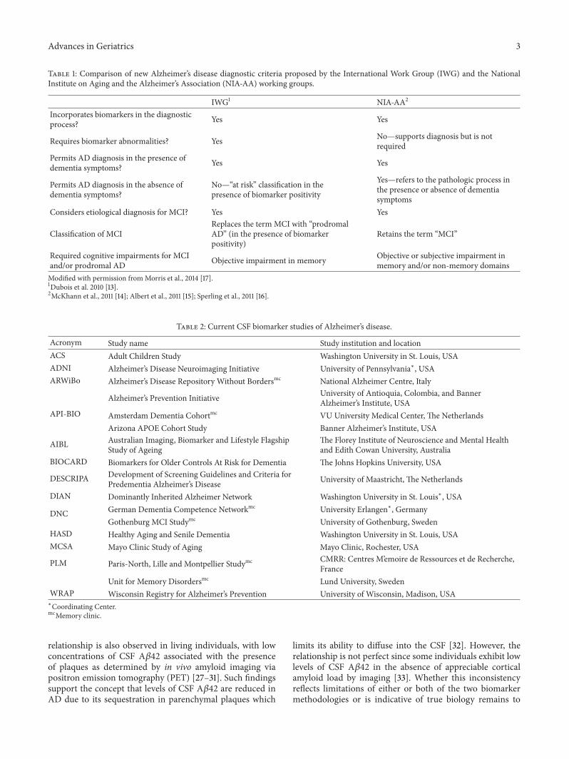
Advances in Geriatrics 3
Table 1: Comparison of new Alzheimer’s disease diagnostic criteria proposed by the International Work Group (IWG) and the NationalInstitute on Aging and the Alzheimer’s Association (NIA-AA) working groups.
IWG1 NIA-AA2
Incorporates biomarkers in the diagnosticprocess? Yes Yes
Requires biomarker abnormalities? Yes No—supports diagnosis but is notrequired
Permits AD diagnosis in the presence ofdementia symptoms? Yes Yes
Permits AD diagnosis in the absence ofdementia symptoms?
No—“at risk” classification in thepresence of biomarker positivity
Yes—refers to the pathologic process inthe presence or absence of dementiasymptoms
Considers etiological diagnosis for MCI? Yes Yes
Classification of MCIReplaces the term MCI with “prodromalAD” (in the presence of biomarkerpositivity)
Retains the term “MCI”
Required cognitive impairments for MCIand/or prodromal AD Objective impairment in memory Objective or subjective impairment in
memory and/or non-memory domainsModified with permission fromMorris et al., 2014 [17].1Dubois et al. 2010 [13].2McKhann et al., 2011 [14]; Albert et al., 2011 [15]; Sperling et al., 2011 [16].
Table 2: Current CSF biomarker studies of Alzheimer’s disease.
Acronym Study name Study institution and locationACS Adult Children Study Washington University in St. Louis, USAADNI Alzheimer’s Disease Neuroimaging Initiative University of Pennsylvania∗, USAARWiBo Alzheimer’s Disease Repository Without Bordersmc National Alzheimer Centre, Italy
API-BIO
Alzheimer’s Prevention Initiative University of Antioquia, Colombia, and BannerAlzheimer’s Institute, USA
Amsterdam Dementia Cohortmc VU University Medical Center, The NetherlandsArizona APOE Cohort Study Banner Alzheimer’s Institute, USA
AIBL Australian Imaging, Biomarker and Lifestyle FlagshipStudy of Ageing
The Florey Institute of Neuroscience and Mental Healthand Edith Cowan University, Australia
BIOCARD Biomarkers for Older Controls At Risk for Dementia The Johns Hopkins University, USA
DESCRIPA Development of Screening Guidelines and Criteria forPredementia Alzheimer’s Disease University of Maastricht, The Netherlands
DIAN Dominantly Inherited Alzheimer Network Washington University in St. Louis∗, USA
DNC German Dementia Competence Networkmc University Erlangen∗, GermanyGothenburg MCI Studymc University of Gothenburg, Sweden
HASD Healthy Aging and Senile Dementia Washington University in St. Louis, USAMCSA Mayo Clinic Study of Aging Mayo Clinic, Rochester, USA
PLM Paris-North, Lille and Montpellier Studymc CMRR: Centres M’emoire de Ressources et de Recherche,France
Unit for Memory Disordersmc Lund University, SwedenWRAP Wisconsin Registry for Alzheimer’s Prevention University of Wisconsin, Madison, USA∗Coordinating Center.mcMemory clinic.
relationship is also observed in living individuals, with lowconcentrations of CSF A𝛽42 associated with the presenceof plaques as determined by in vivo amyloid imaging viapositron emission tomography (PET) [27–31]. Such findingssupport the concept that levels of CSF A𝛽42 are reduced inAD due to its sequestration in parenchymal plaques which
limits its ability to diffuse into the CSF [32]. However, therelationship is not perfect since some individuals exhibit lowlevels of CSF A𝛽42 in the absence of appreciable corticalamyloid load by imaging [33]. Whether this inconsistencyreflects limitations of either or both of the two biomarkermethodologies or is indicative of true biology remains to
4 Advances in Geriatrics
be determined. Each amyloid biomarker modality has itsown strengths and weaknesses. There are currently threeamyloid PET tracers approved for clinical use by the UnitedStates Food and Drug Administration (FDA) (florbetapir(Amyvid), Eli Lilly and Company; flutemetamol (Vizamyl),GE Healthcare; florbetaben (Neuraceq), Piramal Imaging).While a PET scan is viewed by many as less invasive thanlumbar puncture, it is muchmore costly and poses additionalrisks due to injection of a radioactive tracer. However,unlike CSF, amyloid imaging is able to provide informationregarding regional patterns of pathology. In comparison,CSF is able to provide information regarding multiple AD-related biomarkers (e.g., amyloid, tangles, neuroinflamma-tion, neuronal injury/death, and other processes yet to bedetermined), albeit without regional specificity. AlthoughAD CSF biomarkers have not yet been approved by theFDA, clinical samples are routinely assessed for diagnosticpurposes in several European countries as well as the US,albeit much less frequently than in Europe. Efforts to obtainFDA approval for the use of CSF biomarkers in US clinicalsettings are currently underway.
Tau is the primary component of neurofibrillary tanglesin AD, but tau aggregates are the defining features of severalother tauopathies including frontotemporal dementia (FTD),progressive supranuclear palsy (PSP), chronic traumaticencephalopathy (also known as dementia pugilistica), cor-ticobasal degeneration, and Pick’s disease. However, resultsfrom studies evaluating CSF levels of tau in the varioustauopathies have been mixed likely due to the large hetero-geneity among these clinical syndromes [34]. In contrast,CSF tau is consistently reported to be elevated in conditionsinvolving robust neuronal cell death, including acute stroke[35, 36], traumatic brain injury (TBI) [36, 37], Creutzfeldt-Jakob disease (CJD) [38–40], and AD [18], and thus isconsidered to be a marker of neuronal injury and/or death.However, both soluble and aggregated forms of tau have beenshown to be secreted from healthy cells in culture [41], andevidence from animal studies suggests tau is continuouslysecreted from healthy neurons into the brain interstitial fluidspace [42], perhaps driven by synaptic activity [43].
Although levels of ptau are typically highly correlatedwith levels of tau in the CSF, ptau is considered to be amore specific biomarker for AD since elevations in ptau arenot observed following traumatic brain injury and strokeor in CJD [44]. Instead, ptau is likely more a marker ofneurofibrillary tangles since positive correlations have beenobserved between levels of ptau (p-tau
181and p-tau
231and
tau phosphorylated at residues 181 and 231, resp.) and neo-cortical tangle pathology at autopsy [25, 45]. Future studiesevaluating the association between tangle load via tau PETimaging [46] and CSF levels of tau and ptau will be veryinformative in defining the pathological etiology of elevatedCSF tau and ptau. Curiously, levels of ptau in other neurode-generative conditions known to have tangle pathology, whiletypically elevated in comparison to nondiseased controls,do not reach the levels observed in AD [34]. Whetherthis apparent discrepancy reflects the inherent heterogeneityin the many tauopathies (with differences in tangle loadand/or regional distributions and its pathological course)
that impacts the ability of ptau to reach the CSF, potentiallydifferent phosphorylation states in these other tauopathiesthat are not detected by the current ptau assays, or otherfactors remains to be determined.
Unbiased proteomics screens and targeted multianalytesurveys of CSF have identifiedmany proteins that are elevatedor reduced in AD compared to cognitively normal controls[47]. Many candidate proteins are presumed markers ofoxidative stress, synapse loss or neurodegeneration, andneuroinflammation, processes known to be involved in AD,albeit nonspecifically. It is likely that these markers will bemore useful in disease staging than for disease diagnosis.For example, visinin-like protein 1 (VILIP-1) and YKL-40(also known as chitinase-3-like protein 1), presumedmarkersof neurodegeneration and neuroinflammation, respectively,have been shown to be elevated in AD CSF compared tocontrols [48–53], although with overlap between the clinicalgroups. However, these markers, in the presence of lowlevels of CSF A𝛽42, are strong predictors of future cogni-tive decline/dementia (within 3-4 years) in elders who arecognitively normal or who have MCI/very mild AD [49–54],suggesting they are able to identify a stage of AD just priorto symptomatic expression. Despite such promise, until thesecandidate markers are validated in additional large researchcohorts, the three core biomarkers remain the gold standards.
4. Diagnostic and Prognostic Performance ofCSF Biomarkers
Biomarker sensitivity and specificity are both important forAD diagnosis. A high sensitivity is required to identify thehighest proportion of individuals with AD and minimizefalse negatives, whereas a high specificity is necessary fordiagnostic accuracy to minimize false positives and excludesymptomatic or prodromal cases due to non-ADpathologicalprocesses. However, the criteria for what is considered “high”or even “acceptable” will likely differ depending on thepurpose for which the biomarkers will be used, such asproviding a disease diagnosis, primary care screening forreferral to specialized memory clinics, clinical trial enroll-ment, or eventual treatment with disease-modifying drugsthat may be expensive and/or have unwanted side effects.The high prevalence of mixed pathologies in individuals withAD will also likely impact biomarker criteria. All of theseconsiderations are topics of ongoing investigation.
4.1. AD Dementia. Since the current criteria for the clinicaldiagnosis of AD require the presence of dementia [7], themajority of biomarker studies over the years have evaluatedthe ability of candidate markers to distinguish individualsclinically diagnosed with AD from those without dementia.In such analyses, elevations in CSF tau and ptau (∼300%)and reductions in A𝛽42 (∼50%) are observed in AD, typicallywith sensitivities and specificities greater than 80% [55]. Thisbiomarker accuracy, while good, is not better than currentclinical accuracies, at least in specialized dementia centersand clinics [8]. This suboptimal accuracy is likely due to thefact that 25–30% of cognitively normal elders are known to
Advances in Geriatrics 5
have enough plaques and tangles to warrant a neuropatho-logical diagnosis of AD (presumed “preclinical” AD) [56, 57],thus “contaminating” the cognitively normal groupwith indi-viduals with underlying AD pathology. Despite this caveat,a combined measure of these biomarkers (e.g., high levelsof tau(s) and low levels of A𝛽42) performs better than thesingle biomarkers on their own [58, 59]. Importantly, thesemarkers are normal in several other disorders which are oftenaccompanied by cognitive impairment such as depressionand Parkinson’s disease (PD) [37]. In addition, CSF ptau, inparticular, can aid in the differentiation of AD from otherdementias, such as FTD and Lewy body dementia (DLB)[44]. However, the overall diagnostic performance of theseCSF biomarkers to discriminate ADdementia fromdementiadue to other etiologies is not optimal, likely reflecting therelative abundance ofmixed pathologies in people presentingwith dementia [60, 61], as well as inaccuracies in differ-ential clinical diagnosis, especially at early disease stages.Combined with the presence of preclinical pathology in asubstantial percentage of nondemented elderly individuals,assessing biomarkers based on clinical diagnoses precludesthe possibility of finding a disease biomarker with 100%sensitivity and specificity for AD. Nonetheless, proposalshave been made to include biomarker profiles in diagnosticformulations for probable and possible AD dementia for usein research and clinical settings [13, 14].
4.2. Prodromal AD/Mild Cognitive Impairment. Longitudinalstudy of older individuals over time has been critical for ourunderstanding of AD as a disease continuum. Furthermore,given the consistently poor results in AD clinical trials to datewhich have enrolled individuals who already have dementia,it has been proposed that development of disease-modifyingdrugs will likely require testing in patient populations atvery early (predementia) stages. This critical interest in earlydiagnosis, coupled with the appreciation of the long timeperiod during which AD pathology develops prior to thesymptoms of dementia (see below), has fueled efforts toidentify biomarkers of AD pathology at earlier disease stages.Mild cognitive impairment (MCI) has been recognized asa possible intermediate stage between normal aging andAD dementia [62, 63] and, as such, has been the subjectof intense interest in the AD biomarker field. As a group,individuals with MCI (defined by impairments in cognitiveabilities compared to age-matched normative values butat levels that are below the threshold considered to be“dementia”) typically display mean AD biomarker profilesof CSF A𝛽42, tau, and ptau intermediate to those withAD dementia and nondemented controls [55, 64]. However,there is significant overlap among individuals in the MCIand the other diagnostic groups, with approximately two-thirds of individuals with MCI displaying profiles consistentwith AD pathology and one-third appearing in the normalrange [65]. Such heterogeneity likely reflects differences in theunderlying etiologies of the cognitive impairments. Indeed,amyloid plaques, as visualized by PET imaging, are presentin ∼60% of individuals with MCI [66], and longitudinalstudies in several international cohorts have demonstrated
that the combination of low CSF A𝛽42 and high CSF tau andptau is highly predictive of which MCI cases will progressto AD dementia (i.e., prodromal AD) [65, 67–69], as wellas their rate of cognitive decline [54, 70]. Such studies haveprompted the proposed use of these CSF biomarkers to define(probabilistically) the underlying etiology of MCI cases inresearch as well as clinical settings [15].
4.3. Asymptomatic/Preclinical AD. Prevention of cognitivedecline (prior to irrevocable neuronal cell loss) is the ultimategoal of AD therapeutics. However, implementation of sucha strategy provides a unique challenge since, by definition,clinical measures will be unable to identify individuals in thispresymptomatic phase.Therefore, the use of validated diseasebiomarkers will be required for such patient identification.Data from clinicopathological and biomarker studies haveconverged to support the existence of a long preclinicalperiod stage (estimated to be at least 10–20 years) duringwhich the pathologies of AD develop before the appearanceof any cognitive symptoms of dementia. Roughly one-third ofcognitively normal elderly individuals who come to autopsymeet the histopathological criteria for AD [56, 71, 72]. Datafrom more recent biomarker studies in large cohorts ofcognitively normal individuals confirm this finding; a similarproportion exhibits low levels of CSFA𝛽42 that are indicativeof the presence of cortical amyloid as detected by PET amy-loid imaging [27–31]. While some asymptomatic individualsexhibit high levels of CSF tau in the absence of amyloidpositivity (defined by CSF A𝛽42) [73], the etiology(ies) ofsuch elevations and their impact on clinical performance arenot clear.
The critical element required for establishing biomarkervalidity and utility in the preclinical period is its abilityto predict future dementia. Low levels of CSF A𝛽42 havebeen shown to predict future cognitive decline and/or ADdementia in cognitively normal elders [75–77]. In those samestudies, high levels of tau and ptau were not useful predictorsof cognitive decline. However, more recent studies haveshown that the combination of these CSF pathologic markers(e.g., low A𝛽42 and high tau(s) or a high tau(s)/A𝛽42 ratio)in cognitively normal elders is highly predictive of futurecognitive decline (within a few years) [49, 50, 78, 79], similarto its ability to predict cognitive decline and/or AD dementiainMCI cohorts [70, 80]. Visinin-like protein 1, a neuronal cal-cium sensor protein that is currently receiving a lot of interestas a novel, tau-independent marker of neurodegeneration,is elevated in AD [48, 53] and has similarly been shown tobe a strong predictor of cognitive decline and/or dementiain cognitively normal elders in the presence of low levels ofA𝛽42 [50, 54].
Arguably the most compelling support for the utilityof biomarkers in predicting cognitive decline and dementiacomes from studies of rare families with known AD-causingmutations (autosomal-dominantAD (ADAD) that comprises∼1% of all AD cases). These individuals have been excludedfrom observational studies and clinical trials due to thegenetic mechanism of their disease. However, despite poten-tial differences in the mechanisms by which A𝛽 accumulates
6 Advances in Geriatrics
to formamyloid in the brain (likely due to the increase inA𝛽1-42 production in ADAD compared to more complex and stillpoorly understood mechanism(s) in late-onset AD (LOAD)[81]), the hallmark pathologies and clinical symptoms aresimilar between the two disease groups [82]. Study of ADADis particularly well-suited to investigation of biomarker util-ity, especially during the preclinical/asymptomatic period,since the 100% penetrance of the mutations and relativeconsistency of age at symptom onset within families allowsinvestigators to knowwith certaintywhowill develop demen-tia and the approximate timing of symptom appearance.These characteristics overcome the limitations of diseaseuncertainty and unpredictability of symptom onset inherentin studies of LOAD. Symptomatic mutation carriers exhibitlow levels of CSF A𝛽42 compared to mutation noncarriers[83–85] (as do individuals with Down syndrome who aredestined to develop AD due to the presence of three copiesof the gene for the amyloid precursor protein, APP; [86]) andhigh levels of tau [84, 85, 87]. Furthermore, recent reportsfrom the Dominantly Inherited Alzheimer Network (DIAN)indicate that these AD biomarker signatures are observedat least ∼10 to 20 years prior to carriers’ predicted age ofsymptom onset (defined as the age at which their parentdeveloped clinical symptoms) [84, 85]. The precise timing ofsuch preclinical changes in the more common form of thedisease (LOAD) and when they are first detectable are topicsof current investigation and will be critical to consider if anAD diagnosis is to ever be made in the absence of cognitivesymptoms [16].
5. Biomarker Use in TrackingDisease Progression
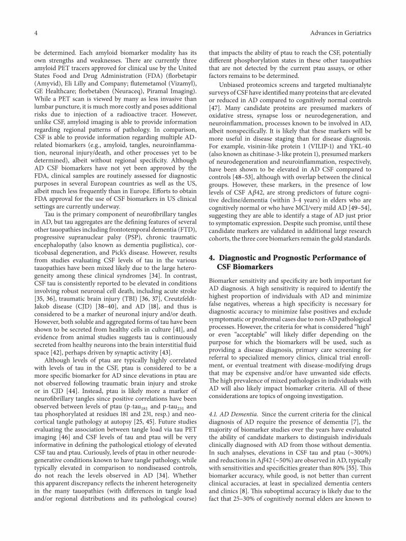
Over the past decade great progress has been made in theAD biomarker field in identifying markers that not onlyyield a more accurate disease diagnosis and prognosis inresearch cohorts, but also provide insight into the normaltrajectories of neuropathologic changes over the course ofthe disease, especially during the early and preclinical stages[88]. Results from many cross-sectional studies support amodel in which dynamic reductions in CSF A𝛽42 (indicativeof amyloid plaques) occur in the early and preclinical phase,with levels remaining fairly constant during the subsequentsymptomatic phase, followed by elevations in markers ofneuronal injury (such as tau and ptau and, more recently,VILIP-1) later in the disease and that continue as peopleapproach and move through the symptomatic phase [89, 90](Figure 1(a)). Consistent with this model, cognitive declineis more closely related to markers of neuronal injury thanamyloid load [91–93].
Recent preliminary findings from within-person longi-tudinal biomarker analyses in DIAN support inclusion ofan eventual slowing down of the rate of neuronal injurywith symptomatic disease progression. While still cogni-tively normal, mutation carriers exhibit increases in CSF tau(and ptau
181and VILIP-1), consistent with results of cross-
sectional analyses [84, 85]. However, these same markers ofneuronal injury actually decrease in mutation carriers who
are symptomatic [85]. Although relatively small in number,studies investigating within-person change in LOAD havedemonstrated little or no change in biomarker levels overrelatively short time periods (6 months to 2 years) [94–97]. A closer review of the data, however, reveals between-subject variability in the patterns of tau changes that maybe biologically relevant. For example, tau was observed toincrease in persons with LOAD who had low tau at baseline(presumably early in the course of the disease) but showedno difference or even decreases in those with high baselinelevels (presumed to be later in the disease process) [98,99]. Consistent with this finding, statistical modeling in theAlzheimer’s Disease Neuroimaging Initiative (ADNI) cohortrevealed similar reductions in tau inADcases but not in thosewith MCI [100]. Longitudinal biomarker studies of middle-aged individuals who are followed from cognitive normalitythrough mild cognitive impairment to AD dementia willprovide critical information regarding biomarker changesover the entire course of the disease, from preclinical toMCI/prodromal AD to AD dementia. Such information willbe important for patient care in the long run and currentlyfor informing the design and interpretation of clinical trialsof disease-modifying therapies.
6. The Potential Use of Biomarkers inClinical Practice
Although measurements of CSF A𝛽42, tau, and ptau in clinicpatients can already be obtained by physicians in the US andin many European countries, these measures are not part ofthe clinical criteria for the diagnosis of AD dementia. How-ever, this may soon change. The current clinical diagnosticcriteria for AD were developed over 25 years ago by theNINCDS-ADRDA Work Group [7]. These criteria dependlargely on the exclusion of causes other thanAD for dementiaand state that a diagnosis of AD cannot be made until thepatient has dementia, defined as “cognitive symptoms severeenough to interfere with social or occupational activities” [7].TheDiagnostic and Statistical Manual fourth revision (DSM-IV) and International Classification of Disease tenth revision(ICD-10) criteria, which are used for routine diagnosis,also require that a patient demonstrate dementia before adiagnosis of AD is possible. Given that we now know thatdementia is the end stage of a long disease process and predictthat disease-modifying therapies will likely bemost beneficialif administered earlier in the disease course, new criteria fordifferent stages of AD across the disease continuum haverecently been proposed by three work groups convened bytheNIA-AA [14–16]. Similar (but not identical) revisions havealso been proposed by the IWG [13, 17]. These new criteriadiffer from the ones published in 1984 in the fact that theyincorporate biomarkers of the underlying disease state anduse them to formalize the different disease stages [101]. Thesecriteria have been developed, in principal, to increase theconfidence of AD as the underlying etiology of a clinicalimpairment and permit a diagnosis of AD at earlier diseasestages, even before the emergence of symptoms. Efforts to
Advances in Geriatrics 7
DementiaMCIPreclinicalNormal
Abno
rmal
CSF A𝛽42
Amyloid PETFDG-
PET/fMRICSF tau(s)
vMRI
CognitionClinical function
AmyloidSynaptic dysfunctionNeuronal injury/tangles
Brain structure
Cognition
Clinical function
Time
(a)
DementiaMCINormal
Abno
rmal
Time
1 2 3
Preclinical stages
CognitionClinical function
AmyloidSynaptic dysfunctionNeuronal injury/tangles
Brain structure
(b)
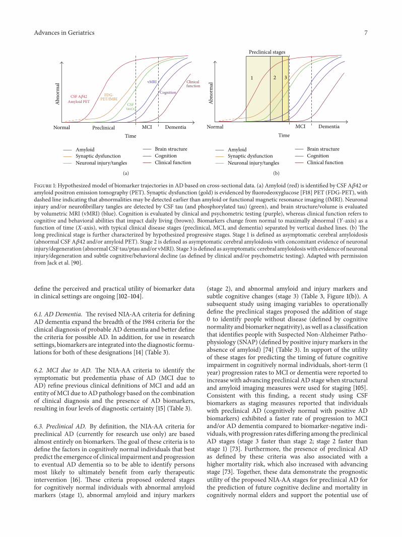
Figure 1: Hypothesized model of biomarker trajectories in AD based on cross-sectional data. (a) Amyloid (red) is identified by CSF A𝛽42 oramyloid positron emission tomography (PET). Synaptic dysfunction (gold) is evidenced by fluorodeoxyglucose [F18] PET (FDG-PET), withdashed line indicating that abnormalities may be detected earlier than amyloid or functional magnetic resonance imaging (fMRI). Neuronalinjury and/or neurofibrillary tangles are detected by CSF tau (and phosphorylated tau) (green), and brain structure/volume is evaluatedby volumetric MRI (vMRI) (blue). Cognition is evaluated by clinical and psychometric testing (purple), whereas clinical function refers tocognitive and behavioral abilities that impact daily living (brown). Biomarkers change from normal to maximally abnormal (Y-axis) as afunction of time (X-axis), with typical clinical disease stages (preclinical, MCI, and dementia) separated by vertical dashed lines. (b) Thelong preclinical stage is further characterized by hypothesized progressive stages. Stage 1 is defined as asymptomatic cerebral amyloidosis(abnormal CSF A𝛽42 and/or amyloid PET). Stage 2 is defined as asymptomatic cerebral amyloidosis with concomitant evidence of neuronalinjury/degeneration (abnormalCSF tau/ptau and/or vMRI). Stage 3 is defined as asymptomatic cerebral amyloidosiswith evidence of neuronalinjury/degeneration and subtle cognitive/behavioral decline (as defined by clinical and/or psychometric testing). Adapted with permissionfrom Jack et al. [90].
define the perceived and practical utility of biomarker datain clinical settings are ongoing [102–104].
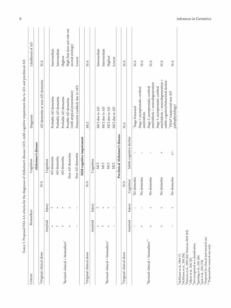
6.1. AD Dementia. The revised NIA-AA criteria for definingAD dementia expand the breadth of the 1984 criteria for theclinical diagnosis of probable AD dementia and better definethe criteria for possible AD. In addition, for use in researchsettings, biomarkers are integrated into the diagnostic formu-lations for both of these designations [14] (Table 3).
6.2. MCI due to AD. The NIA-AA criteria to identify thesymptomatic but predementia phase of AD (MCI due toAD) refine previous clinical definitions of MCI and add anentity ofMCI due to ADpathology based on the combinationof clinical diagnosis and the presence of AD biomarkers,resulting in four levels of diagnostic certainty [15] (Table 3).
6.3. Preclinical AD. By definition, the NIA-AA criteria forpreclinical AD (currently for research use only) are basedalmost entirely on biomarkers. The goal of these criteria is todefine the factors in cognitively normal individuals that bestpredict the emergence of clinical impairment and progressionto eventual AD dementia so to be able to identify personsmost likely to ultimately benefit from early therapeuticintervention [16]. These criteria proposed ordered stagesfor cognitively normal individuals with abnormal amyloidmarkers (stage 1), abnormal amyloid and injury markers
(stage 2), and abnormal amyloid and injury markers andsubtle cognitive changes (stage 3) (Table 3, Figure 1(b)). Asubsequent study using imaging variables to operationallydefine the preclinical stages proposed the addition of stage0 to identify people without disease (defined by cognitivenormality and biomarker negativity), aswell as a classificationthat identifies people with Suspected Non-Alzheimer Patho-physiology (SNAP) (defined by positive injurymarkers in theabsence of amyloid) [74] (Table 3). In support of the utilityof these stages for predicting the timing of future cognitiveimpairment in cognitively normal individuals, short-term (1year) progression rates to MCI or dementia were reported toincrease with advancing preclinical AD stage when structuraland amyloid imaging measures were used for staging [105].Consistent with this finding, a recent study using CSFbiomarkers as staging measures reported that individualswith preclinical AD (cognitively normal with positive ADbiomarkers) exhibited a faster rate of progression to MCIand/or AD dementia compared to biomarker-negative indi-viduals, with progression rates differing among the preclinicalAD stages (stage 3 faster than stage 2; stage 2 faster thanstage 1) [73]. Furthermore, the presence of preclinical ADas defined by these criteria was also associated with ahigher mortality risk, which also increased with advancingstage [73]. Together, these data demonstrate the prognosticutility of the proposed NIA-AA stages for preclinical AD forthe prediction of future cognitive decline and mortality incognitively normal elders and support the potential use of
8 Advances in GeriatricsTa
ble3:Prop
osed
NIA-AAcriteria
forthe
diagno
seso
fAlzh
eimer’sdisease(AD),mild
cogn
itive
impairm
entd
ueto
AD,and
preclin
icalAD.
Criteria
Biom
arkers
Cognitio
nDiagn
osis
Likelih
oodof
AD
Alzheim
er’sdisease
1
Orig
inal-clin
icalalon
eN/A
ADdementia
orno
n-ADdementia
N/A
Amyloid
Injury
Cognitio
n
2 Revise
d-clinical+
biom
arkers∗
+?
ADdementia
Prob
ableADdementia
Interm
ediate
?+
ADdementia
Prob
ableADdementia
Interm
ediate
++
ADdementia
Prob
ableADdementia
Highest
++
Non
-ADdementia
PossibleADdementia
(with
atypicalpresentatio
n)High(but
does
notruleo
utsecond
etiology)
−−
Non
-ADdementia
Dem
entia
(unlikely
duetoAD)
Lowest
Mild
cogn
itive
impa
irment
3 Orig
inal-clin
icalalon
eN/A
MCI
N/A
Amyloid
Injury
Cognitio
n
4 Revise
d-clinical+
biom
arkers∗
+?
MCI
MCI
duetoAD
Interm
ediate
?+
MCI
MCI
duetoAD
Interm
ediate
++
MCI
MCI
duetoAD
Highest
−−
MCI
MCI
duetoAD
Lowest
Preclin
icalAlzheim
er’sdisease
5 Orig
inal-clin
icalalon
eN/A
N/A
N/A
Amyloid
Injury
Cognitio
nSubtlecogn
itive
decline
6 Revise
d-clinical+
biom
arkers∗∗
−−
Nodementia
−
7 Stage
0/no
rmal
N/A
+−
Nodementia
−
Stage1:asymptom
aticcerebral
amyloido
sisN/A
++
Nodementia
−
Stage2
:asymptom
aticcerebral
amyloido
sis+neurod
egeneration
N/A
++
Nodementia
+Stage3
:asymptom
aticcerebral
amyloido
sis+neurod
egeneration+
subtlecogn
itive/behaviorald
eclin
eN/A
−+
Nodementia
+/−
7 SNAP(suspected
non-AD
pathop
hysio
logy)
N/A
1
McK
hann
etal.,1984
[7].
2 McK
hann
etal.,2011[14
].3 Petersenetal.,1999
[62];Petersen2004
[63].
4 Albertetal.,2011[15].
5 Not
recogn
ized
asac
lassificatio
n.6 Sperling
etal.,2011[16].
7 Jacketal.,2012
[74].
∗
Prop
osed
forc
linicalandresearch
use.
∗∗
Prop
osed
forresearchuseo
nly.
Advances in Geriatrics 9
NIA-AA criteria in research studies as well as in AD clinicaltrials. Whether such criteria will eventually be consideredin primary care settings remains to be determined and willmost likely depend on the availability of effective disease-modifying therapies.
7. Use of Biomarkers in Clinical Trials
To date all clinical trials of putative disease-modifying ther-apies in individuals with AD dementia have failed to showmeaningful clinical benefit. Such failures may be due toseveral factors including inadequacies of the specific drugstested, inappropriate pathobiological targets, inclusion ofmisdiagnosed patients, use of insensitive cognitive outcomemeasures, and/or enrollment of individuals who are too farprogressed in the disease (i.e., demented) and thus havealready undergone substantial neuronal cell death [106], inother words, perhaps the wrong drug, the wrong target, thewrong people, and/or the wrong stage [107].
To address these potential shortcomings, biomarkers arebeing used in current and planned AD secondary preventiontrials of potential disease-modifying therapies [108]: (1)Anti-Amyloid Treatment in Asymptomatic Alzheimer’sDisease (A4 Study) (http://www.clinicaltrials.gov/ct2/show/NCT02008357?term=A4&rank=1) [109]; (2) Alzheimer’sPrevention Initiative (API) (http://www.clinicaltrials.gov/ct2/show/NCT01998841?term=alzheimer%27s+prevention+initiative&rank=1) [110]; and (3) Dominantly InheritedAlzheimer Network Trial Unit (DIAN TU) (http://www.clin-icaltrials.gov/ct2/show/NCT01760005?term=DIAN&rank=2)[111]. For example, levels of CSF A𝛽42, tau, and/or ptau(and/or imaging measures) are being used to (1) enrichthe number of patients who actually have underlying ADpathology; (2) stratify patients according to the presenceand/or the amount of underlying pathology; (3) assesstherapeutic target engagement; and (4) monitor the effectsof treatment on downstream pathogenic processes. Notonly will this approach result in an overall reduction inrequired patient numbers, trial duration, and cost, but it isalso expected to provide information particularly suitable formaking go/no-go decisions as to whether to move candidatetherapies forward into large and expensive Phase II and IIItrials. There is also growing interest in the role biomarkerscould play as surrogate endpoints in treatment trials, suchthat treatment effects on the biomarkers themselves could beused as a replacement for clinical endpoints in the acceleratedapproval of AD therapies by regulatory agencies [18, 106].
8. Challenges and Opportunities
8.1. Biomarker. The AD biomarker field is faced with severalchallenges thatmust be overcome in order tomove promisinganalytes into clinical practice. From a methodological per-spective, biomarker candidates must be validated in large,well-characterized research cohorts, with care taken to eval-uate the potential impact of important AD-related covariatessuch as age, gender, and APOE genotype (the strongestgenetic risk factor for AD). The field is close to achieving
that. However, protocol and assay standardization muststill be attained in order to maximize biomarker sensitivityand reliability. Although biological and within-laboratoryvariability for CSF markers are low, there is variation inbiomarker levels between laboratories. Furthermore, thereare no available calibrators (certified reference standards) forthese biomarkers and no consensus on what assays shouldbe used [18]. Worldwide biomarker standardization effortsare currently underway [112–114], and in pharmaceutical andbiotech laboratories, emphasis is being placed on develop-ment of commercial assay platforms with automated, high-throughput capabilities.
Given the long preclinical stage of AD, biomarker valida-tion should define sensitivity and specificity (and associatedpositive and negative predictive values) for underlying ADpathology, not merely clinical diagnosis that is often inac-curate. To date, very few studies have been able to correlatebiomarkers in antemortem samples with results of histologicpostmortem examination, and the time interval betweenthe two evaluations can be large. As in vivo amyloid PETimaging provided the opportunity to validate CSF A𝛽42as a marker of amyloid, methods to image neurofibrillarytangles [46, 115] and neuroinflammatory responses [116] arein development and will be very informative for providingpathologic validation for CSF tau and ptau and potentialmarkers of neuroinflammation such as YKL-40. Establishingvalid cut-points to define biomarker positivity versus nega-tivity [117] will ultimately be required in order to eventuallybring biomarkers to the clinic. Such cut-points will likelyalso depend on covariates such as age, genetic risk factors(e.g., APOE genotype), and or family history of AD. Inaddition, studies evaluating the change in biomarker profileswithin individuals with disease progression will be importantfor disease staging purposes, as will be the developmentof statistical methods to determine cut-points for suchchanges over time [118]. Evaluation of biomarker utility fordifferential diagnosis in cohorts with known underlying non-AD pathologies (FTLD, DLB, vascular dementia, etc.) is alsoneeded, especially since misdiagnoses often occur at earlyclinical stages. It is likely that combinations of biomarkerswill prove most useful for disease diagnosis (presence versusabsence of AD pathology) and prognosis (predicting cogni-tive decline), either within modalities or between modalities(e.g., fluids and imaging) [89, 119, 120]. Ongoing studies thatcollect multiple types of biomarker data will be critical forsuch assessments. Finally, although considered to be the HolyGrail, it remains to be determined whether any viable plasmabiomarkers will emerge. Many research groups are activelyengaged in the search given the clinical appeal of diagnosticblood tests [121].
8.2. Clinical. Lumbar puncture is considered to be moreinvasive than blood draw, and negative attitudes towards LPprovide a challenge to implementing AD biomarkers in bothresearch and clinical settings. The most prevalent adverseside effects of LPs aremild-moderate headache and backache,with more severe headaches considered to be the primarysafety concern. However, use of small caliber “noncutting”
10 Advances in Geriatrics
atraumatic spinal needles (e.g., Sprotte or Whitacre 22–25gauge) has been shown to significantly reduce the incidenceof post-LP headaches [21, 23, 122] such that their use is nowrecommended by experts in the field [123].
On the clinical front, ethical, policy, and social challengesexist regarding provision of a preclinical diagnosis, especiallyin the absence of an available disease-modifying therapy.Challenges for the physician include understanding if andwhen a cognitively normal patient should undergo testingand the potential harm (e.g., anxiety, depression) a patientmay experience upon receiving a diagnosis of preclinicalAD [124]. Challenges also exist regarding the translation ofdiagnosis of preclinical AD into practice and policy. Privacyand confidentiality laws, regulations, and professional prac-tices should address how to minimize the possibility thatinsurance, pharmacy, and medical records disclose that aperson has preclinical AD [124]. From a practical point ofview, insurance coverage may provide a challenge, especiallyin the absence of symptoms. Efforts are ongoing to developguidelines for the design of studies able to provide robustevidence for insurer coverage determinations [125].
9. Summary and Conclusions
The AD field is experiencing an exciting evolution, in bothits appreciation of the dynamic nature of the disease and thedevelopment of therapeutic strategies aimed atmodifying theunderlying disease processes (as opposed to simply amelio-rating dementia symptoms), and biomarkers have been at theforefront of these paradigm shifts. The core CSF analytes,A𝛽42, tau, and ptau, have withstood the test of time inidentifying persons with AD dementia and have also beenshown to strongly predict the development of future cognitivedecline in individuals at very early and predementia as wellas preclinical stages. In response to these findings, revi-sions to AD diagnostic criteria so to incorporate biomarkerresults have recently been proposed in order to increase theconfidence of AD as the underlying etiology of a clinicalimpairment and to permit a diagnosis of AD across thecontinuum of the disease, eventually perhaps in the asymp-tomatic period. Although currently proposed for use only inresearch settings, a diagnosis of preclinical ADwould providea potential window for therapeutic interventions aimed atpreventing cognitive decline not just delaying or halting itonce impairments are already clinically apparent. Informa-tion regarding biomarker trajectories over the normal courseof the disease is being used to inform the design of clinicaltrials and the evaluation of efficacy of proposed disease-modifying therapies. Eventually the hope is to bring validatedbiomarkers to clinical practice in order to provide a moreaccurate diagnosis, prognostic information regarding futurecognitive decline, and evaluation of therapeutic efficacy oncedisease-modifying treatments become available. Realizationof this goal requires worldwide biomarker standardizationefforts, consensus among researchers and clinicians regardingthe clinical utility of assessing biomarkers in patient caresettings, and eventually the endorsement and adoption ofsuch procedures and practices into global health care systems.
Given the large and rapidly expanding aging population, suchefforts should be considered a top international priority.
Conflict of Interests
The author declares that there is no conflict of interestsregarding the publication of this paper.
References
[1] B. D. James, S. E. Leurgans, L. E. Hebert et al., “Contribution ofAlzheimer disease to mortality in the United States,”Neurology,vol. 82, pp. 1045–1050, 2014.
[2] S. S. Mirra, A. Heyman, D. McKeel et al., “The Consortiumto Establish a Registry for Alzheimer’s Disease (CERAD).Part II. Standardization of the neuropathologic assessment ofAlzheimer’s disease,”Neurology, vol. 41, no. 4, pp. 479–486, 1991.
[3] Z. S. Khachaturian, “Diagnosis of Alzheimer’s disease,”Archivesof Neurology, vol. 42, no. 11, pp. 1097–1105, 1985.
[4] B. T. Hyman and J. Q. Trojanowski, “Consensus recommen-dations for the postmortem diagnosis of Alzheimer diseasefrom the National Institute on Aging and the Reagan InstituteWorkingGroup on diagnostic criteria for the neuropathologicalassessment ofAlzheimer disease,” Journal of neuropathology andexperimental neurology, vol. 56, no. 10, pp. 1095–1097, 1997.
[5] T. J. Montine, C. H. Phelps, T. G. Beach et al., “Nationalinstitute on aging-Alzheimer’s association guidelines for theneuropathologic assessment of Alzheimer’s disease: a practicalapproach,” Acta Neuropathologica, vol. 123, no. 1, pp. 1–11, 2012.
[6] D.M.Holtzman, J. C.Morris, andA.M.Goate, “Alzheimer’s dis-ease: the challenge of the second century,” Science TranslationalMedicine, vol. 3, no. 77, Article ID 77sr1, 2011.
[7] G. McKhann, D. Drachman, and M. Folstein, “Clinical diagno-sis of Alzheimer’s disease: report of the NINCDS-ADRDAworkgroup under the auspices of Department of Health and HumanServices Task Force on Alzheimer’s disease,” Neurology, vol. 34,no. 7, pp. 939–944, 1984.
[8] T. G. Beach, S. E. Monsell, L. E. Phillips, and W. Kukull,“Accuracy of the clinical diagnosis of Alzheimer disease atnational institute on aging Alzheimer disease centers, 2005–2010,” Journal of Neuropathology and Experimental Neurology,vol. 71, no. 4, pp. 266–273, 2012.
[9] R. Handels, P. Aalten, C. A. Wolfs et al., “Diagnostic andeconomic evaluation of new biomarkers for Alzheimers disease:the research protocol of a prospective cohort study,” BMCNeurology, vol. 12, p. 72, 2012.
[10] J. L. Cummings and D. V. Jeste, “Alzheimer’s disease and itsmanagement in the year 2010,” Psychiatric Services, vol. 50, no.9, pp. 1173–1177, 1999.
[11] C. S. Teel, “Rural practitioners’ experiences in dementia diag-nosis and treatment,” Aging and Mental Health, vol. 8, no. 5, pp.422–429, 2004.
[12] J. Hassenstab, J. Burns, and J. Morris, “Clinical and neuropsy-chological features of Alzheimer's disease,” in Neurobiology ofMental Illness, D. Charney and E. J. Nestler, Eds., pp. 791–804,Oxford University Press, Oxford, UK, 4th edition, 2013.
[13] B. Dubois, H. H. Feldman, C. Jacova et al., “Revising the defi-nition of Alzheimer’s disease: a new lexicon,” Lancet Neurology,vol. 9, no. 11, pp. 1118–1127, 2010.
Advances in Geriatrics 11
[14] G. M. McKhann, D. S. Knopman, H. Chertkow et al., “Thediagnosis of dementia due to Alzheimers disease: Recom-mendations from the National Institute on Aging and theAlzheimers Association Workgroup,” Alzheimer’s & Dementia,vol. 7, pp. 263–269, 2011.
[15] M. Albert, S. T. DeKosky, and D. Dickson, “The diagnosisof Mild Cognitive Impairment due to Alzheimer’s disease:recommendations from the National Institute on Aging andAlzheimer’s Association Workgroup,” Alzheimer’s & Dementia,vol. 7, no. 3, pp. 270–279, 2011.
[16] R. A. Sperling, P. S. Aisen, L. A. Beckett et al., “Toward definingthe preclinical stages of Alzheimer’s disease: recommendationsfrom the National Institute on Aging-Alzheimer’s Associationworkgroups on diagnostic guidelines for Alzheimer’s disease,”Alzheimer’s & Dementia, vol. 7, no. 3, pp. 280–292, 2011.
[17] J.Morris, K. Blennow, L. Froelich et al., “Harmonized diagnosticcriteria for Alzheimer’s disease: recommendations,” Journal ofInternal Medicine, vol. 275, pp. 204–213, 2014.
[18] K. Blennow, “Biomarkers in Alzheimer’s disease drug develop-ment,” Nature Medicine, vol. 16, no. 11, pp. 1218–1222, 2010.
[19] S. T. Vilming, H. Schrader, and I. Monstad, “The significanceof age, sex, and cerebrospinal fluid pressure in post-lumbar-puncture headache,” Cephalalgia, vol. 9, no. 2, pp. 99–106, 1989.
[20] S. V. Ahmed, C. Jayawarna, and E. Jude, “Post lumbar punctureheadache: diagnosis and management,” Postgraduate MedicalJournal, vol. 82, no. 973, pp. 713–716, 2006.
[21] K. Blennow, A. Wallin, and O. Hager, “Low frequency ofpost-lumbar puncture headache in demented patients,” ActaNeurologica Scandinavica, vol. 88, no. 3, pp. 221–223, 1993.
[22] E. R. Peskind, R. Riekse, J. F. Quinn et al., “Safety and accept-ability of the research lumbar puncture,” Alzheimer Disease andAssociated Disorders, vol. 19, no. 4, pp. 220–225, 2005.
[23] E. Peskind, A. Nordberg, T. Darreh-Shori, and H. Soini-nen, “Safety of lumbar puncture procedures in patients withAlzheimer’s disease,” Current Alzheimer Research, vol. 6, no. 3,pp. 290–292, 2009.
[24] D. Strozyk, K. Blennow, L. R. White, and L. J. Launer, “CSFAbeta 42 levels correlate with amyloid-neuropathology in apopulation-based autopsy study,” Neurology, vol. 60, no. 4, pp.652–656, 2003.
[25] T. Tapiola, I. Alafuzoff, S. Herukka et al., “Cerebrospinal fluid𝛽-amyloid 42 and tau proteins as biomarkers of Alzheimer-typepathologic changes in the brain,” Archives of Neurology, vol. 66,no. 3, pp. 382–389, 2009.
[26] C. M. Clark, S. Xie, J. Chittams et al., “Cerebrospinal fluid tauand 𝛽-amyloid: how well do these biomarkers reflect autopsy-confirmed dementia diagnoses?” Archives of Neurology, vol. 60,no. 12, pp. 1696–1702, 2003.
[27] A. Fagan, M. A. Mintun, R. H. Mach et al., “Inverse relationbetween in vivo amyloid imaging load and cerebrospinal fluidA𝛽42in humans,”Annals of Neurology, vol. 59, no. 3, pp. 512–519,
2006.[28] A. Forsberg, H. Engler, O. Almkvist et al., “PET imaging of amy-
loid deposition in patients with mild cognitive impairment,”Neurobiology of Aging, vol. 29, no. 10, pp. 1456–1465, 2008.
[29] T. Grimmer, M. Riemenschneider, H. Forstl et al., “ Betaamyloid in Alzheimer’s disease: Increased deposition in brainis reflected in reduced concentration in cerebrospinal fluid,”Biological Psychiatry, vol. 65, no. 11, pp. 927–934, 2009.
[30] N. Tolboom, W. M. van der Flier, M. Yaqub et al., “Relationshipof cerebrospinal fluidmarkers to 11C-PiB and 18F-FDDNPbind-ing,” Journal of Nuclear Medicine, vol. 50, no. 9, pp. 1464–1470,2009.
[31] W. J. Jagust, S. M. Landau, L. M. Shaw et al., “Relationshipsbetween biomarkers in aging and dementia,”Neurology, vol. 73,no. 15, pp. 1193–1199, 2009.
[32] S. Hong, O. Quintero-Monzon, B. L. Ostaszewski et al.,“Dynamic analysis of amyloid 𝛽-protein in behaving micereveals opposing changes in isf versus parenchymal A𝛽 duringage-related plaque formation,” Journal of Neuroscience, vol. 31,no. 44, pp. 15861–15869, 2011.
[33] A. M. Fagan, M. A. Mintun, A. R. Shah et al., “Cerebrospinalfluid tau and ptau
181increase with cortical amyloid deposition
in cognitively normal individuals: Implications for future clin-ical trials of Alzheimer’s disease,” EMBO Molecular Medicine,vol. 1, no. 8-9, pp. 371–380, 2009.
[34] D. Irwin, J. Trojanowski, and M. Grossman, “Cerebrospinalfluid biomarkers for differentiation of frontotemporal lobardegeneration from Alzheimer’s disease,” Frontiers in AgingNeuroscience, vol. 5, no. 6, 2013.
[35] C. Yao, A. J. Williams, A. K. Ottens et al., “Detection of proteinbiomarkers using high-throughput immunoblotting followingfocal ischemic or penetrating ballistic-like brain injuries in rats,”Brain Injury, vol. 22, no. 10, pp. 723–732, 2008.
[36] M. Ost, K. Nylen, L. Csajbok et al., “Initial CSF total taucorrelates with 1-year outcome in patients with traumatic braininjury,” Neurology, vol. 67, no. 9, pp. 1600–1604, 2006.
[37] K. Blennow, “Cerebrospinal fluid protein biomarkers forAlzheimer’s disease,” NeuroRx, vol. 1, no. 2, pp. 213–225, 2004.
[38] M. Otto, J. Wiltfang, H. Tumani et al., “Elevated levels of tau-protein in cerebrospinal fluid of patients with Creutzfeldt-Jakobdisease,” Neuroscience Letters, vol. 225, no. 3, pp. 210–212, 1997.
[39] M. B. Coulthart, G. H. Jansen, E. Olsen et al., “Diagnosticaccuracy of cerebrospinal fluid protein markers for sporadicCreutzfeldt-Jakob disease in Canada: a 6-year prospectivestudy,” BMC Neurology, vol. 11, article 133, 2011.
[40] T. Skillback, C. Rosen, F. Asztely, N. Mattsson, K. Blennow, andH. Zetterberg, “Diagnostic performance of cerebrospinal fluidtotal tau and phosphorylated tau in Creutzfeldt-Jakob disease:results from the Swedish Mortality Registry,” JAMA Neurology,vol. 71, no. 4, pp. 476–483, 2014.
[41] N. Kfoury, B. B. Holmes, H. Jiang, D. M. Holtzman, and M.I. Diamond, “Trans-cellular propagation of Tau aggregation byfibrillar species,” The Journal of Biological Chemistry, vol. 287,no. 23, pp. 19440–19451, 2012.
[42] K. Yamada, J. R. Cirrito, F. R. Stewart et al., “In vivo micro-dialysis reveals age-dependent decrease of brain interstitial fluidtau levels in P301S human tau transgenic mice,” Journal ofNeuroscience, vol. 31, no. 37, pp. 13110–13117, 2011.
[43] K. Yamada, J. K.Holth, F. Liao et al., “Neuronal activity regulatesextracellular tau in vivo,”The Journal of Experimental Medicine,vol. 211, no. 3, pp. 387–393, 2014.
[44] H. Hampel, K. Blennow, L. M. Shaw, Y. C. Hoessler, H.Zetterberg, and J. Q. Trojanowski, “Total and phosphorylatedtau protein as biological markers of Alzheimer’s disease,” Exper-imental Gerontology, vol. 45, no. 1, pp. 30–40, 2010.
[45] K. Buerger, M. Ewers, T. Pirttila et al., “CSF phosphorylated tauprotein correlates with neocortical neurofibrillary pathology inAlzheimer’s disease,” Brain, vol. 129, no. 11, pp. 3035–3041, 2006.
12 Advances in Geriatrics
[46] D. Chien, A. K. Szardenings, S. Bahri et al., “Early clinicalPET imaging results with the novel PHF-tau radioligand [F18]-T808,” Journal of Alzheimer’s Disease, vol. 38, no. 1, pp. 171–184,2014.
[47] A. M. Fagan and R. J. Perrin, “Upcoming candidate cere-brospinal fluid biomarkers of Alzheimer’s disease,” Biomarkersin Medicine, vol. 6, no. 4, pp. 455–476, 2012.
[48] J. Lee, K. Blennow, N. Andreasen et al., “The brain injurybiomarker VLP-1 is increased in the cerebrospinal fluid ofAlzheimer disease patients,” Clinical Chemistry, vol. 54, no. 10,pp. 1617–1623, 2008.
[49] R. Craig-Schapiro, R. J. Perrin, C.M.Roe et al., “YKL-40: a novelprognostic fluid biomarker for preclinical Alzheimer’s disease,”Biological Psychiatry, vol. 68, pp. 903–912, 2010.
[50] R. Tarawneh, G. D’Angelo, E. MacY et al., “Visinin-like protein-1: diagnostic and prognostic biomarker in Alzheimer disease,”Annals of Neurology, vol. 70, no. 2, pp. 274–285, 2011.
[51] R. J. Perrin, R. Craig-Schapiro, J. P. Malone et al., “Identificationand validation of novel cerebrospinal fluid biomarkers forstaging early Alzheimer’s disease,” PLoS ONE, vol. 6, no. 1,Article ID e16032, 2011.
[52] B. Olsson, J. Hertze, R. Lautner et al., “Microglial markers areelevated in the prodromal phase of Alzheimer’s disease andvascular dementia,” Journal of Alzheimer’s Disease, vol. 33, no.1, pp. 45–53, 2013.
[53] X. Luo, L. Hou, H. Shi et al., “CSF levels of the neuronalinjury biomarker visinin-like protein-1 in alzheimer’s diseaseand dementia with lewy bodies,” Journal of Neurochemistry, vol.127, no. 5, pp. 681–690, 2013.
[54] R. Tarawneh, J.-M. Lee, J. H. Ladenson, J. C. Morris, and D. M.Holtzman, “CSF VILIP-1 predicts rates of cognitive decline inearly Alzheimer disease,”Neurology, vol. 78, no. 10, pp. 709–719,2012.
[55] K. Blennow, H. Hampel, M. Weiner, and H. Zetterberg, “Cere-brospinal fluid and plasma biomarkers in Alzheimer disease,”Nature Reviews Neurology, vol. 6, no. 3, pp. 131–144, 2010.
[56] C. M. Hulette, K. A. Welsh-Bohmer, M. G. Murray, A. M.Saunders, D. C. Mash, and L. M. McIntyre, “Neuropathologicaland neurolasychological changes in “normal” aging: evidencefor preclinical AlzheimerDisease in cognitively normal individ-uals,” Journal of Neuropathology and Experimental Neurology,vol. 57, no. 12, pp. 1168–1174, 1998.
[57] J. L. Price and J. C. Morris, “Tangles and plaques in nonde-mented aging and “preclinical” Alzheimer’s disease,” Annals ofNeurology, vol. 45, pp. 358–368, 1999.
[58] D. Galasko, L. Chang, R. Motter et al., “High cerebrospinalfluid tau and low amyloid 𝛽42 levels in the clinical diagnosis ofAlzheimer 's disease and relation to apolipoprotein E genotype,”Archives of Neurology, vol. 55, no. 7, pp. 937–945, 1998.
[59] A.Maddalena, A. Papassotiropoulos, B.Muller-Tillmanns et al.,“Biochemical diagnosis of Alzheimer disease by measuring thecerebrospinal fluid ratio of phosphorylated tau protein to 𝛽-amyloid peptide42,” Archives of Neurology, vol. 60, no. 9, pp.1202–1206, 2003.
[60] G. M. Halliday, J. L. Holton, T. Revesz, and D. W. Dickson,“Neuropathology underlying clinical variability in patients withsynucleinopathies,” Acta Neuropathologica, vol. 122, no. 2, pp.187–204, 2011.
[61] N. S. M. Schoonenboom, F. E. Reesink, N. A. Verwey et al.,“Cerebrospinal fluid markers for differential dementia diagno-sis in a largememory clinic cohort,”Neurology, vol. 78, no. 1, pp.47–54, 2012.
[62] R. C. Petersen, G. E. Smith, S. C. Waring, R. J. Ivnik, E. G.Tangalos, and E. Kokmen, “Mild cognitive impairment: clinicalcharacterization and outcome,” Archives of Neurology, vol. 56,pp. 303–308, 1999.
[63] R. C. Petersen, “Mild cognitive impairment as a diagnosticentity,” Journal of Internal Medicine, vol. 256, no. 3, pp. 183–194,2004.
[64] M. Weiner, D. P. Veitch, P. S. Aisen et al., “The Alzheimer’sdisease neuroimaging initiative: a review of papers publishedsince its inception,” Alzheimer’s & Dementia, vol. 9, no. 5, pp.e111–e194, 2013.
[65] L. M. Shaw, H. Vanderstichele, M. Knapik-Czajka et al.,“Cerebrospinal fluid biomarker signature in alzheimer’s diseaseneuroimaging initiative subjects,” Annals of Neurology, vol. 65,no. 4, pp. 403–413, 2009.
[66] C. C. Rowe, S. Ng, U. Ackermann et al., “Imaging 𝛽-amyloidburden in aging and dementia,” Neurology, vol. 68, no. 20, pp.1718–1725, 2007.
[67] N. Mattsson, H. Zetterberg, O. Hansson et al., “CSF biomarkersand incipient Alzheimer disease in patients with mild cognitiveimpairment,” Journal of the American Medical Association, vol.302, no. 4, pp. 385–393, 2009.
[68] P. J. Visser, F. Verhey, D. L. Knol et al., “Prevalence and prog-nostic value of CSFmarkers of Alzheimer’s disease pathology inpatients with subjective cognitive impairment ormild cognitiveimpairment in the DESCRIPA study: a prospective cohortstudy,”The Lancet Neurology, vol. 8, no. 7, pp. 619–627, 2009.
[69] P. Buchhave, L. Minthon, H. Zetterberg, A. K. Wallin, K.Blennow, and O. Hansson, “Cerebrospinal fluid levels of 𝛽-amyloid 1-42, but not of tau, are fully changed already 5 to10 years before the onset of Alzheimer dementia,” Archives ofGeneral Psychiatry, vol. 69, no. 1, pp. 98–106, 2012.
[70] B. J. Snider, A. M. Fagan, C. Roe et al., “Cerebrospinal fluidbiomarkers and rate of cognitive decline in very mild dementiaof the Alzheimer type,” Archives of Neurology, vol. 66, no. 5, pp.638–645, 2009.
[71] H. Braak and E. Braak, “Frequency of stages of Alzheimer-related lesions in different age categories,” Neurobiology ofAging, vol. 18, no. 4, pp. 351–357, 1997.
[72] J. L. Price, D. W. McKeel Jr., V. D. Buckles et al., “Neu-ropathology of nondemented aging: presumptive evidence forpreclinical Alzheimer disease,” Neurobiology of Aging, vol. 30,no. 7, pp. 1026–1036, 2009.
[73] S. Vos, C. Xiong, P. J. Visser et al., “Preclinical Alzheimer’sdisease and its outcome: a longitudinal cohort study,”TheLancetNeurology, vol. 12, no. 10, pp. 957–965, 2013.
[74] C. R. Jack Jr., D. S. Knopman, S. D. Weigand et al., “An oper-ational approach to National Institute on Aging-Alzheimer’sAssociation criteria for preclinical Alzheimer disease,” Annalsof Neurology, vol. 71, no. 6, pp. 765–775, 2012.
[75] I. Skoog, P. Davidsson, O. Aevarsson, H. Vanderstichele, E.Vanmechelen, and K. Blennow, “Cerebrospinal fluid beta-amyloid 42 is reduced before the onset of sporadic dementia:a population-based study in 85-year-olds,” Dementia and Geri-atric Cognitive Disorders, vol. 15, no. 3, pp. 169–176, 2003.
[76] D. R. Gustafson, I. Skoog, L. Rosengren, H. Zetterberg, andK. Blennow, “Cerebrospinal fluid 𝛽-amyloid 1-42 concentrationmay predict cognitive decline in older women,” Journal ofNeurology, Neurosurgery and Psychiatry, vol. 78, no. 5, pp. 461–464, 2007.
[77] E. Stomrud, O. Hansson, K. Blennow, L. Minthon, and E.Londos, “Cerebrospinal fluid biomarkers predict decline in
Advances in Geriatrics 13
subjective cognitive function over 3 years in healthy elderly,”Dementia and Geriatric Cognitive Disorders, vol. 24, no. 2, pp.118–124, 2007.
[78] A. M. Fagan, C. M. Roe, C. Xiong, M. A. Mintun, J. C. Morris,and D. M. Holtzman, “Cerebrospinal fluid tau/𝛽-amyloid42ratio as a prediction of cognitive decline in nondemented olderadults,” Archives of Neurology, vol. 64, no. 3, pp. 343–349, 2007.
[79] G. Li, I. Sokal, J. F. Quinn et al., “CSF tau/A𝛽42
ratio forincreased risk of mild cognitive impairment: a follow-up study,”Neurology, vol. 69, no. 7, pp. 631–639, 2007.
[80] O.Hansson,H. Zetterberg, P. Buchhave, E. Londos, K. Blennow,and L. Minthon, “Association between CSF biomarkers andincipient Alzheimer’s disease in patients with mild cognitiveimpairment: a follow-up study,”TheLancet Neurology, vol. 5, no.3, pp. 228–234, 2006.
[81] K. G. Mawuenyega, W. Sigurdson, V. Ovod et al., “Decreasedclearance of CNS 𝛽-amyloid in Alzheimer’s disease,” Science,vol. 330, no. 6012, p. 1774, 2010.
[82] R. J. Bateman, P. S. Aisen, B. de Strooper et al., “Autosomal-dominant Alzheimer’s disease: a review and proposal for theprevention of Alzheimer’s disease,” Alzheimer’s Research andTherapy, vol. 2, no. 6, article 35, 2011.
[83] M. Moonis, J. M. Swearer, M. P. E. Dayaw et al., “FamilialAlzheimer disease: decreases in CSF A𝛽42 levels precedecognitive decline,” Neurology, vol. 65, no. 2, pp. 323–325, 2005.
[84] R. Bateman, C. Xiong, T. L. S. Benzinger et al., “Clinicaland biomarker changes in dominantly inherited Alzheimer’sdisease,”TheNewEngland Journal ofMedicine, vol. 367, pp. 795–804, 2012.
[85] A. Fagan, “Longitudinal change in CSF biomarkers in autos-omal-dominant Alzheimer disease,” Science Translational Med-icine, vol. 6, no. 226, p. 226ra30, 2014.
[86] T. Tapiola, H. Soininen, andT. Pirttila, “CSF tau andA𝛽42 levelsin patients with Down’s syndrome,”Neurology, vol. 56, no. 7, pp.979–980, 2001.
[87] J. M. Ringman, S. G. Younkin, D. Pratico et al., “Biochemicalmarkers in persons with preclinical familial Alzheimer disease,”Neurology, vol. 71, no. 2, pp. 85–92, 2008.
[88] C. R. Jack, D. S. Knopman, W. J. Jagust et al., “Tracking patho-physiological processes in Alzheimer's disease: an updatedhypothetical model of dynamic biomarkers,” The Lancet Neu-rology, vol. 12, no. 2, pp. 207–216, 2013.
[89] R. J. Perrin, A. M. Fagan, and D. M. Holtzman, “Multimodaltechniques for diagnosis and prognosis of Alzheimer’s disease,”Nature, vol. 461, no. 7266, pp. 916–922, 2009.
[90] C. R. Jack Jr., D. S. Knopman, W. J. Jagust et al., “Hypotheticalmodel of dynamic biomarkers of the Alzheimer’s pathologicalcascade,”The Lancet Neurology, vol. 9, no. 1, pp. 119–128, 2010.
[91] R. S. Desikan, L. K. McEvoy, W. K. Thompson et al., “Amyloid-𝛽—associated clinical decline occurs only in the presence ofelevated P-tau.,” Archives of Neurology, vol. 69, no. 6, pp. 709–713, 2012.
[92] M. I. Kester, A. E. Van Der Vlies, M. A. Blankenstein et al.,“CSF biomarkers predict rate of cognitive decline in Alzheimerdisease,” Neurology, vol. 73, no. 17, pp. 1353–1358, 2009.
[93] I. A. van Rossum, S. J. B. Vos, L. Burns et al., “Injury markerspredict time to dementia in subjects with MCI and amyloidpathology,” Neurology, vol. 79, no. 17, pp. 1809–1816, 2012.
[94] K. Blennow, H. Zetterberg, L. Minthon et al., “Longitudinal sta-bility of CSF biomarkers in Alzheimer’s disease,” NeuroscienceLetters, vol. 419, no. 1, pp. 18–22, 2007.
[95] H. Zetterberg, M. Pedersen, K. Lind et al., “Intra-individualstability of CSF biomarkers for Alzheimer’s disease over twoyears,” Journal of Alzheimer’s Disease, vol. 12, no. 3, pp. 255–260,2007.
[96] L. A. Beckett, D. J. Harvey, A. Gamst et al., “The Alzheimer’sDisease neuroimaging initiative: annual change in biomarkersand clinical outcomes,” Alzheimer’s and Dementia, vol. 6, no. 3,pp. 257–264, 2010.
[97] P. Vemuri, H. J.Wiste, S. D.Weigand et al., “Serial MRI and CSFbiomarkers in normal aging, MCI, and AD,” Neurology, vol. 75,no. 2, pp. 143–151, 2010.
[98] M. Kanai, E. Matsubara, K. Isoe et al., “Longitudinal study ofcerebrospinal fluid levels of tau, A beta1-40, and A beta1-42(43)in Alzheimer’s disease: a study in Japan,” Annals of Neurology,vol. 44, pp. 17–26, 1998.
[99] T. Sunderland, B. Wolozin, D. Galasko et al., “Longitudinalstability of CSF tau levels in Alzheimer patients,” BiologicalPsychiatry, vol. 46, no. 6, pp. 750–755, 1999.
[100] J. B. Toledo, S. X. Xie, J. Q. Trojanowski, and L. M. Shaw,“Longitudinal change in CSF Tau and A𝛽 biomarkers for up to48 months in ADNI,” Acta Neuropathologica, vol. 126, pp. 659–670, 2013.
[101] C. R. Jack, M. S. Albert, D. S. Knopman et al., “Introductionto the recommendations from the National Institute on Aging-Alzheimer’s Association workgroups on diagnostic guidelinesfor Alzheimer’s disease,” Alzheimer’s & Dementia, vol. 7, no. 3,pp. 257–262, 2011.
[102] M. I. Kester, L. Boelaarts, F. H. Bouwman et al., “Diagnosticimpact of CSF biomarkers in a local hospital memory clinic,”Dementia and Geriatric Cognitive Disorders, vol. 29, no. 6, pp.491–497, 2010.
[103] F. Mouton-Liger, D. Wallon, A. C. Troussiere et al., “Impactof cerebro-spinal fluid biomarkers of Alzheimer’s disease inclinical practice: a multicentric study,” Journal of Neurology, vol.261, no. 1, pp. 144–151, 2014.
[104] A. Troussiere, D. Wallon, F. Mouton-Liger et al., “Who needscerebrospinal biomarkers? A national survey in clinical prac-tice,” Journal of Alzheimer’s Disease, vol. 40, pp. 857–861, 2014.
[105] D. S. Knopman, C. R. Jack Jr., H. J. Wiste et al., “Short-termclinical outcomes for stages of NIA-AA preclinical Alzheimerdisease,” Neurology, vol. 78, no. 20, pp. 1576–1582, 2012.
[106] J. C. Morris and D. J. Selkoe, “Recommendations for theincorporation of biomarkers into Alzheimer clinical trials: anoverview,” Neurobiology of Aging, vol. 32, supplement 1, pp. S1–S3, 2011.
[107] R. A. Sperling, C. R. Jack Jr., and P. S. Aisen, “Testing the righttarget and right drug at the right stage,” Science TranslationalMedicine, vol. 3, no. 111, Article ID 111cm33, 2011.
[108] J. Langbaum, A. S. Fleisher, K. Chen et al., “Ushering in thestudy and treatment of preclinical Alzheimer disease,” NatureReviews Neurology, vol. 9, no. 7, pp. 371–381, 2013.
[109] R. Sperling, D. M. Rentz, K. A. Johnson et al., “The A4 study:stopping AD before symptoms begin?” Science TranslationalMedicine, vol. 6, no. 228, p. 228fs13, 2014.
[110] E. M. Reiman, J. B. S. Langbaum, A. S. Fleisher et al.,“Alzheimers prevention initiative: a plan to accelerate the eval-uation of presymptomatic treatments,” Journal of Alzheimer’sDisease, vol. 26, supplement 3, no. 3, pp. 321–329, 2011.
[111] S. Mills, J. Mallmann, A. M. Santacruz et al., “Preclinical trialsin autosomal dominant AD: implementation of the DIAN-TUtrial,” Revue Neurologique, vol. 169, no. 10, pp. 737–743, 2013.
14 Advances in Geriatrics
[112] N. Mattsson, U. Andreasson, S. Persson et al., “The Alzheimer’sAssociation external quality control program for cerebrospinalfluid biomarkers,” Alzheimer’s and Dementia, vol. 7, no. 4, pp.386.e6–395.e6, 2011.
[113] N. Mattsson, U. Andreasson, S. Persson et al., “CSF biomarkervariability in the Alzheimer's association quality control pro-gram,”Alzheimer's andDementia, vol. 9, no. 3, pp. 251–261, 2013.
[114] H. M. Vanderstichele, L. Shaw, M. Vandijck et al., “Alzheimerdisease biomarker testing in cerebrospinal fluid: a methodto harmonize assay platforms in the absence of an absolutereference standard,” Clinical Chemistry, vol. 59, no. 4, pp. 710–712, 2013.
[115] M. T. Fodero-Tavoletti, S. Furumoto, L. Taylor et al., “AssessingTHK523 selectivity for tau deposits in Alzheimer’s disease andnon Alzheimer’s disease tauopathies,” Alzheimer’s Research &Therapy, vol. 6, no. 1, p. 11, 2014.
[116] P. Giannetti, M. Politis, P. Su et al., “Microglia activation inmultiple sclerosis black holes predicts outcome in progressivepatients: an in vivo [(11)C](R)-PK11195-PET pilot study,” Neu-robiology of Disease, vol. 65, pp. 203–210, 2014.
[117] J. W. Bartlett, C. Frost, N. Mattsson et al., “Determining cut-points for Alzheimer’s disease biomarkers: statistical issues,methods and challenges,” Biomarkers in Medicine, vol. 6, no. 4,pp. 391–400, 2012.
[118] J. Luo and C. Xiong, “Youden index and associated cut-points for three ordinal diagnostic groups,” Communications inStatistics. Simulation and Computation, vol. 42, no. 6, pp. 1213–1234, 2013.
[119] S. Vos, I. van Rossum, L. Burns et al., “Test sequence of CSF andMRI biomarkers for prediction of AD in subjects with MCI,”Neurobiology of Aging, vol. 33, no. 10, pp. 2272–2281, 2012.
[120] C. Xiong, G. van Belle, K. Chen et al., “Combining multiplemarkers to improve the longitudinal rate of progression—application to clinical trials on the early stage of Alzheimer’sdisease,” Statistics in Biopharmaceutical Research, vol. 5, no. 1,2013.
[121] C. Bazenet and S. Lovestone, “Plasma biomarkers for Alz-heimers disease: much needed but tough to find,” Biomarkersin Medicine, vol. 6, no. 4, pp. 441–454, 2012.
[122] D. Alcolea, P. Martınez-Lage, A. Izagirre et al., “Feasibility oflumbar puncture in the study of cerebrospinal fluid biomarkersfor Alzheimer’s disease: a multicenter study in Spain,” Journal ofAlzheimer’s Disease, vol. 39, pp. 719–726, 2014.
[123] H. Vanderstichele, M. Bibl, S. Engelborghs et al., “Standardiza-tion of preanalytical aspects of cerebrospinal fluid biomarkertesting for Alzheimer’s disease diagnosis: a consensus paperfrom the Alzheimer’s Biomarkers Standardization Initiative,”Alzheimer’s and Dementia, vol. 8, no. 1, pp. 65–73, 2012.
[124] J. Karlawish, “Addressing the ethical, policy, and social chal-lenges of preclinical Alzheimer disease,” Neurology, vol. 77, no.15, pp. 1487–1493, 2011.
[125] S. Pearson, D. A. Ollendorf, J. A. Colby et al., “Biomarker testsfor the diagnosis of Alzheimer’s disease: generating evidenceto inform insurance coverage determinations,” Alzheimer’s &Dementia, vol. 9, no. 7, pp. 745–752, 2013.

Submit your manuscripts athttp://www.hindawi.com
Stem CellsInternational
Hindawi Publishing Corporationhttp://www.hindawi.com Volume 2014
Hindawi Publishing Corporationhttp://www.hindawi.com Volume 2014
MEDIATORSINFLAMMATION
of
Hindawi Publishing Corporationhttp://www.hindawi.com Volume 2014
Behavioural Neurology
EndocrinologyInternational Journal of
Hindawi Publishing Corporationhttp://www.hindawi.com Volume 2014
Hindawi Publishing Corporationhttp://www.hindawi.com Volume 2014
Disease Markers
Hindawi Publishing Corporationhttp://www.hindawi.com Volume 2014
BioMed Research International
OncologyJournal of
Hindawi Publishing Corporationhttp://www.hindawi.com Volume 2014
Hindawi Publishing Corporationhttp://www.hindawi.com Volume 2014
Oxidative Medicine and Cellular Longevity
Hindawi Publishing Corporationhttp://www.hindawi.com Volume 2014
PPAR Research
The Scientific World JournalHindawi Publishing Corporation http://www.hindawi.com Volume 2014
Immunology ResearchHindawi Publishing Corporationhttp://www.hindawi.com Volume 2014
Journal of
ObesityJournal of
Hindawi Publishing Corporationhttp://www.hindawi.com Volume 2014
Hindawi Publishing Corporationhttp://www.hindawi.com Volume 2014
Computational and Mathematical Methods in Medicine
OphthalmologyJournal of
Hindawi Publishing Corporationhttp://www.hindawi.com Volume 2014
Diabetes ResearchJournal of
Hindawi Publishing Corporationhttp://www.hindawi.com Volume 2014
Hindawi Publishing Corporationhttp://www.hindawi.com Volume 2014
Research and TreatmentAIDS
Hindawi Publishing Corporationhttp://www.hindawi.com Volume 2014
Gastroenterology Research and Practice
Hindawi Publishing Corporationhttp://www.hindawi.com Volume 2014
Parkinson’s Disease
Evidence-Based Complementary and Alternative Medicine
Volume 2014Hindawi Publishing Corporationhttp://www.hindawi.com