Embed Size (px)
Citation preview
1680
AJNR Am J Neuroradiol 22:1680–1685, October 2001
Changes in Brain Morphology in Alzheimer Disease andNormal Aging: Is Alzheimer Disease an Exaggerated
Aging Process?
Takashi Ohnishi, Hiroshi Matsuda, Takeshi Tabira, Takashi Asada, and Masatake Uno
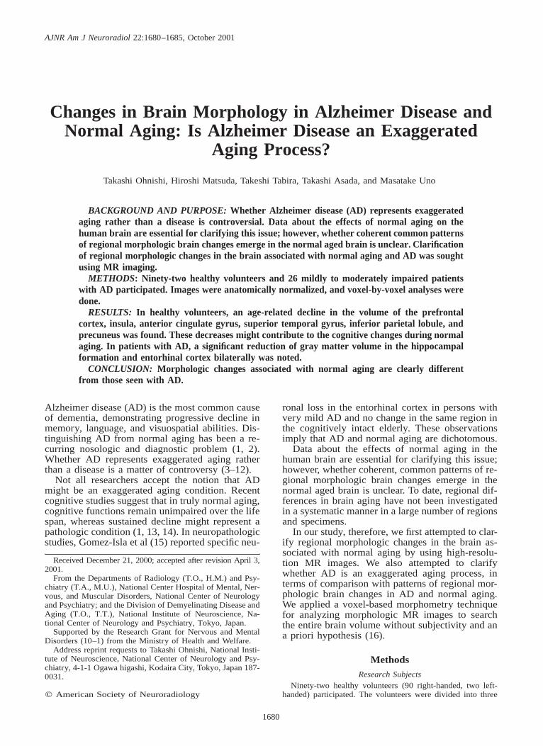
BACKGROUND AND PURPOSE: Whether Alzheimer disease (AD) represents exaggeratedaging rather than a disease is controversial. Data about the effects of normal aging on thehuman brain are essential for clarifying this issue; however, whether coherent common patternsof regional morphologic brain changes emerge in the normal aged brain is unclear. Clarificationof regional morphologic changes in the brain associated with normal aging and AD was soughtusing MR imaging.
METHODS: Ninety-two healthy volunteers and 26 mildly to moderately impaired patientswith AD participated. Images were anatomically normalized, and voxel-by-voxel analyses weredone.
RESULTS: In healthy volunteers, an age-related decline in the volume of the prefrontalcortex, insula, anterior cingulate gyrus, superior temporal gyrus, inferior parietal lobule, andprecuneus was found. These decreases might contribute to the cognitive changes during normalaging. In patients with AD, a significant reduction of gray matter volume in the hippocampalformation and entorhinal cortex bilaterally was noted.
CONCLUSION: Morphologic changes associated with normal aging are clearly differentfrom those seen with AD.
Alzheimer disease (AD) is the most common causeof dementia, demonstrating progressive decline inmemory, language, and visuospatial abilities. Dis-tinguishing AD from normal aging has been a re-curring nosologic and diagnostic problem (1, 2).Whether AD represents exaggerated aging ratherthan a disease is a matter of controversy (3–12).
Not all researchers accept the notion that ADmight be an exaggerated aging condition. Recentcognitive studies suggest that in truly normal aging,cognitive functions remain unimpaired over the lifespan, whereas sustained decline might represent apathologic condition (1, 13, 14). In neuropathologicstudies, Gomez-Isla et al (15) reported specific neu-
Received December 21, 2000; accepted after revision April 3,2001.
From the Departments of Radiology (T.O., H.M.) and Psy-chiatry (T.A., M.U.), National Center Hospital of Mental, Ner-vous, and Muscular Disorders, National Center of Neurologyand Psychiatry; and the Division of Demyelinating Disease andAging (T.O., T.T.), National Institute of Neuroscience, Na-tional Center of Neurology and Psychiatry, Tokyo, Japan.
Supported by the Research Grant for Nervous and MentalDisorders (10–1) from the Ministry of Health and Welfare.
Address reprint requests to Takashi Ohnishi, National Insti-tute of Neuroscience, National Center of Neurology and Psy-chiatry, 4-1-1 Ogawa higashi, Kodaira City, Tokyo, Japan 187-0031.
q American Society of Neuroradiology
ronal loss in the entorhinal cortex in persons withvery mild AD and no change in the same region inthe cognitively intact elderly. These observationsimply that AD and normal aging are dichotomous.
Data about the effects of normal aging in thehuman brain are essential for clarifying this issue;however, whether coherent, common patterns of re-gional morphologic brain changes emerge in thenormal aged brain is unclear. To date, regional dif-ferences in brain aging have not been investigatedin a systematic manner in a large number of regionsand specimens.
In our study, therefore, we first attempted to clar-ify regional morphologic changes in the brain as-sociated with normal aging by using high-resolu-tion MR images. We also attempted to clarifywhether AD is an exaggerated aging process, interms of comparison with patterns of regional mor-phologic brain changes in AD and normal aging.We applied a voxel-based morphometry techniquefor analyzing morphologic MR images to searchthe entire brain volume without subjectivity and ana priori hypothesis (16).
MethodsResearch Subjects
Ninety-two healthy volunteers (90 right-handed, two left-handed) participated. The volunteers were divided into three
AJNR: 22, October 2001 ALZHEIMER DISEASE 1681
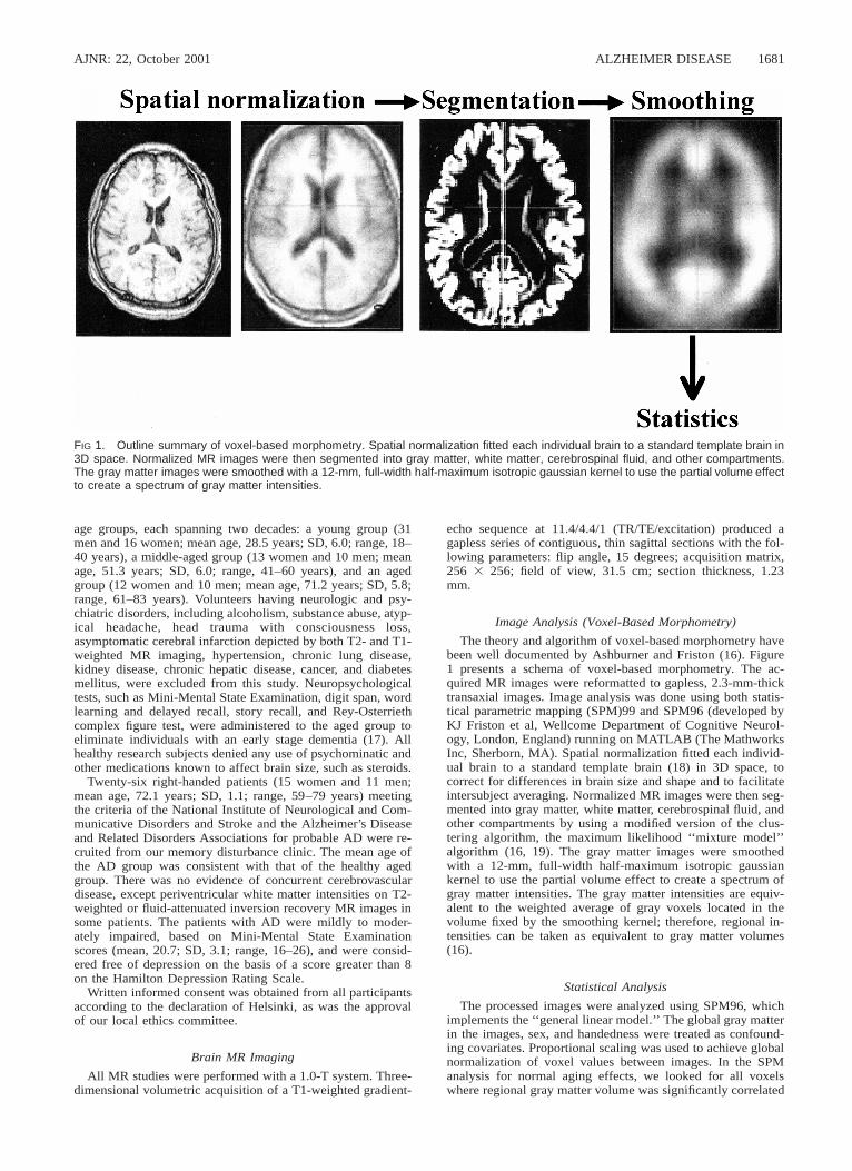
FIG 1. Outline summary of voxel-based morphometry. Spatial normalization fitted each individual brain to a standard template brain in3D space. Normalized MR images were then segmented into gray matter, white matter, cerebrospinal fluid, and other compartments.The gray matter images were smoothed with a 12-mm, full-width half-maximum isotropic gaussian kernel to use the partial volume effectto create a spectrum of gray matter intensities.
age groups, each spanning two decades: a young group (31men and 16 women; mean age, 28.5 years; SD, 6.0; range, 18–40 years), a middle-aged group (13 women and 10 men; meanage, 51.3 years; SD, 6.0; range, 41–60 years), and an agedgroup (12 women and 10 men; mean age, 71.2 years; SD, 5.8;range, 61–83 years). Volunteers having neurologic and psy-chiatric disorders, including alcoholism, substance abuse, atyp-ical headache, head trauma with consciousness loss,asymptomatic cerebral infarction depicted by both T2- and T1-weighted MR imaging, hypertension, chronic lung disease,kidney disease, chronic hepatic disease, cancer, and diabetesmellitus, were excluded from this study. Neuropsychologicaltests, such as Mini-Mental State Examination, digit span, wordlearning and delayed recall, story recall, and Rey-Osterriethcomplex figure test, were administered to the aged group toeliminate individuals with an early stage dementia (17). Allhealthy research subjects denied any use of psychominatic andother medications known to affect brain size, such as steroids.
Twenty-six right-handed patients (15 women and 11 men;mean age, 72.1 years; SD, 1.1; range, 59–79 years) meetingthe criteria of the National Institute of Neurological and Com-municative Disorders and Stroke and the Alzheimer’s Diseaseand Related Disorders Associations for probable AD were re-cruited from our memory disturbance clinic. The mean age ofthe AD group was consistent with that of the healthy agedgroup. There was no evidence of concurrent cerebrovasculardisease, except periventricular white matter intensities on T2-weighted or fluid-attenuated inversion recovery MR images insome patients. The patients with AD were mildly to moder-ately impaired, based on Mini-Mental State Examinationscores (mean, 20.7; SD, 3.1; range, 16–26), and were consid-ered free of depression on the basis of a score greater than 8on the Hamilton Depression Rating Scale.
Written informed consent was obtained from all participantsaccording to the declaration of Helsinki, as was the approvalof our local ethics committee.
Brain MR Imaging
All MR studies were performed with a 1.0-T system. Three-dimensional volumetric acquisition of a T1-weighted gradient-
echo sequence at 11.4/4.4/1 (TR/TE/excitation) produced agapless series of contiguous, thin sagittal sections with the fol-lowing parameters: flip angle, 15 degrees; acquisition matrix,256 3 256; field of view, 31.5 cm; section thickness, 1.23mm.
Image Analysis (Voxel-Based Morphometry)
The theory and algorithm of voxel-based morphometry havebeen well documented by Ashburner and Friston (16). Figure1 presents a schema of voxel-based morphometry. The ac-quired MR images were reformatted to gapless, 2.3-mm-thicktransaxial images. Image analysis was done using both statis-tical parametric mapping (SPM)99 and SPM96 (developed byKJ Friston et al, Wellcome Department of Cognitive Neurol-ogy, London, England) running on MATLAB (The MathworksInc, Sherborn, MA). Spatial normalization fitted each individ-ual brain to a standard template brain (18) in 3D space, tocorrect for differences in brain size and shape and to facilitateintersubject averaging. Normalized MR images were then seg-mented into gray matter, white matter, cerebrospinal fluid, andother compartments by using a modified version of the clus-tering algorithm, the maximum likelihood ‘‘mixture model’’algorithm (16, 19). The gray matter images were smoothedwith a 12-mm, full-width half-maximum isotropic gaussiankernel to use the partial volume effect to create a spectrum ofgray matter intensities. The gray matter intensities are equiv-alent to the weighted average of gray voxels located in thevolume fixed by the smoothing kernel; therefore, regional in-tensities can be taken as equivalent to gray matter volumes(16).
Statistical Analysis
The processed images were analyzed using SPM96, whichimplements the ‘‘general linear model.’’ The global gray matterin the images, sex, and handedness were treated as confound-ing covariates. Proportional scaling was used to achieve globalnormalization of voxel values between images. In the SPManalysis for normal aging effects, we looked for all voxelswhere regional gray matter volume was significantly correlated
AJNR: 22, October 20011682 OHNISHI
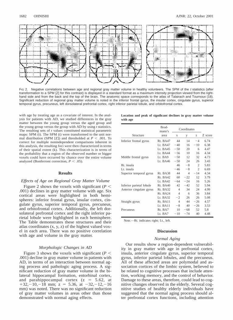
FIG 2. Negative correlations between age and regional gray matter volume in healthy volunteers. The SPM of the t statistics (aftertransformation to a SPM [Z] for this contrast) is displayed in a standard format as a maximum intensity projection viewed from the right-hand side and from the back and the top of the brain. The anatomic space corresponds to the atlas of Talairach and Tournoux (18).Significant reduction of regional gray matter volume is noted in the inferior frontal gyrus, the insular cortex, cingulate gyrus, superiortemporal gyrus, precuneus, left dorsolateral prefrontal cortex, right inferior parietal lobule, and orbitofrontal cortex.
Location and peak of significant declines in gray matter volumewith age
Structure
Brod-mann’s
area
Coordinates
x y z Z score
Inferior frontal gyrus Rt. BA47Lt. BA47Lt. BA45Lt. BA44
44240250256
14162010
24210
616
6.746.384.474.34
Middle frontal gyrus Lt. BA9Lt. BA46
250250
1224
3226
4.733.43
Rt. insulaLt. insula
46246
2828
22
5.836.69
Superior temporal gyrus Rt. BA38Rt. BA42Lt. BA42
4460
264
4222224
2141216
4.343.795.26
Inferior parietal lobule Rt. BA40 42 242 52 5.94Anterior cingulate gyrus Rt. BA32
Rt. BA24Lt. BA32
44
22
346
26
243626
4.993.845.08
Straight gyrus Rt. BA11Lt. BA11
428
4440
226226
4.573.53
Precuneus Rt. BA7Lt. BA7
10210
268274
5240
5.84.48
Note.—Rt. indicates right; Lt., left.
with age by treating age as a covariate of interest. In the anal-ysis for patients with AD, we studied differences in the graymatter between the young group versus the aged group andthe young group versus the group with AD by using t statistics.The resulting sets of t values constituted statistical parametricmaps: SPM (t). The SPM (t) were transformed to the unit nor-mal distribution (SPM [Z]) and thresholded at P , .001. Tocorrect for multiple nonindependent comparisons inherent inthis analysis, the resulting foci were then characterized in termsof their spatial extent (k). This characterization is in terms ofthe probability that a region of the observed number or biggervoxels could have occurred by chance over the entire volumeanalyzed (Bonferroni correction, P , .05).
Results
Effects of Age on Regional Gray Matter VolumeFigure 2 shows the voxels with significant (P ,
.001) declines in gray matter volume with age. Sixcortical areas were highlighted in both hemi-spheres: inferior frontal gyrus, insular cortex, cin-gulate gyrus, superior temporal gyrus, precuneus,and orbitofrontal cortex. Additionally, the left dor-solateral prefrontal cortex and the right inferior pa-rietal lobule were highlighted in each hemisphere.The Table demonstrates these structures and theiratlas coordinates (x, y, z) of the highest valued vox-el in each area. There was no positive correlationwith age and volume in the gray matter.
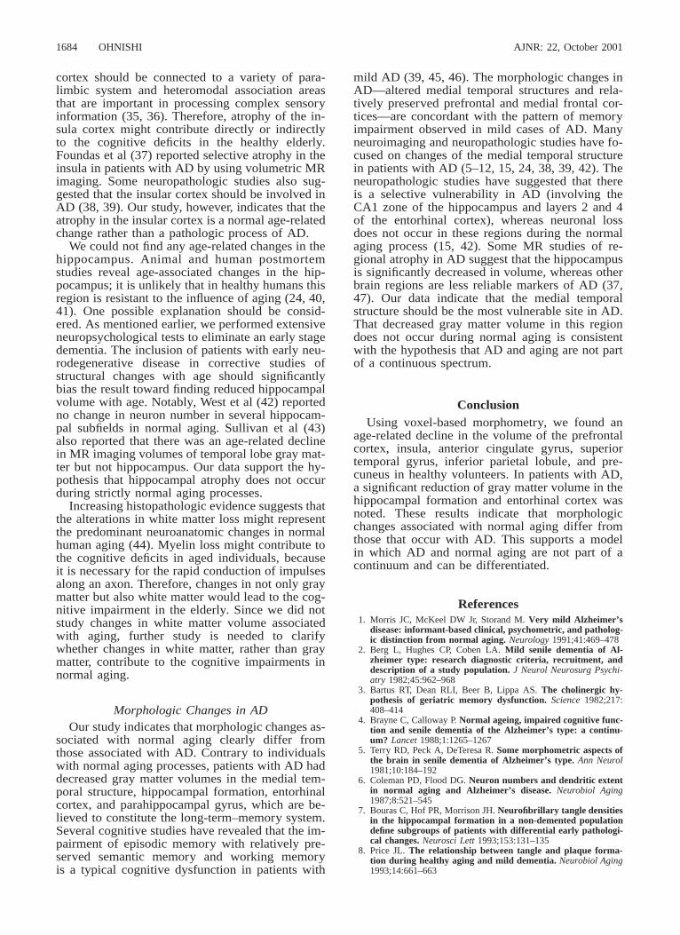
Morphologic Changes in ADFigure 3 shows the voxels with significant (P ,
.001) decline in gray matter volume in patients withAD, in terms of an interaction between normal ag-ing process and pathologic aging process. A sig-nificant reduction of gray matter volume in the bi-lateral hippocampal formation, entorhinal cortex,and parahippocampal cortex (z 5 5.62, at132,210,218 mm; z 5 5.36, at 232,212,216mm) was noted. There was no significant reductionof gray matter volumes in areas other than thosedemonstrated with normal aging effects.
Discussion
Normal AgingOur results show a region-dependent vulnerabil-
ity in gray matter with age in prefrontal cortex,insula, anterior cingulate gyrus, superior temporalgyrus, inferior parietal lobules, and the precuneus.All of these affected areas are polymodal and as-sociation cortices of the limbic system, believed tobe related to cognitive processes that include atten-tion, working memory, and the control of behavior.Damage to these areas, therefore, could lead to cog-nitive changes observed in the elderly. Several cog-nitive studies of healthy elderly individuals havesuggested that the normal aging process should al-ter prefrontal cortex functions, including attention

AJNR: 22, October 2001 ALZHEIMER DISEASE 1683
FIG 3. Significant reduction of regional gray matter volumes in AD, which was noted in the bilateral hippocampal formation, entorhinalcortex, and parahippocampal cortex.
(20, 21). Rabbitt (21) was one of the first research-ers to demonstrate experimentally that elderly sub-jects were less able to ignore task-irrelevant infor-mation. When aged people were tested with abattery of neuropsychological tests, frontal lobe def-icits seemed to be a primary component of theircognitive impairments (22). The results of thisstudy provide some support for these behavioraldata.
Several postmortem and in vivo neuroimagingstudies have suggested that prefrontal, entorhinal,and temporal cortices should be the most severelyaffected, whereas primary visual and somatosen-sory cortices might be more resistant to the influ-ence of aging (12, 23–26). Postmortem studieshave several limitations, such as technical and fix-ation artifacts, selection bias, and the influence ofthe illness and cause of death. In contrast, using invivo brain imaging techniques can avoid theseproblems and provide an opportunity to examinenormal brain morphology in healthy subjects. Todate, the most comprehensive volumetric MR studythat focused on regional changes in brain paren-chyma was reported by Raz et al (25). Because ofthe voxel-based morphometric method applied inthis study, it is difficult to directly compare ourresults with those from earlier volumetric MR stud-ies using region-of-interest analysis. Although thelatter approach has gained general acceptance, ithas a limitation: the sample selection is dependenton the observer’s a priori choice and hypothesis. Itleaves large areas of the brain unexplored. Our
voxel-based morphometric method is used to avoidsubjectivity and dependence on a priori hypothesisand to adopt the principle of data-driven analysis.Raz et al also reported that the most substantialage-related decline was found in the volume of theprefrontal gray matter. There are, however, somediscrepancies between their study and ours. Ourstudy indicates an age-related decline in gray mat-ter volume in the anterior cingulate gyri and insularcortices. Recent investigations with functional neu-roimaging have revealed that the anterior cingulategyrus should be involved in attentional processing,especially for divided attention (27, 28). Behavioralresearchers have established that changes in atten-tion functioning occur during aging (31, 32). Aconsistent finding from this research is that an age-related decline in performance is magnified whenattention must be divided among several relevantstimuli (29–32). In this context, it is reasonable tospeculate that the age-related change in the anteriorcingulate gyri shown in this study should be relatedto these cognitive changes.
To our knowledge, neither an in vivo morpho-logic neuroimaging study nor a postmortem studyhas reported age-related changes in the insular cor-tex. The changes in the insular cortex have beenignored, because of limitations of the methodolo-gies applied in earlier studies. Similar age-relatedchanges in regional cerebral blood flow and re-gional glucose metabolism, including insular de-cline, were demonstrated by voxel-based analysis(33, 34). Several studies suggest that the insular
AJNR: 22, October 20011684 OHNISHI
cortex should be connected to a variety of para-limbic system and heteromodal association areasthat are important in processing complex sensoryinformation (35, 36). Therefore, atrophy of the in-sula cortex might contribute directly or indirectlyto the cognitive deficits in the healthy elderly.Foundas et al (37) reported selective atrophy in theinsula in patients with AD by using volumetric MRimaging. Some neuropathologic studies also sug-gested that the insular cortex should be involved inAD (38, 39). Our study, however, indicates that theatrophy in the insular cortex is a normal age-relatedchange rather than a pathologic process of AD.
We could not find any age-related changes in thehippocampus. Animal and human postmortemstudies reveal age-associated changes in the hip-pocampus; it is unlikely that in healthy humans thisregion is resistant to the influence of aging (24, 40,41). One possible explanation should be consid-ered. As mentioned earlier, we performed extensiveneuropsychological tests to eliminate an early stagedementia. The inclusion of patients with early neu-rodegenerative disease in corrective studies ofstructural changes with age should significantlybias the result toward finding reduced hippocampalvolume with age. Notably, West et al (42) reportedno change in neuron number in several hippocam-pal subfields in normal aging. Sullivan et al (43)also reported that there was an age-related declinein MR imaging volumes of temporal lobe gray mat-ter but not hippocampus. Our data support the hy-pothesis that hippocampal atrophy does not occurduring strictly normal aging processes.
Increasing histopathologic evidence suggests thatthe alterations in white matter loss might representthe predominant neuroanatomic changes in normalhuman aging (44). Myelin loss might contribute tothe cognitive deficits in aged individuals, becauseit is necessary for the rapid conduction of impulsesalong an axon. Therefore, changes in not only graymatter but also white matter would lead to the cog-nitive impairment in the elderly. Since we did notstudy changes in white matter volume associatedwith aging, further study is needed to clarifywhether changes in white matter, rather than graymatter, contribute to the cognitive impairments innormal aging.
Morphologic Changes in ADOur study indicates that morphologic changes as-
sociated with normal aging clearly differ fromthose associated with AD. Contrary to individualswith normal aging processes, patients with AD haddecreased gray matter volumes in the medial tem-poral structure, hippocampal formation, entorhinalcortex, and parahippocampal gyrus, which are be-lieved to constitute the long-term–memory system.Several cognitive studies have revealed that the im-pairment of episodic memory with relatively pre-served semantic memory and working memoryis a typical cognitive dysfunction in patients with
mild AD (39, 45, 46). The morphologic changes inAD—altered medial temporal structures and rela-tively preserved prefrontal and medial frontal cor-tices—are concordant with the pattern of memoryimpairment observed in mild cases of AD. Manyneuroimaging and neuropathologic studies have fo-cused on changes of the medial temporal structurein patients with AD (5–12, 15, 24, 38, 39, 42). Theneuropathologic studies have suggested that thereis a selective vulnerability in AD (involving theCA1 zone of the hippocampus and layers 2 and 4of the entorhinal cortex), whereas neuronal lossdoes not occur in these regions during the normalaging process (15, 42). Some MR studies of re-gional atrophy in AD suggest that the hippocampusis significantly decreased in volume, whereas otherbrain regions are less reliable markers of AD (37,47). Our data indicate that the medial temporalstructure should be the most vulnerable site in AD.That decreased gray matter volume in this regiondoes not occur during normal aging is consistentwith the hypothesis that AD and aging are not partof a continuous spectrum.
ConclusionUsing voxel-based morphometry, we found an
age-related decline in the volume of the prefrontalcortex, insula, anterior cingulate gyrus, superiortemporal gyrus, inferior parietal lobule, and pre-cuneus in healthy volunteers. In patients with AD,a significant reduction of gray matter volume in thehippocampal formation and entorhinal cortex wasnoted. These results indicate that morphologicchanges associated with normal aging differ fromthose that occur with AD. This supports a modelin which AD and normal aging are not part of acontinuum and can be differentiated.
References1. Morris JC, McKeel DW Jr, Storand M. Very mild Alzheimer’s
disease: informant-based clinical, psychometric, and patholog-ic distinction from normal aging. Neurology 1991;41:469–478
2. Berg L, Hughes CP, Coben LA. Mild senile dementia of Al-zheimer type: research diagnostic criteria, recruitment, anddescription of a study population. J Neurol Neurosurg Psychi-atry 1982;45:962–968
3. Bartus RT, Dean RLI, Beer B, Lippa AS. The cholinergic hy-pothesis of geriatric memory dysfunction. Science 1982;217:408–414
4. Brayne C, Calloway P. Normal ageing, impaired cognitive func-tion and senile dementia of the Alzheimer’s type: a continu-um? Lancet 1988;1:1265–1267
5. Terry RD, Peck A, DeTeresa R. Some morphometric aspects ofthe brain in senile dementia of Alzheimer’s type. Ann Neurol1981;10:184–192
6. Coleman PD, Flood DG. Neuron numbers and dendritic extentin normal aging and Alzheimer’s disease. Neurobiol Aging1987;8:521–545
7. Bouras C, Hof PR, Morrison JH. Neurofibrillary tangle densitiesin the hippocampal formation in a non-demented populationdefine subgroups of patients with differential early pathologi-cal changes. Neurosci Lett 1993;153:131–135
8. Price JL. The relationship between tangle and plaque forma-tion during healthy aging and mild dementia. Neurobiol Aging1993;14:661–663
AJNR: 22, October 2001 ALZHEIMER DISEASE 1685
9. Price JL, Davis PB, Morris JC, White DL. The distribution oftangle, plaque and related immunohistochemical markers inhealthy aging and Alzheimer’s disease. Neurobiol Aging 1991;12:295–312
10. Delaere P, Duyckaerts C, Masters C. Large amounts of neocor-tical beta A4 deposits without neuritic plaques nor tangles ina psychometrically assessed, non-demented person. NeurosciLett 1990;166:87–93
11. Arnold SE, Hyman BT, Flory J. The topographical and neuro-anatomical distribution of neurofibrillary tangles and neuriticplaques in cerebral cortex of patients with Alzheimer’s disease.Cereb Cortex 1991;1:103–116
12. Arriagada PV, Marzoloff K, Hyman BT. Distribution of Alzhei-mer-type pathological changes in nondemented elderly match-es the pattern in Alzheimer’s disease. Neurology 1992;42:1681–1688
13. Morris JC, Fulling K. Early Alzheimer’s disease: diagnosticconsideration. Arch Neurol 1988;45:345–349
14. Linn RT, Wolf PA, Bachman DL. The ‘‘preclinical phase’’ ofprobable Alzheimer’s disease. Arch Neurol 1995;52:485–490
15. Gomez-Isla T, Price JL, McKeel DW, Morris JC, Growdon JH,Hyman BT. Profound loss of layer II entorhinal cortex neuronsoccurs in very mild Alzheimer’s disease. J Neurosci 1996;16:4491–4500
16. Ashburner J, Friston KJ. Voxel-based morphometry: the meth-ods. Neuroimage 2000;11:805–821
17. Kogure D, Matsuda H, Ohnishi T, et al. Longitudinal evaluationof early Alzheimer’s disease using brain perfusion SPECT. JNucl Med 2000;41:1155–1162
18. Talairach J, Tournoux P. Co-planar Stereotactic Atlas of the Hu-man Brain. Stuttgart, Germany: Thieme Verlag; 1988:
19. Hamilton M. A rating scale for depression. J Neurol NeurosurgPsychiatry 1960;23:56–62
20. Parkin AJ, Walter BM. Recollective experience, normal agingand frontal dysfunction. Psychol Aging 1992;7:340–345
21. Rabbitt PMA. An age decrement in the ability to ignore irrel-evant information. J Gerontol 1965;20:233–238
22. McDowd JM, Oseas-Kreger DM. Aging, inhibitory processesand negative priming. J Gerontol 1991;46:340–345
23. Haug H. Are neurons of the human cerebral cortex really lostduring aging? A morphometric examination. In: Tarber J, Gis-pen WH, eds. Senile Dementia of Alzheimer Type. Berlin, Ger-many: Springer-Verlag; 1985; 150–163
24. Kemper TL. Neuroanatomical and neuropathological changesduring aging and in dementia. In: Albert ML, Knoepfel EJE,eds. Clinical Neurology of Aging 2nd ed. New York, NY: OxfordUniversity Press; 1994:3–67
25. Raz N, Gunning FM, Head D, et al. Selective aging of the humancerebral cortex observed in vivo: differential vulnerability ofthe prefrontal gray matter. Cereb Cortex 1997;7:268–282
26. Heinsen H, Henn R, Eisenner W, et al. Quantitative investigationon human entorhinal cortex: left-right asymmetry and age-related changes. Anat Embryol 1994;190:181–194
27. Corbetta M, Meizin FM, Dobmeyer S, Shulman GL, Petersen SE.Selective and divided attention during visual discriminationsof shape, color and speed: functional anatomy by positronemission tomography. J Neurosci 1991;11:2383–2402
28. Pardo JV, Pardo PJ, Janer KW, Raichle ME. The anterior cin-gulate cortex mediates processing selection in the Stroop at-
tentional conflict paradigm. Proc Natl Acad Sci U S A 1990;87:256–259
29. Madden DJ, Plude DJ. Selective preservation of selective atten-tion. In: Cerebella J, Rybash J, Hoyer W, Commons ML, eds.Adult Information Processing: Limits on Loss. San Diego, Calif:Academic Press; 1993;273–300
30. MacDowd JM, Birren JE. Aging and attentional processes. In:Birren JE, Schaie KW, eds. Handbook of the Psychology of Aging3 rd ed. San Diego, Calif: Academic Press; 1990;222–233
31. Plude DJ, Hoyer WJ. Age and selectivity of visual informationprocessing. Psychol Aging 1986;1:4–10
32. Rabbitt P. Age and discrimination between complex stimuli. In:Welford AT, Birren JE, eds. Behavior, Aging, and the NervousSystem. Springfield, Ill: Charles C Thomas; 1965:35–53
33. Petit-Taboue MC, Landeau B, Desson JF, Desgranges B, BaronJC. Effects of healthy aging on the regional cerebral metabolicrate of glucose assessed with statistical parametric mapping.Neuroimage 1998;7:176–184
34. Martin AJ, Friston KJ, Colebatch JG, Frackowiak RS. Decreasesin regional cerebral blood flow with normal aging. J CerebBlood Flow Metab 1991;11:684–689
35. Augustine JR. Circuitry and functional aspects of the insularlobe in primates including humans. Brain Res Rev 1996;22:229–244
36. Mesulam MM, Mufson EJ. Neural inputs into the nucleus ba-salis of the substantia innominata (Ch4) in the rhesus monkey.Brain 1984;107:253–274
37. Foundas AL, Leonard CM, Mahoney SM, Agee OF, Heilman KM.Atrophy of the hippocampus, parietal cortex, and insula inAlzheimer’s disease: a volumetric magnetic resonance imagingstudy. Neuropsychiatr Neuropsychol Behav Neurol 1997;10:81–89
38. Braak H, Braak E. Neuropathological staging of Alzheimer-re-lated changes. Acta Neuropathol 1991;82:239–259
39. Braak H, Braak E. Staging of Alzheimer’s disease-related neu-rofibrillary changes. Neurobiol Aging 1995;16:271–278
40. Flood DG, Coleman PD. Neuron numbers and size in agingbrain: comparison of human, monkey and rodent data. Neu-robiol Aging 1988;9:453–463
41. Raz N. Neuroanatomy of aging brain: evidence from structuralMRI. In: Bigler ED, ed. Neuroimaging, II: Clinical ApplicationNew York, NY: Plenum Press; 1996:
42. West MJ, Coleman PD, Flood DG, Troncoso JC. Differences inthe pattern of hippocampal neuronal loss in normal ageing andAlzheimer’s disease. Lancet 1994;344:769–772
43. Sullivan EV, Marsh L, Mathalon DH, Lim KO, Pfefferbaum A.Age-related decline in MR volumes of temporal gray matterbut not hippocampus. Neurobiol Aging 1995;16:591–606
44. Wickelgren I. The aging brain: for the cortex, neuron loss maybe less than thought. Science 1996;273:48–50
45. Becker JT. Working memory and secondary memory deficitsin Alzheimer’s disease. J Clin Exp Neuropsychol 1988;10:739–753
46. Becker JT, Mintun MA, Diehl DJ. Functional neuroanatomy ofverbal free recall: a replication study. Hum Brain Map 1994;1:284–292
47. Deweer B, Lehericy S, Pillon B, Baulac M. Memory disorder inprobable Alzheimer’s disease: the role of hippocampal atrophyas shown with MRI. J Neurol Neurosurg Psychiatry 1995;58:590–597