Embed Size (px)
Citation preview
© 2013 Orza et al, publisher and licensee Dove Medical Press Ltd. This is an Open Access article which permits unrestricted noncommercial use, provided the original work is properly cited.
International Journal of Nanomedicine 2013:8 689–702
International Journal of Nanomedicine
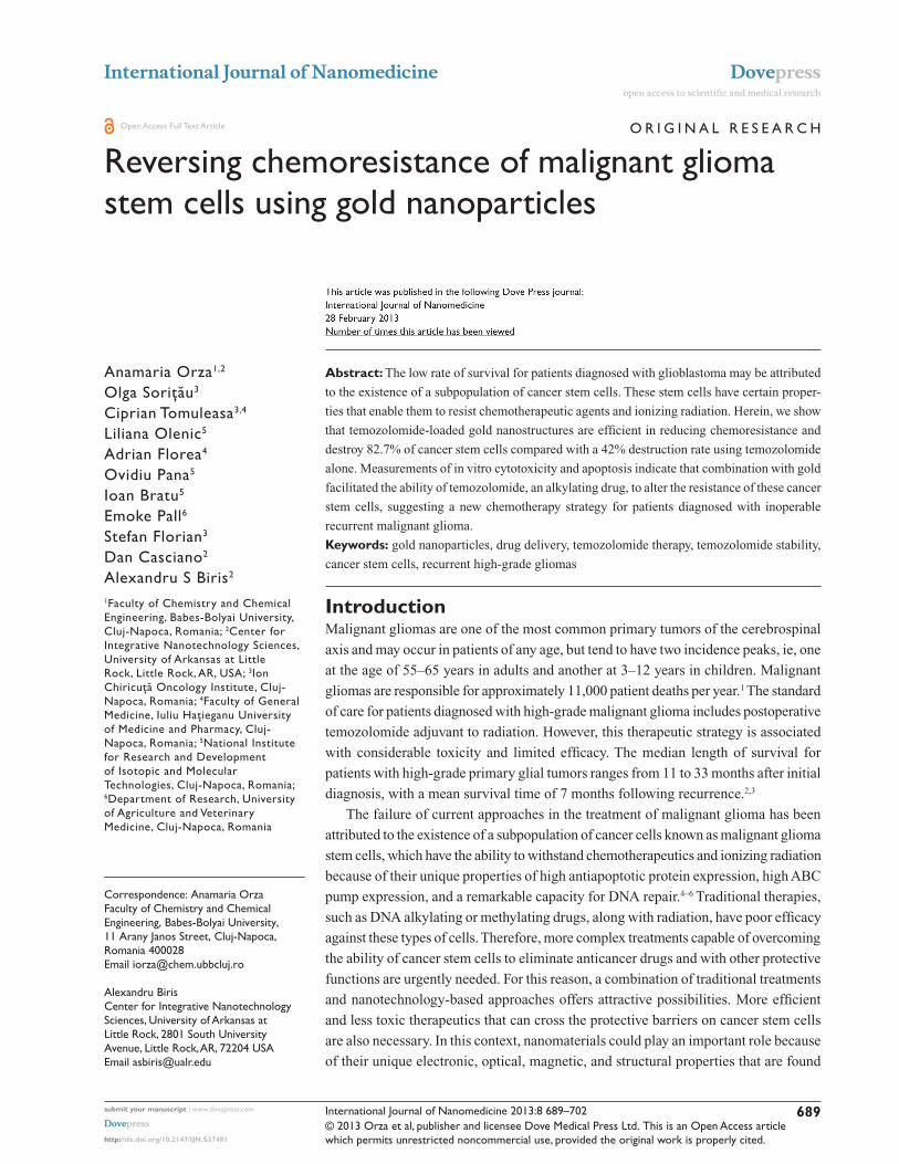
Reversing chemoresistance of malignant glioma stem cells using gold nanoparticles
Anamaria Orza1,2
Olga Soriţău3
Ciprian Tomuleasa3,4
Liliana Olenic5
Adrian Florea4
Ovidiu Pana5
Ioan Bratu5
Emoke Pall6
Stefan Florian3
Dan Casciano2
Alexandru S Biris2
1Faculty of Chemistry and Chemical Engineering, Babes-Bolyai University, Cluj-Napoca, Romania; 2Center for Integrative Nanotechnology Sciences, University of Arkansas at Little Rock, Little Rock, AR, USA; 3Ion Chiricuţă Oncology Institute, Cluj-Napoca, Romania; 4Faculty of General Medicine, Iuliu Haţieganu University of Medicine and Pharmacy, Cluj-Napoca, Romania; 5National Institute for Research and Development of Isotopic and Molecular Technologies, Cluj-Napoca, Romania; 6Department of Research, University of Agriculture and Veterinary Medicine, Cluj-Napoca, Romania
Correspondence: Anamaria Orza Faculty of Chemistry and Chemical Engineering, Babes-Bolyai University, 11 Arany Janos Street, Cluj-Napoca, Romania 400028 Email [email protected] Alexandru Biris Center for Integrative Nanotechnology Sciences, University of Arkansas at Little Rock, 2801 South University Avenue, Little Rock, AR, 72204 USA Email [email protected]
Abstract: The low rate of survival for patients diagnosed with glioblastoma may be attributed
to the existence of a subpopulation of cancer stem cells. These stem cells have certain proper-
ties that enable them to resist chemotherapeutic agents and ionizing radiation. Herein, we show
that temozolomide-loaded gold nanostructures are efficient in reducing chemoresistance and
destroy 82.7% of cancer stem cells compared with a 42% destruction rate using temozolomide
alone. Measurements of in vitro cytotoxicity and apoptosis indicate that combination with gold
facilitated the ability of temozolomide, an alkylating drug, to alter the resistance of these cancer
stem cells, suggesting a new chemotherapy strategy for patients diagnosed with inoperable
recurrent malignant glioma.
Keywords: gold nanoparticles, drug delivery, temozolomide therapy, temozolomide stability,
cancer stem cells, recurrent high-grade gliomas
IntroductionMalignant gliomas are one of the most common primary tumors of the cerebrospinal
axis and may occur in patients of any age, but tend to have two incidence peaks, ie, one
at the age of 55–65 years in adults and another at 3–12 years in children. Malignant
gliomas are responsible for approximately 11,000 patient deaths per year.1 The standard
of care for patients diagnosed with high-grade malignant glioma includes postoperative
temozolomide adjuvant to radiation. However, this therapeutic strategy is associated
with considerable toxicity and limited efficacy. The median length of survival for
patients with high-grade primary glial tumors ranges from 11 to 33 months after initial
diagnosis, with a mean survival time of 7 months following recurrence.2,3
The failure of current approaches in the treatment of malignant glioma has been
attributed to the existence of a subpopulation of cancer cells known as malignant glioma
stem cells, which have the ability to withstand chemotherapeutics and ionizing radiation
because of their unique properties of high antiapoptotic protein expression, high ABC
pump expression, and a remarkable capacity for DNA repair.4–6 Traditional therapies,
such as DNA alkylating or methylating drugs, along with radiation, have poor efficacy
against these types of cells. Therefore, more complex treatments capable of overcoming
the ability of cancer stem cells to eliminate anticancer drugs and with other protective
functions are urgently needed. For this reason, a combination of traditional treatments
and nanotechnology-based approaches offers attractive possibilities. More efficient
and less toxic therapeutics that can cross the protective barriers on cancer stem cells
are also necessary. In this context, nanomaterials could play an important role because
of their unique electronic, optical, magnetic, and structural properties that are found
Dovepress
submit your manuscript | www.dovepress.com
Dovepress 689
O R I G I N A L R E S E A R C h
open access to scientific and medical research
Open Access Full Text Article
http://dx.doi.org/10.2147/IJN.S37481
International Journal of Nanomedicine 2013:8
neither in bulk materials nor in single molecules and which
are necessary to develop advanced cancer treatments.
The ability of various gold nanoparticles to cross the
blood–brain barrier after intravenous administration has been
reported. Particles with the greatest ability to cross the blood–
brain barrier are those in the 15–50 nm size range.7,8 Herein,
we demonstrate binding of temozolomide to the surface of
55 nm gold triangle wires and uptake of the drug in the first
few hours, with progressive accumulation in the perinuclear
region of glioma stem cells. This gold nanoparticle vector is
a promising approach for increasing the efficacy of various
chemotherapies, could increase the active life of temozolo-
mide under physiological conditions, and may also facilitate
crossing of the blood–brain barrier, thereby facilitating greater
accumulation of the drug in brain cells and tissues.
Recently, gold nanoparticles have developed into an
attractive candidate for the specific delivery of complex
drugs, molecules, and/or biomolecules (proteins, DNA, and
RNA) to their targets. Because of their ability to accumulate
selectively in tumor tissue, gold nanoparticles can serve
as detectors of cancer cells9,10 or as targeted drug delivery
agents.11,12 Targeted delivery of anticancer drugs specifically
to the tumor site will maximize the efficacy of the drug and
minimize collateral damage by reducing systemic toxicity13
and hence reduce the side effects typically associated with
use of cytostatic agents in the treatment of cancer.
It is well known that a number of drugs when combined
with gold nanoparticles significantly attenuate toxicity in
various tumor cell lines. Several articles describing nano-
particle conjugations and their effects on different cancer
types have been published.14,15 Kahn et al have studied
decoration of gold nanoparticles with anti-epidermal growth
factor receptor in nasopharyngeal carcinoma (CNE2) and
normal human lung fibroblast cells.16 Localization of gold
nanoparticles through confocal imaging increases the reflec-
tance characteristics of the CNE2 cells, and the regions with
higher reflectance values, represent areas with high epidermal
growth factor receptor expression in cells. GNPs have also
been conjugated with anticancer drugs, such as methotrex-
ate and gemcitabine-epidermal growth factor receptor.17,18
In vitro tests have demonstrated that these gold nanoparticle
conjugates induce accumulation of methotrexate at higher
rates and in higher concentrations in tumor cells, and inhibit
proliferation of gemcitabine-treated pancreatic tumor cells.
Methotrexate-loaded gold nanoparticles have been shown
to achieve a higher cytotoxic response in various cancer cell
lines when compared to similar doses of free methotrexate.
Further, Liu has shown that gold nanoparticles modified with
polyethylene glycol can induce toxicity in CT26 tumor cells
exposed to irradiation.19
The novel properties of nanomaterials need to be explored
in order to improve traditional oncology management tech-
nologies and to develop high-profile treatments with low
overall toxicity to the organism and high efficacy in target-
ing and destroying cancer stem cells. In the present study,
we aimed to synthesize a drug delivery vector, by physically
coupling an anticancer drug, temozolomide, to the surface
of gold nanostructures treated with L-aspartate (GNP-L-
aspartate-TMZ) and to test their ability to kill malignant
glioma-derived cancer stem cells.20–22 The carboxyl group
from L-aspartate reduced Au3+ from tetrachloroauric acid
(HAuCl4 ) salt to Au, and the amine group capped the nano-
particles and organized them into high order GNP-L-aspartate
nanostructures. Recently, Kora et al demonstrated that func-
tional groups, such as COOH and OH from gum ghatti, have
the ability to reduce silver nanoparticles.23 Temozolomide
was conjugated with gold nanoparticles in order to obtain the
GNP-L-aspartate-TMZ delivery vector of interest. The mor-
phological, optical, and structural characteristics of the vector
were then investigated by transmission electron microscopy
(TEM), along with Fourier transform infrared spectroscopy
(FTIR), and x-ray photoelectron spectroscopy (XPS).
The in vitro effect of the GNP-L-aspartate-TMZ vector
on malignant glioma stem cells20 was tested using MTT
proliferation and Annexin V/propidium iodide apoptosis
assays and compared with the effect of temozolomide alone.
Our results indicated that the GNP-L-aspartate-TMZ system
produced highly statistically significant results. The conju-
gated drug appeared to be taken up by the cells in a pattern
very different from that of unbound temozolomide, as visu-
alized by optical microscopy. By binding the gold structure
to the drug, we improved the treatment of World Health
Organization grade III and IV tumors in the central nervous
system owing to the increased efficacy of the newly formed
compound. The dimer formed by the gold nanoparticles and
temozolomide was shown to be more readily absorbed intra-
cellularly by cancerous tissue than normal tissue because of
the surrounding edema and increased neovascularization. It
also had increased anticancer properties in comparison with
the current standard of care.
Materials and methodsGNP-L-aspartate nanostructure preparationPurified L-aspartate was purchased from Merck (White-
house Station, NJ, USA) and HAuCl4 from Fluka (Sigma-
submit your manuscript | www.dovepress.com
Dovepress
Dovepress
690
Orza et al

International Journal of Nanomedicine 2013:8
Aldrich Inc, St Louis, MO, USA). Pure temozolomide was
obtained from Sigma-Aldrich (Munich, Germany). HAuCl4
and L-aspartic acid solutions were prepared in concentrations
of 0.5 × 10⁻³ M and 1.5 × 10⁻³ M, respectively. We slowly
reduced a 50 mL solution of HAuCl4 at room temperature by
adding 15 mL of aspartic acid solution. During the reaction,
three color changes were observed, ie, slightly pink, red, and,
finally, after 9 hours, a persistent violet color. This indicates a
complex reduction mechanism involving formation of small
particles that act as nuclei for growth of triangular nanostruc-
tures on their {111} facets. The resulting product was then
centrifuged at 15,000 rpm for 45 minutes and washed with
Milli Q water (Millipore, Bedford, MA, USA). The puri-
fied nanostructures were redispersed in phosphate-buffered
saline to give a final concentration of 2 × 10⁻⁹ M. The purified
aspartate nanostructures (GNP-L-aspartate) were analyzed by
TEM, FTIR, UV-Vis and XPS spectroscopy.
Conjugation of temozolomide with gold nanostructuresThe simple method described below provides highly monodis-
persed GNP-L-aspartate nanostructures that are required as
intermediates in the preparation of the target delivery vector,
GNP-L-aspartate-TMZ. First, 200 µL of temozolomide
(50 µL/mL) was added to the GNP-L-aspartate nanostructure
solution (10 mL, 2 nM) and stirred for one hour. The result-
ing vector (GNP-L-aspartate-TMZ) was analyzed by TEM,
FTIR, UV-Vis and XPS spectroscopy.
Analytical characterization of multicomponent nanosystemsThe optical properties of the GNP-L-aspartate nanostructures
and GNP-L-aspartate-TMZ vector were monitored using an
ultraviolet visible spectrophotometer (JASCO V-570, Jasco
Inc, Easton, MD). The morphology of the GNP-L-aspartate
and GNP-L-aspartate-TMZ was investigated by TEM under
conventional beam conditions. Next, 30 µL droplets of
suspension were pipetted on copper grids (3 mm diameter,
300 meshes) previously covered with parlodion and carbon
films. After 2 minutes, the liquid was absorbed with filter paper.
Examination of the grids was performed using TEM (JEM
1010, JEOL, Tokyo, Japan). The images were captured using
a Mega VIEW III camera (Olympus, Soft Imaging System,
Münster, Germany) and entered into a database using Soft
Imaging System software (Münster, Germany). The diameter
of the nanoparticles was analyzed using CellD software
(Olympus Soft Imaging Solutions GMBH). Statistical analyses
(mean value, standard deviation, and Student’s t-test) were
performed using Microsoft Office Excel software (Microsoft
Corporation, Redmond, WA, USA). Fourier transform infrared
spectroscopy measurements were performed with a JASCO
6100 spectrometer in the 4000–500 cm−1 spectral region
with a resolution of 4 cm−1 using the KBr pellet technique.
X-ray photoelectron spectroscopy associated with argon ion
etching was used for qualitative and quantitative analysis of
the nanocomposites using a SPECS custom-built system.
Excitation was produced using the aluminum anode of the
x-ray source (hυ = 1486.6 eV).
Cancer stem cellsThe gold standard assay to determine whether a stem cell is or
is not a cancer stem cell involves a series of transplantations
in animal models. Potential surface markers of cancer stem
cells include CD133, aldehyde dehydrogenase 1, CD44, and
CD24. Efflux of Hoechst or rhodamine dyes (also referred to
as side populations) have also been employed for the iden-
tification of putative cancer stem cells. Still, these markers
have significant limitations given the fact that they fail to
identify all cancer stem cells and mostly recognize a cell
subpopulation with increased clonogenic and tumorigenic
activity. Moreover, not all cells presenting cancer stem cell
marker phenotypes present the same behavior as the cancer
stem cells. Most markers for the selection of cancer stem cells
are chosen because of being expressed in normal stem cells
and, most interestingly, there are quite a few molecules which
are commonly expressed in both normal and cancer stem
cells and which guide to various phenomena in dependency
to the particular environment.24
The cancer stem cells used in this study were isolated
from a glioblastoma multiforme biopsy as previously
described.20 Briefly, after mechanical dissociation of the
tumor tissue, the fragments were placed in 1 mL of fetal
calf serum. After 3 hours, 3 mL of Dulbecco’s modified
Eagle’s medium/F-12 medium supplemented with 15% fetal
calf serum was added to the dish. After reaching a subcon-
fluent monolayer, the cells were detached using trypsin-
ethylenediamine tetra-acetic acid (Merck) and resuspended
in a serum-free medium, ie, Dulbecco’s modified Eagle’s
medium/F-12 (1:1) medium supplemented with 15 ng/mL
basic fibroblast growth factor, 20 ng/mL epidermal growth
factor, 2 mM/L L-glutamine, 4 U/L insulin growth factor-1,
and B-27 supplement 1:50 (Sigma Aldrich). Isolated and
expanded cells revealed some stem cell-specific features,
including expression of cellular markers (CD133, CD105,
CD90, Nanog, Oct 3/4), immunocytochemistry expression
of specific genes, ie, CXCR4, nestin, glial fibrillary acidic
submit your manuscript | www.dovepress.com
Dovepress
Dovepress
691
Reversing chemoresistance of malignant glioma stem cells

International Journal of Nanomedicine 2013:8
protein, and neurofilament protein (reverse transcriptase
polymerase chain reaction). The cells also displayed a high
proliferative potential despite chemotherapy and irradiation,
and also had the ability to form spheroids in suspension.
All the studies were done by using exponentially growing
cells with a doubling time of approximately 24 hours. For the
passage, the medium was discarded and cells were washed
with phosphate-buffered solution and afterwards detached
with trypsin-ethylenediamine tetra-acetic acid 0.25%.
Proliferation assayCancer stem cells isolated from a high-grade glioblastoma
were cultivated in Dulbecco’s modified Essential medium
and Ham’s F-12 (Dulbecco’s modified Eagle’s medium/
F-12, ratio 1:1) medium supplemented with 15% fetal calf
serum, 100 U/mL penicillin, 100 µg/mL streptomycin,
1% nonessential amino acids, 2 mM glutamine, 55 µM
beta-mercaptoethanol, and 1 mM natrium pyruvate in a 37°C
humidified incubator containing a mixture of 95% air and
5% carbon dioxide.
Cell survival was assessed using the 3-(4,5-dimethylth-
iazol-2-yl)-2,5-diphenyltetrazolium bromide (MTT) assay,
for which cells in a monolayer culture were incubated in
complete medium before being washed twice with phosphate-
buffered solution. Cells were then incubated with trypsin-
ethylenediamine tetra-acetic acid, resuspended in culture
medium with fetal calf serum, counted, and plated in 100 µL
of medium at 15 × 103 cells/well in 96-well microtiter plates.
After 24 hours, the cells were washed and treated with temo-
zolomide alone or GNP-L-aspartate-TMZ. Temozolomide
was added at a concentration of 5 µg/mL. The MTT activity of
the GNP-L-aspartate-TMZ was compared with that of temo-
zolomide in identical concentrations. Absorbance of the MTT
was measured at 492 nm using a fluorescence microplate
reader (Synergy 2, BioTek, Winooski, VT, USA).
Annexin V/propidium iodide staining apoptosis assayGlioblastoma-derived stem cells were cultivated at subcon-
fluence in complete medium in 6 cm Petri dishes and were
exposed for 24 hours to temozolomide at a concentration
of 5 µg/mL or to GNP-L-aspartate-TMZ. The controls
were untreated cells and cells treated with GNP-L-aspartate
alone.
Apoptosis was evaluated using flow cytometry and optical
microscopy by staining with a FITC Annexin V and propidium
iodide kit (Invitrogen-Molecular Probes, Carlsbad, CA, USA).
After 24 hours of drug exposure, the cells were harvested
by trypsinization and washed with cold phosphate-buffered
solution and then resuspended in 100 µL of binding buffer.
Next, 5 µL of FITC Annexin V and 1 µL of propidium iodide
was added to each sample. The mixture was then incubated at
room temperature in the dark for 15 minutes. Early and late
apoptotic cells were identified using a BD FACSCanto™ II
flow cytometer (BD Biosciences, Franklin Lakes, NJ, USA)
measuring fluorescence emission at 530 nm and 575 nm with
488 nm excitation. A similar protocol was used for staining
adherent cells, and the samples were examined using an
inverted phase microscope with a 488 nm fluorescence filter
(Axiovert, Zeiss, Oberkochen, Germany). Image acquisition
was performed using an AxioCam MRC camera, and cell
counting of positive cells was performed using Axiovision
Rel 4.6 image analysis software. Cells positive for Annexin V
only are in an early stage of apoptosis, and cells positive for
both Annexin V and propidium iodide are either dead or in a
late stage of apoptosis.
Statistical analysisStatistically significant values were obtained by using one-
way variance analysis and a confidence level of 95% with a
GraphPad Prism 5 statistics program (La Jolla, CA, USA).
Data was analyzed with the Bonferroni multiple com-
parison test (Kruskal-Wallis as nonparametric). Statistical
significance was set at P , 0.05, and all experiments were
performed in triplicate.
Results and discussionSynthesis and characterization of GNP-L-aspartate-TMZ delivery vectorAspartate-stabilized gold nanoparticles were used for bind-
ing of temozolomide in order to form the desired final drug
delivery system. Aspartate was used for reduction and stabili-
zation in the synthesis of the nanostructures. Shape, size, and
the surface ligand are important parameters with distinctive
properties that must be chosen very carefully because they
influence cellular binding, incorporation, and/or cytotoxicity.
In order to establish the structure of the drug delivery system,
we studied the conjugation of temozolomide with the GNP-L-
aspartate nanostructures. The binding of temozolomide to the
surface of GNP-L-aspartate is an important parameter with a
significant impact on the overall stability of the nanostructural
multicomponent system.
First, the gold nanostructures were synthesized by reduc-
ing the gold salt, HAuCl4, in a strong acidic medium (pH 2)
in the presence of L-aspartate molecules. Briefly, under
continuous stirring, 0.25 × 10⁻⁵ moles of HAuCl4 were
submit your manuscript | www.dovepress.com
Dovepress
Dovepress
692
Orza et al

International Journal of Nanomedicine 2013:8
reduced with 2.25 × 10⁻⁴ moles of L-aspartate at room
temperature for 24 hours. Mixed chlorohydroxo complexes
(AuCl2O
2−3 or AuClO
34−) were present around the L-aspartate
molecules. Nucleation of the gold atoms took place very
slowly, with reduction occurring after 9 hours of reaction,
and was evident visually by a change of color from pale yel-
low to pink to red violet. Triangular nanoparticles having a
diameter of approximately 55 nm and aspartate as a ligand
were synthesized by this method and used as a delivery
vector for temozolomide. The resulting GNP-L-aspartate
nanostructures were purified by centrifugation and used fur-
ther in the reaction with temozolomide in order to obtain the
desired compound, ie, GNP-L-aspartate. TEM (Figure 1A)
and vibrational spectroscopy (Figure S1) were used to
characterize the structures. Further, these nanostructures were
used as building blocks for formation of the delivery vec-
tor, GNP-L-aspartate. Temozolomide was conjugated with
GNP-L-aspartate by formation of electrostatic bonds between
temozolomide and the aspartate molecules. Attachment was
confirmed by TEM, vibrational spectroscopy, and x-ray pho-
toelectron spectroscopy. TEM analysis along with diameter
histograms are shown in Figure 1A–C. The images confirmed
formation of the nanostructures and their triangular shape.
The length of the chains ranged from 100 nm to 600 nm, with
the majority in the range of 600 nm, and a mean diameter of
55 nm (see Figure 1A). A typical image showing the drug
delivery system is seen in Figure 1B. A thin shell of temo-
zolomide was seen to cover the nanostructure surface. It is
interesting to note that, after coupling, no secondary effects
such as nanoparticle aggregation appeared even one year after
807570656050 55Gold nanoparticles diameter (nm)
Nu
mb
er o
f p
arti
cles
454035300
2
4
6
8
10
12
600500
TMZD- 55 nmS.D- 3.8
GNPs-Asp-TMZ 50 µLGNPs-Asp
GNPs-Asp-TMZ 100 µLGNPs-Asp-TMZ 150 µL
λ (nm)
Wavenumber (1/cm)
Ab
sorb
ance
(au
)
Ab
sorb
ance
(au
)
400
200 nm 100 nm
3000.0
0.5
1.0
1.5
2.0
2.5
1000
TMZGNP-L-aspartate-TMZ
200030004000
1.5
1.2
0.9
0.6
0.3
0.0
GNPs-Asp-TMZ 200 µL
A B
C D
E
Figure 1 Transmission electron microscopic images of GNP-L-aspartate, showing a median diameter of 55 nm with a standard deviation of 3.8 (A), the GNP-L-aspartate-TMZ delivery system (B), a representative size distribution histogram of the gold nanoparticles (C), an ultraviolet-visible absorption spectrum confirming adsorption of the drug into the nanostructure (D), and Fourier transform infrared spectra of (black) pure temozolomide and (green) GNP-L-aspartate-TMZ (E).Abbreviations: GNP, gold nanoparticles; TMZ, temozolomide.
submit your manuscript | www.dovepress.com
Dovepress
Dovepress
693
Reversing chemoresistance of malignant glioma stem cells

International Journal of Nanomedicine 2013:8
their preparation. The presence of temozolomide on the gold
surfaces was also demonstrated by ultraviolet-visible spec-
troscopy (Figure 1D) and vibrational spectroscopy (Fourier
transform infrared, Figure 1E).
Ultraviolet visible spectroscopy clearly showed absorp-
tion of the drug onto the surfaces of the nanostructure
complexes. One peak at around 520 nm corresponds to
the gold nanoparticles, and the other two at approximately
300 nm and 350 nm are attributed to aspartic acid and
temozolomide, respectively. From the spectrum, we can also
see that the drug absorbs to the nanostructure surface until
equilibrium is reached. The quantity of drug that results in
equilibrium is 200 µL (Figure 1D).
The Fourier transform infrared spectrum for pure temozo-
lomide has three broad bands at 3339, 3381, and 3526 cm−1
given by the stretching vibration modes of NH2 and OH−,
while the two bands at around 2935 cm−1 and 2899 cm−1 are
related to the stretching vibrations of the aliphatic methylene
groups. After interaction with gold, two single bands appear
in this region, with one at 3389 cm−1 from the stretching
vibration of NH3+ groups and the other at 2934 cm−1 from the
vibration of C−H and CH3. This change is due to electrostatic
interaction of these groups with the gold nanoparticles.
In the middle of the spectrum at 1746 cm−1 is a four-
member ring from the ketone band which is similar to that of
pure temozolomide but more intense due to the presence of
gold in the structure. The amide I is indirectly influenced by
the NH2-nanostructure linkage and has a peak at 1682 cm−1,
whereas in the pure temozolomide the peak appears at
1672 cm−1. There are no similarities between the Fourier
transform infrared spectrum of pure temozolomide and that of
the temozolomide-loaded gold nanoparticle sample (all peaks
are rounded and shifted), suggesting that strong electrostatic
interactions, such as hydrogen bonds, Van der Waals forces,
or other mechanical or halogen bonds, can occur between
temozolomide molecules and gold nanostructures.
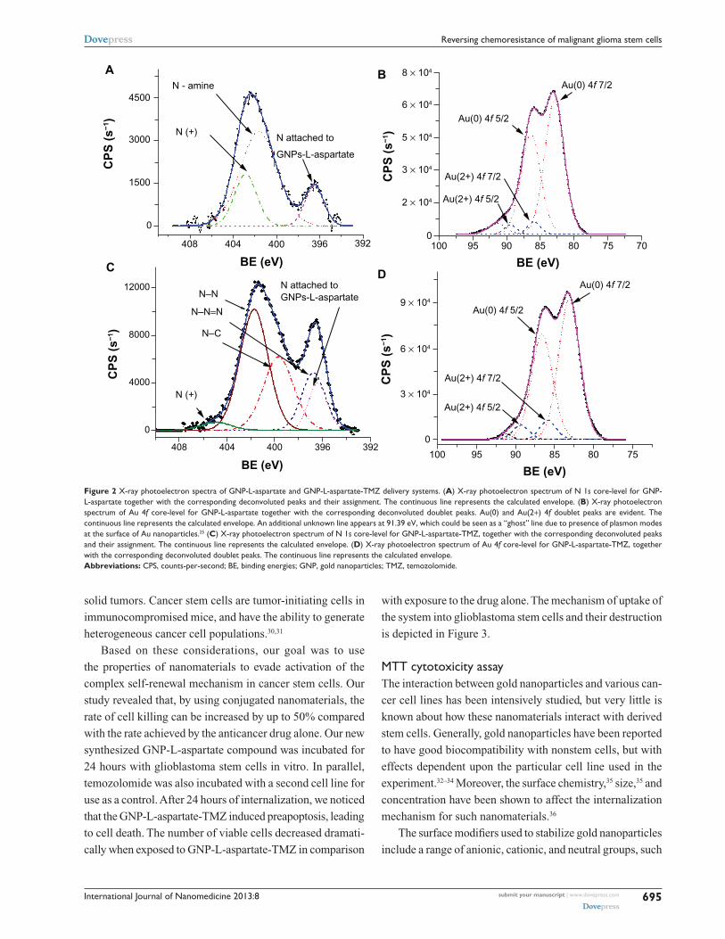
X-ray photoelectron spectroscopic analysis was uti-
lized to clarify the binding of temozolomide to the gold
surface by studying the N 1s and Au 4f core-level lines.
The deconvoluted x-ray photoelectron spectroscopic lines
of N 1s core-level 1 for the GNP-L-aspartate sample are
presented in Figure 2A, while Figure 2B shows the decon-
volution lines of Au 4f core-levels. For GNP-L-aspartate,
the recorded x-ray photoelectron spectroscopic spectrum
of N 1s core-level deconvoluted peaks and their assign-
ment are shown in Figure 2C, while the corresponding
Au 4f core-level deconvoluted doublet peaks are shown
in Figure 2D.
The following lines are identifiable in Figure 2A: a depro-
tonated N of the amine group bound (attached) to the gold
surface at 396.52 eV; an unattached N of the amine group
at 401.67 eV; and a deprotonated positive N (not bound) at
402.88 eV.25,26 Peak positions, line widths, and quantified area
are summarized in Table S1.
Quantification of the data was done by taking into account
real sensitivity, transmission, and mean free path factors. The
molar ratio of aspartic acid to gold nanoparticles was found
to be 0.22 by taking into account the attenuation lengths
for L-aspartic acid (about 2.1 nm) and gold nanoparticles
(1 nm). These values were calculated following the method
of Cumpson and Seah.27 The x-ray photoelectron spectra for
GNP-L-aspartate-TMZ are presented in Figure 2C and D for N
1s core-level 1 and Au 4f core-levels, respectively. Comparing
the x-ray photoelectron spectrum of the N 1s core-level
from GNP-L-aspartate (Figure 2A) with the corresponding
x-ray photoelectron spectrum from GNP-L-aspartate-TMZ
(Figure 2C), we can see that the intensities of the peaks and
the positions of the spectra have changed. These data led
us to conclude that the drug had bound to the surface of the
nanostructured gold by forming electrostatic bonds.
The molar ratio value obtained for GNP-L-aspartate-
TMZ was found to be 0.047. For its calculation, we took into
consideration the molar ratio value of aspartic acid to gold
nanoparticles (0.22) and the integrated intensity values. We
also used the lengths corresponding to the total integrated
intensity of Au 4f and N 1s.
Testing efficacy of GNP-L-aspartate-TMZ in glioblastoma stem cellsThe resemblances of the self-renewal mechanisms of the
cancer and stem cells, have resulted in the new concept of
cancer stem cells. Over the course of the past 15 years, there
has been increasing evidence to support the hypothesis of
cancer stem cells, which indicates that these cells might be
responsible for initiation, of tumors, metastasis, and their
complex resistance to treatment. Therefore, it could be pos-
sible that a tumor might have its origins in the cancer stem
cells, which could be originated from modified stem cells
or progenitor cells with re-obtained self-renewal activity.28
These rare cancer stem cells could play a major role in the
control and treatment of this disease: due to asymmetric divi-
sion, tumor development along with evade therapy given the
characteristics shared by normal stem cells, that include qui-
escence, self-renewal capacity, and drug resistance through
complex pump activity.29 These cells were first identified in
hematologic cancers, but have been recently isolated from
submit your manuscript | www.dovepress.com
Dovepress
Dovepress
694
Orza et al
International Journal of Nanomedicine 2013:8
solid tumors. Cancer stem cells are tumor-initiating cells in
immunocompromised mice, and have the ability to generate
heterogeneous cancer cell populations.30,31
Based on these considerations, our goal was to use
the properties of nanomaterials to evade activation of the
complex self-renewal mechanism in cancer stem cells. Our
study revealed that, by using conjugated nanomaterials, the
rate of cell killing can be increased by up to 50% compared
with the rate achieved by the anticancer drug alone. Our new
synthesized GNP-L-aspartate compound was incubated for
24 hours with glioblastoma stem cells in vitro. In parallel,
temozolomide was also incubated with a second cell line for
use as a control. After 24 hours of internalization, we noticed
that the GNP-L-aspartate-TMZ induced preapoptosis, leading
to cell death. The number of viable cells decreased dramati-
cally when exposed to GNP-L-aspartate-TMZ in comparison
with exposure to the drug alone. The mechanism of uptake of
the system into glioblastoma stem cells and their destruction
is depicted in Figure 3.
MTT cytotoxicity assayThe interaction between gold nanoparticles and various can-
cer cell lines has been intensively studied, but very little is
known about how these nanomaterials interact with derived
stem cells. Generally, gold nanoparticles have been reported
to have good biocompatibility with nonstem cells, but with
effects dependent upon the particular cell line used in the
experiment.32–34 Moreover, the surface chemistry,35 size,35 and
concentration have been shown to affect the internalization
mechanism for such nanomaterials.36
The surface modifiers used to stabilize gold nanoparticles
include a range of anionic, cationic, and neutral groups, such
392396400
BE (eV)
CP
S (
s−1)
404408
0
1500
3000
4500
392396400
BE (eV)
CP
S (
s−1)
404408
0
4000
8000
12000
707585 80
BE (eV)9095100
0
2 × 10⁴
3 × 10⁴
5 × 10⁴
6 × 10⁴
8 × 10⁴
7585 80
BE (eV)
CP
S (
s−1)
9095100
0
3 × 10⁴
6 × 10⁴
9 × 10⁴
A
C
B
D
N - amine
N (+)
N–C
N–N=N
N–N
Au(0) 4f 5/2
Au(0) 4f 7/2
Au(2+) 4f 7/2
Au(2+) 4f 5/2
Au(0) 4f 5/2
Au(0) 4f 7/2
Au(2+) 4f 7/2
Au(2+) 4f 5/2N (+)
N attached to
GNPs-L-aspartate
N attached toGNPs-L-aspartate
CP
S (
s−1)
Figure 2 X-ray photoelectron spectra of GNP-L-aspartate and GNP-L-aspartate-TMZ delivery systems. (A) X-ray photoelectron spectrum of N 1s core-level for GNP-L-aspartate together with the corresponding deconvoluted peaks and their assignment. The continuous line represents the calculated envelope. (B) X-ray photoelectron spectrum of Au 4f core-level for GNP-L-aspartate together with the corresponding deconvoluted doublet peaks. Au(0) and Au(2+) 4f doublet peaks are evident. The continuous line represents the calculated envelope. An additional unknown line appears at 91.39 eV, which could be seen as a “ghost” line due to presence of plasmon modes at the surface of Au nanoparticles.25 (C) X-ray photoelectron spectrum of N 1s core-level for GNP-L-aspartate-TMZ, together with the corresponding deconvoluted peaks and their assignment. The continuous line represents the calculated envelope. (D) X-ray photoelectron spectrum of Au 4f core-level for GNP-L-aspartate-TMZ, together with the corresponding deconvoluted doublet peaks. The continuous line represents the calculated envelope.Abbreviations: CPS, counts-per-second; BE, binding energies; GNP, gold nanoparticles; TMZ, temozolomide.
submit your manuscript | www.dovepress.com
Dovepress
Dovepress
695
Reversing chemoresistance of malignant glioma stem cells

International Journal of Nanomedicine 2013:8
Dead GCSCs
Uptake
Apoptosis
- Glioblastoma cancer stem cell (GCSCs)
- GNP-L-aspartate-TMZ
Exposure
Figure 3 Drug delivery system uptake and cancer cell destruction.Abbreviations: GNP, gold nanoparticles; TMZ, temozolomide.
as citrate, amine, and glucose. Thus, it has been concluded
that the interaction depends upon how the biological cells
respond to foreign materials in general, ie, the basic mecha-
nism of incorporation, degradation, or bioaccumulation caus-
ing cell damage and changes in gene expression.37,38
A very common method of evaluating the effects of dif-
ferent substances in cell cultures is the MTT assay, which is
used to measure the mitochondrial function of cells in vitro
(Figure 4A). Herein, glioma cancer stem cells were found
to interact strongly with our gold nanoparticle-based drug
delivery vectors. Phase contrast microscopy showed that
GNP-L-aspartate was internalized within the first hour (data
not shown here). After 24 hours, the drug delivery vector
affected the number of cells, their rate of proliferation, and
their shape, suggesting that the cells entered a preapoptotic
stage, as shown in Figure 4B. Hence, cells cultivated in the
presence of the nonconjugated drug were not yet preapoptotic
(Figure 4C). Glioma-derived stem cells were initially resis-
tant to temozolomide but, when GNP-L-aspartate-TMZ was
added to the culture medium, the results showed a reduction
in the rate of survival of the tumor cells.
Using Bonferroni’s multiple comparison test, we found
statistically significant (P < 0.05) decrease in proliferation for
cells treated with the conjugated vector, GNP-L-aspartate-
TMZ, compared with the cells treated with TMZ alone (95%
confidence interval 0.09214–0.5345). The killing efficiency
of the GNP-L-aspartate-TMZ system in glioblastoma-derived
stem cells is approximately 50% higher compared with that
of the alkaline drug, temozolomide, used alone. When one or
several initiating genetic changes appeared at the progenitor
level, all of the downstream cells continued this change. In
one particular case, it is possible that a daughter cell acquired
not only the characteristics of the stem cell, but also underwent
additional alterations that allowed the glioma to progress further
and invade adjacent tissues. Due to their small population of
glioblastoma-derived stem cells, malignant gliomas have a
negative response to various conventional treatments.39
Flow cytometry apoptosis testAnnexin V binding to the cell membrane by conjugation of
FITC was measured to identify and quantify apoptotic cells
at the single-cell level by flow cytometry or fluorescence
microscopy. Staining cells simultaneously with FITC-
Annexin V (green fluorescence) and the nonvital dye, propidium
iodide (red fluorescence) enables (bivariate analysis) to distin-
guish between intact cells (FITC negative, propidium iodide
negative), early apoptotic (FITC positive, propidium iodide
negative), and late apoptotic or necrotic cells (FITC positive,
propidium iodide positive). We performed the apoptotic tests
using the flow cytometry method and fluorescence microscopy.
submit your manuscript | www.dovepress.com
Dovepress
Dovepress
696
Orza et al

International Journal of Nanomedicine 2013:8
1.0
0.8
OD
0.4
0.6
0.2
0.0
CSC
CSC-TM
Z
CSC-GNPs-
L-as
parta
te-T
MZ
CSC-GNPs-
L-as
parta
te
BA
C
***
Figure 4 MTT viability assay results showing the response of glioma-derived stem cells to GNP-L-aspartate, to temozolomide, and to GNP-L-aspartate-TMZ. (A) Typical preapoptotic cell after 24 hours of incubation with GNP-L-aspartate-TMZ (PlasDIC phase contrast, magnification 400×), (B) in comparison with control cells treated with temozolomide alone, and (C) control cells cultured without GNP-L-aspartate-TMZ (white light microscopy, PlasDIC contrast phase, magnification 400×).Abbreviations: CSC, cancer stem cells; GNPs, gold nanoparticles; TMZ, temozolomide.
Table 1 Comparison of double staining with Annexin V/propidium iodide obtained by flow cytometry: percentage of early, late, and total apoptotic glioblastoma-derived cancer stem cells
Sample % early apoptotic cells
% late apoptotic + dead cells
Total
CSC control 0.4 0.4 0.8CSC control with GNP-L-aspartate
6.6 4.1 10.7
CSC treated with temozolomide
10.8 31.1 41. 9
CSC treated with GNP-L-aspartate-TMZ
4.4 78.3 82. 7
Abbreviations: CSC, cancer stem cells; GNP, gold nanoparticles; TMZ, temozolomide.
Analyzing the results of the flow cytometry apoptosis assay,
we observed a slight increase in apoptosis among cells treated
with gold nanoparticles (apoptotic cells represent 10.7% of total
cells by comparison with the level of 0.8% found in control
cells), a fact that was not found in the microscopy apoptosis
test (0.9% versus 1.24% for control cells). This observation
might be explained by the mechanical stress affecting the cells
loaded with nanoparticles during the centrifugation step for the
flow cytometry method, a step which was not included in the
microscopy test. Both apoptosis tests revealed an increase in
apoptosis among cells exposed to gold nanoparticles conjugated
with temozolomide. We noticed a dramatic enhancement of late
apoptosis in the flow cytometry studies by 78.3%, represents
a 1.9-fold increase within 24 hours compared with the rate of
apoptosis in cells treated with temozolomide alone (31.1%).
Similar results were obtained for the microscopic analysis,
with the observation that the apoptotic index of cells treated
with temozolomide-conjugated with gold nanoparticles was
3.41 times higher than that of cells treated with temozolomide
alone (Table 1).
Treatment with GNP-L-aspartate-TMZ induced
translocation of plasma membrane phosphatidylserine as an
early apoptosis event in cancer stem cells, and binding of
Annexin V-FITC was visualized by fluorescence microscopy
as shown in Figure 5. However, these cells retained a
morphology and proliferation rate close to that observed in the
control group. GNP-conjugated temozolomide-treated cells
showed positivity for Annexin V-FITC, a rounded shape, and
a decrease in cell numbers and adhesion (see Figure 5).
Our proposed nanoscale drug delivery system offers a
promising chemotherapeutic strategy for patients diagnosed
with unresectable recurrent malignant glioma. Current
therapies are not yet curative, because cancer stem cells may
survive as a result of the increased efflux of chemotherapy
agents due to ABCG2 cell membrane proteins and enhanced
DNA repair.40 Multiple proteins of the ATP-binding cassette
superfamily are being overexpressed by the cancer stem cells
submit your manuscript | www.dovepress.com
Dovepress
Dovepress
697
Reversing chemoresistance of malignant glioma stem cells

International Journal of Nanomedicine 2013:8
Control Control GNP TMZ GNP-TMZ
Figure 5 Morphologic aspects of cellular apoptosis studies for Annexin V FITC/propidium iodide staining observed in contrast phase and fluorescent microscopy (magnification 400×).Note: The upper panel shows images in fluorescence corresponding to those obtained in white light microscopy (lower panel).Abbreviations: GNP, gold nanoparticles; TMZ, temozolomide.
and produce a side population, with the capacity for enhanced
efflux of the fluorescent dyes (Hoechst 33342 or rhodamine
123) and which are transported by the same ABC family
proteins.41 Because therapy-resistant, high-grade gliomas are
one of the common causes of death due to cancer, and as a
result, there is an urgent need for identification of alternative
targeted and more efficient therapies. Such is the case with
specific drug-delivery systems using nanoparticles. Further
combination of various approaches to killing of individual
cancer cells, such as photothermolysis42,43 and radiofre-
quency-induced heating,44,45 which combine more accurate
drug release with additional biological damage produced by
heated nanomaterials, could further increase the ability of
nanostructured materials to serve as the foundation for more
sophisticated approaches to cancer treatment.
ConclusionCancer cells are very complex biological structures that per-
form a variety of functions, ranging from invasion or metasta-
sis to elimination of anticancer drugs from the cell membrane.
Although the exact mechanisms need to be explored further,
combining advances in fundamental oncology and nanotech-
nology offers the opportunity to impact future diagnostics
and therapeutics in a significant way. We have shown that
drug delivery vectors based on gold nanoparticles have the
ability to deliver temozolomide, a cytostatic drug, to treat
unresectable recurrent malignant glioma. Our studies have
also shown that a novel drug delivery vector based on gold
nanoparticles has low toxicity and the ability to internalize
temozolomide. MTT and flow cytometry studies have clearly
indicated the high bioactivity of the GNP-L-aspartate-TMZ
nanostructure system, with the ability to induce apoptosis
in almost 90% of high-grade glioma-derived cancer stem
cells. Temozolomide alone induced death in only 42% of
the cells. Our results indicate synergistic enhancement of
the apoptosis-inducing activity of GNP-L-aspartate-TMZ
vectors compared with GNP-L-aspartate or temozolomide
alone. This approach could constitute a novel method for
treating incurable recurrent malignant gliomas, and could
be developed further for other types of cancer models, and
potentially with great success.
AcknowledgmentsThis research was supported by a grant from the Romanian
Ministry of Research and Education (contract ID 1161).
ASB acknowledges the financial support of the US Army
Telemedicine and Advanced Technology Research Center
program. The editorial assistance of Marinelle Ringer is also
acknowledged.
DisclosureThe authors report no conflicts of interest in this work.
References1. Shapiro WR, Shapiro JR. Biology and treatment of malignant glioma.
Oncology. 1998;12:233–240.2. Henke G, Paulsen F, Steinbach JP, et al. Hypofractionated reirradiation
for recurrent malignant glioma. Strahlenther Onkol. 2009;185:113–119. German.
3. Buie LW, Valgus JM. Current treatment options for the management of glioblastoma multiforme. Hematol Oncol Pharm. 2012;2:57–63.
4. Eyler CE, Rich JN. Survival of the fittest: cancer stem cells in therapeutic resistance and angiogenesis. J Clin Oncol. 2008;26:2839–2845.
5. Li X, Lewis MT, Huang J, et al. Intrinsic resistance of tumorigenic breast cancer cells to chemotherapy. J Natl Cancer Inst. 2008;100:672–679.
6. Diehn M, Cho RW, Lobo NA, et al. Association of reactive oxygen species levels and radioresistance in cancer stem cells. Nature. 2009;458: 780–783.
7. De Jong WH, Hagens WI, Krystek P, Burger MC, Sips AJ, Geertsma RE. Particle size-dependent organ distribution of gold nanoparticles after intravenous administration. Biomaterials. 2008;29:1912–1919.
submit your manuscript | www.dovepress.com
Dovepress
Dovepress
698
Orza et al

International Journal of Nanomedicine 2013:8
8. Sonavane G, Tomoda K, Makino K. Biodistribution of colloidal gold nanoparticles after intravenous administration: effect of particle size. Colloids Surf B Biointerfaces. 2008;66:274–280.
9. Huang X, Jain PK, El-Sayed IH, El-Sayed MA. Gold nanoparticles: interesting optical properties and recent applications in cancer diag-nostics and therapy. Nanomedicine (Lond). 2007;2:681–693.
10. Kim BYS, Rutka JT, Chan WCW. Nanomedicine. N Engl J Med. 2010;363:2434–2443.
11. Lammers T, Subr V, Ulbrich K, et al. HPMA-based polymer therapeutics improve the efficacy of surgery, of radiotherapy and of chemotherapy combinations. Nanomedicine (Lond). 2010;5:1501–1523.
12. Grimm J, Scheinberg DA. Will nanotechnology influence targeted cancer therapy? Semin Radiat Oncol. 2011;21:80–87.
13. Dharap SS, Wang Y, Chandna P, et al. Tumor-specific targeting of an anticancer drug delivery system by LHRH peptide. Proc Natl Acad Sci U S A. 2005;102:12962–12967.
14. Han G, Ghosh P, Rotello VM. Functionalized gold nanoparticles for drug delivery. Nanomedicine. 2007;2:113–123.
15. Liong M, Lu J, Kovochich M, et al. Multifunctional inorganic nano-particles for imaging, targeting, and drug delivery. ACS Nano. 2008;2: 889–896.
16. Khan JA, Kudgus RA, Szabolcs A, et al. Designing nanoconjugates to effectively target pancreatic cancer cells in vitro and in vivo. PLoS One. 2011;6:e20347.
17. Chen YH, Tsai CY, Huang PY, et al. Methotrexate conjugated to gold nanoparticles inhibits tumor growth in a syngeneic lung tumor model. Mol Pharm. 2007;4:713–722.
18. Patra CR, Bhattacharya R, Wang E, et al. Targeted delivery of gem-citabine to pancreatic adenocarcinoma using cetuximab as a targeting agent. Cancer Res. 2008;68:1970–1978.
19. Liu C-J. Enhanced x-ray irradiation-induced cancer cell damage by gold nanoparticles treated by a new synthesis method of polyethylene glycol modification. Nanotechnology. 2008;19:295104.
20. Tomuleasa C, Soritau O, Rus-Ciuca D, et al. Functional and molecular characterization of glioblastoma multiforme derived cancer stem cells. J BUON. 2010;15:583–591.
21. Torcuator RG, Thind R, Patel M, et al. The role of salvage reirradia-tion for malignant gliomas that progress on bevacizumab. Neurooncol. 2010;97(3):401–407.
22. Stupp R, Mason WP, van den Bent MJ, et al. European Organisation for Research and Treatment of Cancer Brain Tumor and Radiotherapy Groups, National Cancer Institute of Canada Clinical Trials Group. Radiotherapy plus concomitant and adjuvant temozolomide for glioblastoma. N Engl J Med. 2005;352:987–996.
23. Kora AJ, Beedu SR, Jayaraman A. Size-controlled green synthesis of silver nanoparticles mediated by gum ghatti (Anogeissus latifolia) and its biological activity. Org Med Chem Lett. 2012;2:17.
24. Iwasaki H, Suda T. Cancer stem cells and their niche. Cancer Sci. 2009;100:1166–1172.
25. Leiro J, Minni E, Suoninen E. Study of plasmon structure in XPS spectra of silver and gold. J Phys F Met Phys. 1983;13:215–221.
26. Kang ET, Neoh KG, Tan KL. X-ray photoelectron spectroscopic studies of electroactive polymers advances. Polymer Science. 1993;106: 135–190.
27. Cumpson PJ, Seah MP. Elastic scattering corrections in AES and XPS. II. Estimating attenuation lengths and conditions required for their valid use in overlayer/substrate experiments. Surf Interface Anal. 1997;25:430–446.
28. Dittmar T, Nagler C, Schwitalla S, Reith G, Niggemann B, Zänker KS. Recurrence cancer stem cells – made by cell fusion? Med Hypotheses. 2009;73:542–547.
29. Fabian A, Barok M, Vereb G, Szollosi J. Die hard: are cancer stem cells the Bruce Willises of tumor biology? Cytometry A. 2009;75A: 67–74.
30. Keysar SB, Jimeno A. More than markers: biological significance of cancer stem cell-defining molecules. Mol Cancer Ther. 2010;9: 92450–92457.
31. Butterworth K, McMahon S, Currell F, Prise M. Physical basis and biological mechanisms of gold nanoparticle radiosensitization. Nanoscale. 2012;4:4830–4838.
32. Orza A, Soritau O, Olenic L, et al. Electrically conductive gold-coated collagen nanofibers for placental-derived mesenchymal stem cells enhanced differentiation and proliferation. ACS Nano. 2011;5: 4490–4500.
33. Sathuluri RR, Yoshikawa H, Shimizu E, Saito M, Tamiya E. Gold nanoparticle-based surface-enhanced Raman scattering for noninvasive molecular probing of embryonic stem cell differentiation. PLoS One. 2011;6:e22802.
34. Ricles LM, Nam SY, Sokolov K, Emelianov SY, Suggs LJ. Function of mesenchymal stem cells following loading of gold nanotracers. Int J Nanomedicine. 2011;6:407–416.
35. Wang Y, Wang Y, Wang L, Che Y, Li Z, Kong D. Preparation and evaluation of magnetic nanoparticles for cell labeling. J Nanosci Nanotechnol. 2011;11:3749–3756.
36. Nativo P, Prior IA, Brust M. Uptake and intracellular fate of surface-modified gold nanoparticles. ACS Nano. 2008;2:1639–1644.
37. Maurer-Jones MA, Bantz KC, Love SA, Marquis BJ, Haynes CL. Toxicity of therapeutic nanoparticles. Nanomedicine. 2009;4:219–241.
38. Zhang Y, Xu Y, Li Z, et al. Mechanistic toxicity evaluation of pristine and PEGylated single-walled carbon nanotubes in neuronal PC12 cells. ACS Nano. 2011;5:7020–7033.
39. Florian IS, Tomuleasa C, Soritau O, et al. Cancer stem cells and malignant gliomas. From pathophysiology to targeted molecular therapy. J BUON. 2011;16:16–23.
40. Frosina G. DNA repair and resistance of gliomas to chemotherapy and radiotherapy. Mol Cancer Res. 2009;7:989–999.
41. Baguley BC. Multidrug resistance in cancer. Methods Mol Biol. 2010;596:1–14.
42. Huang X, Kang B, Qian W, et al. Comparative study of photothermo-lysis of cancer cells with nuclear-targeted or cytoplasm-targeted gold nanospheres: continuous wave or pulsed lasers. J Biomed Opt. 2010;15: 058002.
43. Zharov VP, Galitovsky V, Viegas, M. Photothermal detection of local thermal effects during selective nanophotothermolysis. Appl Phys Lett. 2003;83:4897–4899.
44. Kruse DE, Stephens DN, Lindfors HA, Ingham ES, Paoli EE, Ferrara KW. A radio-frequency coupling network for heating of citrate-coated gold nanoparticles for cancer therapy: design and analysis. IEEE Trans Biomed Eng. 2011;58:2002–2012.
45. Maksimova IL, Akchurin GG, Khlebtsov BN, et al. Near-infrared laser photothermal therapy of cancer by using gold nanoparticles: computer simulations and experiment. Med Laser Appl. 2007;22:199–206.
46. Porter MD, Bright TB, Allara DL, Chidsey CE. Spontaneously orga-nized molecular assemblies. 4. Structural characterization of N-alkyl thiol monolayers on gold by optical ellipsometry, infrared spectroscopy, and electrochemistry. J Am Chem Soc. 1987;109:3559–3568.
submit your manuscript | www.dovepress.com
Dovepress
Dovepress
699
Reversing chemoresistance of malignant glioma stem cells

International Journal of Nanomedicine 2013:8
0.0
0.7
1.4
Ab
sorb
ance
Wavenumber (1/cm)
0.0
0.4
0.8
1.2
Ab
sorb
ance
Wavenumber (1/cm)
4000 3500 3000 2500
2000 1900 1800 1700 1600 1500 1400 1300
1200 1100 1000 900 800 700 600 500 400
0.0
0.4
0.8
1.2
Ab
sorb
ance
Wavenumber (1/cm)
TMZ GNPs-L-aspartate-TMZ GNPs-L-aspartate
TMZ GNPs-L-aspartate-TMZ
GNPs-L-aspartate-TMZ
GNPs-L-aspartate
TMZ
GNPs-L-aspartate
A
B
C
Figure S1 Fourier transform infrared spectra of the pure temozolomide (black), GNP-L-aspartate (green) and GNP-L-aspartate-TMZ (red) region. (A) 4000–2500 cm−1 region, (B) 2000–1300 cm−1 region, and (C) 1200–400 cm−1 region.Abbreviations: GNP, gold nanoparticles; TMZ, temozolomide.
Supplementary informationFourier transform infrared spectra of pure temozolomide versus GNP-L-aspartate and GNP-L-aspartate-TMZThe C−H stretching vibration groups at around 2750 cm−1
disappear when the drug is coupled with the nanostructures,
and only one peak appears in the spectra. This explains the
interaction of the drug with the gold surface. This is due to the
chain length of the amino acids and its molecules which have
a parallel orientation on the gold surface. These molecules
pack into densities sufficient to form high-quality barriers for
both electron and ion transfer processes.46 Also, from the other
regions of the Fourier transform infrared spectra, it is clear
that the drug is linked directly to the gold surface. We are not
denying the existence of aspartic acid in the molecules because
there may be some interaction. Please see the other two regions
of the Fourier transform infrared spectra (Figure S1A–C).
Fourier transform infrared spectra of GNP-L-aspartate vectorIn the middle of the spectrum (2000 cm−1), we can see only a
shoulder that could be related to a carbonyl group, the bending
submit your manuscript | www.dovepress.com
Dovepress
Dovepress
700
Orza et al

International Journal of Nanomedicine 2013:8
vibration of water at approximately 1650 cm−1, and the amide
and C−C double bond around 1500 cm−1. These peaks are
shifted to the right and their height is smaller compared with
the pure amino acid. The bending deformation vibrations
of C−H (methyl group) are given by a very weak peak at
1425 cm−1 (again because of their orientation parallel to the
gold surface). As can be seen in Figure S2A and B, there is no
similarity between the infrared spectrum of pure amino acid
and the one that is bound to the gold nanoparticle surface on
the gold nanoparticle-amino acid sample, suggesting that the
L-aspartate is bound to the surface of the gold nanoparticles,
most likely through the amino groups from the molecule.
4000 3500 3000 2500
0.0
0.5
1.0
Ab
sorb
ance
Wavenumber (1/cm)
Aspartic acid Aspartic acid + GNP
Aspartic acid Aspartic acid + GNP
A
2000 1500 1000 500
0.0
0.5
1.0
Ab
sorb
ance
Wavenumber (1/cm)
B
Figure S2 Fourier transform infrared spectra of (black) pure L-aspartate, (red) GNP-L-aspartate. (A) 4000–2500 cm−1 region and (B) 2000–500 cm−1 region.
submit your manuscript | www.dovepress.com
Dovepress
Dovepress
701
Reversing chemoresistance of malignant glioma stem cells

International Journal of Nanomedicine
Publish your work in this journal
Submit your manuscript here: http://www.dovepress.com/international-journal-of-nanomedicine-journal
The International Journal of Nanomedicine is an international, peer-reviewed journal focusing on the application of nanotechnology in diagnostics, therapeutics, and drug delivery systems throughout the biomedical field. This journal is indexed on PubMed Central, MedLine, CAS, SciSearch®, Current Contents®/Clinical Medicine,
Journal Citation Reports/Science Edition, EMBase, Scopus and the Elsevier Bibliographic databases. The manuscript management system is completely online and includes a very quick and fair peer-review system, which is all easy to use. Visit http://www.dovepress.com/ testimonials.php to read real quotes from published authors.
International Journal of Nanomedicine 2013:8
Table S1
Name BE (eV) FWHM (eV) Area (cps eV)
GNP-L-aspartateN 1s-attached to Au 396.53 1.801 308.555N 1s-amine 401.5 3.954 1415.19N(deprotoned) 1s 402.8 2.683 833.197Au(0) 4f 7/2 82.89 3.436 4919.83Au(0) 4f 5/2 86.39 3.436 4691.03Au(2+) 4f 7/2 85.99 2.334 323.185
Au(2+) 4f 5/2 89.49 2.334 308.156Unknown 91.39 3.409GNP-L-aspartate-TMZN 1s attached to Au 396.41 1.843 958.012N 1s N-N = N imide 396.78 2.34 1472.74N 1s N-C 399.7 2.991 2410.35N 1s N-N 401.76 2.794 3695.92N 1s (deprotoned) 404.95 2.999 267.455Au 4f (0)7/2 83.06 3.146 6637.89Au 4f (0)5/2 86.56 3.146 6329.2Au 4f (2+)7/2 89.19 2.576 776.509
Au 4f (2+)5/2 85.69 2.576 814.38Unknown 91.13 1.594
Notes: Peak positions, for GNP-L-aspartate and GNP-L-aspartate-TMZ, line-widths and quantified area are summarized.
submit your manuscript | www.dovepress.com
Dovepress
Dovepress
Dovepress
702
Orza et al


![Proteomic Cluster Analysis of Malignant Gliomas in Humansmejc.sums.ac.ir/article_42026_1aef94f007e5138fdf4bf14ea... · 2020. 9. 5. · 4% 3-[(3-cholamidopropyl) dimethylammonio]-1-propanesulfonate](https://img.pdfslide.us/doc/110x75/607bd1081041ba48f77df487/proteomic-cluster-analysis-of-malignant-gliomas-in-2020-9-5-4-3-3-cholamidopropyl.jpg)

![Molecular Cytogenetics | Home page - CASE REPORT Open … · 2017. 8. 25. · marked mitotic activity [3,4]. The molecular biology of adult malignant gliomas is now well defined for](https://img.pdfslide.us/doc/110x75/613a90260051793c8c011cd9/molecular-cytogenetics-home-page-case-report-open-2017-8-25-marked-mitotic.jpg)