Embed Size (px)
Citation preview
Parker et al. Skeletal Muscle 2012, 2:6http://www.skeletalmusclejournal.com/content/2/1/6
RESEARCH Open Access
Retracted: MyoD-dependent regulation of NF-κBactivity couples cell-cycle withdrawal to myogenicdifferentiationMaura H Parker1,2,3, Julia von Maltzahn2, Nadine Bakkar4, Ban Al-Joubori2, Jeff Ishibashi2, Denis Guttridge4
and Michael A Rudnicki5*
A notice has been published about this article. See full information at: http://www.skeletalmusclejournal.com/content/3/1/15
DRACT
EAbstract
Background: Mice lacking MyoD exhibit delayed skeletal muscle regeneration and markedly enhanced numbers ofsatellite cells. Myoblasts isolated from MyoD-/- myoblasts proliferate more rapidly than wild type myoblasts, display adramatic delay in differentiation, and continue to incorporate BrdU after serum withdrawal.
Methods: Primary myoblasts isolated from wild type and MyoD-/- mutant mice were examined by microarrayanalysis and further characterized by cell and molecular experiments in cell culture.
Results: We found that NF-κB, a key regulator of cell-cycle withdrawal and differentiation, aberrantly maintainsnuclear localization and transcriptional activity in MyoD-/- myoblasts. As a result, expression of cyclin D is maintainedduring serum withdrawal, inhibiting expression of muscle-specific genes and progression through thedifferentiation program. Sustained nuclear localization of cyclin E, and a concomitant increase in cdk2 activitymaintains S-phase entry in MyoD-/- myoblasts even in the absence of mitogens. Importantly, this deficit was rescuedby forced expression of IκBαSR, a non-degradable mutant of IκBα, indicating that inhibition of NF-κB is sufficient toinduce terminal myogenic differentiation in the absence of MyoD.
Conclusion: MyoD-induced cytoplasmic relocalization of NF-κB is an essential step in linking cell-cycle withdrawalto the terminal differentiation of skeletal myoblasts. These results provide important insight into the uniquefunctions of MyoD in regulating the switch from progenitor proliferation to terminal differentiation.
Keywords: Skeletal muscle, Myoblasts, MyoD, NF-κB, IKK, IκB, Differentiation, Myogenesis
ET
BackgroundCell survival and differentiation is regulated by NF-κB, afamily of ubiquitously expressed transcription factorscomprising RelA/p65, c-Rel, RelB, p50 (processed formof p105), and p52 (processed form of p100) [1]. NF-κBproteins function as homo- or heterodimers, the mostcommon of which is the p50/p65 heterodimer. All fam-ily members contain a DNA-binding domain, a protein-protein dimerization domain, and a nuclear localizationsequence (NLS). However, only RelA/p65, c-Rel, andRelB have a transactivation domain [2].
R* Correspondence: [email protected] Hospital Research Institute, 501 Smyth Rd, Ottawa, ON K1H 8L6,CanadaFull list of author information is available at the end of the article
© 2012 Parker et al; licensee BioMed Central.Commons Attribution License (http://creativecreproduction in any medium, provided the or
Sub-cellular localization of NF-κB is regulated by ‘in-hibitor of κB’ proteins: IκBα, IκBβ, and IκB [3]. IκB pro-teins bind NF-κB, mask the nuclear localization signal,and sequester NF-κB in the cytoplasm as an inactiveprotein. Upon induction, IκB kinases (IKKs) phosphoryl-ate IκB, releasing NF-κB and targeting IκB for degrad-ation. The released NF-κB enters the nucleus, bindsDNA, and regulates gene transcription. This process isnormally activated by molecules such as cytokines,growth factors, and bacterial products [4].Myogenic specification and differentiation requires the
myogenic regulatory factors (MRFs), namely MyoD, Myf5,myogenin, and MRF4/Myf6 [5]. The MRFs share a highlyhomologous basic helix-loop-helix (bHLH) domain, which
This is an Open Access article distributed under the terms of the Creativeommons.org/licenses/by/2.0), which permits unrestricted use, distribution, andiginal work is properly cited.
Parker et al. Skeletal Muscle 2012, 2:6 Page 2 of 12http://www.skeletalmusclejournal.com/content/2/1/6
RETR
is required for DNA binding and dimerization with theE-protein family of transcription factors. MRF-E-proteinheterodimers bind to the consensus E-box sequence,CANNTG, in gene promoters and regulate transcriptionalactivation.In particular, MyoD and Myf5 are essential for murine
skeletal muscle development [6]. However, mice lackingMyoD are viable and fertile, and display no overt pheno-type [7]. This indicates that myogenic specification anddifferentiation during embryonic and fetal developmentoccurs in the absence of MyoD, due to the presence ofother myogenic regulatory factors, such as Myf5. Ana-lysis of regeneration in MyoD-/- muscle established anessential role for MyoD in regulating adult myogenesis.In particular, increased numbers of satellite cells and adeficient muscle regenerative process in mice lackingMyoD (MyoD-/-), or MyoD and dystrophin (MyoD-/-:mdx), suggests that in the absence of MyoD, satellitecells have an increased propensity for self-renewal [8].Analysis of the differentiation kinetics of cultured
MyoD-/- satellite cell derived myoblasts revealed a markeddelay in differentiation, characterized by reduced expres-sion of differentiation specific markers such as myosinheavy chain, myogenin, MRF4, α-actins and acetylcholinereceptor-δ [9-11]. Although a portion of MyoD-/- cells ex-press myosin heavy chain (MyHC), these myocytes fail tofuse and remain primarily mononuclear. Moreover, themajority of MyoD-/- myoblasts display continued incorpor-ation of bromodeoxyuridine (BrdU) into DNA after serumwithdrawal, indicating DNA synthesis is maintained in theabsence of mitogen stimulation.In this study, we examined the role MyoD plays in regu-
lating cell-cycle withdrawal during terminal differentiationin adult myogenesis by undertaking a closer investigationof the molecular phenotype of MyoD-/- myoblasts. Weobserved that MyoD-/- myoblasts maintained nuclearlocalization of NF-κB after serum withdrawal, and dis-played altered expression of NF-κB target genes. In par-ticular, MyoD-/- myoblasts failed to down-regulate cyclinD1, an NF-κB target gene and key mediator of cell-cyclewithdrawal and differentiation in myoblasts [12,13]. Im-portantly, inhibition of NF-κB activity, through expressionof a mutant form of IκBα (IκB-SR), rescued the differenti-ation of MyoD-/- myoblasts. Therefore, we conclude thatMyoD controls cell-cycle withdrawal by regulating thesubcellular localization of the NF-κB family of transcrip-tion factors.
MethodsMyoblast isolation and cell cultureMyoblasts were isolated from 6 to 8 week old wild type(WT) Balb/C mice and MyoD-/- mice and cultured as pre-viously described [9]. To induce differentiation, the cellswere washed once with PBS and transferred to
ACTE
D
differentiation medium (DMEM supplemented with 5%horse serum (Invitrogen), and 2X penicillin/streptomycin).C2C12 murine myoblasts were cultured and differentiatedas previously described [14].
Transfections and luciferase assayC2C12 and MyoD-/- myoblasts were transfected in lowserum Opti-MEM using Lipofectamine (Invitrogen,Carlsbad, CA, USA) according to the manufacturer. Re-porter and expression plasmids were previously described[12], and all transfections were normalized to CMV-βGALexpression. For luciferase assays, cells were lysed in MPER(Pierce) and assays were performed as previouslydescribed [12]. MyoD siRNA was obtained from Santa-Cruz and transfections were performed using Lipofecta-mine 2000 (Invitrogen, Carlsbad, CA, USA).
RNA isolation and RNase protection assayRNA was isolated using TriZol Reagent (Invitrogen,Carlsbad, CA, USA) according to the manufacturer’sinstructions. The RNase protection assay was performedusing the RiboQuant kit according to the manufacturer’sinstructions (BD Bioscience, Franklin Lakes, NJ, USA).The relative amount of radioactivity present in eachband was quantitated using a Phosphorimager (GEHealthcare, Buckinghamshire, England), and the valuesobtained for each cyclin were normalized to the valuefor the GAPDH control.
Immunoblotting and antibodiesProteins were separated on 10% or 12% SDS-polyacryl-amide gel, and transferred to a polyvinylidene fluoridemembrane (Immobilon-P; Millipore, Billerica, MA, USA)according to established protocols. The antibodies usedwere all from Santa Cruz Biotechnology (Santa Cruz. CA,USA): anti- cyclin D1 (C-20), anti-cyclin D2 (H-289), anti-cyclin D3 (C-16), anti-cdk4 (C-22), anti-cyclin A1 (C-19),anti-cyclin E (C-19), anti-cdk2 (H-298), anti-cyclin H (C-18), anti-cdk7 (C-19), anti-NF-κB p65 (C-20), anti-Myf5(C-19), anti-MyoD (C-20), and anti-IKKγ (FL-419). For im-munoblotting, all antibodies were used according to themanufacturer’s instructions, normally at a dilution of 1:500or 1:1,000. Goat anti-mouse and goat anti-rabbit secondaryantibodies were used at 1:2,000 (BioRad, Hercules, CA,USA). Antibody-bound proteins were detected usingenhanced chemiluminescence (ECL; GE Healthcare,Buckinghamshire, England) and X-OMAT 5 X-ray film.
Kinase assaysEach 10-cm plate from a differentiation time course ofWT and MyoD-/- primary myoblasts were lysed with300 μL of NP-40 Lysis/IP buffer (50 mM Tris–HCl (pH7.5), 150 mM NaCl, 1 mM EDTA, 2.5 mM EGTA,1 mM DTT, 10% glycerol, 0.1 mM Na2VO3, 50 mM
Parker et al. Skeletal Muscle 2012, 2:6 Page 3 of 12http://www.skeletalmusclejournal.com/content/2/1/6
NaF, 20 mM β-glycerophosphate, 50 μg/mL PMSF, 2 μg/mL leupeptin, 1 μg/mL aprotinin, 10 μg/mL pepstatin).Immunoprecipitated cdk2 was incubated with 1 μg ofhistone H1 (Invitrogen) and 5 μCi of γ-32P-ATP (GEHealthcare, Buckinghamshire, England) in kinase buffer,and incubating at 30°C for 20 min. IKK kinase assayswere performed as previously described using a GST-IκBα substrate [15].
Gene expression analysisMouse U74Av2 GeneChip microarrays (Affymetrix, SantaClara, CA, USA) were used to analyse gene expression inwild type and MyoD-/- primary myoblasts [16]. A list ofgenes related to the NF-κB pathway was defined withreference to commercial microarrays (Panomics; Superar-ray Bioscience, Frederick, MD, USA) and the literature.Microarray data is available from StemBase (http://www.
RETR
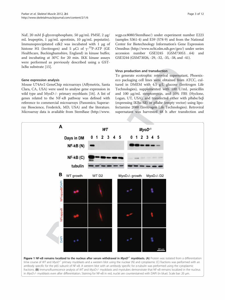
Figure 1 NF-κB remains localized to the nucleus after serum withdrawatime course of WT and MyoD-/- primary myoblasts and a western blot usingantibody specific for the p65 subunit of NF-κB. A western blot with an antifractions. (B) Immunofluorescence analysis of WT and MyoD-/- myoblasts ain MyoD-/- myoblasts even after differentiation. Staining for NF-κB in red, n
scgp.ca:8080/StemBase/) under experiment number E223(samples S361-4) and E59 (S78-9) and from the NationalCenter for Biotechnology Information’s Gene ExpressionOmnibus (http://www.ncbi.nlm.nih.gov/geo/) under seriesaccession number GSE3245 (GSM73053. . .64) andGSE3244 (GSM73026, -29, -32, -35, -38, and -41).
D
Virus production and transductionTo generate ecotrophic retroviral supernatant, Phoenix-eco packaging cell lines were obtained from ATCC, cul-tured in DMEM with 4.5 g/L glucose (Invitrogen LifeTechnologies), supplemented with 100 U/mL penicillinand 100 μg/mL streptomycin, and 10% FBS (Hyclone,Logan, UT, USA), and transfected either with pBabe/Iκβ(expressing IKBα-SR) or pBabe (empty vector) using lipo-fectamine 2000 (Invitrogen Life Technologies). Retroviralsupernatant was harvested 48 h after transfection and
ACTE
l in MyoD-/- myoblasts. (A) Protein was isolated from a differentiationthe nuclear (N) and cytoplasmic (C) fractions was performed with an
body specific for α-tubulin was performed using the cytoplasmicnd myotubes demonstrate that NF-κB remains localized in the nucleusuclei are counterstained with DAPI (in blue). Scale bar: 20 μm.
ACTE
D
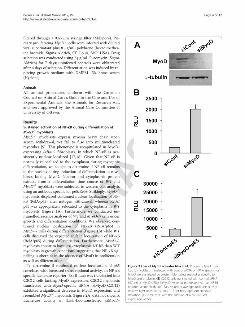
Figure 2 Loss of MyoD activates NF-κB. (A) Protein isolated fromC2C12 myoblasts transfected with control siRNA or siRNA specific forMyoD were analyzed by western blot using antibodies specific toMyoD and α-tubulin. (B) C2C12 cells transfected with control siRNA(siCont) or MyoD siRNA (siMyoD) were co-transfected with an NF-kBreporter vector (3xκB-Luc). Bars represent average luciferase activity(relative light units (RLUs)) (n = 3). Error bars represent standarddeviation. (C) Same as B, with the addition of a p65 (NF-κB)expression vector.
Parker et al. Skeletal Muscle 2012, 2:6 Page 4 of 12http://www.skeletalmusclejournal.com/content/2/1/6
RETR
filtered through a 0.45 μm syringe filter (Millipore). Pri-mary proliferating MyoD-/- cells were infected with dilutedviral supernatant plus 8 μg/mL polybrene (hexadimether-ine bromide, Sigma Aldrich, ST. Louis, MO, USA). Drugselection was conducted using 2 μg/mL Puromycin (SigmaAldrich) for 7 days; uninfected controls were obliteratedafter 4 days of selection. Differentiation was induced by re-placing growth medium with DMEM+5% horse serum(Hyclone).
AnimalsAll animal procedures conform with the CanadianCouncil on Animal Care’s Guide to the Care and Use ofExperimental Animals, the Animals for Research Act,and were approved by the Animal Care Committee atUniversity of Ottawa.
ResultsSustained activation of NF-κB during differentiation ofMyoD-/- myoblastsMyoD-/- myoblasts express myosin heavy chain uponserum withdrawal, yet fail to fuse into multinucleatedmyotubes [9]. This phenotype is recapitulated in MyoD-expressing IκBα-/- fibroblasts, in which NF-κB is per-sistently nuclear localized [17,18]. Given that NF-κB isnormally relocalized to the cytoplasm during myogenicdifferentiation, we sought to determine if NF-κB remainsin the nucleus during induction of differentiation in myo-blasts lacking MyoD. Nuclear and cytoplasmic proteinextracts from a differentiation time course of WT andMyoD-/- myoblasts were subjected to western blot analysisusing an antibody specific for p65/RelA. Strikingly, MyoD-/-
myoblasts displayed continued nuclear localization of NF-κB (RelA/p65) after mitogen withdrawal, whereas RelA/p65 was appropriately relocated to the cytoplasm in WTmyoblasts (Figure 1A). Furthermore we conducted im-munofluorescence analyses of WT and MyoD-/- cells undergrowth and differentiation conditions. We observed con-tinued nuclear localization of NF-κB (RelA/p65) inMyoD-/- cells during differentiation (Figure 1B) while WTcells displayed the expected shift in localization of NF-κB(RelA/p65) during differentiation. Furthermore, MyoD-/-myoblasts appear to have less cytoplasmic NF-kB than WTmyoblasts in growth conditions, suggesting that NF-κB sig-nalling is aberrant in the absence of MyoD in proliferationas well as differentiation.To determine if continued nuclear localization of p65
correlates with increased transcriptional activity, an NF-κBspecific luciferase reporter (3xκB-Luc) was transfected intoC2C12 cells lacking MyoD expression. C2C12 myoblaststransfected with MyoD-specific siRNA (siMyoD-C2C12)exhibited a significant decrease in MyoD expression, andresembled MyoD-/- myoblasts (Figure 2A, data not shown).Luciferase activity in 3xκB-Luc-transfected siMyoD-
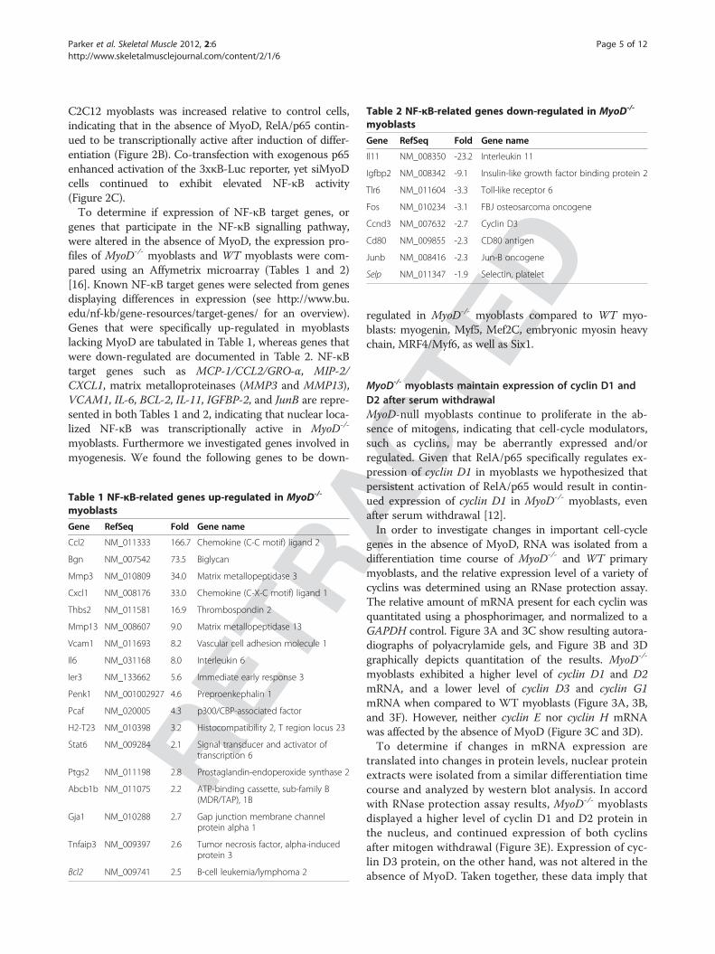
Table 2 NF-κB-related genes down-regulated in MyoD-/-
myoblasts
Gene RefSeq Fold Gene name
Il11 NM_008350 -23.2 Interleukin 11
Igfbp2 NM_008342 -9.1 Insulin-like growth factor binding protein 2
Tlr6 NM_011604 -3.3 Toll-like receptor 6
Fos NM_010234 -3.1 FBJ osteosarcoma oncogene
Ccnd3 NM_007632 -2.7 Cyclin D3
Cd80 NM_009855 -2.3 CD80 antigen
Junb NM_008416 -2.3 Jun-B oncogene
Selp NM_011347 -1.9 Selectin, platelet
Parker et al. Skeletal Muscle 2012, 2:6 Page 5 of 12http://www.skeletalmusclejournal.com/content/2/1/6
C2C12 myoblasts was increased relative to control cells,indicating that in the absence of MyoD, RelA/p65 contin-ued to be transcriptionally active after induction of differ-entiation (Figure 2B). Co-transfection with exogenous p65enhanced activation of the 3xκB-Luc reporter, yet siMyoDcells continued to exhibit elevated NF-κB activity(Figure 2C).To determine if expression of NF-κB target genes, or
genes that participate in the NF-κB signalling pathway,were altered in the absence of MyoD, the expression pro-files of MyoD-/- myoblasts and WT myoblasts were com-pared using an Affymetrix microarray (Tables 1 and 2)[16]. Known NF-κB target genes were selected from genesdisplaying differences in expression (see http://www.bu.edu/nf-kb/gene-resources/target-genes/ for an overview).Genes that were specifically up-regulated in myoblastslacking MyoD are tabulated in Table 1, whereas genes thatwere down-regulated are documented in Table 2. NF-κBtarget genes such as MCP-1/CCL2/GRO-α, MIP-2/CXCL1, matrix metalloproteinases (MMP3 and MMP13),VCAM1, IL-6, BCL-2, IL-11, IGFBP-2, and JunB are repre-sented in both Tables 1 and 2, indicating that nuclear loca-lized NF-κB was transcriptionally active in MyoD-/-
myoblasts. Furthermore we investigated genes involved inmyogenesis. We found the following genes to be down-
RETR
Table 1 NF-κB-related genes up-regulated in MyoD-/-
myoblasts
Gene RefSeq Fold Gene name
Ccl2 NM_011333 166.7 Chemokine (C-C motif) ligand 2
Bgn NM_007542 73.5 Biglycan
Mmp3 NM_010809 34.0 Matrix metallopeptidase 3
Cxcl1 NM_008176 33.0 Chemokine (C-X-C motif) ligand 1
Thbs2 NM_011581 16.9 Thrombospondin 2
Mmp13 NM_008607 9.0 Matrix metallopeptidase 13
Vcam1 NM_011693 8.2 Vascular cell adhesion molecule 1
Il6 NM_031168 8.0 Interleukin 6
Ier3 NM_133662 5.6 Immediate early response 3
Penk1 NM_001002927 4.6 Preproenkephalin 1
Pcaf NM_020005 4.3 p300/CBP-associated factor
H2-T23 NM_010398 3.2 Histocompatibility 2, T region locus 23
Stat6 NM_009284 2.1 Signal transducer and activator oftranscription 6
Ptgs2 NM_011198 2.8 Prostaglandin-endoperoxide synthase 2
Abcb1b NM_011075 2.2 ATP-binding cassette, sub-family B(MDR/TAP), 1B
Gja1 NM_010288 2.7 Gap junction membrane channelprotein alpha 1
Tnfaip3 NM_009397 2.6 Tumor necrosis factor, alpha-inducedprotein 3
Bcl2 NM_009741 2.5 B-cell leukemia/lymphoma 2
Dregulated in MyoD-/- myoblasts compared to WT myo-blasts: myogenin, Myf5, Mef2C, embryonic myosin heavychain, MRF4/Myf6, as well as Six1.
ACTEMyoD-/- myoblasts maintain expression of cyclin D1 and
D2 after serum withdrawalMyoD-null myoblasts continue to proliferate in the ab-sence of mitogens, indicating that cell-cycle modulators,such as cyclins, may be aberrantly expressed and/orregulated. Given that RelA/p65 specifically regulates ex-pression of cyclin D1 in myoblasts we hypothesized thatpersistent activation of RelA/p65 would result in contin-ued expression of cyclin D1 in MyoD-/- myoblasts, evenafter serum withdrawal [12].In order to investigate changes in important cell-cycle
genes in the absence of MyoD, RNA was isolated from adifferentiation time course of MyoD-/- and WT primarymyoblasts, and the relative expression level of a variety ofcyclins was determined using an RNase protection assay.The relative amount of mRNA present for each cyclin wasquantitated using a phosphorimager, and normalized to aGAPDH control. Figure 3A and 3C show resulting autora-diographs of polyacrylamide gels, and Figure 3B and 3Dgraphically depicts quantitation of the results. MyoD-/-
myoblasts exhibited a higher level of cyclin D1 and D2mRNA, and a lower level of cyclin D3 and cyclin G1mRNA when compared to WT myoblasts (Figure 3A, 3B,and 3F). However, neither cyclin E nor cyclin H mRNAwas affected by the absence of MyoD (Figure 3C and 3D).To determine if changes in mRNA expression are
translated into changes in protein levels, nuclear proteinextracts were isolated from a similar differentiation timecourse and analyzed by western blot analysis. In accordwith RNase protection assay results, MyoD-/- myoblastsdisplayed a higher level of cyclin D1 and D2 protein inthe nucleus, and continued expression of both cyclinsafter mitogen withdrawal (Figure 3E). Expression of cyc-lin D3 protein, on the other hand, was not altered in theabsence of MyoD. Taken together, these data imply that
RACT
EDFigure 3 MyoD-/- myoblasts maintain cyclin D expression after serum withdrawal. (A, C) RNA was isolated from a differentiation timecourse of WT and MyoD-/- primary myoblasts and an RNase protection assay was performed with the indicated probes. The protected 32P-labeledfragments were separated on a polyacrylamide gel, the gel dried and exposed to X-ray film. (B, D) The amount of each protected fragment wasquantitated using a phosphorimager. The values obtained were normalized relative to the GAPDH control and the L32 control, and graphed as afunction of the number of days in differentiation medium (DM), n = 3. (E) Nuclear protein was isolated from a differentiation time course of WTand MyoD-/- primary myoblasts and a western blot was performed with the indicated antibodies. (F) Quantitative real-time PCR for Cyclin D3 ofWT and MyoD-/- myoblasts, n = 3.
Parker et al. Skeletal Muscle 2012, 2:6 Page 6 of 12http://www.skeletalmusclejournal.com/content/2/1/6
RETpersistent activation of RelA/p65 is responsible for ex-
pression of cyclin D1 in MyoD-/- myoblasts after serumwithdrawal.pRb is also phosphorylated by cyclin E-cdk2, and this
phosphorylation is required for S-phase entry [19]. More-over, forced expression of cyclin E bypasses the need forcyclin D/cdk4 activity in cell-cycle progression from G1 toS phase [20,21]. Expression of cyclin E mRNA was notaltered in the absence of MyoD, indicating that the in-crease in cyclin D1 expression is not inducing cell-cycleprogression through stimulation of E2F activity on the cyc-lin E promoter (Figure 3C and D).However, overexpression of cyclin D1 may also inhibit
MRF activity and myogenic differentiation through amechanism independent of pRb or MyoD phosphoryl-ation [22-24]. Indeed, a hypophosphorylated mutant of
pRb (pRbΔp35; EF), which induces cell-cycle with-drawal, is unable to rescue cyclin D-dependent inhib-ition of muscle-specific gene expression (MH Parker,MA Rudnicki, unpublished observations).
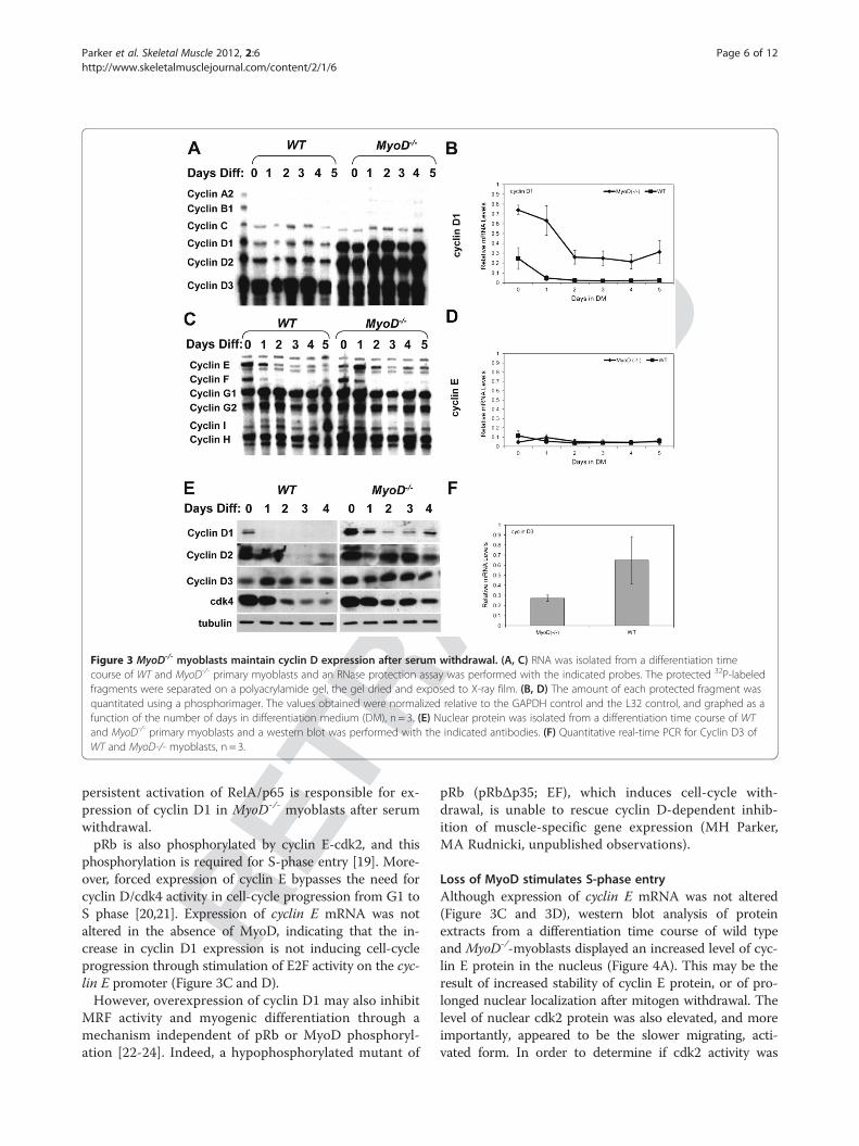
Loss of MyoD stimulates S-phase entryAlthough expression of cyclin E mRNA was not altered(Figure 3C and 3D), western blot analysis of proteinextracts from a differentiation time course of wild typeand MyoD-/-myoblasts displayed an increased level of cyc-lin E protein in the nucleus (Figure 4A). This may be theresult of increased stability of cyclin E protein, or of pro-longed nuclear localization after mitogen withdrawal. Thelevel of nuclear cdk2 protein was also elevated, and moreimportantly, appeared to be the slower migrating, acti-vated form. In order to determine if cdk2 activity was
RACT
EDFigure 4 MyoD-/- myoblasts enter S-phase more readily than WT myoblasts. (A) Western blot was performed using the indicated antibodiesand nuclear protein isolated from a differentiation time course of WT and MyoD-/- primary myoblasts. (B) Cdk2 was immunoprecipitated fromprotein extracts of a differentiation time course of WT and MyoD-/- primary myoblasts, and assayed for kinase activity using histone H1 andγ-32P-ATP as substrate. (C) Proliferating WT and MyoD-/- myoblasts were fixed in ethanol and DNA stained with propidium iodide (PI). Cells wereanalyzed for DNA content by FACS analysis. The percentage of cells in each phase of the cell cycle is indicated.
Parker et al. Skeletal Muscle 2012, 2:6 Page 7 of 12http://www.skeletalmusclejournal.com/content/2/1/6
RETmaintained after mitogen withdrawal, a kinase assay was
employed using protein extracts from the same differenti-ation time course of wild type and MyoD-/- myoblasts. Asexpected, elevated levels of cdk2 correlated with increasedcdk2 kinase activity (Figure 4B).To determine if increased cdk2 kinase activity forced
cells to enter S-phase more readily, proliferating MyoD-/-
and WT myoblasts were fixed and analyzed for DNA con-tent using fluorescence activated cell sorting (FACS). Asexpected, MyoD-/- myoblasts displayed a two-fold increasein the number of cells in S-phase (23% of MyoD-/- myo-blasts compared to 12% of WT myoblasts; Figure 4C).This increase in the proportion of MyoD-/- myoblasts in S-phase was accompanied by a decrease in the proportion ofcells in the G1 phase of the cell cycle (68% of MyoD-/- cells
compared to 84% of WT cells). Therefore, in the absenceof MyoD, myoblasts enter into S-phase more readily, likelyas a result of increased cyclin E-cdk2 activity.
Inhibition of NF-κB restores differentiation of MyoD-/-
myoblastsSkeletal muscle differentiation requires both cell-cyclewithdrawal and muscle-specific gene expression. MyoD-/-
myoblasts fail to withdraw from the cell cycle, asevidenced by continued DNA synthesis after induction ofdifferentiation [9]. Given that MyoD-null myoblasts aber-rantly maintain transcriptionally active RelA/p65 afterserum withdrawal, we hypothesize that persistent activa-tion of RelA/p65 is responsible for the failure to exit thecell cycle. Moreover, we propose that the inability to
Parker et al. Skeletal Muscle 2012, 2:6 Page 8 of 12http://www.skeletalmusclejournal.com/content/2/1/6
induce cell-cycle withdrawal is directly responsible for thedelay in differentiation in MyoD-/- myoblasts.IκB kinases (IKKs) phosphorylate IκB, resulting in deg-
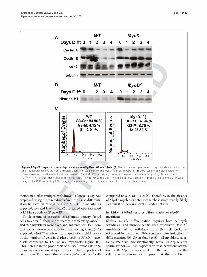
radation of IκB and nuclear localization of RelA/p65 [3].To determine if IKK activity is aberrantly maintained inMyoD-null myoblasts after induction of differentiation,IKK was immunoprecipitated from MyoD-/- and wild typemyoblast protein extracts and kinase activity assayed usingGST-IκBα and γ-32P-ATP as substrate. MyoD-null myo-blasts displayed approximately 2-fold greater IKK activity,as compared to WT myoblasts (Figure 5A).
RETR
Figure 5 Elevated IKK activity in MyoD-/- myoblasts isresponsible for increased NF-κB activity. (A) Kinase assay (KA): IKKwas immunoprecipitated from protein extracts of WT (MyoD+/+) andMyoD-/- primary myoblasts, and assayed for kinase activity usingGST-IκBα and γ-32P-ATP as substrate. Immunoblot (IB): IKK wasimmunoprecipitated from protein extracts of WT (MyoD+/+) andMyoD-/- primary myoblasts, and assayed by western blot using anantibody specific for IKKγ. (B) MyoD-/- myoblasts were transfectedwith vector control, vector expressing a dominant negative mutantIKKβ (IKKβ DN) or a non-phosphorylatable mutant of IkBα (IκB-SR), inaddition to an NF-κB reporter (3xκB-Luc). Bars represent averageluciferase activity (relative light units (RLUs)) (n = 3). Error barsrepresent standard deviation.
ACTE
D
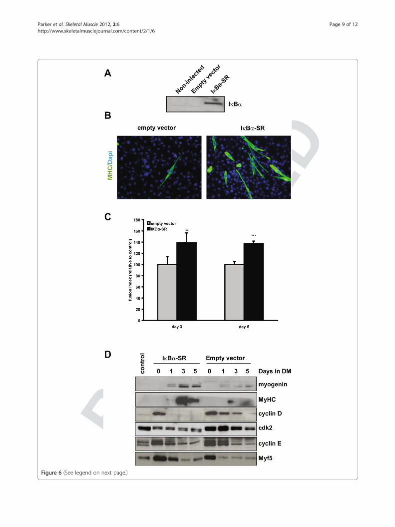
If increased IKK activity results in RelA/p65 activation,then inhibiting IKK should inhibit NF-κB transcriptionalactivity. Treating MyoD-/- myoblasts with a chemical inhibi-tor of IKK (data not shown), or expressing a dominantnegative mutant of IKKβ (IKKβ DN), resulted in decreasedactivity of 3xκB-Luc, indicating that elevated IKK activity isin part responsible for persistent NF-κB transcriptional ac-tivity (Figure 5A). Therefore, to target RelA/p65 directly, anon-phosphorylatable mutant form of IκBα (IκBα-SR) wasexpressed, which is resistant to targeted degradation andprevents nuclear localization of NF-κB. Indeed, expressionof IkBα-SR in MyoD-/- myoblasts inhibited 3xκB-Luc activ-ity, indicating that NF-κB was directly repressed (Figure 5B).Moreover, expression of IκBα-SR in MyoD-/- myoblastsdown-regulated expression of cyclin D1 and cdk2 upon ini-tiation of differentiation (Figure 6C). This strongly suggeststhat sustained nuclear localization of RelA/p65 in theMyoD-null myoblasts is responsible for cellular prolifera-tion after serum withdrawal.Importantly, inhibition of NF-κB activity also induced
expression of myogenin and myosin heavy chain one dayafter serum withdrawal, and resulted in the formation ofmultinucleated myotubes (Figure 6B to 6D). This is incontrast to MyoD-/- myoblasts infected with controlvirus, which up-regulated myogenin and myosin heavychain to a much lower extent, even 5 days after serumwithdrawal (Figure 6D, DM5). Additionally we examinedthe fusion index of myotubes of control and IκBα-SRexpressing MyoD-/- cells at days 3 and 5 after serumwithdrawal. Overexpression of IκBα-SR led to a signifi-cantly increased fusion index as well as to more differen-tiation in general (Figure 6B and 6C).It is interesting to note that IκBαSR-expressing MyoD-/-
cells up-regulate expression of Myf5 during proliferationand early differentiation (Figure 6D). Increased expressionof Myf5 may compensate for the lack of MyoD, and maybe responsible for the induction of differentiation specificgenes, such as myogenin and myosin heavy chain, aftercell-cycle withdrawal. This is consistent with the fact thatembryonic and fetal skeletal muscle development is ableto occur in MyoD-/- mice [7].
DiscussionFinding the specific MyoD-regulated gene product thatlinks cell-cycle withdrawal and terminal myogenic differ-entiation has been hitherto elusive. In this study, we dem-onstrate that continued proliferation and inhibition ofdifferentiation in MyoD-null myoblasts is due to persistentnuclear localization of RelA/p65. Expression of a non-phosphorylatable mutant of IκBα (IκBα-SR), whichinduces cytoplasmic retention of RelA/p65, down-regu-lated expression of cyclin D1 in MyoD-/- myoblasts, andresulted in the formation of multinucleated myotubes.Therefore, inhibition of RelA/p65 activation was able to
RETR
ACTE
D
Figure 6 (See legend on next page.)
Parker et al. Skeletal Muscle 2012, 2:6 Page 9 of 12http://www.skeletalmusclejournal.com/content/2/1/6
Figure 6 Nuclear localization of NF-κB inhibits terminal differentiation. (A) MyoD-/- myoblasts were infected with empty capsid (pBABE) orvirus expressing IκBα-SR (pBABE-IkB). Protein extracts from proliferating infected cells and uninfected cells (control) were analyzed by western blotanalysis using an antibody specific to IκBα. (B) MyoD-/- myoblasts were infected with empty capsid (pBABE) or virus expressing IκBα-SR (pBABE-IkB). Proliferating cells (GM), or cells induced to differentiate for 5 days, were fixed and assessed for myosin heavy chain (MyHC) expression (ingreen). Nuclei were visualized using DAPI (in blue). (C) Fusion index of myotubes from MyoD-/- cells either infected with a control (pBABE) or anIκBα-SR (pBABE-IkB) expressing virus, n = 5, ** = P< 0.01, *** = P< 0.001. (D) MyoD-/- myoblasts were infected with empty capsid (pBABE) or virusexpressing IκBα-SR (pBABE-IkB). Protein extracts from a differentiation time course of infected and uninfected cells were analyzed by western blotanalysis using the antibodies indicated.
(See figure on previous page.)
Parker et al. Skeletal Muscle 2012, 2:6 Page 10 of 12http://www.skeletalmusclejournal.com/content/2/1/6
RETR
substitute for MyoD expression during myogenic differ-entiation. Taken together, this indicates that RelA/p65provides the link between MyoD-induced cell-cyclewithdrawal and differentiation.MyoD is postulated to initiate expression of p21 and
p57, inhibitors of cdk2 and cdk1 kinase activity [25-27].Moreover, mice lacking p21 and p57 are phenotypicallysimilar to myogenin knockout mice, in that they lack dif-ferentiated myofibers [28]. These data suggest that myo-genic cell-cycle withdrawal and differentiation requiresMyoD-dependent induction of cdk inhibitor expression.However, MyoD-/- myoblasts have a similar level of p21mRNA as compared to WT myoblasts, and mice lackingp21 display no apparent muscle abnormalities [9,29].Therefore, we propose that adult satellite cells utilize analternate or additional mechanism for inducing cell-cyclewithdrawal during terminal differentiation: down-regula-tion of RelA/p65 activity.Notably, MIP-2/CXCL1/GRO-α and IL-6, which were
upregulated 33-fold and 8-fold, respectively, in MyoD-/-
myoblasts, are associated with constitutive activation ofNF-κB, and promote tumor growth and progression[30]. Importantly, IGFBP-2, which binds IGFs and spe-cifically inhibits IGF-dependent myogenic cell prolifera-tion, was down-regulated 9.1-fold in MyoD-/- myoblasts[31-35]. Therefore, in the absence of MyoD, myoblastsare programmed to proliferate as a result of maintaininggrowth factor signaling. This is consistent with datademonstrating that MyoD-/- myoblasts have an increasedpropensity for proliferation and self-renewal.Biglycan and thrombospondin 2, two genes that were
up-regulated in MyoD-/- myoblasts (73.5-fold and 16.9-fold, respectively), are extracellular matrix componentsthat play important roles in scaffolding and signal trans-duction during myogenic regeneration. In particular, bigly-can binds TGF-β, and plays an important role inmediating TGF-β signaling in responding cells [36,37].This is important given that TGF-β inhibits myogenic dif-ferentiation and induces expression of cyclin D1 [22].Taken together, these data strongly suggest that NF-κBregulates expression of genes important for inducing cellproliferation.During normal differentiation of myoblasts, NF-κB is
relocalized to the cytoplasm and DNA-binding activity
ACTE
Ddecreases within 24 h of serum withdrawal [12,18]. Differ-entiation is accelerated in myoblasts expressing a non-phosphorylatable form of IκBα (IκBαSR), which is unableto be degraded, thus inhibiting NF-κB (p65) nuclearlocalization [12]. Furthermore, these IκBαSR-expressingmyoblasts proliferate less rapidly and down-regulate ex-pression of cyclin D1. In contrast, IκBα-/- mouse embry-onic fibroblasts (MEFs) infected with a MyoD-expressingretrovirus maintain NF-κB nuclear localization, resultingin the formation of fewer myotubes that are smaller andincorporate fewer nuclei [17]. Our experiments explainmechanistically why MyoD-null myoblasts display aphenotype similar to that of MyoD-infected IκBα-/- MEFs.During myogenic regeneration, satellite cells are acti-
vated and proliferate prior to initiating differentiation.Increased numbers of satellite cells and a deficientmuscle regenerative process in MyoD-/- mice suggestthat in the absence of MyoD, satellite cells have anincreased propensity for self-renewal rather than differ-entiation [38]. In light of the data presented here, weconclude that RelA/p65 plays an important role duringthe myoblast to myotube transition during adult myo-genesis. Indeed, treatment of mdx mice with a cell per-meable peptide inhibitor of IKK, which specificallyinhibits NF-κB activity, restores regeneration, as evi-denced by a greater number of newly formed myofibers,and increased muscle tetanic force [39].Failure to induce myogenic differentiation is also illu-
strated in rhabdomyosarcoma (RDS), one the mostcommon childhood solid tumors. RDS is characterizedby inhibition of MyoD activity and concomitant failureto withdraw from the cell cycle and differentiate. Thisstudy suggests that loss of MyoD activity in RDS cellsmay cause aberrant nuclear localization of NF-κB,resulting in sustained cyclin D1 expression. As such,inhibiting IKK and stabilizing IκBα may play a valuablerole in inhibiting proliferation and inducing differenti-ation in RDS cells.During myogenesis, cytoplasmic re-localization of
RelA/p65 after mitogen withdrawal plays a key role fordown-regulating cyclin D expression, inducing cell-cyclewithdrawal and activating differentiation-specific geneexpression. Therefore, the regulation of NF-κB is essen-tial in the induction of myogenic differentiation. Our
Parker et al. Skeletal Muscle 2012, 2:6 Page 11 of 12http://www.skeletalmusclejournal.com/content/2/1/6
RETR
experiments define the mechanistic link between MyoDand NF-κB that acts to couple cell-cycle withdrawal toterminal differentiation.
ConclusionWe have demonstrated that NF-κB, a key regulator ofcell-cycle withdrawal and differentiation, aberrantlymaintains nuclear localization and transcriptional activ-ity in MyoD-/- myoblasts. Cyclin D is consequently main-tained during serum withdrawal, inhibiting progressionthrough myogenic differentiation. Sustained nuclearlocalization of cyclin E, and a concomitant increase incdk2 activity maintains S-phase entry in MyoD-/- myo-blasts even in the absence of mitogens. Forced expres-sion of IκBαSR, a non-degradable mutant of IκBα,rescued the deficit indicating that inhibition of NF-κB issufficient to induce terminal myogenic differentiation.Therefore, MyoD-induced cytoplasmic relocalization ofNF-κB is an essential step in linking cell-cycle with-drawal to terminal differentiation.
AbbreviationsbHLH: Basic helix-loop-helix; BrdU: Bromodeoxyuridine; FACS: Fluorescenceactivated cell sorting; IKK: IκB kinase; MEF: Mouse embryonic fibroblast;MRF: Myogenic regulatory factors; MyHC: Myosin heavy chain; NLS: Nuclearlocalization sequence; WT: Wild type; siRNA: Small interfering RNA.
Competing interestsThe authors declare no competing interests.
Authors’ contributionsMHP and NB carried out the experiments and drafted the manuscript. BAJand JM carried out the rescue experiments. JI performed the microarrayexperiment. DG provided essential reagents and participated in the designand coordination of the study. MAR conceived of the study, and participatedin its design and coordination and helped to draft the manuscript. Allauthors read and approved the final manuscript.
AcknowledgementsWe thank Mark Gillespie for critical reading of the manuscript. MAR holds theCanada Research Chair in Molecular Genetics and is an InternationalResearch Scholar of the Howard Hughes Medical Institute. This work wassupported by grants to MAR from the Canadian Institutes of Health Research,the Muscular Dystrophy Association, the National Institutes of Health, andthe Canada Research Chair Program, and by grants to DG from the NationalInstitutes of Health.
Author details1Faculty of Health Sciences Graduate Programme, McMaster University,Hamilton, Ontario, Canada. 2Ottawa Hospital Research Institute, MolecularMedicine Program, Ottawa, Ontario, Canada. 3Current address: FredHutchinson Cancer Research Center, Clinical Research Division, Seattle, WA,USA. 4The Ohio State University College of Medicine, Columbus, Ohio, USA.5Ottawa Hospital Research Institute, 501 Smyth Rd, Ottawa, ON K1H 8L6,Canada.
Received: 3 January 2012 Accepted: 21 March 2012Published: 27 April 2012
References1. Hayden MS, Ghosh S: Shared principles in NF-kappaB signalling. Cell 2008,
132:344–362.2. Baldwin AS Jr: The NF-kappaB and I kappa B proteins: new discoveries
and insights. Annu Rev Immunol 1996, 14:649–683.
ACTE
D
3. Karin M, Ben-Neriah Y: Phosphorylation meets ubiquitination: the controlof NF-[kappa]B activity. Annu Rev Immunol 2000, 18:621–663.
4. Skaug B, Jiang X, Chen ZJ: The role of ubiquitin in NF-kappaB regulatorypathways. Annu Rev Biochem 2009, 78:769–796.
5. Parker MH, Seale P, Rudnicki MA: Looking back to the embryo: definingtranscriptional networks in adult myogenesis. Nat Rev Genet 2003,4:497–507.
6. Rudnicki MA, Schnegelsberg PN, Stead RH, Braun T, Arnold HH, Jaenisch R:MyoD or Myf-5 is required for the formation of skeletal muscle. Cell 1993,75:1351–1359.
7. Braun T, Rudnicki MA, Arnold HH, Jaenisch R: Targeted inactivation of themuscle regulatory gene Myf-5 results in abnormal rib development andperinatal death. Cell 1992, 71:369–382.
8. Asakura A, Komaki M, Rudnicki M: Muscle satellite cells are multipotentialstem cells that exhibit myogenic, osteogenic, and adipogenicdifferentiation. Differentiation 2001, 68:245–253.
9. Sabourin LA, Girgis-Gabardo A, Seale P, Asakura A, Rudnicki MA: Reduceddifferentiation potential of primary MyoD-/- myogenic cells derived fromadult skeletal muscle. J Cell Biol 1999, 144:631–643.
10. Yablonka-Reuveni Z, Rudnicki MA, Rivera AJ, Primig M, Anderson JE,Natanson P: The transition from proliferation to differentiation is delayedin satellite cells from mice lacking MyoD. Dev Biol 1999, 210:440–455.
11. Cornelison DD, Olwin BB, Rudnicki MA, Wold BJ: MyoD(-/-) satellite cells insingle-fiber culture are differentiation defective and MRF4 deficient. DevBiol 2000, 224:122–137.
12. Guttridge DC, Albanese C, Reuther JY, Pestell RG, Baldwin AS Jr: NF-kappaBcontrols cell growth and differentiation through transcriptionalregulation of cyclin D1. Mol Cell Biol 1999, 19:5785–5799.
13. Bakkar N, Guttridge DC: NF-kappaB signalling: a tale of two pathways inskeletal myogenesis. Physiol Rev 2010, 90:495–511.
14. Guttridge DC, Mayo MW, Madrid LV, Wang CY, Baldwin AS Jr: NF-kappaB-induced loss of MyoD messenger RNA: possible role in muscle decayand cachexia [see comments]. Science 2000, 289:2363–2366.
15. Didonato JA: Assaying for I kappa B kinase activity. Methods Enzymol 2000,322:393–400.
16. Ishibashi J, Perry RL, Asakura A, Rudnicki MA: MyoD induces myogenicdifferentiation through cooperation of its NH2- and COOH-terminalregions. J Cell Biol 2005, 171:471–482.
17. Ladner KJ, Caligiuri MA, Guttridge DC: Tumor necrosis factor-regulatedbiphasic activation of NF-kappa B is required for cytokine-induced lossof skeletal muscle gene products. J Biol Chem 2003, 278:2294–2303.
18. Bakkar N, Wang J, Ladner KJ, Wang H, Dahlman JM, Carathers M, AcharyyaS, Rudnicki MA, Hollenbach AD, Guttridge DC: IKK/NF-kappaB regulatesskeletal myogenesis via a signalling switch to inhibit differentiation andpromote mitochondrial biogenesis. J Cell Biol 2008, 180:787–802.
19. Harbour JW, Luo RX, Dei Santi A, Postigo AA, Dean DC: Cdkphosphorylation triggers sequential intramolecular interactions thatprogressively block Rb functions as cells move through G1. Cell 1999,98:859–869.
20. Geng Y, Eaton EN, Picon M, Roberts JM, Lundberg AS, Gifford A, Sardet C,Weinberg RA: Regulation of cyclin E transcription by E2Fs andretinoblastoma protein. Oncogene 1996, 12:1173–1180.
21. Geng Y, Whoriskey W, Park MY, Bronson RT, Medema RH, Li T, Weinberg RA,Sicinski P: Rescue of cyclin D1 deficiency by knockin cyclin E. Cell 1999,97:767–777.
22. Rao SS, Kohtz DS: Positive and negative regulation of D-type cyclinexpression in skeletal myoblasts by basic fibroblast growth factor andtransforming growth factor beta. A role for cyclin D1 in control ofmyoblast differentiation. J Biol Chem 1995, 270:4093–4100.
23. Skapek SX, Rhee J, Spicer DB, Lassar AB: Inhibition of myogenicdifferentiation in proliferating myoblasts by cyclin D1-dependent kinase[see comments]. Science 1995, 267:1022–1024.
24. Skapek SX, Rhee J, Kim PS, Novitch BG, Lassar AB: Cyclin-mediated inhibitionof muscle gene expression via a mechanism that is independent of pRBhyperphosphorylation. Mol Cell Biol 1996, 16:7043–7053.
25. Halevy O, Novitch BG, Spicer DB, Skapek SX, Rhee J, Hannon GJ, Beach D,Lassar AB: Correlation of terminal cell cycle arrest of skeletal muscle withinduction of p21 by MyoD. Science 1995, 267:1018–1021.
26. Parker SB, Eichele G, Zhang P, Rawls A, Sands AT, Bradley A, Olson EN,Harper JW, Elledge SJ: p53-independent expression of p21Cip1 in muscleand other terminally differentiating cells [see comments]. Science 1995,267:1024–1027.
ACTE
D
Parker et al. Skeletal Muscle 2012, 2:6 Page 12 of 12http://www.skeletalmusclejournal.com/content/2/1/6
R
27. Figliola R, Maione R: MyoD induces the expression of p57Kip2 in cellslacking p21Cip1/Waf1: overlapping and distinct functions of the two cdkinhibitors. J Cell Physiol 2004, 200:468–475.
28. Zhang P, Wong C, Liu D, Finegold M, Harper JW, Elledge SJ: p21(CIP1) andp57(KIP2) control muscle differentiation at the myogenin step. Genes Dev1999, 13:213–224.
29. Deng C, Zhang P, Harper JW, Elledge SJ, Leder P: Mice lacking p21CIP1/WAF1 undergo normal development, but are defective in G1 checkpointcontrol. Cell 1995, 82:675–684.
30. Frost RA, Lang CH: Skeletal muscle cytokines: regulation by pathogen-associated molecules and catabolic hormones. Curr Opin Clin Nutr MetabCare 2005, 8:255–263.
31. James PL, Jones SB, Busby WH Jr, Clemmons DR, Rotwein P: A highlyconserved insulin-like growth factor-binding protein (IGFBP-5) is expressedduring myoblast differentiation. J Biol Chem 1993, 268:22305–22312.
32. Ewton DZ, Coolican SA, Mohan S, Chernausek SD, Florini JR: Modulation ofinsulin-like growth factor actions in L6A1 myoblasts by insulin-likegrowth factor binding protein (IGFBP)-4 and IGFBP-5: a dual role forIGFBP-5. J Cell Physiol 1998, 177:47–57.
33. Ewton DZ, Florini JR: IGF binding proteins-4, -5 and −6 may playspecialized roles during L6 myoblast proliferation and differentiation.J Endocrinol 1995, 144:539–553.
34. Rotwein P, James PL, Kou K: Rapid activation of insulin-like growth factorbinding protein-5 gene transcription during myoblast differentiation.Mol Endocrinol 1995, 9:913–923.
35. Florini JR, Ewton DZ, Magri KA, Mangiacapra FJ: IGFs and muscledifferentiation. Adv Exp Med Biol 1993, 343:319–326.
36. Droguett R, Cabello-Verrugio C, Riquelme C, Brandan E: Extracellularproteoglycans modify TGF-beta bio-availability attenuating its signallingduring skeletal muscle differentiation. Matrix Biol 2006, 25:332–341.
37. Hayashi Y, Liu CY, Jester JJ, Hayashi M, Wang IJ, Funderburgh JL, Saika S,Roughley PJ, Kao CW, Kao WW: Excess biglycan causes eyelidmalformation by perturbing muscle development and TGF-alphasignalling. Dev Biol 2005, 277:222–234.
38. Megeney LA, Kablar B, Garrett K, Anderson JE, Rudnicki MA: MyoD isrequired for myogenic stem cell function in adult skeletal muscle. GenesDev 1996, 10:1173–1183.
39. Acharyya S, Villalta SA, Bakkar N, Bupha-Intr T, Janssen PM, Carathers M, Li Z-W,Beg A, Ghosh S, Sahenk Z, et al: IKK/NF-kB signalling interplay inmacrophages and myofibers promotes muscle degeneration in Duchennemuscular dystrophy. J Clin Invest 2007, 117:889–901.
doi:10.1186/2044-5040-2-6Cite this article as: Parker et al.: Retracted: MyoD-dependent regulationof NF-κB activity couples cell-cycle withdrawal to myogenicdifferentiation. Skeletal Muscle 2012 2:6.
RET
Submit your next manuscript to BioMed Centraland take full advantage of:
• Convenient online submission
• Thorough peer review
• No space constraints or color figure charges
• Immediate publication on acceptance
• Inclusion in PubMed, CAS, Scopus and Google Scholar
• Research which is freely available for redistribution
Submit your manuscript at www.biomedcentral.com/submit