Embed Size (px)
Citation preview
SC I ENCE S I GNAL ING | R E S EARCH ART I C L E
IMMUNOLOGY
1IGMM, CNRS, Université de Montpellier, Montpellier, France. 2CNC–Centre for Neu-roscience and Cell Biology, University of Coimbra, Coimbra, Portugal. 3INSERMU1170,Gustave Roussy Cancer Center, Villejuif, France. 4Faculty ofMedicine, Université Paris-Sud, Le Kremlin-Bicêtre, France.*These authors contributed equally to this work.†Corresponding author. Email: [email protected] (V.D.); [email protected] (N.T.)
Craveiro et al., Sci. Signal. 10, eaal3024 (2017) 17 October 2017
Copyright © 2017
The Authors, some
rights reserved;
exclusive licensee
American Association
for the Advancement
of Science. No claim
to original U.S.
Government Works
Dow
nloade
Resveratrol stimulates the metabolic reprogramming ofhuman CD4+ T cells to enhance effector functionMarco Craveiro,1,2* Gaspard Cretenet,1* Cédric Mongellaz,1 Maria I. Matias,1 Olivier Caron,3,4
Maria C. Pedroso de Lima,2 Valérie S. Zimmermann,1 Eric Solary,3,4 Valérie Dardalhon,1
Vjekoslav Dulić,1† Naomi Taylor1†
The polyphenol resveratrol activates the deacetylase Sirt1, resulting in various antioxidant, chemoprotectant,neuroprotective, cardioprotective, and anti-inflammatory properties. We found that at high concentrations ofresveratrol, human CD4+ T cells showed defective antigen receptor signaling and arrest at the G1 stage of thecell cycle, whereas at low concentrations, cells were readily activated and exhibited enhanced Sirt1 deacetylaseactivity. Nevertheless, low-dose resveratrol rapidly stimulated genotoxic stress in the T cells, which resulted inengagement of a DNA damage response pathway that depended on the kinase ATR [ataxia telangiectasia–mutated(ATM) and Rad3-related], but not ATM, and subsequently in premitotic cell cycle arrest. The concomitant activationof p53 was coupled to the expression of gene products that regulate cell metabolism, leading to a metabolicreprogramming that was characterized by decreased glycolysis, increased glutamine consumption, and a shiftto oxidative phosphorylation. These alterations in the bioenergetic homeostasis of CD4+ T cells resulted inenhanced effector function, with both naïve and memory CD4+ T cells secreting increased amounts of the in-flammatory cytokine interferon-g. Thus, our data highlight the wide range of metabolic adaptations that CD4+
T lymphocytes undergo in response to genomic stress.
d fr
on May 4, 2018http://stke.sciencem
ag.org/om
INTRODUCTIONResveratrol (3,5,4′-trihydroxy-trans-stilbene) is a natural polyphenoliccompound that is produced by plants in response to environmentalstress, providing them with protection from microbial infections (1–4).Resveratrol appears to mimic the effects of caloric restriction, increasinglife span in lower organisms (5). Furthermore, this pharmacologicalagent has elicited much interest because of its potential to modulate adiverse array of pathological conditions, and it is associated with anti-cancer, antiaging, and anti-inflammatory properties (6–10). On the basisof the promising data emerging from ex vivo studies and preclinicalanimal models, resveratrol has been tested in more than 30 clinical trialsinvolving more than 1000 individuals. Nevertheless, the specific pathol-ogies inwhich resveratrol has substantial clinical benefits are not yet clear(11–16).The pharmacological properties of resveratrol have been attributed,at least in part, to its activation of the nicotinamide adenine dinucleotide(NAD+)–dependent silent information regulator 2 (Sir2) deacetylase(17) both in vitro (18, 19) and in vivo (20). Overexpression of themammalian Sir2 homolog sirtuin-1 (Sirt1) in mice extends their lifespan (21, 22) and protects them from a diverse array of diseases (23–28).Conversely, knocking out Sirt1 is associated with autoimmunity (29–32).However, the effects of Sirt1 are likely to be complex. Although Sirt1attenuates murine T cell signaling and effector function (29, 30, 33–35),it also promotes the differentiation of naïve CD4+ T cells into T helper17 (TH17) effector cells in mice (36). Furthermore, physiological mod-ifications of Sirt1 function in human T cell subsets have thus far notbeen evaluated.
T cell activity is of great importance in a wide range of patho-physiological conditions for which resveratrol activity is being clinicallyevaluated. Hence, elucidating the potential on-target and off-targeteffects of resveratrol on T lymphocytes is critical. T cells present acomplex target because their cellular metabolism is altered after activa-tion by a cognate antigen. The capacity of T lymphocytes to respond tostimulation by antigen depends on an extensive proliferative response, aprocess that requires new energetic and biosynthetic components thatare supplied, at least in part, through a metabolic shift from oxidativephosphorylation (OXPHOS) toward glycolytic and glutaminolyticpathways (37–39). This shift from OXPHOS contrasts with the activityof resveratrol, a compound that generally increases mitochondrialactivity and associated OXPHOS (40–42). However, note that resvera-trol leads to a wide range of effects, including decreased, stabilized, andenhanced T cell effector functions (43–46).
Disparate effects of resveratrol on genomic stability have also beenreported. In some studies, resveratrol contributes to genomic stabilityand reduces tumorigenesis by reducing the amount of reactive oxygenspecies (ROS), which leads to oxidative damage (47–51). However, inother studies, resveratrol mediates DNA damage, facilitating antitumortreatments (47, 52–59). One possible reason for these discrepanciescould be that resveratrol has distinct effects on quiescent cells versusproliferating cells. In this regard, T lymphocytes present a challengingtarget. Although they are generally quiescent, exposure to foreign anti-gen rapidly stimulates cell cycle entry and cellular proliferation. Acoordinated response to genotoxic stress is regulated by the kinasesATM (ataxia telangiectasia–mutated) and ATR (ATM and Rad3-related) (60, 61). Of interest are reports that resveratrol activatesone or both of these kinases in different cellular contexts (53–55). Here,we report that resveratrol rapidly stimulates the ATR-dependentdamage pathway in antigen-stimulated human CD4+ T cells, withactivation of the tumor suppressor p53. This genotoxic stress re-sponse links a metabolic reprogramming to an enhanced CD4 T celleffector function characterized by increased production of the cytokineinterferon-g (IFN-g).
1 of 14
SC I ENCE S I GNAL ING | R E S EARCH ART I C L E
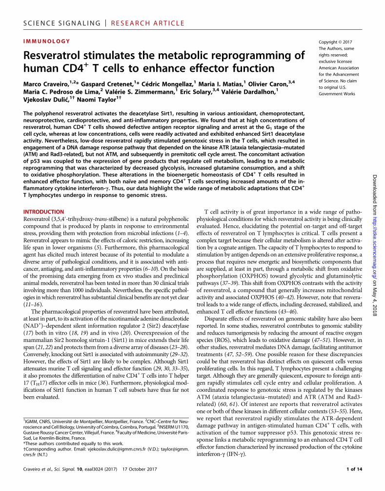
RESULTSLow-dose resveratrol increases the activity of theNAD+-dependent deacetylase Sirt1 in primary humanCD4+ T cellsTo gain insight into the role of resveratrol in modulating Sirt1 functionin humanCD4+ T lymphocytes, we first examined its expression profilein response to T cell receptor (TCR) stimulation. We found that TCRengagement resulted in a substantial increase in Sirt1 abundance, withaugmented nuclear localization and aggregation (Fig. 1A). Low-doseresveratrol (20 mM) further increased the mean fluorescence inten-sity (MFI) of Sirt1 staining by about twofold (Fig. 1A and fig. S1A).However, high-dose resveratrol (100 mM) attenuated the TCR-mediated increase in Sirt1 abundance, and these CD4+ lymphocytesdid not undergo blast formation (Fig. 1A and fig. S2A). This differedmarkedly from treatment with low-dose resveratrol, which augmentedblast size (fig. S2A). Separating subsets of TCR-stimulated CD4+ T cellsbased on their forward scatter (FSC) and side scatter (SSC) profiles
Craveiro et al., Sci. Signal. 10, eaal3024 (2017) 17 October 2017
demonstrated that Sirt1 abundance paralleled increases in cell sizeand granularity (Fig. 1B). As expected from these data, low-doseresveratrol substantially increased Sirt1 activity in TCR-stimulatedlymphocytes, as monitored by the generation of O-acetyl–adenosinediphosphate–ribose (OAADPr), a reaction product of the Sirt-catalyzed,NAD+-dependent deacetylation of target proteins (Fig. 1C). Thus, TCRstimulation combined with low-dose, but not high-dose, resveratrolaugments Sirt1 activity in human T lymphocytes.
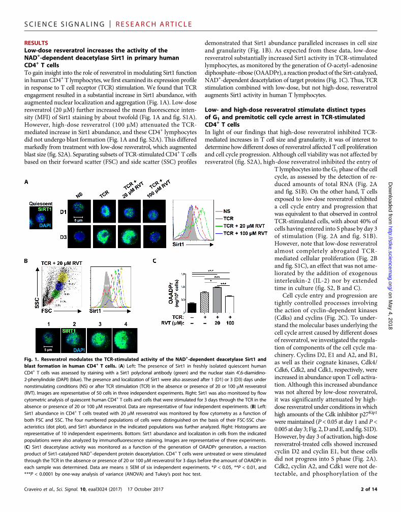
Low- and high-dose resveratrol stimulate distinct typesof G1 and premitotic cell cycle arrest in TCR-stimulatedCD4+ T cellsIn light of our findings that high-dose resveratrol inhibited TCR-mediated increases in T cell size and granularity, it was of interest todetermine how different doses of resveratrol affected T cell proliferationand cell cycle progression. Although cell viability was not affected byresveratrol (fig. S2A), high-dose resveratrol inhibited the entry of
on May 4, 2018
http://stke.sciencemag.org/
Dow
nloaded from
T lymphocytes into theG1 phase of the cellcycle, as assessed by the detection of re-duced amounts of total RNA (Fig. 2Aand fig. S1B). On the other hand, T cellsexposed to low-dose resveratrol exhibiteda cell cycle entry and progression thatwas equivalent to that observed in controlTCR-stimulated cells, with about 40% ofcells having entered into S phase by day 3of stimulation (Fig. 2A and fig. S1B).However, note that low-dose resveratrolalmost completely abrogated TCR-mediated cellular proliferation (Fig. 2Band fig. S1C), an effect that was not ame-liorated by the addition of exogenousinterleukin-2 (IL-2) nor by extendedtime in culture (fig. S2, B and C).
Cell cycle entry and progression aretightly controlled processes involvingthe action of cyclin-dependent kinases(Cdks) and cyclins (Fig. 2C). To under-stand the molecular bases underlying thecell cycle arrest caused by different dosesof resveratrol, we investigated the regula-tion of components of the cell cycle ma-chinery. Cyclins D2, E1 and A2, and B1,as well as their cognate kinases, Cdk4/Cdk6, Cdk2, and Cdk1, respectively, wereincreased in abundance upon T cell activa-tion. Although this increased abundancewas not altered by low-dose resveratrol,it was significantly attenuated by high-dose resveratrol under conditions inwhichhigh amounts of the Cdk inhibitor p27Kip1
were maintained (P < 0.05 at day 1 and P <0.005 at day 3; Fig. 2,D andE, and fig. S1D).However, by day 3 of activation, high-doseresveratrol-treated cells showed increasedcyclin D2 and cyclin E1, but these cellsdid not progress into S phase (Fig. 2A).Cdk2, cyclin A2, and Cdk1 were not de-tectable, and phosphorylation of the
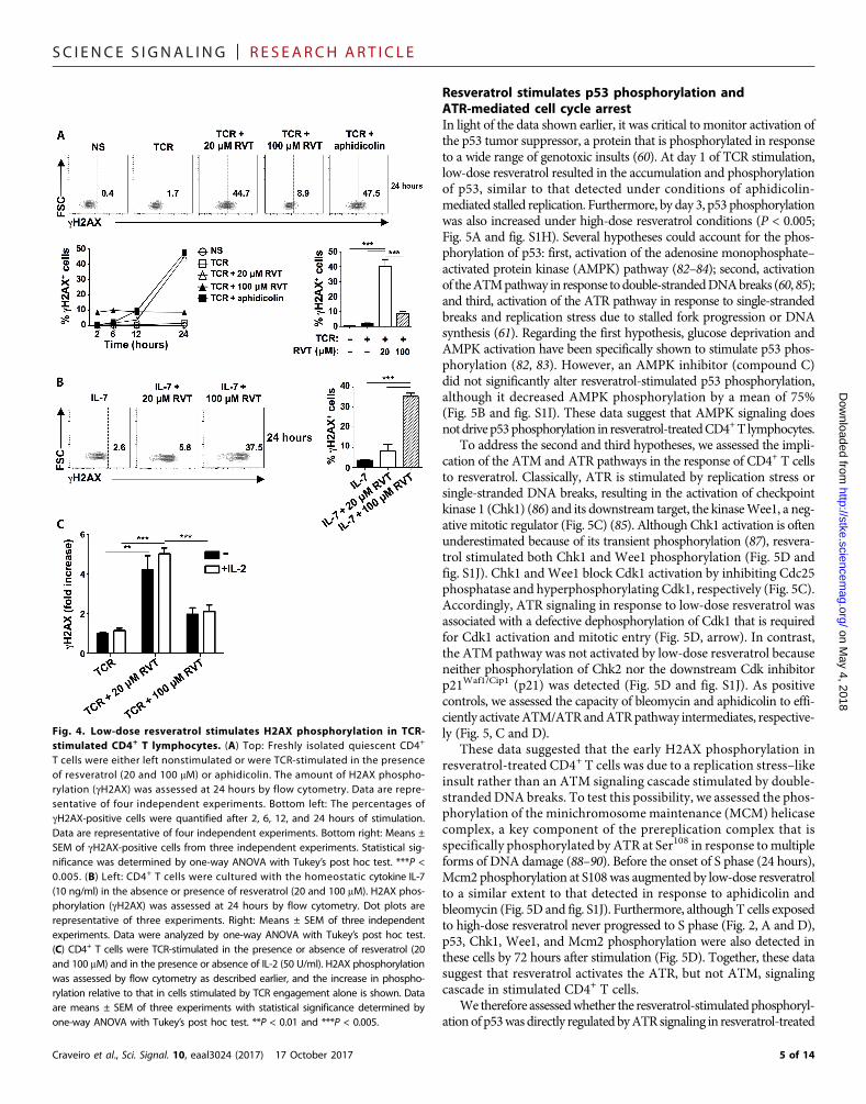
Fig. 1. Resveratrol modulates the TCR-stimulated activity of the NAD+-dependent deacetylase Sirt1 andblast formation in human CD4+ T cells. (A) Left: The presence of Sirt1 in freshly isolated quiescent humanCD4+ T cells was assessed by staining with a Sirt1 polyclonal antibody (green) and the nuclear stain 4′,6-diamidino-2-phenylindole (DAPI) (blue). The presence and localization of Sirt1 were also assessed after 1 (D1) or 3 (D3) days undernonstimulating conditions (NS) or after TCR stimulation (TCR) in the absence or presence of 20 or 100 mM resveratrol(RVT). Images are representative of 50 cells in three independent experiments. Right: Sirt1 was also monitored by flowcytometric analysis of quiescent human CD4+ T cells and cells that were stimulated for 3 days through the TCR in theabsence or presence of 20 or 100 mM resveratrol. Data are representative of four independent experiments. (B) Left:Sirt1 abundance in CD4+ T cells treated with 20 mM resveratrol was monitored by flow cytometry as a function ofboth FSC and SSC. The four numbered populations of cells were distinguished on the basis of their FSC-SSC char-acteristics (dot plot), and Sirt1 abundance in the indicated populations was further analyzed. Right: Histograms arerepresentative of 10 independent experiments. Bottom: Sirt1 abundance and localization in cells from the indicatedpopulations were also analyzed by immunofluorescence staining. Images are representative of three experiments.(C) Sirt1 deacetylase activity was monitored as a function of the generation of OAADPr generation, a reactionproduct of Sirt1-catalyzed NAD+-dependent protein deacetylation. CD4+ T cells were untreated or were stimulatedthrough the TCR in the absence or presence of 20 or 100 mM resveratrol for 3 days before the amount of OAADPr ineach sample was determined. Data are means ± SEM of six independent experiments. *P < 0.05, **P < 0.01, and***P < 0.0001 by one-way analysis of variance (ANOVA) and Tukey’s post hoc test.
2 of 14
SC I ENCE S I GNAL ING | R E S EARCH ART I C L E
on May 4, 2018
http://stke.sciencemag.org/
Dow
nloaded from
pocket proteins pRb (retinoblastoma protein) and p130, hallmarksof S-phase progression, was also not observed (Fig. 2, D and E, andfig. S1E). Furthermore, the Cdks regulating the G1-S phase progres-sion (Cdk4, Cdk6, Cdk2, and Cdk1) were not increased in abun-dance (Fig. 2D). We found that this was likely because of the
Craveiro et al., Sci. Signal. 10, eaal3024 (2017) 17 October 2017
reduced abundance and phosphoryl-ation of the F-box protein Skp2 (Fig.2E, upper band, and fig. S1E), therate-limiting component responsiblefor p27Kip1 ubiquitination and degrada-tion (62, 63). The premitotic cell cyclearrest that was triggered by low-doseresveratrol did not result in senescence,as shown by the enhanced phosphoryl-ation of pocket proteins, increasedamounts of cyclins A2 and B1 andthe proliferation marker Ki-67, as wellas decreased p27Kip1 abundance (Fig.2, D and E, and fig. S1, D and E). Fur-thermore, accumulation of hyperphos-phorylated Cdk1 (Fig. 2D, arrow)suggests that resveratrol blocks the G2-Mtransition of the cell cycle by abrogatingthe Cdc25-mediated activation of Cdk1(Fig. 2C).
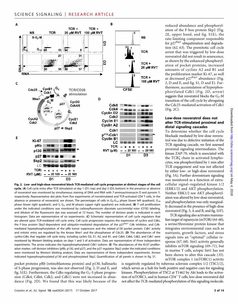
Low-dose resveratrol does notalter TCR-stimulated proximal anddistal signaling cascadesTo determine whether the cell cycleblockade mediated by low-dose resvera-trol was due to defective initiation of theTCR signaling cascade, we first assessedproximal signaling intermediates. Thekinase ZAP-70, which is associated withthe TCRz chain in activated lympho-cytes, was phosphorylated by 1 min afterTCR engagement and was not affectedby either low- or high-dose resveratrol(Fig. 3A). Further downstream signalingwas monitored as a function of extra-cellular signal–regulated kinase 1/2(ERK1/2) and AKT phosphorylation.Neither ERK1/2 nor AKT phosphoryl-ationwas altered by low-dose resveratrol,and phosphorylation was only marginal-ly decreased in the presence of high-doseresveratrol (Fig. 3, A and B, and fig. S1F).
TCR signaling also activatesmamma-lian target of rapamycin (mTOR) (64–66),a serine/threonine protein kinase thatintegrates environmental cues such asnutrients, growth factors, and stresssignals into an “optimal” cellular re-sponse (67, 68). Sirt1 activity generallyinhibits mTOR signaling (69–71), butin murine T cells, ectopic Sirt1 has notbeen shown to alter this cascade (35).mTOR complex 1 (mTORC1) activity
is negatively regulated by tuberous sclerosis complex 1/2 (TSC1/2),which serves as a hub for both positive and negative cues for signalingkinases. Phosphorylation of TSC2 at T1462 by Akt leads to the activa-tion of mTORC1, and in human CD4+ T cells, low-dose resveratrol didnot affect the TCR-mediated phosphorylation of this signalingmolecule.
Fig. 2. Low- and high-dose resveratrol block TCR-mediated cell cycle progression at distinct stages of the cellcycle. (A) Cell cycle entry after TCR stimulation at day 1 (D1; top) and day 3 (D3; bottom) in the presence or absenceof resveratrol was monitored by simultaneous staining of DNA and RNA with 7-aminoactinomycin D and pyronin Y,respectively. Representative dot plots from five experiments of nonstimulated and TCR-activated CD4 T cells, in theabsence or presence of resveratrol, are shown. The percentages of cells in G0-G1A phase (lower left quadrant), G1B
phase (lower right quadrant), and S, G2, and M phases (upper right quadrant) are indicated. (B) T cell proliferationunder the indicated conditions was monitored by carboxyfluorescein diacetate succinimidyl ester (CFSE) labeling,and dilution of the fluorescent dye was assessed at 72 hours. The number of division peaks is indicated in eachhistogram. Data are representative of six experiments. (C) Schematic representation of cell cycle regulators thatare altered upon TCR-mediated cell cycle entry. Cell cycle progression requires the expression of cyclins and Cdks,the F-box protein Skp2-dependent and ubiquitin-mediated degradation of the p27Kip1 Cdk inhibitor, and Cdk-mediated hyperphosphorylation of the pRb tumor suppressor and the related p130 pocket protein. Cdk1 activityand mitotic entry are regulated by the kinase Wee1 and the phosphatase of Cdc25. (D) The abundances of thecyclins-Cdks that regulate cell cycle entry, including cyclins D2, E1, A2, and B1, and Cdk4, Cdk6, Cdk2, and Cdk1 weremonitored by Western blotting analysis on days 1 and 3 of activation. Data are representative of three independentexperiments. The arrow indicates the hyperphosphorylated Cdk1 isoform. (E) The abundances of the Ki-67 prolifer-ation marker, cell division inhibitors (pRb, p130, and p27), and the p27 regulator Skp2 under the indicated conditionswere monitored by Western blotting analysis. Data are representative of three independent experiments. Arrowsindicated hyperphosphorylated p130 and phosphorylated Skp2. Quantification of all panels is shown in fig. S1.
3 of 14
SC I ENCE S I GNAL ING | R E S EARCH ART I C L E
on May 4, 2018
http://stke.sciencemag.org/
Dow
nloaded from
Furthermore, neither phosphorylation of mTOR itself nor S6 ribosomalprotein, a downstream mTOR substrate, was altered by low-doseresveratrol. Note that phosphorylation was significantly decreased inthe presence of 100 mM resveratrol (P < 0.01; Fig. 3C and fig. S1G).Thus, mTOR signaling in TCR-stimulated T cells is attenuated byhigh-dose resveratrol, whereas at low doses, the activity resulting fromTCR engagement is maintained.
We next assessed whether distal TCR signaling was altered byresveratrol, monitored as a function of the cell surface abundanceof the CD69, CD25 (IL-2Ra subunit), and CD71 (transferrin receptor)activation markers. Surface abundance of CD69, due to the trans-location of intracellular stores to the cell membrane without a require-ment for protein synthesis (72, 73), was increased in most of the
Craveiro et al., Sci. Signal. 10, eaal3024 (2017) 17 October 2017
activated cells, irrespective of the presenceof resveratrol (Fig. 3D). In marked con-trast, induction of CD25 and CD71, bothof which are dependent on de novo pro-tein synthesis, was significantly attenu-ated by high-dose resveratrol but wasunaffected by low doses of the polyphenol(P < 0.001; Fig. 3D). Thus, only high-doseresveratrol impedes mTOR and distalTCR signaling cascades.
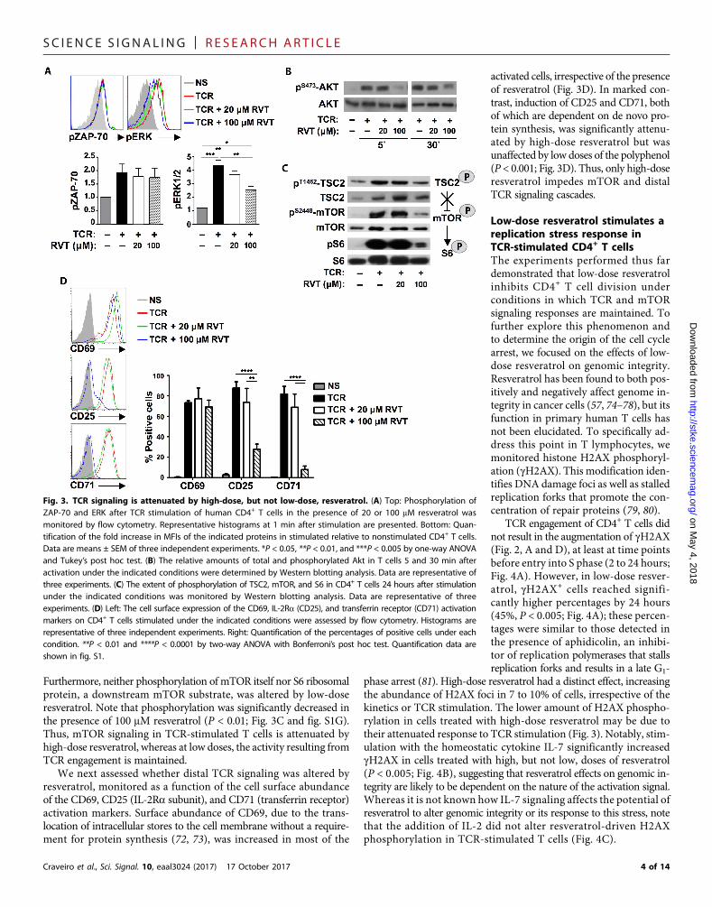
Low-dose resveratrol stimulates areplication stress response inTCR-stimulated CD4+ T cellsThe experiments performed thus fardemonstrated that low-dose resveratrolinhibits CD4+ T cell division underconditions in which TCR and mTORsignaling responses are maintained. Tofurther explore this phenomenon andto determine the origin of the cell cyclearrest, we focused on the effects of low-dose resveratrol on genomic integrity.Resveratrol has been found to both pos-itively and negatively affect genome in-tegrity in cancer cells (57, 74–78), but itsfunction in primary human T cells hasnot been elucidated. To specifically ad-dress this point in T lymphocytes, wemonitored histone H2AX phosphoryl-ation (gH2AX). This modification iden-tifies DNA damage foci as well as stalledreplication forks that promote the con-centration of repair proteins (79, 80).
TCR engagement of CD4+ T cells didnot result in the augmentation of gH2AX(Fig. 2, A and D), at least at time pointsbefore entry into S phase (2 to 24 hours;Fig. 4A). However, in low-dose resver-atrol, gH2AX+ cells reached signifi-cantly higher percentages by 24 hours(45%, P < 0.005; Fig. 4A); these percen-tages were similar to those detected inthe presence of aphidicolin, an inhibi-tor of replication polymerases that stallsreplication forks and results in a late G1-
phase arrest (81). High-dose resveratrol had a distinct effect, increasingthe abundance of H2AX foci in 7 to 10% of cells, irrespective of thekinetics or TCR stimulation. The lower amount of H2AX phospho-rylation in cells treated with high-dose resveratrol may be due totheir attenuated response to TCR stimulation (Fig. 3). Notably, stim-ulation with the homeostatic cytokine IL-7 significantly increasedgH2AX in cells treated with high, but not low, doses of resveratrol(P < 0.005; Fig. 4B), suggesting that resveratrol effects on genomic in-tegrity are likely to be dependent on the nature of the activation signal.Whereas it is not known how IL-7 signaling affects the potential ofresveratrol to alter genomic integrity or its response to this stress, notethat the addition of IL-2 did not alter resveratrol-driven H2AXphosphorylation in TCR-stimulated T cells (Fig. 4C).
Fig. 3. TCR signaling is attenuated by high-dose, but not low-dose, resveratrol. (A) Top: Phosphorylation ofZAP-70 and ERK after TCR stimulation of human CD4+ T cells in the presence of 20 or 100 mM resveratrol wasmonitored by flow cytometry. Representative histograms at 1 min after stimulation are presented. Bottom: Quan-tification of the fold increase in MFIs of the indicated proteins in stimulated relative to nonstimulated CD4+ T cells.Data are means ± SEM of three independent experiments. *P < 0.05, **P < 0.01, and ***P < 0.005 by one-way ANOVAand Tukey’s post hoc test. (B) The relative amounts of total and phosphorylated Akt in T cells 5 and 30 min afteractivation under the indicated conditions were determined by Western blotting analysis. Data are representative ofthree experiments. (C) The extent of phosphorylation of TSC2, mTOR, and S6 in CD4+ T cells 24 hours after stimulationunder the indicated conditions was monitored by Western blotting analysis. Data are representative of threeexperiments. (D) Left: The cell surface expression of the CD69, IL-2Ra (CD25), and transferrin receptor (CD71) activationmarkers on CD4+ T cells stimulated under the indicated conditions were assessed by flow cytometry. Histograms arerepresentative of three independent experiments. Right: Quantification of the percentages of positive cells under eachcondition. **P < 0.01 and ****P < 0.0001 by two-way ANOVA with Bonferroni’s post hoc test. Quantification data areshown in fig. S1.
4 of 14
SC I ENCE S I GNAL ING | R E S EARCH ART I C L E
Craveiro et al., Sci. Signal. 10, eaal3024 (2017) 17 October 2017
on May 4, 2018
http://stke.sciencemag.org/
Dow
nloaded from
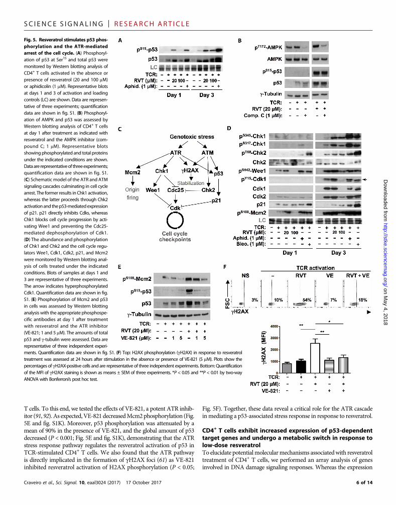
Resveratrol stimulates p53 phosphorylation andATR-mediated cell cycle arrestIn light of the data shown earlier, it was critical to monitor activation ofthe p53 tumor suppressor, a protein that is phosphorylated in responseto a wide range of genotoxic insults (60). At day 1 of TCR stimulation,low-dose resveratrol resulted in the accumulation and phosphorylationof p53, similar to that detected under conditions of aphidicolin-mediated stalled replication. Furthermore, by day 3, p53 phosphorylationwas also increased under high-dose resveratrol conditions (P < 0.005;Fig. 5A and fig. S1H). Several hypotheses could account for the phos-phorylation of p53: first, activation of the adenosine monophosphate–activated protein kinase (AMPK) pathway (82–84); second, activationof theATMpathway in response todouble-strandedDNAbreaks (60,85);and third, activation of the ATR pathway in response to single-strandedbreaks and replication stress due to stalled fork progression or DNAsynthesis (61). Regarding the first hypothesis, glucose deprivation andAMPK activation have been specifically shown to stimulate p53 phos-phorylation (82, 83). However, an AMPK inhibitor (compound C)did not significantly alter resveratrol-stimulated p53 phosphorylation,although it decreased AMPK phosphorylation by a mean of 75%(Fig. 5B and fig. S1I). These data suggest that AMPK signaling doesnotdrive p53phosphorylation in resveratrol-treatedCD4+T lymphocytes.
To address the second and third hypotheses, we assessed the impli-cation of the ATM and ATR pathways in the response of CD4+ T cellsto resveratrol. Classically, ATR is stimulated by replication stress orsingle-stranded DNA breaks, resulting in the activation of checkpointkinase 1 (Chk1) (86) and its downstream target, the kinaseWee1, a neg-ative mitotic regulator (Fig. 5C) (85). Although Chk1 activation is oftenunderestimated because of its transient phosphorylation (87), resvera-trol stimulated both Chk1 and Wee1 phosphorylation (Fig. 5D andfig. S1J). Chk1 andWee1 block Cdk1 activation by inhibiting Cdc25phosphatase and hyperphosphorylating Cdk1, respectively (Fig. 5C).Accordingly, ATR signaling in response to low-dose resveratrol wasassociated with a defective dephosphorylation of Cdk1 that is requiredfor Cdk1 activation and mitotic entry (Fig. 5D, arrow). In contrast,the ATM pathway was not activated by low-dose resveratrol becauseneither phosphorylation of Chk2 nor the downstream Cdk inhibitorp21Waf1/Cip1 (p21) was detected (Fig. 5D and fig. S1J). As positivecontrols, we assessed the capacity of bleomycin and aphidicolin to effi-ciently activateATM/ATRandATRpathway intermediates, respective-ly (Fig. 5, C and D).
These data suggested that the early H2AX phosphorylation inresveratrol-treated CD4+ T cells was due to a replication stress–likeinsult rather than an ATM signaling cascade stimulated by double-stranded DNA breaks. To test this possibility, we assessed the phos-phorylation of the minichromosome maintenance (MCM) helicasecomplex, a key component of the prereplication complex that isspecifically phosphorylated by ATR at Ser108 in response tomultipleforms of DNA damage (88–90). Before the onset of S phase (24 hours),Mcm2 phosphorylation at S108was augmented by low-dose resveratrolto a similar extent to that detected in response to aphidicolin andbleomycin (Fig. 5D and fig. S1J). Furthermore, although T cells exposedto high-dose resveratrol never progressed to S phase (Fig. 2, A and D),p53, Chk1, Wee1, and Mcm2 phosphorylation were also detected inthese cells by 72 hours after stimulation (Fig. 5D). Together, these datasuggest that resveratrol activates the ATR, but not ATM, signalingcascade in stimulated CD4+ T cells.
We therefore assessedwhether the resveratrol-stimulatedphosphoryl-ationof p53was directly regulatedbyATRsignaling in resveratrol-treated
Fig. 4. Low-dose resveratrol stimulates H2AX phosphorylation in TCR-stimulated CD4+ T lymphocytes. (A) Top: Freshly isolated quiescent CD4+
T cells were either left nonstimulated or were TCR-stimulated in the presenceof resveratrol (20 and 100 mM) or aphidicolin. The amount of H2AX phospho-rylation (gH2AX) was assessed at 24 hours by flow cytometry. Data are repre-sentative of four independent experiments. Bottom left: The percentages ofgH2AX-positive cells were quantified after 2, 6, 12, and 24 hours of stimulation.Data are representative of four independent experiments. Bottom right: Means ±SEM of gH2AX-positive cells from three independent experiments. Statistical sig-nificance was determined by one-way ANOVA with Tukey’s post hoc test. ***P <0.005. (B) Left: CD4+ T cells were cultured with the homeostatic cytokine IL-7(10 ng/ml) in the absence or presence of resveratrol (20 and 100 mM). H2AX phos-phorylation (gH2AX) was assessed at 24 hours by flow cytometry. Dot plots arerepresentative of three experiments. Right: Means ± SEM of three independentexperiments. Data were analyzed by one-way ANOVA with Tukey’s post hoc test.(C) CD4+ T cells were TCR-stimulated in the presence or absence of resveratrol (20and 100 mM) and in the presence or absence of IL-2 (50 U/ml). H2AX phosphorylationwas assessed by flow cytometry as described earlier, and the increase in phospho-rylation relative to that in cells stimulated by TCR engagement alone is shown. Dataare means ± SEM of three experiments with statistical significance determined byone-way ANOVA with Tukey’s post hoc test. **P < 0.01 and ***P < 0.005.
5 of 14
SC I ENCE S I GNAL ING | R E S EARCH ART I C L E
Craveiro et al., Sci. Signal. 10, eaal3024
on May 4, 2018
http://stke.sciencemag.org/
Dow
nloaded from
-
-
fr)
-
-
-
.;.
,
--.
-
-
.
.
-trl
-. Simrepea
T cells. To this end, we tested the effects of VE-821, a potent ATR inhib-itor (91, 92). As expected,VE-821decreasedMcm2phosphorylation (Fig.5E and fig. S1K). Moreover, p53 phosphorylation was attenuated by amean of 90% in the presence of VE-821, and the global amount of p53decreased (P < 0.001; Fig. 5E and fig. S1K), demonstrating that the ATRstress response pathway regulates the resveratrol activation of p53 inTCR-stimulated CD4+ T cells. We also found that the ATR pathwayis directly implicated in the formation of gH2AX foci (61) as VE-821inhibited resveratrol activation of H2AX phosphorylation (P < 0.05;
(2017) 17 October 2017
Fig. 5F). Together, these data reveal a critical role for the ATR cascadeinmediating a p53-associated stress response in response to resveratrol.
CD4+ T cells exhibit increased expression of p53-dependenttarget genes and undergo a metabolic switch in response tolow-dose resveratrolTo elucidate potentialmolecularmechanisms associatedwith resveratroltreatment of CD4+ T cells, we performed an array analysis of genesinvolved in DNA damage signaling responses. Whereas the expression
(F) Top: H2AX phosphorylation (gH2AX) in response to resveratrolation in the absence or presence of VE-821 (5 mM). Plots show thesentative of three independent experiments. Bottom: Quantification± SEM of three experiments. *P < 0.05 and **P < 0.01 by two-way
Fig. 5. Resveratrol stimulates p53 phosphorylation and the ATR-mediatedarrest of the cell cycle. (A) Phosphorylation of p53 at Ser15 and total p53 weremonitored by Western blotting analysis oCD4+ T cells activated in the absence opresence of resveratrol (20 and 100 mMor aphidicolin (1 mM). Representative blotsat days 1 and 3 of activation and loadingcontrols (LC) are shown. Data are representative of three experiments; quantificationdata are shown in fig. S1. (B) Phosphorylation of AMPK and p53 was assessed byWestern blotting analysis of CD4+ T cellsat day 1 after treatment as indicated withresveratrol and the AMPK inhibitor (compound C; 1 mM). Representative blotsshowing phosphorylated and total proteinsunder the indicated conditions are shownDataare representativeof threeexperimentsquantification data are shown in fig. S1(C) Schematic model of the ATR and ATMsignaling cascades culminating in cell cyclearrest. The former results in Chk1 activationwhereas the latter proceeds through Chk2activationand thep53-mediatedexpressionof p21. p21 directly inhibits Cdks, whereasChk1 blocks cell cycle progression by activating Wee1 and preventing the Cdc25mediated dephosphorylation of Cdk1(D) The abundance and phosphorylationof Chk1 and Chk2 and the cell cycle regulators Wee1, Cdk1, Cdk2, p21, and Mcm2were monitored by Western blotting analysis of cells treated under the indicatedconditions. Blots of samples at days 1 and3 are representative of three experimentsThe arrow indicates hyperphosphorylatedCdk1. Quantification data are shown in figS1. (E) Phosphorylation of Mcm2 and p53in cells was assessed by Western blottinganalysis with the appropriate phosphospecific antibodies at day 1 after treatmenwith resveratrol and the ATR inhibito(VE-821; 1 and 5 mM). The amounts of totap53 and g-tubulin were assessed. Data arerepresentative of three independent experi
ments. Quantification data are shown in fig 1.treatment was assessed at 24 hours after st ulpercentages of gH2AX-positive cells and are reof the MFI of gH2AX staining is shown as m nsANOVA with Bonferroni’s post hoc test.6 of 14
SC I ENCE S I GNAL ING | R E S EARCH ART I C L E
on May 4, 2018
http://stke.sciencemag.org/
Dow
nloaded from
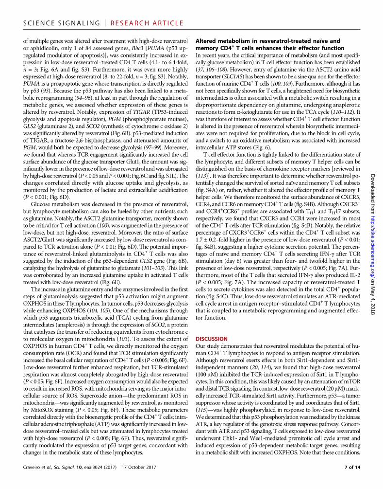
of multiple genes was altered after treatment with high-dose resveratrolor aphidicolin, only 1 of 84 assessed genes, Bbc3 [PUMA (p53 up-regulated modulator of apoptosis)], was consistently increased in ex-pression in low-dose resveratrol–treated CD4 T cells (4.1- to 6.4-fold,n = 3; Fig. 6A and fig. S3). Furthermore, it was even more highlyexpressed at high-dose resveratrol (8- to 22-fold, n = 3; fig. S3). Notably,PUMA is a proapoptotic gene whose transcription is directly regulatedby p53 (93). Because the p53 pathway has also been linked to a meta-bolic reprogramming (94–96), at least in part through the regulation ofmetabolic genes, we assessed whether expression of these genes isaltered by resveratrol. Notably, expression of TIGAR (TP53-inducedglycolysis and apoptosis regulator), PGM (phosphoglycerate mutase),GLS2 (glutaminase 2), and SCO2 (synthesis of cytochrome c oxidase 2)was significantly altered by resveratrol (Fig. 6B). p53-mediated inductionof TIGAR, a fructose-2,6-bisphosphatase, and attenuated amounts ofPGM, would both be expected to decrease glycolysis (97–99). Moreover,we found that whereas TCR engagement significantly increased the cellsurface abundance of the glucose transporter Glut1, the amount was sig-nificantly lower in the presence of low-dose resveratrol andwas abrogatedbyhigh-dose resveratrol (P<0.05 andP<0.001; Fig. 6Cand fig. S1L). Thechanges correlated directly with glucose uptake and glycolysis, asmonitored by the production of lactate and extracellular acidification(P < 0.001; Fig. 6D).
Glucose metabolism was decreased in the presence of resveratrol,but lymphocyte metabolism can also be fueled by other nutrients suchas glutamine. Notably, the ASCT2 glutamine transporter, recently shownto be critical for T cell activation (100), was augmented in the presence oflow-dose, but not high-dose, resveratrol. Moreover, the ratio of surfaceASCT2/Glut1was significantly increased by low-dose resveratrol as com-pared to TCR activation alone (P < 0.01; Fig. 6D). The potential impor-tance of resveratrol-linked glutaminolysis in CD4+ T cells was alsosuggested by the induction of the p53-dependent GLS2 gene (Fig. 6B),catalyzing the hydrolysis of glutamine to glutamate (101–103). This linkwas corroborated by an increased glutamine uptake in activated T cellstreated with low-dose resveratrol (Fig. 6E).
The increase in glutamine entry and the enzymes involved in the firststeps of glutaminolysis suggested that p53 activation might augmentOXPHOS in theseT lymphocytes. In tumor cells, p53 decreases glycolysiswhile enhancing OXPHOS (104, 105). One of the mechanisms throughwhich p53 augments tricarboxylic acid (TCA) cycling from glutamineintermediates (anaplerosis) is through the expression of SCO2, a proteinthat catalyzes the transfer of reducing equivalents from cytochrome cto molecular oxygen in mitochondria (103). To assess the extent ofOXPHOS in human CD4+ T cells, we directly monitored the oxygenconsumption rate (OCR) and found that TCR stimulation significantlyincreased the basal cellular respiration ofCD4+T cells (P<0.005; Fig. 6F).Low-dose resveratrol further enhanced respiration, but TCR-stimulatedrespiration was almost completely abrogated by high-dose resveratrol(P<0.05; Fig. 6F). Increased oxygen consumptionwould also be expectedto result in increased ROS, withmitochondria serving as themajor intra-cellular source of ROS. Superoxide anion—the predominant ROS inmitochondria—was significantly augmented by resveratrol, asmonitoredby MitoSOX staining (P < 0.05; Fig. 6F). These metabolic parameterscorrelated directly with the bioenergetic profile of the CD4+ T cells; intra-cellular adenosine triphosphate (ATP) was significantly increased in low-dose resveratrol–treated cells but was attenuated in lymphocytes treatedwith high-dose resveratrol (P < 0.005; Fig. 6F). Thus, resveratrol signifi-cantly modulated the expression of p53 target genes, concordant withchanges in the metabolic state of these lymphocytes.
Craveiro et al., Sci. Signal. 10, eaal3024 (2017) 17 October 2017
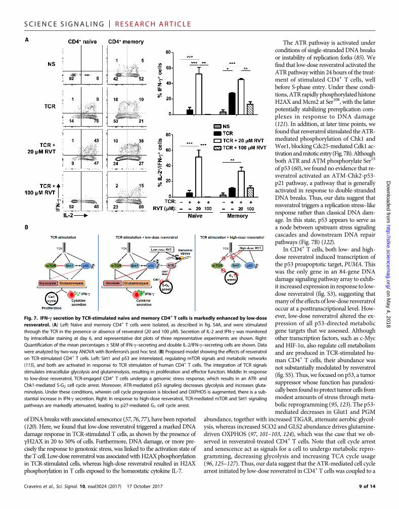
Altered metabolism in resveratrol-treated naïve andmemory CD4+ T cells enhances their effector functionIn recent years, the critical importance of metabolism (and most specifi-cally glucose metabolism) in T cell effector function has been established(37, 106–108). However, entry of glutamine via the ASCT2 amino acidtransporter (SLC1A5) has been shown to be a sine qua non for the effectorfunction of murine CD4+ T cells (100, 109). Furthermore, although it hasnot been specifically shown for T cells, a heightened need for biosyntheticintermediates is often associated with a metabolic switch resulting in adisproportionate dependency on glutamine, undergoing anapleroticreactions to form a-ketoglutarate for use in the TCA cycle (110–112). Itwas therefore of interest to assess whether CD4+ T cell effector functionis altered in the presence of resveratrol wherein biosynthetic intermedi-ates were not required for proliferation, due to the block in cell cycle,and a switch to an oxidative metabolism was associated with increasedintracellular ATP stores (Fig. 6).
T cell effector function is tightly linked to the differentiation state ofthe lymphocyte, and different subsets of memory T helper cells can bedistinguished on the basis of chemokine receptor markers [reviewed in(113)]. It was therefore important to determine whether resveratrol po-tentially changed the survival of sorted naïve andmemory T cell subsets(fig. S4A) or, rather, whether it altered the effector profile of memory Thelper cells. We therefore monitored the surface abundance of CXCR3,CCR4, andCCR6onmemoryCD4+T cells (fig. S4B). AlthoughCXCR3+
and CCR4+CCR6+ profiles are associated with TH1 and TH17 subsets,respectively, we found that CXCR3 and CCR4 were increased in mostof the CD4+ T cells after TCR stimulation (fig. S4B). Notably, the relativepercentage of CXCR3+CCR6+ cells within the CD4+ T cell subset was1.7 ± 0.2–fold higher in the presence of low-dose resveratrol (P < 0.01;fig. S4B), suggesting a higher cytokine secretion potential. The percen-tages of naïve and memory CD4+ T cells secreting IFN-g after TCRstimulation (day 6) was greater than four- and twofold higher in thepresence of low-dose resveratrol, respectively (P < 0.005; Fig. 7A). Fur-thermore, most of the T cells that secreted IFN-g also produced IL-2(P < 0.005; Fig. 7A). The increased capacity of resveratrol-treated Tcells to secrete cytokines was also detected in the total CD4+ popula-tion (fig. S4C). Thus, low-dose resveratrol stimulates anATR-mediatedcell cycle arrest in antigen receptor–stimulated CD4+ T lymphocytesthat is coupled to a metabolic reprogramming and augmented effec-tor function.
DISCUSSIONOur study demonstrates that resveratrol modulates the potential of hu-man CD4+ T lymphocytes to respond to antigen receptor stimulation.Although resveratrol exerts effects in both Sirt1-dependent and Sirt1-independent manners (20, 114), we found that high-dose resveratrol(100 mM) inhibited the TCR-induced expression of Sirt1 in T lympho-cytes. In this condition, this was likely caused by an attenuation ofmTORanddistal TCR signaling. In contrast, low-dose resveratrol (20mM)mark-edly increasedTCR-stimulated Sirt1 activity. Furthermore, p53—a tumorsuppressor whose activity is coordinated by and coordinates that of Sirt1(115)—was highly phosphorylated in response to low-dose resveratrol.Wedetermined that this p53phosphorylationwasmediatedby the kinaseATR, a key regulator of the genotoxic stress response pathway. Concor-dant with ATR and p53 signaling, T cells exposed to low-dose resveratrolunderwent Chk1- and Wee1-mediated premitotic cell cycle arrest andinduced expression of p53-dependent metabolic target genes, resultingin ametabolic shift with increased OXPHOS. Note that these conditions,
7 of 14
SC I ENCE S I GNAL ING | R E S EARCH ART I C L E
on May 4, 2018
http://stke.sciencemag.org/
Dow
nloaded from
whichpromoted an enhanced bioenergetic profile, endowedCD4+T cellswith a substantially enhanced cytokine secretion potential (Fig. 7B).
The role of resveratrol in protecting against carcinogenesis has beenthe subject of intense study, andmultiple reports showed that resveratrolfunctions bypreventingDNAdamage formation aswell as by improvingDNA damage repair (47, 50, 116). Resveratrol affects multiple aspects ofDNA metabolism, including DNA replication, recombination, repair,
Craveiro et al., Sci. Signal. 10, eaal3024 (2017) 17 October 2017
and telomeremaintenance, as well as the redox state, thereby promotingthe integrity of genomic DNA. However, in vitro, resveratrol mediatesDNA cleavage in a process requiring DNA-bound copper [Cu(II)] ions(49, 57, 59, 117–119). On the basis of diverse studies, it is nearly im-possible to draw clear-cut conclusions about the effects of sirtuins or re-sveratrol on genomic stability. Both reduction of DNA breaks and aninhibition of replicative senescence (57, 74–78), as well as the generation
Fig. 6. TCR-stimulated CD4+ T cells exhibit in-creased transcriptionof p53-dependentmetabol-ic target genes and an altered T cell metabolismafter exposure to resveratrol. (A) The expressionof 84 genes involved in DNA damage signalingpathways was evaluated in CD4+ T cells activatedby TCR engagement alone as compared to TCR en-gagement in the presence of resveratrol (20 mM)using a polymerase chain reaction (PCR) array pro-file (Qiagen; fig. S3). Representative data in one ofthree samples at day 1 of stimulation are shownwith only Bbc3 (PUMA) significantly induced in thelatter conditions (4.2- to 6.4-fold induction, n = 3).(B) Transcripts of p53 metabolic target genes includ-ing TIGAR, PGM, GLS2, and SCO2 were monitored byquantitative reverse transcription PCR analysis ofcells under the indicated conditions (day 3) andwere normalized against the abundance of 18Sribosomal RNA. Data are means ± SEM of fourindependent experiments. *P < 0.05 and **P < 0.01by paired t test. (C) Glut1 surface expression wasmonitored by flow cytometry, and representativehistograms from three experiments under the in-dicated conditions (day 3) are shown, with quan-tifications shown in fig. S1. (D) 2-Deoxy-D[1-3H]glucose (2DG) uptake (n = 3), lactate production(n = 5), and extracellular acidification (n = 5) weremonitored in cells under the indicated conditions atday 3. Data are means ± SEM of the indicated num-ber of experiments. *P < 0.05, **P < 0.01, and ***P <0.005 by one-way ANOVAwith Tukey’s post hoc test.(E) Left: ASCT2 surface expression was monitored byflow cytometry, and representative histograms areshown. Middle: The ratio ± SEM of ASCT2 to Glut1abundance in cells stimulated through the TCR inthe presence or absence of resveratrol are presentedas a function of the MFI of both transporters (n =3; paired t test). Right: Uptake of L-2,3,4-[3H]gluta-mine was performed for 10 min, and mean countsper minute ± SEM for triplicate samples from threeindependent experiments at day 3 are presented.Data were analyzed by one-way ANOVA withTukey’s post hoc test. **P < 0.01. (F) Left: Cellularrespiration was monitored on a Seahorse XF-24analyzer, and OCRs of triplicate samples underbasal conditions and in response to the indicatedmitochondrial inhibitors are presented for cells onday 3 of activation. Mean basal consumption rates(OCR; picomoles/min per 106 cells) ± SEM of trip-licate samples from three independent experimentsare shown (upper right). Right: ATP was measuredin cells under the same conditions by luminescentdetection, and mean intracellular amounts ± SEM
from data obtained in 10 independent experiments are presented. Data were analyzed by one-way ANOVA with Tukey’s post hoc test. Mitochondrial superoxide anionwas monitored using MitoSOX Red reagent, and the MFI ± SEM of triplicate samples are presented. Data were analyzed by paired t test. FCCP, carbonyl cyanide p-trifluoromethoxyphenylhydrazone; Ant.A, antimycin A; Rot., rotenone. *P < 0.05, **P < 0.01, and ***P < 0.005.8 of 14
SC I ENCE S I GNAL ING | R E S EARCH ART I C L E
on May 4, 2018
http://stke.sciencemag.org/
Dow
nloaded from
ofDNAbreakswith associated senescence (57, 76, 77), have been reported(120). Here, we found that low-dose resveratrol triggered a marked DNAdamage response in TCR-stimulated T cells, as shown by the presence ofgH2AX in 20 to 50% of cells. Furthermore, DNA damage, or more pre-cisely the response to genotoxic stress, was linked to the activation state ofthe T cell. Low-dose resveratrol was associatedwithH2AXphosphorylationin TCR-stimulated cells, whereas high-dose resveratrol resulted in H2AXphosphorylation in T cells exposed to the homeostatic cytokine IL-7.
Craveiro et al., Sci. Signal. 10, eaal3024 (2017) 17 October 2017
The ATR pathway is activated underconditions of single-stranded DNA breaksor instability of replication forks (85). Wefind that low-dose resveratrol activated theATRpathwaywithin 24 hours of the treat-ment of stimulated CD4+ T cells, wellbefore S-phase entry. Under these condi-tions, ATR rapidly phosphorylated histoneH2AX andMcm2 at Ser108, with the latterpotentially stabilizing prereplication com-plexes in response to DNA damage(121). In addition, at later time points, wefound that resveratrol stimulated theATR-mediated phosphorylation of Chk1 andWee1, blocking Cdc25-mediated Cdk1 ac-tivationandmitoticentry (Fig.7B).Althoughboth ATR and ATM phosphorylate Ser15
of p53 (60), we found no evidence that re-sveratrol activated an ATM-Chk2-p53-p21 pathway, a pathway that is generallyactivated in response to double-strandedDNA breaks. Thus, our data suggest thatresveratrol triggers a replication stress–likeresponse rather than classical DNA dam-age. In this state, p53 appears to serve asa node between upstream stress signalingcascades and downstream DNA repairpathways (Fig. 7B) (122).
In CD4+ T cells, both low- and high-dose resveratrol induced transcription ofthe p53 proapoptotic target, PUMA. Thiswas the only gene in an 84-gene DNAdamage signaling pathway array to exhib-it increased expression in response to low-dose resveratrol (fig. S3), suggesting thatmany of the effects of low-dose resveratroloccur at a posttranscriptional level. How-ever, low-dose resveratrol altered the ex-pression of all p53-directed metabolicgene targets that we assessed. Althoughother transcription factors, such as c-Mycand HIF-1a, also regulate cell metabolismand are produced in TCR-stimulated hu-man CD4+ T cells, their abundance wasnot substantially modulated by resveratrol(fig. S5). Thus, we focused on p53, a tumorsuppressor whose function has paradoxi-cally been found toprotect tumor cells frommodest amounts of stress through meta-bolic reprogramming (95, 123). The p53-mediated decreases in Glut1 and PGM
abundance, together with increased TIGAR, attenuate aerobic glycol-ysis, whereas increased SCO2 and GLS2 abundance drives glutamine-driven OXPHOS (97, 101–103, 124), which was the case that we ob-served in resveratrol-treated CD4+ T cells. Note that cell cycle arrestand senescence act as signals for a cell to undergo metabolic repro-gramming, decreasing glycolysis and increasing TCA cycle usage(96, 125–127). Thus, our data suggest that theATR-mediated cell cyclearrest initiated by low-dose resveratrol in CD4+ T cells was coupled to a
Fig. 7. IFN-g secretion by TCR-stimulated naïve and memory CD4+ T cells is markedly enhanced by low-doseresveratrol. (A) Left: Naïve and memory CD4+ T cells were isolated, as described in fig. S4A, and were stimulatedthrough the TCR in the presence or absence of resveratrol (20 and 100 mM). Secretion of IL-2 and IFN-g was monitoredby intracellular staining at day 6, and representative dot plots of three representative experiments are shown. Right:Quantification of the mean percentages ± SEM of IFN-g–secreting and double IL-2/IFN-g–secreting cells are shown. Datawere analyzed by two-way ANOVA with Bonferroni’s post hoc test. (B) Proposed model showing the effects of resveratrolon TCR-stimulated CD4+ T cells. Left: Sirt1 and p53 are interrelated, regulating mTOR signals and metabolic networks(115), and both are activated in response to TCR stimulation of human CD4+ T cells. The integration of TCR signalsstimulates intracellular glycolysis and glutaminolysis, resulting in proliferation and effector function. Middle: In responseto low-dose resveratrol, TCR-engaged CD4+ T cells undergo a genomic stress response, which results in an ATR- andChk1-mediated S-G2 cell cycle arrest. Moreover, ATR-mediated p53 signaling decreases glycolysis and increases gluta-minolysis. Under these conditions, wherein cell cycle progression is blocked and OXPHOS is augmented, there is a sub-stantial increase in IFN-g secretion. Right: In response to high-dose resveratrol, TCR-mediated mTOR and Sirt1 signalingpathways are markedly attenuated, leading to p27-mediated G1 cell cycle arrest.
9 of 14
SC I ENCE S I GNAL ING | R E S EARCH ART I C L E
http://stke.sciencemag.org
Dow
nloaded from
metabolic shift, adjusting the balance between glycolysis and OXPHOS(Fig. 7B).
The bioenergetic profile of resveratrol-treated T cells was altered bythis skewing of metabolism away from glycolysis and toward a settingcharacterized by an increasedASCT2-to-Glut1 ratio, with an augmentedglutamine transport, substantially increasedmitochondrial ROS produc-tion, and increased OXPHOS. How this would affect T cell effectorfunction is unclear because glycolysis can increase and decrease thepotential of T cells to secrete effector cytokines, such as IFN-g and IL-17(128–133). Note that amino acid metabolism is essential for effector T celldifferentiation (100, 109, 134, 135), and memory cells rely more onOXPHOS than on glycolysis (38, 130). In both naïve and memoryCD4+ T cells, low-dose resveratrol markedly augmented the amount ofIFN-g secreted, but this increase was even higher for naïve cells than formemory cells (means of 10- and 2-fold, respectively). Furthermore, inboth cell types, resveratrol substantially increased the number of cells thatproduced both IL-2 and IFN-g. Thus, our data suggest that resveratrol isan agent that, by altering the metabolic fitness of T lymphocytes, en-hances their cytokine effector potential.
Adjusting the balance between glycolysis and OXPHOS can alsohave substantial effects in other cell types. DecreasingOXPHOS inmiceexpressing amutant p53markedly attenuates tumorigenesis (136). Thus,generating a context that is the converse of that shaped by resveratrol,that is, inhibiting a p53-mediated shift to mitochondrial metabolism,may be beneficial for individuals with an increased risk of developingcancers, such as Li-Fraumeni syndrome patients with germline muta-tions in the TP53 gene. The potential use of resveratrol as a therapyfor the treatment of neurological, cardiovascular, hepatic, and metabolicpathologies therefore necessitates a critical evaluation of its effect on Tlymphocytes in vivo, especially in an autoimmune setting. The datashown here reveal a complex network of resveratrol-stimulated changesin cell cycle progression andmetabolism, altering the potential of T lym-phocytes to respond to foreign antigens.
on May 4, 2018
/
MATERIALS AND METHODST cell isolation and cultureCD4+ T cells were isolated from adult peripheral blood, obtained fromhealthy donors after informed consent. Cells were purified using negative-selection Rosette tetramers (STEMCELL Technologies), and the purity ofthe cell population was monitored on a FACSCanto II (BD Biosciences).Purities were always greater than 94%. Naïve and memory CD4+ T cellswere sorted on a FACSAria after staining with anti-CD4, anti-CD45RA,anti-CD45RO,CD62L,CD127, andCD25 antibodies (fig. S4A). Lympho-cytes (1 × 10⁶ per well in a 24-well plate) were cultured in RPMI 1640 +GlutaMAX (Gibco, Life Technologies) supplemented with 10% fetal calfserum (FCS) and 2% penicillin/streptomycin (Gibco, Life Technologies).For TCR stimulation, 24-well plates were coated with anti-CD3 (cloneOKT3,BioLegend) and anti-CD28 (clone 9.3, providedbyC. June)mono-clonal antibodies (mAbs) at a concentration of 1 mg/ml, and recombinantIL-2 (rIL-2) (50U/ml)was added as indicated. T cellswere also cultured inthe presence of rIL-7 (10 ng/ml). As indicated, resveratrol (20 or 100 mM;Sigma-Aldrich), compound C1 (1 mM; Sigma-Aldrich), VE-821 (1 and5mM;Euromedex), aphidicolin (1mM), andbleomycin (1mM)were addedto T cell cultures 1 hour before TCR stimulation.
ImmunofluorescenceCells were collected and coated on poly-L-lysine–treated slides. Cells werefixed in a 4% paraformaldehyde (PFA) solution [phosphate-buffered sa-
Craveiro et al., Sci. Signal. 10, eaal3024 (2017) 17 October 2017
line (PBS), 4%PFA] at 37°C for 15min, permeabilized in PBS containing3% bovine serum albumin (BSA)/0.1% saponin for 10 min, and blockedfor nonspecific protein binding with 10% FCS. Stainingwith primary anti-Sirt1 antibody (Ab) (Santa Cruz Biotechnology) and a secondary AlexaFluor 488–coupled anti-rabbit immunoglobulin G (Invitrogen) was per-formed inPBS containing 3%BSA for 1hour at room temperature.Nucleiwere then labeled by DAPI staining for 10 min at room temperature.
Flow cytometric analysesTo detect cell surfacemarkers, cells were incubatedwith the appropriatefluorochrome-conjugated mAbs, and expression was monitored incomparison with isotype controls. Antibodies against CD4, CD25,CD69, and CD71 were from Beckman Coulter. Y319-phosphorylatedZAP-70 (BD Biosciences), T202/Y204-phosphorylated ERK1/2 (BDBiosciences), and phosphorylated H2AX (BioLegend) were detected af-ter cell fixation and permeabilization. Surface Glut1 and ASCT2 weredetected by binding to their respective retroviral envelope ligands fusedto enhanced green fluorescent protein or recombinant rabbit fragmentcrystallizable (rFc) (Metafora Biosystems), as previously described(72, 137–139). The presence of mitochondrial superoxide was assessedby staining with MitoSOX Red indicator (1 mM; Invitrogen). Proliferationwasmonitored as a function of carboxyfluorescein diacetate succinimidylester (Invitrogen) or violet proliferation dye (Invitrogen) dilution.Before staining for intracellular IFN-g and IL-2 (BD Biosciences), cellswere activatedwithphorbol 12-myristate 13-acetate (100ng/ml; Sigma-Al-drich) and ionomycin (1 mg/ml; Sigma-Aldrich) in the presence of brefel-din A (10 mg/ml; Sigma-Aldrich) for 3.5 to 4 hours at 37°C. Cell cycleanalysiswas performedby simultaneous staining forDNAandRNAusing7-aminoactinomycin D (20 mM; Sigma-Aldrich) and pyronin Y (5 mM;Sigma-Aldrich), respectively. Cells were assessed on a FACSCanto II orBDLSR II Fortessa (BDBiosciences), and datawere analyzed using FACS-Diva (BD Biosciences) or FlowJo (Tree Star) software.
Metabolic assaysOCRs were measured on an XF-24 Extracellular Flux Analyzer (Sea-horse Bioscience). TCR-stimulated T cells with and without low-doseresveratrol (20 mM) were seeded at a concentration of 1.5 × 10⁶ cells,whereas nonstimulated and high-dose resveratrol-stimulated cells wereseeded at a concentration of 2.0 × 10⁶ cells in XFmedium (nonbufferedDulbecco’smodified Eagle’smediumcontaining 2.5mMglucose, 2mML-glutamine, and 1 mM sodium pyruvate). Oxygen consumption wasmonitored under basal conditions and in response to oligomycin (1 mM),carbonyl cyanide p-trifluoromethoxyphenylhydrazone (1.5 mM), rotenone(100 nM), and antimycin A (1 mM; Sigma-Aldrich). The basal respirationrate was calculated as the difference between basal OCR and the OCR afterinhibitionofmitochondrial complexes 1 and3with rotenoneandantimycinA, respectively. ATP and L-lactate weremeasured according to the stan-dard procedures of theATPlite kit (PerkinElmer) and L-lactate kit (EtonBioscience), respectively. Extracellular pH was measured immediatelyafter harvesting of medium using a standard pH meter.
Glucose and glutamine uptake assaysCells (2 × 10⁶)were starved by incubation at 37°C in serum and glucose-or glutamine-free RPMI 1640 for 30 min. Radiolabeled 2-deoxy-D-[1-3H] glucose or glutamine-L-[3,4-3H(N)] (PerkinElmer) was added toa final concentration of 0.1 mM (2 mCi/ml). Cells were incubated for10min at room temperature, washed in cold serum/glucose/glutamine-free RPMI 1640, and solubilized in 500 ml of 0.1% SDS. Radioactivitywas measured by liquid scintillation.
10 of 14
SC I ENCE S I GNAL ING | R E S EARCH ART I C L E
on May 4, 2018
http://stke.sciencemag.org/
Dow
nloaded from
Total protein extraction and analysesCells were lysed in lysis buffer containing 20 mM Hepes (pH 7.6),100mMKCl, 0.1mMEDTA, 1mMNaF, 1mM sodium orthovanadate,1% Triton X-100, 0.5% NP-40, 0.15 mM spermine, 0.5 mM spermidine,1 mMdithiothreitol, and a protease inhibitor cocktail. After a 30-min in-cubation on ice, extracts were centrifuged, and supernatants were har-vested. Extracts (20 mg) were resolved on SDS–polyacrylamide gelelectrophoresis gels (8.5 to 12%) and transferred electrophoretically ontopolyvinylidene difluoride (PVDF) membranes. PVDF membranes wereincubatedwith the indicated antibodies (table S1) for 1hour at room tem-perature or overnight at 4°C andwithhorseradishperoxidase–conjugatedanti-goat, anti-rabbit, or anti-mouse secondaryAbs, and immunoreactiveproteins were visualized using enhanced chemiluminescence (ECL,Amersham) according to the manufacturer’s instructions. Proteins werequantified with ImageJ software and normalized to amido black–stainedproteins (loading controls) or tubulin as indicated.
Gene expression analysis by PCR arrayTotal RNA was isolated with the RNeasy Mini Kit (Qiagen) and wasreverse-transcribed with QuantiTect Reverse Transcription Kit (Qiagen).SYBR Green–based (SYBR Green I Master, Roche) real-time quantitativePCR (qPCR) for TIGAR, PGM, GLS2, SCO2, HIF1A, and RNA18S wasperformedwith the LightCycler 480 Real-Time PCR System (Roche), andall primers are shown in table S2. To determine relative expression,samples for each experimental condition were run in duplicate and werenormalized to 18S. Primer sequences used for amplificationwere designedwith Primer3 and NetPrimer (PREMIER Biosoft) software packages. Theexpression of 84 genes involved in DNA damage signaling pathways wasanalyzed with the RT2 Profiler PCR Array (SABiosciences, Qiagen)according to the manufacturer’s instructions. Briefly, RNA was preparedfrom 5 million cells after 24 hours in culture under the indicated con-ditions using the Qiagen RNeasy kit. First-strand complementary DNAwas then prepared and used in the PCR array in combination with SYBRGreen qPCR master mixes on a Roche Light Cycler 480. Data were nor-malized toACTB (actin beta), B2M (b2-microglobulin), and RPLP0 usingthe SABiosciences DNA template analysis software.
Statistical analysesData were analyzed with GraphPad Software (GraphPad Prism), andP values were calculated by one-way ANOVA (Tukey’s post hoc test),two-way ANOVA (with Tukey’s post hoc or Bonferroni’s tests), orpaired t tests, as indicated.
SUPPLEMENTARY MATERIALSwww.sciencesignaling.org/cgi/content/full/10/501/eaal3024/DC1Fig. S1. Quantification and statistical analyses of main data panels.Fig. S2. Effects of resveratrol on the formation of CD4+ T cell blasts and cell counts in responseto TCR engagement.Fig. S3. Effect of resveratrol treatment on the expression of DNA damage signaling pathway genes.Fig. S4. Sorting of naïve and memory CD4+ T cells for the assessment of phenotype andcytokine secretion profiles.Fig. S5. TCR-mediated induction of HIF1A and c-Myc in human CD4+ cells is not altered by low-dose resveratrol.Table S1. Antibody list.Table S2. Primer sequences.
REFERENCES AND NOTES1. B. B. Aggarwal, A. Bhardwaj, R. S. Aggarwal, N. P. Seeram, S. Shishodia, Y. Takada, Role of
resveratrol in prevention and therapy of cancer: Preclinical and clinical studies.Anticancer Res. 24, 2783–2840 (2004).
Craveiro et al., Sci. Signal. 10, eaal3024 (2017) 17 October 2017
2. B. P. Hubbard, D. A. Sinclair, Small molecule SIRT1 activators for the treatment of agingand age-related diseases. Trends Pharmacol. Sci. 35, 146–154 (2014).
3. K. S. Bhullar, B. P. Hubbard, Lifespan and healthspan extension by resveratrol.Biochim. Biophys. Acta 1852, 1209–1218 (2015).
4. A. Chalkiadaki, L. Guarente, Sirtuins mediate mammalian metabolic responses tonutrient availability. Nat. Rev. Endocrinol. 8, 287–296 (2012).
5. B. Agarwal, J. A. Baur, Resveratrol and life extension. Ann. N. Y. Acad. Sci. 1215, 138–143(2011).
6. B. Poussier, A. C. Cordova, J.-P. Becquemin, B. E. Sumpio, Resveratrol inhibits vascularsmooth muscle cell proliferation and induces apoptosis. J. Vasc. Surg. 42, 1190–1197(2005).
7. Z. Estrov, S. Shishodia, S. Faderl, D. Harris, Q. Van, H. M. Kantarjian, M. Talpaz,B. B. Aggarwal, Resveratrol blocks interleukin-1b-induced activation of the nucleartranscription factor NF-kB, inhibits proliferation, causes S-phase arrest, and inducesapoptosis of acute myeloid leukemia cells. Blood 102, 987–995 (2003).
8. P. Signorelli, R. Ghidoni, Resveratrol as an anticancer nutrient: Molecular basis, openquestions and promises. J. Nutr. Biochem. 16, 449–466 (2005).
9. S. Das, D. K. Das, Resveratrol: A therapeutic promise for cardiovascular diseases.Recent Pat. Cardiovasc. Drug Discov. 2, 133–138 (2007).
10. S. Das, D. K. Das, Anti-inflammatory responses of resveratrol. Inflamm. AllergyDrug Targets 6, 168–173 (2007).
11. O. Vang, What is new for resveratrol? Is a new set of recommendations necessary?Ann. N. Y. Acad. Sci. 1290, 1–11 (2013).
12. J. Tomé-Carneiro, M. Larrosa, A. González-Sarrías, F. A. Tomás-Barberán,M. T. García-Conesa, J. C. Espín, Resveratrol and clinical trials: The crossroad from in vitrostudies to human evidence. Curr. Pharm. Des. 19, 6064–6093 (2013).
13. J. M. Smoliga, E. S. Colombo, M. J. Campen, A healthier approach to clinical trialsevaluating resveratrol for primary prevention of age-related diseases in healthypopulations. Aging 5, 495–506 (2013).
14. R. S. Turner, R. G. Thomas, S. Craft, C. H. van Dyck, J. Mintzer, B. A. Reynolds, J. B. Brewer,R. A. Rissman, R. Raman, P. S. Aisen; Alzheimer’s Disease Cooperative Study, Arandomized, double-blind, placebo-controlled trial of resveratrol for Alzheimer disease.Neurology 85, 1383–1391 (2015).
15. O. Vang, Resveratrol: Challenges in analyzing its biological effects. Ann. N. Y. Acad. Sci.1348, 161–170 (2015).
16. R. H. X. Wong, R. S. Nealon, A. Scholey, P. R. C. Howe, Low dose resveratrol improvescerebrovascular function in type 2 diabetes mellitus. Nutr. Metab. Cardiovasc. Dis.26, 393–399 (2016).
17. S.-J. Lin, P.-A. Defossez, L. Guarente, Requirement of NAD and SIR2 for life-spanextension by calorie restriction in Saccharomyces cerevisiae. Science 289, 2126–2128(2000).
18. K. T. Howitz, K. J. Bitterman, H. Y. Cohen, D. W. Lamming, S. Lavu, J. G. Wood, R. E. Zipkin,P. Chung, A. Kisielewski, L.-L. Zhang, B. Scherer, D. A. Sinclair, Small moleculeactivators of sirtuins extend Saccharomyces cerevisiae lifespan. Nature 425, 191–196(2003).
19. M. Gertz, G. T. Nguyen, F. Fischer, B. Suenkel, C. Schlicker, B. Fränzel, J. Tomaschewski,F. Aladini, C. Becker, D. Wolters, C. Steegborn, A molecular mechanism for directsirtuin activation by resveratrol. PLOS ONE 7, e49761 (2012).
20. J. A. Baur, K. J. Pearson, N. L. Price, H. A. Jamieson, C. Lerin, A. Kalra, V. V. Prabhu,J. S. Allard, G. Lopez-Lluch, K. Lewis, P. J. Pistell, S. Poosala, K. G. Becker, O. Boss,D. Gwinn, M. Wang, S. Ramaswamy, K. W. Fishbein, R. G. Spencer, E. G. Lakatta,D. Le Couteur, R. J. Shaw, P. Navas, P. Puigserver, D. K. Ingram, R. de Cabo,D. A. Sinclair, Resveratrol improves health and survival of mice on a high-calorie diet.Nature 444, 337–342 (2006).
21. A. Satoh, C. S. Brace, N. Rensing, P. Cliften, D. F. Wozniak, E. D. Herzog, K. A. Yamada,S.-i. Imai, Sirt1 extends life span and delays aging in mice through the regulation of Nk2homeobox 1 in the DMH and LH. Cell Metab. 18, 416–430 (2013).
22. Y. Kanfi, S. Naiman, G. Amir, V. Peshti, G. Zinman, L. Nahum, Z. Bar-Joseph,H. Y. Cohen, The sirtuin SIRT6 regulates lifespan in male mice. Nature 483, 218–221(2012).
23. A. S. Banks, N. Kon, C. Knight, M. Matsumoto, R. Gutiérrez-Juárez, L. Rossetti, W. Gu,D. Accili, SirT1 gain of function increases energy efficiency and prevents diabetes inmice. Cell Metab. 8, 333–341 (2008).
24. D. Herranz, M. Serrano, SIRT1: Recent lessons from mouse models. Nat. Rev. Cancer 10,819–823 (2010).
25. D. Herranz, M. Muñoz-Martin, M. Cañamero, F. Mulero, B. Martinez-Pastor,O. Fernandez-Capetillo, M. Serrano, Sirt1 improves healthy ageing and protects frommetabolic syndrome-associated cancer. Nat. Commun. 1, 3 (2010).
26. G. Donmez, L. Guarente, Aging and disease: Connections to sirtuins. Aging Cell 9,285–290 (2010).
27. G. Donmez, D. Wang, D. E. Cohen, L. Guarente, SIRT1 suppresses b-amyloid productionby activating the a-secretase gene ADAM10. Cell 142, 320–332 (2010).
11 of 14
SC I ENCE S I GNAL ING | R E S EARCH ART I C L E
on May 4, 2018
http://stke.sciencemag.org/
Dow
nloaded from
28. P. T. Pfluger, D. Herranz, S. Velasco-Miguel, M. Serrano, M. H. Tschöp, Sirt1 protectsagainst high-fat diet-induced metabolic damage. Proc. Natl. Acad. Sci. U.S.A. 105,9793–9798 (2008).
29. J. Sequeira, G. Boily, S. Bazinet, S. Saliba, X. He, K. Jardine, C. Kennedy, W. Staines,C. Rousseaux, R. Mueller, M. W. McBurney, sirt1-null mice develop an autoimmune-likecondition. Exp. Cell Res. 314, 3069–3074 (2008).
30. J. Zhang, S.-M. Lee, S. Shannon, B. Gao, W. Chen, A. Chen, R. Divekar, M. W. McBurney,H. Braley-Mullen, H. Zaghouani, D. Fang, The type III histone deacetylase Sirt1 isessential for maintenance of T cell tolerance in mice. J. Clin. Invest. 119, 3048–3058(2009).
31. A. Purushotham, T. T. Schug, Q. Xu, S. Surapureddi, X. Guo, X. Li, Hepatocyte-specificdeletion of SIRT1 alters fatty acid metabolism and results in hepatic steatosis andinflammation. Cell Metab. 9, 327–338 (2009).
32. H.-L. Cheng, R. Mostoslavsky, S. Saito, J. P. Manis, Y. Gu, P. Patel, R. Bronson, E. Appella,F. W. Alt, K. F. Chua, Developmental defects and p53 hyperacetylation in Sir2homolog (SIRT1)-deficient mice. Proc. Natl. Acad. Sci. U.S.A. 100, 10794–10799 (2003).
33. S. Kong, S.-J. Kim, B. Sandal, S.-M. Lee, B. Gao, D. D. Zhang, D. Fang, The type IIIhistone deacetylase Sirt1 protein suppresses p300-mediated histone H3 lysine 56acetylation at Bclaf1 promoter to inhibit T cell activation. J. Biol. Chem. 286,16967–16975 (2011).
34. V. K. Nimmagadda, C. T. Bever, N. R. Vattikunta, S. Talat, V. Ahmad, N. K. Nagalla,D. Trisler, S. I. V. Judge, W. Royal III, K. Chandrasekaran, J. W. Russell, T. K. Makar,Overexpression of SIRT1 protein in neurons protects against experimentalautoimmune encephalomyelitis through activation of multiple SIRT1 targets.J. Immunol. 190, 4595–4607 (2013).
35. Y. Wang, Y. Bi, C. Li, Y. Li, Z. Zhang, J. Wang, Y. Lu, Q. Yu, H. Su, H. Yang, G. Liu, Histonedeacetylase SIRT1 negatively regulates the differentiation of interleukin-9-producingCD4+ T Cells. Immunity 44, 11337–11349 (2016).
36. H. W. Lim, S. G. Kang, J. K. Ryu, B. Schilling, M. Fei, I. S. Lee, A. Kehasse, K. Shirakawa,M. Yokoyama, M. Schnölzer, H. G. Kasler, H.-S. Kwon, B. W. Gibson, H. Sato, K. Akassoglou,C. Xiao, D. R. Littman, M. Ott, E. Verdin, SIRT1 deacetylates RORgt and enhancesTh17 cell generation. J. Exp. Med. 212, 607–617 (2015).
37. N. J. Maciver, R. D. Michalek, J. C. Rathmell, Metabolic regulation of T lymphocytes.Annu. Rev. Immunol. 31, 259–283 (2013).
38. G. J. W. van der Windt, E. L. Pearce, Metabolic switching and fuel choice during T-celldifferentiation and memory development. Immunol. Rev. 249, 27–42 (2012).
39. E. L. Pearce, E. J. Pearce, Metabolic pathways in immune cell activation and quiescence.Immunity 38, 633–643 (2013).
40. R. H. Houtkooper, E. Pirinen, J. Auwerx, Sirtuins as regulators of metabolism andhealthspan. Nat. Rev. Mol. Cell Biol. 13, 225–238 (2012).
41. C. Cantó, R. H. Houtkooper, E. Pirinen, D. Y. Youn, M. H. Oosterveer, Y. Cen,P. J. Fernandez-Marcos, H. Yamamoto, P. A. Andreux, P. Cettour-Rose, K. Gademann,C. Rinsch, K. Schoonjans, A. A. Sauve, J. Auwerx, The NAD+ precursor nicotinamideriboside enhances oxidative metabolism and protects against high-fat diet-inducedobesity. Cell Metab. 15, 838–847 (2012).
42. R. Cerutti, E. Pirinen, C. Lamperti, S. Marchet, A. A. Sauve, W. Li, V. Leoni, E. A. Schon,F. Dantzer, J. Auwerx, C. Viscomi, M. Zeviani, NAD+-dependent activation of Sirt1corrects the phenotype in a mouse model of mitochondrial disease. Cell Metab. 19,1042–1049 (2014).
43. G. Xuzhu, M. Komai-Koma, B. P. Leung, H. S. Howe, C. McSharry, I. B. McInnes, D. Xu,Resveratrol modulates murine collagen-induced arthritis by inhibiting Th17 and B-cellfunction. Ann. Rheum. Dis. 71, 129–135 (2012).
44. R. Falchetti, M. P. Fuggetta, G. Lanzilli, M. Tricarico, G. Ravagnan, Effects of resveratrol onhuman immune cell function. Life Sci. 70, 81–96 (2001).
45. Y. Yang, J. H. Paik, D. Cho, J.-A. Cho, C.-W. Kim, Resveratrol induces the suppression oftumor-derived CD4+CD25+ regulatory T cells. Int. Immunopharmacol. 8, 542–547 (2008).
46. S.-L. Wu, L. Yu, X.-Y. Jiao, K.-W. Meng, C.-E. Pan, The suppressive effect of resveratrolon protein kinase Cq in peripheral blood T lymphocytes in a rat liver transplantationmodel. Transplant. Proc. 38, 3052–3054 (2006).
47. S. A. Gatz, L. Wiesmüller, Take a break—Resveratrol in action on DNA. Carcinogenesis 29,321–332 (2008).
48. N. G. Denissova, C. M. Nasello, P. L. Yeung, J. A. Tischfield, M. A. Brenneman, Resveratrolprotects mouse embryonic stem cells from ionizing radiation by acceleratingrecovery from DNA strand breakage. Carcinogenesis 33, 149–155 (2012).
49. M. J. Burkitt, J. Duncan, Effects of trans-resveratrol on copper-dependent hydroxyl-radical formation and DNA damage: Evidence for hydroxyl-radical scavenging and anovel, glutathione-sparing mechanism of action. Arch. Biochem. Biophys. 381, 253–263(2000).
50. R.-H. Wang, K. Sengupta, C. Li, H.-S. Kim, L. Cao, C. Xiao, S. Kim, X. Xu, Y. Zheng,B. Chilton, R. Jia, Z.-M. Zheng, E. Appella, X. W. Wang, T. Ried, C.-X. Deng, ImpairedDNA damage response, genome instability, and tumorigenesis in SIRT1 mutant mice.Cancer Cell 14, 312–323 (2008).
Craveiro et al., Sci. Signal. 10, eaal3024 (2017) 17 October 2017
51. H. D. Halicka, H. Zhao, J. Li, Y. S. Lee, T. C. Hsieh, J. M. Wu, Z. Darzynkiewicz, Potentialanti-aging agents suppress the level of constitutive mTOR- and DNA damage- signaling.Aging 4, 952–965 (2012).
52. A. Tyagi, R. P. Singh, C. Agarwal, S. Siriwardana, R. A. Sclafani, R. Agarwal, Resveratrolcauses Cdc2-tyr15 phosphorylation via ATM/ATR-Chk1/2-Cdc25C pathway as acentral mechanism for S phase arrest in human ovarian carcinoma Ovcar-3 cells.Carcinogenesis 26, 1978–1987 (2005).
53. D. J. Colin, E. Limagne, K. Ragot, G. Lizard, F. Ghiringhelli, É. Solary, B. Chauffert,N. Latruffe, D. Delmas, The role of reactive oxygen species and subsequent DNA-damage response in the emergence of resistance towards resveratrol in colon cancermodels. Cell Death Dis. 5, e1533 (2014).
54. A. Rashid, C. Liu, T. Sanli, E. Tsiani, G. Singh, R. G. Bristow, I. Dayes, H. Lukka, J. Wright,T. Tsakiridis, Resveratrol enhances prostate cancer cell response to ionizing radiation.Modulation of the AMPK, Akt and mTOR pathways. Radiat. Oncol. 6, 144 (2011).
55. E. H. Heiss, Y. D. C. Schilder, V. M. Dirsch, Chronic treatment with resveratrol inducesredox stress- and ataxia telangiectasia-mutated (ATM)-dependent senescence inp53-positive cancer cells. J. Biol. Chem. 282, 26759–26766 (2007).
56. A. R. Hussain, S. Uddin, R. Bu, O. S. Khan, S. O. Ahmed, M. Ahmed, K. S. Al-Kuraya,Resveratrol suppresses constitutive activation of AKT via generation of ROS and inducesapoptosis in diffuse large B cell lymphoma cell lines. PLOS ONE 6, e24703 (2011).
57. H. Luo, A. Yang, B. A. Schulte, M. J. Wargovich, G. Y. Wang, Resveratrol inducespremature senescence in lung cancer cells via ROS-mediated DNA damage.PLOS ONE 8, e60065 (2013).
58. K. Fukuhara, M. Nagakawa, I. Nakanishi, K. Ohkubo, K. Imai, S. Urano, S. Fukuzumi,T. Ozawa, N. Ikota, M. Mochizuki, N. Miyata, H. Okuda, Structural basis for DNA-cleavingactivity of resveratrol in the presence of Cu(II). Bioorg. Med. Chem. 14, 1437–1443 (2006).
59. N. Ahmad, V. M. Adhami, F. Afaq, D. K. Feyes, H. Mukhtar, Resveratrol causesWAF-1/p21-mediated G1-phase arrest of cell cycle and induction of apoptosis inhuman epidermoid carcinoma A431 cells. Clin. Cancer Res. 7, 1466–1473 (2001).
60. T. Sperka, J. Wang, K. L. Rudolph, DNA damage checkpoints in stem cells, ageing andcancer. Nat. Rev. Mol. Cell Biol. 13, 579–590 (2012).
61. M. K. Zeman, K. A. Cimprich, Causes and consequences of replication stress.Nat. Cell Biol. 16, 2–9 (2014).
62. A. C. Carrano, E. Eytan, A. Hershko, M. Pagano, SKP2 is required for ubiquitin-mediateddegradation of the CDK inhibitor p27. Nat. Cell Biol. 1, 193–199 (1999).
63. H. Sutterlüty, E. Chatelain, A. Marti, C. Wirbelauer, M. Senften, U. Muller, W. Krek,p45SKP2 promotes p27Kip1 degradation and induces S phase in quiescent cells.Nat. Cell Biol. 1, 207–214 (1999).
64. S. Colombetti, V. Basso, D. L. Mueller, A. Mondino, Prolonged TCR/CD28 engagementdrives IL-2-independent T cell clonal expansion through signaling mediated by themammalian target of rapamycin. J. Immunol. 176, 2730–2738 (2006).
65. S. Sauer, L. Bruno, A. Hertweck, D. Finlay, M. Leleu, M. Spivakov, Z. A. Knight, B. S. Cobb,D. Cantrell, E. O’Connor, K. M. Shokat, A. G. Fisher, M. Merkenschlager, T cell receptorsignaling controls Foxp3 expression via PI3K, Akt, and mTOR. Proc. Natl. Acad. Sci. U.S.A.105, 7797–7802 (2008).
66. G. M. Delgoffe, T. P. Kole, Y. Zheng, P. E. Zarek, K. L. Matthews, B. Xiao, P. F. Worley,S. C. Kozma, J. D. Powell, The mTOR kinase differentially regulates effector andregulatory T cell lineage commitment. Immunity 30, 832–844 (2009).
67. M. Laplante, D. M. Sabatini, mTOR signaling at a glance. J. Cell Sci. 122, 3589–3594(2009).
68. S. Sengupta, T. R. Peterson, D. M. Sabatini, Regulation of the mTOR complex 1 pathwayby nutrients, growth factors, and stress. Mol. Cell 40, 310–322 (2010).
69. H. S. Ghosh, M. McBurney, P. D. Robbins, SIRT1 negatively regulates the mammaliantarget of rapamycin. PLOS ONE 5, e9199 (2010).
70. S. Hong, B. Zhao, D. B. Lombard, D. C. Fingar, K. Inoki, Cross-talk between sirtuin andmammalian target of rapamycin complex 1 (mTORC1) signaling in the regulation of S6kinase 1 (S6K1) phosphorylation. J. Biol. Chem. 289, 13132–13141 (2014).
71. Y. Wang, Y. Bi, X. Chen, C. Li, Y. Li, Z. Zhang, J. Wang, Y. Lu, Q. Yu, H. Su, H. Yang, G. Liu,Histone deacetylase SIRT1 negatively regulates the differentiation of interleukin-9-producing CD4+ T cells. Immunity 44, 1337–1349 (2016).
72. N. Manel, S. Kinet, J.-L. Battini, F. J. Kim, N. Taylor, M. Sitbon, The HTLV receptor is anearly T-cell activation marker whose expression requires de novo protein synthesis.Blood 101, 1913–1918 (2003).
73. M. Lavanya, S. Kinet, A. Montel-Hagen, C. Mongellaz, J.-L. Battini, M. Sitbon, N. Taylor,Cell surface expression of the bovine leukemia virus-binding receptor on B and Tlymphocytes is induced by receptor engagement. J. Immunol. 181, 891–898 (2008).
74. Y. Tang, J. Xu, W. Qu, X. Peng, P. Xin, X. Yang, C. Ying, X. Sun, L. Hao, Resveratrol reducesvascular cell senescence through attenuation of oxidative stress by SIRT1/NADPHoxidase-dependent mechanisms. J. Nutr. Biochem. 23, 1410–1416 (2012).
75. X. B. Wang, L. Zhu, J. Huang, Y. G. Yin, X. Q. Kong, Q. F. Rong, A. W. Shi, K. J. Cao,Resveratrol-induced augmentation of telomerase activity delays senescence ofendothelial progenitor cells. Chin Med J (Engl) 124, 4310–4315 (2011).
12 of 14
SC I ENCE S I GNAL ING | R E S EARCH ART I C L E
on May 4, 2018
http://stke.sciencemag.org/
Dow
nloaded from
76. Z. Gao, M. S. Xu, T. L. Barnett, C. W. Xu, Resveratrol induces cellular senescence withattenuated mono-ubiquitination of histone H2B in glioma cells. Biochem. Biophys.Res. Commun. 407, 271–276 (2011).
77. L. Peltz, J. Gomez, M. Marquez, F. Alencastro, N. Atashpanjeh, T. Quang, T. Bach, Y. Zhao,Resveratrol exerts dosage and duration dependent effect on human mesenchymalstem cell development. PLOS ONE 7, e37162 (2012).
78. S. Yamashita, K. Ogawa, T. Ikei, M. Udono, T. Fujiki, Y. Katakura, SIRT1 prevents replicativesenescence of normal human umbilical cord fibroblast through potentiating thetranscription of human telomerase reverse transcriptase gene. Biochem. Biophys.Res. Commun. 417, 630–634 (2012).
79. O. Fernandez-Capetillo, A. Celeste, A. Nussenzweig, Focusing on foci: H2AX and therecruitment of DNA-damage response factors. Cell Cycle 2, 426–427 (2003).
80. M. Fragkos, J. Jurvansuu, P. Beard, H2AX is required for cell cycle arrest via the p53/p21pathway. Mol. Cell. Biol. 29, 2828–2840 (2009).
81. I. M. Ward, J. Chen, Histone H2AX is phosphorylated in an ATR-dependent manner inresponse to replicational stress. J. Biol. Chem. 276, 47759–47762 (2001).
82. Z. Feng, H. Zhang, A. J. Levine, S. Jin, The coordinate regulation of the p53 and mTORpathways in cells. Proc. Natl. Acad. Sci. U.S.A. 102, 8204–8209 (2005).
83. R. G. Jones, D. R. Plas, S. Kubek, M. Buzzai, J. Mu, Y. Xu, M. J. Birnbaum, C. B. Thompson,AMP-activated protein kinase induces a p53-dependent metabolic checkpoint.Mol. Cell 18, 283–293 (2005).
84. K. H. Vousden, K. M. Ryan, p53 and metabolism. Nat. Rev. Cancer 9, 691–700 (2009).85. J. Smith, L. M. Tho, N. Xu, D. A. Gillespie, The ATM–Chk2 and ATR–Chk1 pathways in DNA
damage signaling and cancer. Adv. Cancer Res. 108, 73–112 (2010).86. L. I. Toledo, M. Murga, P. Gutierrez-Martinez, R. Soria, O. Fernandez-Capetillo, ATR
signaling can drive cells into senescence in the absence of DNA breaks. Genes Dev. 22,297–302 (2008).
87. G. Lossaint, E. Besnard, D. Fisher, J. Piette, V. Dulic, Chk1 is dispensable for G2 arrest inresponse to sustained DNA damage when the ATM/p53/p21 pathway is functional.Oncogene 30, 4261–4274 (2011).
88. C. Evrin, P. Clarke, J. Zech, R. Lurz, J. Sun, S. Uhle, H. Li, B. Stillman, C. Speck, A double-hexameric MCM2-7 complex is loaded onto origin DNA during licensing of eukaryoticDNA replication. Proc. Natl. Acad. Sci. U.S.A. 106, 20240–20245 (2009).
89. D. Cortez, G. Glick, S. J. Elledge, Minichromosome maintenance proteins are directtargets of the ATM and ATR checkpoint kinases. Proc. Natl. Acad. Sci. U.S.A. 101,10078–10083 (2004).
90. K. A. Cimprich, D. Cortez, ATR: An essential regulator of genome integrity. Nat. Rev.Mol. Cell Biol. 9, 616–627 (2008).
91. R. Prevo, E. Fokas, P. M. Reaper, P. A. Charlton, J. R. Pollard, W. G. McKenna, R. J. Muschel,T. B. Brunner, The novel ATR inhibitor VE-821 increases sensitivity of pancreaticcancer cells to radiation and chemotherapy. Cancer Biol. Ther. 13, 1072–1081 (2012).
92. J. Vávrová, L. Zárybnická, E. Lukášová, M. Řezáčová, E. Novotná, Z. Šinkorová, A. Tichý,J. Pejchal, K. Ďurišová, Inhibition of ATR kinase with the selective inhibitor VE-821 resultsin radiosensitization of cells of promyelocytic leukaemia (HL-60). Radiat. Environ.Biophys. 52, 471–479 (2013).
93. K. Nakano, K. H. Vousden, PUMA, a novel proapoptotic gene, is induced by p53.Mol. Cell 7, 683–694 (2001).
94. C. R. B. Berkers, O. D. K. Maddocks, E. C. Cheung, I. Mor, K. H. Vousden, Metabolicregulation by p53 family members. Cell Metab. 18, 617–633 (2013).
95. E. Gottlieb, K. H. Vousden, p53 regulation of metabolic pathways. Cold Spring Harb.Perspect. Biol. 2, a001040 (2010).
96. K. M. Aird, R. Zhang, Nucleotide metabolism, oncogene-induced senescence and cancer.Cancer Lett. 356, 204–210 (2015).
97. K. Bensaad, A. Tsuruta, M. A. Selak, M. N. C. Vidal, K. Nakano, R. Bartrons, E. Gottlieb,K. H. Vousden, TIGAR, a p53-inducible regulator of glycolysis and apoptosis. Cell 126,107–120 (2006).
98. H. Kondoh, M. E. Lleonart, J. Gil, J. Wang, P. Degan, G. Peters, D. Martinez, A. Carnero,D. Beach, Glycolytic enzymes can modulate cellular life span. Cancer Res. 65, 177–185(2005).
99. A. M. Puzio-Kuter, The role of p53 in metabolic regulation. Genes Cancer 2, 385–391(2011).
100. M. Nakaya, Y. Xiao, X. Zhou, J.-H. Chang, M. Chang, X. Cheng, M. Blonska, X. Lin, S.-C. Sun,Inflammatory T cell responses rely on amino acid transporter ASCT2 facilitation ofglutamine uptake and mTORC1 kinase activation. Immunity 40, 692–705 (2014).
101. W. Hu, C. Zhang, R. Wu, Y. Sun, A. Levine, Z. Feng, Glutaminase 2, a novel p53 targetgene regulating energy metabolism and antioxidant function. Proc. Natl. Acad. Sci. U.S.A.107, 7455–7460 (2010).
102. S. Suzuki, T. Tanaka, M. V. Poyurovsky, H. Nagano, T. Mayama, S. Ohkubo, M. Lokshin,H. Hosokawa, T. Nakayama, Y. Suzuki, S. Sugano, E. Sato, T. Nagao, K. Yokote, I. Tatsuno,C. Prives, Phosphate-activated glutaminase (GLS2), a p53-inducible regulator ofglutamine metabolism and reactive oxygen species. Proc. Natl. Acad. Sci. U.S.A. 107,7461–7466 (2010).
Craveiro et al., Sci. Signal. 10, eaal3024 (2017) 17 October 2017
103. S. Matoba, J.-G. Kang, W. D. Patino, A. Wragg, M. Boehm, O. Gavrilova, P. J. Hurley,F. Bunz, P. M. Hwang, p53 regulates mitochondrial respiration. Science 312, 1650–1653(2006).
104. W. Ma, H. Sung, J. Park, S. Matoba, P. Hwang, A pivotal role for p53: Balancing aerobicrespiration and glycolysis. J. Bioenerg. Biomembr. 39, 243–246 (2007).
105. X.-d. Zhang, Z.-h. Qin, J. Wang, The role of p53 in cell metabolism. Acta Pharmacol. Sin.31, 1208–1212 (2010).
106. E. L. Pearce, M. C. Poffenberger, C.-H. Chang, R. G. Jones, Fueling immunity: Insights intometabolism and lymphocyte function. Science 342, 1242454 (2013).
107. G. M. Delgoffe, K. N. Pollizzi, A. T. Waickman, E. Heikamp, D. J. Meyers, M. R. Horton,B. Xiao, P. F. Worley, J. D. Powell, The kinase mTOR regulates the differentiation of helperT cells through the selective activation of signaling by mTORC1 and mTORC2.Nat. Immunol. 12, 295–303 (2011).
108. M. D. Buck, D. O’Sullivan, E. L. Pearce, T cell metabolism drives immunity. J. Exp. Med.212, 1345–1360 (2015).
109. D. Klysz, X. Tai, P. A. Robert, M. Craveiro, G. Cretenet, L. Oburoglu, C. Mongellaz, S. Floess,V. Fritz, M. I. Matias, C. Yong, N. Surh, J. C. Marie, J. Huehn, V. Zimmermann, S. Kinet,V. Dardalhon, N. Taylor, Glutamine-dependent a-ketoglutarate production regulates thebalance between T helper 1 cell and regulatory T cell generation. Sci. Signal. 8, ra97(2015).
110. R. J. DeBerardinis, A. Mancuso, E. Daikhin, I. Nissim, M. Yudkoff, S. Wehrli,C. B. Thompson, Beyond aerobic glycolysis: Transformed cells can engage inglutamine metabolism that exceeds the requirement for protein and nucleotidesynthesis. Proc. Natl. Acad. Sci. U.S.A. 104, 19345–19350 (2007).
111. C. A. Lyssiotis, J. Son, L. C. Cantley, A. C. Kimmelman, Pancreatic cancers rely on a novelglutamine metabolism pathway to maintain redox balance. Cell Cycle 12, 1987–1988(2013).
112. J. Son, C. A. Lyssiotis, H. Ying, X. Wang, S. Hua, M. Ligorio, R. M. Perera, C. R. Ferrone,E. Mullarky, N. Shyh-Chang, Y. Kang, J. B. Fleming, N. Bardeesy, J. M. Asara, M. C. Haigis,R. A. DePinho, L. C. Cantley, A. C. Kimmelman, Glutamine supports pancreatic cancergrowth through a KRAS-regulated metabolic pathway. Nature 496, 101–105 (2013).
113. E. Lubberts, The IL-23-IL-17 axis in inflammatory arthritis. Nat. Rev. Rheumatol. 11,415–429 (2015).
114. N. L. Price, A. P. Gomes, A. J. Y. Ling, F. V. Duarte, A. Martin-Montalvo, B. J. North,B. Agarwal, L. Ye, G. Ramadori, J. S. Teodoro, B. P. Hubbard, A. T. Varela, J. G. Davis,B. Varamini, A. Hafner, R. Moaddel, A. P. Rolo, R. Coppari, C. M. Palmeira, R. de Cabo,J. A. Baur, D. A. Sinclair, SIRT1 is required for AMPK activation and the beneficial effectsof resveratrol on mitochondrial function. Cell Metab. 15, 675–690 (2012).
115. T. F. Liu, C. E. McCall, Deacetylation by SIRT1 reprograms inflammation and cancer.Genes Cancer 4, 135–147 (2013).
116. M. Jang, L. Cai, G. O. Udeani, K. V. Slowing, C. F. Thomas, C. W. Beecher, H. H. Fong,N. R. Farnsworth, A. D. Kinghorn, R. G. Mehta, R. C. Moon, J. M. Pezzuto, Cancerchemopreventive activity of resveratrol, a natural product derived from grapes.Science 275, 218–220 (1997).
117. K. Fukuhara, N. Miyata, Resveratrol as a new type of DNA-cleaving agent. Bioorg. Med.Chem. Lett. 8, 3187–3192 (1998).
118. M. Subramanian, U. Shadakshari, S. Chattopadhyay, A mechanistic study on thenuclease activities of some hydroxystilbenes. Bioorg. Med. Chem. 12, 1231–1237 (2004).
119. S. A. Gatz, M. Keimling, C. Baumann, T. Dörk, K.-M. Debatin, S. Fulda, L. Wiesmuller,Resveratrol modulates DNA double-strand break repair pathways in an ATM/ATR–p53-and –Nbs1-dependent manner. Carcinogenesis 29, 519–527 (2008).
120. L. Bosch-Presegue, A. Vaquero, The dual role of sirtuins in cancer. Genes Cancer 2,648–662 (2011).
121. B. E. Stead, C. J. Brandl, M. K. Sandre, M. J. Davey, Mcm2 phosphorylation and theresponse to replicative stress. BMC Genet. 13, 36 (2012).
122. S. Sengupta, C. C. Harris, p53: Traffic cop at the crossroads of DNA repair andrecombination. Nat. Rev. Mol. Cell Biol. 6, 44–55 (2005).
123. E. Kim, A. Giese, W. Deppert, Wild-type p53 in cancer cells: When a guardian turns into ablackguard. Biochem. Pharmacol. 77, 11–20 (2009).
124. O. D. K. Maddocks, K. H. Vousden, Metabolic regulation by p53. J. Mol. Med. 89, 237–245(2011).
125. S. Mazurek, W. Zwerschke, P. Jansen-Dürr, E. Eigenbrodt, Metabolic cooperationbetween different oncogenes during cell transformation: Interaction between activatedras and HPV-16 E7. Oncogene 20, 6891–6898 (2001).
126. J. Kaplon, L. Zheng, K. Meissl, B. Chaneton, V. A. Selivanov, G. Mackay, S. H. van der Burg,E. M. E. Verdegaal, M. Cascante, T. Shlomi, E. Gottlieb, D. S. Peeper, A key role formitochondrial gatekeeper pyruvate dehydrogenase in oncogene-induced senescence.Nature 498, 109–112 (2013).
127. M. Li, K. R. Durbin, S. M. Sweet, J. D. Tipton, Y. Zheng, N. L. Kelleher, Oncogene-inducedcellular senescence elicits an anti-Warburg effect. Proteomics 13, 2585–2596 (2013).
128. R. D. Michalek, V. A. Gerriets, S. R. Jacobs, A. N. Macintyre, N. J. MacIver, E. F. Mason,S. A. Sullivan, A. G. Nichols, J. C. Rathmell, Cutting edge: Distinct glycolytic and lipid
13 of 14
SC I ENCE S I GNAL ING | R E S EARCH ART I C L E
http://stke.sciencemD
ownloaded from
oxidative metabolic programs are essential for effector and regulatory CD4+ T cellsubsets. J. Immunol. 186, 3299–3303 (2011).
129. C.-H. Chang, J. D. Curtis, L. B. Maggi Jr., B. Faubert, A. V. Villarino, D. O’Sullivan,S. C.-C. Huang, G. J. W. van der Windt, J. Blagih, J. Qiu, J. D. Weber, E. J. Pearce, R. G. Jones,E. L. Pearce, Posttranscriptional control of T cell effector function by aerobic glycolysis.Cell 153, 1239–1251 (2013).
130. M. Sukumar, J. Liu, Y. Ji, M. Subramanian, J. G. Crompton, Z. Yu, R. Roychoudhuri,D. C. Palmer, P. Muranski, E. D. Karoly, R. P. Mohney, C. A. Klebanoff, A. Lal, T. Finkel,N. P. Restifo, L. Gattinoni, Inhibiting glycolytic metabolism enhances CD8+ T cellmemory and antitumor function. J. Clin. Invest. 123, 4479–4488 (2013).
131. C.-H. Chang, J. Qiu, D. O’Sullivan, M. D. Buck, T. Noguchi, J. D. Curtis, Q. Chen, M. Gindin,M. M. Gubin, G. J. W. van der Windt, E. Tonc, R. D. Schreiber, E. J. Pearce, E. L. Pearce,Metabolic competition in the tumor microenvironment is a driver of cancer progression.Cell 162, 1229–1241 (2015).
132. P.-C. Ho, J. D. Bihuniak, A. N. Macintyre, M. Staron, X. Liu, R. Amezquita, Y.-C. Tsui, G. Cui,G. Micevic, J. C. Perales, S. H. Kleinstein, E. D. Abel, K. L. Insogna, S. Feske, J. W. Locasale,M. W. Bosenberg, J. C. Rathmell, S. M. Kaech, Phosphoenolpyruvate is a metaboliccheckpoint of anti-tumor T cell responses. Cell 162, 1217–1228 (2015).
133. G. Cretenet, I. Clerc, M. Matias, S. Loisel, M. Craveiro, L. Oburoglu, S. Kinet, C. Mongellaz,V. Dardalhon, N. Taylor, Cell surface Glut1 levels distinguish human CD4 andCD8 T lymphocyte subsets with distinct effector functions. Sci. Rep. 6, 24129(2016).
134. L. V. Sinclair, J. Rolf, E. Emslie, Y.-B. Shi, P. M. Taylor, D. A. Cantrell, Control ofamino-acid transport by antigen receptors coordinates the metabolic reprogrammingessential for T cell differentiation. Nat. Immunol. 14, 500–508 (2013).
135. A. N. Macintyre, V. A. Gerriets, A. G. Nichols, R. D. Michalek, M. C. Rudolph, D. Deoliveira,S. M. Anderson, E. D. Abel, B. J. Chen, L. P. Hale, J. C. Rathmell, The glucosetransporter Glut1 is selectively essential for CD4 T cell activation and effectorfunction. Cell Metab. 20, 61–72 (2014).
136. P.-y. Wang, J. Li, F. L. Walcott, J.-G. Kang, M. F. Starost, S. L. Talagala, J. Zhuang,J.-H. Park, R. D. Huffstutler, C. M. Bryla, P. L. Mai, M. Pollak, C. M. Annunziata,S. A. Savage, A. T. Fojo, P. M. Hwang, Inhibiting mitochondrial respiration prevents cancer ina mouse model of Li-Fraumeni syndrome. J. Clin. Invest. 127, 132–136 (2017).
137. F. J. Kim, N. Manel, E. N. Garrido, C. Valle, M. Sitbon, J.-L. Battini, HTLV-1 and -2envelope SU subdomains and critical determinants in receptor binding.Retrovirology 1, 41 (2004).
138. L. Swainson, S. Kinet, N. Manel, J.-L. Battini, M. Sitbon, N. Taylor, Glucose transporter1 expression identifies a population of cycling CD4+ CD8+ human thymocytes
Craveiro et al., Sci. Signal. 10, eaal3024 (2017) 17 October 2017
with high CXCR4-induced chemotaxis. Proc. Natl. Acad. Sci. U.S.A. 102, 12867–12872(2005).
139. S. Kinet, L. Swainson, M. Lavanya, C. Mongellaz, A. Montel-Hagen, M. Craveiro,N. Manel, J.-L. Battini, M. Sitbon, N. Taylor, Isolated receptor binding domains ofHTLV-1 and HTLV-2 envelopes bind Glut-1 on activated CD4+ and CD8+ T cells.Retrovirology 4, 31 (2007).
Acknowledgments: We thank all members of our laboratories for discussions, scientificcritique, and continual support. We are grateful to Montpellier RIO Imaging for support incytometry experiments and the RAM (Animal House Network of Montpellier) animal facility ofthe Institut de Génétique Moléculaire de Montpellier. We are indebted to T. Gostan of theService d’Analyse de Données Biologiques Complexes platform for data and biostatisticalanalyses and to M. Sitbon and A. Singer for critical discussions and reading of the manuscript.Funding: M.C. was supported by a fellowship from the Portuguese Foundation forScience and Technology. G.C. was supported by a fellowship from the Ligue Contre le Cancer.M.I.M. received funding from the French Ministry of Education. C.M., V. Dardalhon, V.S.Z.and V. Dulić are supported by CNRS. N.T. is supported by INSERM. This work was supportedby funding from the Association de la Recherche contre le Cancer, French national (AgenceNationale de la Recherche) research grants (PolarATTACK and GlutStem), Institut National duCancer, the European Community (contracts LSHC-CT-2005-018914 “ATTACK” and PIRG5-GA-2009-249227 “T cell homeostasis”), and the French laboratory consortiums (Labex) EpiGenMed andGR-Ex and support by the Labex EpiGenMed and GR-Ex programs, reference ANR-10-LABX-12-01and ANR-11-LABX-0051. Author contributions: M.C., G.C., C.M., V. Dulić, and N.T. conceivedthe project and designed the experiments. M.C., G.C., C.M., M.I.M., V.S.Z., V. Dardalhon, andV. Dulić executed the experiments, and all authors contributed to data analyses andinterpretations of the results. M.C., G.C., C.M., V. Dulić, and N.T. wrote the manuscript, andall authors added critical reviews and modifications. Competing interests: The authorsdeclare that they have no competing interests.
Submitted 1 November 2016Accepted 29 September 2017Published 17 October 201710.1126/scisignal.aal3024
Citation: M. Craveiro, G. Cretenet, C. Mongellaz, M. I. Matias, O. Caron, M. C. P. de Lima,V. S. Zimmermann, E. Solary, V. Dardalhon, V. Dulić, N. Taylor, Resveratrol stimulates themetabolic reprogramming of human CD4+ T cells to enhance effector function. Sci. Signal.10, eaal3024 (2017).
ag
14 of 14
on May 4, 2018
.org/
effector function T cells to enhance+Resveratrol stimulates the metabolic reprogramming of human CD4
Zimmermann, Eric Solary, Valérie Dardalhon, Vjekoslav Dulic and Naomi TaylorMarco Craveiro, Gaspard Cretenet, Cédric Mongellaz, Maria I. Matias, Olivier Caron, Maria C. Pedroso de Lima, Valérie S.
DOI: 10.1126/scisignal.aal3024 (501), eaal3024.10Sci. Signal.
carefully assessed, especially in an autoimmune setting.. These data suggest that the use of resveratrol to treat various pathologies should beγinflammatory cytokine interferon-
the metabolism of the cells to make them more responsive to antigens and produce increased amounts of the T cells to antigens. However, low doses of the drug reprogrammed+resveratrol inhibited the responses of human CD4
. found that high doses ofet alproduced conflicting evidence of the effects of resveratrol in different contexts. Craveiro because of its potential therapeutic effects against cancer, aging, and inflammation. However, many studies have
Resveratrol is a polyphenolic plant compound, which has attracted much interest as a pharmacological agentResveratrol and lymphocyte responses
ARTICLE TOOLS http://stke.sciencemag.org/content/10/501/eaal3024
MATERIALSSUPPLEMENTARY http://stke.sciencemag.org/content/suppl/2017/10/13/10.501.eaal3024.DC1
CONTENTRELATED
http://science.sciencemag.org/content/sci/354/6317/1260.fullhttp://stke.sciencemag.org/content/sigtrans/11/512/eaas9172.fullhttp://stm.sciencemag.org/content/scitransmed/7/298/298ra117.fullhttp://stke.sciencemag.org/content/sigtrans/10/510/eaan4931.fullhttp://stke.sciencemag.org/content/sigtrans/10/507/eaar5580.fullhttp://stke.sciencemag.org/content/sigtrans/10/467/eaaf8823.fullhttp://stke.sciencemag.org/content/sigtrans/10/475/eaah4273.full
REFERENCES
http://stke.sciencemag.org/content/10/501/eaal3024#BIBLThis article cites 138 articles, 35 of which you can access for free
PERMISSIONS http://www.sciencemag.org/help/reprints-and-permissions
Terms of ServiceUse of this article is subject to the
registered trademark of AAAS.is aScience Signaling Association for the Advancement of Science. No claim to original U.S. Government Works. The title
York Avenue NW, Washington, DC 20005. 2017 © The Authors, some rights reserved; exclusive licensee American (ISSN 1937-9145) is published by the American Association for the Advancement of Science, 1200 NewScience Signaling
on May 4, 2018
http://stke.sciencemag.org/
Dow
nloaded from