Embed Size (px)
Citation preview
Resveratrol Protects Chondrocytes from Apoptosis viaAltering the Ultrastructural and BiomechanicalProperties: An AFM StudyHua Jin1., Qian Liang1., Tongsheng Chen2, Xiaoping Wang1*
1 Department of Pain Management, The First Affiliated Hospital of Jinan University, Guangzhou, China, 2 MOE Key Laboratory of Laser Life Science & Institute of Laser Life
Science, South China Normal University, Guangzhou, China
Abstract
Osteoarthritis (OA), a degenerative joint disease with high prevalence among older people, occurs from molecular ornanometer level and extends gradually to higher degrees of the ultrastructure of cartilage, finally resulting in irreversiblestructural and functional damages. This report aims to use atomic force microscopy (AFM) to investigate the protectiveeffects of resveratrol (RV), a drug with good anti-inflammatory properties, on cellular morphology, membrane architecture,cytoskeleton, cell surface adhesion and stiffness at nanometer level in sodium nitroprusside (SNP)-induced apoptoticchondrocytes, a typical cellular OA model. CCK-8 assay showed that 100 mM RV significantly prevented SNP-inducedcytotoxicity. AFM imaging and quantitative analysis showed that SNP potently induced chondrocytes changes includingshrunk, round, lamellipodia contraction and decrease in adherent junctions among cells, as well as the destruction ofbiomechanics: 90% decrease in elasticity and 30% decrease in adhesion. In addition, confocal imaging analysis showed thatSNP induced aggregation of the cytoskeleton and decrease in the expression of cytoskeletal proteins. More importantly,these SNP-induced damages to chondrocytes could be potently prevented by RV pretreatment. Interestingly, thebiomechanical changes occurred before morphological changes could be clearly observed during SNP-induced apoptosis,indicating that the biomechanics of cellular membrane may be a more robust indicator of cell function. Collectively, ourdata demonstrate that RV prevents SNP-induced apoptosis of chondrocytes by regulating actin organization, and that AFM-based technology can be developed into a powerful and sensitive method to study the interaction mechanisms betweenchondrocytes and drugs.
Citation: Jin H, Liang Q, Chen T, Wang X (2014) Resveratrol Protects Chondrocytes from Apoptosis via Altering the Ultrastructural and Biomechanical Properties:An AFM Study. PLoS ONE 9(3): e91611. doi:10.1371/journal.pone.0091611
Editor: Etienne Dague, LAAS-CNRS, France
Received November 27, 2013; Accepted February 12, 2014; Published March 14, 2014
Copyright: � 2014 Jin et al. This is an open-access article distributed under the terms of the Creative Commons Attribution License, which permits unrestricteduse, distribution, and reproduction in any medium, provided the original author and source are credited.
Funding: This work was supported by the National Natural Science Foundation of China (Grant No. 81071491 and 61178078), and Key Project of the Departmentof Education and Finance of Guangdong Province (cxzd115). The funders had no role in study design, data collection and analysis, decision to publish, orpreparation of the manuscript.
Competing Interests: The authors have declared that no competing interests exist.
* E-mail: [email protected]
. These authors contributed equally to this work.
Introduction
Osteoarthritis (OA) is known as a degenerative arthritis or
degenerative joint disease, which affects 20 million people in U.S.
[1]. At present, the treatment for OA mainly focuses on relieving
pains and symptoms, and improving function of cartilage.
However, there are no treatments to cure OA or reduce the
degradation of cartilage. Current treatments for OA are restricted
to anti-inflammatory drugs which bring numerous side effects and
are only temporarily effective to the patients. To find safe and
highly effective drugs for OA treatment are therefore very urgent.
Resveratrol (3,5,49 -trihydroxystilbene, RV), a polyphenol
derived from grapes, berries, peanuts and other plants, has been
shown to possess anti-proliferative, anti-oxidative and anti-
inflammatory properties [2], and these effects are associated with
the suppression of inflammation, arthritis and cardiovascular
diseases [3]. It is reported that RV protects chondrocytes from
apoptosis via preventing mitochondrial depolarization and ATP
consumption [4] or suppressing ROS and p53-production [5]. RV
also can be as a potent safe drug for OA treatment, but the
mechanisms are still unclear.
Apoptosis of chondrocytes is regarded as a feature of progressive
cartilage degeneration in OA [6]. Sodium nitroprusside (SNP) was
widely used as the donor of nitric oxide (NO) to study the
molecular mechanism of NO-induced chondrocytes apoptosis
[7,8]. Although NOC-12 may be a more effective NO donor in
OA metastasis [9], it could not effectively induce apoptosis of
chondrocytes [10]. Eo and co-workers [11] reported that RV
could rescue SNP-induced degradation of I-kappa B alpha mainly
through SN50 peptide-mediated inhibition of NF-kappa B activity,
thus blocking SNP-induced caspase-3 activation and apoptosis.
However, the effects of RV on morphological and biomechanical
properties of chondrocytes at subcelluar or nanometer-level have
not been studied.
Nanobiomechanics of cells have been identified as a vital
characteristic to distinguish normal cells from diseased cells which
differ physically from healthy cells [12]. Diseases can not only
cause biological and functional alterations but also induce
abnormalities in physical and structural characteristics of cells.
PLOS ONE | www.plosone.org 1 March 2014 | Volume 9 | Issue 3 | e91611
Therefore, research into biomechanics at the cellular and
molecular levels of some human diseases can provide a better
elucidation on the mechanisms behind disease progression [13],
thereby providing important information for treatment of these
diseases as well. Due to the nanometer resolution, AFM has been
extensively used in detection of some diseases at cellular or
subcellular level [14]. The ultrastructural and biomechanical
properties have been altered a lot in disease or cancerous cells, and
these alterations can be used as target to diagnose or distinguish
diseased cells from healthy cells [15,16]. Furthermore, biome-
chanics of cell membrane is always changed in the context of
drugs. Therefore, detecting these changes at nanometer level is
very important for evaluating curative effect and elucidating
mechanisms of drugs.
In this work, we used the rabbit chondrocytes as the cell model
to detect the protective effects of RV on SNP-induced chondro-
cytes apoptosis. Rabbit chondrocytes have been extensively used in
the basic research of mechanisms of chondrocytes or OA [17–24],
and we have gained ripe experimental experiences [25]. Alter-
ations in ultrastructure and biomechanics of cellular membrane of
chondrocytes with or without RV pretreatment were investigated
using AFM at nanometer scale. Our results showed that RV could
effectively protect chondrocytes from apoptosis through altering
the cytoskeleton arrangements and biomechanical properties
including cellular stiffness and adhesion force.
Materials and Methods
MaterialsTrypsin and type II collagenase, DMEM, fetal bovine serum,
Cell Counting Kit-8 were purchased from Invitrogen (California,
USA), Hyclone (Logan, Utah, USA), Sijiqing (Hangzhou, China)
and Dojindo (Kumamoto, Japan), respectively. Actin-Tracker
Green (phalloidin-FITC) and Tubulin-Tracker Red (a-Tubulin-
Alexa Fluor 555) were both obtained from Beyotime Institute of
Biotechnology (Naijing, China). Dulbecco’s modified Eagle
medium (DMEM) was from Gibco (Carlsbad, California, USA),
fetal bovine serum (FBS) was from Sijiqing (Hangzhou, China).
Isolation and culture of chondrocytesNew Zealand rabbits were purchased from Experimental
Animal Center of Guangzhou (China). As Tonomura, et al [26]
described, articular cartilage was derived from knee, hip and
shoulder joints of 6-week-old New Zealand white rabbits. The
utilization of rabbit articular cartilage has been approved by the
Animal Ethics Committee of Guangdong province, China. The
extracted cartilages were firstly minced into small pieces and
Chondrocytes were isolated by enzymatic digestion of 0.25%
Trypsin in phosphate buffered solution (PBS) for 1 h and 0.2%
type II collagenase in DMEM for 4–6 h. After collection by
centrifugation, chondrocytes were resuspended in DMEM sup-
plemented with 10% FBS and antibiotics (100 U/ml penicillin and
100 U/ml streptomycin) and 4.5% glucose. The cells were
transferred when confluent monolayer cells reached to 85–90%,
the transferred density was 56104 cells/cm2. The growth medium
was changed every other day. The second and third generations of
chondrocytes were used in our study.
CCK-8 assay to analyze cell viabilityChondrocytes were cultured in 96-well plates for 24 h, and then
exposed to different concentrations of SNP for different periods.
Cell viability was assessed using Cell Counting Kit assay according
to the manufacturer’s instructions. All experiments were per-
formed three times.
AFM measurements of cell morphologyFor all topographic images, the cells were fixed with 2.5%
paraformaldehyde, and imaged by a tapping mode AFM (Park
Scientific Instruments) in air. The silicon nitride tips (UL20B) used
in all AFM measurements were irradiated with ultraviolet in air for
15 min to remove any organic contaminates prior to use. The
curvature radius of the tips is less than 10 nm, and the length,
width and thickness of the cantilevers are 115, 30, and 3.5 mm,
respectively, with the oscillation frequency of 255 kHz and a force
constant of 0.03 N/m.
Surface roughness of cell membraneThe average surface roughness (Ra) is defined as the arithmetic
mean of the deviations in height from the line mean value, and Rq
is the root mean square. As the roughness has a dependence on the
sampling size, Ra and Rq were analyzed in two different areas: 10
randomly selected 4 mm2 (2 mm62 mm) and 10 randomly selected
25 mm2 (5 mm65 mm). P,0.05 were considered as statistical
significance.
Determination of nanomechanical properties ofchondrocytes
The force spectroscopy of cells was detected using an AFM
(Agilent 5500) in the near physiological environment. The
methods here were according to Kim’s procedure [27]. In brief,
the cells were firstly fixed with 2.5% glutaraldehyde and kept in
PBS (PH = 7.4) during AFM tip indentation. All force measure-
ments were performed at the same loading rate (1.26105 pN/s).
The deflection-vs-displacement curves were obtained by the
instrument, and to convert the deflection-vs-displacement curves
into the force-vs-distance curves, we adopted Cappella’s method
[28]. In each group, over 20 cells were measured. The data of
stiffness and adhesion forces were processed using SPSS13.0 to
gain the Gaussian distribution (or normal distribution) histograms.
The Young’s modulus was calculated using Hertz model which
shows the relationship between the applied force F and the
indentation d:
F~4
3
ER1=2d
3=2
1�u2ð Þ
In the equation, n is the poisson ratio, F the loading force, d the
indentation, E the Young’s modulus, and R the curvature radius of
the AFM tip, respectively. A Poisson ratio of 0.5 is appropriate for
cells [29–31]. Young’s modulus during the calculations to obtain
the best fit to the model considering the least-squares method as
proposed by Dimitriadis et al [32].
Immunofluorescence stainingThe characterizations of cytoskeleton were evaluated by
staining with phalloidin-FITC and Tubulin-Tracker, separately.
The chondrocytes were fixed with 4% paraformaldehyde for
30 min and incubated with 1 mM phalloidin-FITC or 1 mM
Tubulin-Tracker for 60 min in dark at room temperature,
separately, and then washed twice with PBS. After that, the
cytoskeleton organization was imaged by a laser scanning confocal
microscope (LCM 510 Meta Duo Scan, Carl Zeiss, Germeny).
The resulting fluorescence was also measured by flow cytometer at
excitation wavelength 488 nm, emission wavelength 530 nm to
quantitatively elucidate the alterations of cytoskeleton proteins.
AFM Studied Effect of Resveratrol on Chondrocytes
PLOS ONE | www.plosone.org 2 March 2014 | Volume 9 | Issue 3 | e91611
Results and Discussions
The changes in cell viability induced by SNP and RVTo detect the protecting effects of RV on chondrocytes, we
firstly established the OA model by exposure of chondrocytes to
SNP, an inorganic compound with the formula Na2[Fe(CN)5-
NO]N2H2O. SNP has been used as anti-hypertensive treatments
for decades and it has not obvious side effects. In vitro, SNP could
be as an external NO donor to induce apoptosis.
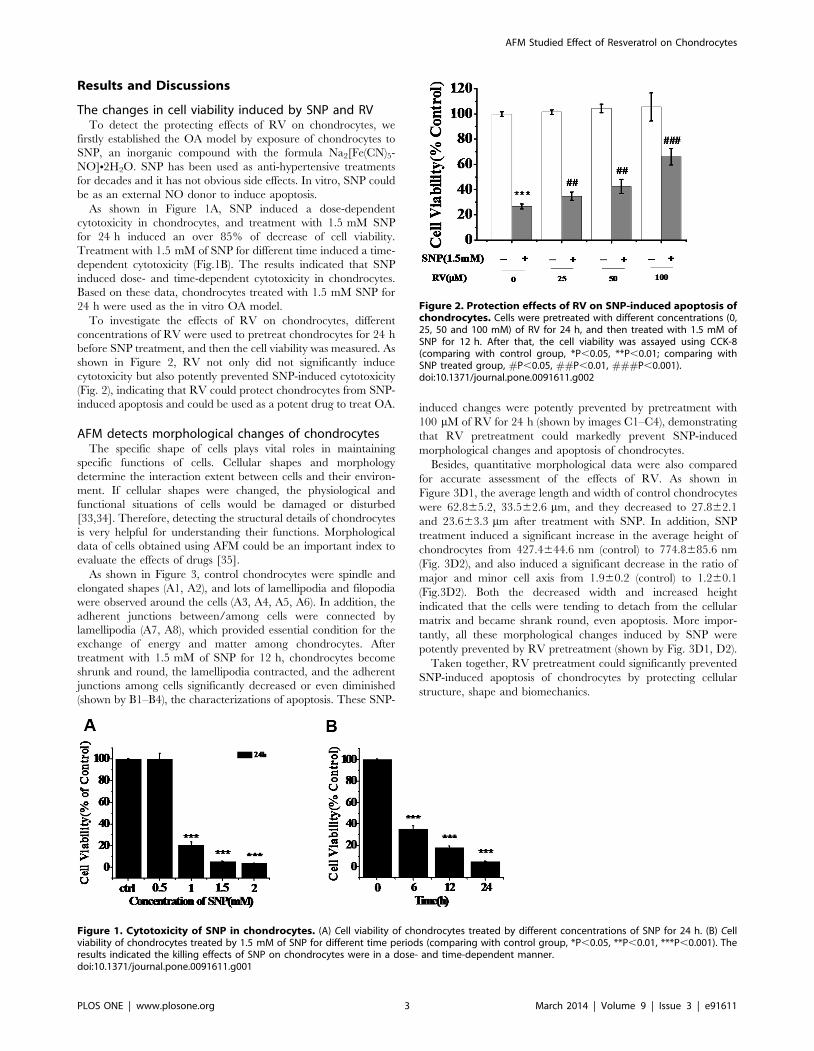
As shown in Figure 1A, SNP induced a dose-dependent
cytotoxicity in chondrocytes, and treatment with 1.5 mM SNP
for 24 h induced an over 85% of decrease of cell viability.
Treatment with 1.5 mM of SNP for different time induced a time-
dependent cytotoxicity (Fig.1B). The results indicated that SNP
induced dose- and time-dependent cytotoxicity in chondrocytes.
Based on these data, chondrocytes treated with 1.5 mM SNP for
24 h were used as the in vitro OA model.
To investigate the effects of RV on chondrocytes, different
concentrations of RV were used to pretreat chondrocytes for 24 h
before SNP treatment, and then the cell viability was measured. As
shown in Figure 2, RV not only did not significantly induce
cytotoxicity but also potently prevented SNP-induced cytotoxicity
(Fig. 2), indicating that RV could protect chondrocytes from SNP-
induced apoptosis and could be used as a potent drug to treat OA.
AFM detects morphological changes of chondrocytesThe specific shape of cells plays vital roles in maintaining
specific functions of cells. Cellular shapes and morphology
determine the interaction extent between cells and their environ-
ment. If cellular shapes were changed, the physiological and
functional situations of cells would be damaged or disturbed
[33,34]. Therefore, detecting the structural details of chondrocytes
is very helpful for understanding their functions. Morphological
data of cells obtained using AFM could be an important index to
evaluate the effects of drugs [35].
As shown in Figure 3, control chondrocytes were spindle and
elongated shapes (A1, A2), and lots of lamellipodia and filopodia
were observed around the cells (A3, A4, A5, A6). In addition, the
adherent junctions between/among cells were connected by
lamellipodia (A7, A8), which provided essential condition for the
exchange of energy and matter among chondrocytes. After
treatment with 1.5 mM of SNP for 12 h, chondrocytes become
shrunk and round, the lamellipodia contracted, and the adherent
junctions among cells significantly decreased or even diminished
(shown by B1–B4), the characterizations of apoptosis. These SNP-
induced changes were potently prevented by pretreatment with
100 mM of RV for 24 h (shown by images C1–C4), demonstrating
that RV pretreatment could markedly prevent SNP-induced
morphological changes and apoptosis of chondrocytes.
Besides, quantitative morphological data were also compared
for accurate assessment of the effects of RV. As shown in
Figure 3D1, the average length and width of control chondrocytes
were 62.865.2, 33.562.6 mm, and they decreased to 27.862.1
and 23.663.3 mm after treatment with SNP. In addition, SNP
treatment induced a significant increase in the average height of
chondrocytes from 427.4644.6 nm (control) to 774.8685.6 nm
(Fig. 3D2), and also induced a significant decrease in the ratio of
major and minor cell axis from 1.960.2 (control) to 1.260.1
(Fig.3D2). Both the decreased width and increased height
indicated that the cells were tending to detach from the cellular
matrix and became shrank round, even apoptosis. More impor-
tantly, all these morphological changes induced by SNP were
potently prevented by RV pretreatment (shown by Fig. 3D1, D2).
Taken together, RV pretreatment could significantly prevented
SNP-induced apoptosis of chondrocytes by protecting cellular
structure, shape and biomechanics.
Figure 1. Cytotoxicity of SNP in chondrocytes. (A) Cell viability of chondrocytes treated by different concentrations of SNP for 24 h. (B) Cellviability of chondrocytes treated by 1.5 mM of SNP for different time periods (comparing with control group, *P,0.05, **P,0.01, ***P,0.001). Theresults indicated the killing effects of SNP on chondrocytes were in a dose- and time-dependent manner.doi:10.1371/journal.pone.0091611.g001
Figure 2. Protection effects of RV on SNP-induced apoptosis ofchondrocytes. Cells were pretreated with different concentrations (0,25, 50 and 100 mM) of RV for 24 h, and then treated with 1.5 mM ofSNP for 12 h. After that, the cell viability was assayed using CCK-8(comparing with control group, *P,0.05, **P,0.01; comparing withSNP treated group, #P,0.05, ##P,0.01, ###P,0.001).doi:10.1371/journal.pone.0091611.g002
AFM Studied Effect of Resveratrol on Chondrocytes
PLOS ONE | www.plosone.org 3 March 2014 | Volume 9 | Issue 3 | e91611
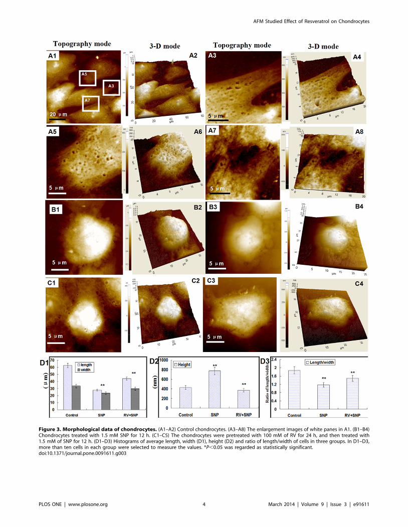
Figure 3. Morphological data of chondrocytes. (A1–A2) Control chondrocytes. (A3–A8) The enlargement images of white panes in A1. (B1–B4)Chondrocytes treated with 1.5 mM SNP for 12 h. (C1–C5) The chondrocytes were pretreated with 100 mM of RV for 24 h, and then treated with1.5 mM of SNP for 12 h. (D1–D3) Histograms of average length, width (D1), height (D2) and ratio of length/width of cells in three groups. In D1–D3,more than ten cells in each group were selected to measure the values. *P,0.05 was regarded as statistically significant.doi:10.1371/journal.pone.0091611.g003
AFM Studied Effect of Resveratrol on Chondrocytes
PLOS ONE | www.plosone.org 4 March 2014 | Volume 9 | Issue 3 | e91611
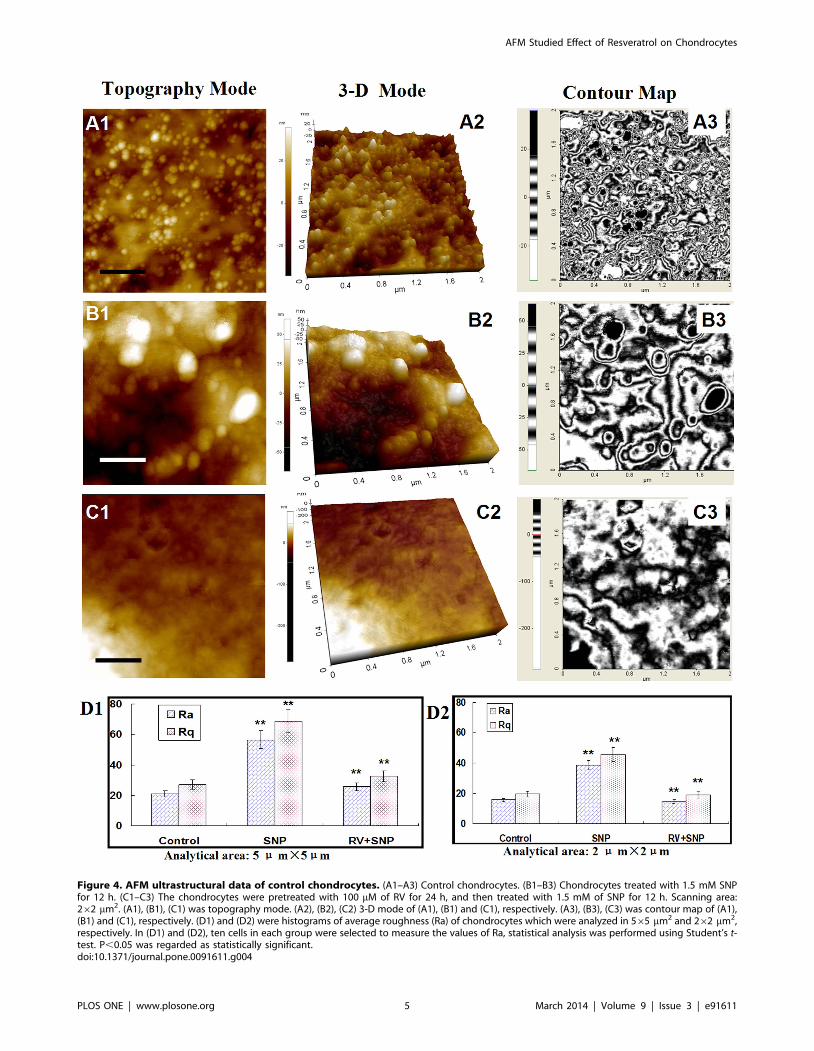
Figure 4. AFM ultrastructural data of control chondrocytes. (A1–A3) Control chondrocytes. (B1–B3) Chondrocytes treated with 1.5 mM SNPfor 12 h. (C1–C3) The chondrocytes were pretreated with 100 mM of RV for 24 h, and then treated with 1.5 mM of SNP for 12 h. Scanning area:262 mm2. (A1), (B1), (C1) was topography mode. (A2), (B2), (C2) 3-D mode of (A1), (B1) and (C1), respectively. (A3), (B3), (C3) was contour map of (A1),(B1) and (C1), respectively. (D1) and (D2) were histograms of average roughness (Ra) of chondrocytes which were analyzed in 565 mm2 and 262 mm2,respectively. In (D1) and (D2), ten cells in each group were selected to measure the values of Ra, statistical analysis was performed using Student’s t-test. P,0.05 was regarded as statistically significant.doi:10.1371/journal.pone.0091611.g004
AFM Studied Effect of Resveratrol on Chondrocytes
PLOS ONE | www.plosone.org 5 March 2014 | Volume 9 | Issue 3 | e91611
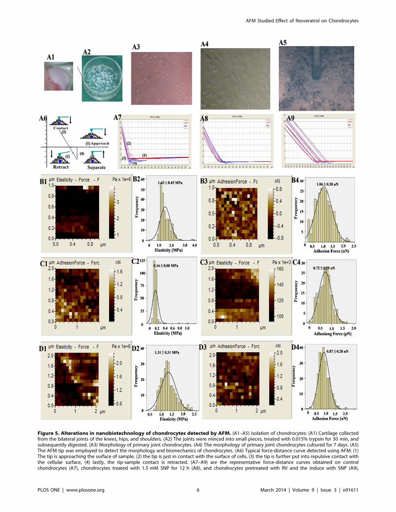
Figure 5. Alterations in nanobiotechnology of chondrocytes detected by AFM. (A1–A5) isolation of chondrocytes: (A1) Cartilage collectedfrom the bilateral joints of the knees, hips, and shoulders. (A2) The joints were minced into small pieces, treated with 0.015% trypsin for 30 min, andsubsequently digested. (A3) Morphology of primary joint chondrocytes. (A4) The morphology of primary joint chondrocytes cultured for 7 days. (A5)The AFM tip was employed to detect the morphology and biomechanics of chondrocytes. (A6) Typical force-distance curve detected using AFM: (1)The tip is approaching the surface of sample, (2) the tip is just in contact with the surface of cells, (3) the tip is further put into repulsive contact withthe cellular surface, (4) lastly, the tip-sample contact is retracted. (A7–A9) are the representative force-distance curves obtained on controlchondrocytes (A7), chondrocytes treated with 1.5 mM SNP for 12 h (A8), and chondrocytes pretreated with RV and the induce with SNP (A9),
AFM Studied Effect of Resveratrol on Chondrocytes
PLOS ONE | www.plosone.org 6 March 2014 | Volume 9 | Issue 3 | e91611
Alterations in cellular membrane architecture detected atnanometer level
Figure 4 showed the ultrastructural data of chondrocytes. The
membrane architecture of control chondrocytes (Figs.4A1–A3)
showed uniform structures and granular morphology with the
surface particles of 50,100 nm. Figures 4B1–B3 showed the
surface architecture of SNP-induced chondrocytes which became
heterogeneous, and the sizes of the membrane particles increased
to 150,200 nm. The ultrastructure of SNP-treated chondrocytes
pretreated by RV became smooth and homogeneous but the
granular morphology on cellular membrane diminished
(Figs.4C1–C3), implying that RV could significantly protect
chondrocytes from SNP-induced apoptosis and changes in
morphological properties, but could not completely prevented
SNP-induced changes in nanostructure of cellular membrane.
Therefore, AFM with nanometer-scale resolution provides us new
insights about cellular structure-function.
Additionally, the average roughness of cell membrane is directly
or indirectly sensitive to the membrane-skeleton integrity [36].
The average roughness (Ra) and root mean square roughness (Rq)
were measured for comparison. As shown in Figures 4D1 and D2,
the values of both Ra and Rq of SNP-induced chondrocytes
increased significantly compared with that of control chondro-
cytes. While Ra and Rq of the -chondrocytes pretreated with RV
decreased 50% than that of SNP group, and their values were
similar to that of control chondrocytes. These data suggested that
RV protected chondrocytes from SNP-induced damages via
altering the membrane architecture.
Taken together, all these morphological data revealed that SNP
could successfully induced apoptosis in chondrocytes, and RV
could protect the chondrocytes from damaging or apoptosis via
changing their morphological properties and architectures.
Alterations in nanomechanical properties ofchondrocytes
Since the biomechanical properties of cells can potentially
indicate their function and health, it is therefore very important to
study the cellular biomechanics. Lots of literatures have shown
that study on cellular mechanics is very helpful for clinical
diagnostics and even the formulation of suitable strategies towards
effective therapeutic treatments of human diseases. Although AFM
measures the nanobiomechanics of single cell, it has been used to
diagnose some diseases [16,37]. Although the potent protection of
RV on chondrocytes has been reported [38,39], the effects on the
nanobiomechanics, particularly on cell function and growth, is
poor.
Here, the nanobiomechanical properties including elasticity and
adhesion force were detected at levels of nanometer and pN,
respectively. Figures 5A1–A4 showed the isolation of chondro-
cytes. Figures A5 and A6 indicated that the AFM tip was
employed to detect the morphology and biomechanics of
chondrocytes. After positioning the AFM tip over the cell center
(Fig.5A5), the tip was brought to contact and pressed against the
respectively. The elasticity maps, histogram of elasticity, adhesion force map and histogram of adhesion force of control chondrocytes (B1–B4),chondrocytes treated with 1.5 mM SNP for 12 h (C1–C4), and chondrocytes pretreated with RV and then cotreated with SNP (A4), respectively.doi:10.1371/journal.pone.0091611.g005
Figure 6. Organization of cytoskeleton chondrocytes. Control chondrocytes (A1–A6), chondrocytes treated with 100 mM RV for 12 h (B1–B6),chondrocytes treated with 1.5 mM SNP for 24 h (C1–C6) and chondrocytes pretreated with RV and then treated with SNP (D1–D6), respectively. A4–A6, B4–B6, C4–C6, D4–D6 were enlarged images of cellular cytoskeleton in A1–A3, B1–B3, C1–C3, D1–D3, respectively. Bars in A1–A3, B1–B3, C1–C3,D1–D3 and A4–A6, B4–B6, C4–C6, D4–D6 were 50 and 20 mm, respectively.doi:10.1371/journal.pone.0091611.g006
AFM Studied Effect of Resveratrol on Chondrocytes
PLOS ONE | www.plosone.org 7 March 2014 | Volume 9 | Issue 3 | e91611
cell surface (Fig.5A6). During the tip retraction from the cell
surface rupture events, the retraction events in the force-distance
curves revealed the general tip-cell-surface adhesive interactions.
Figs 5A7–A9 showed the typical force-distance curves obtained
from control chondrocytes, SNP and RV+SNP groups, respec-
tively.
As shown in Figs 5B1 and B2, the elasticity/stiffness of control
chondrocytes was 1.4360.45 MPa. After treatment with 1.5 mM
SNP for 12 h, the elasticity of the chondrocytes decreased to
0.1660.08 MPa (Figs.5C1 andC2), indicating that SNP obviously
destructed the rigidity and chemical compositions of chondrocytes
membrane. Nevertheless, if the chondrocytes pretreated with
100 mM of RV for 24 h before exposure to 1.5 mM SNP for 12 h,
the elasticity was 1.3160.31 MPa (Fig.5D1,D2), indicating that
RV could effectively protect the elasticity/stiffness of chondro-
cytes. The damage to the cell envelope and the changes in the
composition of chondrocytes induced by SNP were suspected to be
the major causes of the decreases in the cell stiffness/elasticity. As
reported by Cai and co-workers [40], the damage and destruction
of the cytoskeleton directly led to the decrease of cellular rigidity.
Moreover, adhesion of cellular membrane plays a very
important role in cell physiological and pathological processes
[41]. Here, we employed force spectroscopy of AFM to measure
the non-specific adhesion force between AFM tip and cellular
membrane as a function of the nanomechanical properties of the
existing surface adhesive molecules. The adhesion force of control
chondrocytes was 1.0660.38 pN (Figs.5B3 and B4), but it
decreased to 0.7260.29 pN after SNP treatment (Figs.5C3 and
C4), indicating that membrane proteins were damaged by SNP
treatment. However, the adhesion force of SNP-treated chondro-
cytes pretreated with RV only increased to 0.87 60.28 pN,
demonstrating that RV could partially protect the membrane
proteins. Furthermore, RV significantly increased the number of
actins (A1–A4), and the particles with nano-meter scale were
mainly distributed on/around actins (B1–B3) (Fig.S1). Notably,
the nanomechanical properties of cellular membrane, including
adhesion force and stiffness, were both enhanced with RV
treatment (Figs.S1 C1 and C2).
Taken together, these results showed that SNP induced the
destruction of biomechanics in chondrocytes, including 90%
decrease in elasticity and 30% decrease in adhesion. Notably, RV
pretreatment could recover the elasticity/stiffness closely to that of
control chondrocytes, but RV possessed only a little protecting
effect on membrane proteins, implying that RV could not entirely
protect the physiological and functional properties of chondro-
cytes.
Alterations in cytoskeletal proteins F-actin and a-tubulinFrom the biomechanical data, we can see that RV protects
chondrocytes from SNP-induced apoptosis mainly through main-
taining their elasticity/stiffness. As cytoskeleton is very important
to maintain the biomechanics of cells, we further qualitatively
investigated the organization of cytoskeleton, including F-actin
and a-tubulin using confocal microscopy. The cytoskeleton is the
mesh-like structure beneath the cell membrane, which is an
important reflection of cellular structure organization [42].
Particularly, F-actin cytoskeleton is extensively regarded as the
key factor to regulate the shapes and generate the mechanical
forces of cells, and it plays vital roles during cellular physiological
and pathological behaviors.
As shown in Figure 6, control chondrocytes presented well-
spreading shapes, and F-actin and a-Tubulin were well-organized
uniformly assembly (Figs.6A1–A6). The F-actins were of paral-
leled-like organization and a-Tubulin microtubules were of mesh-
like alignment, which showed a well-grown station of control
chondrocytes. After exposure of cells to 100 mM RV for 24 h, the
fluorescence intensity increased significantly (Figs.6B1–B6) indic-
ative of the increase of both F-actins and a-Tubulin, suggesting
that RV treatment may protect the cellular cytoskeleton and
promote the expression of cytoskeletal proteins. While after
treatment with SNP, the chondrocytes became shrunk and round,
and both the F-actin and a-Tubulin cytoskeleton were reorganized
and polymerized (Figs.6C1–C6 showed). Interestingly, in the RV-
pretreated chondrocytes, we found that the cytoskeleton could
recover to some extent and represented well-spreading organiza-
tions (shown by Figs.6D1–D6), indicating that RV protected
chondrocytes from SNP-induced apoptosis mainly through alter-
ing the organization of cytoskeleton. For further detecting the
expression of cytoskeletal proteins induced by SNP and RV, we
also measured the MFI of F-actin and a-tubulin using fluores-
cence-based flow cytometry, and found that SNP induced
apoptosis and stiffness decrease mainly through the reorganization
and decrease the cytoskeletal proteins—F-actin instead of a-
tubulin (Fig.S2).
All these data demonstrated that the biomechanical properties
were heavily influenced by the organization and expression levels
of actin microfilaments instead of a-tubulin. SNP induced
aggregation of cytoskeleton, decrease in the expression of
cytoskeletal proteins, 90% decrease of elasticity and 30% decrease
of adhesion force in chondrocytes. While RV pretreatment could
protect the elasticity/stiffness close to that of control chondrocytes
through protecting the integrity and organization of cell cytoskel-
eton. These data demonstrate that AFM can be as a promising
nanodevice to study cells cytoskeleton integrity and arrangement
in vitro models of apoptosis and migration.
It is very imperative to evaluate the effects of RV on
chondrocytes when it is used in tissue engineering or OA
treatment. The evidence provided in this study showed that RV
could protect chondrocytes from SNP-induced apoptosis by
regulating actin organization, but it had only a little effect on
adhesive molecules or proteins in/on cell membrane, and that
AFM-based technique provides us an effective and feasible tool to
detect the changes and underlying mechanism of cells induced by
drugs at nanometer level.
Supporting Information
Figure S1 The morphological and nanomechanical properties
of chondrocytes treated with 100 nM of RV for 24 h, which
detected using AFM. A1–A2 Morphology of chondrocytes. A3–A4
the enlargement of white frame in A1. B1–B3 The ultrastructure
of membrane on chondrocytes. C1,C2 The adhesion force and
stiffness modulus of cellular membrane was 2.1360.66 nN and
5.0562.43 MPa.
(TIF)
Figure S2 The alterations in expression of F-actin anda-tubulin
in chondrocytes treated with SNP, RV and SNP+RV, respectively,
which detected by fluorescence-based flow cytometry. The F-
actins and microtubule were stained with FITC- phalloidine and
Alexa Fluor 555-a-Tubulin antibody, respectively.
(TIF)
Author Contributions
Conceived and designed the experiments: XPW. Performed the experi-
ments: HJ QL. Analyzed the data: HJ AL TSC. Contributed reagents/
materials/analysis tools: HJ. Wrote the paper: HJ TSC.
AFM Studied Effect of Resveratrol on Chondrocytes
PLOS ONE | www.plosone.org 8 March 2014 | Volume 9 | Issue 3 | e91611
References
1. Anderson AS, Loeser RF (2010) Why is osteoarthritis an age-related disease?
Best Pract Res Cl Rh 24(1):15–26.2. Wadsworth TL, Koop DR (1999) Effects of the wine polyphenolics quercetin
and resveratrol on pro-inflammatory cytokine expression in RAW 264.7macrophages. Biochem Pharmacol 57(8): 941–949.
3. Shakibaei M, Harikumar KB, Aggarwal BB (2009) Reseratrol addiction: to die
or not to die. Mol Nutr Food Res 53(1): 115–128.4. Dave M, Attur M, Palmer G, Al-Mussawir HE, Kennish L, et al. (2008) The
antioxidant resveratrol protects against chondrocyte apoptosis via effects onmitochondrial polarization and ATP production. Arthritis Rheum 58(9): 2786–
2797.
5. Csaki C, Keshishzadeh N, Fischer K, Shakibaei M (2008) Regulation ofinflammation signalling by resveratrol in human chondrocytes in vitro. Biochem
Pharmacol 75(3): 677–687.6. Del Carlo M Jr, Loeser RF (2008) Cell death in osteoarthritis. Curr Rheumatol
Rep 10(1): 37–42.7. Wu GJ, Chen TG, Chang HC, Chiu WT, Chang CC, et al. (2007) Nitric oxide
from both exogenous and endogenous sources activates mitochondria-dependent
events and induces insults to human chondrocytes. J Cell Biochem 101(6):1520–1531.
8. Nakagawa S, Arai Y, Mazda O, Kishida T, Takahashi KA, et al. (2010) N-acetylcysteine prevents nitric oxide-induced chondrocyte apoptosis and cartilage
degeneration in an experimental model of osteoarthritis. J Orthopaed Res
28(2):156–163.9. Andres MC de, Maneiro E, Martın MA, Arenas J, Blanco FJ (2013) Nitric oxide
compounds have different effects profiles on human articular chondrocytemetabolism. Arthritis Res Ther 15(5):R115.
10. Carlo MD, Loeser RF (2003) Increased oxidative stress with aging reduceschondrocyte survival: correlation with intracellular glutathione levels. Arthritis
Rheum 48(12):3419–3430.
11. Eo SH, Cho H, Kim SJ (2013) Resveratrol Inhibits Nitric Oxide-InducedApoptosis via the NF-Kappa B Pathway in Rabbit Articular Chondrocytes.
Biomol Ther 21(5):364–370.12. Lee GY, Lim CT (2007) Biomechanics approaches to studying human diseases.
Trends Biotechnol 25(3): 111–118.
13. Stolz M, Gottardi R, Raiteri R, Miot S, Martin I, et al. (2009) Early detection ofaging cartilage and osteoarthritis in mice and patient samples using atomic force
microscopy. Nat nanotechnol 4(3): 186–192.14. Stolz M, Raiteri R, Daniels AU, VanLandingham MR, Baschong W, et al.
(2004) Dynamic elastic modulus of porcine articular cartilage determined at twodifferent levels of tissue organization by indentation-type atomic force
microscopy. Biophys J 86(5): 3269–3283.
15. Iyer S, Gaikwad RM, Subba-Rao V, Woodworth CD, Sokolov I (2009) Atomicforce microscopy detects differences in the surface brush of normal and
cancerous cells. Nat Nanotechnol 4(6): 389–393.16. Cross SE, Jin YS, Tondre J, Wong R, Rao J, et al. (2008) AFM-based analysis of
human metastatic cancer cells. Nanotechnology 19: 384003–384011.
17. Kim SJ, Ju JW, Oh CD, Yoon YM, Song WK, et al. (2002) ERK-1/2 and p38kinase oppositely regulate nitric oxide-induced apoptosis of chondrocytes in
association with p53, caspase-3, and differentiation status. J Biol Chem277(2):1332–1339.
18. Kim SJ, Kim HG, Oh CD, Hwang SG, Song WK, et al. (2002) p38 kinase-dependent and -independent inhibition of protein kinase C f and -a regulates
nitric oxide-induced apoptosis and dedifferentiation of articular chondrocytes.
J Biol Chem 277(33):30375–30381.19. Kim SJ, Hwang SG, Shin DY, Kang SS, Chun JS (2002) p38 Kinase Regulates
Nitric Oxide-induced Apoptosis of Articular Chondrocytes by Accumulating p53via NFkappaB-dependent Transcription and Stabilization by Serine 15
Phosphorylation. J Biol Chem 277(36):33501–33508.
20. Oh CD, Chun JS (2003) Signaling mechanisms leading to the regulation ofdifferentiation and apoptosis of articular chondrocytes by insulin-like growth
factor-1. J Biol Chem 278(38):36563–3657121. Kim SJ, Hwang SG, Kim IC, Chun JS (2003) Actin cytoskeletal architecture
regulates nitric oxide-induced apoptosis, dedifferentiation, and cyclooxygenase-2
expression in articular chondrocytes via mitogen-activated protein kinase and
protein kinase C pathways. J Biol Chem 278(43):42448–42456.22. Yoon JB, Kim SJ, Hwang SG, Chang S, Kang SS, et al. (2003) Non-steroidal
anti-inflammatory drugs inhibit nitric oxide-induced apoptosis and dedifferen-tiation of articular chondrocytes independent of cyclooxygenase activity. J Biol
Chem 278(17):15319–15325.
23. Hwang SG, Ryu JH, Kim IC, Jho EH, Jung HC, et al. (2004) Wnt-7a causes lossof differentiated phenotype and inhibits apoptosis of articular chondrocytes via
different mechanisms. J Biol Chem 279(25):26597–26604.24. Kim JS, Park ZY, Yoo YJ, Yu SS, Chun JS (2005) p38 kinase mediates nitric
oxide-induced apoptosis of chondrocytes through the inhibition of protein kinase
C by blocking autophosphorylation. Cell Death Differ 12(3):201–212.25. Zhuang CP, Wang XP, Chen TS (2013) H2O2 induces apoptosis of rabbit
chondrocytes via both the extrinsic and the caspase-independent intrinsicpathways. J Innov Opt Health Sci 6(3):1350022.
26. Tonomura H, Takahashi KA, Mazda O, Arai Y, Inoue A, et al. (2006)Glutamine protects articular chondrocytes from heat stress and NO-induced
apoptosis with HSP70 expression. Osteoarthritis Cartilage 14(6): 545–553.
27. Kim KS, Cho CH, Park EK, Jung MH, Yoon KS, et al. (2012) AFM-detectedapoptotic changes in morphology and biophysical property caused by paclitaxel
in Ishikawa and HeLa cells. PloS one 7(1):e30066.28. Cappella B, Baschieri P, Frediani C, Miccoli P, Ascoli C (1997) Force-distance
curve by AFM. IEEE Eng Med Biol 16(2): 58–65.
29. Laney DE, Garcia RA, Parson SM, Hansma HG (1997) Changes in the ElasticProperties of Cholinergic Synaptic Vesicles as Measured by Atomic Force
Microscopy. Biophys J 72: 806–813.30. Radmacher M (2002). Measuring the elastic properties of living cells by the
atomic force microscope. Methods Cell Biol 68: 67–90.31. Liang X, Mao G, Simon Ng KY (2004) Probing small unilamellar EggPC
vesicles on mica surface by atomic force microscopy. Colloids Surf B:
Biointerfaces 34(1): 41–5.32. Dimitriadis EK, Horkay F, Maresca J, Kachar B, Chadwick RS (2002)
Determination of Elastic Moduli of Thin Layers of Soft Material Using theAtomic Force Microscope. Biophys J 82(5): 2798–2810.
33. Yin Z, Sadok A, Sailem H, McCarthy A, Xia X, et al. (2013) A screen for
morphological complexity identifies regulators of switch-like transitions betweendiscrete cell shapes. Nat Cell Biol 15(7): 860–871.
34. McBeath R, Pirone DM, Nelson CM, Bhadriraju K, Chen CS (2004) Cell shape,cytoskeletal tension, and RhoA regulate stem cell lineage commitment. Dev Cell
6(4): 483–495.35. Cai X, Yang X, Cai J, Wu S, Chen Q (2010) Atomic force microscope-related
study membrane-associated cytotoxicity in human pterygium fibroblasts induced
by mitomycin C. J Phys Chem B 114(11): 3833–3839.36. Girasole M, Pompeo G, Cricenti A, Congiu-Castellano A, Andreola F, et al.
(2007) Roughness of the plasma membrane as an independent morphologicalparameter to study RBCs: A quantitative atomic force microscopy investigation.
Biochim Biophys Acta 1768(5): 1268–1276.
37. Teck Chwee Lim (2005) Single Cell Mechanics and its connections to HumanDiseases. Asia-Pacific Biotech News 09: 674–675.
38. Csaki C, Mobasheri A, Shakibaei M (2009) Synergistic chondroprotective effectsof curcumin and resveratrol in human articular chondrocytes: inhibition of IL-
1beta-induced NF-kappaB-mediated inflammation and apoptosis. Arthritis ResTher 11(6): R165.
39. Liu FC, Hung LF, Wu WL, Chang DM, Huang CY, et al. (2010)
Chondroprotective effects and mechanisms of resveratrol in advanced glycationend products-stimulated chondrocytes. Arthritis Res Ther 12(5): R167.
40. Cai X, Yang X, Cai J, Wu S, Chen Q (2010) Atomic force microscope-relatedstudy membrane-associated cytotoxicity in human pterygium fibroblasts induced
by mitomycin C. J Phys Chem B 114(11):3833–3839.
41. Geiger B (2001) Cell biology: encounters in space. Science 294(5547):1661–1663.
42. Hsieh CH, Lin YH, Lin S, Tsai-Wu JJ, Herbert Wu CH, et al. (2008) Surfaceultrastructure and mechanical property of human chondrocyte revealed by
atomic force microscopy. Osteoarthritis Cartilage 16(4):480–488.
AFM Studied Effect of Resveratrol on Chondrocytes
PLOS ONE | www.plosone.org 9 March 2014 | Volume 9 | Issue 3 | e91611