Embed Size (px)
Citation preview

1
Resuscitation Seminar
June 2011 11
2
INDEX:
www.developinganaesthesia.org
Approach to the critically ill patient
(Dr Amanda Baric)
4
ECG interpretation
(Dr Rachel Cowell)
8
Reversible causes of cardiac arrest
(Dr Sam Kennedy)
17
Basic Life Support and Advanced Life Support
(Dr Brendan Muzel)
20
Acute Coronary Syndromes
(Dr Amanda Baric)
29
Post resuscitation care
(Dr Roni Krieser)
34
Maternal Resuscitation
(Dr Ginette Falcone)
46
Newborn Resusciation
(Rhonda Keenan)
40
3
Defibrillation
(Dr Amanda Baric)
52
The views expressed in this publication are those of the authors alone.
Every effort has been made to trace and acknowledge copyright. However should any infringement have occurred, the authors tender their apologies and invite copyright owners to contact them.
Creative commons licence developing nations 2.O
4
APPROACH TO THE CRITICALLY UNWELL PATIENT
Many patients who have a cardiac arrest have experienced a period of physiologic instability for a period of time prior to their arrest. It is important to recognise such a patient and manage the cause of deterioration. A systematic approach to such a patient is required. The ABCDE approach is recommended.
Early recognition of the deteriorating patient is an important link in the chain of survival. It prevents cardiac arrest, reduces the number of admissions to ICU and can help to identify those patients in whom attempts at resuscitation are futile.
Features of deterioration
There are periods of deterioration in 50-80% of patients who go on to have a cardiac arrest. Hypoxia and hypotension are common events prior to a cardiac arrest. Sometimes the period of deterioration can extend over several hours. If there is a delay in referral to critical care providers, the patient will proceed to an arrest and the cardiac rhythm in such a patient is Pulseless Electrical Activity (PEA) or asystole, both of which carry a worse prognosis.
Traditionally, cardiac arrest teams are only called after the cardiac arrest has occurred. Many hospitals around the world now have a medical emergency team (MET) that is comprised of critical care doctors and nurses. The early involvement of a medical emergency team who respond to calls to patients with significant physiologic derangement may decrease the incidence of cardiac arrest.
There are usually defined criteria for calling the medical emergency team. Any member of the health care team can initial a MET call. The calling criteria may include:
A – Threatened Airway
B – All respiratory arrests, respiratory rate less than 5 breaths per minute or greater than 36 breaths per minute
C – All cardiac arrests, pulse rate less than 40 beats per minute or greater than 140 beats per minute, systolic blood pressure less than 90 mmHg
D – Sudden fall in level of conscious state (Glasgow coma score of > 2 points) or repeated or prolonged seizures
Other – Any patient that the staff are concerned about but does not fit the above criteria
Causes of cardiorespiratory arrest:
Airway problems most commonly arise due to obstruction. It may be partial or complete obstruction. Complete airway obstruction will rapidly cause a cardiorespiratory arrest. Partial airway obstruction may cause cerebral or pulmonary oedema, apnoea, hypoxic brain injury and will eventually cause a cardiac arrest.
5
Airway obstruction can be due to central nervous system depression and loss of airway reflexes, foreign material in the airway, including blood, vomit, other foreign bodies, trauma to the airway, infection and inflammation that cause swelling in the airway and laryngeal or bronchial spasm.
Breathing problems that lead to cardiac arrest can be classified as those that decrease respiratory drive, decreased respiratory effort and lung disorders. All will lead to hypoxaemia and eventual cardiac arrest if left untreated. Decreased respiratory drive occurs due to central nervous system depression secondary to drugs, trauma and metabolic disorders. Decreased respiratory effort occurs due to muscle weakness, nerve damage (such as spinal cord disorders), restrictive chest defects and pain from fractured ribs. Lung disorders to consider include those secondary to trauma such as pneumothorax and haemothorax with contusion, infection, pulmonary embolism, acute exacerbations of airways disease and asthma and adult respiratory distress syndrome.
Circulatory problems that lead to cardiac arrest include primary cardiac disease and disease secondary to reduced oxygen delivery to the myocardium. The commonest manifestation of primary cardiac problems is ventricular fibrillation or other arrhythmia. Examples of primary cardiac disease include ischaemia due to acute coronary syndromes, hypertensive disease, valvular disease, hereditary cardiac disease such as long QT syndromes, electrolyte disturbance and electrocution. Conditions that lead to secondary cardiac problems include asphyxia, hypoxaemia, blood loss, hypothermia and septic shock.
ABCDE approach to the critically unwell patient:
A complete assessment of the patient is required. A systematic approach is best so that all causes of deterioration are considered and managed rapidly. During the initial assessment, life-threatening problems are treated and the patient is reassessed after interventions and at frequent intervals thereafter. It is important to call for help from experts early and identify the need for higher-level care.
After establishing safety of the rescuer and patient, a rapid assessment of the airway, breathing and circulation, conscious state and exposure of the patient is made. Attach monitoring of the ECG and oxygen saturation monitor (oximeter) and establish intravenous access. Perform a 12 lead ECG and electrolyte, haemoglobin and arterial blood gases.
Airway:
It is important to recognize obstruction of the airway. If the patient is talking in sentences, it is unlikely that there is a major obstruction of the airway. Check for noisy breathing and ask the patient how he feels. If there is dyspnoea, particularly if there is a choking sensation and changes with posture, there may be obstruction of the airway. Obstructive airway noise can arise from pharyngeal obstruction (snoring), laryngeal obstruction (stridor) or bronchial obstruction (wheeze). A paradoxical movement of the chest and use of accessory muscles of respiration suggests the presence of upper airway obstruction.
6
Treat airway obstruction with simple airway manoeuvres such as head tilt, chin lift, jaw thrust first. If this does not relieve the problem, use simple adjuncts. The Guedel oropharyngeal airway can be used if the patient has no airway reflexes, but may induce gagging or vomiting in the patient who is not unconscious. A nasopharyngeal airway can be useful to relieve obstruction arising at the level of the nasopharynx, but may cause epistaxis.
Careful suctioning of the airway is performed if there is gurgling sounds or evidence of foreign material in the mouth.
An advanced airway will be required if the above measures do not relieve the obstruction. A laryngeal mask is simple to insert, but insertion of an endotracheal tube will require an anaesthetist or someone who is has experience in intubation.
Breathing:
In most situations, high flow oxygen given by a face mask is indicated. The recognition of breathing problems requires a careful examination of the patient. Look for signs of respiratory distress. Count the respiratory rate. The normal rate is between 12 to 20 breaths per minute. Check for the use of accessory muscles and for any obvious chest deformity, particularly in the presence of trauma. The patient may be cyanosed due to hypoxia or may have confusion related to hypoxia. Check for noisy breathing and listen to the breath sounds. There may be crackles, wheeze of reduced air entry. Feel for the expansion of the chest and check the position of the trachea. Surgical emphysema may represent a pneumothorax or pneumomediastinum. If oximetry is available, it should be attached. If the saturation is less than 90%, significant hypoxia may occur rapidly if the situation worsens. It will need to be corrected rapidly.
Breathing problems are addressed initially with airway opening manoeuvres, administration of oxygen and treating the underlying cause. If there are ongoing problems with oxygenation and breathing, the patient will need ventilatory support with a bag-valve mask apparatus, supraglottic airway or intubation and mechanical ventilation. In some patients, non-invasive ventilation may be appropriate.
Circulation:
Problems with the circulation will often manifest as problems related to hypoperfusion of vital organs. The patient may appear pale and sweaty, have cool peripheries with prolonged capillary refill time and be confused and disoriented. Reduced organ perfusion may result in chest pain, low urine output and shortness of breath.
Check for the pulse rate. It may be slow <40 or fast >100 beats per minute. The capillary refill time is checked by putting pressure on the finger tip and releasing it. It should be less than 2 seconds in a normal patient, but may be prolonged if there is peripheral vasoconstriction as occurs in shock or if the patient is cold. Take the blood pressure. Hypotension needs to be assessed and treated. In the collapsed patient, it is most commonly due to hypovolaemia, but may be due to cardiogenic or septic shock.
Circulatory problems are treated after the establishment of a clear airway and correction of breathing problems. Oxygen is administered to help with delivery of oxygen to the tissues. Intravenous access is established with a wide bore, short
7
cannula (to allow for rapid infusion of fluids). Blood tests are taken at the same time as intravenous access is established.
If it is evident that there is a primary cardiac cause for circulatory problems, treat with oxygen, morphine, nitrates and aspirin as for acute coronary syndrome. Take care with nitrates if there is hypotension or evidence of right heart infarction. A fluid bolus is given if hypovolaemia may be contributing to circulatory compromise. It is important to establish cardiac monitoring, blood pressure and pulse oximetry. Ionotropes and vasopressors may be indicated if there has been no response to a fluid challenge in order to increase perfusion to the vital organs.
Disability:
The conscious state needs to be assessed. A reduction in consciousness can occur due to a primary cerebral problem or be secondary to hypoxia, hypercarbia, reduced cerebral perfusion, metabolic disorders or medications. A quick way of assessing the conscious state is to check the AVPU score – A =alert, V=responding to verbal stimuli, P=responding to painful stimuli, U=unconscious.
The management and assessment occur simultaneously, but involve securing the airway, breathing and circulation, followed by treatment of the underlying cause of the reduced conscious state. If the patient is unable to control the airway, turn them into the lateral position and consider intubation for those with a Glasgow coma score of <8.
Exposure:
The patient needs to be exposed so as to be properly examined for an obvious cause of deterioration. There may be a rash in anaphylaxis, bleeding or obvious injuries. Once the patient has been properly examined from head to toe, cover him so as to avoid heat loss and to maintain dignity. Further history should be sought from the patient, relatives or carers and the medical record. Check the results of investigations and refer for high level (intensive or high dependency) care if there is no clear cause and treatment for the deterioration of the patient’s condition. Documentation of all your findings is important and a clear plan is recorded in the history.
Conclusion:
The early recognition of the patient at risk of cardiac arrest may prevent cardiorespiratory arrest. Many patients will exhibit warning signs of physiologic deterioration before cardiac arrest. The causes of cardiac arrest may be due to airway, breathing or circulatory problems. A systematic approach to the assessment and treatment of the critically ill patient is required.
References:
1. Australian Resuscitation Council. Advanced Life Support, 5th edition, 2006.
2. Australian Resuscitation Council Website. www.resus.org.au
3. UptoDate. www.uptodate.com 2011.
8
INTERPRETING ECGs
Electrical Activity of the Heart:
Cardiac pacemaker cells (typically found in the Sinoatrial SA node in the right atrium), initiate the electrical currents in the heart. The depolarisation triggered by the pacemaker cells is transmitted along conducting fibres that travel through the atria to the Atrio-‐Ventricular node (AV node). Here, there is a delay prior to the rapid depolarisation of specialised fibres in the ventricular septum – from the bundle of His, to the left and right bundle branches – before a slower depolarisation of the Purkinje fibres that lie in the ventricular myocardium.
Myocardial cells are polarised (carry an electric charge on their surface) due to transmembrane ion concentration differences (primarily Na+ and K+). Normal charge is negative 90mV, with the inside of the cell being negatively charged relative to the outside. When cells are stimulated to above their ‘threshold’ potential, they depolarise and become transiently positively charged. The process of depolarisation then spreads in one direction down the conducting pathways of the heart, followed by repolarisation, returning the electrical potential back to its resting state of –90mV.
Principles of the ECG:
The ECG leads display the differences in electrical potential between the electrodes.
Leads:
There are six limb leads (3 bipolar leads: I, II, III and 3 unipolar leads: avR, aVL, aVF) and six chest leads (V1 to V6). Each bipolar lead measures the difference between two electrodes, whereas the unipolar leads measure the voltage at one point relative to an electrode that has zero action potential (avR looks at right arm, aVL looks at left arm, and aVF looks at left leg or foot). The letter “a” that precedes these three leads refers to the fact that these are electrically augmented by 50%. The limb leads look at the heart in a vertical plane.
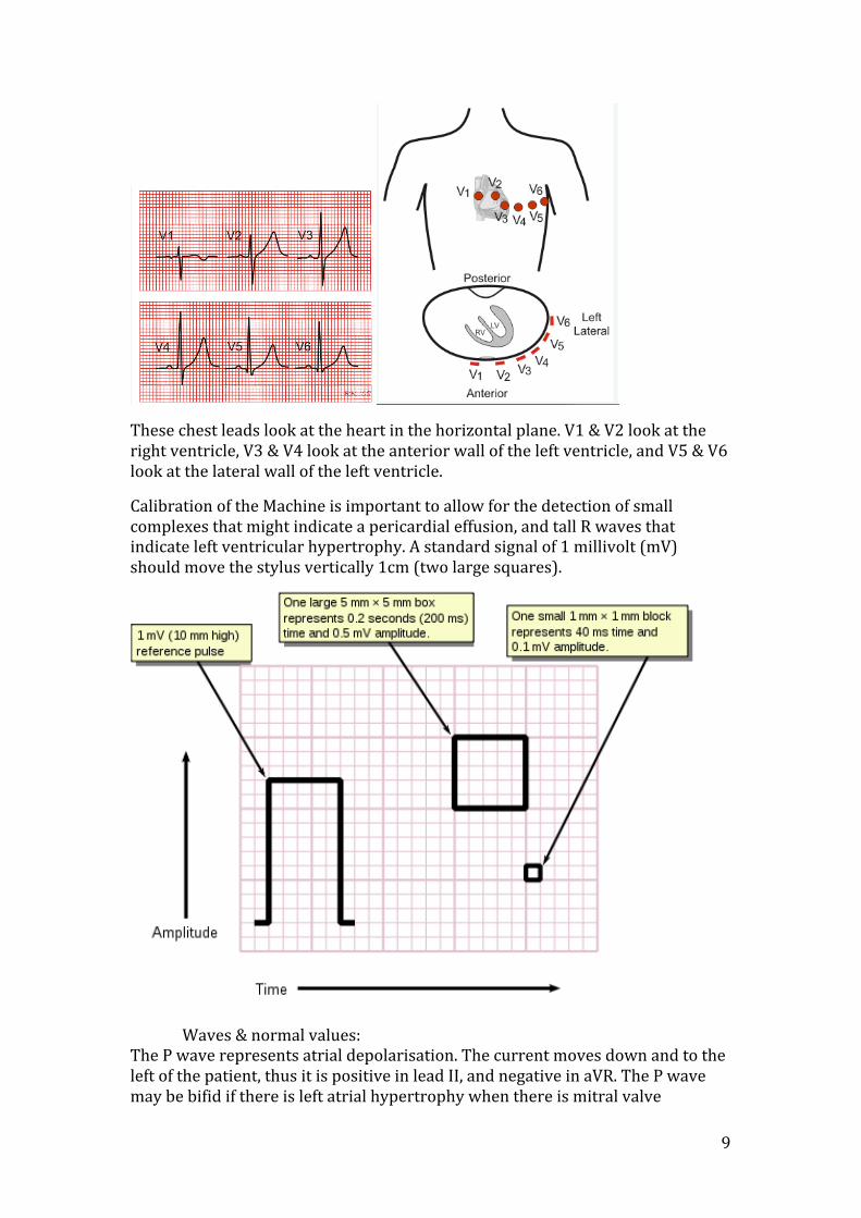
The six chest leads are unipolar and positioned from V1 (fourth intercostal space adjacent to the right side of the patient’s sternum) V2 same space on the left side of the sternum, V4, V5 and V6 are all in the 5th intercostal space, but in the midclavicular, anterior axillary and midaxillary lines, respectively.
9
These chest leads look at the heart in the horizontal plane. V1 & V2 look at the right ventricle, V3 & V4 look at the anterior wall of the left ventricle, and V5 & V6 look at the lateral wall of the left ventricle.
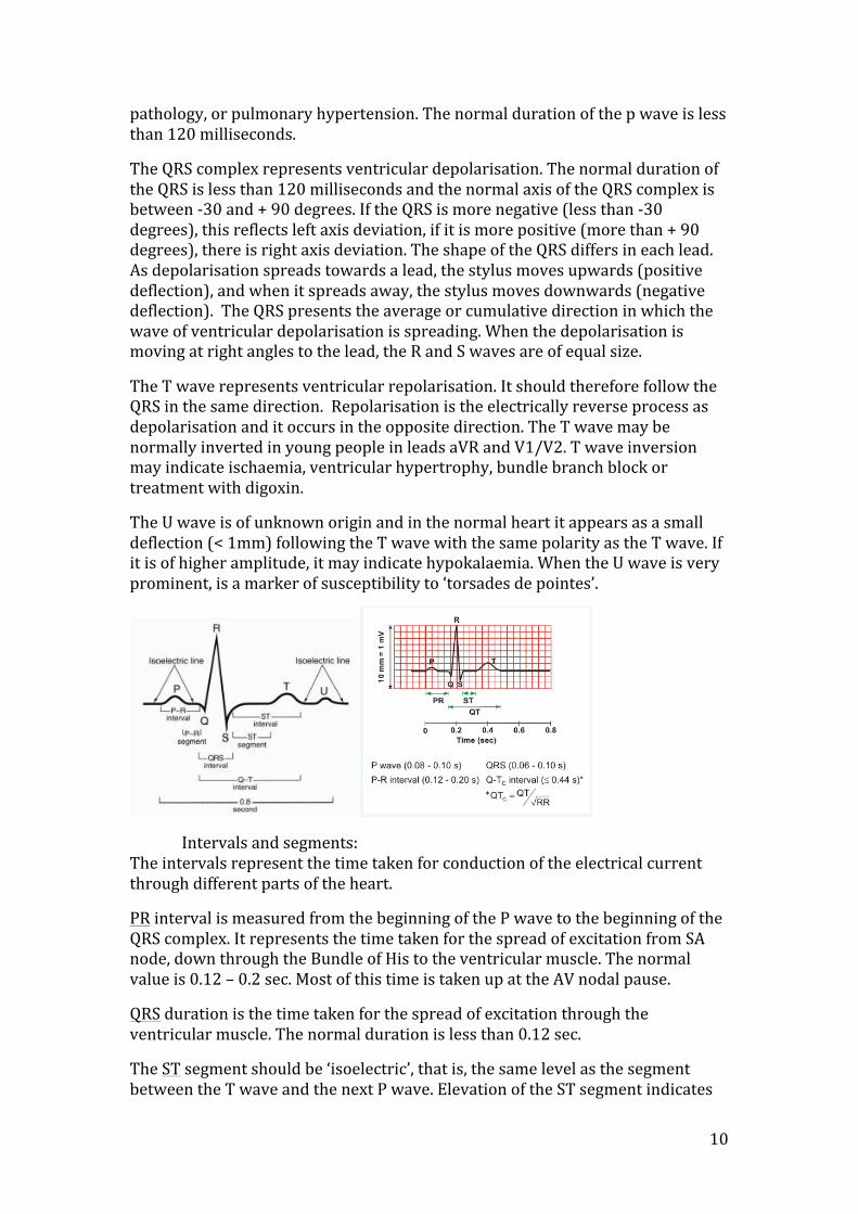
Calibration of the Machine is important to allow for the detection of small complexes that might indicate a pericardial effusion, and tall R waves that indicate left ventricular hypertrophy. A standard signal of 1 millivolt (mV) should move the stylus vertically 1cm (two large squares).
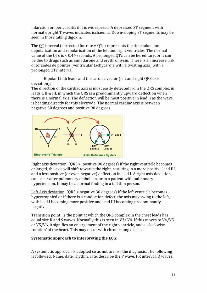
Waves & normal values: The P wave represents atrial depolarisation. The current moves down and to the left of the patient, thus it is positive in lead II, and negative in aVR. The P wave may be bifid if there is left atrial hypertrophy when there is mitral valve
10
pathology, or pulmonary hypertension. The normal duration of the p wave is less than 120 milliseconds.
The QRS complex represents ventricular depolarisation. The normal duration of the QRS is less than 120 milliseconds and the normal axis of the QRS complex is between -‐30 and + 90 degrees. If the QRS is more negative (less than -‐30 degrees), this reflects left axis deviation, if it is more positive (more than + 90 degrees), there is right axis deviation. The shape of the QRS differs in each lead. As depolarisation spreads towards a lead, the stylus moves upwards (positive deflection), and when it spreads away, the stylus moves downwards (negative deflection). The QRS presents the average or cumulative direction in which the wave of ventricular depolarisation is spreading. When the depolarisation is moving at right angles to the lead, the R and S waves are of equal size.
The T wave represents ventricular repolarisation. It should therefore follow the QRS in the same direction. Repolarisation is the electrically reverse process as depolarisation and it occurs in the opposite direction. The T wave may be normally inverted in young people in leads aVR and V1/V2. T wave inversion may indicate ischaemia, ventricular hypertrophy, bundle branch block or treatment with digoxin.
The U wave is of unknown origin and in the normal heart it appears as a small deflection (< 1mm) following the T wave with the same polarity as the T wave. If it is of higher amplitude, it may indicate hypokalaemia. When the U wave is very prominent, is a marker of susceptibility to ‘torsades de pointes’.
Intervals and segments: The intervals represent the time taken for conduction of the electrical current through different parts of the heart.
PR interval is measured from the beginning of the P wave to the beginning of the QRS complex. It represents the time taken for the spread of excitation from SA node, down through the Bundle of His to the ventricular muscle. The normal value is 0.12 – 0.2 sec. Most of this time is taken up at the AV nodal pause.
QRS duration is the time taken for the spread of excitation through the ventricular muscle. The normal duration is less than 0.12 sec.
The ST segment should be ‘isoelectric’, that is, the same level as the segment between the T wave and the next P wave. Elevation of the ST segment indicates
11
infarction or, pericarditis if it is widespread. A depressed ST segment with normal upright T waves indicates ischaemia. Down-‐sloping ST segments may be seen in those taking digoxin.
The QT interval (corrected for rate = QTc) represents the time taken for depolarisation and repolarisation of the left and right ventricles. The normal value of the QTc is < 0.44 seconds. A prolonged QTc can be hereditary, or it can be due to drugs such as amiodarone and erythromycin. There is an increase risk of torsades de pointes (ventricular tachycardia with a twisting axis) with a prolonged QTc interval.
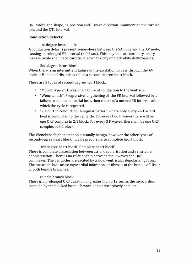
Bipolar Limb leads and the cardiac vector (left and right QRS axis deviation): The direction of the cardiac axis is most easily detected from the QRS complex in leads I, II & III, in which the QRS is a predominantly upward deflection when there is a normal axis. The deflection will be most positive in lead II as the wave is heading directly for this electrode. The normal cardiac axis is between negative 30 degrees and positive 90 degrees.
Right axis deviation: (QRS > positive 90 degrees) If the right ventricle becomes enlarged, the axis will shift towards the right, resulting in a more positive lead III, and a less positive (or even negative) deflection in lead I. A right axis deviation can occur after pulmonary embolism, or in a patient with pulmonary hypertension. It may be a normal finding in a tall thin person.
Left Axis deviation: (QRS < negative 30 degrees) If the left ventricle becomes hypertrophied or if there is a conduction defect, the axis may swing to the left, with lead I becoming more positive and lead III becoming predominantly negative.
Transition point: Is the point at which the QRS complex in the chest leads has equal size R and S waves. Normally this is seen in V3/ V4. If this moves to V4/V5 or V5/V6, it signifies an enlargement of the right ventricle, and a ‘clockwise rotation’ of the heart. This may occur with chronic lung disease.
Systematic approach to interpreting the ECG:
A systematic approach is adopted so as not to miss the diagnosis. The following is followed: Name, date, rhythm, rate, describe the P wave, PR interval, Q waves,
12
QRS width and shape, ST position and T wave direction. Comment on the cardiac axis and the QTc interval.
Conduction defects:
1st degree heart block: A conduction delay is present somewhere between the SA node and the AV node, causing a prolonged PR interval (> 0.2 sec). This may indicate coronary artery disease, acute rheumatic carditis, digoxin toxicity or electrolyte disturbances.
2nd degree heart block: When there is an intermittent failure of the excitation to pass through the AV node or Bundle of His, this is called a second-‐degree heart block.
There are 3 types of second-‐degree heart block:
• “Mobitz type 2”: Occasional failure of conduction to the ventricle. • “Wenckebach”: Progressive lengthening of the PR interval followed by a
failure to conduct an atrial beat, then return of a normal PR interval, after which the cycle is repeated.
• “2:1 or 3:1” conduction: A regular pattern where only every 2nd or 3rd beat is conducted to the ventricle. For every two P waves there will be one QRS complex in 2:1 block. For every 3 P waves, there will be one QRS complex in 3:1 block.
The Wenckebach phenomenon is usually benign, however the other types of second-‐degree heart block may be precursors to complete heart block.
3rd degree heart block “Complete heart block”: There is complete dissociation between atrial depolarisation and ventricular depolarisation. There is no relationship between the P waves and QRS complexes. The ventricles are excited by a slow ventricular depolarising focus. The causes include acute myocardial infarction, or fibrosis of the bundle of His or of both bundle branches.
Bundle branch block: There is a prolonged QRS duration of greater than 0.12 sec, as the myocardium supplied by the blocked bundle branch depolarises slowly and late.
13
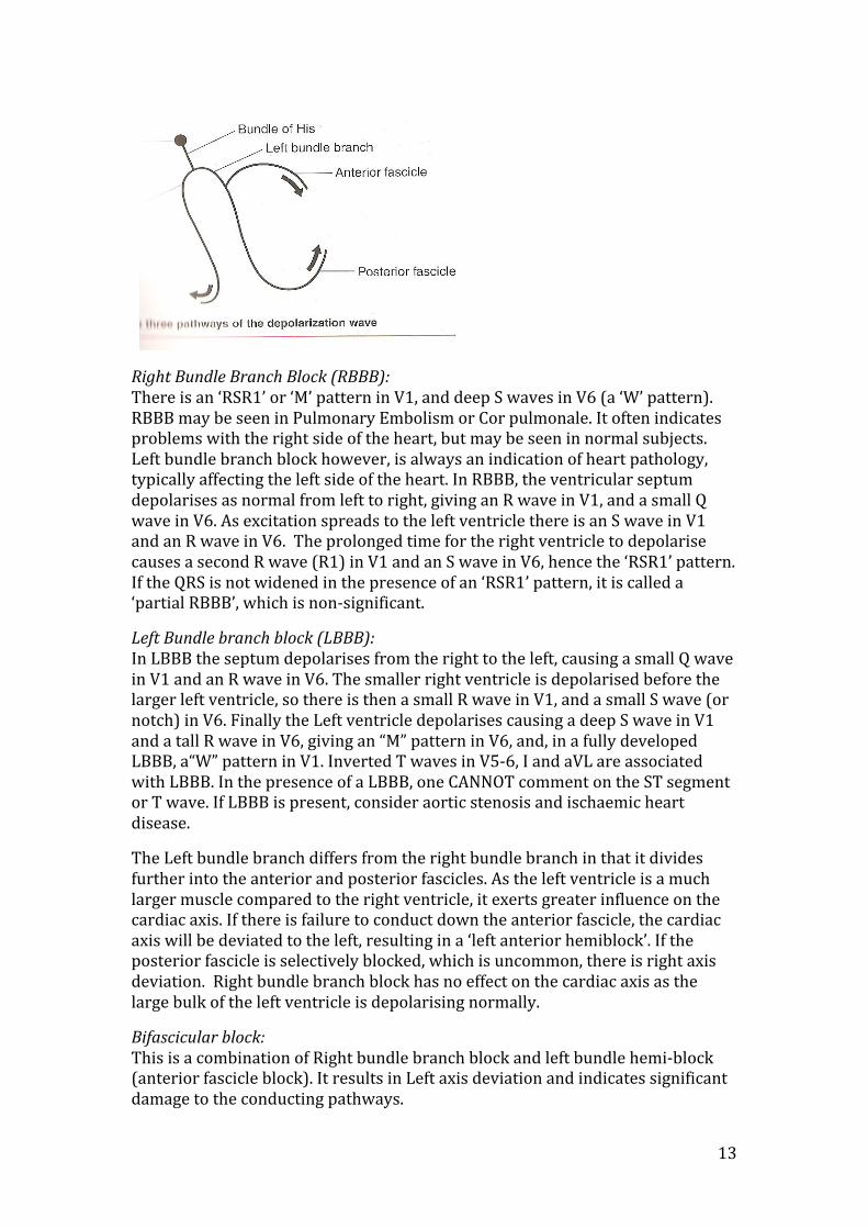
Right Bundle Branch Block (RBBB): There is an ‘RSR1’ or ‘M’ pattern in V1, and deep S waves in V6 (a ‘W’ pattern). RBBB may be seen in Pulmonary Embolism or Cor pulmonale. It often indicates problems with the right side of the heart, but may be seen in normal subjects. Left bundle branch block however, is always an indication of heart pathology, typically affecting the left side of the heart. In RBBB, the ventricular septum depolarises as normal from left to right, giving an R wave in V1, and a small Q wave in V6. As excitation spreads to the left ventricle there is an S wave in V1 and an R wave in V6. The prolonged time for the right ventricle to depolarise causes a second R wave (R1) in V1 and an S wave in V6, hence the ‘RSR1’ pattern. If the QRS is not widened in the presence of an ‘RSR1’ pattern, it is called a ‘partial RBBB’, which is non-‐significant.
Left Bundle branch block (LBBB): In LBBB the septum depolarises from the right to the left, causing a small Q wave in V1 and an R wave in V6. The smaller right ventricle is depolarised before the larger left ventricle, so there is then a small R wave in V1, and a small S wave (or notch) in V6. Finally the Left ventricle depolarises causing a deep S wave in V1 and a tall R wave in V6, giving an “M” pattern in V6, and, in a fully developed LBBB, a“W” pattern in V1. Inverted T waves in V5-‐6, I and aVL are associated with LBBB. In the presence of a LBBB, one CANNOT comment on the ST segment or T wave. If LBBB is present, consider aortic stenosis and ischaemic heart disease.
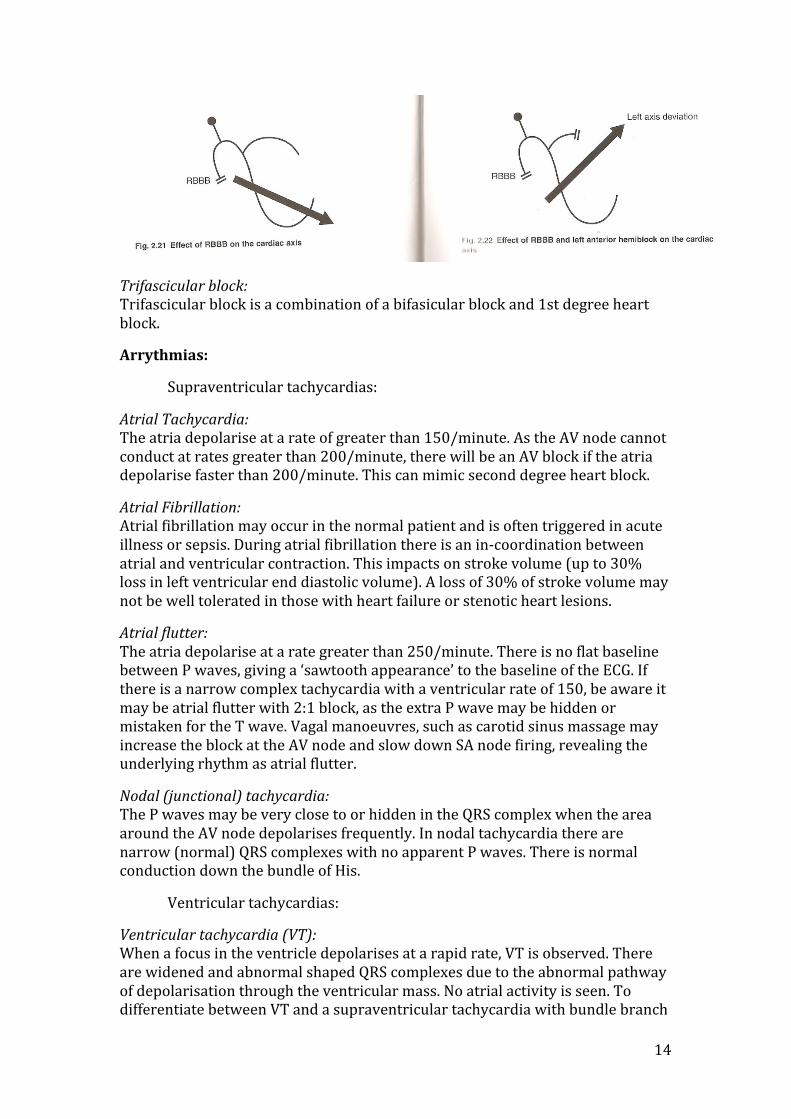
The Left bundle branch differs from the right bundle branch in that it divides further into the anterior and posterior fascicles. As the left ventricle is a much larger muscle compared to the right ventricle, it exerts greater influence on the cardiac axis. If there is failure to conduct down the anterior fascicle, the cardiac axis will be deviated to the left, resulting in a ‘left anterior hemiblock’. If the posterior fascicle is selectively blocked, which is uncommon, there is right axis deviation. Right bundle branch block has no effect on the cardiac axis as the large bulk of the left ventricle is depolarising normally.
Bifascicular block: This is a combination of Right bundle branch block and left bundle hemi-‐block (anterior fascicle block). It results in Left axis deviation and indicates significant damage to the conducting pathways.
14
Trifascicular block: Trifascicular block is a combination of a bifasicular block and 1st degree heart block.
Arrythmias:
Supraventricular tachycardias:
Atrial Tachycardia: The atria depolarise at a rate of greater than 150/minute. As the AV node cannot conduct at rates greater than 200/minute, there will be an AV block if the atria depolarise faster than 200/minute. This can mimic second degree heart block.
Atrial Fibrillation: Atrial fibrillation may occur in the normal patient and is often triggered in acute illness or sepsis. During atrial fibrillation there is an in-‐coordination between atrial and ventricular contraction. This impacts on stroke volume (up to 30% loss in left ventricular end diastolic volume). A loss of 30% of stroke volume may not be well tolerated in those with heart failure or stenotic heart lesions.
Atrial flutter: The atria depolarise at a rate greater than 250/minute. There is no flat baseline between P waves, giving a ‘sawtooth appearance’ to the baseline of the ECG. If there is a narrow complex tachycardia with a ventricular rate of 150, be aware it may be atrial flutter with 2:1 block, as the extra P wave may be hidden or mistaken for the T wave. Vagal manoeuvres, such as carotid sinus massage may increase the block at the AV node and slow down SA node firing, revealing the underlying rhythm as atrial flutter.
Nodal (junctional) tachycardia: The P waves may be very close to or hidden in the QRS complex when the area around the AV node depolarises frequently. In nodal tachycardia there are narrow (normal) QRS complexes with no apparent P waves. There is normal conduction down the bundle of His.
Ventricular tachycardias:
Ventricular tachycardia (VT): When a focus in the ventricle depolarises at a rapid rate, VT is observed. There are widened and abnormal shaped QRS complexes due to the abnormal pathway of depolarisation through the ventricular mass. No atrial activity is seen. To differentiate between VT and a supraventricular tachycardia with bundle branch

15
block, one needs to consider the clinical presentation. VT is commonly found in the setting of acute myocardial infarction. It is also helpful to look for P waves and the relationship with the QRS complexes. The clinician should find previous ECGs and compare the shape of the QRS complexes. (They should be the same shape if LBBB was present previously). Look at the width of the QRS (if it is wider than 160 milliseconds, it is likely to be ventricular in origin), and look at previous ECGs for a change in the cardiac axis (which would suggest a ventricular origin of the tachycardia).
Ventricular fibrillation: In ventricular fibrillation the ventricular muscle fibres contract independently from each other in a completely disorganised fashion, resulting in no QRS complexes, and thus no ability to eject blood from the ventricles.
Other ECG findings:
Long QT: (QTc >0.44 seconds): May be congenital or acquired. It may result from medications such as amiodarone, quinidine and erythromycin, or in electrolyte disturbances such as hypocalcaemia, hypomagnesaemia or hypothermia. Prolonged QT is associated with Torsades de Pointes (polymorphic ventricular tachycardia).
Acute Cor pulmonale: Acute cor pulmonale (typically from pulmonary embolus) is diagnosed on the ECG by the presence of a sinus tachycardia, a right shift of the QRS axis, and the classical ‘S1, Q3, T3’ pattern (prominent S waves in lead I, Q waves in lead III and T wave inversion in lead III). Acute Right Ventricular dilatation may occur with T wave inversion in V1 to V4 and poor R wave progression, which may resemble an acute anterior infarction.
Wolf Parkinson White (WPW) syndrome: WPW is characterised by wide QRS complexes associated with a short PR interval, and a slurring of the initial part of the QRS complex (called the delta wave). These changes occur due to aberrant activation of the ventricular myocardium by a ‘bypass’ tract or ‘accessory bundle’, resulting in ‘pre-‐excitation’ of the ventricles. This increases the risk of re-‐entrant supraventricular tachy-‐arrhythmias. In this situation, depolarisation can spread down the Bundle of His and return back up into the atria via the accessory bundle, causing reactivation of the atria, and a sustained tachycardia.
Toxicity: Drug toxicity from phenytoin, tricyclic antidepressants and electrolyte disturbances produce ECG changes that are typical of each condition.
References:
1. Hampton JR. The ECG made easy. Churchill Livingstone, 2003 2. Ganong, Review of Medical Physiology 22nd edition. p 547 3. Braunwald et al. Harrison’s Principles of Internal Medicine 15th edition. p
1238 -‐ 42 4. Pathophysiology of Heart Disease

16
5. Current Emergency Diagnosis and Management 6. Brown, Emergency Medicine (Diagnosis & Management) 7. http://www.cvphysiology.com/Arrhythmias/A009.htm 8. http://en.wikipedia.org/wiki/Electrocardiography
17
REVERSIBLE CAUSES OF CARDIAC ARREST
Dr. Sam Kennedy, General Practitioner (GP) and GP Anaesthetist
The reversible causes of cardiac arrest are important to consider during a cardiac arrest. They are also important to consider after successful cardiopulmonary resuscitation +/-‐ defibrillation when cardiac output is restored. Furthermore, a review of the reversible causes of cardiac arrest is an important means by which to identify patients who are at risk of cardiac arrest.
The focus of this discussion is the application of a system that readily and efficiently recalls the reversible causes of cardiac arrest, especially in the time-‐critical scenario of cardiac arrest. The causes of cardiac arrest can be divided into the “4Hs and 4Ts”. The diagnosis of the “4Hs and 4Ts” is covered, as well as the management of each cause and when to consider the reversible causes of cardiac arrest during cardiopulmonary resuscitation.
When discussing the reversible causes of cardiac arrest, it is important to acknowledge that most important part of resuscitation is that cardiac compression and defibrillation is performed well and according to protocol.
Please refer to previous presentations for a refresher and update on the management of adult cardiopulmonary arrest – CPR and defibrillation protocols.
Cardiac disease is the cause of 65% of sudden cardiopulmonary arrests. Ischaemic heart disease is the most common cardiac cause, accounting for 60-‐70% of the cardiac causes. Other cardiac causes include: cardiomyopathy and congestive cardiac failure, cardiac rhythm disturbances, hypertensive disease, and hypertrophic cardiomyopathy. Non-‐cardiac causes include: trauma, non-‐trauma related bleeding (gastrointestinal haemorrhage, aortic rupture, intracranial haemorrhage), overdose, drowning, and pulmonary embolism.
While performing CPR and between defibrillation attempts (if indicated), it is important to consider the reversible causes of cardiopulmonary arrest, which can be divided into the “4Hs and 4Ts”.
“4Hs and 4Ts” -‐ diagnosis and the necessary interventions:
Hypoxia – apply 100% oxygen and ensure adequate airway and ventilation.
Hypovolaemia – gain large bore IV access. Give a rapid infusion of colloid or crystalloid, which increase and maintain intravascular volume (avoiding glucose containing solutions which rapidly distribute volume across all fluid compartments of the body after hepatic glucose metabolism). Rapid infusion of blood if bleeding is suspected. Give Adrenaline 1mg to increase venous return and cardiac output. Monitor blood pressure.
Hypothermia – raise the body temperature to 34 degrees Celsius. Hypothermia contributes to arrhythmias, coagulopathy and reduced immune function while shivering increases oxygen consumption. This is balanced against the cerebral
18
protection that induced hypothermia provides in the post arrest management. Consider use of “space blanket” or forced air warming device.
Hypokalaemia/hyperkalaemia and metabolic disorders – arterial blood gas can direct therapy, while a review of the patient’s history may provide clues.
Hyperkalaemia – in the “pre-‐arrest” setting, one should treat K+ levels at > 6.5 mmol. Insulin 10 units intravenously and a 50% dextrose bolus will facilitate intracellular transfer of potassium to reduce serum K+, resonium will impair the absorption of potassium from the gut, while salbutamol may also help lower plasma K+. If ECG changes are present (peaked T-‐waves), the priority is to stabilise the myocardium with calcium gluconate 1g IV (10ml of 10%) over 2 minutes peripherally.
Hypokalaemia – in the “pre-‐arrest” setting, 10mmol of KCL in 100ml N/saline can be given peripherally over 1 hour and can be repeated and titrated against a plasma K+ level. MgSO4 is also given IV. At arrest, KCL 5 mmol is given as an intravenous push plus Magnesium sulphate 4ml of 50% solution.
Metabolic disorders:
Acidosis – Hyperventilate to reduce PaCO2. HCO3 (1mmol/kg) is a controversial intervention, but unlikely to do any harm.
Hyperglycaemia – Insulin and IV fluid.
Hypoglycaemia – 50% dextrose bolus.
Tension Pneumothorax – diagnosed with deviated trachea/mediastinum, decreased breath sounds and hyper resonant percussion note associated with pulseless electrical activity. Place large bore cannula in 2nd intercostal space in mid clavicular line and organise a chest tube thereafter.
Tamponade (cardiac) – diagnosed with elevated jugular venous pressure (JVP) and muffled breath sounds +/-‐ low voltage ECG reading and pulseless electrical activity. Place large bore cannula under the xiphisternum at 45 degrees to skin aiming for left shoulder, aspirating with insertion. Arrange pericardiocentesis thereafter.
Thrombosis
Pulmonary Embolism – in the arrest setting is a clinical diagnosis (pleuritic pain and tachycardia preceding pulseless electrical activity). Risk factor assessment is also important (bed rest, post operative, thrombotic tendency). Computer Tomography Pulmonary Angiogram (CTPA) or Ventilation/Perfusion (V/Q) scan may confirm the diagnosis. Thrombolysis can be considered after successful resuscitation.
Coronary artery thrombosis – is a clinical diagnosis with diagnostic ECG changes, preceding VF/VT. Thrombolysis or percutaneous intervention post successful resuscitation.
19
(In the context of anaesthesia, the management of air embolism is covered here. Air embolism most commonly can occur at laparoscopy or central line insertion. Air may return to the right atrium via a large vein causing decreased cardiac output. Management is to remove the cause, put patient head down +/-‐ aspirate air from the right atrium via a central venous catheter -‐ CVC.)
Toxins – diagnosed on clinical suspicion and with review of environmental exposures and medications. Correct any metabolic disturbances and provide antidotes where applicable.
In the context of local anaesthetic toxicity, in addition to advanced life support, give Intralipid 20% 1.5ml/kg bolus over 1 min, then infuse Intralipid 20% at 0.25 ml/kg/min over 20 min. Repeat bolus at 5 minute intervals as required +/-‐ increase rate of infusion to 0.5ml/kg/min over 10 min.)
20
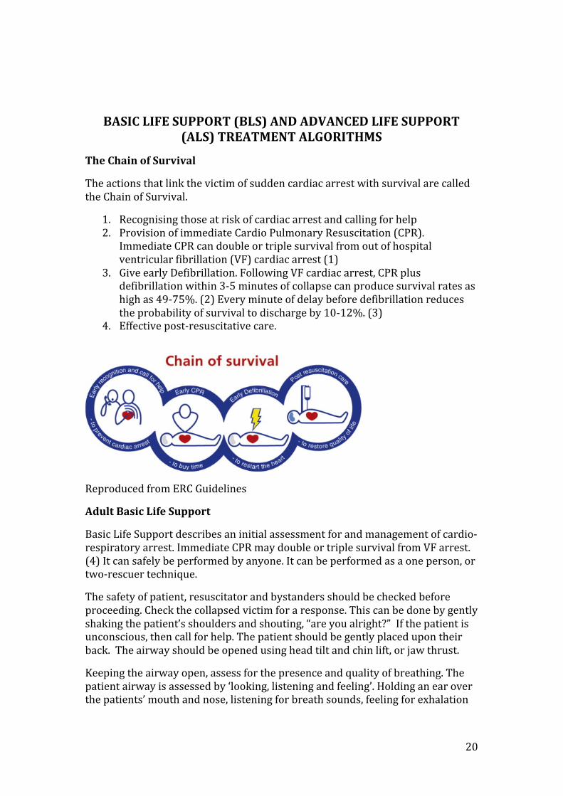
BASIC LIFE SUPPORT (BLS) AND ADVANCED LIFE SUPPORT (ALS) TREATMENT ALGORITHMS
The Chain of Survival
The actions that link the victim of sudden cardiac arrest with survival are called the Chain of Survival.
1. Recognising those at risk of cardiac arrest and calling for help 2. Provision of immediate Cardio Pulmonary Resuscitation (CPR).
Immediate CPR can double or triple survival from out of hospital ventricular fibrillation (VF) cardiac arrest (1)
3. Give early Defibrillation. Following VF cardiac arrest, CPR plus defibrillation within 3-‐5 minutes of collapse can produce survival rates as high as 49-‐75%. (2) Every minute of delay before defibrillation reduces the probability of survival to discharge by 10-‐12%. (3)
4. Effective post-‐resuscitative care.
Reproduced from ERC Guidelines
Adult Basic Life Support
Basic Life Support describes an initial assessment for and management of cardio-‐respiratory arrest. Immediate CPR may double or triple survival from VF arrest. (4) It can safely be performed by anyone. It can be performed as a one person, or two-‐rescuer technique.
The safety of patient, resuscitator and bystanders should be checked before proceeding. Check the collapsed victim for a response. This can be done by gently shaking the patient’s shoulders and shouting, “are you alright?” If the patient is unconscious, then call for help. The patient should be gently placed upon their back. The airway should be opened using head tilt and chin lift, or jaw thrust.
Keeping the airway open, assess for the presence and quality of breathing. The patient airway is assessed by ‘looking, listening and feeling’. Holding an ear over the patients’ mouth and nose, listening for breath sounds, feeling for exhalation
21
on the cheek, and observing any rise/fall of the chest. Gasping is an early sign of cardiac arrest, and should be recognised.
If breathing normally, the patient should be placed in the recovery position (left lateral), monitored, and help should be summoned. If breathing is abnormal or absent, help should be summoned. If there are two people present, the first should leave to call for help, whilst the second continues with the BLS algorithm. A single resuscitator should quickly call for help. The patients’ side should only be left if there is no other option. Checking for pulses is an inaccurate method of confirming the presence or absence of circulation, both for laypersons and professionals.
High quality chest compressions are essential. This should be performed over the lower half of the sternum at a rate of 100 compressions per minute and at a ratio of 30:2 (compression to respiration). Chest compressions should be performed in a comfortable position kneeling over the patient either from the right or left side of the patient. The heel of the hand is placed over the lower half of the sternum. The other hand is placed on top, and fingers interlaced. Elbows are extended, and power generated from flexing the hips.
Chest compressions are combined with rescue breaths. After 30 compressions, the airway is opened with head-‐tilt and chin-‐lift. Pinch the soft part of the nose closed. Take a normal breath and place your lips around the patients’ mouth with a good seal. Blow steadily until the chest rises for 1 second. This is an effective rescue breath. Take your mouth away, and watch the chest fall as air comes out. This should be repeated a second time to achieve two effective rescue breaths. Chest compressions and rescue breaths should be conducted simultaneously in two-‐person resuscitation.
Chest compressions should be un-‐interrupted during connection of the defibrillator, or during other advanced life support manoeuvres (including laryngoscopy if appropriate). There should be only a brief pause in the cycle during ALS for rhythm assessment, defibrillator discharge and the passage of an endotracheal tube (if appropriate). It is a good idea to change over the person doing chest compression every 2 minute cycle to avoid fatigue.
The use of an AED (automated external defibrillator), which is a biphasic defibrillating device, is included in European Resuscitation Resuscitation Council (ERC) and Australian Resuscitation Council (ARC) basic life support protocols. However, this is subject to device availability, and has been omitted from this discussion.
Advanced Life Support
Advanced life support is the algorithm for assessment and early intervention of cardio-‐respiratory arrest. Advanced life support is a continuum from adult basic life support. The recommended sequence will depend on the location, staffing experience, and available equipment. Out-‐of-‐hospital cardiac arrest is usually the result of a primary cardiac event (coronary artery disease, ischaemic cardiomyopathy, valvular heart disease). In contrast, inpatient cardiac arrest is usually preceeded by slow physiological deterioration (hypoxia, hyotension),
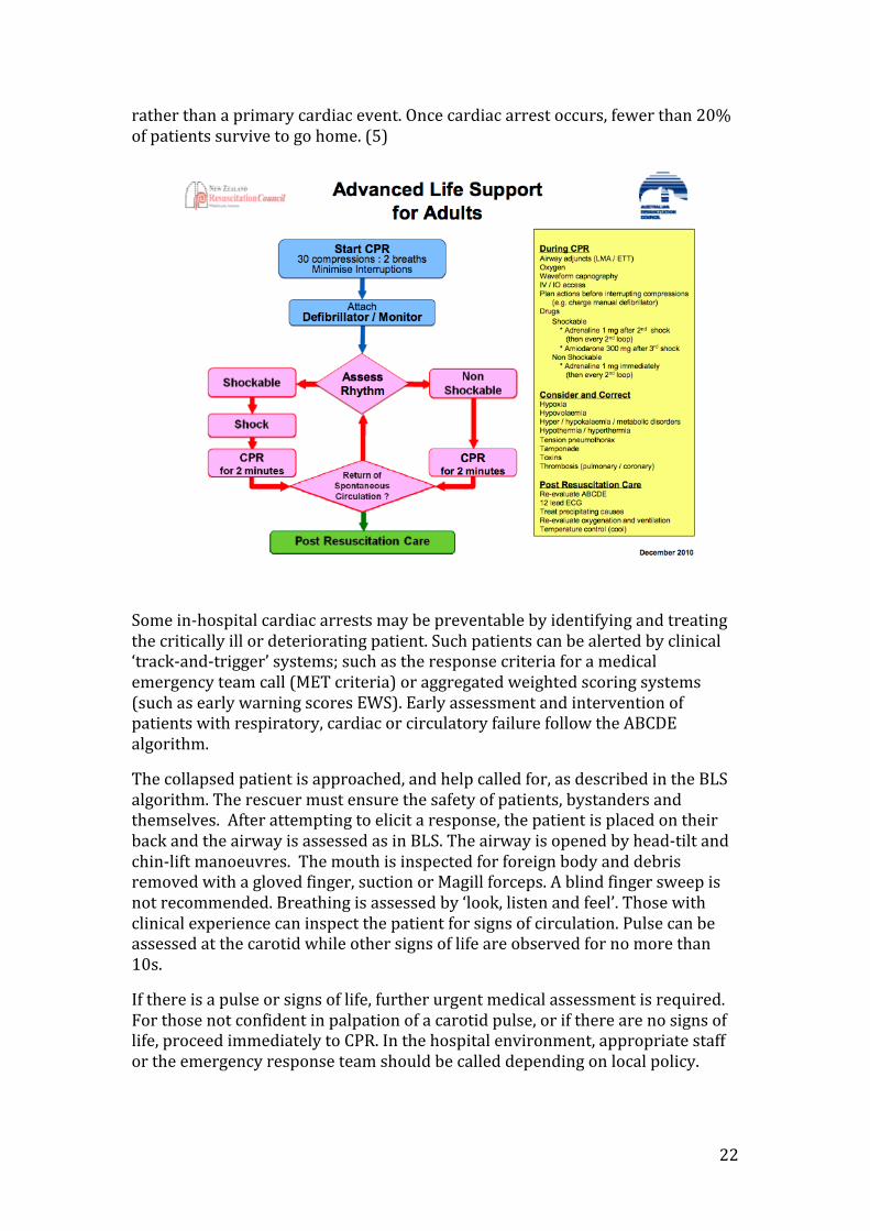
22
rather than a primary cardiac event. Once cardiac arrest occurs, fewer than 20% of patients survive to go home. (5)
Some in-‐hospital cardiac arrests may be preventable by identifying and treating the critically ill or deteriorating patient. Such patients can be alerted by clinical ‘track-‐and-‐trigger’ systems; such as the response criteria for a medical emergency team call (MET criteria) or aggregated weighted scoring systems (such as early warning scores EWS). Early assessment and intervention of patients with respiratory, cardiac or circulatory failure follow the ABCDE algorithm.
The collapsed patient is approached, and help called for, as described in the BLS algorithm. The rescuer must ensure the safety of patients, bystanders and themselves. After attempting to elicit a response, the patient is placed on their back and the airway is assessed as in BLS. The airway is opened by head-‐tilt and chin-‐lift manoeuvres. The mouth is inspected for foreign body and debris removed with a gloved finger, suction or Magill forceps. A blind finger sweep is not recommended. Breathing is assessed by ‘look, listen and feel’. Those with clinical experience can inspect the patient for signs of circulation. Pulse can be assessed at the carotid while other signs of life are observed for no more than 10s.
If there is a pulse or signs of life, further urgent medical assessment is required. For those not confident in palpation of a carotid pulse, or if there are no signs of life, proceed immediately to CPR. In the hospital environment, appropriate staff or the emergency response team should be called depending on local policy.
23
In Advanced Life Support, the importance of good quality chest compressions with minimal interruption is again stressed. Whilst waiting for the clinical response team, CPR should be administered as described in the BLS algorithm. Positive pressure ventilation, with high flow oxygen, should be performed with the most appropriate equipment at hand. This may be bag-‐valve-‐mask or supraglottic airway depending on availability. Ventilation can be aided by airway adjuncts such as an oropharyngeal or nasopharyngeal airway. Tracheal intubation should be attempted only by those who are trained, competent and experienced in this skill. Waveform capnography should be available for confirming tube placement. If the trachea has been intubated, cardiac compression can proceed uninterrupted, and the patient ventilated at a rate of 10 breaths per minute. Each breath should be sufficient to allow the chest to rise for 1 second. Avoid aggressive or over-‐ventilation.
Appropriate monitoring should be attached to the patient, including a defibrillator (pads or paddles) to expedite rhythm assessment. This should occur without interruption to CPR. Once attached, CPR should be paused briefly for rhythm assessment.
A patient found to be in ventricular fibrillation (VF) or pulseless ventricular tachycardia (VT) is said to have a shockable rhythm. The defibrillator is charged whilst CPR continues. Once the defibrillator is charged, pause chest compression, ensure that all rescuers are clear of the patient and that high flow oxygen is removed. After a rapid, thorough safety check, a shock can be given. Manual defibrillation using a monophasic defibrillator should be set at 360J, without dose escalation. Defibrillation is delivered as a single shock. The precordial thump in this setting is no longer recommended. It is no longer recommended that a fixed period of CPR precede defibrillation of the shockable rhythm.
After defibrillation, CPR is immediately restarted without rhythm assessment or pulse check. Ideally, the pause between stopping and restarting chest compressions should be less than 5 seconds. Once resuscitation has resumed, prepare intravenous cannulae and drugs likely to be used by the resuscitation team. Circulatory access should be obtained. Large calibre peripheral cannulae are preferable for drug and fluid boluses. If peripheral cannulation proves difficult, the intraosseous route should be considered. Central venous cannulation is a good alternative, if able to be performed quickly by experienced operators. Once access is established, blood is taken for further investigation and a fluid bolus can be administered. After two minutes of resuscitation, CPR is paused, and the rhythm assessed. If the patient is still in VT/VF, give a second shock at 360J. Without reassessing the rhythm or feeling for a pulse, resume CPR at a ratio 30:2.
This cycle is continued. After the second shock has been delivered, and CPR recommenced, Adrenaline can be administered at a dose of 1mg intravenously or via the intraosseous route. This can be repeated every 3-‐5 minutes (alternate cycles) until return of spontaneous circulation. After the third shock, Amiodarone should be administered at a dose of 300mg. The algorithm continues until return of spontaneous circulation. If a narrow complex regular
24
rhythm is present, try to palpate a pulse. If return of spontaneous circulation has been achieved, begin post-‐resuscitation care.
The patient found to be in asystole or pulseless electrical activity, is said to have a non-‐shockable rhythm. CPR should continue with delivery of high concentrations of inspired oxygen, with 2 minutely brief interruption for rhythm re-‐assessment. Adrenaline should be administered immediately at a dose of 1mg IV or IO. Subsequent doses are given at 3-‐5 minutely intervals. An intravenous fluid bolus of 20ml kg-‐1 (up to 1L) should be given. The underlying pathology must be identified and treated. The 4Hs and 4Ts, should be aggressively sought and treated. These include hypoxia, hypovolemia, disorders of metabolism (hyperkalaemia, hypokalaemia, hypocalcaemia, acidaemia) and hypothermia, tension pneumothorax, cardiac tamponade, toxins (or medications), and thromboembolism (pulmonary embolism, myocardial infarction).
During advanced life support, a team leader should be identified. This person is responsible for monitoring progress through the ALS algorithm, the quality of chest compression, alternating CPR providers and communicating with members of the team.
There are a number of important therapeutic goals for patients who have a return to spontaneous circulation following resuscitation. This post-‐resuscitative phase aims to make the diagnosis and provide definitive treatment of ischaemic heart disease, reduced cardiac complications, and reduce neurological sequelae. Appropriate level monitoring, arrhythmia prevention, fluid and electrolyte therapies, and glycaemic control are other important objectives. Some patients will require ventilatory support and sedation. Other post-‐resuscitation treatment considerations include therapeutic hypothermia and seizure prevention.
Paediatric Basic Life Support
Adult cardiac arrest is most often the result of a primary (or cardiac) cause. Contrastingly, paediatric cardiac arrest is most often secondary to hypoxia and respiratory failure or circulatory failure. Paediatric basic life support improves patient survival after cardiac arrest.
Initially, ensure the safety of the child and rescuer. Check the child’s responsiveness by gently stimulating/shaking the child, and asking loudly “are you alright?” If the child responds by answering or moving, leave in the position found. Continue to assess, monitor, and ensure that appropriate help is on the way.
If the child does not respond, call for help, gently place them onto their back and open the airway. The paediatric airway is opened by tilting the head and providing chin-‐lift. For infants the airway is opened by maintaining a neutral head position, and proving gentle chin lift and mouth opening. Care should be taken to ensure that chin lift is performed from the bony mandible, and that soft tissue under the chin is not pushed back as this may obstruct the airway. If this manoeuvre is ineffective, a jaw thrust should be tried.
25
Keep the airway open whilst assessing for the presence and quality of breathing. Look, listen and feel for normal breathing. If breathing normally, place the child into a recovery position, and send for help. If breathing is abnormal or absent, carefully remove any visible obstruction. A blind finger sweep is not recommended. Keep the airway open and give 5 rescue breaths.
For a child over one, the nose can be pinched closed, a normal breath taken by the rescuer, and a good seal made over the patients mouth. This is followed by a gentle breath, observing a chest rise for 1-‐1.5 seconds. For infants, the head is maintained in neutral position with chin lift to open the airway. After taking a normal breath, a seal is made over the mouth and nose of the infant. A gentle breath is delivered, observing the chest rise for 1-‐1.5 seconds. Chest fall is observed as the seal is removed, and the airway maintained. Difficulty in delivery of effective rescue breaths should alert to the possibility of airway obstruction. The airway should be re-‐inspected, and visible obstruction cleared.
After administration of 5 rescue breaths, the rescuer should perform a rapid assessment of the circulation and for signs of life. Signs of life include spontaneous breathing, movement, choking/gagging during rescue breaths, infant/child colour. Healthcare providers cannot reliably detect the presence or absence of a central pulse. Pulse palpation can be incorporated for circulation assessment by healthcare providers confident in this technique. Children over one year are assessed at the carotid or femoral pulses. Infants are assessed at the brachial or femoral pulses. The decision to begin CPR must be made in less than 10s.
If the rescuer is confident that signs of life and a pulse of over 60 beats per minute can be detected then rescue breaths should be continued. After resumption of spontaneous effective breathing, the child can be placed into the recovery position. Help should be summoned, and the child’s condition monitored.
If there are no signs of life, or unless the rescuer is certain of a definite pulse of greater than 60 then chest compressions should be commenced. The lower half of the sternum is identified by locating the xiphisternum, and placing the hand one finger breadth above it. Infant chest compressions can be performed by placing two finger tips over the lower half of the sternum. Alternatively, the chest can be encircled with both hands, with thumbs placed over the lower half of the sternum. In children over one year, the heel of one hand can be placed over the lower half of the sternum. The arm is held straight as compressions are delivered. For larger children, compressions can be administered with the two handed technique as in adults. Compressions should be sufficient to depress the sternum by 1/3 (4cm in infants, 5cm in children) at a rate of 100 to 120 per minute.
The compression ventilation ratio used for children should be based on whether one, or more than one rescuer is present. For two person resuscitation, after 15 chest compressions, two rescue breaths should be delivered. Resuscitation continues at a ratio of 15:2 until the child shows signs of life, or further qualified help arrives to take over. For single-‐rescuer resuscitation, the guidelines suggest a ratio of 30:2.
26
It is vital for rescuers get help quickly when a child collapses. When more than one rescuer is available, the first starts resuscitation, whilst the second sends for assistance. If only one rescuer is present, undertake resuscitation for one minute before seeking assistance. If possible, carry the child while summoning help.
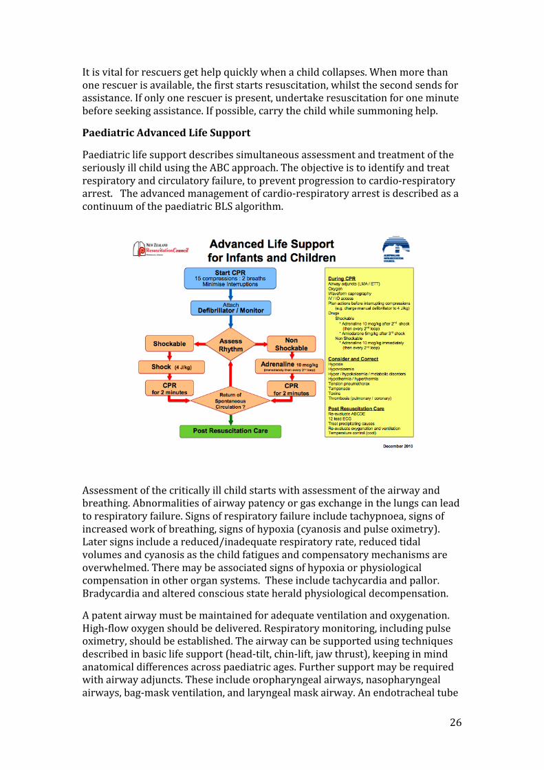
Paediatric Advanced Life Support
Paediatric life support describes simultaneous assessment and treatment of the seriously ill child using the ABC approach. The objective is to identify and treat respiratory and circulatory failure, to prevent progression to cardio-‐respiratory arrest. The advanced management of cardio-‐respiratory arrest is described as a continuum of the paediatric BLS algorithm.
Assessment of the critically ill child starts with assessment of the airway and breathing. Abnormalities of airway patency or gas exchange in the lungs can lead to respiratory failure. Signs of respiratory failure include tachypnoea, signs of increased work of breathing, signs of hypoxia (cyanosis and pulse oximetry). Later signs include a reduced/inadequate respiratory rate, reduced tidal volumes and cyanosis as the child fatigues and compensatory mechanisms are overwhelmed. There may be associated signs of hypoxia or physiological compensation in other organ systems. These include tachycardia and pallor. Bradycardia and altered conscious state herald physiological decompensation.
A patent airway must be maintained for adequate ventilation and oxygenation. High-‐flow oxygen should be delivered. Respiratory monitoring, including pulse oximetry, should be established. The airway can be supported using techniques described in basic life support (head-‐tilt, chin-‐lift, jaw thrust), keeping in mind anatomical differences across paediatric ages. Further support may be required with airway adjuncts. These include oropharyngeal airways, nasopharyngeal airways, bag-‐mask ventilation, and laryngeal mask airway. An endotracheal tube
27
can be placed for a definitive airway, but is not essential. Cuffed tubes may be considered in paediatric patients where lung compliance is poor. In untrained hands, intubation may lead to delays in ventilation, delays in chest compression, and worse patient outcomes. Failure to obtain an airway with techniques described above may rarely require a surgical airway. Monitoring of end-‐tidal carbon dioxide is important for intubated patients, and can provide clues to tube position, adequacy of circulation/ventilation and effectiveness of chest compressions in arrested patients.
Healthcare providers commonly provide excessive ventilation during CPR and this may be harmful. Hyperventilation causes increased intra-‐thoracic pressure, decreased cerebral and coronary perfusion, and poorer survival. (6) Hyperventilation increases the likelihood of gastric insufflation, which can impede ventilation, increase aspiration risk and have negative circulatory effects.
Circulatory failure (shock) is characterised by a mismatch between metabolic demand by the tissues and delivery of oxygen and nutrients by the circulation. Physiological compensation will lead to changes in heart rate and systemic vascular resistance. The signs of circulatory failure included increased heart rate (bradycardia suggests physiological decompensation), decreased systolic blood pressure, decreased peripheral perfusion (prolonged capillary refill, decreased skin temperature, mottled skin), weak/absent peripheral pulses, decreased urine output and metabolic acidosis.
The main priorities in circulatory management include establishing first line cardiac monitoring (ECG, non invasive blood pressue, SpO2). Secure intravenous access should be obtained. This may be by peripheral intravenous or intraosseous cannulation. Central venous cannulation is a good alternative, if it is able to be obtained quickly. Once access to the circulation is secured, give a bolus of fluid (20ml kg-‐1). Isotonic crystalloids are recommended as initial resuscitation in infants and children with any type of shock. Subsequent fluid boluses can be given, guided by clinical findings and clinical response. Give drugs (inotropes, vasopressors, anti-‐arrhythmics) as required.
The child should be assessed, and reassessed continuously, commencing with the airway before moving onto breathing and then the circulation. Whilst the focus should be on rapid identification and rapid intervention of respiratory and circulatory failure, simultaneous attempts must be made to identify the underlying cause.
Advanced management of the paediatric cardio-‐respiratory arrest is a continuum of the BLS protocol. The focus is on good quality and uninterrupted CPR (15:2) following 5 rescue breaths, delivery of positive pressure ventilation with high inspired oxygen concentration, early defibrillator attachment and rhythm assessment, identification and early defibrillation of the shockable rhythm, and treatment of the underlying cause in non-‐shockable rhythms.
Arrested patients found to have asystole, pulseless electrical activity, or electromechanical dissociation on rhythm assessment are said to have non-‐shockable rhythms. CPR should continue with high inspired oxygen concentration delivery, with 2 minutely brief interruptions for rhythm re-‐
28
assessment. Adrenaline should be administered immediately at a dose of 10 micrograms per kg intravenously or via the intraosseous route. Subsequent doses are given at 3-‐5 minutely intervals. An intravenous fluid bolus of 20ml per kg should be given. The underlying pathology must be identified and treated. The 4H’s and 4T’s, previously discussed, should be sought and treated.
Arrested patients found to have ventricular fibrillation or ventricular tachycardia on rhythm assessment are said to have a shockable rhythm. Defibrillation is attempted immediately after a thorough but rapid safety check. A single-‐shock strategy using a non-‐escalating dose of 4 Joules per kg is recommended for defibrillation in children (biphasic or monophasic). CPR is immediately recommenced. Re-‐charge the defibrillator as CPR continues. At two minutes, CPR is interrupted for a further rhythm check. A second shock is given if VF/pulseless VT are identified. CPR recommences, and the cycle continues. After the second shock has been delivered, and CPR has recommenced, Adrenaline 10 micrograms per kg is administered. Amiodarone 5mg per kg intravenously or via the intraosseous route is administered after the third shock. Adrenaline 10 microgram per kg is given every alternate cycle (3-‐5 minutely).
If signs of life become evident, check the monitor for an organised rhythm. If present, check for signs of life and a central pulse and evaluate the patients’ haemodynamics (blood pressure, capillary return, pulses). Any reversible causes should be identified and treated. The first 2 H’s (hypoxia and hypovolaemia) have the highest prevalence in critically ill or injured children.
Dr Brendan Munzel
(Summarised from ERS and ARC resuscitation guidelines)
References
1. Valenzuela TD, et.al. Estimating effectiveness of cardiac arrest interventions: a logistic regression survival model. Circulation 1997; 96:3308-13.
2. Steil IG, et.al. Modifiable factors associated with improved cardiac arrest survival in a multicentre BLS/defibrillator system: OPALS study phase I results. Ann Emerg Med 1999; 33:44-50.
3. Waalewijn RA, et.al. Survival models for out-of hospital cardiopulmonary resuscitation from the perspective so the bystander, the first responder, and the paramedic. Resuscitation 2001; 51:113-22.
4. Weistfeld ML, et.al. Survival after application of AED before arrival of EMS: evaluation in resuscitation outcomes consortium population of 21 million, JAmCollCardiol 2010;55:1713-20.
5. Gwinnutt CL, et.al. Outcome after cardiac arrest in adults in UK hospitals. Resuscitation 2000; 47125-35.
6. Aufderheide TP, et.al. Death by hyperventilation: a common and life threatening problem during CPR. CritCareMed 2004; 32:S345-‐51.
29
ACUTE CORONARY SYNDROMES
Coronary artery disease consists of a clinical spectrum of disease that can present in different ways. The management of the different presentations of acute coronary syndrome will be discussed.
Clinical presentation of acute coronary syndrome:
The cause of acute coronary syndrome is ischaemia of the coronary myocytes. If the delivery of oxygen and substrate to the myocytes is prolonged, myocardial necrosis will occur in the region supplied by the relevant coronary artery. Coronary artery narrowing and occlusion occurs due to atheromatous plaque rupture, constriction of smooth muscle, or thrombosis formation in a coronary artery. Atheromatous plaques can fissure, releasing tissue factor and activating platelets and the coagulation cascade to occlude the artery.
Acute coronary syndromes arise due to the same disease process. They are, unstable angina, non-ST segment elevation myocardial infarction and ST segment myocardial infarction. All of these can lead to arrhythmias and cardiac muscle dysfunction. It is important that patients with any of these three syndromes present to hospital and are monitored as soon as possible. Life threatening arrhythmia will require defibrillation or cardioversion.
Stable angina is NOT an acute coronary syndrome. It typically occurs in patients with known risk factors for coronary artery disease and presents with tightness across the chest that may radiate to the neck, arms, back or epigastrium that is provoked by exercise and is relieved by rest.
Unstable angina is angina that occurs at rest (usually of more than 20 minutes duration), new onset angina that markedly limits physical activity and increasing angina that is more frequent, longer in duration and occurs with less exertion than previous angina.
Non-ST elevation myocardial infarction (NSTEMI) may present in the same way as unstable angina. The principal difference is that the ischaemia is severe enough to cause damage to the myocardium such that there is an elevation in troponinsm (chemical biomarkers of myocardial damage). Since the elevation in troponins may not be detectable for hours after presentation, the initial management of unstable angina and NSTEMI is the same.
ST elevation MI (STEMI) needs to be recognized promptly so as to enable referral to cardiology and early reperfusion of the blocked coronary artery (arteries).
Some patients have atypical presentation of acute coronary syndromes. Women, diabetics and the elderly may have atypical chest pain (sharp, intermittent), tend to present later and may have cardiac failure as the presenting symptom. Confusion may be a presenting feature in the elderly patient.
30
Cocaine associated myocardial infarction may present in the same way as for other patients, but a history of cocaine use is useful, as the treatment is significantly different. Beta blockers are CONTRINDICATED as they can intensify coronary artery vasoconstriciton, and benzodiazepines are useful.
Diagnostic features:
Unstable angina
The ECG may be normal in unstable angina. There are some ECG features that correlate with increasing risk and may indicate the need for further investigation with coronary angiography. The cardiac enzymes (troponin and CK levels in the blood) are usually normal, but an elevated troponin suggests that the patient is at high risk.
Non-ST elevation MI
The ECG shows non specific abnormalities such as ST segment depression or T wave inversion. The cardiac enzymes are usually elevated with an elevated CK and troponin.
ST segment elevation MI
The ECG shows acute ST segment elevation. Q waves are likely to develop, but may not develop in the patients who are re-perfused with thrombolysis or angioplasty and coronary artery stenting (PCI- percutaneous coronary intervention). The cardiac enzymes and troponins are elevated (but this may take up to 4-6 hours after the onset of chest pain). The diagnostic features on ECG are ST elevation of 1mm or more in 2 contiguous chest leads or 2mm of elevation in 2 contiguous chest leads, or a new left bundle branch block in a patient with a typical history of myocardial infarction.
Immediate treatment of all acute coronary artery syndromes:
A rapid assessment of the airway, breathing and circulation, conscious state and exposure of the patient to detect any precipitating causes or complications (systemic hypoperfusion, left heart failure with hypoxia and arrhythmias) of the coronary syndrome is made. Attach monitoring of the ECG and oxygen saturation monitor (oximeter) and establish intravenous access. A 12 lead ECG will be required as soon as practical and repeated at regular intervals (every 10 minutes) until the chest pain resolves.
Administer oxygen if it is indicated. If there is hypoxia (SpO2 < 90%), respiratory distress and if the patient is at high risk for hypoxia, it is recommended that oxygen be administered. The role of supplemental oxygen for those patients without hypoxia is less clear. A review in 2010 found a suggestion of harm in patients with acute coronary syndrome with normoxia. Hyperoxia has been shown to have a direct vasoconstrictor effect on the coronary arteries. In normoxic patients there is a weak reommendation for the use of oxygen in acute coronary syndromes. (Up to Date 2011)
Morphine is administered for control of pain and to relieve anxiety. Intravenous dosing of 2-4 mg as an initial dose is recommended followed by repeat doses at intervals of 5-15 minutes.
31
Aspirin is used as the initial antiplatelet agent to prevent platelets adhering to exposed endothelium in the coronary arteries. It is recommended for all patients with acute coronary syndrome and is given in doses of 300mg orally.
Nitrates such as sublingual nitroglycerin are used for chest pain. Three initial doses are given and then if pain is ongoing, an intravenous infusion at an initial dose of 40mcg per minute is commenced. There are some important indications and contraindications to its use. The indications (aside from ongoing chest pain) include the presence of heart failure and hypertension. They are to be used with caution in those with right ventricular infarction, severe aortic stenosis, hypotension and in those patients who have taken a phospodiesterase inhibitor (for erectile dysfunction or pulmonary hypertension) in the previous 24 hours.
Beta blocker therapy is administered using a cardioselective agent such as metoprolol or atenolol. Beta blockers are useful to help reduce myocardial oxygen demand and to help reduce arrhythmias. The contraindications to beta blockers include bradycardia, shock heart block and reactive airways disease. They should be used with caution in those with heart failure and in consultation with a cardiologist.
Atorvastatin is recommended by the AHA/ACC in patients who are not already on a statin for lowering cholesterol.
Monitoring of electrolytes is important in order to minimize the risk of cardiac arrhythmia. A serum potassium and magnesium is performed in order to identify those patients that may need to have replacement therapy.
Treatment for unstable angina and non ST elevation myocardial infarction:
The above measures are taken and in addition to aspirin, another antiplatelet agent is recommended: either a thieopyridine such as clopidogrel or a glycoprotein IIb/IIIa inhibitor such as eptifibatide or tirofiban.
Anticoagulation therapy is commenced. Unfractionated heparin is preferred as an intravenous bolus of 50-60 units per kg followed by 12 units/kg/hr intravenously (as an infusion) with monitoring of the aPTT (aim for 1.5-2 times normal). Enoxaparin is an alternative, but care is taken in those patients with renal compromise.
An assessment of risk is made to facilitate decision making about proceeding to coronary angiography. The features that increase risk include:
• The presence of ST depression and the extent of ST depression
• Elevated cardiac biomarkers
• Evidence of hemodynamic instability
• Persistent chest pain in spite of appropriate therapy
A risk score has been devised (TIMI score) and a score of 4-7 indicated a high risk for a poor outcome in patients with unstable angina or non ST MI:
• Age over 65 years
32
• The presence of at least three risk factors for IHD (hypertension, diabetes, dyslipidaemia, smoking or positive family history for early MI)
• Prior coronary stenosis of over 50%
• Presence of ST segment deviation on admission ECG
• At least two anginal episodes in the previous 24 hours
• Elevated serum cardiac biomarkers
• Use of aspirin in the previous 7 days (probably a marker of more severe coronary disease)
Patients considered to be at high risk are referred to cardiology for early angiography with percutaneous intervention. They may also benefit from angiotensin converting enzyme inhibitor therapy. There is NO evidence for benefit from thrombolysis in unstable angina and non ST MI, and thrombolysis MAY CAUSE HARM.
Treatment of ST elevation myocardial infarction (STEMI):
After initial therapy as for all acute coronary syndromes, it is important to identify the patients who will need acute re-perfusion either via thrombolytic therapy or percutaneous coronary intervention (PCI) via cardiac catheterization.
In order to detect STEMI, continuous cardiac monitoring (ECG monitoring) is recommended to detect arrhythmias and serial 12 lead ECGs are required to detect ST segment elevation. ST elevation of 1mm or more in limb leads or 2mm or more in chest leads or a NEW left bundle branch block are features of a STEMI and acute reperfusion is indicated.
In general, early PCI (within 90 minutes of presentation) is recommended, but where this is not possible, reperfusion needs to be achieved with thrombolytic therapy. Thrombolysis is indicated in patients who present within the first 12 hours of the onset of symptoms of a myocardial infarction. If there are symptoms of pain in the time from 12 to 24 hours after the onset of acute chest pain, it may be considered.
Before thrombolysis, a dose of clopidogrel is indicated. Thrombolysis is achieved with intravenous agents such as streptokinase, reteplase, tenecteplase. There are some important contraindications to thrombolysis and they need to be excluded before administration. They include, previous haemorrhagic stroke, other stroke within 6 months, central nervous damage or neoplasm, active internal bleeding, aortic dissection, recent major surgery or trauma and a known bleeding disorder.
Following reperfusion either with thrombolysis or PCI, antithrombotic therapy is commenced (heparin, low molecular weight heparin), beta blockers and angiotensin converting enzyme inhibitors.
Conclusion:
Acute coronary syndromes are grouped into STEMI and non-STEMI or unstable angina. It is important to identify those patients with a STEMI to enable acute reperfusion either with thrombolytic agents or percutaneous coronary intervention. All patients with acute coronary syndromes are assessed with the ABCDE approach,

33
given oxygen when indicated, nitrates, morphine and aspirin. An early 12 lead ECG, continuous cardiac monitoring (for arrhythmia), intravenous access and serial blood tests for biomarkers of cardiac infarction and electrolytes is indicated. The time to reperfusion after first presentation to medical care needs to be minimized in order to improve outcome after myocardial infarction.
References:
1. Australian Resuscitation Council. Advanced Life Support, 5th edition, 2006.
2. Australian Resuscitation Council Website. www.resus.org.au/
3. UptoDate. www.uptodate.com 2011.
34
POST RESUSCITATION CARE
Dr. Roni Krieser
Following return of spontaneous circulation, there is a significant risk of morbidity and mortality due to cerebral and cardiac dysfunction secondary to whole body ischaemia. Post cardiac arrest management aims to provide support to organ systems affected by systemic ischaemic and reperfusion injury.
Post Cardiac Arrest Syndrome.
Prolonged whole body ischaemia causes global tissue and organ injury, with additional damage occurring during reperfusion following successful resuscitation. Post arrest pathophysiology, is superimposed on the pathology that caused the arrest and the patient’s other co-morbidities.
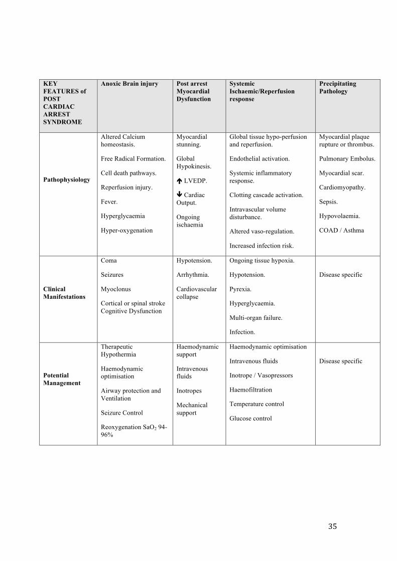
The post cardiac arrest syndrome is characterised by:
• Anoxic brain injury • Arrest related myocardial dysfunction • Systemic ischaemic/reperfusion response. • Ongoing pathology that lead to cardiac arrest.
Initial objectives of Post cardiac arrest care is
• Optimize cardiopulmonary function and vital organ perfusion. • Where possible, patients should be treated in hospitals with intensive care
facilities. • Identify and treat the precipitating cause of the arrest.
Secondary Objectives
• Control body temperature to optimize survival and neurological recovery. • Identify and treat acute coronary syndromes. • Optimize mechanical ventilation to reduce lung injury. • Support organ function and reduce the risk of multi-organ injury. • Assess prognosis • Ensure appropriate rehabilitation for survivors.
35
KEY FEATURES of POST CARDIAC ARREST SYNDROME
Anoxic Brain injury Post arrest Myocardial Dysfunction
Systemic Ischaemic/Reperfusion response
Precipitating Pathology
Pathophysiology
Altered Calcium homeostasis.
Free Radical Formation.
Cell death pathways.
Reperfusion injury.
Fever.
Hyperglycaemia
Hyper-oxygenation
Myocardial stunning.
Global Hypokinesis.
LVEDP.
Cardiac Output.
Ongoing ischaemia
Global tissue hypo-perfusion and reperfusion.
Endothelial activation.
Systemic inflammatory response.
Clotting cascade activation.
Intravascular volume disturbance.
Altered vaso-regulation.
Increased infection risk.
Myocardial plaque rupture or thrombus.
Pulmonary Embolus.
Myocardial scar.
Cardiomyopathy.
Sepsis.
Hypovolaemia.
COAD / Asthma
Clinical Manifestations
Coma
Seizures
Myoclonus
Cortical or spinal stroke Cognitive Dysfunction
Hypotension.
Arrhythmia.
Cardiovascular collapse
Ongoing tissue hypoxia.
Hypotension.
Pyrexia.
Hyperglycaemia.
Multi-organ failure.
Infection.
Disease specific
Potential Management
Therapeutic Hypothermia
Haemodynamic optimisation
Airway protection and Ventilation
Seizure Control
Reoxygenation SaO2 94-96%
Haemodynamic support
Intravenous fluids
Inotropes
Mechanical support
Haemodynamic optimisation
Intravenous fluids
Inotrope / Vasopressors
Haemofiltration
Temperature control
Glucose control
Disease specific
36
Therapeutic Hypothermia
Anoxic brain injury accounts for the majority of morbidity and mortality in the post arrest period.
Early institution of therapeutic hypothermia after out of hospital VF arrest, has shown improved outcomes in patients who remained comatose following return of spontaneous circulation. In addition to improved cerebral outcomes, therapeutic hypothermia may also improve myocardial function and reduce myocardial infarct size. Therapeutic hypothermia improves the survival and functional outcome in one in every 6 cardiac arrest survivors. There is uncertainty regarding the use of Therapeutic Hypothermia post Non-VF arrests, that is those arrests due to asystole or pulseless electrical activity.
Induction of therapeutic hypothermia should occur early and can be achieved through internal and external techniques, which include: Ice-Cold intravenous fluids (30ml/kg of 3-4Co Saline or Hartmanns solution), External Ice packs or cooling mats. Cold air. Neuromuscular blockade may be used to treat shivering. he aim is to reach a temperature of 32-34Co. Ideally, Core Temperature should be measured continuously using oesophageal, bladder [not in anuric patients] or pulmonary artery temperature catheters.
Hypothermia is maintained for 12-24 hours.
Rewarming can be achieved through internal and external techniques. The rate of warming should be 0.25-0.5Co per hour. Throughout induction and warming phases, monitoring and management of electrolyte and haemodynamic disturbances is important.
Complications of Therapeutic Hypothermia:
Shivering is common, with the increased muscle activity both increasing oxygen consumption, and preventing temperature reduction. Sedation with neuromuscular blockade may be required to control shivering. A rise in systemic vascular resistance is seen with hypothermia and may decrease cardiac output. Other complications include: arrhythmias [most commonly bradycardia], diuresis with subsequent hypovolaemia and electrolyte imbalance. Both coagulation and platelet function are impaired. Unstable glucose levels may occur.
Hyperthermia
After resuscitation, hyperthermia can impair brain recovery. Post resuscitation release of inflammatory cytokines may be the cause of hyperthermia. Patients treated with therapeutic hypothermia may also develop pyrexia after re-warming. Although evidence is lacking, extrapolation from post stroke patients suggests that maintaining normothermia may be worthwhile. If therapeutic hypothermia is not undertaken, antipyretics and external cooling should be used in patients who develop fever within the first 72 hours post arrest.
37
Pulmonary System Evaluation and Support
Lung dysfunction following cardiac arrest is common. This may be due to :
• Pulmonary Oedema secondary to left ventricular failure • Non cardiogenic pulmonary oedema due to inflammation, infection or
physical injury. • Aspiration • Severe atelectasis • Significant ventilation-perfusion (VQ) mismatch- resulting in a functional
shunt.
Assessment of lung function should include Chest X-ray and Arterial blood gases.
The chest x-ray should be assessed for: signs of pulmonary oedema, infection, endotracheal and central line placement, rib fractures secondary to chest compressions and pneumothorax. The blood gases should be used to assess Alveolar-Arterial oxygen gradient, PaO2 and Pa CO2.
Hyperventilation and reduced Pa CO2, may result in cerebral vasoconstriction exacerbating cerebral hypoxic injury. Tidal volumes should be kept between 6-8ml/Kg [ideal weight], with inspiratory plateau pressures kept below 30cmH20 to reduce ventilator associated lung injury. PEEP should be used to reduce atelectasis and may be supplemented with “recruitment techniques”. Minute ventilation should be titrated to achieve a PaCO2 of 40-45 mmHg.
Following the return of spontaneous circulation, oxygen administration should be titrated to achieve arterial saturations above 94%. Hyperoxia may be associated with the formation of Oxygen free radicals and exacerbate neurological injury.
Cardiovascular System
Acute coronary syndromes are a common cause of cardiac arrest. A 12 lead ECG and blood markers of myocardial infarction should be obtained as soon as possible after return of spontaneous circulation to assess for signs of infarction. The presence of an ST elevation myocardial infarction should be treated aggressively. Where possible, emergency percutaneous angiography and angioplasty should be undertaken to improve myocardial function and improve neurological outcomes. Where angiographic intervention is not available, thrombolysis should be considered for post arrest patients with ST elevation acute myocardial infarcts.
Haemodynamic instability is common following cardiac arrest. Myocardial ischaemia, reperfusion, and defibrillation may all result in myocardial dysfunction. Vasodilatation may result from loss of sympathetic tone and acidosis and require vasopressor support.
Fluid administration together with vasoactive, inotropic and inodilator agents may be required to optimize blood pressure, cardiac output and systemic perfusion. A mean arterial pressure >65-100mmHg, and a mixed venous oxygen >70% are reasonable aims.
38
Glucose Control
Metabolic derangement following cardiac arrest is common. Hyperglycaemia has been associated with worse neurological outcomes. The optimum blood glucose level is unknown. Tight glucose control is associated with higher rates of hypoglycaemia. Moderate glucose control of 8-10mmol/L is reasonable in the post arrest period.
Seizure Management
Seizures are common in the post arrest period with an incidence of 5-20%. Seizure activity increases cerebral metabolism and hence oxygen requirements. Comatose patients may require an EEG for seizure recognition. Prolonged seizures should be treated with benzodiazepines, Phenytoin, Sodium Valproate or a Barbiturate.
Prognostic Markers
Neurological Assessment: The sudden interruption of cerebral blood flow during an arrest results in rapid loss of higher neurological function, that is, consciousness. Brain stem function is lost more slowly. Absence of neurological function following return of spontaneous circulation does not reliably predict poor outcome. A prognostic clinical examination can only be conducted in the absence of confounding factors: hypotension, seizures, sedatives or paralysis. No clinical signs can reliably predict poor outcome <24 hours post arrest.
In comatose patients who have NOT undergone therapeutic hypothermia, the absence of both papillary light and corneal reflexes at >72hours post arrest, have a poor prognosis. Likewise, a low Glasgow Coma Score or myoclonic activity is associated with a poor outcome.
In patients who have been treated with therapeutic hypothermia, prognostic testing is less reliable and may require observation for more than 72 hours post arrest.
Investigations looking at prognosis post arrest include: EEG, Somatosensory-evoked potentials, CT and MRI scans
Conclusion
Optimisation of systemic perfusion, metabolic homeostasis and support of struggling organ function should be implemented early in the post arrest period. Early diagnosis and treatment of acute coronary events may increase the chance of survival. Therapeutic hypothermia may reduce neurological injury. All organ systems may be affected by Post cardiac arrest syndrome. A system based approach to assessment and treatment, in the most comprehensive medical facility available, is important to achieve the best possible functional outcome for patients.

39
References
1. American Heart Association (AHA) Guidelines on Post Resus Support. Circulation 2005; 112 IV-84-IV-88
2. Post Cardiac Arrest Care 2010 AHA guidelines for CPR and Emergency cardiovascular care. Circulation 2010; 122 [suppl 3]:S768-786
3. ILCOR Consensus Statement on Post cardiac Arrest Syndrome Resuscitation (2008) 79, 350-379
4. Post Cardiac Arrest Syndrome: A review of Therapeutic Strategies. Stub et al. Circulation. 2011; 123:1428-1435
40
BASIC NEWBORN RESUSCITATION
Rhonda Keenan
NEWBORN
The term “newborn” refers to an infant from birth to the first hours of life. The neonatal period extends from birth until 28 days of life. Most newborns establish spontaneous effective respirations after birth. Approximately 10% require some assistance to begin breathing at birth, with less than 1% requiring extensive resuscitation. (Australian & New Zealand statistics)
Infants born at term have low or nil risk factors requiring resuscitation. They commence breathing or crying spontaneously, have good muscle tone and heart rate above 100 beats per minute. They must be dried and kept warm.
TRANSITION FROM FETAL TO EXTRAUTERINE LIFE.
Following birth the lungs change from fluid filled to air filled. Pulmonary blood flow increases dramatically, and intra-cardiac and extra-cardiac shunts reverse directions and subsequently close.
Oxygen levels rise over several minutes in normal term newborns, with a heart rate range between 110-160 beats per minute, whilst it takes 5-10 minutes for oxygen (O2) saturations to reach 90%.
Adaption to extra uterine life depends on many coordinated and interdependent physiological events.
REQUIREMENTS FOR RESUSCITATION OF THE NEWBORN
Communication is fundamental and must occur between those caring for the mother and those responsible for the infant. Details of any maternal medical conditions and treatment that may affect resuscitation and management of the infant should be conveyed, including intrapartum events that informs about fetal wellbeing.
Paediatric staff should be informed whether the pregnancy was a singleton or multiple gestation, gestational age and anticipated birthing risk factors such as the presence of meconium in the liquor, congenital abnormalities, maternal infections and medications and assessments of foetal heart rate during the labour.
The environment should be a warm, draft-free area as newborns are at risk of hypothermia and prevention of heat loss is important. To maintain normothermia (36.50C- 37.20C), reduce heat loss of the infant by drying and removing wet linen, wrapping the infant with pre-warmed linen and placing the babe under a radiant warmer.
Newborns less than 1500grams birth weight become cold very easily. It is recommended to place such a babe in a polythene bag with the body completely covered and head kept outside bag. This reduces heat loss effectively during resuscitation but may occasionally cause slight hyperthermia.
Equipment required for active resuscitation should ideally include a firm padded resuscitation surface, overhead warmer and light, timing clock, oxygen /air supply
41
with flow meter, tubing and equipment for administering air/oxygen, suction tubing and catheters, neonatal laryngoscopes and blades, stethoscope and note paper for documentation.
A manual ventilation device that can be used with a face mask or endotracheal tube is essential. Self inflating bags do not depend on a medical gas source but may be attached to an oxygen/air supply. This device re-expands following compression and has a pressure relief valve that may be over-ridden where greater pressures are required. It does not provide a “Positive-End-Expiratory-Pressure” and it is difficult to deliver consistent inflating pressures.
Personnel who attend births should be trained in basic neonatal resuscitation skills including airway support, ventilation via a face mask and chest compressions. A person with advanced neonatal resuscitation training will have the additional skills enabling tracheal intubation and ventilation, peripheral/umbilical vein cannulation and drug and fluid administration knowledge.
KEY POINTS
All newborns experience a degree of hypoxia during labour and birth, with most healthy infants tolerating this well. Some infants may require additional assistance to establish normal breathing at birth. The primary aim of newborn resuscitation is inflation of the lungs with air or oxygen. Ventilation supports extra-uterine circulation to pump oxygenated blood to and from the heart to initiate recovery.
The time of birth should be noted, the umbilical cord should be securely clamped, and the infant dried and wrapped in warm towels. This provides stimulation to breathe and gives time for a full assessment of tone, respiratory effort and heart rate.
The primary assessment of the healthy infant will reflect good muscle tone with flexed posture and limbs moving, crying or breathing within seconds of delivery, rising heart rate more than 100 beats per minute within the first minute of life and becoming well perfused quickly.
The compromised infant will present with poor tone, extended posture, no movement, pallor and no respiratory effort or absent breathing, persistently low heart rate less than 100 beats per minute and will require assisted ventilation.
INITIAL ASSESSMENT OF THE NEWBORN INFANT
Assessment of muscle tone is subjective and gestational age dependent. Observe for movement of limbs and breathing. Brisk gently drying is usually sufficient to stimulate term newborns to breathe. If there is no response to this stimulation the infant will require further assessment and intervention.
Breathing following the initial effort may pause for a few seconds and then establish as regular breaths to maintain the heart rate above 100 beats per minute (bpm). If the heart rate is not maintained above 100 bpm positive pressure ventilation should be instigated. Recession and retraction of ribs and sternum indicates difficulty in expanding the lungs. Persistent apnoea requires positive pressure ventilation.
Heart rate is determined by auscultation with a stethoscope. The normal newborn range is 110-160 bpm soon after birth. An obvious increasing or decreasing heart rate
42
is an indication of the infant’s condition. Assisted ventilation is required where the heart rate is persistently below 100 bpm. A newborn with a heart rate less than 60 bpm despite adequate assisted ventilation requires chest compressions.
Colour of the newborn infant is not used to guide interventions. Normal newborns are blue at birth and with the onset of breathing; oxygenation will change the appearance over approximately 10 minutes so that the infant then looks a normal colour. No interventions are required if the infant is breathing effectively and has good muscle tone. Central cyanosis may be determined by examining gums and mucous membranes for signs of inadequate perfusion.
ADDITIONAL CONSIDERATIONS
The timing of umbilical cord clamping should not be delayed in the compromised infant as no benefit has been demonstrated by delaying clamping and it causes a delay in commencement of active resuscitation.
Supplemental oxygen during resuscitation has been extensively researched. There is increasing evidence that excessive oxygenation can be harmful to the newborn infant during and after resuscitation. Trials comparing neonatal resuscitation initiated in room air versus 100% oxygen have showed increased survival of those infants who were initially resuscitated in air. Infants receiving intermittent positive pressure ventilation in 100% oxygen had increased time to first breath/cry and also experienced oxidative stress for up to 4 weeks.
Airway management of newborns does not require suctioning as they clear their airways very effectively. If secretions are obvious they may be cleared with a wide bore suction catheter. Pharyngeal suctioning can cause laryngeal spasm, trauma to soft tissue, bradycardia and delay the onset of spontaneous respiration.
If respiratory efforts are present but not producing effective ventilation, the airway may be obstructed. If this is suspected, suction should only be performed under direct vision with a laryngoscope. Generally, suctioning should not be used except where there are obvious signs of obstruction.
Meconium stained liquor and suctioning of the mouth prior to completion of birth makes no difference to outcomes and is not recommended. Routine endotracheal suctioning is not recommended if the infant is actively breathing at birth. It does not alter the outcome and may cause harm.
If the amniotic fluid contains thick meconium and the infant is born in poor condition, suction the mouth/pharynx using a large bore suction catheter at 5 cm depth for 2-5 seconds only. Laryngoscopy and suctioning around the vocal cords under direct vision or by intubation may be performed if person with expertise in this practice is present.
Australian Resuscitation Council Neonatal Life Support Guideline 13.4, December 2010 states that “in general, nasal, oral and pharyngeal suctioning should not be used except when babies show obvious signs of obstruction either to spontaneous breathing or to positive pressure ventilation, or where removal of secretions is needed to visualise the vocal cords during intubation”.
43
BIRTH RESUSCITATION
Immediately at birth where the infant breathes spontaneously and has good muscle tone, dry and provide warmth with subsequent assessments of heart rate, breathing, tone and oxygenation.
If the infant is not breathing, dry and stimulate, position the head and neck to open the airway, provide warmth and assess breathing and heart rate. If the newborn fails to breathe, transfer to a resuscitation surface, turn on timer clock and ensure heating source is switched on. Dry skin but do not cover the baby. Evaluate respirations then heart rate. Suction the mouth only in the presence of obvious blood mucous or meconium briefly and only to a depth of 5 centimetres. Maintain the airway by correct positioning of the infant; supine with the head in a neutral position and some support under the shoulders if moulding during birth has caused a very prominent occiput.
BREATHING AND HEART RATE
If after tactile stimulation there is inadequate breathing or the infant remains apnoeic or the heart rate is less than 100 bpm, commence positive pressure ventilation (PPV) and call for help. Communicate clearly and delegate roles.
POSITIVE PRESSURE VENTILATION (PPV)
For effective PPV, select the correct size mask. With the infant’s head in the neutral position and the operator’s finger under the chin to keep airway open, gently place the mask over the face, being careful not to cover the eyes, and apply gentle even downward pressure to create a seal and minimise gas leakage.
Inflate the lungs with sufficient pressure that the chest and abdomen move slightly, with a ventilation rate of 40-60 breaths per minute. Use a counting pattern, “breathe-two-three”, with lung inflation on “breathe” and allow the infant to exhale on the “two-three”. An infant who has gasped at birth will be easier to ventilate than one who has never breathed.
Effective ventilation will be confirmed by improvement in heart rate increasing above 100 bpm, and an observed slight rise of chest and abdomen with each inflation. Oxygenation will also improve.
If these signs are not seen, readjust the head/neck position of the infant, check for malfunctioning equipment, improve the seal between face and mask and increase the inflation pressures by squeezing the bag of the manual ventilation device a little harder. Higher inflation pressures may be required to open the lungs during the first few inflations.
Check the infant’s heart rate with a stethoscope or palpate the base of the umbilical cord, (though this is not a reliable sign that heart rate is absent) or use pulse oximeter if available. If the heart rate remains less than 100 bpm, continue positive pressure ventilation and check the heart rate every 30 seconds
Newborns requiring resuscitation generally have healthy hearts that are depressed by hypoxaemia. Effective positive pressure ventilation alone will normally result in a rapid improvement in heart rate and is the key to successful resuscitating the newly born infant.
44
EXTERNAL CHEST COMPRESSIONS
Indications for commencing external chest compressions are based on heart rate, time already elapsed and the infant’s response to ventilation. Resuscitators should ensure that assisted ventilation is being delivered optimally prior to commencing external chest compressions. If the newborn’s heart rate is less than 60 bpm despite adequate assisted ventilation for 30 seconds, chest compressions should be commenced.
CHEST COMPRESSION TECHNIQUE
With the operator’s fingers surrounding the thorax, the two thumbs should be superimposed on the lower half of the sternum, one finger breath below the nipple line. Compressions depth should be approximately 1/3rd of the chest wall diameter. This is the preferred method to improve peak systolic and coronary perfusion pressure.
A two finger technique with the tip of the fingers on the lower half of the sternum is an alternative method that may be used in very small preterm infants or where other interventions require access for treatment such as umbilical vein cannulation.
Synchronised compressions and inflations must be achieved to provide effective resuscitation. Compress the chest quickly and firmly to a depth of about 1/3rd of the distance from sternum to spine. The recommended ratio of compressions to breaths in a newborn is 3:1 to achieve 90 compressions and 30 breaths in 1 minute. Ensure the chest is inflating with each ventilation breath. The person performing the compressions must relax the tension on the chest wall to allow an effective inflation of the lungs.
The person performing the compressions should count out loud in a pattern such as “one-and-two-and-three-and-breathe-and”, where one-and-two-and-three are compressions and-breathe provides an effective inflation to lungs.
Progress of resuscitation must be assessed by checking the heart rate every 30 seconds. If the heart rate does not increase above 60 bpm, reassess ventilation technique and continue chest compressions with positive pressure ventilation at 3:1 ratio.
Compressions are ceased once the heart rate exceeds 60 bpm and continues to rise. In a small number of newborns lung inflation and chest compressions will not be sufficient to generate an effective circulation and drugs and intravenous fluid boluses may be required.
EMERGENCY DRUGS
A senior neonatologist should be in attendance by this stage of resuscitation, and responsible for ongoing management. If available, pulse oximetry should be commenced with the sensor applied pre-ductally (to the right hand). Tracheal intubation may be required if ventilation has been unsuccessful, the heart rate has not increased to 100 bpm or no adequate breathing efforts have been achieved by the newborn.
Adrenaline is recommended if the heart rate remains less than 60 bpm. The dosage is 0.1-0.3 ml/kg of a 1:10,000 solution, (10-30 micrograms per kg body weight)
45
administered intravenously and may be repeated if heart rate remains below 60 bpm despite effective ventilation and cardiac compressions. If intravenous access is unavailable, adrenaline may be administered via the tracheal route at 30-100 micrograms/kg, though the efficacy and safety of these doses has not been studied.
DOCUMENTATION AND CONTINUING CARE
After the resuscitation of a newborn infant, all findings and actions must be fully documented. The report must include a description of the interventions and times, and the report must be accurate and comprehensive.
The infant who has required intensive resuscitation should be cared for in a nursery where monitoring and appropriate evaluation and care can be provided. Every effort should be made to keep the parents informed as much as possible during and after the resuscitation.
REFERENCES
1. Australian Resuscitation Council. Neonatal Life Support Guidelines. December 2010. http://www.resus.org.au/
2. neoResus. The Victorian Newborn Resuscitation Project. Updated January 2011. http://www.neoresus.org.au
3. NETS Education. Guidelines for Group Leaders. Resuscitation Skills Workshops. 2008
4. The Royal Women’s Hospital. Vic. Australia. Neonatal Resuscitation. Reviewed by Sudholz, H.,Clinical Education Team. Patterson, H., NeonatalEducator. 2010
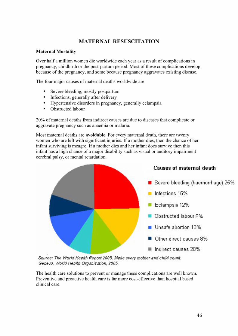
46
MATERNAL RESUSCITATION
Maternal Mortality
Over half a million women die worldwide each year as a result of complications in pregnancy, childbirth or the post-partum period. Most of these complications develop because of the pregnancy, and some because pregnancy aggravates existing disease.
The four major causes of maternal deaths worldwide are
• Severe bleeding, mostly postpartum • Infections, generally after delivery • Hypertensive disorders in pregnancy, generally eclampsia • Obstructed labour
20% of maternal deaths from indirect causes are due to diseases that complicate or aggravate pregnancy such as anaemia or malaria.
Most maternal deaths are avoidable. For every maternal death, there are twenty women who are left with significant injuries. If a mother dies, then the chance of her infant surviving is meagre. If a mother dies and her infant does survive then this infant has a high chance of a major disability such as visual or auditory impairment cerebral palsy, or mental retardation.
The health care solutions to prevent or manage these complications are well known. Preventive and proactive health care is far more cost-effective than hospital based clinical care.
47
Haemorrhage
Even a small amount of bleeding can cause haemorrhagic shock and death
How can we prevent or proactively manage obstetric bleeding?
• Antenatal care clinics are important and can check the mother’s haemoglobin level. Iron tablets are of great benefit to help prevent iron deficiency anaemia. The average blood loss from a normal vaginal delivery is 500 millilitres and that from a caesarean section is 1 litre. If a mother is anaemic at the time of delivery then any blood loss can be significant and cause increased morbidity. A large blood loss will increase her chance of dying.
• Provision of a skilled health professional at the time of birth to give parental oxytocic after the delivery, significantly decreases blood loss. After a vaginal delivery give an intramuscular injection of syntometrine (50 micrograms ergometrine and 5 international units syntocinon). If the mother is hypertensive give an intramuscular injection of 10 international units of syntocinon. For a caesarean section, after the delivery of the baby, give 5 international units of syntocinon intravenously.
How do we manage active bleeding?
• Early recognition of the clinical signs of bleeding is important so that an active plan of management can be instigated and help obtained from a team of professionals. The early clinical signs of bleeding include tachycardia and oliguria. As bleeding progresses the peripheral pulse becomes weak; the patient’s skin feels cold and clammy; and anuria can occur. A late sign of haemorrhage is hypotension because the blood pressure is actively defended until vasoconstriction can no longer compensate for the blood loss. If a patient has a gravid uterus then she must lie on her left lateral side to help promote venous return through the Inferior Vena Cava (IVC).
• An intravenous cannula is inserted and this needs to be a large calibre if possible. The maximum flow rate from a 20-gauge cannula is 60 millilitres a minute, from an 18-gauge is 105 millilitres a minute, from a 16-gauge is 215 millilitres a minute and from a 14-gauge is 325 millilitres a minute.
• The maximum flow rates through the cannula are achieved by elevating the height of the infusing fluid above the patient and using a pump set to help squeeze the fluid through the line.
• Normal saline is an excellent crystalloid fluid to infuse to compensate for volume loss. Balanced salt solution is an acceptable alternative but may contain calcium. Blood that is infused via the same line after administration of calcium containing fluid will potentially coagulate in the line.
• Fluids and blood need to be warmed through a blood warmer as they are infused. Cold fluids decrease the core temperature of the patient and this aggravates further bleeding through platelet dysfunction.
• If an urgent blood transfusion is required because the patient is shocked, give type O negative blood. This blood group is the universal donor. Keep giving this blood group until group specific blood or fully cross-matched blood is
48
available. A blood bank needs to be available to issue this blood 24 hours a day.
• Massive obstetric haemorrhage will require infusion of clotting factors in the form of fresh frozen plasma. Cryoprecipitate is required to replenish fibrinogen levels. Platelets may also need to be given, particularly in the hypertensive diseases of pregnancy - pre-eclampsia and the syndrome of “Haemolysis, Elevated Liver enzymes and Low Platelets”(HELLP).
• Insertion of a “Bakri” ballon into the uterus per vaginum can help tamponarde the uterus and reduce bleeding. The Bakri balloon is connected to a 3-way catheter with a port for insufflation with 600 millilitres of warm normal saline; a port for irrigation; and a port for drainage. The catheter can be used to help conserve the uterus, to reduce bleeding whist preparations are made for a hysterectomy or to transport the patient to another hospital that can provide the surgery needed.
• 1 milligram of misoprostol can be inserted into the vagina to help contract the uterus and reduce bleeding if the uterus can be conserved.
Infection
Even a small infection can cause septicaemia and death. How can we prevent or proactively manage obstetric infection?
• Prevent anaemia developing in the mother • Scrupulous aseptic techniques at the time of delivery • Scrupulous aseptic techniques at the time of caesarean section. The skin
preparation with disinfectant must be painted on the skin and allowed to dry before proceeding to the surgical cut. Surgical disinfectant solutions include those that contain iodine. Antibiotics are given intravenously as prophylactic protection against gram-positive organisms of the skin.
How do we actively manage infection?
• Parental antibiotics • Bacteraemia can develop into septicaemia. Septicaemia has a high mortality
rate as multi-organ failure can develop. Critical care facilities to support the patient will be required.
Hypertensive diseases of pregnancy
Even a small rise in blood pressure can cause eclampsia and death. How can we prevent or proactively manage hypertension?
• Antenatal clinics to regularly measure the blood pressure and check urine for the presence of protein using simple urine analysis. Treatment of pre-existing blood pressure is treated with medications during the pregnancy. Blood pressure normally decreases in the second trimester of pregnancy, so any increase in pressure must be closely monitored.
• Pre-eclampsia is defined as a diastolic blood pressure of greater than 90 mmHg and develops after 20 weeks gestation.
• Women with pre-eclampsia do not feel unwell until the condition becomes severe and then the disease is life threatening. The early signs are hypertension
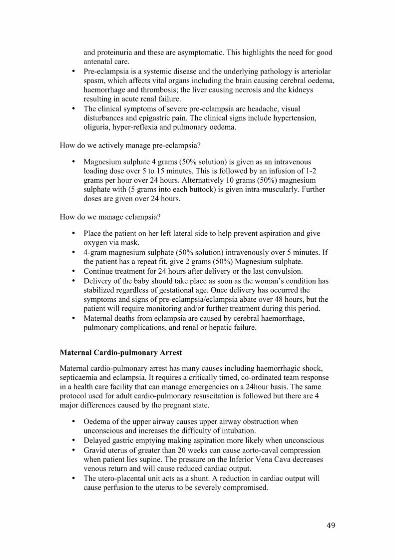
49
and proteinuria and these are asymptomatic. This highlights the need for good antenatal care.
• Pre-eclampsia is a systemic disease and the underlying pathology is arteriolar spasm, which affects vital organs including the brain causing cerebral oedema, haemorrhage and thrombosis; the liver causing necrosis and the kidneys resulting in acute renal failure.
• The clinical symptoms of severe pre-eclampsia are headache, visual disturbances and epigastric pain. The clinical signs include hypertension, oliguria, hyper-reflexia and pulmonary oedema.
How do we actively manage pre-eclampsia?
• Magnesium sulphate 4 grams (50% solution) is given as an intravenous loading dose over 5 to 15 minutes. This is followed by an infusion of 1-2 grams per hour over 24 hours. Alternatively 10 grams (50%) magnesium sulphate with (5 grams into each buttock) is given intra-muscularly. Further doses are given over 24 hours.
How do we manage eclampsia?
• Place the patient on her left lateral side to help prevent aspiration and give oxygen via mask.
• 4-gram magnesium sulphate (50% solution) intravenously over 5 minutes. If the patient has a repeat fit, give 2 grams (50%) Magnesium sulphate.
• Continue treatment for 24 hours after delivery or the last convulsion. • Delivery of the baby should take place as soon as the woman’s condition has
stabilized regardless of gestational age. Once delivery has occurred the symptoms and signs of pre-eclampsia/eclampsia abate over 48 hours, but the patient will require monitoring and/or further treatment during this period.
• Maternal deaths from eclampsia are caused by cerebral haemorrhage, pulmonary complications, and renal or hepatic failure.
Maternal Cardio-pulmonary Arrest
Maternal cardio-pulmonary arrest has many causes including haemorrhagic shock, septicaemia and eclampsia. It requires a critically timed, co-ordinated team response in a health care facility that can manage emergencies on a 24hour basis. The same protocol used for adult cardio-pulmonary resuscitation is followed but there are 4 major differences caused by the pregnant state.
• Oedema of the upper airway causes upper airway obstruction when unconscious and increases the difficulty of intubation.
• Delayed gastric emptying making aspiration more likely when unconscious • Gravid uterus of greater than 20 weeks can cause aorto-caval compression
when patient lies supine. The pressure on the Inferior Vena Cava decreases venous return and will cause reduced cardiac output.
• The utero-placental unit acts as a shunt. A reduction in cardiac output will cause perfusion to the uterus to be severely compromised.
50
The steps to follow in cardio-pulmonary resuscitation are
• Ensure your own safety and wear gloves and goggles. • Assess the level of consciousness of the patient. If the patient is unresponsive
and has abnormal or absent breathing then identify this as a cardiac arrest. Call for help, including an obstetrician and neonatologist.
• Place the patient in the left lateral position with a tilt of 15 – 30 degrees. This is imperative.
• Begin chest compressions at rate of 100 per minute and a depth of
compression of 5 centimetres. • Ensure the upper airway is patent. Protect the airway from aspiration
by early intubation if possible. • Simultaneous compressions and breaths are to be avoided. Rate of 30
compressions to 2 breaths. • 1 milligram of adrenaline is given every 3 minutes • Aorto-caval compression renders external cardiac compression
extremely difficult and the placental bed acts as a shunt, which further decreases tissue perfusion.
• Within 6 minutes of the cardiac arrest the mother will suffer irreversible damage and within 10 minutes the foetus will suffer irreversible damage.
• Within 5 minutes of commencing external cardiac compressions to a woman who is greater than 20 weeks gestation, a caesarean section (peri-mortem) must be performed to relieve the aorto-caval compression and potentially save the woman’s life.
• This is a rare event and 1 out of every 2 mothers who arrest will die.
51
Challenges ahead in Maternal Health in Mongolia
In 2008 the total population of Mongolia was 2.68 million people. The maternal mortality rate per 100,000 live births was 158 in the year 1998 and 49 in 2008. However there is a wide variation in the mortality rate between the urban and rural areas. In some rural and remote areas the mortality rate is 4 to 6 times greater than in Ulaanbaatar. This is very significant when one considers that 50% of the total population live in Ulaanbaatar and 40% live in the rural areas with a nomadic lifestyle. How do you deliver health services to remote areas with very low population density? How do you overcome the problems of poor transportation and communication, hampered by a severe climate, to deliver health services?
The migration from the rural areas to the big cities has imposed great socio-economic and health challenges on these areas. How do you deliver health services in the peri-urban Ger district outside Ulaanbaatar where there is unsafe water to drink and poor sanitation?
How do you educate and train physicians, nurses and midwives in health care prevention and promotion so that they can work at the primary care level?
How do you develop national articulated standards in health care, develop peer review and clinical audits, develop committees and staff designated to quality assurance, and develop protocols and clinical practice guidelines?
“Even a small illness can cause death”
Prevention and health promotion can decrease maternal mortality, decrease maternal morbidity and ultimately ensure the survival and wellbeing of their infants – the next generation.
References 1. Make every Mother Count. World Health Report. Year 2005 2. J.P Nolan et al. Executive summary 2010 International Consensus on
Cardiovascular Care Science with treatment recommendations. Resuscitation 81S. Year 2010; e1-e25
3. Country Cooperation Strategy. World Health Organisation Mongolia. Year 2010
4. Mongolia and WHO Cooperation and Collaboration. World Health Organization Western Pacific Region. Year 2011
52
DEFIBRILLATION
Cardiac defibrillation is indicated for the treatment of ventricular fibrillation or pulseless ventricular tachycardia. Early successful defibrillation is one of the few interventions that have been shown to improve outcome from VF/VT cardiac arrest.
For every minute that passes between collapse and attempted defibrillation, mortality increases 7-‐10% in the absence of cardiopulmonary resuscitation (CPR). If there is any delay in obtaining a defibrillator, start chest compressions and ventilation immediately. If CPR is provided, the decrease in survival form collapse to shock is more gradual (3-‐4% per minute).
Mechanism of defibrillation:
During defibrillation, a current is passed through the myocardium to depolarise a critical mass of the cardiac muscle simultaneously to allow the natural pacemaker tissue to resume control.
A defibrillator has 3 main features. A power source capable of providing a direct current, a capacitor that can be charged to a pre-‐determined energy level and two electrodes to place on the patient’s chest through which the capacitor discharges.
Successful defibrillation requires enough current to be delivered to the myocardium, but the actual delivered current is difficult to determine because it is influenced by thoracic impedance and the position of the electrodes. As little as 4% of the current actually reaches the heart.
Safe defibrillation:
Defibrillation can present a risk of electric shock to the rescuers, burns, sparking and fire. The hazard is increased in the presence of water, metal fixtures, flammable substances and oxygen. All bystanders and rescuers need to be clear of the patient during defibrillation.
Sparks from poorly applied defibrillator pads can cause a fire. The use of self-‐adhesive pads may minimise the risk of sparks. An open oxygen source should be removed to at least one metre from the patient. If a circuit is attached to an endotracheal tube, even at high flow, the danger is eliminated if it is not disconnected from the patient.
Foil-‐backed nitrate patches need to be removed from the patient’s chest to avoid arcing during defibrillation. Similarly, avoid placing the defibrillator pads over ECG electrodes and an implanted device. Permanent pacemakers and implanted defibrillators may have their circuits damaged if defibrillator electrodes are placed over them.
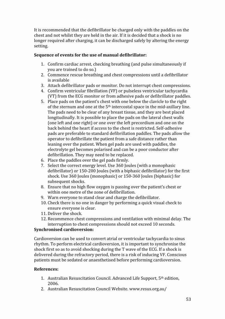
53
It is recommended that the defibrillator be charged only with the paddles on the chest and not whilst they are held in the air. If it is decided that a shock is no longer required after charging, it can be discharged safely by altering the energy setting.
Sequence of events for the use of manual defibrillator:
1. Confirm cardiac arrest, checking breathing (and pulse simultaneously if you are trained to do so.)
2. Commence rescue breathing and chest compressions until a defibrillator is available
3. Attach defibrillator pads or monitor. Do not interrupt chest compressions. 4. Confirm ventricular fibrillation (VF) or pulseless ventricular tachycardia
(VT) from the ECG monitor or from adhesive pads or defibrillator paddles. 5. Place pads on the patient’s chest with one below the clavicle to the right
of the sternum and one at the 5th intercostal space in the mid-‐axillary line. The pads need to be clear of any breast tissue, and they are best placed longitudinally. It is possible to place the pads on the lateral chest walls (one left and one right) or one over the left precordium and one on the back behind the heart if access to the chest is restricted. Self-‐adhesive pads are preferable to standard defibrillation paddles. The pads allow the operator to defibrillate the patient from a safe distance rather than leaning over the patient. When gel pads are used with paddles, the electrolyte gel becomes polarised and can be a poor conductor after defibrillation. They may need to be replaced.
6. Place the paddles over the gel pads firmly. 7. Select the correct energy level. Use 360 Joules (with a monophasic
defibrillator) or 150-‐200 Joules (with a biphasic defibrillator) for the first shock. Use 360 Joules (monophasic) or 150-‐360 Joules (biphasic) for subsequent shocks.
8. Ensure that no high flow oxygen is passing over the patient’s chest or within one metre of the zone of defibrillation.
9. Warn everyone to stand clear and charge the defibrillator. 10. Check there is no one in danger by performing a quick visual check to
ensure everyone is clear. 11. Deliver the shock. 12. Recommence chest compressions and ventilation with minimal delay. The
interruption to chest compressions should not exceed 10 seconds. Synchronised cardioversion:
Cardioversion can be used to convert atrial or ventricular tachycardia to sinus rhythm. To perform electrical cardioversion, it is important to synchronise the shock first so as to avoid shocking during the T wave of the ECG. If a shock is delivered during the refractory period, there is a risk of inducing VF. Conscious patients must be sedated or anaesthetised before performing cardioversion.
References:
1. Australian Resuscitation Council. Advanced Life Support, 5th edition, 2006.
2. Australian Resuscitation Council Website. www.resus.org.au/