Embed Size (px)
Citation preview
extensive metastatic disease. No adverse events were encountered. Meanfollow–up period of the 21 patients was 128 days (range, 3–263). Fourteenpatients have died of progressive disease (at days 3,26,41,45,83,86,118,125,150,150,156,174,196 and 210), 3 remain alive (at days 239,250and 263) and 4 were lost to follow–up (at days 89,90,92 and116). Tumoringrowth or stent occlusion was not observed in any patient during thisfollow–up period.Conclusions: The Wilson–Cook Za metal stent was effective in palliationof inoperable malignant biliary tumors in all patients. Technical problemswith stent deployment were encountered. Overall stent patency, efficacy,and safety profile appear excellent.
189
LABORATORY PREDICTORS OF CHOLEDOCHOLITHIASISPRIOR TO CHOLECYSTECTOMYNejat Kiyici, M.D., Pallavi Agarwal, M.D., Tanaya Nayak, M.D.,Edward Norkus, M.D. and Hilary Hertan, M.D.*. Division ofGastroenterology and Clinical Nutrition, Our Lady of Mercy MedicalCenter, Bronx, NY.
Purpose: To identify laboratory predictors of common bile duct stones bynoninvasive methods prior to cholecystectomy.Methods: In this retrospective study, charts were reviewed in 71 patientswho had ERCP for evaluation of suspected common bile duct (CBD)stone.Patient demographics, admission vital signs, presenting symptoms,comorbid conditions and admission laboratory values were analyzed.Ad-mission and pre–ERCP laboratory values were compared.The mean inter-val between admission date and performance of ERCP was 4.6 days.Sono-gram (US), ERCP, and CT scan findings were noted.CBD diameter of �8mm was considered dilated.Results: CBD stones were identified by ERCP in 37 of 71 patients.Ad-mission ALT level �150 U/L (p�0.003) and 40 % increase in WBC countprior to ERCP (p�0.005) were strong predictors of CBD stones.Chance ofCBD stone increased 26.6 fold whenWBC count increased prior to ERCPand 6.2 fold when admission ALT was �150 U/L.Dilated CBD wasidentified only in 58 % of the patients who underwent US testing.Sensi-tivity was 55 %.When normal CBD size was detected by US, 50 % hadCBD stones.
CBD stonen � 37
No CBD stonen � 34 P–value
Age 63 � 20.2 55.3 � 19 0.0750Sex (F/M) 24/12 20/14 0.622ALT � 150 26 15 0.003Change in WBC 40% increase 30% decrease 0.0225
Conclusions: Admission level of ALT �150 U/L and increasing WBCcount during hospital stay appear to be very strong predictors of CBD stoneprior to cholecystectomy.These predictors may aid in the selection of thepatients who need ERCP prior to cholecystectomy.
190
THE EFFECT OF LIDOCAINE SPRAYED ON THE AMPULLAOF VATER ON THE INCIDENCE OF ERCP ACUTEPANCREATITISJeremy J. Schwartz, M.D., Ronald J. Lew, M.D., Nuzhat A. Ahmad,M.D., Gregory G. Ginsberg, M.D., Michael L. Kochman, M.D., ColleenBrensinger, M.S. and William B. Long, M.D.*. Division ofGastroenterology, University of Pennsylvania, School of Medicine,Philadelphia, PA.
Purpose: To determine whether lidocaine sprayed on the ampulla of Vaterreduces the incidence or severity of ERCP acute pancreatitis (AP).Methods: Prospective, randomized, triple–blinded, placebo–controlledtrial with concealed allocation. Patients aged 18 years or older with anintact ampulla undergoing ERCP at our institution were invited to partic-
ipate. Demographics, historical data, and ERCP indications were recorded.Patients were randomized to have 10 cc of either 1% lidocaine or normalsaline sprayed on the ampulla prior to cannulation. ERCP medications,findings, and therapies were recorded. The attending rated difficulty ofcannulation (easy, average, difficult, unsuccessful). Patients were followedby telephone interview or bedside visit. For patients who developed ERCPAP (abdominal pain beginning within 24 hours post ERCP and serumamylase � 3X normal 24� hours post ERCP) had severity (hospitalizationin days: 1–3 � mild, 4–10 � moderate, � 10 days or organ failure,necrosis, or death � severe) and complication data recorded. The study wasapproved by the IRB.Results: 326 patients were enrolled of whom 32 were excluded prior toanalysis. Of the 294 patients undergoing follow–up, 145 were randomizedto lidocaine and 149 to placebo. No patients were lost to follow–up. Nosignificant differences were noted in demographics, historical variables,ERCP indication, findings, medications, cannulation difficulty, or endo-scopic therapies between the groups. 7 patients in the lidocaine group and5 patients in the placebo group developed ERCP AP (p�0.52). There wasa trend (p�0.08) toward increased severity of ERCP AP in the placebogroup with both severe cases of ERCP AP and the 1 death occurring in theplacebo group. Factors associated with an increased risk of post–ERCP APincluded increased difficulty of biliary (p�0.004) or pancreatic cannulation(p�0.018), use of � 0.5 mg glucagon during ERCP (p�0.046), andperformance of biliary sphincterotomy (p�0.0525). On multivariate anal-ysis, the use of glucagon was not associated with an increased risk of ERCPAP.Conclusions: 10 cc of 1% lidocaine sprayed on the ampulla was not foundto lower the incidence of ERCP AP. The significance of the trend towardincreased severity of ERCP AP in the placebo group is unclear.
191
RESPONSE RATE AND PREDICTORS OF RESPONSE TOCHOLECYSTECTOMY IN BILIARY DYSKINESIAAmit Rastogi, M.D., Rodrigo A. Rodriguex, M.D., Robert E. Schoen,M.D. and Adam Slivka, M.D.*. Gastroenterology, Hepatology andNutrition, University of Pittsburgh Medical Center, Pittsburgh, PA.
Purpose: Upper abdominal pain without cholelithiasis and a negative workup for other common causes of abdominal pain is a vexing problem.Cholecystokinin HIDA scan (CCK HIDA) has been used to diagnosefunctional gall bladder (GB) disease (biliary dyskinesia). Cholecystectomyis recommended if the GB ejection fraction (GB EF) is � 35%. Aim wasto evaluate the response to cholecystectomy in patients (pts) diagnosed withbiliary dyskinesia and to identify clinical, laboratory or radiological pa-rameters that might predict response to cholecystectomy.Methods: Retrospectively 40 pts with biliary dyskinesia (GB EF � 35%)were identified between 01/1996 and 12/2001. The clinical parametersnoted were age, sex, site and periodicity of pain, relation to meals, radiationof pain, reproducibility of pain with CCK HIDA. Laboratory parametersincluded AST, ALT, bilirubin, amylase, lipase. Results of ultrasound, GBEF on CCK HIDA and pathology of the resected GB were noted. Subjectswere contacted a mean of 25 months post cholecystectomy by an unin-volved physician to assess abdominal pain response. Response was gradedas Complete, Partial or No response.
Response to cholecystectomy in relation to baseline variables was eval-uated with Fisher’s exact test and T Test.Results: Of 40 patients, 6 were lost to follow up and were excluded. Themean age was 41 years. 65% were females . At baseline, abdominal painwas episodic in 26 pts and continuous in 8 . It was right sided in 11, midlinein 21 and left sided in 2 pts . Increased symptoms with meals was presentin 28 pts . Pain was reproduced with CCK HIDA in 21 pts . The mean GBEF was 19% . The mean duration of follow up post cholecystectomy was25 months( range 3–66 months). 15 pts had a complete response (44%), 12had a partial response (35%) and 7 had no response (21%). There was nostatistically significant association between any clinical, laboratory or ra-diological variable and response to cholecystectomy.
S63AJG – September, Suppl., 2002 Abstracts
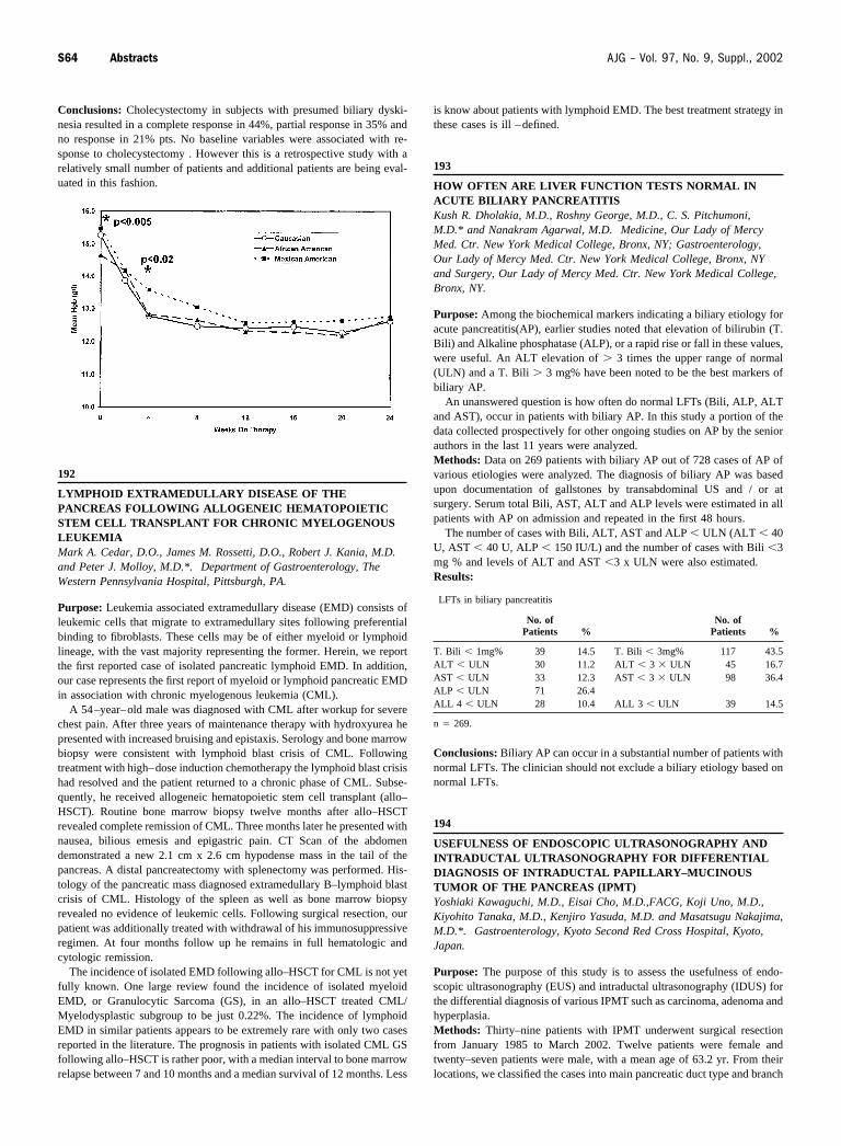
Conclusions: Cholecystectomy in subjects with presumed biliary dyski-nesia resulted in a complete response in 44%, partial response in 35% andno response in 21% pts. No baseline variables were associated with re-sponse to cholecystectomy . However this is a retrospective study with arelatively small number of patients and additional patients are being eval-uated in this fashion.
192
LYMPHOID EXTRAMEDULLARY DISEASE OF THEPANCREAS FOLLOWING ALLOGENEIC HEMATOPOIETICSTEM CELL TRANSPLANT FOR CHRONIC MYELOGENOUSLEUKEMIAMark A. Cedar, D.O., James M. Rossetti, D.O., Robert J. Kania, M.D.and Peter J. Molloy, M.D.*. Department of Gastroenterology, TheWestern Pennsylvania Hospital, Pittsburgh, PA.
Purpose: Leukemia associated extramedullary disease (EMD) consists ofleukemic cells that migrate to extramedullary sites following preferentialbinding to fibroblasts. These cells may be of either myeloid or lymphoidlineage, with the vast majority representing the former. Herein, we reportthe first reported case of isolated pancreatic lymphoid EMD. In addition,our case represents the first report of myeloid or lymphoid pancreatic EMDin association with chronic myelogenous leukemia (CML).
A 54–year–old male was diagnosed with CML after workup for severechest pain. After three years of maintenance therapy with hydroxyurea hepresented with increased bruising and epistaxis. Serology and bone marrowbiopsy were consistent with lymphoid blast crisis of CML. Followingtreatment with high–dose induction chemotherapy the lymphoid blast crisishad resolved and the patient returned to a chronic phase of CML. Subse-quently, he received allogeneic hematopoietic stem cell transplant (allo–HSCT). Routine bone marrow biopsy twelve months after allo–HSCTrevealed complete remission of CML. Three months later he presented withnausea, bilious emesis and epigastric pain. CT Scan of the abdomendemonstrated a new 2.1 cm x 2.6 cm hypodense mass in the tail of thepancreas. A distal pancreatectomy with splenectomy was performed. His-tology of the pancreatic mass diagnosed extramedullary B–lymphoid blastcrisis of CML. Histology of the spleen as well as bone marrow biopsyrevealed no evidence of leukemic cells. Following surgical resection, ourpatient was additionally treated with withdrawal of his immunosuppressiveregimen. At four months follow up he remains in full hematologic andcytologic remission.
The incidence of isolated EMD following allo–HSCT for CML is not yetfully known. One large review found the incidence of isolated myeloidEMD, or Granulocytic Sarcoma (GS), in an allo–HSCT treated CML/Myelodysplastic subgroup to be just 0.22%. The incidence of lymphoidEMD in similar patients appears to be extremely rare with only two casesreported in the literature. The prognosis in patients with isolated CML GSfollowing allo–HSCT is rather poor, with a median interval to bone marrowrelapse between 7 and 10 months and a median survival of 12 months. Less
is know about patients with lymphoid EMD. The best treatment strategy inthese cases is ill –defined.
193
HOW OFTEN ARE LIVER FUNCTION TESTS NORMAL INACUTE BILIARY PANCREATITISKush R. Dholakia, M.D., Roshny George, M.D., C. S. Pitchumoni,M.D.* and Nanakram Agarwal, M.D. Medicine, Our Lady of MercyMed. Ctr. New York Medical College, Bronx, NY; Gastroenterology,Our Lady of Mercy Med. Ctr. New York Medical College, Bronx, NYand Surgery, Our Lady of Mercy Med. Ctr. New York Medical College,Bronx, NY.
Purpose: Among the biochemical markers indicating a biliary etiology foracute pancreatitis(AP), earlier studies noted that elevation of bilirubin (T.Bili) and Alkaline phosphatase (ALP), or a rapid rise or fall in these values,were useful. An ALT elevation of � 3 times the upper range of normal(ULN) and a T. Bili � 3 mg% have been noted to be the best markers ofbiliary AP.
An unanswered question is how often do normal LFTs (Bili, ALP, ALTand AST), occur in patients with biliary AP. In this study a portion of thedata collected prospectively for other ongoing studies on AP by the seniorauthors in the last 11 years were analyzed.Methods: Data on 269 patients with biliary AP out of 728 cases of AP ofvarious etiologies were analyzed. The diagnosis of biliary AP was basedupon documentation of gallstones by transabdominal US and / or atsurgery. Serum total Bili, AST, ALT and ALP levels were estimated in allpatients with AP on admission and repeated in the first 48 hours.
The number of cases with Bili, ALT, AST and ALP � ULN (ALT � 40U, AST � 40 U, ALP � 150 IU/L) and the number of cases with Bili �3mg % and levels of ALT and AST �3 x ULN were also estimated.Results:
LFTs in biliary pancreatitis
No. ofPatients %
No. ofPatients %
T. Bili � 1mg% 39 14.5 T. Bili � 3mg% 117 43.5ALT � ULN 30 11.2 ALT � 3 � ULN 45 16.7AST � ULN 33 12.3 AST � 3 � ULN 98 36.4ALP � ULN 71 26.4ALL 4 � ULN 28 10.4 ALL 3 � ULN 39 14.5
n � 269.
Conclusions: Biliary AP can occur in a substantial number of patients withnormal LFTs. The clinician should not exclude a biliary etiology based onnormal LFTs.
194
USEFULNESS OF ENDOSCOPIC ULTRASONOGRAPHY ANDINTRADUCTAL ULTRASONOGRAPHY FOR DIFFERENTIALDIAGNOSIS OF INTRADUCTAL PAPILLARY–MUCINOUSTUMOR OF THE PANCREAS (IPMT)Yoshiaki Kawaguchi, M.D., Eisai Cho, M.D.,FACG, Koji Uno, M.D.,Kiyohito Tanaka, M.D., Kenjiro Yasuda, M.D. and Masatsugu Nakajima,M.D.*. Gastroenterology, Kyoto Second Red Cross Hospital, Kyoto,Japan.
Purpose: The purpose of this study is to assess the usefulness of endo-scopic ultrasonography (EUS) and intraductal ultrasonography (IDUS) forthe differential diagnosis of various IPMT such as carcinoma, adenoma andhyperplasia.Methods: Thirty–nine patients with IPMT underwent surgical resectionfrom January 1985 to March 2002. Twelve patients were female andtwenty–seven patients were male, with a mean age of 63.2 yr. From theirlocations, we classified the cases into main pancreatic duct type and branch
S64 Abstracts AJG – Vol. 97, No. 9, Suppl., 2002


![Left Sided Laparoscopic Cholecystectomy: Case Report and ...open cholecystectomy - before laparoscopic era [2] and 1 case in 2008 [3] and about 50 cases of laparoscopic cholecystectomy](https://img.pdfslide.us/doc/110x75/5f6509906579645fd7227a11/left-sided-laparoscopic-cholecystectomy-case-report-and-open-cholecystectomy.jpg)