Embed Size (px)
Citation preview
© 2008
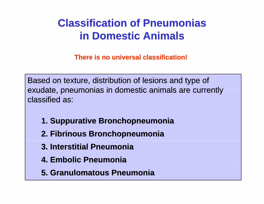
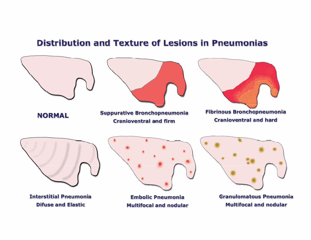
Classification of PneumoniasClassification of Pneumoniasin Domestic Animalsin Domestic Animalsin Domestic Animalsin Domestic Animals
There is no universal classification!There is no universal classification!
Based on texture, distribution of lesions and type of Based on texture, distribution of lesions and type of exudate pneumonias in domestic animals are currentlyexudate pneumonias in domestic animals are currentlyexudate, pneumonias in domestic animals are currently exudate, pneumonias in domestic animals are currently classified as:classified as:
1. Suppurative BronchopneumoniaSuppurative Bronchopneumonia2.2. Fibrinous BronchopneumoniaFibrinous Bronchopneumonia3.3. Interstitial PneumoniaInterstitial Pneumonia4.4. Embolic PneumoniaEmbolic Pneumonia5.5. Granulomatous PneumoniaGranulomatous Pneumonia
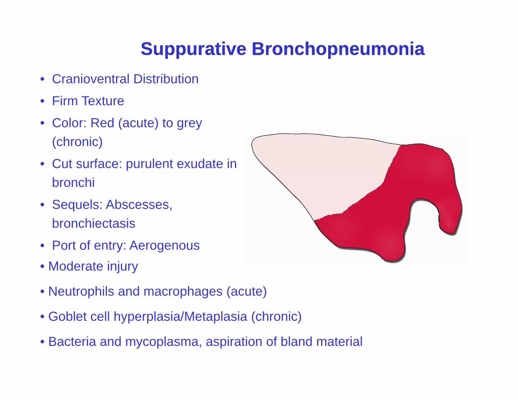
Suppurative BronchopneumoniaSuppurative BronchopneumoniaC i l Di ib i• Cranioventral Distribution
• Firm Texture
• Color: Red (acute) to greyColor: Red (acute) to grey (chronic)
• Cut surface: purulent exudate in bronchi
• Sequels: Abscesses, bronchiectasisbronchiectasis
• Port of entry: Aerogenous• Moderate injury
• Neutrophils and macrophages (acute)
• Goblet cell hyperplasia/Metaplasia (chronic)
• Bacteria and mycoplasma, aspiration of bland material
NNNN
CC
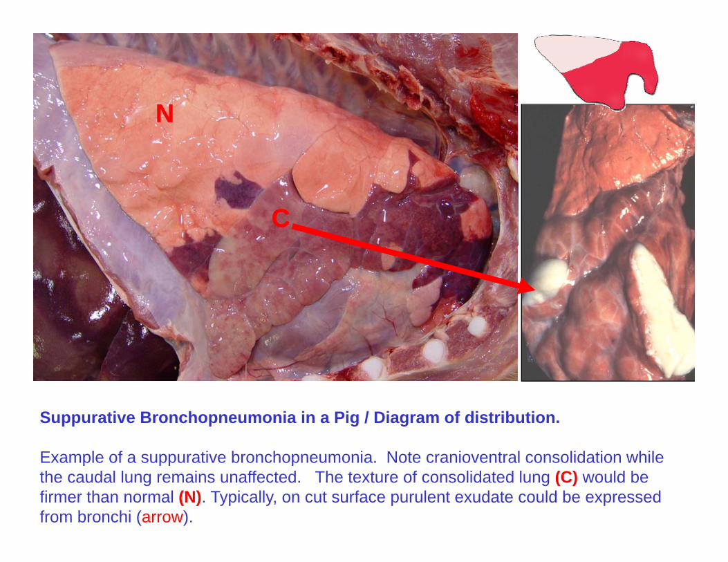
Suppurative Bronchopneumonia in a Pig / Diagram of distribution.
Example of a suppurative bronchopneumonia Note cranioventral consolidation whileExample of a suppurative bronchopneumonia. Note cranioventral consolidation while the caudal lung remains unaffected. The texture of consolidated lung (C) would be firmer than normal (N). Typically, on cut surface purulent exudate could be expressed from bronchi (arrow).
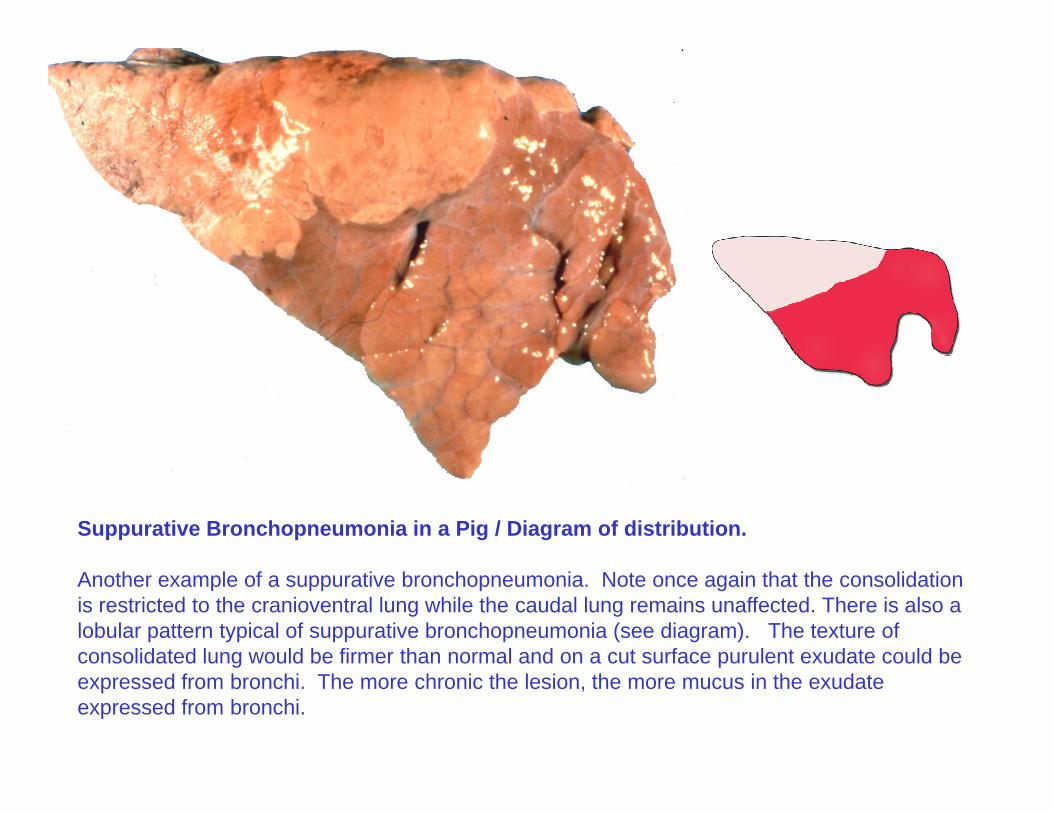
Suppurative Bronchopneumonia in a Pig / Diagram of distribution.
Another example of a suppurative bronchopneumonia. Note once again that the consolidation is restricted to the cranioventral lung while the caudal lung remains unaffected. There is also a lobular pattern typical of suppurative bronchopneumonia (see diagram). The texture of consolidated lung would be firmer than normal and on a cut surface purulent exudate could beconsolidated lung would be firmer than normal and on a cut surface purulent exudate could be expressed from bronchi. The more chronic the lesion, the more mucus in the exudate expressed from bronchi.
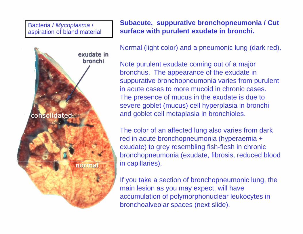
Bacteria / Mycoplasma / aspiration of bland material
Subacute, suppurative bronchopneumonia / Cut surface with purulent exudate in bronchi.
Normal (light color) and a pneumonic lung (dark red).
Note purulent exudate coming out of a major bronchus The appearance of the exudate inbronchus. The appearance of the exudate in suppurative bronchopneumonia varies from purulent in acute cases to more mucoid in chronic cases. The presence of mucus in the exudate is due to severe goblet (mucus) cell hyperplasia in bronchi and goblet cell metaplasia in bronchioles.
The color of an affected lung also varies from darkThe color of an affected lung also varies from dark red in acute bronchopneumonia (hyperaemia + exudate) to grey resembling fish-flesh in chronic bronchopneumonia (exudate, fibrosis, reduced blood i ill i )in capillaries).
If you take a section of bronchopneumonic lung, the main lesion as you may expect, will have y y p ,accumulation of polymorphonuclear leukocytes in bronchoalveolar spaces (next slide).
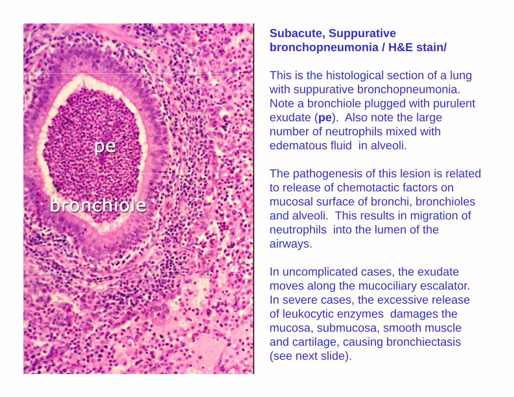
Subacute, Suppurative bronchopneumonia / H&E stain/
This is the histological section of a lungThis is the histological section of a lung with suppurative bronchopneumonia. Note a bronchiole plugged with purulent exudate (pe). Also note the large (p ) gnumber of neutrophils mixed with edematous fluid in alveoli.
The pathogenesis of this lesion is relatedThe pathogenesis of this lesion is related to release of chemotactic factors on mucosal surface of bronchi, bronchioles and alveoli. This results in migration of neutrophils into the lumen of the airways.
In uncomplicated cases the exudateIn uncomplicated cases, the exudate moves along the mucociliary escalator. In severe cases, the excessive release of leukocytic enzymes damages the
b th lmucosa, submucosa, smooth muscle and cartilage, causing bronchiectasis (see next slide).
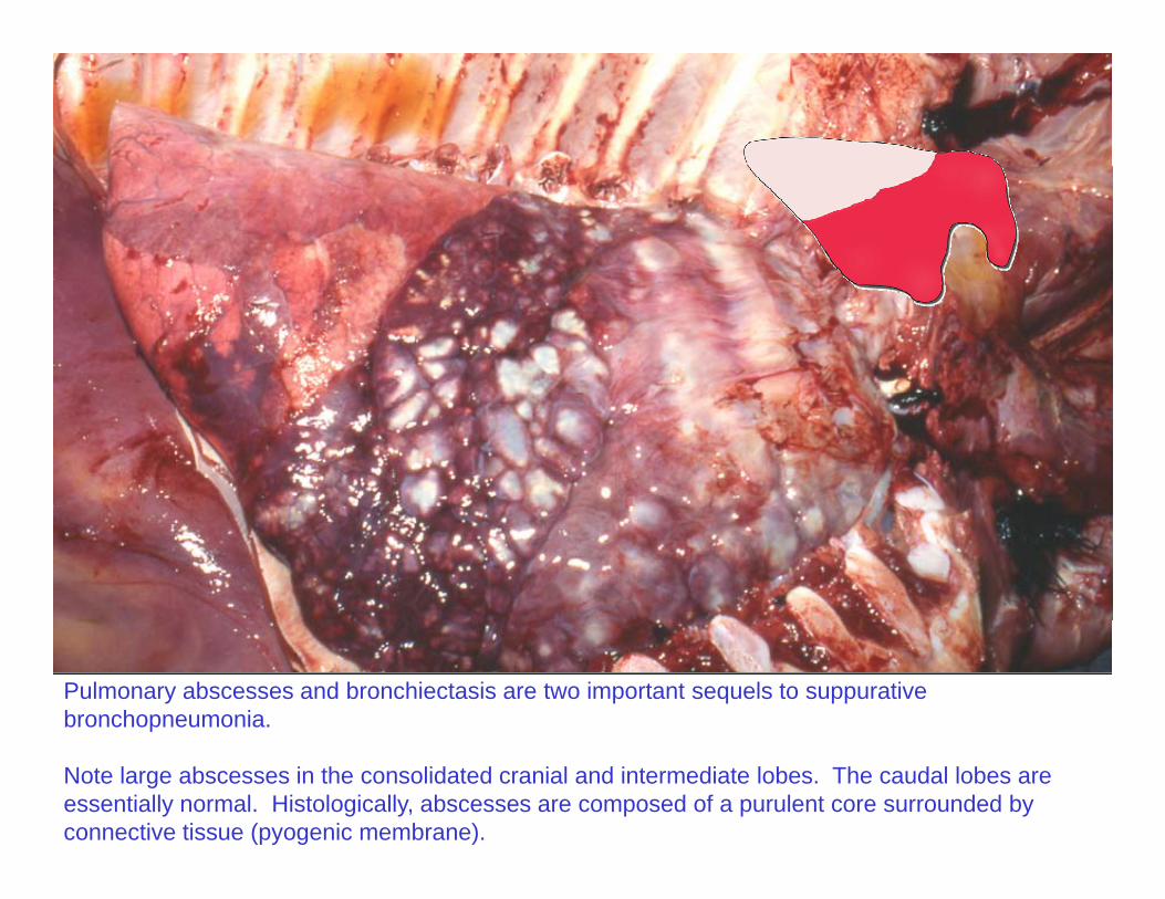
Pulmonary abscesses and bronchiectasis are two important sequels to suppurative bronchopneumonia.
Note large abscesses in the consolidated cranial and intermediate lobes. The caudal lobes are essentially normal. Histologically, abscesses are composed of a purulent core surrounded by connective tissue (pyogenic membrane).
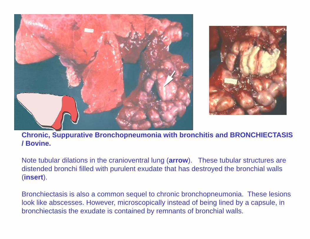
Chronic Suppurative Bronchopneumonia with bronchitis and BRONCHIECTASISChronic, Suppurative Bronchopneumonia with bronchitis and BRONCHIECTASIS / Bovine.
Note tubular dilations in the cranioventral lung (arrow). These tubular structures are distended bronchi filled with purulent exudate that has destroyed the bronchial walls (insert).
Bronchiectasis is also a common sequel to chronic bronchopneumonia These lesionsBronchiectasis is also a common sequel to chronic bronchopneumonia. These lesions look like abscesses. However, microscopically instead of being lined by a capsule, in bronchiectasis the exudate is contained by remnants of bronchial walls.
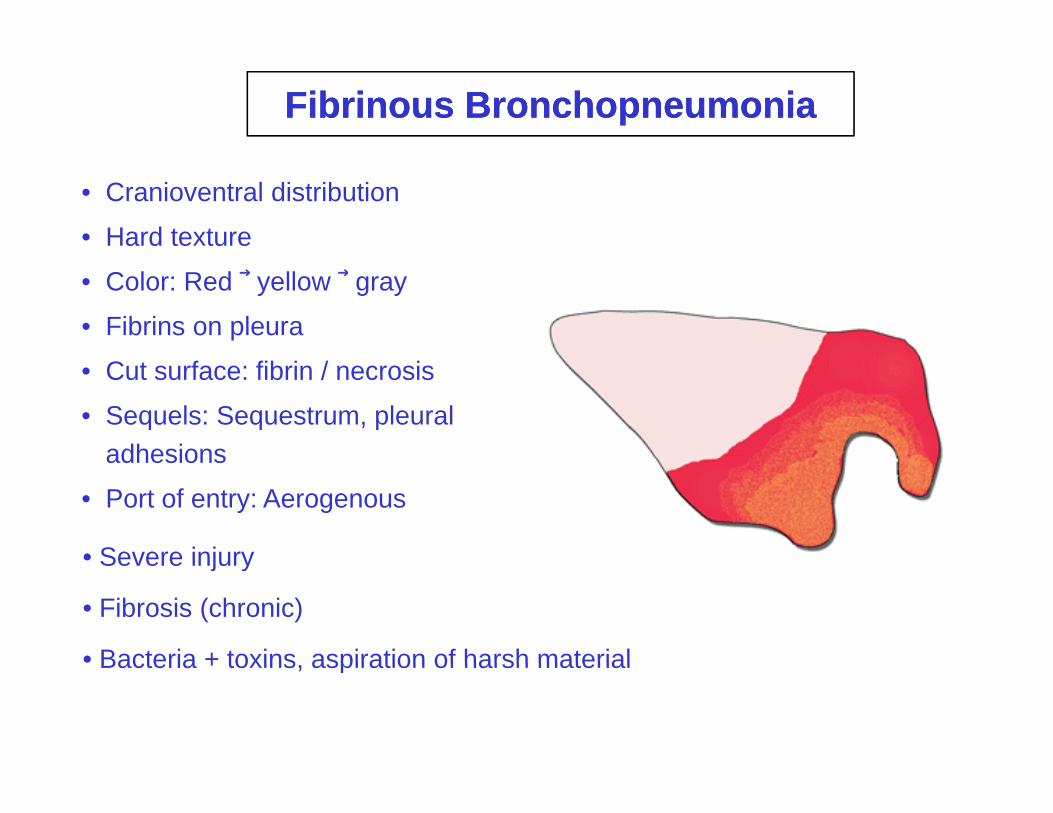
Fibrinous BronchopneumoniaFibrinous Bronchopneumonia
• Cranioventral distribution
• Hard texture
• Color: Red yellow gray
• Fibrins on pleura
• Cut surface: fibrin / necrosis
• Sequels: Sequestrum, pleural adhesionsadhesions
• Port of entry: Aerogenous
• Severe injurySevere injury
• Fibrosis (chronic)
• Bacteria + toxins aspiration of harsh materialBacteria + toxins, aspiration of harsh material
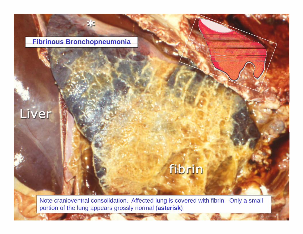
Fibrinous BronchopneumoniaFibrinous Bronchopneumonia
Note cranioventral consolidation. Affected lung is covered with fibrin. Only a small portion of the lung appears grossly normal (asterisk)
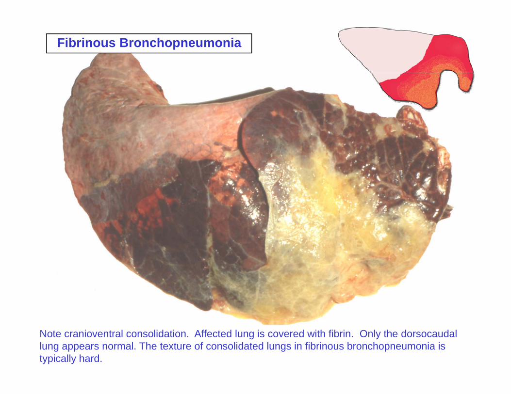
Fibrinous Bronchopneumonia
Note cranioventral consolidation. Affected lung is covered with fibrin. Only the dorsocaudal lung appears normal. The texture of consolidated lungs in fibrinous bronchopneumonia is typically hard.
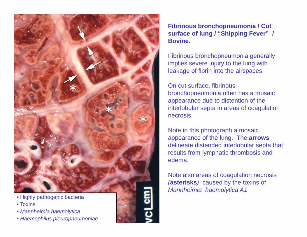
Fibrinous bronchopneumonia / Cut surface of lung / “Shipping Fever” / Bovine.
Fibrinous bronchopneumonia generally implies severe injury to the lung with leakage of fibrin into the airspaces. g p
On cut surface, fibrinous bronchopneumonia often has a mosaic appearance due to distention of the ppinterlobular septa in areas of coagulation necrosis.
Note in this photograph a mosaic p g pappearance of the lung. The arrows delineate distended interlobular septa that results from lymphatic thrombosis and edema.
Note also areas of coagulation necrosis (asterisks) caused by the toxins of Mannheimia haemolytica A1
• Highly pathogenic bacteria• Toxins• Mannheimia haemolytica• Haemophilus pleuropneumoniae
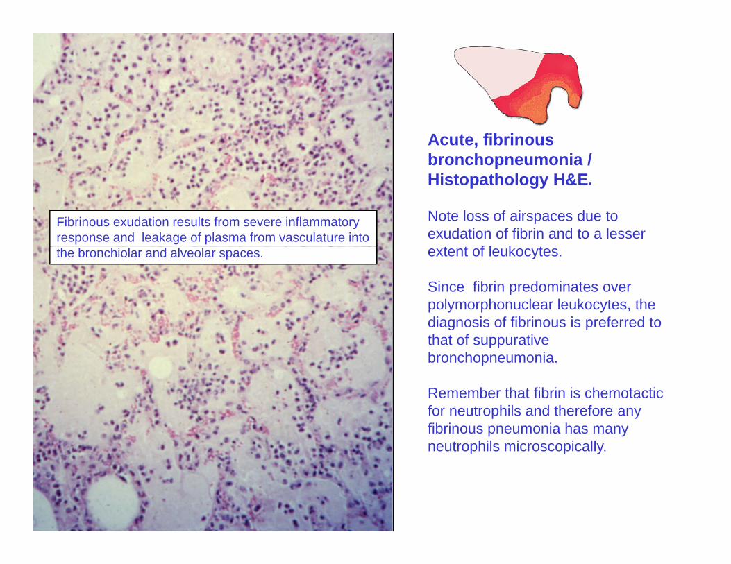
Acute, fibrinous bronchopneumonia / Hi t th l H&EHistopathology H&E.
Note loss of airspaces due to exudation of fibrin and to a lesser
t t f l k t
Fibrinous exudation results from severe inflammatory response and leakage of plasma from vasculature into
extent of leukocytes.
Since fibrin predominates over polymorphonuclear leukocytes, the di i f fib i i f d t
the bronchiolar and alveolar spaces.
diagnosis of fibrinous is preferred to that of suppurative bronchopneumonia.
R b th t fib i i h t tiRemember that fibrin is chemotacticfor neutrophils and therefore any fibrinous pneumonia has many neutrophils microscopically.
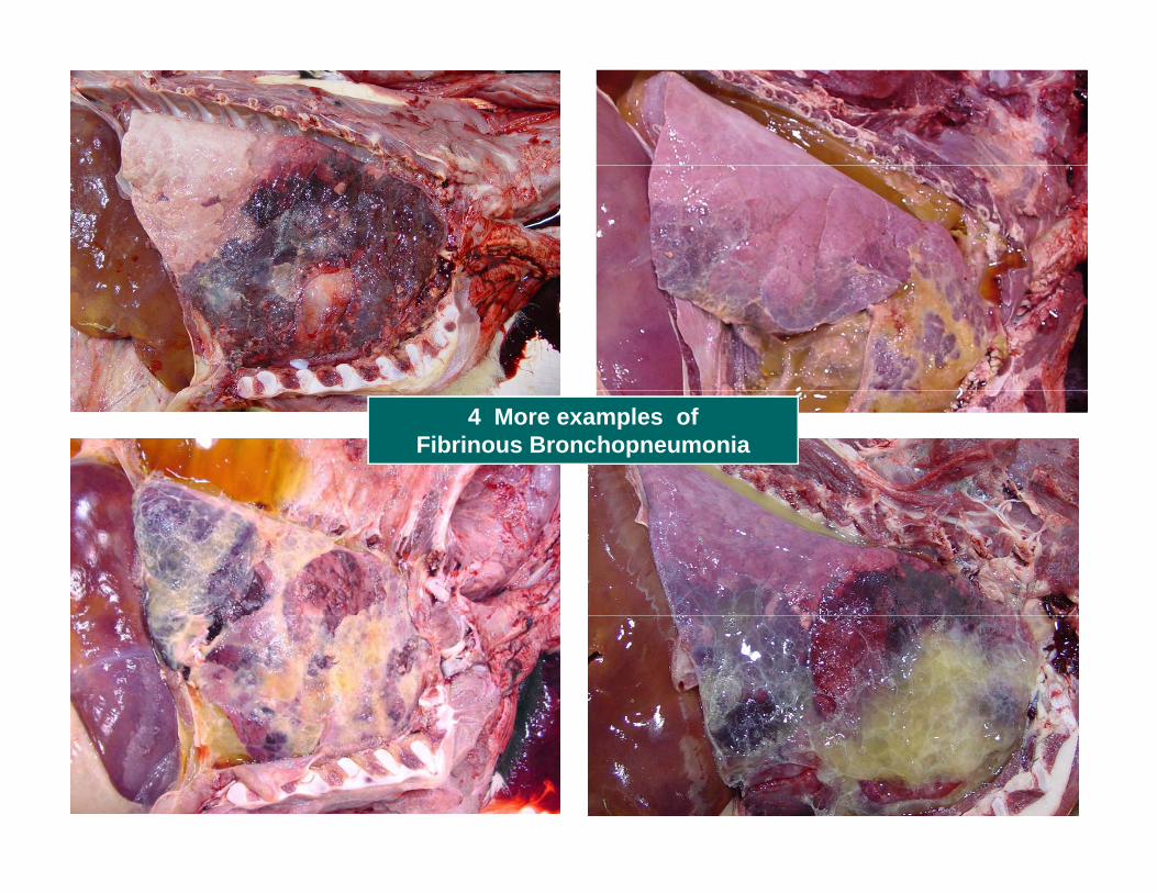
4 More examples ofFibrinous Bronchopneumonia
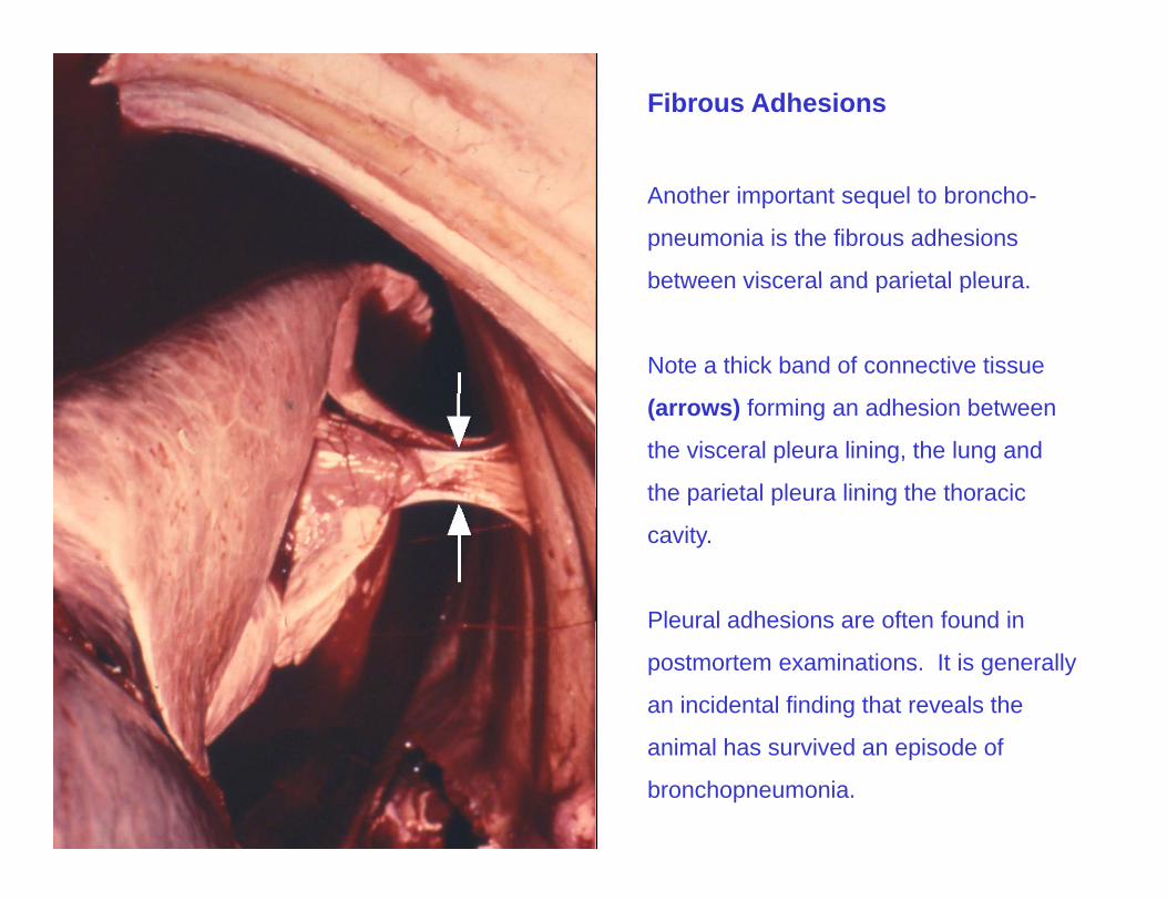
Fibrous Adhesions
Another important sequel to broncho-
pneumonia is the fibrous adhesions
between visceral and parietal pleurabetween visceral and parietal pleura.
Note a thick band of connective tissue
(arrows) forming an adhesion between
the visceral pleura lining, the lung and
the parietal pleura lining the thoracic
cavity.
Pleural adhesions are often found inPleural adhesions are often found in
postmortem examinations. It is generally
an incidental finding that reveals the
animal has survived an episode of
bronchopneumonia.
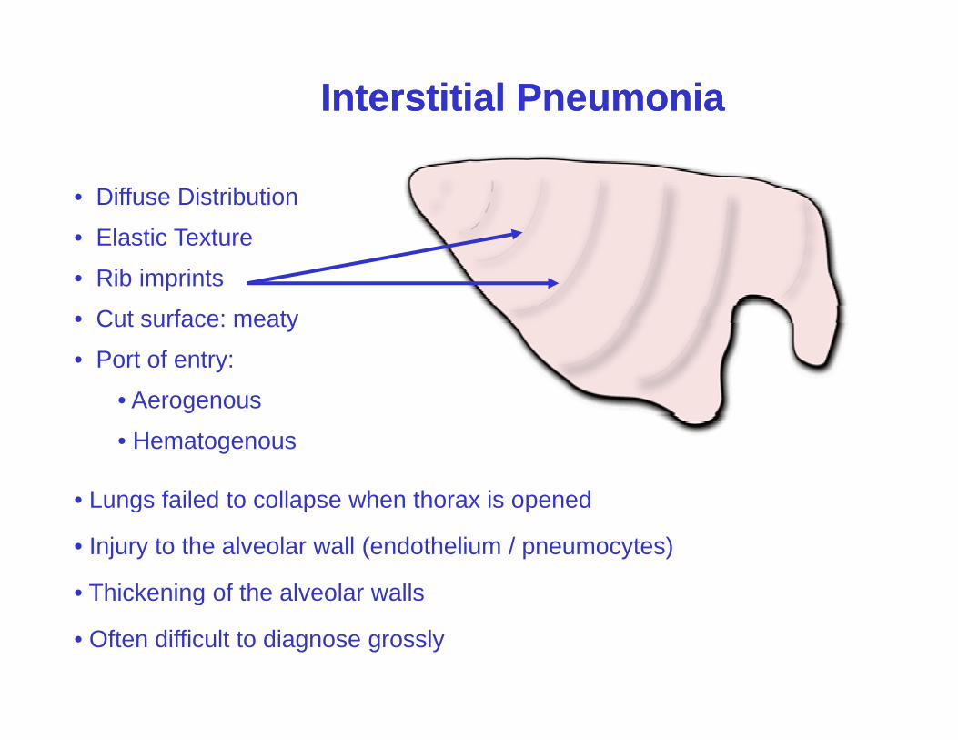
Interstitial PneumoniaInterstitial Pneumonia
• Diffuse Distribution
El ti T t• Elastic Texture
• Rib imprints
• Cut surface: meatyCut surface: meaty
• Port of entry:
• Aerogenous
• Hematogenous
• Lungs failed to collapse when thorax is openedg p p
• Injury to the alveolar wall (endothelium / pneumocytes)
• Thickening of the alveolar wallsg
• Often difficult to diagnose grossly
Interstitial Pneumonia
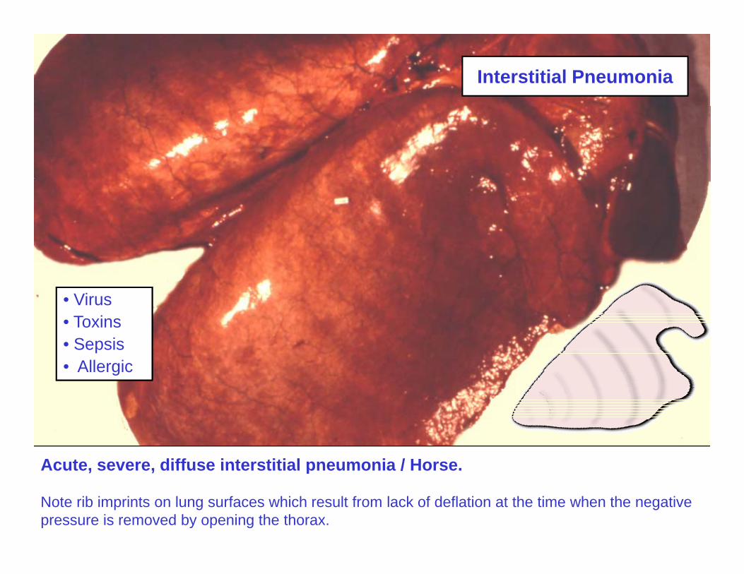
• Virus• Toxins• Toxins• Sepsis• Allergic
Acute severe diffuse interstitial pneumonia / HorseAcute, severe, diffuse interstitial pneumonia / Horse.
Note rib imprints on lung surfaces which result from lack of deflation at the time when the negative pressure is removed by opening the thorax.
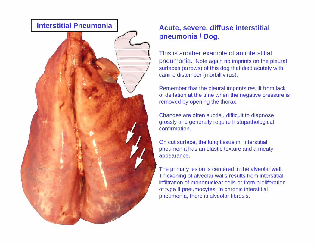
Acute, severe, diffuse interstitial pneumonia / Dog.
Interstitial Pneumonia
This is another example of an interstitial pneumonia. Note again rib imprints on the pleural surfaces (arrows) of this dog that died acutely with canine distemper (morbillivirus).canine distemper (morbillivirus).
Remember that the pleural imprints result from lack of deflation at the time when the negative pressure is removed by opening the thorax.
Changes are often subtle , difficult to diagnose grossly and generally require histopathological confirmation.
On cut surface, the lung tissue in interstitial pneumonia has an elastic texture and a meaty appearance.
The primary lesion is centered in the alveolar wallThe primary lesion is centered in the alveolar wall. Thickening of alveolar walls results from interstitial infiltration of mononuclear cells or from proliferation of type II pneumocytes. In chronic interstitial pneumonia, there is alveolar fibrosis.
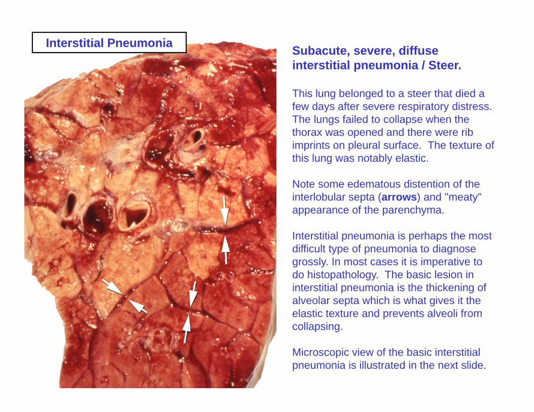
Interstitial Pneumonia Subacute, severe, diffuse interstitial pneumonia / Steer.
This lung belonged to a steer that died a few days after severe respiratory distress. The lungs failed to collapse when the h d d h ibthorax was opened and there were rib imprints on pleural surface. The texture of this lung was notably elastic.
N t d t di t ti f thNote some edematous distention of the interlobular septa (arrows) and "meaty” appearance of the parenchyma.
I t titi l i i h th tInterstitial pneumonia is perhaps the most difficult type of pneumonia to diagnose grossly. In most cases it is imperative to do histopathology. The basic lesion in i t titi l i i th thi k i finterstitial pneumonia is the thickening of alveolar septa which is what gives it the elastic texture and prevents alveoli from collapsing.
Microscopic view of the basic interstitial pneumonia is illustrated in the next slide.
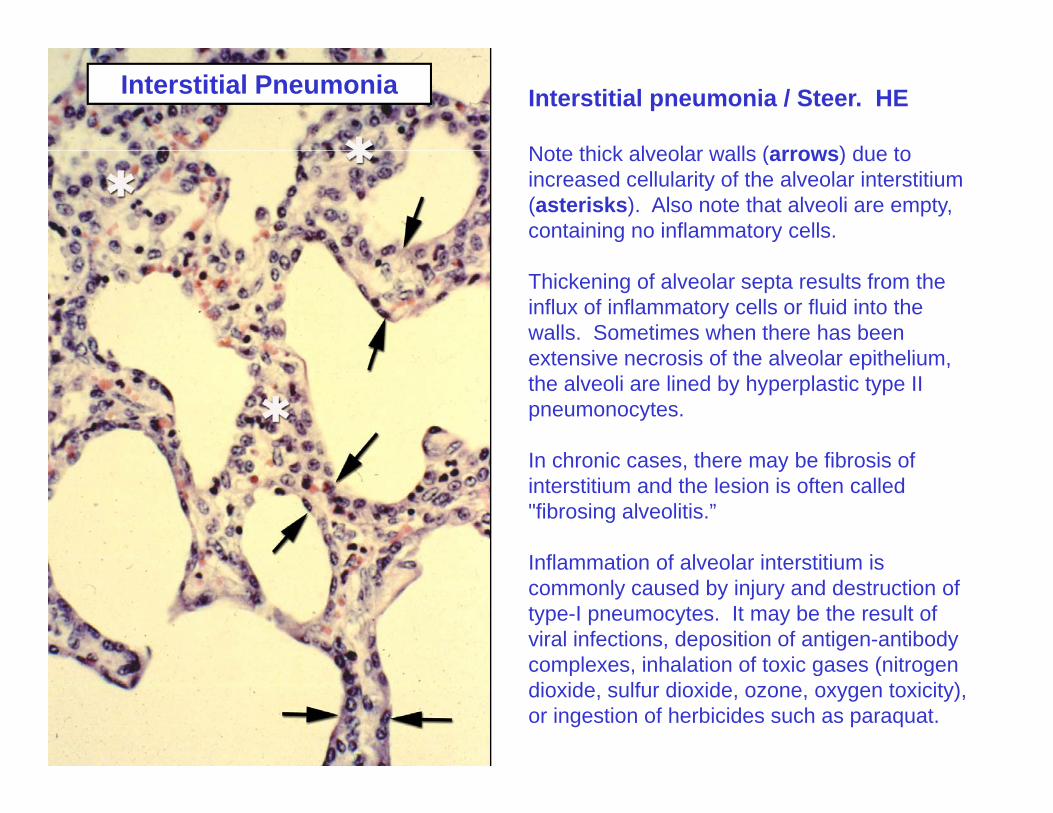
Interstitial Pneumonia Interstitial pneumonia / Steer. HE
Note thick alveolar walls (arrows) due toNote thick alveolar walls (arrows) due to increased cellularity of the alveolar interstitium (asterisks). Also note that alveoli are empty, containing no inflammatory cells.
Thickening of alveolar septa results from the influx of inflammatory cells or fluid into the walls. Sometimes when there has been extensive necrosis of the alveolar epitheliumextensive necrosis of the alveolar epithelium, the alveoli are lined by hyperplastic type II pneumonocytes.
In chronic cases there may be fibrosis ofIn chronic cases, there may be fibrosis of interstitium and the lesion is often called "fibrosing alveolitis.”
Inflammation of alveolar interstitium isInflammation of alveolar interstitium is commonly caused by injury and destruction of type-I pneumocytes. It may be the result of viral infections, deposition of antigen-antibody complexes inhalation of toxic gases (nitrogencomplexes, inhalation of toxic gases (nitrogen dioxide, sulfur dioxide, ozone, oxygen toxicity), or ingestion of herbicides such as paraquat.
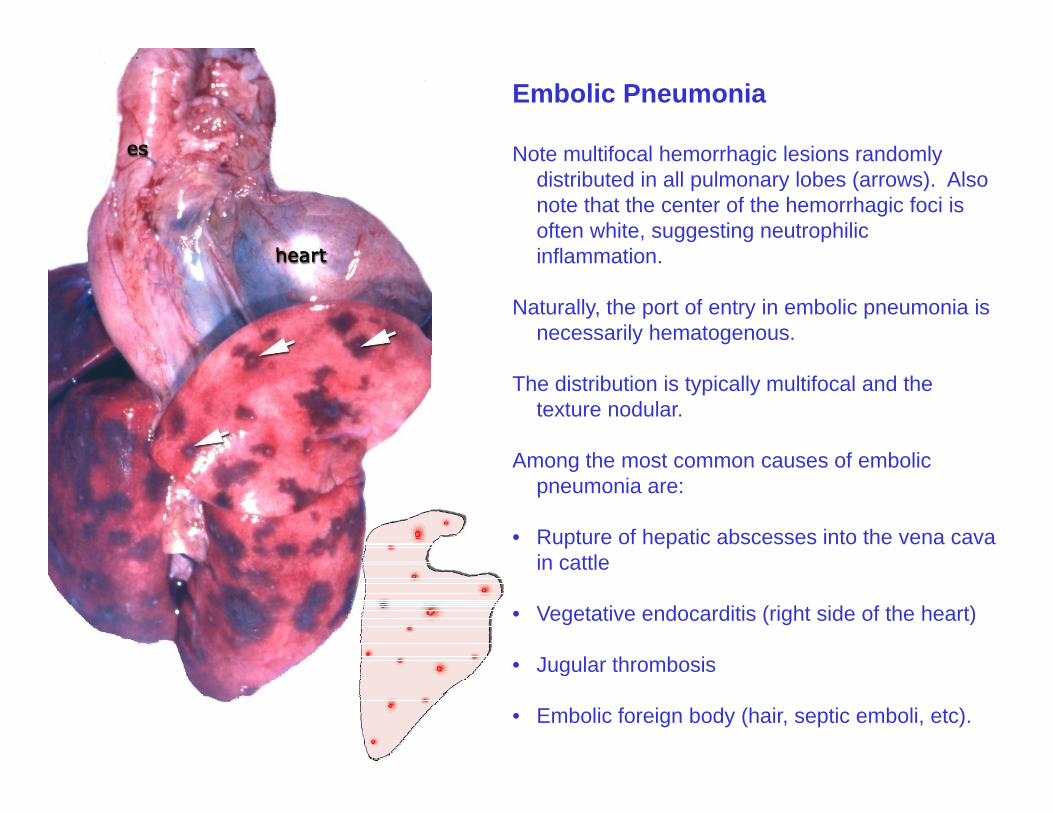
Embolic Pneumonia
Note multifocal hemorrhagic lesions randomlyNote multifocal hemorrhagic lesions randomly distributed in all pulmonary lobes (arrows). Also note that the center of the hemorrhagic foci is often white, suggesting neutrophilic inflammationinflammation.
Naturally, the port of entry in embolic pneumonia is necessarily hematogenous.
The distribution is typically multifocal and the texture nodular.
Among the most common causes of embolicAmong the most common causes of embolic pneumonia are:
• Rupture of hepatic abscesses into the vena cava in cattlein cattle
• Vegetative endocarditis (right side of the heart)
• Jugular thrombosis• Jugular thrombosis
• Embolic foreign body (hair, septic emboli, etc).
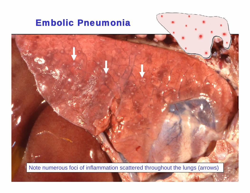
Embolic PneumoniaEmbolic Pneumonia
Note numerous foci of inflammation scattered throughout the lungs (arrows)
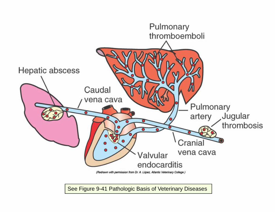
See Figure 9-41 Pathologic Basis of Veterinary Diseases
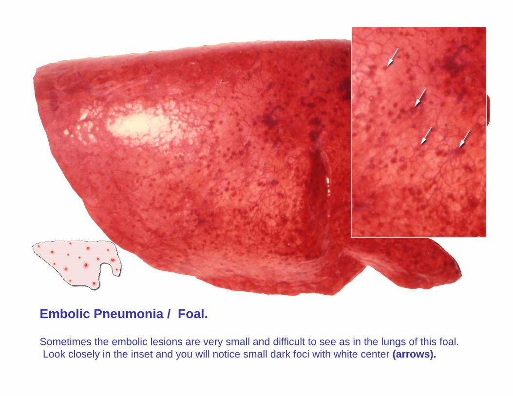
Embolic Pneumonia / Foal.
Sometimes the embolic lesions are very small and difficult to see as in the lungs of this foal. Look closely in the inset and you will notice small dark foci with white center (arrows).
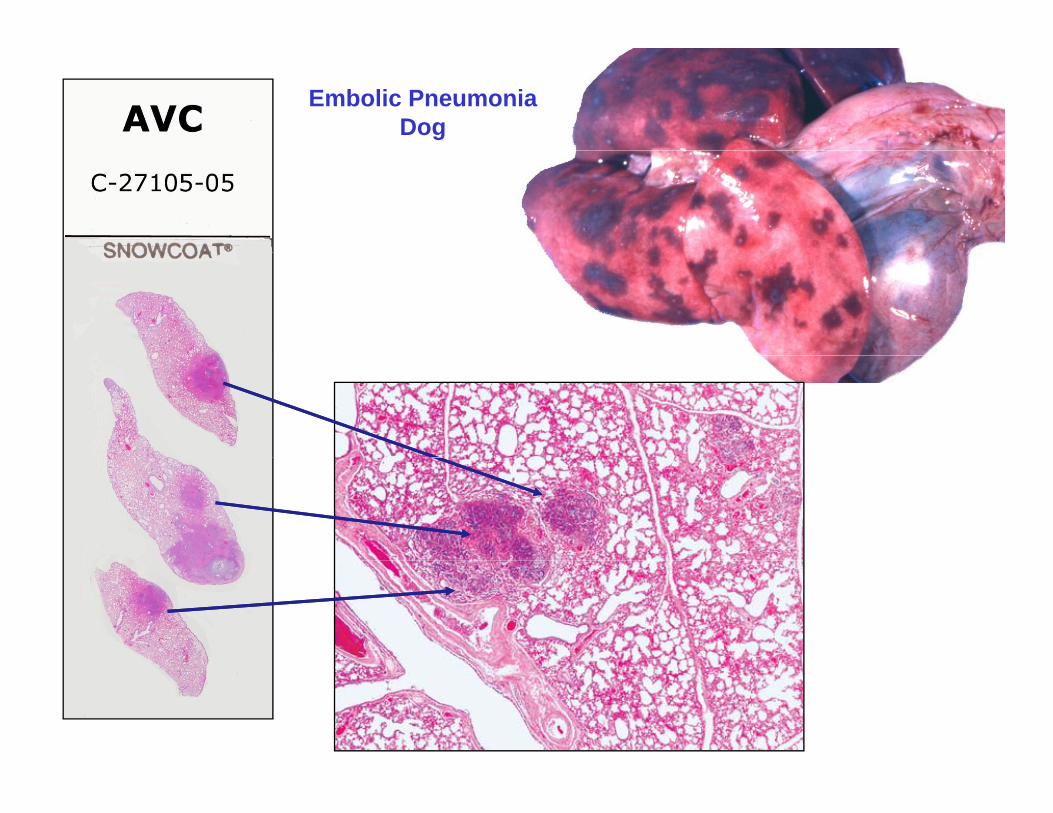
Embolic PneumoniaDog
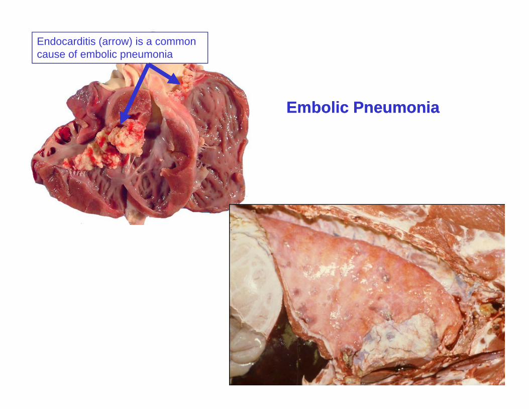
Endocarditis (arrow) is a common cause of embolic pneumonia
Embolic PneumoniaEmbolic Pneumonia
Embolic Pneumonia
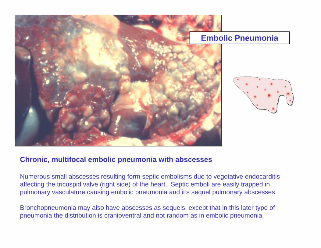
Chronic, multifocal embolic pneumonia with abscesses
Numerous small abscesses resulting form septic embolisms due to vegetative endocarditis affecting the tricuspid valve (right side) of the heart. Septic emboli are easily trapped in pulmonary vasculature causing embolic pneumonia and it’s sequel pulmonary abscesses
Bronchopneumonia may also have abscesses as sequels, except that in this later type of pneumonia the distribution is cranioventral and not random as in embolic pneumonia.
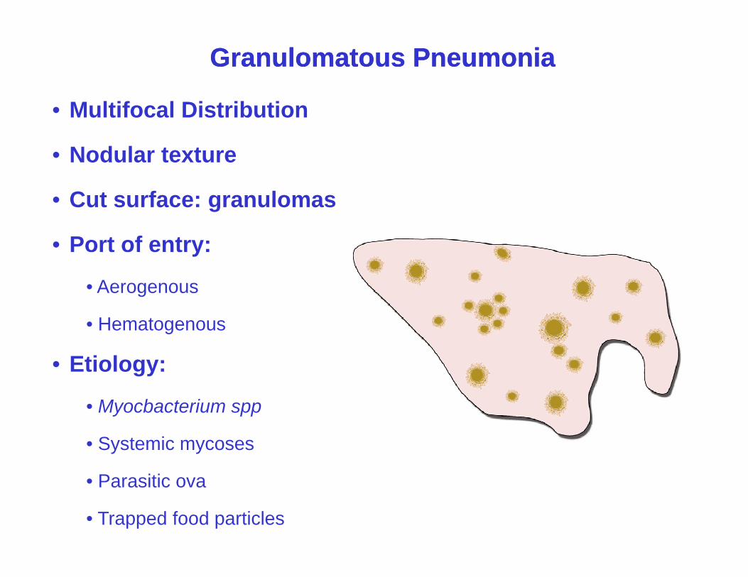
Granulomatous PneumoniaGranulomatous Pneumonia
• Multifocal Distribution• Multifocal Distribution
• Nodular texture
• Cut surface: granulomas
• Port of entry:y
• Aerogenous
• Hematogenous• Hematogenous
• Etiology:
• Myocbacterium spp
• Systemic mycoses
• Parasitic ova
• Trapped food particles
Granulomatous Pneumonia
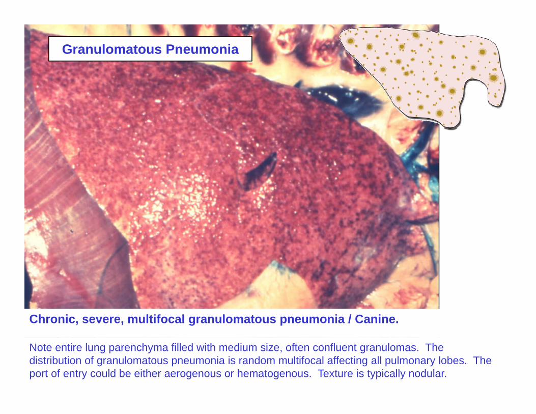
Chronic, severe, multifocal granulomatous pneumonia / Canine.
Note entire lung parenchyma filled with medium size, often confluent granulomas. The distribution of granulomatous pneumonia is random multifocal affecting all pulmonary lobes. The port of entry could be either aerogenous or hematogenous. Texture is typically nodular.
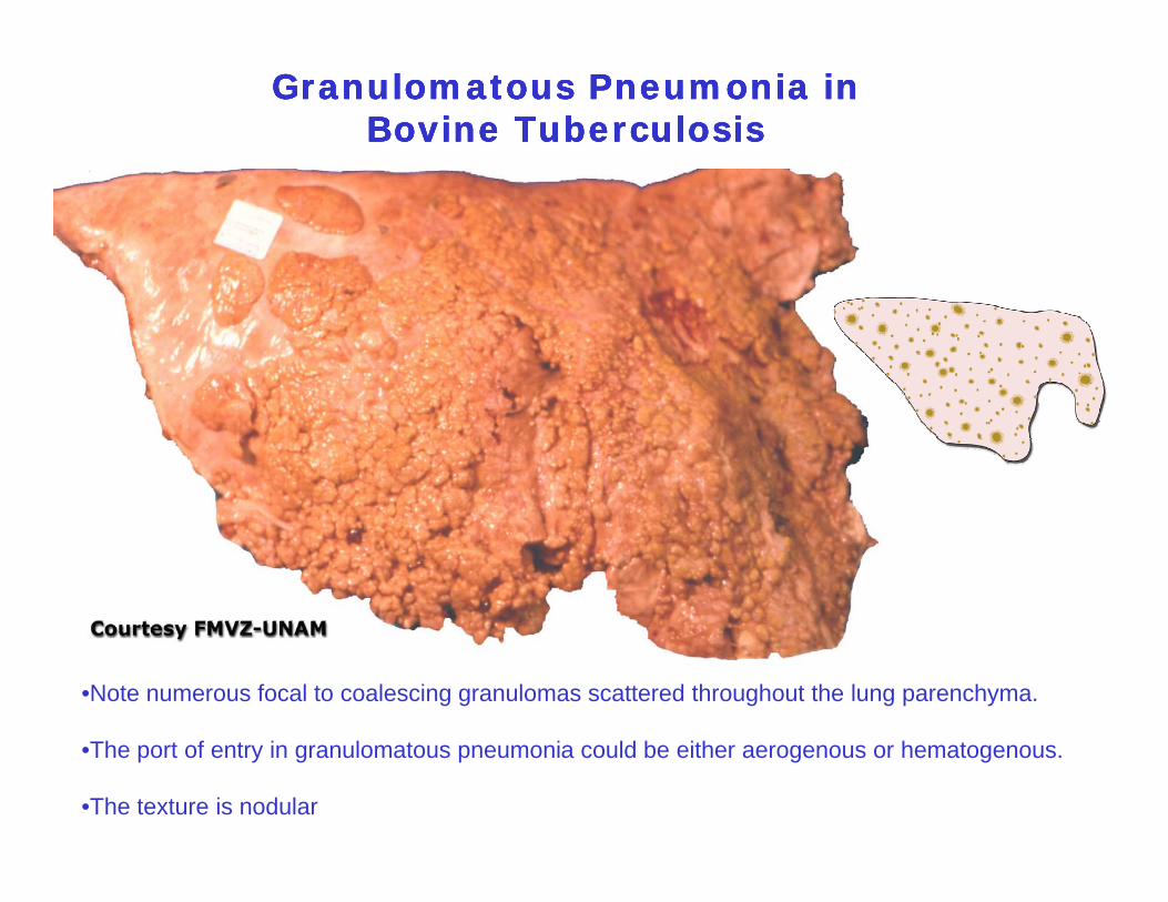
Granulomatous Pneumonia in Granulomatous Pneumonia in Bovine TuberculosisBovine Tuberculosis
Granulomatous Pneumonia in Granulomatous Pneumonia in Bovine TuberculosisBovine Tuberculosis
•Note numerous focal to coalescing granulomas scattered throughout the lung parenchyma.
•The port of entry in granulomatous pneumonia could be either aerogenous or hematogenous.
•The texture is nodular
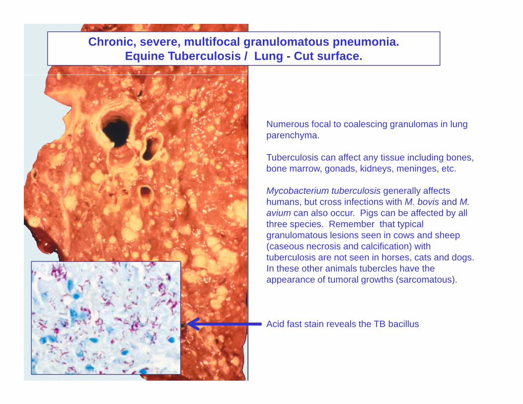
Chronic, severe, multifocal granulomatous pneumonia. Equine Tuberculosis / Lung - Cut surface.
Numerous focal to coalescing granulomas in lungNumerous focal to coalescing granulomas in lung parenchyma.
Tuberculosis can affect any tissue including bones, bone marrow, gonads, kidneys, meninges, etc.
Mycobacterium tuberculosis generally affects humans, but cross infections with M. bovis and M. avium can also occur. Pigs can be affected by all three species. Remember that typical p ypgranulomatous lesions seen in cows and sheep (caseous necrosis and calcification) with tuberculosis are not seen in horses, cats and dogs. In these other animals tubercles have the appearance of tumoral growths (sarcomatous)appearance of tumoral growths (sarcomatous).
Acid fast stain reveals the TB bacillus
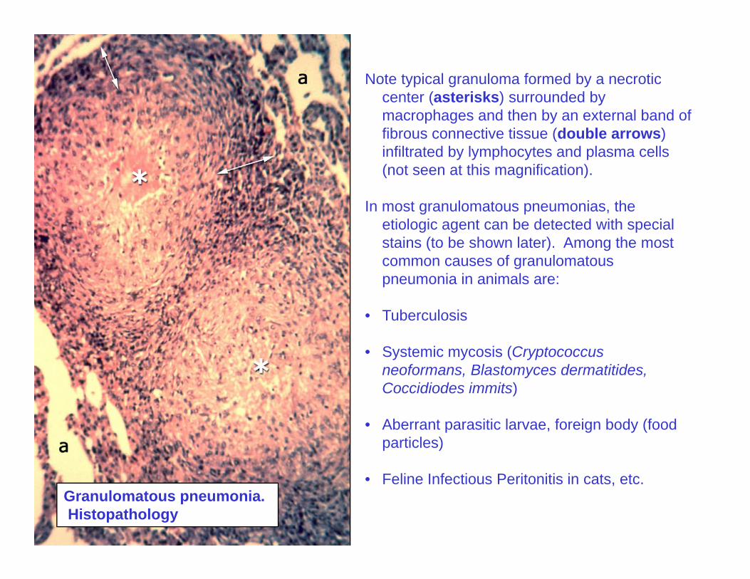
Note typical granuloma formed by a necrotic center (asterisks) surrounded by macrophages and then by an external band of fibrous connective tissue (double arrows) infiltrated by lymphocytes and plasma cells (not seen at this magnification).
In most granulomatous pneumonias, the etiologic agent can be detected with special stains (to be shown later). Among the most common causes of granulomatous pneumonia in animals are:
• Tuberculosis
• Systemic mycosis (Cryptococcus neoformans, Blastomyces dermatitides, Coccidiodes immits)
• Aberrant parasitic larvae, foreign body (food particles)
• Feline Infectious Peritonitis in cats, etc.Granulomatous pneumonia. Histopathology
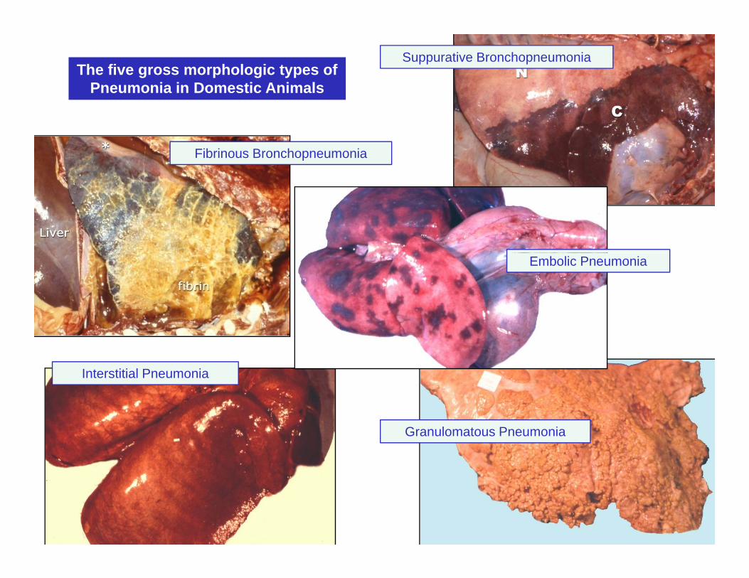
The five gross morphologic types of Pneumonia in Domestic Animals
Suppurative Bronchopneumonia
Fibrinous Bronchopneumonia
Embolic Pneumonia
Interstitial Pneumonia
Granulomatous Pneumonia

THE ENDTHE END