Embed Size (px)
Citation preview

Liver Pathology – Lab 2
Shannon Martinson, 2017 http://people.upei.ca/smartinson/

Case 1
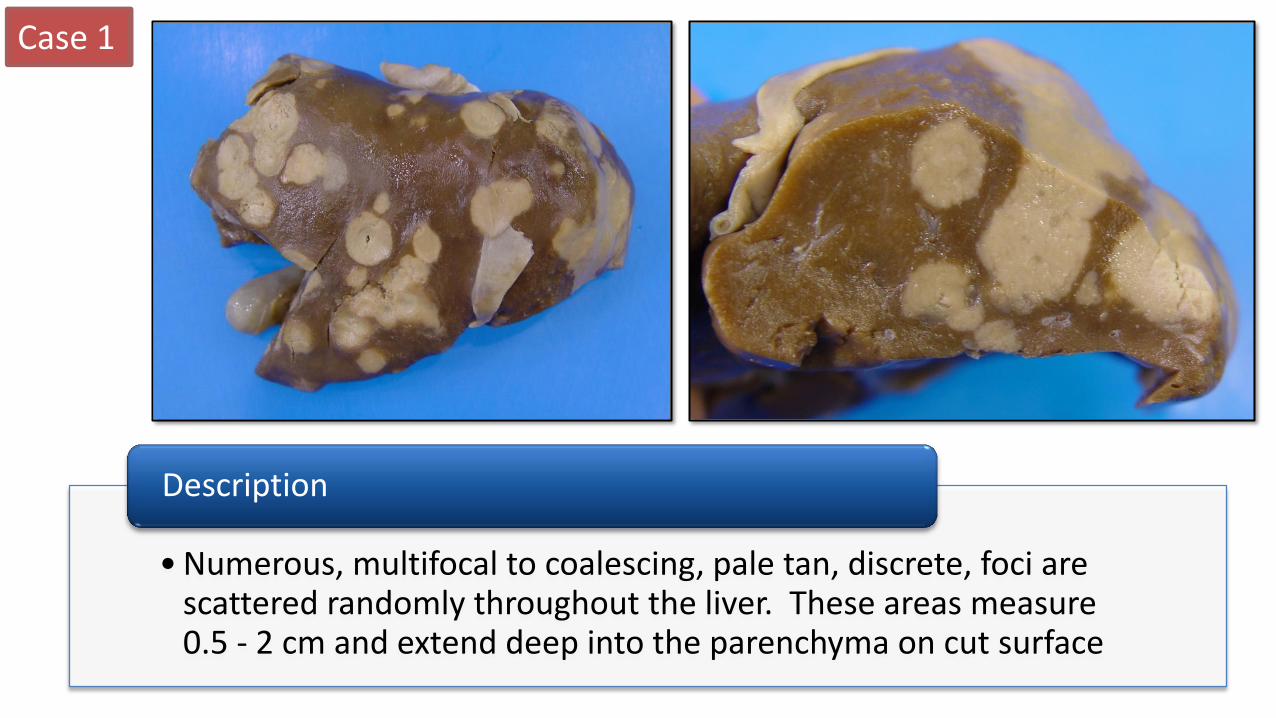
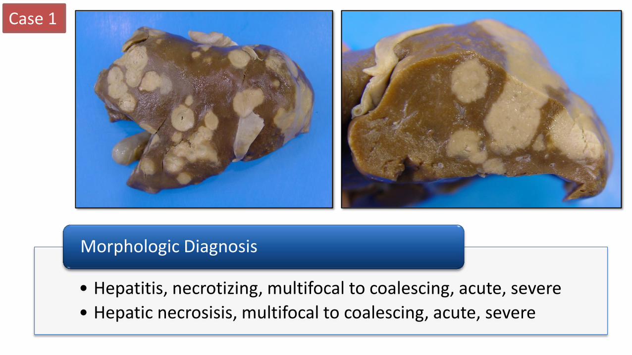
Signalment: • 8 day old male lamb History: • Noted to be weak • Found dead
Case 1
• Numerous, multifocal to coalescing, pale tan, discrete, foci are scattered randomly throughout the liver. These areas measure 0.5 - 2 cm and extend deep into the parenchyma on cut surface
Description
Case 1
• Hepatitis, necrotizing, multifocal to coalescing, acute, severe
• Hepatic necrosisis, multifocal to coalescing, acute, severe
Morphologic Diagnosis

Case 1
• Fusobacterium necrophorum (necrobacillosis)
• Clostridium piliforme (Tyzzer’s disease) • Listeria monocytogenes (Listeriosis) • Campylobacter fetus fetus • Salmonella • Trueperella pyogenes
What are some possible etiologies for this lesion?
Possible routes of infection?
• Hematogenous – umbilical vein • Direct extension from adjacent organ • Ascending via biliary tract
Fusobacterium necrophorum Disease name = Necrobacillosis
How would confirm a diagnosis?
• Bacterial culture • Routine • Anaerobic (F. necrophorum) • Cold enrichment + special media (Listeria) • Special media – Campylobacter
• Histology and silver stain (C. piliforme)
Case 2
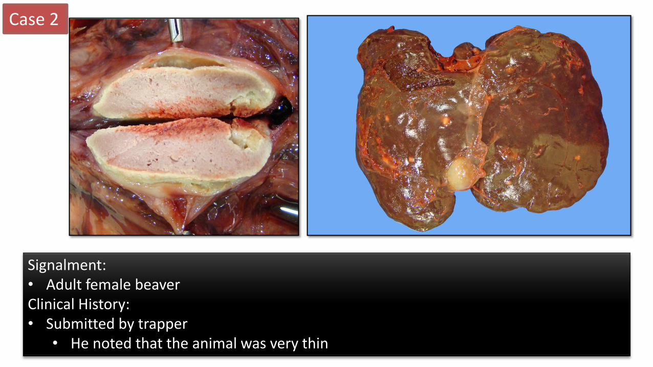
Signalment: • Adult female beaver Clinical History: • Submitted by trapper
• He noted that the animal was very thin

Case 2
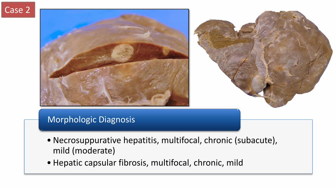
• Several fibrous tags are present on the hepatic capsular surface
• Multiple (5 – 6), round, discrete, yellow to tan, soft/caseous, foci measuring ~ 0.5 – 1.5 cm are scattered randomly throughout the liver
Description
Case 2
• Necrosuppurative hepatitis, multifocal, chronic (subacute), mild (moderate)
• Hepatic capsular fibrosis, multifocal, chronic, mild
Morphologic Diagnosis
Case 2
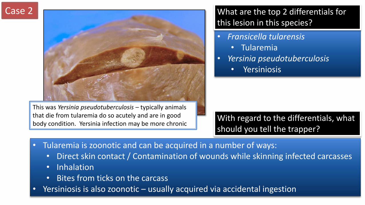
• Fransicella tularensis • Tularemia
• Yersinia pseudotuberculosis • Yersiniosis
What are the top 2 differentials for this lesion in this species?
With regard to the differentials, what should you tell the trapper?
• Tularemia is zoonotic and can be acquired in a number of ways: • Direct skin contact / Contamination of wounds while skinning infected carcasses • Inhalation • Bites from ticks on the carcass
• Yersiniosis is also zoonotic – usually acquired via accidental ingestion
This was Yersinia pseudotuberculosis – typically animals that die from tularemia do so acutely and are in good body condition. Yersinia infection may be more chronic
Case 3
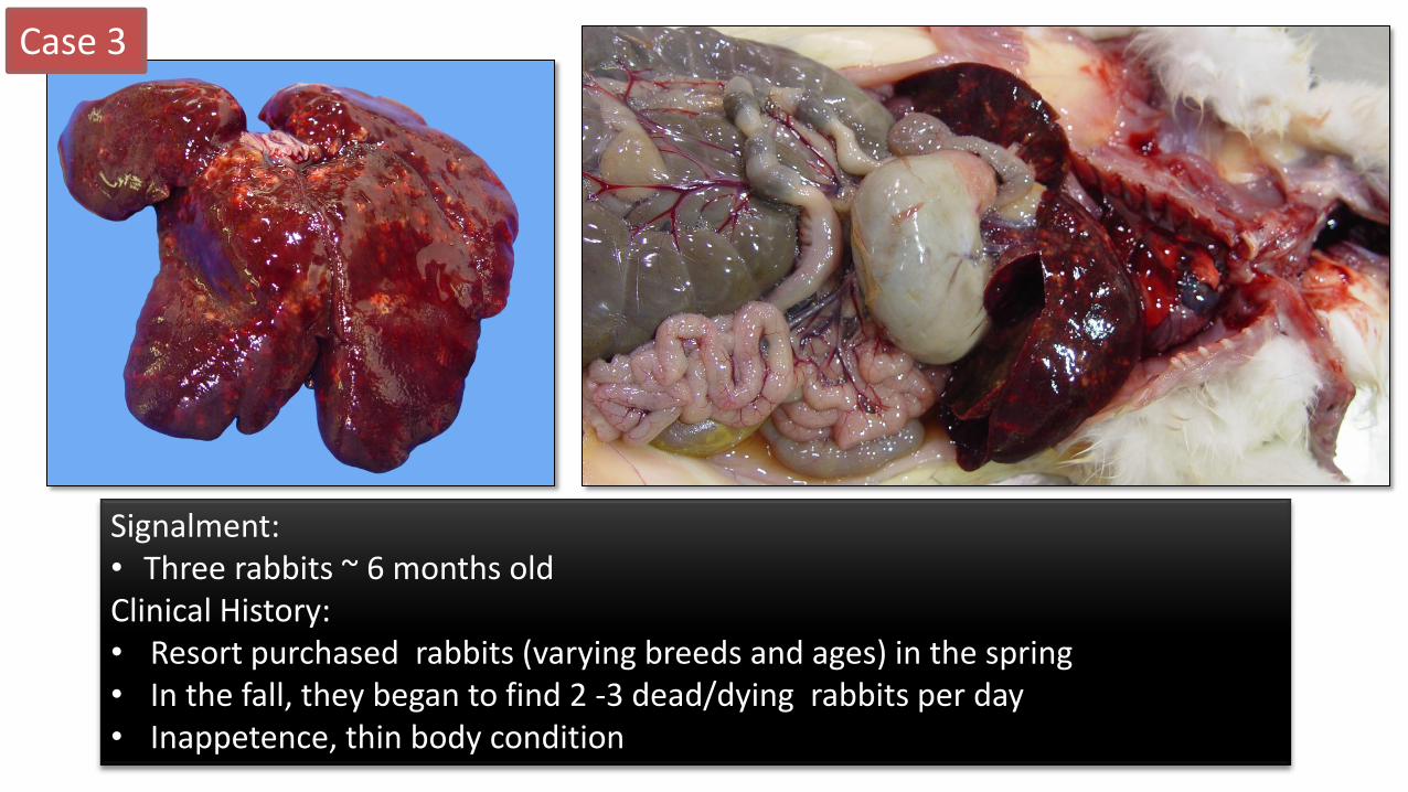
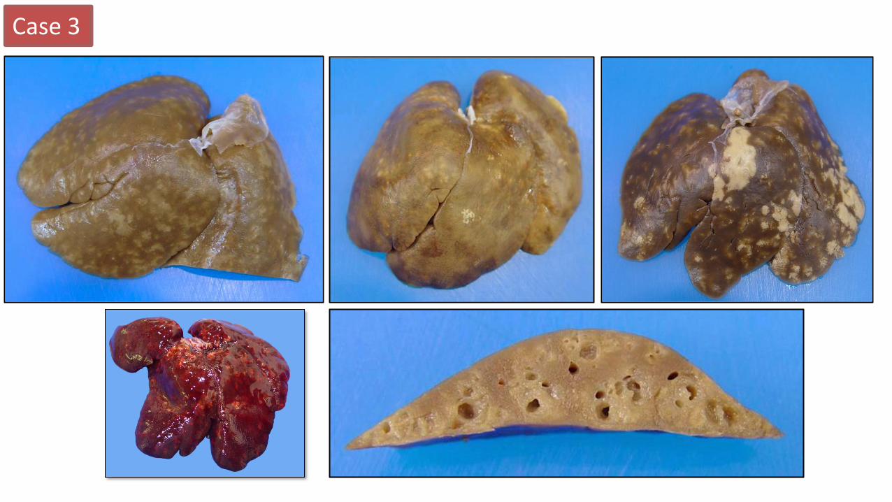
Signalment: • Three rabbits ~ 6 months old Clinical History: • Resort purchased rabbits (varying breeds and ages) in the spring • In the fall, they began to find 2 -3 dead/dying rabbits per day • Inappetence, thin body condition
Case 3
Case 3
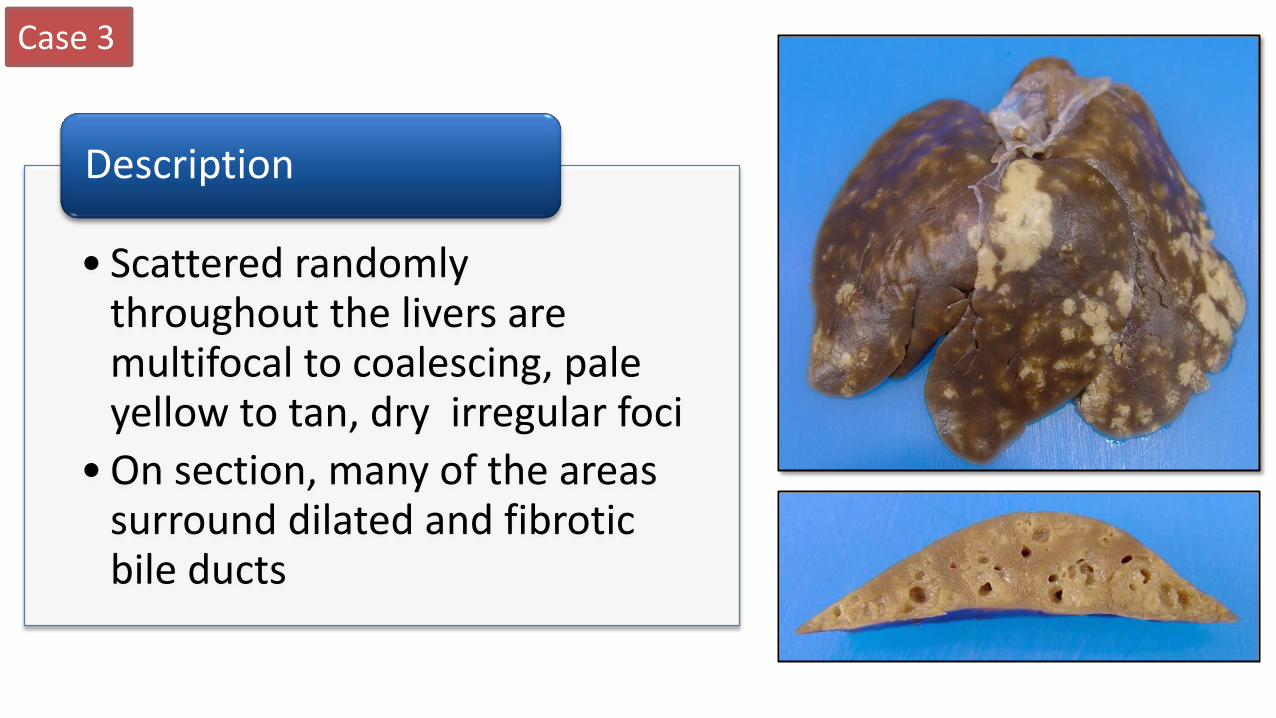
• Scattered randomly throughout the livers are multifocal to coalescing, pale yellow to tan, dry irregular foci
• On section, many of the areas surround dilated and fibrotic bile ducts
Description

Case 3
• Cholangiohepatitis, proliferative and necrotizing, multifocal, chronic, severe, with intralesional coccidia
Morph Diagnosis Histo Images: http://www.askjpc.org/wsco/wsc/images/2014/141801-2.jpg

Case 3
• Eimeria stiedae
Etiology
Histo Images: http://www.askjpc.org/wsco/wsc/images/2014/141801-2.jpg

Case 3
• Black Head
• Histomoniasis
Disease Name
• Histomonas meleagridis
Etiology
Case 4
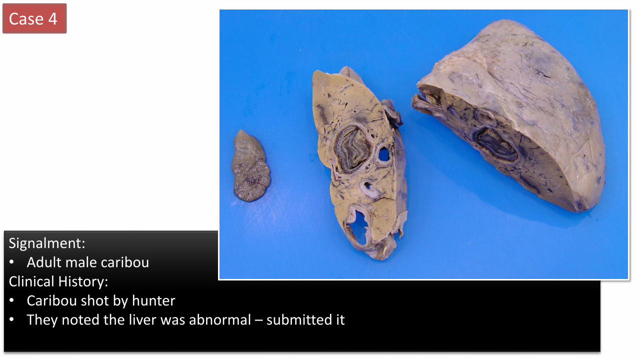
Signalment: • Adult male caribou Clinical History: • Caribou shot by hunter • They noted the liver was abnormal – submitted it

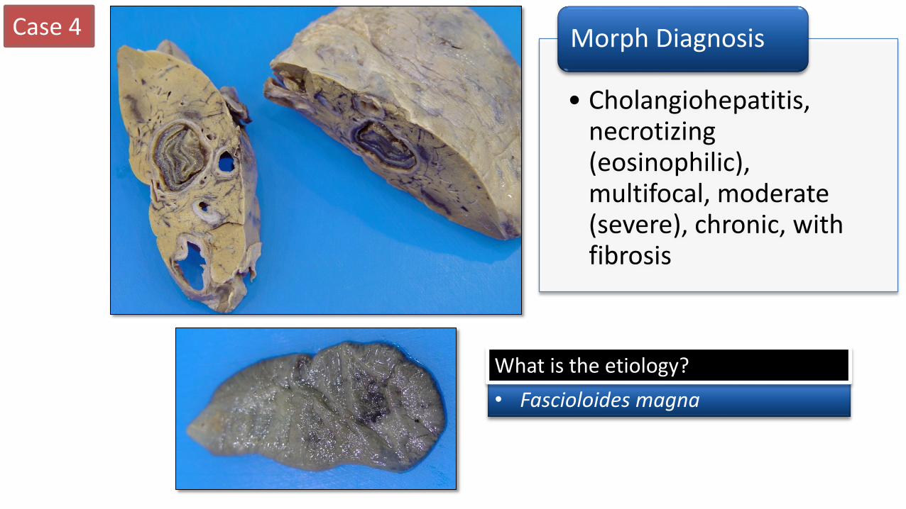
• Several round cyst-like cavities, each surrounded by a thick fibrous capsule, are present multifocally within the liver. Within these structures, there are coiled trematodes
• The flukes are leaf shaped, dorsoventrally flattened, measuring ~ 5 – 8 cm x 3 – 4 cm with an oral and ventral sucker
• Fine black tortuous tracts are present multifocally in the parenchyma
Description Case 4
Case 4
• Cholangiohepatitis, necrotizing (eosinophilic), multifocal, moderate (severe), chronic, with fibrosis
Morph Diagnosis
• Fascioloides magna
What is the etiology?

Case 4
• Bacillary hemoglobinuria • Clostridium hemolyticum
• Black Disease • Clostridium novyii
In sheep and cattle, what 2 diseases may be precipitated by these lesions?
• Hard specimen to morph!
• Cholangiohepatitis, necrotizing (eosinophilic), multifocal, moderate (severe), chronic, with fibrosis and intralesional flukes
Morph Diagnosis
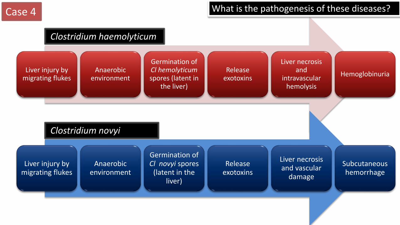
Liver injury by migrating flukes
Anaerobic environment
Germination of Cl hemolyticum spores (latent in
the liver)
Release exotoxins
Liver necrosis and
intravascular hemolysis
Hemoglobinuria
Clostridium haemolyticum
Liver injury by migrating flukes
Anaerobic environment
Germination of Cl novyi spores
(latent in the liver)
Release exotoxins
Liver necrosis and vascular
damage
Subcutaneous hemorrhage
Case 4
Clostridium novyi
What is the pathogenesis of these diseases?

Case 5
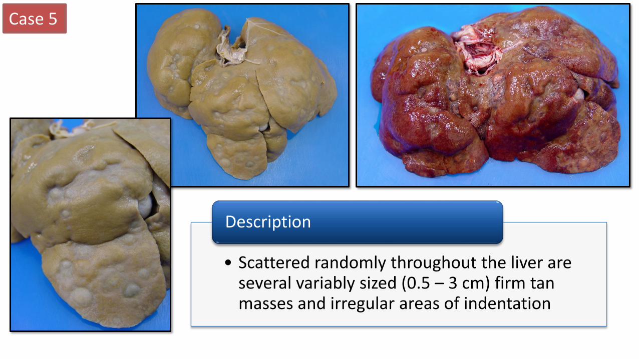
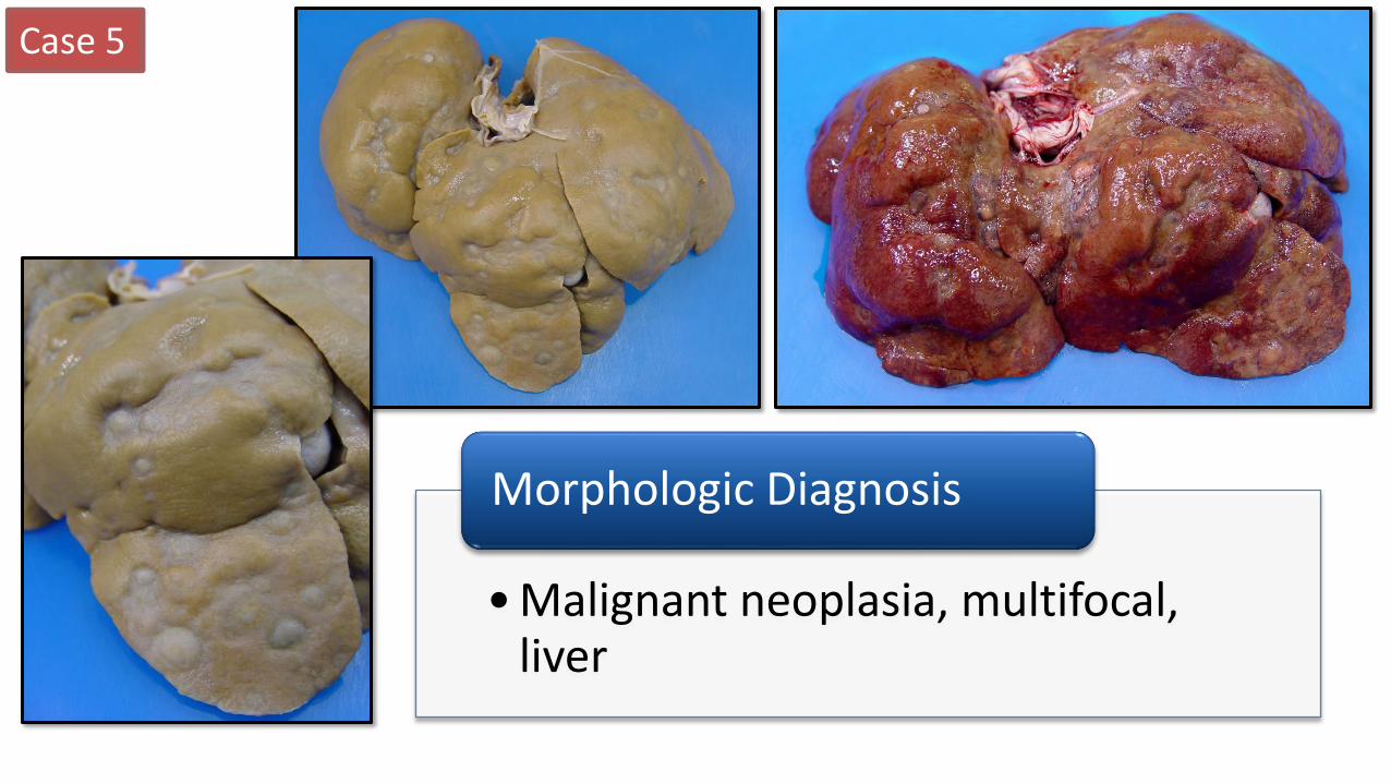
Signalment: • 7 year old FS Old English Sheepdog Clinical History: • Ascites and PU/PD for ~ 1 week • Previous history of urinary incontinence • Radiographs – hepatomegaly and masses in lung
Case 5
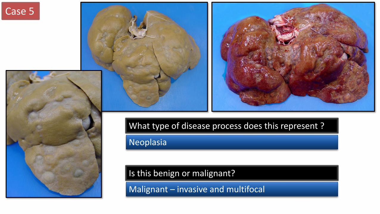
• Scattered randomly throughout the liver are several variably sized (0.5 – 3 cm) firm tan masses and irregular areas of indentation
Description
Case 5
What type of disease process does this represent ?
Neoplasia
Is this benign or malignant?
Malignant – invasive and multifocal
Case 5
• Malignant neoplasia, multifocal, liver
Morphologic Diagnosis
Case 5
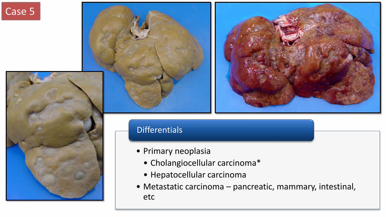
• Primary neoplasia
• Cholangiocellular carcinoma*
• Hepatocellular carcinoma
• Metastatic carcinoma – pancreatic, mammary, intestinal, etc
Differentials
Case 5
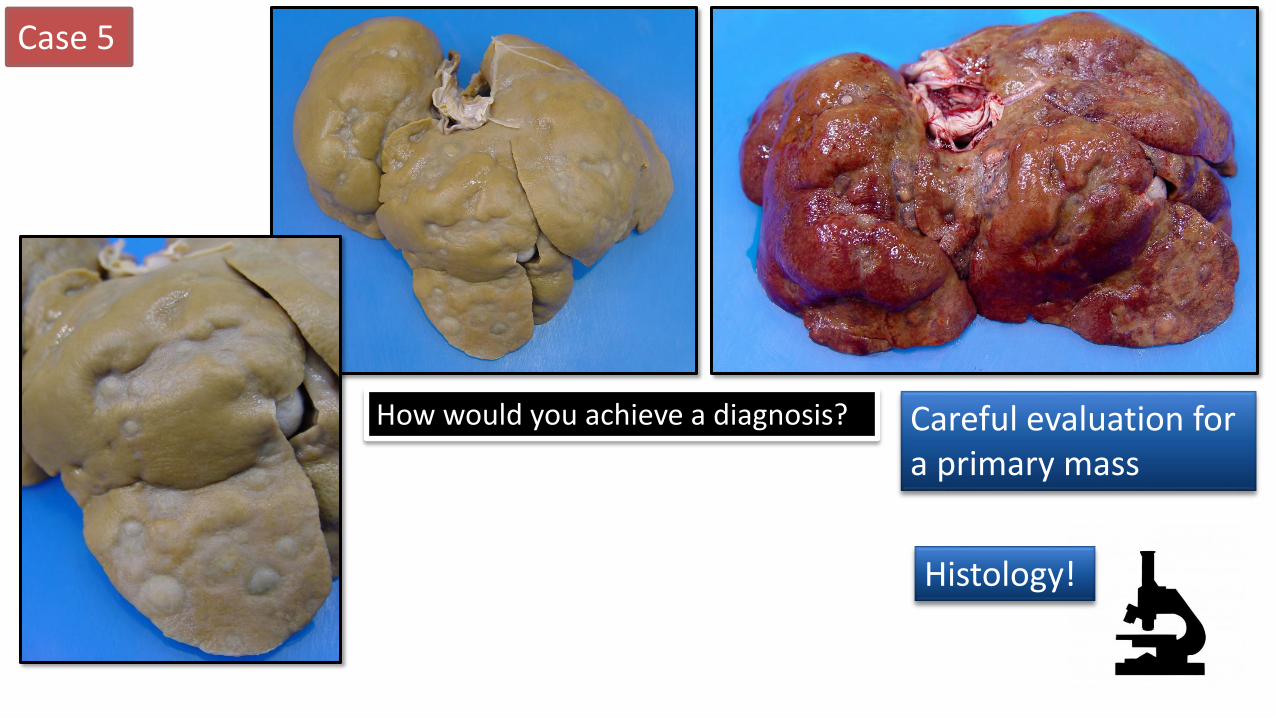
Careful evaluation for a primary mass
How would you achieve a diagnosis?
Histology!

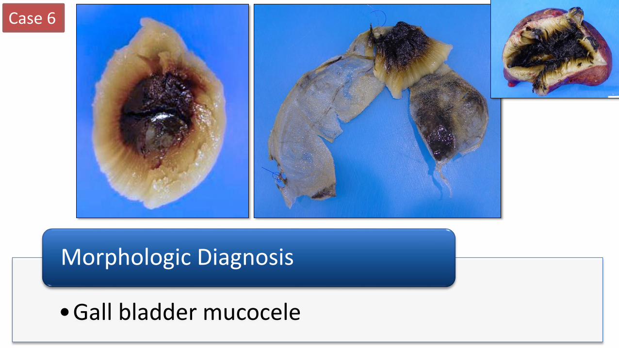
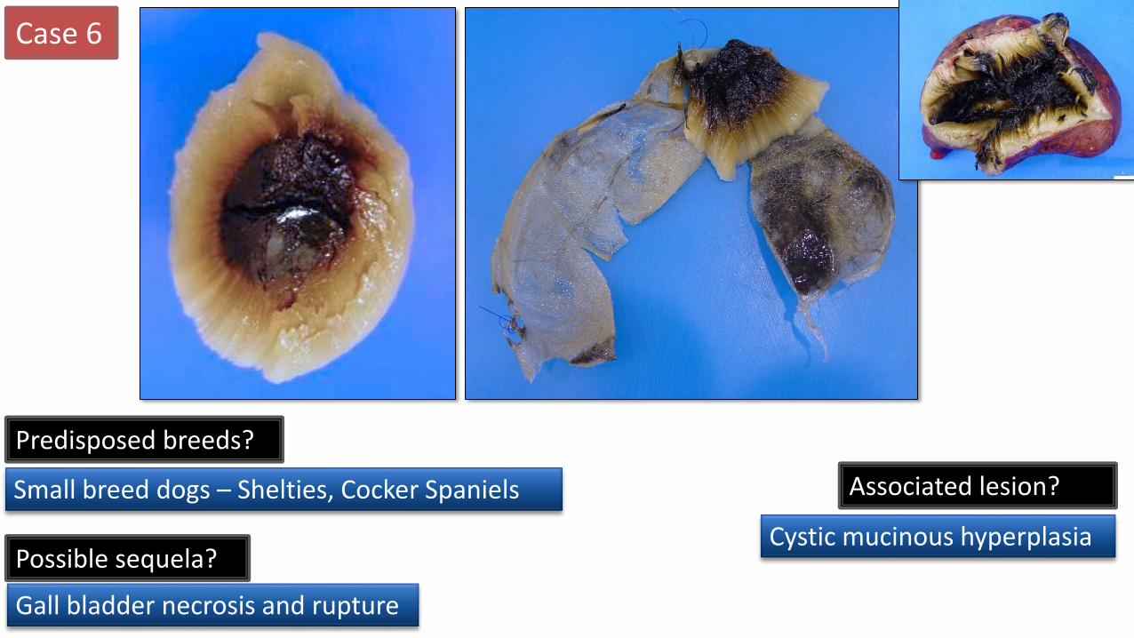
Signalment: • 13 year old, male dog Clinical History: • Acute vomiting/anorexia for 3 days • Intestinal foreign body noted on ultrasound • Gall bladder had a “kiwi pattern” on US
Case 6
http://veteriankey.com/

Case 6
• The gall bladder is markedly dilated with thinning of the wall
• The content is solid and soft (mucoid) and a pale yellow to white with a dark green center and a radiating pattern
Description
Case 6
•Gall bladder mucocele
Morphologic Diagnosis
Case 6
Predisposed breeds?
Small breed dogs – Shelties, Cocker Spaniels
Possible sequela?
Gall bladder necrosis and rupture
Associated lesion?
Cystic mucinous hyperplasia
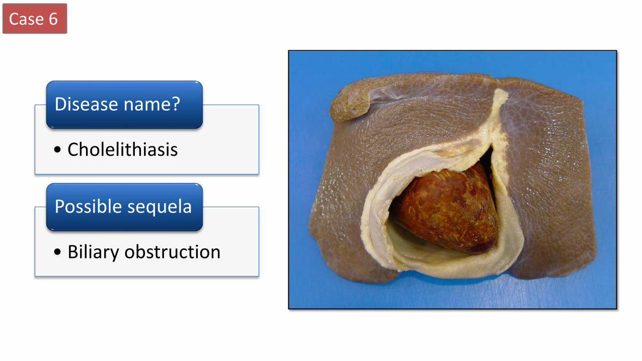
Case 6
• Cholelithiasis
Disease name?
• Biliary obstruction
Possible sequela