Embed Size (px)
Citation preview
ARTICLE
Respiratory syncytial virus infection in children with severemotor and intellectual disabilities
S. Onoyama & T. Hoshina & S. Honjo & K. Ihara & T. Hara
Received: 13 February 2013 /Accepted: 30 April 2013 /Published online: 17 May 2013# Springer-Verlag Berlin Heidelberg 2013
Abstract Children with severe motor intellectual disabil-ities (SMID) are at high risk of death from acute viral lowerrespiratory tract infections (LRTI). Although respiratorysyncytial virus (RSV) is the most common cause of viralLRTI in children, there have been a few reports on therelationship between SMID and the severity of RSV-LRTI.The aim of the present study is to assess the influence ofRSV-LRTI in children with SMID. A case–control studycomposed of children with SMID (n=18) and previouslyhealthy children (n=43) less than 16 years old hospitalizedfor RSV-LRTI was performed during five consecutive RSVseasons. The clinical presentation and the laboratory data inthe SMID group were compared with those in the non-SMID group. In the bivariate analysis, the median age ofthe SMID group was higher than that of the non-SMIDgroup (p=0.002). Children with SMID had an increased riskfor ventilation support (p=0.057). The count of neutrophilsin the SMID group was significantly increased (p=0.012),whereas the proportion of bacterial co-infection was lowerthan that in the non-SMID group (p=0.005). Multivariatelogistic analysis showed that SMID was associated withlonger oxygen usage [>7 days: odds ratio (OR) 5.309, p=0.033]. The present study revealed that children with SMIDwere prone to developing hypoxia by RSV-LRTI. The strat-egies for the treatment and prevention of RSV infection needto be improved in SMID children.
Introduction
Severe motor intellectual disability (SMID) is defined as thecombination of severe physical handicap and intellectualdisability [1, 2]. Children with SMID generally have ob-structive and restrictive respiratory disorders, making itdifficult to clear out sputum from the airways, and sufferfrom multiple-organ disorders, such as muscle weakness,muscle spasticity, seizure, and gastroesophageal reflux, pos-sibly leading to the development of respiratory failure [2, 3].In addition, the patients are susceptible to recurrent andchronic respiratory infections, which may damage the alve-olar tissues. Based on their chronic respiratory condition,these children are susceptible to severe viral lower respira-tory tract infection (LRTI) [2].
Respiratory syncytial virus (RSV) is the most commoncause of viral LRTI in infants. Almost all children have RSVinfection until the age of 2 years, and 2–3 % of them needhospital treatment for RSV-LRTI [4, 5]. It is generallyrecognized that the morbidity and mortality related to RSVinfection is high in premature infants and children withcongenital heart disease (CHD) [6, 7]. On the other hand,there are few reports that have analyzed the relationshipbetween children with SMID and RSV infection. The aimof this study is to investigate the severity of RSV-LRTI inchildren with SMID.
Materials and methods
Study population
Our retrospective case–control study was performed on 61patients less than 16 years of age who were admitted to theDepartment of Pediatrics at Kyushu University Hospitalduring five consecutive seasons from September 1, 2006to April 30, 2011 for RSV-LRTI. We performed a rapid RSV
S. Onoyama : T. Hoshina (*) :K. Ihara : T. HaraDepartment of Pediatrics, Graduate School of Medical Sciences,Kyushu University, 3-1-1 Maidashi, Higashi-ku,Fukuoka 812-8582, Japane-mail: [email protected]
S. HonjoDepartment of Pediatrics, National Hospital Organization FukuokaHospital, Fukuoka, Japan
Eur J Clin Microbiol Infect Dis (2013) 32:1353–1357DOI 10.1007/s10096-013-1893-x
antigen detection test for every patient who was suspected tohave underlying viral LRTI by physical findings and labo-ratory data on admission. Eighteen of 61 patients had SMID(SMID group) and the remaining 43 patients did not haveany underlying diseases (non-SMID group). SMID wasdiagnosed according to the classical criteria (Oshima’scriteria) [1]. We evaluated the psychomotor developmentfor children under 5 years of age by developmental quo-tients (DQ) using the Enjoji developmental test, and forthose over 5 years of age by intelligence quotients (IQ)using the Wechsler Intelligence Scale for Children. Allchildren with SMID were classified as grade 1 or 2, whowere bedridden or able to sit, crawl, or walk with support,and had IQ or DQ lower than 20. The underlying diseases ofSMID were as follows: malformation syndrome (n=3),holoprosencephaly (n=2), 21 trisomy (n=2), sequelae ofmeningitis (n=2), cerebral palsy (n=2), 18 trisomy (n=1),hydrocephalus (n=1), colpocephaly (n=1), lissencephaly (n=1), Aicardi syndrome (n=1), Zellweger syndrome (n=1),and sub-acute sclerosing panencephalitis (n=1). Four ofthem received tracheostomy and three received home oxy-gen therapy. Four had congenital heart diseases of non-hemodynamical significance. In none of the enrolled pa-tients was any primary or secondary immunodeficiencyidentified. Clinical information on each patient was collect-ed using a standardized case report form. Many of theenrolled patients had histories of aspiration pneumonia com-plicated by gastroesophageal reflux disease (GERD), butnone of them had apparent episodes of aspiration by GERDat the onset of RSV-LRTI. All patients had regular dentalexaminations, and none of them were under dental treatmentat the onset of RSV-LRTI. From the laboratory data onadmission were examined peripheral white blood cell(WBC) counts and neutrophil counts, serum C-reactive pro-tein (CRP) levels, the findings of chest X-ray, and thebacteriology results of sputum samples. We diagnosed thepatients as having LRTI when they had cough and sputumproduction as clinical symptoms with auscultatory findingsof abnormal breath sounds, wheezes, or crackles [8]. Noneof the patients had typical symptoms of chronic sinusitis,such as yellow or greenish discharge from their noses.
Confirmation of RSV infection
All the patients were diagnosed as having RSV infection bya rapid antigen detection test (SA Scientific, San Antonio,SA, USA) using a nasopharyngeal aspirate or secretionsuctioned through the tracheostomy orifice. It has beenreported that specimens obtained by endotracheal tube aspi-ration had a higher sensitivity for the RSV antigen detectionthan by nasopharyngeal aspirate specimens in adult patients[9]. To the best of our knowledge, there have been nosimilar studies in child patients. Nevertheless, it would be
rational to speculate that there was little practical differencein the sensitivity of antigen detection by specimens fromnasopharyngeal aspiration and from endotracheal tube aspi-ration, because the RSV viral load in the nasopharyngeallesions was significantly greater in child patients than that inadults. A previous report demonstrated that the specificity ofthe test was equally high by specimens from nasopharyngealor endotracheal tube aspiration [9]. It had also been reportedthat the rapid RSV antigen detection test had a lower sensi-tivity than viral culture or reverse transcriptase polymerasechain reaction (RT-PCR) [10, 11]. To confirm the sensitivityof the rapid antigen RSV detection test, we also performedmultiplex RT-PCR assay by using samples from eight pa-tients. The viral RNA was extracted using a Ribospin vRDkit (GeneAll, Seoul, Korea), in accordance with the manu-facturer’s instructions. Multiplex RT-PCR was conductedusing the Seeplex® RV15 OneStep ACE Detection kit(Seegene Inc., Seoul, Korea). Reverse transcription andPCR amplification were performed on the GeneAmp PCRSystem 9700 (Applied Biosystems, Foster City, CA, USA).Amplicons were separated and detected by the automatedmicrochip electrophoresis system MultiNA (Shimadzu,Kyoto, Japan). RSVA or B were detected with all samples,while eight samples with negative results were detected bythe rapid RSV antigen test, analyzed by RT-PCR, and it wasfound that all of them were negative. Based on this result,the sensitivity of the rapid RSV antigen test correspondedwith that of the RT-PCR assay, indicating that the rapidantigen test would be a reliable diagnosis method.
Sputum collection, bacteriological examination, and itsevaluation
Sputum samples were obtained for the confirmation ofbacterial infection. The sputum collection and the judgmentof their qualities using Geckler’s classification wereperformed as previously described [12]. Smears, classifiedas Geckler’s group 4 or 5, were judged to be suitable forbacterial examination. Only sputum samples suctionedthrough the tracheostomy orifice were judged to be suitable,even when they were classified as Geckler’s group 6. Whenphagocytized bacterial cells were seen on the Gram stainsmear of the sputum sample and corresponding bacteriumwas isolated later, it was identified as a complication ofbacterial infection.
Statistical analysis
The two-sample t-test with unequal variants was used tocalculate the difference in WBC and neutrophils counts.Because WBC and neutrophils counts were distributed withright-skewness, log-transformed values were used in thestatistical analysis. The Wilcoxon Mann–Whitney method
1354 Eur J Clin Microbiol Infect Dis (2013) 32:1353–1357
was used to compare other continuous variables. Fisher’sexact test was applied for the qualitative analysis. Themultivariate logistic regression analysis was performed toestimate odds ratios (ORs) for the association between theindependent variables and outcomes. p-values less than 0.05were considered to be statistically significant.
STATA (version 11.1; StataCorp, College Station, TX,USA) was used for the multivariate logistic regression anal-ysis and JMP (version 9.0; SAS Institute Inc., Cary, NC,USA) was used for the remaining analyses.
Results
The patients’ clinical characteristics and laboratory data areshown in Table 1. Wheezes were detected by chest auscul-tation in all of the enrolled patients. None of these patientsdied in the present study. In the bivariable analysis, themedian age of the SMID group was higher than that of thenon-SMID group (p=0.002). The count of neutrophils wassignificantly increased in the children with SMID (p=0.012). In 29 (55.8 %) of 52 patients from whom sputumsamples could be obtained, the sputum samples wereenough to be evaluated by bacterial examination. In 15(51.7 %) of the 29 sputum samples, one or more bacterialpathogens were identified. One patient (11.1 %) in theSMID group and 14 patients (70 %) in the non-SMID groupwere diagnosed as having bacterial co-infection, respective-ly. Samples from nasopharyngeal aspirate were obtainedfrom six of nine SMID patients for bacterial study, and wefound that only one patient was diagnosed as having bacte-rial co-infection (16.7 %). Compared with the non-SMIDgroup, the ratio of bacterial co-infection was lower in theSMID group (p=0.005). First, an oxygen administrationwas performed for all of the patients with hypoxic state. Arespirator support was introduced next for the patients whocontinued to be in hypoxic and hypercapnic state, even withthe oxygen therapy. The number of children who requiredmechanical ventilation was 4 (22 %) in the SMID group and2 (5 %) in the non-SMID group, respectively. The causes ofrequiring ventilation were acute respiratory failure (n=5)and frequent apnea (n=1, non-SMID group). More childrenwith SMID required mechanical ventilation for respiratorysupport due to RSV-LRTI (p=0.057).
Multivariate logistic regression analysis was performedto exclude the bias due to the differences in clinical charac-teristics. The severity of RSV-LRTI in the patients withSMID was estimated by longer supplemental oxygen (over7 days), longer hospitalization (over 9 days), and require-ment of mechanical ventilation (Table 2). The patients withSMID were significantly associated with longer supplemen-tal oxygen (OR 5.309, p=0.033). The risks of longer hos-pitalization and requiring mechanical ventilation also
became higher, but they did not reach statistical significance(Table 2).
Supplemental oxygen therapy exceeded 7 days in eightpatients with SMID. The underlying diseases of the patientswere malformation syndrome (n=2), sequelae of meningitis(n=1), cerebral palsy (n=1), 18 trisomy (n=1), hydroceph-alus (n=1), colpocephaly (n=1), and sub-acute sclerosingpanencephalitis (n=1). There were no significant differencesin the proportions of patients who received tracheostomyand home oxygen therapy and having congenital heart dis-eases of non-hemodynamical significance between the pa-tients who received supplemental oxygen for more or lessthan 7 days. The proportion of patients diagnosed as havingpneumonia was higher in SMID patients who received sup-plemental oxygen for over 7 days (62.5 %), while thisproportion was also higher in the patients without underly-ing disease (54.5 %).
Discussion
Children with SMID, which is not a worldwide recognizedpopulation, are similar to children with neuromuscular im-pairment (NMI) in having difficulty to cough up sputum.SMID is defined by the presence of severe “neuromuscularimpairment” and intellective disability. NMI was an inde-pendent risk factor with increased risks for pediatric inten-sive care unit (PICU) admission and respiratory failure dueto RSV-LRTI [3]. Keren et al. also showed that neurologicaland neuromuscular disease was a risk factor of respiratoryfailure in children with influenza [13]. In the present study,we investigated whether children with SMID were prone todeveloping severe LRTI in the patients with RSV infection,the most common cause of viral LRTI in childhood. Theduration of supplemental oxygen was longer by RSV-LRTIin children with SMID compared with patients withoutunderlying diseases. In SMID patients, hypoxemia can eas-ily occur by increased sputum production on acute viralrespiratory infection because of their inadequate coughingto expectorate sputum.
Bacterial co-infection had been a risk factor for severeRSV-LRTI among previously healthy children [14]. Up to40 % of the patients with RSV bronchiolitis, who requiredPICU admission and mechanical ventilation, had bacterialco-infection [14]. In the present study, only 11 % of SMIDpatients hospitalized for RSV-LRTI had bacterial co-infection, while 70 % of the patients without underlyingdiseases had co-infection. To our best knowledge, therehas been no report in the literature that has examined therelationship between bacterial co-infection and the severityof RSV-LRTI in patients with SMID or NMI. There is alimitation to the utility of sputum sample because of thedifficulty to expectorate adequate sputum in pediatric
Eur J Clin Microbiol Infect Dis (2013) 32:1353–1357 1355
patients. However, some reports showed the usefulness ofthe identification of the causative pathogen using sputumsamples taken using special procedures [11, 15]. In ourprevious study, many samples suctioned from the hypophar-ynx through the nose were also classified as being suitablefor bacterial examination without being washed [12]. It ispossible that even RSV infection without bacterial co-infection will easily cause severe LRTI in children withSMID.
A multicenter randomized double-blind placebo-controlled trial showed the efficacy of the humanized mono-clonal antibody palivizumab to prevent severe RSV infec-tion in infants born before 35 weeks of gestation andchildren with hemodynamically significant congenitalheart disease [6, 7]. Palivizumab prophylaxis is alsorecommended for children with NMI [3]. However, theusage of palivizumab for that population is still off-label inmost countries. Even beyond infancy, SMID patients areprone to developing severe LRTI. It is difficult to determineuntil what age palivizumab prophylaxis should continue.Many researchers have tried to develop a vaccine againstRSV but none have been successful to date [16]. It is
expected that a safe and effective vaccine can be developedto prevent severe RSV infection.
The neutrophil count was significantly increased in chil-dren with SMID, in spite of the low bacterial co-infectionrate. According to the clinical records for the SMID andnon-SMID patients, the neutrophil counts in the steady statewere not increased in both groups. Although the exactreason for neutrophilia in SMID remains unknown, it mightbe possible that an acute stress by RSV infection induced thetransient increase of neutrophils in peripheral blood for theSMID patients, who are considerably sensitive to acutestress by viral infection or respiratory failure.
This retrospective study has some limitations. First, wedid not perform other virological tests in order to detectother respiratory viruses except RSV. In infants with RSVbronchiolitis, some reports showed similar clinical progres-sion for co-infections and single infection, whereas otherssuggested that co-infection might increase the severity of thedisease [17, 18]. Further investigations are warranted inorder to evaluate the severity of RSV-LRTI with viral co-infection in SMID children. Second, only the inpatients withRSV infection were included in this study. We could not
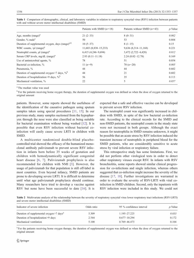
Table 1 Comparison of demographic, clinical, and laboratory variables in relation to respiratory syncytial virus (RSV) infection between patientswith and without severe motor intellectual disabilities (SMID)
Patients with SMID (n=18) Patients without SMID (n=43) p-Value
Age, months (range)a 21 (2–33) 8 (0–31) 0.002
Gender, % malea 50 53 0.804
Duration of supplemental oxygen, days (range)a,b 10 (5–37) 8 (1–15) 0.15
WBC counts, /μl (range)a 11,603 (8,838–15,233) 9,636 (8,314–11,168) 0.221
Neutrophil counts, μl (range)a 6,415 (4,246–9,694) 3,472 (2,722–4,428) 0.012
Serum CRP levels, mg/dL (range)a 2.93 (0.11–11.18) 2.24 (0.02–12.78) 0.169
Use of antimicrobial agents, % 78 51 0.054
Bacterial co-infection, % 11 (n=9) 70 (n=20) 0.005
Pneumonia, % 44 44 0.602
Duration of supplemental oxygen>7 days, %b 44 21 0.602
Duration of hospitalization>9 days, %b 56 35 0.113
Mechanical ventilation, % 22 5 0.057
a The median value was usedb For the patients receiving home oxygen therapy, the duration of supplemental oxygen was defined as when the dose of oxygen returned to theoriginal amount
Table 2 Multivariate analysis of the relationship between the severity of respiratory syncytial virus lower respiratory tract infections (RSV-LRTI)and severe motor intellectual disabilities (SMID)
Indicator of severe infection Odds ratio 95 % confidence interval p-Value
Duration of supplemental oxygen>7 daysa 5.309 1.195–27.225 0.033
Duration of hospitalization>9 days 2.544 0.677–10.294 0.172
Mechanical ventilation 5.100 0.769–46.473 0.104
a For the patients receiving home oxygen therapy, the duration of supplemental oxygen was defined as when the dose of oxygen returned to theoriginal amount
1356 Eur J Clin Microbiol Infect Dis (2013) 32:1353–1357
perform a population-based study and compare the overallclinical manifestation between these groups. Third, the studyin our hospital alone may bias the patient population becauseKyushu University hospital is a tertiary referral hospital in theFukuoka area. Finally, the present study was performed in asingle medical center, and the total number of patients in thestudy population was small. The small number of children ineach group may lead to inaccuracy of the statistic analysis todetect differences between the groups.
In conclusion, children with SMID were at high risk ofdeveloping hypoxia by RSV-LRTI. We should treat thesechildren based on the recognition that they are prone todeveloping hypoxia by viral infections because of theirchronic respiratory failure. A large-scale prospective studyis warranted to validate the severity of single RSV infectionin children with SMID.
Acknowledgments We thank Tetsuyoshi Sugita (Shimadzu) for thetechnical assistance with the RT-PCR assay. We also thank Deana Tatafor her significant advice regarding the manuscript.
Conflict of interest The authors declare that they have no conflict ofinterest.
References
1. Oshima K (1971) Basic problem of severely mentally and physi-cally disabled children (in Japanese). Koshu Eisei (Tokyo) 35:648–655
2. Hanaoka T, Mita K, Hiramoto A, Suzuki Y, Maruyama S,Nakadate T, Kishi R, Okada K, Egusa Y (2010) Survival prognosisof Japanese with severe motor and intellectual disabilities living inpublic and private institutions between 1961 and 2003. JEpidemiol 20:77–81
3. Wilkesmann A, Ammann RA, Schildgen O, Eis-Hübinger AM,Müller A, Seidenberg J, Stephan V, Rieger C, Herting E, WygoldT, Hornschuh F, Groothuis JR, Simon A; DSM RSV Ped StudyGroup (2007) Hospitalized children with respiratory syncytialvirus infection and neuromuscular impairment face an increasedrisk of a complicated course. Pediatr Infect Dis J 26:485–491
4. Welliver RC (2003) Review of epidemiology and clinical riskfactors for severe respiratory syncytial virus (RSV) infection. JPediatr 143:S112–S117
5. Hall CB, Weinberg GA, Iwane MK, Blumkin AK, Edwards KM,Staat MA, Auinger P, Griffin MR, Poehling KA, Erdman D,Grijalva CG, Zhu Y, Szilagyi P (2009) The burden of respiratorysyncytial virus infection in young children. N Engl J Med360:588–598
6. The IMpact-RSV Study Group (1998) Palivizumab, a humanizedrespiratory syncytial virus monoclonal antibody, reduces hospital-ization from respiratory syncytial virus infection in high-risk in-fants. Pediatrics 102:531–537
7. Feltes TF, Cabalka AK, Meissner HC, Piazza FM, Carlin DA, TopFH Jr, Connor EM, Sondheimer HM; Cardiac Synagis StudyGroup (2003) Palivizumab prophylaxis reduces hospitalizationdue to respiratory syncytial virus in young children with hemody-namically significant congenital heart disease. J Pediatr 143:532–540
8. Greene G, Hood K, Little P, Verheij T, Goossens H, Coenen S,Butler CC (2011) Towards clinical definitions of lower respiratorytract infection (LRTI) for research and primary care practice inEurope: an international consensus study. Prim Care Respir J20:299–306
9. Englund JA, Piedra PA, Jewell A, Patel K, Baxter BB, Whimbey E(1996) Rapid diagnosis of respiratory syncytial virus infections inimmunocompromised adults. J Clin Microbiol 34:1649–1653
10. Goodrich JS, Miller MB (2007) Comparison of Cepheid’s analyte-specific reagents with BD Directigen for detection of respiratorysyncytial virus. J Clin Microbiol 45:604–606
11. Yoo SJ, Kuak EY, Shin BM (2007) Detection of 12 respiratoryviruses with two-set multiplex reverse transcriptase-PCR assayusing a dual priming oligonucleotide system. Korean J Lab Med27:420–427
12. Hoshina T, Kusuhara K, Takimoto T, Saito M, Hara T (2010)Identification of bacterial pathogens in pediatric community-acquired lower respiratory tract infection using a simplified proce-dure of sputum sampling and examination: comparison betweenhospitalized children with and without underlying diseases. Eur JClin Microbiol Infect Dis 29:519–525
13. Keren R, Zaoutis TE, Bridges CB, Herrera G, Watson BM, WheelerAB, Licht DJ, Luan XQ, Coffin SE (2005) Neurological and neuro-muscular disease as a risk factor for respiratory failure in childrenhospitalized with influenza infection. JAMA 294:2188–2194
14. Thorburn K, Harigopal S, Reddy V, Taylor N, van Saene HK(2006) High incidence of pulmonary bacterial co-infection in chil-dren with severe respiratory syncytial virus (RSV) bronchiolitis.Thorax 61:611–615
15. Hishiki H, Ishiwada N, Fukasawa C, Abe K, Hoshino T, Aizawa J,Ishikawa N, Kohno Y (2011) Incidence of bacterial coinfectionwith respiratory syncytial virus bronchopulmonary infection inpediatric inpatients. J Infect Chemother 17:87–90
16. Power UF (2008) Respiratory syncytial virus (RSV)vaccines—two steps back for one leap forward. J Clin Virol41:38–44
17. Semple MG, Cowell A, Dove W, Greensill J, McNamara PS,Halfhide C, Shears P, Smyth RL, Hart CA (2005) Dual infectionof infants by human metapneumovirus and human respiratorysyncytial virus is strongly associated with severe bronchiolitis. JInfect Dis 191:382–386
18. De Paulis M, Gilio AE, Ferraro AA, Ferronato AE, do SacramentoPR, Botosso VF, Oliveira DB, Marinheiro JC, Hársi CM, DurigonEL, Vieira SE (2011) Severity of viral coinfection in hospitalizedinfants with respiratory syncytial virus infection. J Pediatr (Rio J)87:307–313
Eur J Clin Microbiol Infect Dis (2013) 32:1353–1357 1357